Chapter 3 Dose units and dosimetry
Several different terms and units have been used in dosimetry over the years. The conversion to SI units has made this subject even more confusing. However, it is essential that these terms and units are understood to appreciate what is meant by radiation dose and to allow meaningful comparisons between different investigations to be made. In addition to explaining the various units, this chapter also summarizes the various sources of ionizing radiation and the magnitude of radiation doses that are encountered.
The more important terms in dosimetry include:
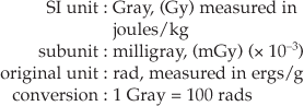
RADIATION-ABSORBED DOSE (D)
This is a measure of the amount of energy absorbed from the radiation beam per unit mass of tissue.
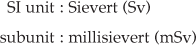
EQUIVALENT DOSE (H)
This is a measure which allows the different radiobiological effectiveness (RBE) of different types of radiation to be taken into account.
For example, alpha particles (see Ch. 19) penetrate only a few millimetres in tissue, lose all their energy and are totally absorbed, whereas X-rays penetrate much further, lose some of their energy and are only partially absorbed. The biological effect of a particular radiation-absorbed dose of alpha particles would be considerably more severe than a similar radiation-absorbed dose of X-rays.
By introducing a numerical value known as the radiation weighting factor WR which represents the biological effects of different radiations, the unit of equivalent dose (H) provides a common unit allowing comparisons to be made between one type of radiation and another, for example:
| X-rays, gamma rays and beta particles | WR = 1 |
| Fast neutrons (10 keV–100 keV) and protons | WR = 10 |
| Alpha particles | WR = 20 |
Equivalent dose (H) = radiation-absorbed dose (D) × radiation weighting factor (WR)
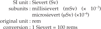
(For X-rays, the radiation weighting factor (WR factor) = 1, therefore the equivalent dose (H), measured in Sieverts, is equal to the radiation-absorbed dose (D), measured in Grays.)
EFFECTIVE DOSE (E)
This measure allows doses from different investigations of different parts of the body to be compared, by converting all doses to an equivalent whole body dose.
This is necessary because some parts of the body are more sensitive to radiation than others. The International Commission on Radiological Protection (ICRP) has allocated each tissue a numerical value, known as the tissue weighting factor (WT), based on its radiosensitivity, i.e. the risk of the tissue being damaged by radiation — the greater the risk, the higher the tissue weighting factor. The sum of the individual tissue weighting factors represents the weighting factor for the whole body. The tissue weighting factors recommended by the ICRP in 1990 and updated in 2005 are shown in Table 3.1.
Table 3.1 The tissue weighting factors (WT) recommended by the ICRP in 1990 and in 2007
| Tissue | 1990 WT | 2007 WT |
|---|---|---|
| Bone marrow | 0.12 | 0.12 |
| Breast | 0.05 | 0.12 |
| Colon | 0.12 | 0.12 |
| Lung | 0.12 | 0.12 |
| Stomach | 0.12 | 0.12 |
| Bladder | 0.05 | 0.04 |
| Oesophagus | 0.05 | 0.04 |
| Gonads | 0.20 | 0.08 |
| Liver | 0.05 | 0.04 |
| Thyroid | 0.05 | 0.04 |
| Bone surface | 0.01 | 0.01 |
| Brain | * | 0.01 |
| Kidneys | * | 0.01 |
| Salivary glands | 0.01 | |
| Skin | 0.01 | 0.01 |
| Remainder tissues | 0.05* | 0.12+ |
* Adrenals, brain, upper large intestine, small intestine, kidney muscle, pancreas, spleen, thymus and uterus
+ Adipose tissue, adrenals, connective tissue, extrathoracic airways, gall bladder, heart wall, lymphatic nodes, muscle, pancreas, prostate, SI wall, spleen, thymus and uterus/cervix
Effective dose (E) = equivalent dose (H) × tissue weighting factor (WT)
When the simple term dose is applied loosely, it is the effective dose (E) that is usually being described. Effective dose can thus be thought of as a broad indication of the risk to health from any exposure to ionizing radiation, irrespective of the type or energy of the radiation or the part of the body being irradiated. A comparison of effective doses from different investigations is shown in Table 3.2.
Table 3.2 Typical effective doses for a range of dental and routine medical examinations
| X-ray examination | Effective dose (mSv) |
|---|---|
| CT chest | 8.0 |
| CT head | 2.0 |
| Barium swallow | 1.5 |
| Barium enema | 7.0 |
| Lumbar spine (AP) | 0.7 |
| Skull (PA) | 0.03 |
| Skull (Lat) | 0.01 |
| Chest (PA) | 0.02 |
| Chest (Lat) | 0.04 |
| Bitewing/periapical | 0.001–0.008 |
| Upper standard occlusal | 0.008 |
| Panoramic | 0.004–0.03 |
| Lateral cephalometric | 0.002–0.003 |
| CT mandible | 0.36–1.2 |
| CT maxilla | 0.1–3.3 |
COLLECTIVE EFFECTIVE DOSE OR COLLECTIVE DOSE
This measure is used when considering the total effective dose to a population, from a particular investigation or source of radiation.


DOSE RATE
This is a measure of the dose per unit time, e.g. dose/hour, and is sometimes a more convenient, and measurable, figure than, for example, a total annual dose limit (see Ch. 8).

ESTIMATED ANNUAL DOSES FROM VARIOUS SOURCES OF RADIATION
Everyone is exposed to some form of ionizing radiation from the environment in which we live. Sources include:
• Natural background radiation
• Artificial background radiation
The Radiation Protection Division of the Health Protection Agency (formerly the National Radiological Protection Board (NRPB)) has estimated the annual doses from these various sources in the UK. Table 3.3. gives a summary of the data.
Table 3.3 HPA(NRPB)-estimated average annual doses to the UK population from various sources of radiation
| Radiation source | Average annual dose (μSv) | Approximate % |
|---|---|---|
| Natural background | ||
| Cosmic rays | 300 | |
| External exposure from the earth’s crust | 400 | |
| Internal radiation from certain foodstuffs | 370 | |
| Exposure to radon and its decay products | 700 | |
| Total | 2.7 mSv (approx.) | 87% |
| Artificial background | ||
| Fallout | 10 | |
| Radioactive waste | 2 | >1% |
| Medical and dental diagnostic radiation | 250 | 12% |
| Occupational exposure | 9 | >1% |
An individual’s average dose from background radiation is estimated at approximately 2.7 mSv per year in the UK, while in the USA it is estimated at approximately 3.6 mSv. These figures are useful to remember when considering the magnitude of the doses associated with various diagnostic procedures (see later).
TYPICAL DOSES ENCOUNTERED IN DIAGNOSTIC RADIOLOGY
The European Guidelines on Radiation Protection in Dental Radiology published in 2004 were based on an extensive review of the available evidence on all aspects of radiation protection in dentistry. They concluded that although many studies have measured doses of radiation for dental radiography, only a few had estimated effective dose. For some techniques there is no published data available and some for which very different results have been reported. The typical effective doses shown earlier in Table 3.2 are based broadly on their findings, together with a selection of typical effective doses from various medical diagnostic procedures published the in NRPB document Guidelines on Patient Dose to Promote the Optimisation of Protection for Diagnostic Medical Exposures in 1999.
It must be stressed that these are typical values and that a considerable range of effective doses exists in dental radiography. The main reasons for this variation are kV of equipment used, shape and size of beam, speed and type of image receptor used and the tissues included in the calculations. These factors are of great importance in radiation protection and are discussed in more detail in Chapter 8.
However, the figures do provide an indication of the comparative sizes of the various effective doses. The individual doses encountered in dental radiology may appear very small, but it must be remembered that the diagnostic burden, however small, is an additional radiation burden to that which the patient is already receiving from background radiation. This additional dose may be considerable for any individual patient. The enormous number of dental radiographs (intraoral and extraoral) taken per year (estimated at approximately 20–25 million in the UK alone) means that the collective dose from dental radiography is quite substantial. The risks associated with some of the diagnostic investigations are discussed in Chapter 4.