Pica is the ingestion of materials other than normal food and varies from licking to actual eating or drinking. It is associated in most cases with dietary deficiency, either of bulk or, in some cases, more specifically fiber, or of individual nutrients, particularly salt, cobalt or phosphorus. It is considered as normal behavior in rabbits and foals, where it is thought to be a method of dietary supplementation or refection of the intestinal bacterial flora. Boredom, in the case of animals closely confined, often results in the development of pica. Chronic abdominal pain due to peritonitis or gastritis and central nervous system disturbances, including rabies and nervous acetonemia, are also causes of pica.
The type of pica may be defined as follows: osteophagia is the chewing of bones; infantophagia is the eating of young; coprophagia is the eating of feces. Other types include wood-eating in sheep, bark-eating, the eating of carrion and cannibalism. Salt hunger can result in coat-licking, leather-chewing, earth-eating and the drinking of urine. Urine drinking may also occur if the urine is mixed with palatable material such as silage effluent. Bark-eating is a common vice in horses, especially when their diet is lacking in fiber, e.g. when they are grazing irrigated pasture.
Cannibalism may become an important problem in housed animals, particularly swine, which bite one another’s tails, often resulting in severe local infections. Although some cases may be due to protein, iron or bulk deficiency in the diet, many seem to be the result of boredom in animals given insufficient space for exercise. A high ambient temperature and generally limited availability of food also appear to contribute. Male castrates are much more often affected than females and the bites are also much more severe in males. Provision of larger pens or a hanging object to play with, removal of incisor teeth and the avoidance of mixing animals of different sizes in the same pen are common control measures in pigs. In many instances only one pig in the pen has the habit and his removal may prevent further cases. One common measure that is guaranteed to be successful in terms of tail-biting is surgical removal of all tails with scissors during the first few days of life, when the needle teeth are removed. Unfortunately the cannibalistic tendency may then be transferred to ears. As in all types of pica, the habit may survive the correction of the causative factor.
Infantophagia can be important in pigs in two circumstances. In intensively housed sows, especially young gilts, hysterical savaging of each pig as it is born can cause heavy losses. When sows are grazed and housed at high density on pasture it is not uncommon to find ‘cannibal’ sows who protect their own litters but attack the young pigs of other sows. This diagnosis should be considered when there are unexplained disappearances of young pigs.
Pica may have serious consequences: cannibalism may be the cause of many deaths; poisonings, particularly lead poisoning and botulism, are common sequelae; foreign bodies lodging in the alimentary tract or accumulations of wool, fiber or sand may cause obstruction; perforation of the esophagus or stomach may result from the ingestion of sharp foreign bodies; grazing time is often reduced and livestock may wander away from normal grazing. In many cases the actual cause of the pica cannot be determined and corrective measures may have to be prescribed on a basis of trial and error.
Complete deprivation of food causes rapid depletion of glycogen stores and a changeover in metabolism to fat and protein. In the early stages there is hunger, increase in muscle power and endurance, and a loss of body weight. In sheep there is often a depression of serum calcium levels sufficient to cause clinical hypocalcemia. The development of ketosis and acidosis follows the increased fat utilization. A marked reduction in feed intake in pony mares in late pregnancy is often a precursor of hyperlipemia, a highly fatal disease discussed in Chapter 28 on metabolic diseases. The most pronounced biochemical change in ponies occurring as a result of experimental food deprivation is a lipemia, which reaches a peak by the eighth day of fasting but quickly returns to normal when feeding is resumed. This degree of change in blood lipids appears to be a characteristic of ponies and horses; it is much higher than occurs in pigs.
In lactating cows, a short period of starvation results in depression of plasma glucose and an increase in plasma lipid concentrations. Milk yield falls by 70%. On refeeding most levels return to normal in 5 days but blood lipid and milk yield may take as long as 49 days to recover to normal levels. In horses, fecal output falls to zero at day 4 and water intake is virtually nil from that time on, but urine volume is maintained. In spite of the apparent water imbalance there is no appreciable dehydration, plasma protein levels and PCV staying at normal levels. A significant loss of skin turgor (increase in skin tenting) due to the disappearance of subcutaneous fat as cachexia develops may occur. Muscular power and activity decrease and the loss of body weight may reach as high as 50–60%. The metabolic rate falls and is accompanied by a slowing of the heart and a reduction in stroke volume, amplitude of the pulse and blood pressure. The circulation is normal as indicated by mucosal color and capillary refill.
In the final stages, when fat stores are depleted, massive protein mobilization occurs and a premortal rise in total urinary nitrogen is observed, whereas blood and urine ketones are likely to diminish from their previous high level. Great weakness of skeletal and cardiac musculature is also present in the terminal stages and death is due to circulatory failure. During the period of fat utilization there is a considerable reduction in the ability of tissues to utilize glucose and its administration in large amounts is followed by glycosuria. In such circumstances readily assimilated carbohydrates and proteins should be given in small quantities at frequent intervals but fatty foods may exacerbate the existing ketosis. Diets for animals that have been through a period of great nutritional stress because of deprivation of food or because of illness are described below under inanition.
Starvation of farm livestock is an animal welfare issue with economic and ethical considerations. When starving animals are identified by a neighboring farmer or veterinarian they are commonly reported to the appropriate authorities, which may be provincial or state appointed inspectors (animal care officers) who have the authority to take appropriate action. The animals are examined and corrective action is taken, including possession of the animals and relocating them to a commercial feeding facility.1 Predicting survival of starved animals is a major challenge. Economics becomes an important aspect because the financial costs of stabilizing a group of starved horses may exceed their free market price. Responsible management of chronically starved commercial animals should include options for immediate euthanasia. Ethical considerations include deciding if certain severely starved animals should be euthanized. In some cases, enforcement officers may be reluctant to recommend mass euthanasia of otherwise healthy horses based on personal aversion.1
Chronically starved horses lose body weight, become weak and their body condition score may decline to below 2 on the basis of 1–9, and death is common, especially during cold weather.1 Chronically starved horses frequently respond poorly to refeeding. About 20% of severely malnourished horses can be expected to die in spite of attempts at refeeding.2 Recovery of severely malnourished horses to an average body condition score may require 6–10 months.3
Incomplete starvation – inanition or malnutrition – is a more common field condition than complete starvation. The diet is insufficient in quantity; all essential nutrients are present but in suboptimal amounts. The condition is compatible with life, and in general the same pattern of metabolic change occurs as in complete starvation but to a lesser degree. Thus ketosis, loss of body weight and muscular power and a fall in metabolic rate occur. As a result of the reduction in metabolic activity there is a fall in body temperature and respiratory and heart rates. In addition there is mental depression, anestrus in cows but not ewes, and increased susceptibility to infection. This increased susceptibility to infection that occurs in some cases of malnutrition cannot be accepted as a general rule. In the present state of knowledge it can only be said that ‘some nutritional influences affect resistance to some forms of infection’.
A significantly reduced food intake also increases susceptibility to some poisons, and this has been related to the effects of starvation on hepatic function. In ruminants, the effects of starvation on the activity of liver enzymes is delayed compared to the effects in monogastric animals, due apparently to the ability of the ruminal store of feed to cushion the effect of starvation for some days. The most striking effect of short-term malnutrition in sheep and cattle compared to rats was the very rapid and large accumulation of neutral fat in hepatocytes. If there is a relative lack of dietary protein over a long period of time, anasarca occurs, particularly in the intermandibular space.
Malnutrition makes a significant contribution to a number of quasi-specific diseases, ‘weaner ill-thrift’ and ‘thin sow syndrome’ among them, and these are dealt with elsewhere.
Controlled malnutrition in the form of providing submaintenance diets to animals during periods of severe feed shortage is now a nutritional exercise with an extensive supporting literature. For pastured animals it is a fact of economic life that significant loss of body weight is planned and tolerated for some parts of each year because the well-known phenomenon of compensatory growth enables the animal to make up the lost weight, with no disadvantage, during the times of plenty. Animals fed on submaintenance diets undergo metabolic changes reflected in blood and tissue values as well as the more significant changes in weight. Experimental restriction of feed intake to 65% of normal levels in nonlactating, nonpregnant heifers does not cause significant falls in serum calcium and phosphorus levels, nor in plasma glutamic oxaloacetic transaminase (GOT), aspartate transferase (AST), lactate dehydrogenase (LDH) or creatine phosphokinase (CPK) activities. Serum alkaline phosphatase (AP) activity was also maintained. In sheep that are losing weight because of undernutrition there is a significant decrease in plasma creatinine concentration.
Experimental feed restriction, followed by fasting, followed by ad libitum access to feed, such as might occur in nature, had no serious ill-effects on goats. The goats lost weight significantly but did not overeat on being allowed access to feed.
A deficiency of one or more specific dietary essentials also causes a form of partial starvation and is dealt with in Chapter 30.
Outbreaks of incomplete starvation may occur in cattle, sheep and horses that are kept outdoors during the cold winter months in regions of the northern hemisphere. The feed usually consists of poor-quality grass hay or cereal grain straw and no grain supplementation. During prolonged exposure to the cold environment the animals will increase their daily intake in an attempt to satisfy maintenance requirements and, in cattle, abomasal impaction with a high case mortality may occur. Animals affected with severe inanition are usually weak and recumbent and may or may not eat when offered a palatable feed.
Malnutrition and starvation may occur in calves under 1 month that are fed poor-quality milk replacers containing excessive quantities of nonmilk carbohydrates and proteins. The diet is not well digested by young calves and chronic diarrhea and gradual malnutrition occur. Affected calves recover quickly when fed cows’ whole milk for several days. At necropsy there is a marked reduction in muscle mass, lack of depot fat and serious atrophy of fat. Starvation may also occur in beef calves sucking poorly nourished heifer dams with an insufficient supply of milk. The mortality will be high during cold weather when the maintenance requirements are increased. Affected calves will initially suck vigorously and persistently, they will attempt to eat dry feed, drink surface water and urine and bawl for several hours. Eventually they lie in sternal recumbency with their head and neck turned into their flanks and die quietly. The response to therapy is usually unsatisfactory and the case fatality rate is high. The convalescence period in survivors is prolonged and treatment is usually uneconomic. Affected animals must be brought indoors and kept warm and well bedded during treatment and realimentation. Initially, fluid therapy using balanced electrolyte solutions containing glucose and amino acids may be necessary to restore the animal’s strength and appetite. This is followed by the provision of controlled amounts of a highly palatable digestible diet. High-quality legume hay is excellent, small amounts of ground grain are of value and the daily administration of a multiple B vitamin and mineral mixture will replenish those lost during inanition. Skim-milk powder is an excellent source of carbohydrate and protein for young animals that have been partially starved. Adult animals cannot digest large quantities of milk powder because of the relative lack of the appropriate digestive enzymes.
Horses that have been ill with a poor appetite should be tempted with green grass first, and failing that tried with good-quality hay – preferably alfalfa. It is best to dilute it with good grass hay to begin with, and increase the mix to 100% legume hay over a week. An average horse will require 1.5–2 kg BW/day. Grain can be added, mixed with molasses or as a mash. Low-fiber diets are recommended to ensure maximum digestibility. A supplement of B vitamins may be advantageous until full appetite and intake are regained. Horses with broken jaws or that are unable to eat at all for some reason can be allowed to go without food for 3 days, but beyond that time they should be fed by stomach tube. A suitable ration is:
• Electrolyte mixture (NaCl, 10 g; NaHCO3, 15 g; KCl, 75 g; K2HPO4, 60 g; CaCl2, 45 g; MgO, 24 g) 210 g
• Dextrose, increased from 300 g/d in 7 days to 900 g
• Dehydrated cottage cheese, increased from 300 g/day in 7 days to 900 g
The ration is divided into two or three equal amounts and fed during one day. Adult horses that are weak and recumbent may be supported in a sling to avoid decubitus ulceration and other secondary complications associated with prolonged recumbency.
Thirst is an increased desire for water manifested by excessive water intake (polydipsia). There are two important causes of thirst: dryness of the pharyngeal and oral mucosae increases the desire for water, irrespective of the water status of body tissues; in addition, cellular dehydration due to a rise in blood osmotic pressure causes increased thirst. Specific observations in ponies have shown that water intake is increased in response to either an increase in the osmotic pressure of tissue fluid or a decrease in the volume of their body fluids.
Cellular dehydration occurs commonly in many cases of dehydration due to vomiting, diarrhea, polyuria and excessive sweating. Increased thirst in early fever is due to changes in cell colloids leading to increased water retention. A marked polydipsia and polyuria occur in salt deficiency in lactating dairy cattle, in addition to weight loss, a fall in milk production and salt hunger. Salivary sodium levels are best used for diagnosis. A similar syndrome occurs in the ‘thin sow syndrome’.
In humans, several other factors appear to exert some effect on water intake: a deficiency of potassium and an excess of calcium in tissue fluid both increase thirst; an increased thirst also occurs in uremia irrespective of the body’s state of hydration. It has been suggested that these chemical factors may cause direct stimulation of the thirst center in the hypothalamus. Clinically, diabetes insipidus produces by far the most exaggerated polydipsia.
The clinical syndrome produced by water deprivation is not well defined. Animals supplied with saline water will drink it with reluctance and, if the salinity is sufficiently great, die of salt poisoning. Cattle at pasture that are totally deprived of water usually become quite excited and are likely to knock down fences and destroy watering points in their frenzy. On examination they exhibit a hollow abdomen, sunken eyes and the other signs of dehydration. There is excitability with trembling and slight frothing at the mouth. The gait is stiff and uncoordinated and recumbency follows. Abortion of decomposed calves, with dystocia due to failure of the cervix to dilate, may occur for some time after thirst has been relieved and cause death in survivors. At necropsy there is extensive liquefaction of fat deposits, dehydration and early fetal death in pregnant cows.
Experimental water deprivation has been recorded in camels and lactating and nonlactating dairy cows. In camels death occurred on the seventh to ninth day of total deprivation; body weight loss was about 25%. Lactating cows allowed access to only 50% of their regular water supply become very aggressive about the water trough, spend more time near it and lie down less. After 4 days milk yield is depressed to 74% and body weight to 86% of original figures. There is a significant increase in serum osmolality with increased concentrations of urea, sodium, total protein and copper. The PCV is increased, as are activities of creatinine kinase and serum AST. With complete deprivation for 72 hours, the changes are similar but there are surprisingly few clinical signs at that time. The composition of the milk does not change markedly and blood levels return to normal in 48 hours. After deprivation of half of their water intake, cattle reduced their water loss by all routes, but plasma and total blood volumes were unchanged.
Sheep, even pregnant ewes, are capable of surviving even though access to water is limited to only once each 72 hours, but there is a significant loss (26%) of body weight. Deprivation of water that allows access to water only once every 96 hours is not compatible with maintaining the pregnancy.
Weight loss or failure to gain weight (ill-thrift)
This section is concerned with the syndrome of weight loss in the presence of an adequate food supply and a normal appetite. In the absence of any primary disease, an animal or group of animals that presents with this as the problem is a major diagnostic dilemma. Several poorly identified diseases in this category are ‘weaner ill-thrift’, ‘thin sow syndrome’, ‘thin ewe syndrome’, ‘weak calf syndrome’ (see Ch. 36).
Errors by the owner in the estimation of body weight can lead to inadequate feeding if the ration is based on the requirements needed for growth and maintenance per unit of body weight. Scales are rarely available and estimations by weight-bands are generally inaccurate and subject to too much variability. A reasonably satisfactory alternative used in cattle, sheep and horses is a body condition score estimated on the basis of the amount of body covering of muscle, fat and connective tissue.
Detailed below is a checklist of causes that should be considered when an animal has a weight loss problem in the absence of signs indicative of a primary wasting disease.
‘Hobby farm malnutrition’ is a surprisingly common cause, especially in companion horses. Inexperienced owners keep their animals where they are not able to graze pasture and are entirely dependent on stored feed, but underfeed for economy’s sake. A knowledge of the animals’ needs and of the approximate energy and protein values of feeds are necessary to prepare an appropriate ration. In a hospital situation any horse presented with a weight loss problem and without a potential diagnosis on first examination should be weighed, fed an energy-rich diet ad libitum for 4 days, then re-weighed. A horse that has previously been underfed will gain 3–5 kg in weight per day.
The feed must be inspected. Mature meadow hay may be efficient only as a filler, and poorly filled oat grain may be very poorly nutritive on a weight basis. Gentle animals that are fed in a group with others may be physically prevented from getting a fair share of available feed, especially if trough space is inadequate.
This problem is also common when urban people try to raise a few veal calves or sheep to help defray the costs of their rural acreage. It is common in these circumstances to equate rough meadow grass with proper nutrition for young or pregnant ruminants.
Other considerations are as follows:
• Diets that are inadequate in total energy because they cannot replace the energy loss caused by the animal’s level of production can be important causes of weight loss in heavy-producing animals. This subject is discussed under the heading of production disease. An example is acetonemia of high-producing cows in which body stores of fat and protein are raided to repair the energy deficiency of the diet
• Malnutrition as a result of a ration that is deficient in an essential trace element is unusual in the management situation being discussed. A nutritional deficiency of cobalt does cause weight loss in ruminants but is likely to have an area effect rather than cause weight loss in single animals. Copper, salt, zinc, potassium, selenium, phosphorus, calcium and vitamin D deficiencies are also in this category. Experimental nutritional deficiencies of riboflavin, nicotinic acid, pyridoxine and pantothenic acid in calves and pigs can also be characterized by ill-thrift
• Inadequate intake of an adequate supply of feed is dealt with under diseases of the mouth and pharynx and is not repeated here, but it is emphasized that the first place for a clinician to look in a thin animal is its mouth. The owner may have forgotten just how old the animal is and one often finds a cow without any incisor teeth attempting to survive on pasture
• Other factors that reduce an animal’s food intake when it is available in adequate amounts include anxiety, the excitement of estrus, new surroundings, loss of newborn, bad weather, tick or other insect worry and abomasal displacement.
• Glucose loss in the urine in diabetes mellitus or chronic renal disease, the former indicated by hyperglycemia and both by glycosuria, are obvious examples of weight loss as a result of excessive metabolic loss of energy
• Protein loss in the feces. Cases of protein-losing gastroenteropathy are unusual and are difficult to identify without access to a radioactive isotope laboratory. The loss may occur through an ulcerative lesion, via a generalized vascular discontinuity or by exudation through intact mucosa as a result of hydrostatic pressure in blood vessels, e.g. in verminous aneurysm, or lymphatics in cases of lymphangiectasia of the intestine. The identification of a neoplasm (lymphosarcoma or intestinal or gastric adenocarcinoma are the usual ones) or of granulomatous enteritis is not possible without laparotomy and biopsy of the alimentary segment. One is usually led to the possibility of this as a diagnosis by either a low serum total protein or low albumin level in a normal total protein level, and in the absence of other protein loss as set out below
• Proteinuria for a lengthy period can cause depletion of body protein stores, resulting in weight loss. Chronic glomerulonephritis is the usual cause. Examination of the urine should be part of every clinical examination of a patient, but is not commonly so in horses because of the difficulty of obtaining a specimen without recourse to catheterization. Moving the horse into a box stall with fresh straw, or the intravenous injection of furosemide, are possible methods when straightforward collection is not possible. The latter provides an abnormally dilute sample
• Internal and external parasitoses in which blood sucking is a significant pathogenetic mechanism can result in severe protein loss, as well as anemia per se.
Faulty digestion and absorption are commonly manifested by diarrhea, and diseases that have this effect are dealt with under the heading of malabsorption syndromes (see Enteriti). In grazing ruminants, the principal causes are the nematode worms Ostertagia, Nematodirus, Trichostrongylus, Chabertia, Cooperia and Oesophagostomum and the flukes Fasciola and Paramphistomum. In cattle there are, in addition, tuberculosis, coccidiosis, sarcosporidiosis and enzootic calcinosis. In sheep and goats there are Johne’s disease, viral pneumonia without clinical pulmonary involvement, and hemonchosis. In horses there are strongylosis, habronemiasis and heavy infestations with botfly larvae. In pigs there are stephanuriasis, hyostrongylosis (including the ‘thin sow syndrome’), infestation with Macracanthorhynchus hirudinaceus, and ascariasis. Gastrointestinal neoplasia must also be considered as a possible cause
• Chronic villous atrophy occurs most severely with intestinal parasitism or as a result of a viral infection
• Other lesions caused by parasitic invasion that affect digestion and absorption are gastric granuloma associated with Habronema spp. in horses and verminous arteritis, also in horses
• Abnormal physical function of the alimentary tract, as in vagus indigestion of cattle and grass sickness in horses, can be a potent cause of failure to absorb nutrients, but the syndrome is usually manifested by poor food intake and grossly abnormal feces
• Inadequate utilization of absorbed nutrients is a characteristic of chronic liver disease. It is usually distinguishable by a low serum albumin level, by liver function tests and by serum enzyme estimations. A clinical syndrome including edema, jaundice, photosensitization and weight loss is a common accompaniment
• Neoplasia in any organ. The metabolism of the body as a whole is often unbalanced by the presence of a neoplasm so that the animal wastes even though its food intake seems adequate
• Chronic infection, including specific diseases such as tuberculosis, sarcocystosis, East Coast fever, trypanosomiasis (nagana), maedi-visna, caprine arthritis–encephalitis, enzootic pneumonia of swine and nonspecific infections such as atrophic rhinitis of pigs, abscess, empyema and chronic peritonitis have the effect of reducing metabolic activity generally as well as reducing appetite. Both effects are the result of the toxemia caused by tissue breakdown and of toxins produced by the organisms present. Less well understood are the means by which systemic infections, e.g. equine infectious anemia, scrapie in sheep and other slow viruses, produce a state of weight loss progressing to emaciation
• Food refusal is a well-recognized syndrome in pigs, due in some cases to mycotoxins in the feed, and ‘off feed effects’ are similarly encountered in feedlot cattle on rations containing a large proportion of wheat grain
• Many diseases of other systems, e.g. congestive heart failure, are manifested by weight loss because of inadequate oxygenation of tissues.
Determination of the specific cause of weight loss in an individual animal depends first on differentiation into one of the three major groups:
• Nutritional causes, diagnosed by assessment of the animal’s total food intake
• Protein or carbohydrate loss in the animal’s excretions, diagnosed by clinicopathological laboratory tests
• Faulty absorption of the food ingested, diagnosed by tests of digestion as set out in Chapter 5.
The present-day emphasis on the need for economically efficient performance by farm animals introduces another set of criteria, besides freedom from disease, to be taken into consideration when deciding an animal’s future. The same comment applies, and much more importantly, when a herd’s productivity is being assessed. This is usually done by comparing the subject herd’s performances to that of peer herds, or animals in similar environmental and management conditions.
It is usual to use the production indexes that are the essential outputs of the particular enterprise as the criteria of productivity. Thus, in dairy herds the criteria could be:
• Milk or butterfat production per cow per lactation (liters per cow or liters per hectare)
• Reproductive efficiency as mean intercalving interval
• Percentage calf survival to 1 year of age
• Longevity as percentage mortality per year or average age of cows in herd plus culling rate per year
• The culling rate needs to differentiate between sale because of disease or poor production and sale as a productive animal
• Acceptability of product at sale – as indicated by bulk tank milk somatic cell count, rejection of milk because of poor-quality, low-fat content, low solids-not-fat content.
If it is decided that performance falls too far short of the target, an investigation is warranted. Some targets for productivity in each of the animal industries are available, but they vary a great deal between countries depending on the levels of agriculture practiced and the standards of performance expected. For this reason, they are not set down here; nor is the degree of shortfall from the target that is acceptable – this depends heavily on the risk aversion or acceptability in the industry in that country. For example, if the enterprise is heavily capitalized by high-cost housing and land, the standard of performance would be expected to be higher than in a more exploitative situation where cattle are pastured all year. In the latter, a reasonable flexibility could be included in the assessment of productivity by permitting it to fall within the scope of 2 SD of the mean productivity established by peer herds.
If it is decided that performance is below permissible standards an investigation should be conducted and should include the following groups of possible causes:
• Nutrition – its adequacy in terms of energy, protein, minerals, vitamins and water
• Inheritance – the genetic background of the herd and the quality of its heritable performance
• Accommodation – to include protection from environmental stress by buildings for housed animals and terrain and tree cover for pastured animals; also consideration of population density as affecting access to feed, water and bedding areas
• General managerial expertise – the degree of its application to the individual flock or herd. This is difficult to assess and then only indirectly, e.g. the efficiency of heat detection, achievement of planned calving pattern
• Disease wastage – as clinical disease or, more particularly, subclinical disease. The latter may include such things as quarter infection rate as an index of mastitis, fecal egg counts relative to parasite burden, metabolic profile relative to metabolic disease prevalence rate, etc.
These investigations tend to require special techniques in addition to the clinical examination of individual animals. They are mostly self-evident, but attention is drawn to the section on examination of a herd or flock in Chapter 1. It will be apparent that there is a great deal of merit in having herds and flocks under constant surveillance for productivity and freedom from disease, as is practiced in modern herd health programs. Monitoring performance and comparing it with targets is the basis of that system.
The specific syndromes that fall within this category of disease, and which are dealt with elsewhere in this book are ill-thrift of weaner sheep, ‘thin sow syndrome’, ‘weak calf syndrome’, ‘poor performance syndrome’ of horses, ‘low butterfat syndromes’ and ‘summer slump’ of milk cows. Two performance shortfalls encountered commonly by field veterinarians are ill-thrift in all species and poor performance syndrome in horses, presented in the two sections following. More specialized problems are dealt with in Herd health (details below).
Physical exercise and associated disorders
The act of performing physical work requires expenditure of energy at rates above the resting metabolic rate. Increases in metabolic rate can be supported by anaerobic metabolism through the use of intramuscular adenosine triphosphate stores and conversion of glycogen or glucose to lactate for short periods of time. Ultimately, however, all energy is derived by aerobic metabolism and is limited by the rate of delivery of oxygen to tissue and its utilization in mitochondria. To support the increased energy expenditure required to perform work such as racing, carrying a rider or pulling a cart, the metabolic rate is increased. Increases in metabolic rate are supported by increases in oxygen delivery to tissue and carbon dioxide removal. Increased oxygen consumption is dependent upon an increase in oxygen delivery to tissues which is possible by increases in cardiac output, muscle blood flow and, in horses, an increase in hemoglobin concentration with a concomitant increase in the oxygen-carrying capacity of blood. The increased transport of oxygen from the air to the blood is accomplished principally by increases in respiratory rate and tidal volume. Factors that affect oxygen transport from the air to the mitochondria have the potential to impair performance. For instance, laryngeal hemiplegia reduces minute ventilation and exacerbates the normal exercise-associated hypoxemia in horses, atrial fibrillation decreases cardiac output and hence oxygen delivery to tissues and anemia reduces the oxygen-carrying capacity of the blood.
The increase in cardiac output with exercise of maximal intensity in horses is very large – horses have a cardiac output of about 75 (mL/min)/kg at rest and 750 (mL/min)/kg (300 L/min for a 400 kg horse) during maximal exercise. Associated with the increase in cardiac output are increases in right atrial, pulmonary arterial and aortic blood pressures. Systemic arterial blood pressure during exercise increases as the intensity of exercise increases with values for systolic, mean and diastolic pressures increasing from 115, 100 and 80 mmHg (15.3, 13.3 and 10.6 kPa) at rest to 205, 160 and 120 mmHg (27.3, 21.3 and 16 kPa), respectively, during intense exercise.
Pulmonary artery pressure increases from a mean of approximately 25 mmHg (3.3 kPa) to almost 100 mmHg (13.3 kPa) during intense exercise. The increase in pulmonary artery pressure with exercise may contribute to exercise-induced pulmonary hemorrhage.
The increase in metabolic rate during exercise causes a marked increase in metabolic heat generation with a subsequent increase in body temperature. The increase in body temperature is dependent on the intensity and duration of exercise and the ability of the horse to dissipate heat from the body. Intense exercise of short duration is associated with marked increases in body temperature but such increases rarely cause disease. However, prolonged exercise of moderate intensity, especially if performed in hot and humid conditions, may be associated with rectal temperatures in excess of 42.5°C (108.5°F). Heat is dissipated primarily by evaporation of sweat from the skin surface. Sweating results in a loss of body water and electrolytes, including sodium, potassium, calcium and chloride. The size of these losses can be sufficient to cause dehydration and abnormalities of serum electrolyte concentrations and also impaired cardiovascular and thermoregulatory function.
Recovery from exercise is influenced by the fitness of the individual, with fitter horses recovering more rapidly, the intensity and duration of the exercise bout, and activity during recovery. Horses allowed to walk after a bout of intense exercise recuperate more quickly than do horses that are not allowed to walk. Recovery is delayed if the horse cannot drink to replenish body water or in hot and humid conditions.
The definition of poor racing performance is difficult. Horses that have a proven record of performing well and that then fail to perform at their previous level are readily apparent and a physical cause of the reduction in performance can often be identified. More difficult are the horses that do not have a history of satisfactory performance and are best labeled as ‘failure to perform to expectation’. Horses in this group may indeed have a clinical abnormality but commonly the reason is lack of innate ability or inadequate training – both causes that must be raised with the owner and trainer carefully and tactfully, and only after a thorough examination of the horse.
Exercise intolerance in race horses is best defined as the inability to race at speeds previously attained by that horse or attained by peers. In its most extreme form exercise intolerance is evident as failure to complete the race, whereas its mildest form is evident as a slight decrement in performance, such as losing a race by several lengths or one or two seconds, or failure to perform to expectation.
Horses with a history of a recent decrement in performance or those that are not performing to expectation should be examined in a systematic fashion.
A detailed history should be collected that focuses on documenting the reduction in performance, its time course and the presence and evolution of any clinical signs. This can be accomplished by asking the following questions of the owner or trainer:
• What evidence is there of poor performance? This query should focus on providing objective evidence of a reduction in performance through examination of race times or results. This also allows the severity of the reduction in performance to be documented
• What is the horse’s training schedule? The training regimen should be appropriate for the horse’s level of competition
• Describe the horse’s exercise intolerance. Does it start the race strongly and ‘fade’ in the last part of the race, or is it unable to maintain a suitable speed for the complete race? Is the horse slow to recover its normal respiratory rate after exercise? Can it sweat? Does it consistently veer or ‘pull’ towards one side?
• Is there any history of illness in this horse or other horses in the same stable or at the race track ? Has the horse had a fever or been inappetent? Is the horse on any medication? Specific attention should be paid to any history of respiratory disease
• Does the horse make an unusual noise associated with respiration when running? Horses with upper airway obstructions almost always make an abnormal noise during exercise
• Does the horse cough either at rest, during or after exercise? Coughing may be an indication of lower respiratory tract disease
• Has the horse ever had blood at the nostrils after exercise or has it been diagnosed as having exercise-induced pulmonary hemorrhage?
• Is the horse lame? Does it ever show signs of muscle stiffness or abnormal gait?
A thorough clinical examination should be performed. The physical examination should include a detailed examination of the musculoskeletal, cardiovascular and respiratory systems and may include the collection of samples of body fluids for laboratory analysis. Ancillary testing, such as radiography, endoscopy, nuclear scintigraphy and stress testing, may be available at larger centers.
The horse should be examined at rest for evidence of musculoskeletal disease and then should be observed at the walk and trot for signs of lameness. Subtle lameness that is sufficient to impair performance may be difficult to detect in a horse slowly trotting, and other examinations, such as observation during and after high-speed running at a track, radiography and nuclear scintigraphy, may be necessary. The major muscle groups, including the quadriceps, should be palpated for firmness or pain suggestive of rhabdomyolysis.
The heart should be auscultated carefully for evidence of valvular incompetence or arrhythmias. Mild (grade II–III/VI) systolic ejection murmurs heard loudest on the left thorax are common in fit race horses and should not be mistaken for evidence of valvular disease. Electrocardiography to diagnose abnormalities of rhythm or echocardiography to demonstrate the extent of valvular lesions are indicated if abnormalities are detected on cardiac auscultation.
The respiratory system should be carefully examined by auscultation of the thorax in a quiet area. The thorax should be auscultated initially with the horse at rest; if no abnormalities are detected the horse’s tidal volume should be increased by rebreathing air from a large bag held over its nose, or by exercise. Radiography of the thorax may demonstrate changes consistent with exercise-induced pulmonary hemorrhage, recurrent airway obstruction or pneumonia. Aspirates of tracheal fluid or bronchoalveolar lavage fluid should be examined for evidence of inflammation or hemorrhage. The upper respiratory tract, including pharynx, larynx, trachea and carina, should be examined with a flexible endoscope.
Collection of blood and urine samples for laboratory analysis are indicated if specific abnormalities are detected on physical examination. For instance, exercise-associated rhabdomyolysis can be confirmed by measurement of serum creatine kinase and aspartate aminotransferase activity. However, blood samples are often submitted for analysis as a matter of routine. Specific attention should be paid to the hemogram, in particular the white blood cell count, for evidence of inflammation and the hematocrit for evidence of anemia. Care should be taken to not assign minor abnormalities an undue significance until corroborating evidence is obtained. Tracheal or bronchoalveolar lavage fluid may provide evidence of lower respiratory tract disease. Examination of feces for helminth ova may demonstrate parasitism.
Examination of horses during and after high-speed exercise on a treadmill is now routine in many referral centers. Values of a number of performance-related variables have been determined for Standardbred and Thoroughbred race horses, with better athletes having greater aerobic capacity. However, at this time the main use of high-speed exercise testing is detection of exercise-induced arrhythmia, such as paroxysmal ventricular tachycardia or atrial fibrillation, rhabdomyolysis and upper airway obstruction. Upper airway obstruction is a common cause of poor performance that can often be diagnosed by rhinolaryngoscopic examination of horses at rest or after brief nasal occlusion. However, some causes of obstruction are best diagnosed using rhinolaryngoscopy during exercise.
Any disease that adversely affects the normal function of a horse has the potential to impair performance. Listed below are some common causes of exercise intolerance in race horses.
Poor performance attributable to cardiovascular disease may be caused by:
• Atrial fibrillation, usually readily diagnosed by electrocardiographic examination. Paroxysmal atrial fibrillation induced by exercise that resolves soon after exercise ceases causes poor performance and is difficult to diagnose
• Valvular incompetence, such as mitral or tricuspid regurgitation secondary to acquired or congenital disease. Endocarditis is rare in horses
• Congenital anomalies including ventricular septal defect
• Parasitism, especially caused by Strongylus sp. and cyathostomes
• Chronic disease, such as the presence of an abscess
• Gastric ulceration (anemia is an unusual manifestation of this disease)
• Administration of inhibitors of folic acid synthesis or prolonged oral administration of inactive folic acid
Treatment should be directed towards correcting the underlying disease. Routine administration of hematinics to horses with a normal hemogram is unnecessary. If after careful and comprehensive examination an organic cause for the poor performance is not found, attention should be given to the horse’s training program. Training programs for horses are described elsewhere (see below).
Many exercise-induced diseases are associated with specific activities. For instance, heat stroke and exhaustion are very rare in Standardbred and Thoroughbred horses raced over distances of up to 3 miles (5 km) but common in horses participating in endurance races (50–100 km) or the second day of three-day event competitions. Conversely, exercise-induced pulmonary hemorrhage occurs only in horses that race at high speed. The exercise-associated diseases exertional rhabdomyolysis, synchronous diaphragmatic flutter, hyperthermia and exercise-induced pulmonary hemorrhage are dealt with in other sections of this book.
All physical work, if of sufficient intensity and duration, causes fatigue. The mechanisms underlying fatigue vary with the type of work or exercise performed. Thus fatigue in a race horse running 3 km at high speed has a different genesis from fatigue in an endurance horse that has run 100 km at low speed. Typically, Standardbred and Thoroughbred racehorses recovery quickly and exhaustion rarely occurs. However, horses performing endurance exercise require longer to recover, and the processes associated with fatigue may progress to the extent that recovery is delayed or impossible without treatment. The failure to recover and the clinical and clinicopathologic signs associated with this have been labeled ‘exhausted horse syndrome’.
The exhausted horse syndrome is associated with endurance races, three-day eventing, trail riding and fox and bird hunting – all activities in which there is prolonged submaximal exercise. The likelihood of the disorder is increased in unfit horses or when horses are exercised in hot and humid conditions, especially if they are not accustomed to such conditions.1
The pathogenesis of exhaustion is complicated but probably involves depletion of body glycogen and electrolytes, especially sodium, chloride and potassium, hypovolemia due to large losses of water in sweat, hyperthermia and acid–base disturbances. Endurance exercise is associated with the production of large amounts of heat, which are dissipated primarily by evaporation of sweat.2 Approximately 11 L of sweat are lost each hour during submaximal exercise, and this loss causes a significant decline in total body water, sodium, potassium and chloride content and serum concentrations of these ions.3 Loss of chloride causes a metabolic alkalosis. Hypovolemia impairs thermoregulation by reducing blood flow to the skin and probably results in a reduction in gastrointestinal blood flow contributing to intestinal ischemia and development of ileus.4 Body temperature increases to danger-ous levels (43°C, 109°F) and the horse cannot continue to exercise. If the exercise-induced abnormalities are sufficiently severe then the combination of hyperthermia and dehydration may initiate a cascade of events terminating in shock, multiple organ failure and death.1
The clinical signs of the exhausted horse syndrome include failure to continue to exercise, depression, weakness, failure to eat and drink, delayed return of heart rate and rectal temperature to normal values, poor skin turgor and capillary refill time, a stiff stilted gait consistent with rhabdomyolysis, and decrease or absent borborygmi.1 Urine is concentrated and the horse ceases to urinate.
Clinicopathologic examination reveals hemoconcentration, hypochloremia, hypokalemia and variable changes in serum sodium concentration. There is usually a metabolic alkalosis (increased blood bicarbonate concentration), although some severely affected horses will also have a metabolic acidosis associated with increased blood lactate concentration. Serum creatinine and urea nitrogen concentrations are increased because of dehydration and/or renal disease. Serum creatine kinase activity may be markedly increased in horses with rhabdomyolysis.
Treatment consists of rapid restoration of hydration status, correction of electrolyte and acid–base abnormalities and reduction in body temperature. Fluid therapy is addressed in detail elsewhere. Suitable fluids for administration to exhausted horses are Ringer’s solution, isotonic sodium chloride with added potassium chloride (10 mEq/L) and calcium gluconate (10–20 mL of 24% solution per liter), or lactated Ringer’s solution. Theoretically, lactated Ringer’s solution should not be given to horses with metabolic alkalosis, but clinical experience indicates its safety and efficacy.1
Horses should be aggressively cooled by application of cold water or water and ice. In spite of folk lore to the contrary, application of ice cold water to hyperthermic horses is not dangerous or associated with rhabdomyolysis.5 NSAIDs, for pain relief and prophylaxis of the effects of endotoxemia, can be given when the horse is no longer hypovolemic.
Prevention rests in ensuring that participating horses are adequately trained for the event and acclimated to the environmental conditions. Horses should be healthy, preferably as determined by a veterinary examination before the race, and should be monitored during the event for signs of excessive fatigue, dehydration or hyperthermia.
Diagnosis and care of recumbent adult horses
Diagnosis and management of adult horses that are recumbent can be challenging. The large size of adult horses, the variety of conditions that can cause recumbency, the difficulty in performing a thorough clinical examination and the need for prolonged and intensive care all present formidable obstacles to management of recumbent horses. Causes of prolonged (>8 h) recumbency in horses are listed in Table 2.7. Other causes of acute recumbency of shorter duration are usually obvious on initial examination and include septic or hemorrhagic shock, such as occurs in horses with colic or internal or external hemorrhage.
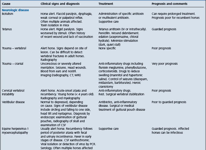
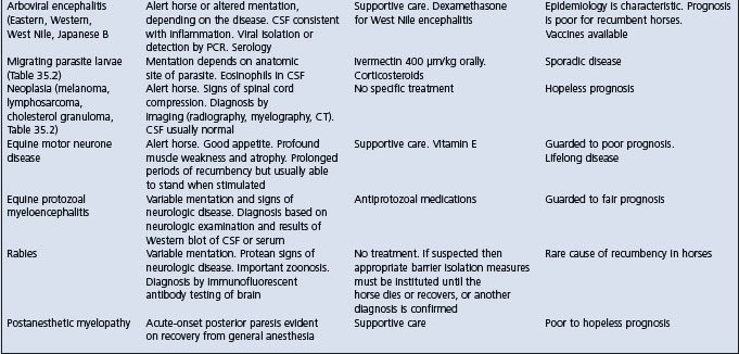
Table 2.7 Causes and diagnostic features of recumbency of more than 8 hours duration in adult horses
Careful questioning of the horse’s attendants can reveal valuable information regarding the cause of recumbency. Causes such as observed trauma, foaling and excessive unaccustomed exercise are readily determined from the history. In addition to inquiries about the cause of the recumbency, estimates of the duration of recumbency should be obtained from the attendants. This can often be best elicited by asking when the horse was last observed to be standing. A history of recent illness, abnormal behavior or unusual use immediately before the horse became recumbent is useful. The horse’s age, sex, breed and use should be determined. Information regarding management, vaccination and deworming status, feeding and health of other horses can be revealing. Outbreaks of recumbency suggest either an infectious (equine herpesvirus-1) or toxic (botulism, ionophore) cause. Questions should be directed toward discerning the cause of the horse’s recumbency rather than collecting information.
Physical examination of recumbent horses is challenging but should be as complete as practical and safe. The examination should begin with a general assessment of the horse and its surroundings and can be directed at answering a series of questions:
• Are the surrounding conditions safe for the horse and people? Is the footing sound?
• Is there evidence of the horse struggling or thrashing?
• Has the horse defecated and urinated recently?
• Is there evidence of exposure to toxins or physical evidence of the reason for recumbency?
Examination of the horse should begin with measurement of heart rate, respiratory rate and temperature (rectal temperature might not be accurate if there is dilation of the anus), examination of mucous membranes and an assessment of its hydration, body condition and level of consciousness. The horse should be thoroughly examined for evidence of trauma. Although the examination should be complete, initial examination of cases for which the cause of recumbency is not immediately obvious should focus on the nervous and musculoskeletal systems.
• Is the horse alert and able to sit in sternal recumbency or is it unconscious and in lateral recumbency? Can the horse rise with assistance?
• Is the horse’s mentation normal?
• Are there any spontaneous voluntary or involuntary movements?
• Can the horse eat and drink?
• Are the cranial nerves normal?
• Is there evidence of trauma to the head or neck?
• Is there evidence of paresis or paralysis? Are only the hind limbs involved or are both the hind limbs and forelimbs involved?
• Are the peripheral reflexes normal (withdrawal, patellar, cervicofacial, cutaneous, anal, penile)?
• Is cutaneous sensation present in all regions? If not, what are the anatomic boundaries of desensitized areas?
• Is the position of the limbs normal? Is there evidence of crepitus, swelling or unusual shape of the limbs or axial skeleton?
• Are the horse’s feet normal? Does it have laminitis? What is the response to application of hoof testers?
• Are abnormalities detected on rectal examination (fractured pelvis, distended bladder, fecal retention, pregnancy), provided that it is safe to perform one?
Other body systems should be evaluated as indicated or necessary. The heart and lungs should be auscultated, although detecting abnormal lung sounds in a recumbent horse is difficult. The horse should be rolled so that a complete examination can be performed. Assisting the horse to stand using a rope tied to the tail and thrown over a rafter, or preferably using a sling, can be useful in assessing the severity of the horse’s illness (can it stand at all?) and in facilitating a complete physical examination. If there is a suspicion that the horse has colic a nasogastric tube should be placed to check for accumulation of liquid gastric contents, a rectal examination performed and peritoneal fluid collected.
Ancillary diagnostic testing includes radiography of limbs and/or axial spine as indicated by the history or physical examination; myelography if a compressive lesion of the cervical spinal cord is suspected; endoscopic examination of the pharynx and guttural pouches (especially in horses with a history of falling, see Rupture of the longus capitus muscle, ultrasonography of the chest and abdomen; collection of cerebrospinal fluid; and electromyography.
Hematologic abnormalities are sometimes reflective of the causative disease. Serum biochemical abnormalities are reflective of the causative disease and in addition are influenced by muscle damage caused by the horse being recumbent (increased creatine kinase and aspartate aminotransferase activity), inappetent (increased total and indirect bilirubin, and triglyceride concentrations), and unable to drink or gain access to water (increased serum urea nitrogen, creatinine, sodium, chloride, total protein and albumin concentrations). Cerebrospinal fluid is reflective of any inciting disease but is usually normal.
The principles of care are treatment of the primary disease, prevention of further illness or injury, assisting the horse to stand, and provision of optimal nutrition and hydration.
Treatment of the primary disease is covered in other sections of this book. Similarly, maintenance of hydration and electrolyte status is covered elsewhere. Maintenance of normal hydration is sometimes problematic in recumbent horses because of limited access to water and unwillingness to drink. Provision of fresh, palatable water is essential. Intravenous or enteral (nasogastric intubation) administration of fluids and electrolyte solutions might be necessary in some recumbent horses, especially early in their illness.
Horses with diseases that cause recumbency often have problems with fecal and urinary incontinence or retention. Catheterization of the urinary bladder might be necessary to relieve distension in horses with neurogenic upper motor bladder or lower motor bladder dysfunction, or in male horses that are reluctant to urinate when recumbent. Catheterization of the bladder is often repeated. To minimize the risk of iatrogenic cystitis, the procedure should be performed aseptically. Administration of bethanechol might increase detrusor muscle tone and aid urination, and phenoxybenzamine (0.5 mg/kg intravenously over 15 min) might decrease sphincter tone in horses with upper motor neurone bladder.
Horses that can eat should be fed a balanced, palatable and nutritious diet. Tempting horses with reduced appetite with treats such as apples, carrots and horse treats might stimulate appetite for hay and grain. Horses that are unable to eat should be fed through a nasogastric tube. Slurries of alfalfa pellets or commercial diets can be administered through nasogastric tubes. The maintenance needs of a sedentary 425 kg horse are approximately 15–18 Mcal/d. The maintenance needs of a recumbent horse are unknown, but are probably less than that of normal sedentary horses.
A major challenge in managing recumbent horses is preventing further injury. Recumbent horses often make repeated efforts to stand, which, while encouraging to all involved, can result in further injury. Horses attempting to stand can injure their head, especially the periorbital regions, and skin over bony prominences such as over the wing of the ilium. Minimizing further injury is achieved by use of a sling or tail rope to assist horses to stand, housing in a padded stall with deep, soft bedding (although this can interfere with the horse’s ability to stand), and protection of the head and distal limbs with a helmet and bandages, respectively. Recumbent horses kept in well-grassed pasture often do well and have minimal self-inflicted trauma.
Decubital ulcers occur over pressure points such as the wing of the ilium, point of the shoulder and zygomatic arch, and can become severe. Recumbent horses that paddle can abrade the skin over limb joints with subsequent increased risk of septic arthritis. Bandages, helmets, ointments such as silver sulfadiazine paste, and soft bedding minimize but do not eliminate these abrasions. Recumbent horses that cannot or do not voluntarily move from side to side should be rolled every 2–4 hours.
Peripheral pressure neuropathy can occur in recumbent horses. The radial nerve and facial nerve are most often affected. Prevention is achieved by use of padded bedding, slings, frequent rolling and a helmet.
Recumbent horses can sustain muscle damage from pressure on large muscle groups. For large or well-muscled horses this can result in large increases in serum creatine kinase activity and myoglobinuria. Myoglobinuria can cause acute renal failure, although this degree of myoglobinuria in recumbent horses is unusual.
Pneumonia can occur as a result of recumbency. Horses that are dysphagic are at increased risk of aspiration of feed material and saliva, and hence development of aspiration pneumonia. Horses receiving corticosteroids are at increased risk of bacterial and fungal (Aspergillus spp.,) pneumonia. While not every recumbent horse should be administered antimicrobials, this is indicated in horses at increased risk of developing pneumonia. Antimicrobials should have a broad spectrum, including activity against Streptococcus spp., such as a combination of penicillin and an aminoglycoside.
Slinging horses is labor-intensive and requires the use of a sling that is designed for use with horses. Horses should not be lifted using hip slings intended for use with cattle. Use of these slings to lift horses by grasping over the wing of each ilium is inhumane and unsuccessful. Horses in slings should be closely monitored and not allowed to hang in the sling. The horses should be assisted to stand in the sling every 6 or 8 hours. The sling should be used to help the horse to get up and provide some support while it is standing, but the horse should not have all its weight borne by the sling for more than a few minutes. Horses that have an excessive amount of weight borne by the sling for a prolonged period of time have trouble breathing and are likely to develop colic, rupture of the urinary bladder, diaphragmatic hernia or rectal prolapse.
Potentially catastrophic complications include septic arthritis, radial nerve injury, bladder rupture, diaphragmatic hernia, rectal prolapse, colon torsion and long bone fracture. The risk of these complications can be minimized by the practices detailed above, but cannot be eliminated.
Chandler K. Clinical approach to the recumbent adult horse. In Practice. June: 2000:308.
Davis EG, et al. Treatment and supportive care of recumbent horses. Compend Contin Educ Pract Vet. 2004;26:216.
Rush BR, et al. Compend Contin Educ Pract Vet. 2004;26:256.
Nout YS, Reed SM. Management and treatment of the recumbent horse. Equine Vet Educ. 2005;17:324.
Sudden or unexpected death
When an animal is found dead without having been previously observed to be ill, a diagnosis, even after necropsy examination, is often difficult because of the absence of a detailed history and clinical findings. A checklist of diseases for consideration when sudden or unexpected death occurs in a single animal or group of animals is provided below. Details of each of the diseases listed are available in other sections of this book. The list applies particularly to cattle, but some occurrences in other species are noted. It is necessary to point out the difference between ‘found dead’ and ‘sudden death’.
When animals are observed infrequently, for example at weekly intervals, it is possible for them to be ill with obvious clinical signs for some days without being observed. In these circumstances the list of possible diagnoses is very large. It is also correspondingly large when animals are kept together in large groups and are not observed as individuals. This is likely to happen in beef cattle, especially in feedlots or as calves with dams at pasture, when the animals are unaccustomed to human presence and move away when approached. The list below refers to animals that are closely observed as individuals at least once daily.
This condition could be due to cardiac tamponade in cows, ruptured aorta or atrium, inherited aortic aneurysm or verminous mesenteric arterial aneurysm in horses and esophagogastric ulcer or intestinal hemorrhagic syndrome in pigs.
This condition may occur secondary to mycosis of guttural pouch of the horse. In one survey of sudden deaths in horses while racing, most (68%) were undiagnosed, although it was assumed that they died of exercise-associated ventricular arrhythmias. Of those that were diagnosed, most were due to spontaneous hemorrhage.1 Similar conclusions have resulted from other surveys.2 Most reported cases of sudden death in the horse are the result of cardiovascular accidents.3,4
Fracture of the pelvis can result in fatal hemorrhage within the gluteal muscles of the horse5 and rupture of the middle uterine artery at parturition in cattle may occur with uterine prolapse.
This condition can arise from rupture of the stomach of horses, abomasum of cows and colon in mares at foaling. Large amounts of gastrointestinal contents are deposited rapidly into the peritoneal cavity. In newborn animals, especially foals, fulminating infections are the commonest cause.
Peracute exogenous toxemia in a single animal could be as a result of snakebite, but the snake would have to be very poisonous and the animal of small body weight to cause death without any observable illness.
Trauma may cause death by either internal hemorrhage or damage to the central nervous system, especially the brain or atlanto-occipital joint sufficient to damage the medulla oblongata. In most cases the trauma is evident: there has been fighting, or a fall has occurred, or the animal has attempted to jump an obstacle. In horses a free gallop downhill may result in a serious fall or collision with, for example, a wall, especially if the ground is slippery.
Inapparent trauma usually occurs when animals are tied up by halter and rush backwards when frightened or are startled by an electric fence and the halter shank is long. Sometimes the animal will plunge forward and hit its forehead between the eyes on a protruding small object such as a bolt used in a fence. Sadism, especially by the insertion of whip handles or pitchfork handles into the anus or vulva, may also be inapparent.
Gastric rupture in the horse may occur following overeating highly fermentable feed, administration of excessive quantities of fluids by nasogastric tube, gastric impaction or when gastric motility is markedly reduced in acute grass sickness or gastric distension with fluid. Peracute enteritis in the horse can cause rapid unexpected death.
Volvulus or gastrointestinal accidents account for almost 50% of sudden deaths in sows, followed next by gastric ulceration, retained fetuses and toxemia.6
Recumbent cattle that become lodged in a small hollow in the ground may die of bloat7 because the cardia becomes covered with ruminal fluid and eructation is not possible.
These may be due to overdose with intravenous solutions of calcium salts in an excited cow, too-rapid fluid infusion in an animal with pulmonary edema, intravenous injection of procaine penicillin suspension, and intravenous injections of ivermectin in horses. These are not hard to diagnose and the producer or veterinarian is usually obviously embarrassed.
One of the most sudden death occurrences is the anaphylactoid reaction in a horse to an intravenous injection of an allergen such as crystalline penicillin. Death occurs in about 60 seconds. Intra-arterial injections of penicillin or phenothiazine tranquilizers have also been reported to cause sudden death.3
An analysis was made of the causes of death in horses and ponies over 1 year of age that died suddenly and unexpectedly.8 No cause of death was found in 31% of cases and 16% died from the following causes: hemorrhage in the respiratory tract, central nervous system and adverse drug reactions. Cardiovascular lesions were the cause in 14% and the remaining 3% had lesions of the gastrointestinal tract.
Sudden death in racehorses is commonly due to massive hemorrhage into the lungs, abdomen or brain.5 In horses that were found dead but appeared normal when last seen, the cause of death was not determined in 33%. Lesions of the gastrointestinal tract were the cause of death in 39% and respiratory tract lesions in 9%. Lesions of both the central nervous system and cardiovascular system were the cause of death in 5%. The remaining 10% had miscellaneous causes.
The diseases listed below could obviously affect single animals if the animals were housed or run singly.
This usually affects a number of animals that are found together in a pile or group. Rarely, electrical current only electrifies a contact object intermittently and deaths will be intermittent. In most cases the history and an examination of the environment reveals the cause.
At pasture, sudden death may come from the sudden exposure of the cattle to plants that cause bloat, hypomagnesemia, cyanide or nitrite poisoning, fluoroacetate poisoning, fast death factor (produced by algae in a lake or pond) or acute interstitial pneumonia. Acute myocardiopathy in young animals on diets deficient in vitamin E or selenium is in this group, as is inherited myocardiopathy in Herefords. Gross nutritional deficiency of copper in cattle causes ‘falling disease’, a manifestation of acute myocardiopathy.
Acute myocardiopathy and heart failure is associated with poisons in Phalaris spp. pasture, grass nematodes on Lolium rigidum, the hemlocks Cicuta and Oenanthe spp. and the weeds Fadogia, Pachystigma, Pavette, Ascelapius and Aeriocarpa, Crystostegia and Albizia, Cassia spp. The trees oleander and yew (Taxus spp.) may also be causes, and those species containing fluoroacetate, such as the gidgee tree and the weeds Gastrolobium, Oxylobium, Dichapetalum and Ixioloena spp. may be implicated. There are a number of plants that cause cardiac irregularity and some sudden deaths, e.g. Urginea, Kalanchoea spp., but more commonly congestive heart failure is caused. Monensin, lasolocid and salinomycin toxicities are increasingly common causes in horses and, to a less extent, cows.
Access to potent poisons may occur in housed animals or in those fed prepared feeds.
There are few poisons that cause sudden death without premonitory signs. Cyanide is one, but is an unlikely poison in these circumstances. Monensin, mixed in a feed for cattle that is then fed to horses, or fed in excess to cattle, does cause death by heart failure. Organophosphates are more likely, but clinical signs are usually apparent. Lead is in a similar category; however, very soluble lead salts can cause death quickly in young animals.
These cause septicemia or toxemia, and include anthrax, blackleg, hemorrhagic septicemia and (especially in sheep, but occasionally in cattle) peracute pasteurellosis. In pigs, mulberry heart disease and perhaps gut edema should be considered. In horses, colitis is probably the only disease that will cause sudden death. In sheep and young cattle, enterotoxemia associated with C. perfringens should be included and this may be involved in rumen overload in feedlot cattle on heavy grain feed. Circumstances, feeding practices, climate and season of the year usually give some clue as to the cause.
In very young, including neonatal, animals, congenital defects that are incompatible with life – prematurity, septicemia because of poor immune status or toxemia associated with particular pathogens, especially E. coli, and hypothyroidism – are important causes of sudden death.
Anaphylaxis after injection of biological materials, including vaccines and sera, is usually an obvious diagnosis, but its occurrence in animals at pasture can cause obscure deaths. In these circumstances it usually affects one animal and clinical illness is often observed. A similar occurrence is sudden death in a high proportion of piglets injected with an iron preparation when their selenium–vitamin E status is low.
• Keep excellent records because of the probability of insurance enquiry or litigation
• Take a careful history, which may indicate changes of feed composition or source, exposure to poisons or administration of potentially toxic preparations
• Make a careful examination of the environment to look for potential sources of pathogens. Be especially careful of your personal welfare if electrocution is possible – wet concrete floors can be lethal when combined with electrical current unless you are wearing rubber boots
• Carefully examine dead animals for signs of struggling, frothy nasal discharge, unclotted blood from natural orifices, bloat, pallor or otherwise of mucosae, burn marks on body, especially on the feet, or signs of trauma or of having been restrained. Pay particular attention to the forehead by palpating the frontal bones – these may have been fractured with a heavy blunt object without much damage to the skin or hair
• Ensure that typical cadavers are examined at necropsy, preferably by specialist pathologists at independent laboratories, where opinions are more likely to be considered authoritative and unbiased
• Collect samples of suspect materials for analysis. Preferably, collect two samples, one to be analyzed and one to be made available to a feed company, if indicated.
1 Gelberg HB, et al. J Am Vet Med Assoc. 1985;187:1354.
2 Platt H. Br Vet J. 1982;138:417.
3 Lucke VM. Equine Vet J. 1987;19:85.
4 Allen JR, et al. Equine Vet J. 1987;19:145.
5 Brown CM, Mullaney TP. In Pract. 1991;13:121.
6 Sanford SE, et al. Can Vet J. 1994;35:388.