Signs and Symptoms of Congestive Heart Failure in Children

Signs of Adrenergic Response

Tachycardia

Tachypnea

Cool skin

Oliguria

Diaphoresis

Signs of Systemic Venous Congestion

Hepatomegaly

Periorbital edema

Ascites (rare)

Pulmonary effusion

Signs of Pulmonary Venous Congestion

Tachypnea

Retractions

Nasal flaring

Pulmonary edema

Nonspecific Signs of Cardiorespiratory Distress

Irritability

Change in responsiveness

Fatigue

Poor feeding, failure to thrive

Table 8-6 Ross Classification of Congestive Heart Failure

Ross Classification System of Pediatric Heart Failure
Class Symptoms
I None
II Mild tachypnea/diaphoresis with feedings in infants
Exertional dyspnea in children
III Marked tachypnea/diaphoresis with feeding in infants
Marked exertional dyspnea in children
IV Symptoms at rest (diaphoresis, tachypnea, tachycardia, retractions)

From Ross RD, Bollinger RO, Pinsky WW: Grading the severity of heart failure in infants. Pediatr Cardiol 13:72-75, 1992.

Adrenergic Stimulation

Adrenergic stimulation produces tachycardia and redistribution of blood flow. The increase in heart rate may succeed in maintaining effective cardiac output despite reduced ventricular function. However, tachycardia reduces ventricular diastolic filling time and stroke volume, and decreases left coronary artery myocardial perfusion time. Tachycardia also increases myocardial oxygen consumption. For these reasons a mild increase in heart rate may succeed in maintaining cardiac output, but significant rises in heart rate may contribute to further deterioration.284 A third heart sound or a summation gallop may be produced by the rapid filling of a noncompliant ventricle. However, extra heart sounds may be difficult to distinguish once the heart rate exceeds 120 to 140/min.

Adrenergic stimulation also produces peripheral vasoconstriction, reduced renal blood flow, and diaphoresis. The child's extremities are often cool (they cool from peripheral to proximal areas), with a pale or mottled color. Decreased renal blood flow results in a urine volume of less than 0.5 to 1.0   mL/kg per hour, despite adequate fluid intake. Urine sodium concentration is usually low, and a microscopic hematuria often is present.841 Diaphoresis may be observed in infants, particularly over the head and neck.

Systemic Venous Congestion

When right ventricular dysfunction develops, right ventricular end-diastolic pressure increases and right atrial and central venous pressures rise. This produces systemic venous congestion. With hepatic venous hypertension the liver sinusoids fill with blood, so the liver enlarges and becomes palpable below the child's right costal margin; this hepatomegaly is one of the earliest signs of systemic venous congestion in children. The infant and child also may demonstrate periorbital edema.

Because jugular venous distension is difficult to perceive in the short, fat neck of the infant, it is not a reliable sign of congestive heart failure until the child is school age or older. Dependent edema, or ascites, is rarely seen in children unless the central venous pressure is extremely high (as may occur after Fontan-type surgical correction of tricuspid atresia), or unless it is associated with other metabolic problems, such as hypoalbuminemia or renal failure.

If ascites does develop it is the result of high central venous and portal venous pressures, and loss of fluid from the surface of the liver or from the surfaces of the gut and mesentery. This high-pressure exudate results in movement of both fluid and protein into the peritoneal cavity. The presence of protein in the ascitic fluid will draw more fluid from the surface of the gut and mesentery, so the ascites often increases.

If ascites is present the child's abdominal girth increases, the abdomen appears full, and the skin is taut and shiny. If two examiners are present a fluid wave may be elicited, and areas of shifting dullness may be noted during percussion. (These and other signs and symptoms of ascites are discussed in detail in Chapter 14.)

Pulmonary Venous Congestion

Signs of respiratory distress are often the first and most noticeable signs of congestive heart failure in the infant. Left ventricular failure results in a rise in left ventricular end-diastolic pressure and an increase in pulmonary venous pressure; pulmonary edema develops once pulmonary venous pressure is 20 to 25   mm Hg, but occurs at lower pressure if capillary permeability is increased. Most commonly, children with CHF demonstrate pulmonary interstitial (rather than alveolar) edema. Pulmonary edema, in turn, reduces lung compliance and increases the work of breathing; these changes result in tachypnea and increased respiratory effort.

Intercostal, subcostal, sternal, supraclavicular, or suprasternal retractions may be noted, and are particularly apparent in the infant. Older children may demonstrate use of accessory muscles of respiration, including use of scapular muscles and the sternocleidomastoid. Because these muscles are inadequately developed in the infant, “head bobbing” may be noted. Nasal flaring is an additional sign of respiratory distress.

The infant or child with severe respiratory distress grunts with expiration. The grunting results from expiration against a closed glottis and is an instinctive attempt to maintain PEEP and prevent atelectasis and collapse of small airways.

Rales (crackles) often are not observed in infants with congestive heart failure, despite the presence of pulmonary interstitial edema. Children with respiratory distress tend to breathe shallowly, so small airway sounds are less likely to be appreciated. In addition, pulmonary interstitial edema is cleared rapidly into lymphatics when tachypnea is present.841 If crackles are noted, the presence of severe congestive heart failure or a concurrent respiratory infection should be suspected. Wheezes may be heard, especially if the child has a large left-to-right shunt. Lobar emphysema or atelectasis occasionally may result from cardiac compression of larger airways.

If the central venous pressure is extremely high, a pleural effusion or chylothorax may develop. These complications produce a decrease in intensity or a change in pitch of breath sounds over the affected chest. Chest expansion (especially that noted during positive pressure ventilation) will be compromised on the involved side, and hypoxemia may be noted.

Nonspecific Signs of Distress

Subtle signs of cardiorespiratory distress in infants and children include a change in disposition or responsiveness; unusual lethargy or irritability is often observed. The infant usually requires prolonged feeding times, sucks poorly, and takes only small amounts of formula. Typically the infant falls asleep during or immediately after the feeding because the work of breathing is significant. The infant usually swallows a large amount of air during feedings, so gastric distension develops. Vomiting is common after feeding.

These infants have high energy requirements and poor caloric intake, therefore failure to thrive is common. Often any weight gain noted results from edema rather than nutrition.

Laboratory Evaluation

The electrocardiogram is not helpful in the diagnosis of congestive heart failure unless associated heart block or an arrhythmia is present. If ventricular dilation is present, cardiomegaly will be apparent on the chest radiograph (see Fig. 10-12). Pulmonary interstitial edema also may be noted. An echocardiogram is helpful in identifying congenital heart disease or pericardial effusion and evaluating cardiac chamber size, ventricular contractility and ejection fraction.

Free water retention (disproportionate to the amount of sodium retention) usually produces a dilutional fall in serum sodium and hemoglobin concentrations. True anemia may result from increased red blood cell destruction or reduced red blood cell production. The bilirubin, lactic dehydrogenase (LDH), and reticulocyte counts are elevated in the patient with accelerated red blood cell destruction. If the anemia is caused by reduced red blood cell production, the reticulocyte count is inappropriately low. Hypoglycemia occasionally is noted in very small infants with congestive heart failure because metabolic needs are high and glycogen stores are minimal.841

Serum natriuretic peptides are increasingly being used as biomarkers to aid in the identification and monitoring of congestive heart failure. Although atrial natriuretic peptide (ANP) was the first of these peptides to be described, brain natriuretic peptide (BNP), initially discovered in brain matter, was later found in significantly higher amounts in ventricular myocardium.713

BNP is released from the heart as a prohormone (pro-BNP), which is subsequently cleaved into the active 32 amino acid polypeptide, BNP, and a metabolically inactive portion, N-terminal pro-BNP (NT pro-BNP). Consequently, serum assays of serum BNP and NT pro-BNP levels have been shown to be both sensitive and specific for the presence or worsening of heart failure in both adult205,560,634,790 and pediatric patients.187,281,345,518

Management

Care of the child with congestive heart failure targets improvement of cardiac function and elimination of excess intravascular fluid. In addition, oxygen delivery must be supported and oxygen demands controlled or minimized. Reversible causes of congestive heart failure also must be treated.

Improvement in Cardiac Function: Digitalis Derivatives

Digitalis may be extremely effective in the treatment of congestive heart failure. It must, however, be used appropriately with careful monitoring for therapeutic and side and toxic effects.

Therapeutic Effects

Digitalis (administered predominantly as digoxin in children) continues to be used in the treatment of congestive heart failure in children, although evidence of its efficacy in pediatric patients is controversial.435 The presumed effect of digoxin in older children is inotropic, improving ventricular contractility. Digoxin also affects the excitability of myocardial cells and slows the heart rate. Pediatric digoxin therapy (particularly during infancy) also appears to relieve some symptoms of congestive heart failure through effects on oxygen consumption.

The inotropic effects of digoxin result from inhibition of the sodium-potassium pump, so that sodium accumulates intracellularly. Sodium then competes with calcium for sites on the sodium-calcium exchange mechanism, raising intracellular calcium levels and improving myocardial contractility. Digitalis also interacts with the sarcolemma to sequester calcium, so that intracellular calcium is increased further.355,841

Digoxin also slows the heart rate by lowering the resting membrane potential, increasing parasympathetic sensitivity, and reducing sensitivity to norepinephrine. It slows conduction velocity and increases recovery time of the atrioventricular (AV) node. These effects result in a decrease in heart rate but also may contribute to increased ectopic activity.

Digitalis has some direct and indirect peripheral and coronary vasoconstrictive effects, resulting in increased systemic vascular resistance.355,735 When digitalis is administered to patients with congestive heart failure, it appears to antagonize peripheral effects of catecholamines, thereby reducing oxygen consumption.

The clinical effects of digoxin administration in infants may be variable,473,789 and positive effects of digoxin may be related to effects on oxygen supply and demand rather than to inotropic effects. Digoxin administration to the premature neonate may not produce significant improvement in contractility, and the incidence of clinical toxicity is significant in these patients.435 For this reason, digoxin may not be useful in the treatment of congestive heart failure in premature neonates.

Digoxin administration often fails to produce measurable echocardiographic evidence of improvement in cardiac contractility in full-term infants with congestive heart failure caused by a ventricular septal defect. However, demonstrable clinical improvement often is noted; this improvement probably is related to a significant fall in oxygen consumption.435 Studies of digoxin's effects on animals during periods of induced anemia and reduced oxygen transport have documented reduced oxygen consumption and improvement in the match between oxygen supply and oxygen demand.769

Dose

When digoxin therapy is initiated, several loading (“digitalizing”) doses typically are administered to provide therapeutic serum levels of the drug. Once these levels are achieved, maintenance doses of digoxin are administered to replace the child's estimated daily renal excretion of the drug, so that therapeutic levels are maintained (Table 8-7).

Table 8-7 Pediatric Digoxin Dose507

image

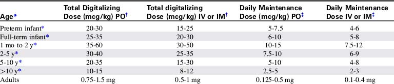
Digoxin doses must be modified based on the child's age and clinical condition. Because digoxin is excreted through the kidneys, neonates who have limited renal tubular function and children with renal failure require reduced dosing. Premature neonates have limited renal tubular function; they are more sensitive to digoxin and require smaller digoxin doses than full-term neonates or older children.355 For this reason the risk of digoxin toxicity is highest in premature neonates.

Infants have a larger volume of digoxin distribution than older children because infants have more red blood cell binding sites for digoxin than do older patients. For this reason, higher loading and maintenance doses of digoxin were recommended for neonates and young infants in the past. Although young infants apparently tolerate high doses and high serum digoxin levels better than adults, it is not clear that the higher dosing is beneficial during infancy. Beyond the neonatal period patients normally excrete approximately one third of the daily administered oral dose.507

Whenever digoxin is administered to a critically ill child, careful consideration of dose and careful monitoring for evidence of toxicity is required. In any patient the appropriate dose of digoxin is the minimum dose necessary to produce therapeutic effects. There is a very small difference between optimal therapeutic levels of digoxin and toxic levels of the drug.

Children with inflammatory cardiac diseases (e.g., myocarditis or cardiomyopathy), chronic hypoxemia, and postoperative cardiovascular patients may demonstrate increased sensitivity to the drug.355 When providing digoxin therapy for the first time to these patients it is prudent to consider providing lower doses of digoxin, or more gradual loading of the drug. Maintenance doses of digoxin may be administered without previous loading doses; this will result in achievement of therapeutic digoxin levels over 4 to 5 days.841

The total oral digitalizing dose for children is approximately 25 to 60   mcg/kg (or 0.025-0.060   mg/kg), and the higher doses in these ranges usually are administered to children 2 years of age or less. The digitalizing or loading dose usually is administered in two to four divided oral doses over 24 to 48   hours.

The maintenance digoxin dose is approximately one eighth (12%) of the total loading dose, administered twice a day. The child receives approximately one fourth of the loading dose daily.

Administration

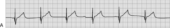
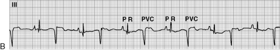
Before administration of the final loading dose, an electrocardiogram or rhythm strip is typically ordered to ensure that arrhythmias have not developed. Because digoxin slows conduction through the AV node, an increased P-R interval often is present following establishment of a therapeutic serum digoxin level. However, second- or third-degree heart block or ectopy should be reported to a physician or the on-call provider before the next dose of digoxin is administered.

In the hospital the child's heart rate usually is checked before administration of any digoxin. If bradycardia is detected the dose usually is held until an electrocardiographic rhythm strip is obtained and toxic heart block or arrhythmias are ruled out. When attempting to determine if digoxin toxicity is present, a particular heart rate is usually less important than assessment of the child's systemic perfusion and evaluation of the electrocardiogram for evidence of heart block (see section, Digoxin Levels).

If the child vomits after administration of an oral dose of digoxin in the hospital, a physician or on-call provider should be consulted before the dose is readministered. It is often difficult to determine how much of the dose was lost, so a repeat dose may result in elevation of serum digoxin levels. In addition, vomiting may be a sign of digoxin toxicity.

It is imperative that the written order for and preparation of the digitalizing doses be double checked before administration because it is very easy to make an error by a factor of 10 or 100 when working with micrograms and milligrams. The order for the digoxin dose should be written (verbal orders should not be used) to avoid miscommunication. The order should be written in both milligrams and micrograms.

Digoxin should be administered with caution to children with decreased renal function, and the dose should be reduced accordingly. Hypokalemia can contribute to the development of clinical signs of digoxin toxicity even in the presence of relatively low serum digoxin levels, so the serum potassium should be monitored and potassium supplementation provided as needed. Hypomagnesemia and hypercalcemia also may aggravate digitalis toxicity, and quinidine may potentiate digoxin toxicity.355 This is especially pertinent as many patients receiving digoxin are also on concomitant diuretics, which may cause electrolyte disturbances.

Digoxin Levels

A serum digoxin level may be monitored when digoxin therapy is instituted (during the “loading phase”), when the child's response to therapy is suboptimal, when toxicity is suspected, or when the drug dose is changed. Therapeutic serum digoxin levels vary from institution to institution but are in the range of 1.1 to 2.2   ng/mL (nontoxic levels in infants may be as high as 3.5   ng/mL). Serum digoxin levels exceeding 3.5   ng/mL are generally considered toxic.

Blood sampling for serum digoxin levels should be performed at prescribed intervals following digoxin administration (consult with the child's physician or ordering provider and hospital clinical laboratory). These levels must be interpreted with caution. As noted, hypokalemia, hypomagnesemia, and hypercalcemia can aggravate digoxin cardiotoxicity even in the presence of “normal” digoxin levels.735 Some children exhibit endogenous digitalis-like substances that can influence serum digoxin levels. In addition, premature neonates may demonstrate bradyarrhythmias even in the presence of “therapeutic” levels of digoxin. For these reasons the presence of clinical symptoms compatible with digoxin toxicity usually is interpreted more strongly than the serum digoxin level alone.

Several common critical care drugs are known to affect digoxin levels. Amiodarone, verapamil, diltiazem, spironolactone, carvedilol and indomethacin all increase serum digoxin levels.735 The digoxin dose should be reduced during concurrent administration of these drugs, and serum digoxin levels should be monitored.

Digoxin Toxicity

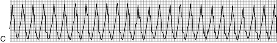
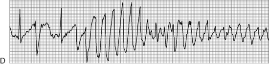
The most serious toxic effects of digoxin in children are arrhythmias, which may be observed in the absence of other clinical signs of toxicity. The most common arrhythmia in young children is bradycardia, although heart block and atrial and ventricular premature contractions (ectopy), and ventricular fibrillation have been reported. Virtually any new arrhythmia appearing after initiation of digoxin therapy may be caused by digoxin toxicity.735

Less specific and less common signs of digoxin toxicity in children include anorexia, nausea, vomiting, and diarrhea. Drowsiness and lethargy are common signs of toxicity in infants and young children.356

If digoxin toxicity is suspected, the physician or on-call provider should be notified; further digoxin usually is held pending the results of serum digoxin-level testing. A blood specimen should be drawn for laboratory analysis. Toxicity may be present at levels as low as 2.5   ng/mL or lower; the serum digoxin level must be evaluated in light of the patient's clinical condition.355

If toxicity is discovered in the asymptomatic child, electrocardiographic monitoring should be instituted and the child should be observed closely for the development of arrhythmias. If large amounts of oral digoxin have recently been ingested or administered, induced emesis soon after the ingestion may succeed in recovering 35% to 40% of the ingested drug. Vomiting should only be induced if the child is alert and demonstrates a cough and gag reflex. If massive amounts of digoxin have been ingested, the insertion of temporary transvenous pacing wires is recommended before the development of symptoms.

Treatment of symptomatic digoxin toxicity requires support of cardiovascular function (including treatment of arrhythmias), prevention of further drug absorption, and enhancement of digoxin excretion.356 Bradycardia usually is treated with atropine or pacemaker therapy. Phenytoin (2-5   mg/kg IV) often is effective in the treatment of digoxin-induced arrhythmias because it increases the sinoatrial node conduction rate and reduces automaticity. Lidocaine does not affect atrial activity but will suppress ventricular automaticity, and so may be effective in the treatment of ventricular tachyarrhythmias.735,841 Synchronized cardioversion may convert ventricular tachycardia to refractory fibrillation,365 so it should not be performed.

When renal function is reduced, digoxin excretion is impaired and toxicity may develop more readily. The digoxin level may remain elevated long after the digoxin therapy is stopped in these patients, so support of cardiorespiratory function may be required for several days.

Digoxin excretion is not improved by the administration of furosemide (or other diuretics), exchange transfusion, or dialysis. Hemoperfusion using activated charcoal has had limited effect because the digoxin usually is distributed and bound extensively in tissue.356,365

Life-threatening digoxin toxicity associated with malignant arrhythmias, hypotension, and poor systemic perfusion is treated with digoxin-specific Fab antibody fragments.507 This antibody binds serum digoxin, rendering it inactive.365 The dose of Fab provided is determined by the total body exposure to digoxin, which can be estimated from the digoxin level or the amount of digoxin ingested (for formulas, see Table 8-7 or package insert). In general, approximately 40   mg of purified digoxin-specific Fab will bind approximately 0.5   mg of digoxin.507 Note that digoxin elixir is considered to be absorbed totally, while digoxin tablets generally are calculated to be 80% absorbed.

Parent Instruction

If the parents will be administering digoxin at home, the parents must be taught how to administer the drug. In addition, the parents must be taught what to do if a dose is omitted or if the child vomits after the medication is administered. It is usually helpful to provide the parents with a specific approximate administration schedule; for example, the digoxin may be given at 8 AM and 8 PM. If the morning or evening dose is forgotten, but remembered by 12 noon or midnight, respectively, it may be given. However, if the drug is forgotten and not remembered until after 12 noon or midnight, that dose should be omitted and should not be “made up” in subsequent doses. If the parents are unsure whether a specific dose was administered, that dose should be omitted. If the child vomits after receiving the digoxin, the dose probably should not be repeated because it is difficult to predict how much of the drug was absorbed before regurgitation.

Parents should be taught to contact the child's physician or on-call provider if more than one dose of digoxin is omitted or if the child appears ill for any reason, because digoxin toxicity may be present. Parents are typically not taught to count the child's pulse routinely before administration of digoxin doses, because this focuses attention on specific numbers rather than on overall assessment of the child, and it can increase the parents' anxiety. Monitoring of heart rate does not ensure better detection of digoxin toxicity than that resulting from general evaluation of the child's condition.

The parents should be aware that a digoxin overdose may cause serious arrhythmias or death. Digoxin must be kept out of reach of children, and the medication bottle should have a “child-proof” cap.

Improvement in Cardiac Function: Additional Inotropic Agents

Several inotropic agents may effectively improve myocardial contractility during the treatment of congestive heart failure. Dopamine, dobutamine, and epinephrine are adrenergic agonists that may be titrated to provide beta-1 sympathomimetic effects (increased heart rate, atrioventricular conduction velocity, and ventricular contractility). Each of these drugs also may produce peripheral vascular effects that must be considered during drug selection and administration. An additional nonadrenergic inodilator, milrinone, improves myocardial contractility by inhibition of phosphodiesterase, so that intracellular effects of circulating catecholamines are prolonged. (For further information see Shock, Chapter 6.)

Vasodilator Therapy

Vasodilator therapy may improve myocardial function by altering both ventricular preload and afterload. Ventricular preload is reduced as a result of venodilation and displacement of blood volume into venous capacitance vessels. Ventricular afterload is reduced as a result of arterial dilation; in addition, ventricular wall stress decreases when ventricular chamber size is reduced. When vasodilator therapy is provided, the patient's ventricular compliance curve is altered, so that higher ventricular end-diastolic volume (and ultimately, stroke volume) is present at lower ventricular end-diastolic pressure (see Fig. 8-13 earlier in chapter).

Obviously the beneficial effects of vasodilators must be balanced with the potential detrimental effects of reduction in venous return and the potential fall in blood pressure. The hypovolemic patient is particularly likely to become hypotensive during vasodilator therapy. Volume expanders always should be readily available during the initial administration of these drugs.

No vasodilator is a pure arterial or venous dilator. However, these drugs often are classified by their primary sites of action. Vasodilators that dilate both arteries and veins include nitroprusside, phentolamine, prazosin, captopril, and nifedipine. Predominant arterial dilators include hydralazine and minoxidil. The most common venodilator is nitroglycerin. For further information about the dose, administration, and effects of these vasodilators the reader is referred to Shock in Chapter 6.

Angiotensin Converting Enzyme (ACE) Inhibitors

Angiotensin-converting-enzyme (ACE) inhibitors block the conversion of angiotensin I to angiotensin II, resulting in potent vasodilation. ACE inhibitors also block the breakdown of bradykinin, a powerful vasodilator, so it prolongs bradykinin action and augments afterload reduction (Fig. 8-14). In patients with left-to-right shunt lesions, reduction in systemic vascular resistance promotes blood flow into the systemic circulation rather than shunting to the pulmonary vascular bed.552

image

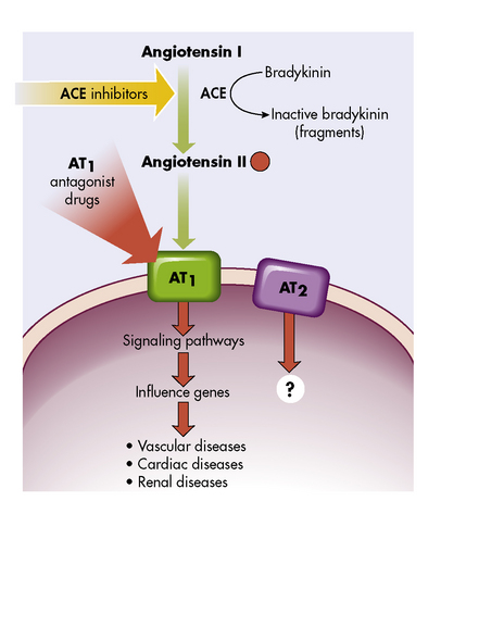
Fig. 8-14 Angiotensins and their receptors, AT1 and AT2. Blocking the angiotensin-converting enzyme (ACE) with ACE inhibitors decreases the amount of angiotensin II. Blocking the receptor AT1 with drugs (AT1 antagonists) blocks the attachment of angiotensin II to the cell, preventing the cellular effects and decreasing the vascular, cardiac, and renal effects.

(From McCance KL, Huether SE: Pathophysiology: The biologic basis for disease in adults and children, ed 6. St Louis, Mosby, 2009.)

ACE inhibitors also inhibit ventricular remodeling modulated by angiotensin II, thereby preventing the development of ventricular hypertrophy.697 Ultimately, these drugs have been shown to reduce mortality in adults with CHF and in children with dilated cardiomyopathy.530,829,850,861 Side effects include hypotension, cough, hyperkalemia, headache, dizziness, fatigue, nausea, and renal impairment.

Careful monitoring of hemodynamic status should be carried out in all patients with CHF when ACE inhibitors are started, because compromised cardiac output increases the risk of significant hypotension. Initial doses of ACE inhibitors are purposely low and then titrated upward to achieve maximal therapeutic effect.

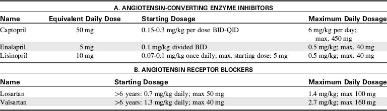
Angiotensin Receptor Blockers

Because of other conversion pathways, ACE inhibitors do not completely prevent the formation of angiotensin II. Therefore, angiotensin receptor blockers (ARBs) are useful for patients who develop side effects from ACE inhibitors. Specifically, the cough associated with ACE inhibitors is caused by elevated bradykinin levels. Because ARBs do not affect bradykinin metabolism, ARBs are a logical choice when this particular side effect is observed (see Table 8-8). Although the benefits of ARBs in adults with heart failure are well described, there is limited information regarding use of ARBs in children with heart failure. Losartan has been used safely in children with Marfan syndrome113 and is the subject of a current multicenter trial.

Table 8-8 Angiotensin-Converting Enzyme (ACE) Inhibitors and Angiotensin-Receptor Blockers (ARBs)

image
Reduction in Intravascular Volume: Diuretic Therapy

Limitation of fluid intake and improvement in systemic perfusion and blood distribution may increase renal perfusion sufficiently in the child with congestive heart failure to prompt a diuresis. However, administration of diuretics is often necessary to aid in the elimination of excess intravascular fluid (Table 8-9).

Table 8-9 Diuretic Therapy for Children

image

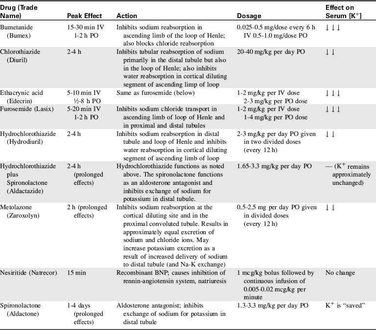
The most common diuretics used in children are the loop diuretics. These drugs block sodium and chloride reabsorption in the ascending limb of the loop of Henle, so that diuresis occurs. However, the increased sodium and chloride excretion may produce hyponatremia and hypochloremia. Potassium excretion in the distal nephron is also typically enhanced by these diuretics, so hypokalemia also may develop.

Hypochloremic or hypokalemic metabolic alkalosis is a significant potential complication of loop-diuretic therapy because either hypokalemia or hypochloremia will enhance renal hydrogen ion excretion and bicarbonate reabsorption. Significant metabolic alkalosis is treated by replacement of potassium and chloride losses. Hypochloremia must be treated effectively because it will prevent sodium excretion and compromise the effectiveness of diuretic therapy. If metabolic alkalosis persists, administration of ammonium chloride (75   mg/kg per day in divided doses) or acetazolamide (Diamox—a carbonic anhydrase inhibitor, administered 5   mg/kg PO or IV once daily) may be indicated.668

Electrolyte and acid-base balance must be monitored closely during diuretic therapy. Concurrent administration of a potassium-sparing diuretic may prevent hypokalemia.

Ototoxicity is a potential complication of these diuretics. In addition, the child's renal function should be monitored closely; these drugs usually are not administered if the blood urea nitrogen (BUN) and creatinine levels rise significantly.

Furosemide (Lasix)

Furosemide is the most popular loop diuretic. It acts rapidly when administered intravenously (within 5-10   min), and it usually results in significant diuresis. Generally an intravenous dose of 1   mg/kg is effective, although the dose may be doubled (or more) in children with severe heart failure who require chronic diuretic therapy. Furosemide also may be administered intramuscularly for rapid action, provided the child's systemic perfusion is adequate. Oral furosemide is administered in doses of 1 to 2   mg/kg when less acute diuresis is required (peak action: 1-2   h). This drug should not be administered to children who are allergic to sulfonamides.

Bumetanide (Bumex)

This diuretic is extremely potent at doses much smaller (approximately 0.025-0.5   mg/kg IV, PO, or IM every 12   hours) than those required for furosemide. It has a relatively rapid onset of action (approximately 10   minutes), and it has actions similar to furosemide at the loop of Henle. In addition, bumetanide may produce renal and peripheral vasodilation, so that the glomerular filtration rate increases temporarily. This drug also may cause ototoxicity, and diuretic effects can be blunted with concomitant administration of indomethacin. Cross-sensitivity to bumetanide may occur in patients with sulfonamide allergy.

Ethacrynic Acid (Edecrin)

This drug is similar in action to furosemide, with a rapid onset of action (1-2   mg/kg IV, slowly). However, it is prescribed less commonly for children because it is associated with a high incidence of gastrointestinal side effects. There is a significant incidence of ototoxicity with pediatric administration of this drug.

Thiazides and Chlorothiazide

Additional diuretics act at the cortical diluting segment, preventing sodium chloride (and water) reabsorption. These drugs include the thiazides, which may be utilized for less acute diuresis. Potassium loss and hypokalemia may result from these diuretics, but not to the degree seen with loop diuretics.

Chlorothiazide (Diuril) is the most popular of the pediatric thiazide diuretics. It is administered orally (20-40   mg/kg over 24   h in divided doses) and has a peak effect within 2 to 4 hours.

Metolazone (Zaroxolyn)

This drug also works by blocking sodium chloride and water reabsorption at the cortical diluting segments. It may be particularly effective when administered with furosemide. Relatively small doses (0.5-2.5   mg/kg per day, orally) are usually effective, with a rapid (2-h) onset of action. This drug may produce hepatic dysfunction.

Aldosterone Inhibitors

Aldosterone inhibition also produces diuresis. This prevents sodium reabsorption and inhibits potassium and hydrogen ion loss. These drugs also are known as “potassium-sparing” drugs because potassium loss is minimal; they should not be administered to patients with hyperkalemia. The effects of these drugs are gradual and do not peak for several days; for this reason it is important to anticipate the need for dosage adjustments. If the child is discharged during diuretic therapy, fluid and electrolyte balances should be well regulated before discharge.

Spironolactone (Aldactone) may be administered once daily (1.3-3.3   mg/kg per day orally) and will produce effective diuresis within 1 to 4 days. It is most effective when administered in conjunction with another diuretic that has a different site of renal action. It is often used to counteract the potassium loss brought on by loop diuretics. In addition to its diuretic properties, it is known to have a protective effect for adults with severe congestive heart failure, reducing mortality by up to 30%, and hospitalization rates by up to 35%.704

Hydrochlorothiazide and spironolactone (Aldactazide) is a combination of diuretics with differing sites of action that may provide extremely effective diuresis on a chronic basis. The potassium-sparing properties of the spironolactone component may prevent the development of hypokalemia. The dose is similar to spironolactone (1.65-3.3   mg/kg per day). Because this drug has a gradual onset and provides diuresis only several days after beginning therapy, it may be necessary to taper concurrent administration of other (short-acting) drugs.

Nesiritide (Natrecor)

Nesiritide is the recombinant form of BNP, the 32 amino acid polypeptide, released from ventricular myocardium. It produces vasodilation and diuresis. Because of reports of increased renal dysfunction and mortality in adult patients, its use is restricted to intravenous administration for patients in decompensated, severe heart failure with dyspnea at rest. It appears to be well-tolerated in pediatric patients, though hypotension and arrhythmias can occur.555

Nursing Implications

When the child receives diuretics it is important for the nurse to monitor the effectiveness of therapy and assess the child carefully for evidence of complications. The precise time of diuretic administration should be noted on the child's medication record and flow sheet, and the timing and quantity of the child's diuretic response also must be noted. It may be helpful to highlight the diuretic response in the nursing record so that it is identified easily. The physician or on-call care provider should be notified immediately if the child fails to respond to a previously effective dose because this may indicate worsening of heart failure (or low cardiac output) or development of renal failure.

Throughout diuretic therapy the child's fluid balance and hydration must be monitored closely. If the child's cardiovascular function is extremely unstable, diuresis may produce acute hypovolemia and a compromise in systemic perfusion. In addition, aggressive diuretic therapy may result in undesirable hemoconcentration.

When the child's congestive heart failure is severe, absorption of and response to oral diuretics may not be satisfactory. It may be necessary to switch to parenteral administration of the drugs until systemic perfusion and gastrointestinal function improve.

Electrolyte balance, particularly serum potassium and chloride ion concentrations, must be monitored during diuretic therapy. Because hypokalemia can potentiate digoxin toxicity, it should be prevented in these children. Potassium replacement of 1 to 4   mEq/kg per day should be sufficient to maintain serum potassium levels of 3.5 to 4.5   mEq/L, despite increased urinary potassium loss. As noted above, potassium-sparing drugs should not be administered in the presence of hyperkalemia, and potassium supplementation should be tapered accordingly when these drugs are added. Acid-base balance also should be monitored, and metabolic alkalosis should be prevented or treated.

If diuretics are administered late in the evening, diuresis may result in sleep disruption (either the child awakens to void, or awakens during diaper or bed-linen change) unless a urinary catheter is in place. Therefore, unless the child's heart failure is severe, some adjustment in scheduling of the evening diuretic dose should be made so that the child experiences diuresis before bedtime.

Parental teaching is required if the child is to receive diuretic therapy at home. Such information should include the technique of administration, potential effects of drug toxicity, flexibility (or lack of it) in administration schedule, and indications for contacting a healthcare provider. If supplementary potassium chloride administration is required the importance of the supplement must be emphasized.

Beta-Adrenergic Blockade

Activation of the sympathetic nervous system and release of catecholamines normally increases cardiac output in acutely ill patients. However, in the setting of chronic congestive heart failure, chronic adrenergic stimulation leads to alterations in myocardial excitation-contraction, gene expression, and eventually, ventricular remodeling and fibrosis.109,441,748 Beta-blockers prevent these maladaptive changes, while also having antiarrhythmic properties, inducing coronary vasodilation and slowing heart rate.121,792

Metoprolol

Metoprolol, a selective beta-1 receptor antagonist, has been shown to be very effective in adults with congestive heart failure.377,794 Metoprolol improves ejection fraction in children with dilated cardiomyopathy, but sometimes is not well tolerated.261

Carvedilol

Carvedilol is a nonselective beta-blocker and alpha-1 blocker. It is gaining acceptance as first line beta-blockade therapy in children with dilated cardiomyopathy. It, too, has been shown to be effective in adults with congestive heart failure,678 as well as in small case series of pediatric patients. A recent randomized controlled trial showed no difference between carvedilol and placebo, but the study may have been underpowered to show a difference.793 Subgroup analyses found that single ventricle patients of left ventricular morphology respond better than those of non-LV morphology. A distinguishing feature of carvedilol is its antioxidant activity, which limits both apoptosis (cell death) and cell proliferation (hypertrophy).32 Similar to ACE inhibitors, carvedilol can cause hypotension and so is started at low doses and cautiously titrated upward. Initial dosing is 0.05 to 0.1   mg/kg per dose given twice daily and is gradually adjusted upward to 0.2 to 0.4   mg/kg per dose twice/day, with a maximum dose of 25   mg.

Fluid Therapy and Nutrition

Accurate measurement and recording of the child's daily weight and intake and output is imperative when congestive heart failure is present. The child should be weighed on the same scale (or in the bed) at the same time of day (preferably by the same nurse) so that weight gain or loss can be evaluated. Significant weight changes (greater than 50   g/24   h in infants, 200   g/24   h in children, or 500   g/24   h in adolescents) should be verified and reported to the on-call provider.

Normal urine output in children should average 1.0 to 2.0   mL/kg body weight per hour if fluid intake is adequate. Sources of fluid loss that are not measured, such as excessive diaphoresis during fever or periods of increased respiratory rate, also should be considered. If a urinary catheter is not in place, all diapers and draw-sheets or pads must be weighed before and after use. One gram of weight increase resulting from urine is counted as 1   mL of urine output.

All sources of fluid intake and output must be totaled to evaluate the child's fluid status and the effectiveness of diuresis. If IV catheters are in place, total IV and oral fluid intake must be considered. Fluids required to flush IV or arterial catheters, to dilute medications, or to obtain cardiac output measurements are often sources of unrecognized fluid intake for the child.

During diuretic therapy the nurse must assess clinical signs of the child's fluid balance. The hypovolemic child characteristically demonstrates urine output of less than 0.5   mL/kg body weight per hour and has dry skin and mucous membranes, a flat or sunken fontanelle (in infants less than 18 months of age), and decreased or normal tearing; the child may demonstrate weight loss. The child's central venous or pulmonary artery wedge pressure is usually low when hypovolemia is present, although congestive heart failure or cardiac dysfunction may cause increased systemic and pulmonary venous pressures.

The child with hypervolemia usually demonstrates signs and symptoms of systemic and/or pulmonary venous congestion. The central venous and/or pulmonary artery wedge pressure is elevated, and the child usually gains weight. In addition, the child's mucous membranes are moist, and periorbital edema and hepatomegaly usually are noted. If an endotracheal tube is in place, it may be necessary to suction the child's airway more frequently as a result of copious pulmonary secretions.

Infants with congestive heart failure often do not tolerate oral feedings. Small, frequent feedings are usually more successful than infrequent, larger ones. If the infant is breathing faster than 60 times/min or is requiring nearly an hour to ingest 1 to 2 oz of formula, it may be better to provide tube feedings (see section, Altered Nutrition and Potential Gastrointestinal Complications in this chapter and section, Enteral and Parenteral Alimentation in Chapter 14) until the heart failure has improved; continued attempts at oral feedings may cause the infant to use more calories breathing and feeding than the child can possibly ingest. The child's daily caloric maintenance requirements should be calculated (Table 8-10), and the nurse should consult with the on-call provider or nutrition therapist if the child's caloric intake is inadequate.

Table 8-10 Calculation of Pediatric Daily Caloric Requirements

Age Daily Requirements* (kcal/kg)
High-risk neonate 120-150
Normal neonate 100-120
1-2   y 90-100
2-6   y 80-90
7-9   y 70-80
10-12   y 50-60

* Ill children (with disease, surgery, fever, or pain) may require additional calories above the maintenance value, and comatose children may require fewer calories (because of lack of movement).

Restriction of fluid intake is often required if heart failure is severe (see Table 8-11 for the formulas necessary for the estimation of daily fluid requirements). If an infant is vigorously demanding more oral fluids than the amount allowed, the nurse should discuss with the healthcare team the possibility of increasing the oral intake and diuretic therapy proportionally.

Table 8-11 Formulas for Estimating Daily Maintenance Fluid and Electrolyte Requirements for Children

  Daily Requirements Hourly Requirements
Fluid Requirements Estimated from Weight*
Newborn (up to 72   hr after birth) 60-100   mL/kg (newborns are born with excess body water)
Up to 10   kg 100   mL/kg (can increase up to 150   mL/kg to provide caloric requirements if renal and cardiac function are adequate) 4   mL/kg
11-20   kg 1000   mL for the first 10   kg + 50   mL/kg for each kg over 10   kg 40   mL for first 10   kg + 2   mL/kg for each kg over 10   kg
21-30   kg 1500   mL for the first 20   kg + 25   mL/kg for each kg over 20   kg 60   mL for first 20   kg + 1   mL/kg for each kg over 20   kg
Fluid Requirements Estimated from Body Surface Area (BSA)
Maintenance 1500   mL/m2 BSA
Insensible losses 300-400   mL/m2 BSA
Electrolytes
Sodium (Na) 2-4   mEq/kg
Potassium (K) 1-2   mEq/kg
Chloride (Cl) 2-3   mEq/kg
Calcium (Ca) 0.5-3   mEq/kg
Phosphorous (Phos) 0.5-2   mmol/kg
Magnesium (Mg) 0.4-0.9   mEq/kg

* The “maintenance” fluids calculated by these formulas must only be used as a starting point to determine the fluid requirements of an individual patient. If intravascular volume is adequate, children with cardiac, pulmonary, or renal failure or increased intracranial pressure should generally receive less than these calculated “maintenance” fluids. The formula utilizing body weight generally results in a generous “maintenance” fluid total.

If congestive heart failure is severe or chronic, consult with the dietician when providing instructions for nutrition after discharge. Low-sodium infant formulas (such as Similac PM 60/40) are available, but their increased cost should be considered when deciding if the infant requires the formula for home care. The child with chronic congestive heart failure should avoid excessively salty foods, such as bacon, ham, sausage, potato chips, and some soft drinks are to be avoided if the child is requiring diuretic therapy. If a low-sodium diet is absolutely necessary for an older child, the child and the child's primary caretaker must be included in the dietary planning.

Comfort Measures and Thermoregulation

The child with congestive heart failure usually is most comfortable if placed in the semi-Fowler or sitting position so that abdominal contents can drop away from the diaphragm; this allows maximal diaphragm excursion and lung expansion. In addition, placement of a small linen roll under the child's shoulders extends the child's airway and may help the child to breathe with less difficulty.

The child's environment should be kept as quiet as possible to reduce stimulation and encourage rest. The nurse must decide when and how to consolidate nursing care so that the child is allowed periods of uninterrupted sleep yet excessive stimulation is avoided.

Premature infants and neonates with little subcutaneous fat have more difficulty maintaining body temperature when environmental temperature is low. In addition, the neonate's oxygen requirements are increased when the environmental temperature is excessively warm or cold. The “neutral thermal environment” is that environmental temperature at which the neonate maintains a rectal temperature of 37° C with the lowest oxygen consumption. In the critical care unit, a warm environmental temperature is maintained with overbed warmers.

The nurse is responsible for maintaining an appropriate environmental temperature while the infant is in the unit or during diagnostic tests or transport. When an overbed warmer is used, the infant's insensible fluid loss is increased by approximately 40% to 50%.

Transfusion Therapy to Treat Severe Anemia

If severe congestive heart failure is produced by anemia, improvement in arterial oxygen-carrying capacity through transfusion is usually necessary. This transfusion therapy improves arterial oxygen content, so that oxygen transport can be maintained without the need for an extremely high cardiac output. However, transfusion therapy must be performed with caution when anemia is profound or compensated because hypervolemia may develop and worsen symptoms of congestive heart failure.

Packed red blood cells usually are administered to children with chronic severe anemia at a rate of approximately 3   mL/kg per hour. This transfusion rate should be sufficiently gradual so that hypervolemia and worsening of congestive heart failure are avoided. Concurrent administration of a diuretic usually is required. If severe congestive heart failure is already present, a partial exchange transfusion will enable simultaneous removal of red cell-poor blood and replacement with packed red blood cells. Immune-mediated hemolytic anemia may not respond to transfusion therapy; steroid administration or splenectomy may be necessary for these patients (see Anemia, in Chapter 15).

Evaluation of Therapy

The nurse must be aware of the signs and symptoms of increasing heart failure, including continued tachycardia, increased peripheral vasoconstriction, decreased urine output, increased hepatomegaly, and increased respiratory rate and effort. Some of these symptoms may be noted easily by monitoring trends in the vital signs and record of intake and output. However, hepatomegaly and respiratory distress may be described less specifically. It is helpful to mark the edge of the liver at the beginning of the day (with another nurse or provider present to validate) so that changes in liver size can be recognized easily throughout the day. Location and severity of any existing retractions always should be recorded with the vital signs so that an increase in respiratory distress will be apparent to even a new nurse caring for the child.

Care of the child with congestive heart failure requires careful monitoring of clinical condition and careful titration of therapy to maximize therapeutic effects and minimize side effects. Advanced concepts in management of congestive heart failure are included in Box 8-10.

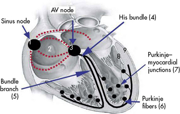
Box 8-10 Advanced Concepts: Resynchronization Therapy for the Treatment of CHF

Biventricular pacing and cardiac resynchronization therapy have been shown to be effective in adults with heart failure and prolonged QRS duration of left bundle branch block morphology.963 The purpose is to restore a more synchronized, “efficient” cardiac contraction in those patients in whom interruption of the ventricular conduction system has caused asynchronous activation and uncoordinated beating of the heart. In pediatric patients, cardiac resynchronization can be successful in decreasing QRS duration, and improving ejection fraction, and ultimately improving NYHA heart failure classification.

Low cardiac output (shock)

This topic is so important to critical care that information was expanded into an entire chapter, Shock, Cardiac Arrest, and Resuscitation (Chapter 6). Postoperative low cardiac output is also addressed in Postoperative Care, later in this chapter.

Altered nutrition and potential gastrointestinal complications

Nancy Rudd

Nutritional challenges are frequently encountered by those caring for critically ill infants and children with cardiac disease. Growth failure is a well-recognized and challenging consequence of congenital heart disease with type and severity of cardiac defect determining the impact on nutritional status and growth. This section reviews the etiology and development of altered nutrition and management strategies. In addition, it summarizes specific gastrointestinal and feeding challenges common to the care of children with cardiovascular problems. The problems addressed include necrotizing enterocolitis, protein-losing enteropathy, chylous effusion, mesenteric arteritis, and vocal cord injury. These conditions and specific nutritional therapy to address each issue are summarized in this section.

Etiology, Pathophysiology and Identification of Growth Failure in Patients with Congenital Heart Disease

Growth failure in children with CHD is multifactoral. A relationship exists between inadequate growth and abnormal hemodynamics in infancy. Physiologic alterations that impact growth include congestive heart failure, cyanosis, and pulmonary hypertension. The type of cardiac lesion present and the severity of hemodynamic impairment also play a role in the undernutrition of infants with CHD.9

The pathology of growth failure in the setting of congestive heart failure (CHF) is complex. The diagnosis of CHF often correlates with clinical findings of tachypnea, hepatomegaly, and tachycardia. Growth disturbances are a consequence of increased myocardial and respiratory work, inadequate caloric intake because of anorexia or fatigability during feeding, increased metabolic rate from increased work of breathing, and alterations in gastrointestinal function and absorption. The presence of significant right heart volume overload from left to right intracardiac shunting is a major factor in the development of CHF. Tachypnea results from excessive pulmonary blood flow as seen with cardiac defects resulting in left to right shunting lesions. This increased work of breathing can lead to difficulty for the infant's coordination of sucking, swallowing, and breathing with the result that oral intake is inadequate.

Decreased gastric capacity is caused by pressure on the stomach from an enlarged, congested liver resulting from systemic congestion. Any delay in gastric emptying may predispose the infant to gastroesophageal reflux or may result in premature satiety. Elevated right atrial and systemic venous pressures can cause intestinal protein losses and fat malabsorption from elevated venous pressure in the mesenteric bed. Gastrointestinal perfusion is reduced by catecholamine-mediated redistribution of blood flow away from skin, gut, and kidney to maintain adequate brain and heart perfusion.

Lesions with potential for excessive pulmonary blood flow, increased pulmonary artery pressure, increased blood return to the left heart, and elevation of left ventricular end-diastolic pressure result in high output hemodynamics and can cause hypermetabolism. Lesions with a large left-to-right shunt typically affect weight rather than height in the early stages of infancy. Cardiac lesions that commonly result in CHF in the neonate include hypoplastic left heart syndrome (HLHS), transposition of the great arteries (TGA), patent ductus arteriosus (PDA), total anomalous pulmonary venous return (TAPVR, particularly if associated with obstructed venous return), critical aortic stenosis (AS) or pulmonary stenosis (PS), coarctation of the aorta (CoA) or other obstruction to left heart or aortic flow, and large ventricular septal defect (VSD).

The role of cyanosis as a cause of growth failure in children with CHD is unclear. Arterial oxygen desaturation alone does not necessarily result in tissue hypoxia, because tissue aerobic metabolism may not be impaired until arterial PaO2 falls below 30   mm Hg. In addition, arterial oxygen content and tissue oxygenation may be preserved at near-normal levels if oxygen carrying capacity is increased by elevated hemoglobin.

If hypoxemia and CHF are both present, growth is most severely affected and results in reduced height and weight. Hypoxemia and edema may induce gastroparesis and gut hypomotility. Some have theorized that the duration of hypoxemia in years, not the severity of hypoxemia, plays a significant role in growth retardation.746 Lesions often resulting in the combination of cyanosis and CHF include double outlet right ventricle (DORV), pulmonary atresia (PA), tricuspid atresia (TA), and hypoplastic left heart syndrome (HLHS).

The presence of pulmonary hypertension also plays a critical role in growth disturbances in infants with both cyanotic and acyanotic CHD. Increased resting oxygen consumption has been demonstrated in patients with pulmonary hypertension. In one study, the presence of pulmonary hypertension appeared to be the most significant factor identified in growth impairment in infants with CHD; the combination of cyanosis and pulmonary hypertension had the most severe impact on growth.907

Other perioperative factors contributing to growth failure include fever and sepsis. Fever alone can increase the caloric expenditure 12% for each degree Celsius reached above 37°. Sepsis and its resulting influence on energy consumption and increased autonomic tone can increase caloric expenditure 25% to 50%.566 Gastrointestinal infections such as Clostridium difficile and rotavirus can also compromise the child's nutritional status.

Each of these physiologic alterations impacts the ability of a child with hemodynamically significant heart disease to match nutrient intake with metabolic demands. Undernutrition occurs when energy expenditure combined with nutrient losses exceed that of nutrient intake and absorption. There are six classes of nutrients that impact an infant's diet and thus growth: carbohydrates, fats, proteins, vitamins, minerals, and water. All six play an important role in maintaining an optimal nutritional state. However, only three yield energy for the body's use. These three are termed energy nutrients and include carbohydrates, fats, and proteins.

During the first 6 months of life for the healthy infant, the suggested level of intake for energy is 91-112   kcal(calories) per kg of body weight to maintain adequate body temperature, growth and activity. Protein needs during infancy are relatively high as a result of rapid skeletal and muscle growth, and average 1.5   g of protein per kilogram of body weight. A neonate weighing less than 3   kg typically requires 120   mL/kg per day of fluids and a 3- to 10-kg infant typically requires 100   mL/kg per day of fluids for maintenance fluid requirements; of course, these requirements must be tailored to the clinical condition.

The infant and child with heart disease may have greater than normal requirements yet have less than normal ability to consume the calories and proper fluid intake. Infants with hemodynamically significant CHD require significantly more nutritional support to sustain growth than their healthy counterparts. Fluid losses in a neonate with congestive heart failure are estimated to be 10% to 15% greater than those of a normal infant because of tachypnea, emesis, diarrhea, and anticongestive management with diuretics. Energy intake required in infants with CHD to sustain normal growth is reported to be between 130 and 150   kcal (calories)/kg per day, and is affected by the type of cardiac lesion and “catch-up” growth needed. Some infants can require as much as 175 to 180   kcal/kg per day.746 Currently, there are no established growth parameters for infants with hemodynamically significant CHD. Current practice targets a weight gain of 10 to 30   g/day when adequate calories are provided.

Protein balance is another key factor in the management of most infants with CHD because serum protein imbalance may play a role in the development of edema. It is important to ensure adequate protein intake and adequate serum protein in this patient population by monitoring serum albumin, transferrin, or prealbumin levels.

Increased metabolic rate or energy expenditure contributes to growth failure particularly when CHF is present. Hypermetabolism is likely related to increased work of respiratory muscles necessary for adequate ventilation in the presence of decreased lung compliance. The basal metabolic rate is elevated in infants with CHD because of cardiac and respiratory work and has been reported to be as much as three to five times higher than the work in infants without heart disease. The presence of dilated or hypertrophied cardiac muscle also increases oxygen consumption. Hypertrophied cardiac muscle uses 20% to 30% of the body's total oxygen consumption instead of the typical 10%.

Nutrient losses also play a role in cardiac undernutrition. Malabsorption significantly limits an infant's tolerance of feedings, compromises the neonate's ability to maximize caloric intake, and decreases nutrient absorption. As noted, nutrient intake is also compromised by altered gastric capacity in the presence of hepatomegaly and by edema of the intestinal wall and mucosal surfaces leading to impaired nutrient absorption. Consequently, the volume of enteral feeding, the caloric concentration, and the delivery time of bolus feedings may need to be adjusted.

Gastroesophageal reflux (GER) describes the movement of gastric contents back into the esophagus. The reflux can be “silent” or it may produce pain or discomfort with signs such as neck arching, coughing, swallowing, or emesis. GER can occur in up to 65% of healthy infants and as many as 40% to 50% of infants age 1-2 months have two episodes of reflux per day.425 GER also plays a role in CHD. The role of GER in CHD seems most prevalent when the infant has a hemodynamically significant lesion and it likely results from delayed gastric emptying secondary to malabsorption. The presence of a nasogastric tube (NG) to supplement enteral feedings may contribute to GER symptoms because the tube may prevent closure of the gastroesophageal sphincter.

Inadequate nutrient intake is felt to be the predominant cause of growth failure in infants with CHD. Oral feeding requires more energy than any other infant activity. For a neonate with symptomatic heart disease, oral feeding can be compared to a “stress test.” Tachypnea and resulting fatigue may not allow an infant to consume all of the calories and volume needed to maintain growth. Oral feeding is challenging because the neonate may feel hungry at the start of the feed, feed eagerly initially, then reach early satiety (so the infant stops feeding). The infant may use a lot of energy feeding and may tire quickly, resulting in suboptimal nutrient intake and increased energy expenditure. Other limiting factors include respiratory infections with resulting tachypnea that can impair an infant's coordination of sucking, swallowing and breathing necessary for successful oral feeding. Additional factors leading to inadequate caloric intake may relate to medical management strategies used to treat underlying cardiac pathology. Fluid restriction and anorexia resulting from diuretic use may limit enteral caloric intake.

The physical growth of infants is a direct reflection of their nutritional well-being and is the single most important parameter used in assessing their nutritional status. Assessments of growth are made by periodic determinations of weight, height, and head circumference. Growth charts for each parameter should be utilized to document and evaluate the infant's growth pattern.

Expected weight gain in a term infant during the first 6 months of life should average 20 to 30   g/day with variation based on gender, age in months, and growth percentile.218 Incremental gain in crown-heel length for full-term infants averages 0.66   cm/week during the first 6 months of life. Healthy infants display rapid increases in head circumference and their head growth correlates well with brain growth. Average gain in head circumference for term infants between birth and 6 months of age is 0.33   cm/week.480

Poor nutritional status can negatively impact both preoperative and postoperative outcomes. Inadequate protein and calories can reduce skeletal muscle function and increase the risk of postoperative pneumonia. The immune system is also adversely affected by undernutrition; impaired nutrition can lead to an impaired immune state, predisposing an infant or child to postoperative infection or poor wound healing.

Several studies have focused on the importance of postsurgical nutrition in infants following repair or palliation. Research suggests that weight gain following neonatal surgery is often suboptimal and greater attention should be focused on nutrition in the postoperative period including rapid advancement of enteral feeding to promote optimal growth and nutritional status. One study demonstrated that rapid advancement of calories to higher concentrations significantly improved energy intake and weight gain (20   g/day gain in control group versus 35   g/day loss in the usual care group). In this study enteral feedings were advanced from 20 to 30 calories per ounce over just a 3-day span. Additionally, postoperative hospital stay for the intervention group of infants was significantly decreased.701

Management: Strategies to Optimize Nutrition and Growth

With knowledge of the multiple causes of growth deficiency, it is the responsibility of the healthcare team to make nutritional management a high priority in the care of infants and children with congenital heart disease. Many options are available to combat the harm nutritional deficiency can create. Discussion of these treatment options follows.

Gastroesophageal Reflux (GER)

To address gastroesophageal reflux and malabsorption issues, several treatment modalities have been used. Smaller, more frequent feedings are recommended along with elevated supine positioning during and after feeding to reduce the reflux of gastric contents into the esophagus. In the past thickening of formula with dry rice cereal was suggested as a means of limiting GER but this practice is somewhat controversial. Although some studies have noted fewer episodes of emesis when thickened formula is introduced, others have shown an increase in gastric emptying time and thus longer interval when reflux episodes were possible. Commercial formulas with added rice have been marketed as a treatment option for infants with symptomatic GER.

Any decision regarding a trial of thickened formula should be left to the individual care provider. Prokinetic agents such as metoclopramide have been used to treat GER during infancy, but there is debate over their effectiveness. Introduction of an antisecretory or acid-neutralizing agent may help prevent erosive esophagitis. Surgical treatment options such as placement of jejunostomy tube, (to bypass the stomach), or the Nissen fundoplication should be reserved for severe GER that does not respond to aggressive medical therapy.

Parenteral Nutrition

Parenteral nutrition is indicated in infants with CHD when the projected time to establish adequate enteral support exceeds the infant's metabolic reserves. Such instances include postoperative patients when oral feedings are not anticipated to commence for more than 1 or 2 days, and infants requiring prolonged intubation, those with marked malnutrition prior to surgery, and infants with any coexisting gastrointestinal abnormalities such as duodenal atresia or gastroschisis.

If enteral nutrition is unlikely to be initiated for greater than 1 or 2 days, a central venous catheter should be placed. This allows maximum dextrose and protein concentrations in the parenteral nutrition as well as administration of intravenous lipid emulsions.763 To prevent essential fatty acid deficiency, administration of 0.5   g/kg of a 20% lipid solution is needed three times per week.

Enteral Feeding

An important adjunct therapy to parenteral nutrition, when possible, is the use of trophic feeding. Continuous administration of formula or breastmilk at a rate of 0.5 to 2   mL/kg per hour uses the gastrointestinal tract and can lessen the risk of systemic bacterial infection by preventing complications related to intestinal mucosal atrophy and loss of functional intestinal barrier.

Enteral feedings are more physiologic and preferable to parenteral nutrition when gastrointestinal function is adequate. In addition, enteral feedings are more accessible, more cost effective, and safer. The goal of enteral therapies is to sustain growth or enable “catch-up” growth without overburdening cardiovascular function or disturbing fluid and electrolyte balance.

Catch-up growth refers to the velocity of growth following a time period of impaired growth (caused by undernutrition). The nutritional needs to achieve catch-up growth can be as high as 1½ to 2 times the required daily allowances for age.

The total energy expenditure in children with CHD is 22% to 29% greater than that of a healthy, age-matched child, so that caloric intake should be targeted. Optimizing caloric intake to meet increased energy needs may be accomplished enterally by increasing the quantity or volume of feedings or by increasing the caloric density of formula.

Once nutrient requirements have been estimated, an enteral feeding regimen should be determined. Most full-term infants receive breast-milk or a cow's milk-based formula. Soy and hydrolyzed casein formulas are reserved for infants who do not tolerate a milk-based formula. Oral is the preferred route for administration of enteral feeding whenever possible. An on-demand feeding regimen that provides adequate nutrients and results in appropriate weight gain is the optimal outcome. In the infant with CHD who successfully feeds on demand, it is not necessary to restrict or supplement enteral intake and standard formula at 20   cal/oz or 0.67   kcal (calorie)/mL is used.

Bottle feeding is often recommended for infants with CHD because it is speculated to be less strenuous than breastfeeding and the infant's intake can be measured more accurately. Studies in breastfed infants with CHD have included reports of higher oxygen saturations during feedings and more rapid weight gain than in bottle-fed infants.818 The degree of hemodynamic impairment does not always correlate with an infant's ability to breastfeed. Therefore breastfeeding should not be excluded for infants with hemodynamically significant CHD. However, ad lib breastfeeding requires careful monitoring until evidence of adequate fluid intake and positive growth are demonstrated.818 Infants being breastfed may be temporarily bottle-fed with expressed breast milk if adequate enteral intake is in question.

The normal diet of an infant consists of either breast milk or commercial formula (with no other supplement needed) for the first months of life. The chemical components found in both breast milk and formula provide two very important benefits; in addition to supplying energy they promote growth and repair of body tissue. Breast milk is low in protein (constituting about 6% of calories) and high in fat (constituting about 52% of calories), compared with formula (about 9% of calories from protein and about 49% from fat). However, the protein types contained in breast milk are more bioavailable than the proteins in formula, so breastmilk actually provides closer to 8% of calories from protein. Once a child is older than 1 year, a 1-kcal/mL formula should be substituted for standard infant formula. Commercially available formulas for this purpose include protein-based PediaSure, and amino-acid based EleCare or Vivonex.

The need to advance feedings to a total volume of 130 to 150   mL/kg per day and goal calories of 120 to 130   kcal/kg per day to achieve target growth is not uncommon in an infant with significant CHD. Noting frequency of emesis, diarrhea, or abdominal distension assists in monitoring for tolerance of enteral feeding. Daily weight, caloric intake, total fluid intake and output, along with laboratory studies are watched closely during the advancement of enteral feedings. If volume intolerance is a limiting factor in meeting caloric requirements, it may be necessary to increase the caloric density of the formula. The caloric density of standard 20   kcal/oz formula or breast milk can be increased to 24, 27, or even 30   kcal/oz by many methods. The simplest method of increasing calories is to add less water to powdered formula than instructions indicate for preparing typical 20   kcal/oz formula. This method results in a higher concentration of calories and nutrients but does reduce free water in the infant's diet. Breast milk contains an estimated 20   kcal/oz and provides nutritional and immunologic advantages over infant formula.

It is important to remember there may be variation in the fat content of breastmilk based on the mother's dietary intake. Creamatocrit Plus and similar devices are available and used in some clinical settings. This portable centrifuge is designed to measure the caloric and lipid content of breast milk and therefore allows more precise estimation of caloric intake of breastfed infants. If an abundant volume of breast milk is available, hind milk (milk released after several minutes of breast feeding is preferred to foremilk (milk released in the initial minutes of breast feeding), because hind milk has higher fat content and the caloric density may be as high as 30   cal/oz. Foremilk contains higher water content and less fat so will be less calorie dense.

Breast milk can be fortified with a commercial powdered formula to densities of 22, 24, 27, or 30   cal/oz based on nutritional needs. There are multiple recipes for fortification and each is based on the nutrient content of the commercial formula being added. It is important to check with a nutritionist for an appropriate recipe. The use of commercial human milk fortifiers is reserved for preterm infants.480 Additionally, fortification may be accomplished using commercial glucose polymers, microlipid or MCT oil, or protein supplements. Glucose polymers such as polycose (2   kcal/mL as liquid and 23   kcal/TBSP as a powder) increase carbohydrate concentrations. Medium chain triglycerides such as microlipid (4.5   kcal/mL) or MCT oil (7.7   kcal/mL) increase the fat content. Lastly, Promod, a protein supplement, increases the protein content by 3g/tablespoon.

Concentration of formula is usually the method of choice for increasing caloric content because this method provides a balanced formula that includes all nutrients, rather than simple addition of only fat, carbohydrate, or protein. Some fortification recipes are easier for families to prepare and this can play a role in the method of fortification chosen for a patient. Families must be educated by a nutritionist to ensure the appropriate nutritional composition of formula or breast milk is provided to the infant.

It is important to remember, increasing caloric density may lead to an increased solute load including electrolytes and minerals. Renal solute load refers to excess nitrogen, which is the metabolic end product of protein metabolism, sodium, chloride, and potassium that are excreted in urine by the kidneys. Such a disturbance may cause dehydration as the kidneys react to the excessive solute load and draw too much water from the body into the urine.

The delivery mode of enteral nutrition for the neonate, infant, or child with heart disease must be carefully selected. The preferred route is oral if achievable. However, fatigue, anorexia, or swallowing problems may make exclusive oral feeding unattainable. Initially, enteral feeding may be best managed as a combination of oral (PO) and nasogastric (NG) feedings. This allows the infant to continue to develop oral feeding skills but also receive the necessary energy required for catch-up growth. The advantages of enteral feeding by NG route include delivery of more nutrients with a tube that is minimally invasive and short term. Potential disadvantages are interference with PO feedings resulting from a tube in one nostril and in the esophagus that makes swallowing and breathing potentially more challenging. The NG tube may also make reflux more prevalent and contribute to esophagitis.566

Nasojejunal delivery of nutrients is an option when delayed gastric emptying or GER is present. Oral gastric (OG) tubes are sometimes utilized in neonatal feeding strategies. An OG tube may be preferred in infants with respiratory distress, as neonates are primarily nose breathers. Disadvantages of OG tubes include difficulty securing the tube and need for removal and then replacement with oral feeding attempts.

The feeding tube chosen should be the smallest possible to safely deliver feedings. Typically a size 6.5 French in a 2.5- to 3.5-kg neonate is used. Long-dwelling tubes are preferred as they remain soft and flexible up to 30 days.

Initially PO feeding time should be no longer than 20 to 30   min thus limiting the energy expended during prolonged feedings. The remainder of the prescribed volume is given via NG by gravity or with a feeding pump. The goal of bolus feeding is to give the entire oral plus nasogastric feeding in less than 30 to 45   min. This allows adequate gastric emptying before the start of the next feeding. With initial bolus feeding, the duration may be extended to 60   min to monitor for tolerance. As a guideline a trial of removal of the NG tube for an all PO attempt may be considered when the infant has tolerated goal calories and volume for 2 or more days, and is taking greater than 50% of target volumes by mouth.

Enteral feedings can be delivered as intermittent boluses, continuous infusion, or a combination of the two. Bolus feedings are felt to be more physiologic than continuous infusions, although continuous infusions may be the delivery mode of choice in patients unable to tolerate bolus feedings. Practically, for the infant requiring continuous infusion, the duration of feedings should be shortened to 20 or 22   hours per day, allowing brief periods of time for activities with the patient not attached to the feeding pump. Ideally, to help maximize oral motor skills, normalize infant feeding patterns, simplify delivery at home, and still meet nutritional goals, if tolerated, it is most beneficial if the infant receives bolus feedings during the day and a continuous feeding at night.

Long-Term Supplementation

Long-term tube supplementation may be required in patients unable to consume necessary calories or volume despite optimizing caloric density and addressing oral feeding skills. Nonpermanent feeding tube options include NG tubes and NJ (nasojejunostomy) tubes. Some institutions reserve the use of nasogastric tubes for the hospital setting and patients are not discharged home with an NG in place. Concerns include potential dislodgement resulting in aspiration, concern that the tube may interfere with normal function of the upper and lower esophageal sphincters and may contribute to gastroesophageal reflux, impairment of the normal airway protective mechanisms of the pharynx and larynx, resulting in an increased likelihood of aspiration, and need for reinsertion inducing a vasovagal response with cardiac decompensation.

When a permanent source of delivering enteral feeds is required to maintain growth, a percutaneous endoscopic gastrostomy tube (PEG tube) or surgical gastrostomy or gastrojejunostomy tube should be inserted.

Infants experiencing tachypnea and muscle weakness often need specific interventions to achieve oral feeding. Supportive strategies include providing chin support or utilizing a slow-flow nipple allowing the infant to more efficiently sequence and coordinate sucking, swallowing, and breathing. As the infant's oral feeding skills improve, transition to a standard flow nipple can be made.

Although somewhat more challenging to use, the Haberman feeding system can be very effective in promoting successful oral feeding in cardiac infants. The one-way valve system in a Haberman allows for control of fluids (low, medium, and high) when concerns exist regarding an infant's ability to handle fluid flow and regulate breathing pauses without breaking the seal on the nipple.

For attainment of successful growth and nutrition, the goal of any strategy or method used is to set an estimated patient goal, assess growth parameters, and make adjustments as needed until acceptable growth is achieved.

Management: Necrotizing Enterocolitis

The clinical problem of necrotizing enterocolitis (NEC) in patients with cardiovascular problems is presumed to be related to impaired hemodynamics and a low perfusion state. Necrotizing enterocolitis can be seen preoperatively or postoperatively in the neonate with complex CHD and is exhibited by ischemic necrosis of the intestinal mucosa. Nutritional care is supportive and includes discontinuation of enteral feedings with bowel rest, and maintenance of intermittent nasogastric suction for gastrointestinal decompression. Nasogastric suction is recommended until the infant's clinical condition improves, the ileus resolves, and pneumatosis is no longer seen on the abdominal radiograph.763

Total parenteral nutrition is used to provide nutritional support during the period of bowel rest. Enteral feedings are cautiously resumed as the infant's clinical condition allows. Breastmilk use is preferable in infants at risk for NEC because of the positive benefits in ease of digestion and absorption as well as added immunologic protection.

Management: Mesenteric Arteritis

Following repair of coarctation of the aorta, infants and children may experience paradoxical hypertension. In severe cases, mesenteric arteritis and even bowel ischemia can develop. Because preoperatively the mesenteric arteries were exposed to a low blood pressure (i.e., typically at a mean pressure rather than pulsatile), the sudden increase in arterial blood pressure is thought to cause acute vessel injury and severe reactive vasoconstriction that can result in inadequate blood flow to the bowel. The clinical occurrence of paradoxical hypertension and mesenteric arteritis are sometimes referred to as postcoarctectomy syndrome.

Older children and adults with severe coarctation of the aorta are felt to be at greatest risk for development of mesenteric arteritis postoperatively. Because of the potential risk of developing mesenteric arteritis, many centers keep patients “NPO” (nothing per os, or nothing by mouth) for the first 48 to 72   h post repair while medically controlling hypertension. Then a clear liquid diet is introduced, with slow advance of diet to solids as tolerated.44 If clinical concerns of mesenteric arteritis or bowel ischemia arise, enteral feedings must be held, nasogastric decompression initiated, and IV fluids provided until symptoms resolve.

Management: Chylous Effusion

Operative injury to the thoracic duct and lymphatics or postoperative systemic venous hypertension or increased right-sided cardiac pressures can lead to chylous effusions. The development of postoperative chylothorax in infants and children with CHD compromises both nutrition and fluid status.

Traditional treatment of postoperative chylous effusion includes chest tube for drainage and diet modification. Nutrition strategies vary from a low-fat diet to complete enteric rest with total parenteral nutrition.650 Both options are less than optimal in providing balanced nutrition.

Careful assessment of the child's nutritional state must be ongoing because diet modification therapy is often continued for 4 to 6 weeks as a conservative means of treating chylous effusions. Formulas with a predominant fat source from medium-chain triglycerides (MCTs) are preferred over human milk or standard infant formulas, which are very high in long-chain triglycerides (LCTs). The benefit in managing chylous effusions with MCT formulas is based on the fact that LCTs require absorption via the lymphatic system whereas MCTs bypass the lymphatics and go directly into the portal system.

A variety of MCT formula choices are available for infants and children and include Portagen, a lactose free formula with 85% of its fat as MCT oil, Pregestimil with 55% of fat as MCT oil, Vivonex Pediatric with 68% of fat as MCT oil, and Alimentum with 50% of fat as MCT oil. Because prolonged use of these formulas may result in inadequate intake of essential fatty acids and trace minerals, it is recommended that a nutritionist be consulted in the care of children requiring a low-fat diet. A commercial formula specifically designed for infants with chylothorax, Enfaport, has been developed and reported benefits include 85% of fat as MCT oil, high protein levels, and all the essentially fatty acids to support the nutritional needs of an infant with chylous effusion.

Guidelines for a toddler or child requiring low-fat diet include limiting food choices to those with 3   g or less of fat per serving and restricting calories from fat to 20% to 25% of total daily intake (see, also, Chapter 14).

Management: Protein-Losing Enteropathy

In children with cardiac disease, protein-losing enteropathy is most frequently reported in patients following Fontan-type surgical intervention. Protein-losing enteropathy (PLE) is typified by excessive loss of serum proteins into the gastrointestinal tract resulting in hypoproteinemia detected as abnormally low serum albumin levels. Clinical signs include edema, fatty stools and a change in bowel habits with the development of diarrhea and abdominal discomfort.

Nutritional management focuses on optimizing protein intake and limiting fat to predominantly MCTs. In normal adults, normal protein requirements are 0.6 to 0.8   g/kg desired body weight per day. In protein-losing enteropathy, this value may increase to 1.5 to 3.0   g/kg per day, and protein supplements may be necessary to achieve positive protein balance.366 Protein intake can be enhanced by consumption of lean meat, low fat milk and cheese, and egg substitutes.

Commercially available protein supplements such as Promod are a reliable source of fat-free dietary protein, but may be expensive and unpalatable. More palatable supplements that provide both protein and nonprotein calories include Ensure, Isocal, Peptamen, and PediaSure.

A reduction in intake of long-chain fatty acids reduces mesenteric lymphatic flow and pressure, thus decreasing the amount of lymph leakage and protein loss. In cases of PLE with severely diminished gastrointestinal motility and absorption, the gastrointestinal tract may not tolerate enteral feedings. Under these circumstances, patients may require parenteral nutrition.

Management: Vocal Cord Injury

Vocal cord, or more appropriately, vocal fold dysfunction, can limit enteral feeding and compromise nutrition. Vocal fold dysfunction is most frequently encountered in children with cardiac disease in the postoperative period and results from surgical intervention. Infrequently, vocal fold injury is caused by endotracheal tube placement. When associated with recent surgery, the vocal fold injury usually results from damage to the left recurrent laryngeal nerve during repair of either coarctation of the aorta or patent ductus arteriosus. Enteral feeding is affected because airway and swallowing problems result from immobility of the affected vocal fold. Vocal fold dysfunction should be suspected if the infant or child has hoarse cry or voice or if coughing and sputtering occur with swallowing of liquids.

Normally the recurrent laryngeal nerve carries signals to the muscles responsible for opening vocal folds during breathing and coughing, and closing vocal folds during swallowing. Damage to the vocal folds can lead to transient or permanent paresis or paralysis. Paresis is the partial interruption of nerve impulses resulting in weak or abnormal motion of laryngeal muscles. Paralysis is the total interruption of nerve impulses resulting in no movement of the muscle.

Direct laryngoscopy is the initial diagnostic method of choice for assessing and documenting vocal cord immobility in the postoperative cardiac patient. Most often the vocal fold immobility is unilateral and involves the left vocal fold, although instances of bilateral injury do occur and can result in significant respiratory difficulty.

The position of the affected vocal fold may vary. A midline position is associated with stridor but adequate swallowing capability. Lateral positioning results in a glottic gap. This puts the child at risk for aspiration because swallowing is not coordinated. Most frequently the affected vocal fold is somewhere between midline and lateral and is in a paramedian position.

Infants with left vocal fold immobility in the paramedian position need to be bottle-fed in a right-side down, side-lying position. Toddlers and children with an affected vocal fold should be taught or instructed to turn their head toward the affected side while swallowing as a way of reducing the risk of aspiration.

In the setting of questionable or documented vocal fold immobility, speech and feeding specialists should be consulted. Their input is essential in addressing the feeding challenges presented by infants with postoperative vocal fold dysfunction. These professionals are skilled in oral-motor assessment and the development of infant-specific feeding strategies designed to promote positive oral feeding outcomes.

Arrhythmias

Debra Hanisch

Pearls

When a cardiac arrhythmia is observed or suspected, the nurse's first response must be to quickly assess the effect of the rhythm on the patient's hemodynamic status. A clear recording of the ECG should be obtained as well to document the arrhythmia.

90% of arrhythmias in pediatric patients are supraventricular tachycardias.

80% of wide QRS tachycardias are supraventricular in origin, but must be regarded as ventricular tachycardias until proved otherwise.

All antiarrhythmic drugs have the potential to be proarrhythmic and warrant careful monitoring of the patient.

Normal Rhythm and Conduction

The normal conduction system of the heart is depicted in Fig. 8-15. In sinus rhythm, the electrical impulse is initiated by the sinoatrial node, located in the right atrium near the junction of the superior vena cava. From there, the impulse is propagated as a wave of depolarization over the atria, converging at the atrioventricular (AV) node, located at the base of the atrial septum just above the tricuspid valve. Conduction is slowed as it moves through the AV node, allowing time for ventricular filling. From there, the impulse proceeds rapidly down the bundle of His, continues simultaneously down the right and left bundle branches, and finally spreads through the Purkinje fibers to depolarize the ventricles.

image

Fig. 8-15 Conduction of the impulse in the heart. The impulse originates in the sinus node (1), continues in the atrial wall (2), and is delayed in the atrioventricular (AV) node (3). Conduction within the ventricles is initially rapid within the rapid conduction system: His bundle (4), right and left bundle branches (5), and Purkinje fibers (6). The impulse is transferred from the rapid conduction system to the working myocardium in the Purkinje-myocardial junctions (7), which are located in the endocardium. Within the slowly conducting working myocardium, the impulse is conducted from endocardium to epicardium.

(From Ellenbogen: Clinical cardiac pacing, defibrillation and resynchronization therapy, ed 3, Philadelphia, Saunders, 2007.)

Normal components of the ECG are illustrated in Fig. 8-16. The P-wave represents atrial depolarization. The isoelectric segment between the P-wave and QRS complex corresponds to the slowed impulse passing through the AV node. The QRS complex, normally narrow, is produced by rapid ventricular depolarization and is followed by the T-wave, signifying ventricular repolarization. Atrial repolarization occurs but is not visible on the surface ECG.

image

Fig. 8-16 Electrocardiogram (ECG) and cardiac electrical activity. A, Normal ECG. Depolarization and repolarization. B, ECG intervals among P, QRS, and T waves. C, Schematic representation of ECG and its relationship to cardiac electrical activity. AV, Atrioventricular; LA, left atrium; LBB, left bundle branch; LV, left ventricle; RA, right atrium; RBB, right bundle branch. RV, right ventricle.

(A and B from Patton KT, Thibodeau GA. Anatomy & physiology, ed 7. St Louis, Mosby, 2010. C from Thibodeau GA. Anatomy & physiology. St Louis, Mosby, 1987.)

Analyzing cardiac rhythms involves an assessment of:

Rhythm

Heart rate

P-waves

QRS complexes

ST-segments

P-QRS relationships

Ectopic and escape beats

Time intervals and heart rate calculations from ECG grid paper are provided in Fig. 8-17, A. Although 12-lead ECG interpretation will not be addressed here, the PR interval, QRS duration, and QT interval are important to note when analyzing rhythm strips. It is also important to identify ST-segment elevation or depression. Table 8-12 displays normal heart rates, and normal PR intervals, and QRS durations in children; these are also depicted in Fig. 8-17, A. QT intervals are corrected (QTc) using Bazett's formula:

image

Fig. 8-17 A, ECG intervals. When evaluating an ECG strip, the PR interval and QRS duration should be measured and compared with normal values. In addition, the corrected QT interval, QTc, should be calculated using Bazette's formula: image
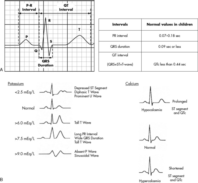
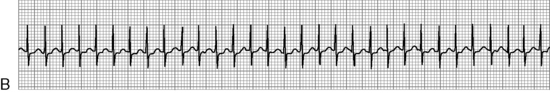
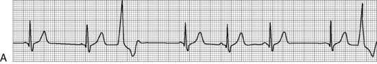
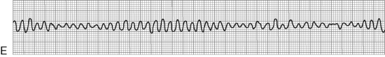
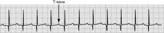
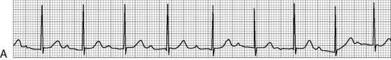
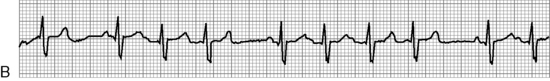
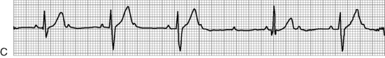
. B, ECG changes caused by electrolyte imbalance.

(Adapted from Park MK, Guntheroth WG. How to read pediatric ECGs, ed 4. Philadelphia, Mosby, 2006.)

Table 8-12 Normal Heart Rates, PR Intervals, and QRS Durations in Children

image

image


where the measured QT interval (QT) in seconds is divided by the square root of the preceding R-R interval (RR) in seconds. In children and infants older than 6 months, the QTc is normally less than 0.44   s.

Etiology

Arrhythmia substrates can result from congenital heart disease or its surgical correction, hypoxia, electrolyte or acid-base imbalances, metabolic derangements, drug toxicity, genetic disorders, myocardial disease, or injury to cardiac tissue. Arrhythmias can be symptomatic or asymptomatic; they may require urgent treatment or have the potential to deteriorate into symptomatic arrhythmias.

Three common classifications of clinically significant pediatric arrhythmias are bradyarrhythmias, tachyarrhythmias, and arrest (pulseless) rhythms. Hypoxia, sinus node dysfunction and AV block are the most common causes of bradyarrhythmias. Supraventricular tachycardia (SVT) caused by a reentrant conduction pathway is the most common cause of pediatric tachyarrhythmias. Sinus tachycardia is another fast heart rhythm that is not actually an abnormal rhythm but rather an indication that a high heart rate and cardiac output are needed to meet the body's demands. Arrest rhythms include ventricular fibrillation (VF) and pulseless ventricular tachycardia (VT). These rhythms may indicate ventricular irritability secondary to hypoxia or a severe metabolic derangement, such as acidosis, significant hyperkalemia or hypokalemia, or drug toxicity. Ventricular tachyarrhythmias may also occur as a consequence of a genetic ion channelopathy (i.e., long QT syndrome), myocarditis, cardiomyopathy, myocardial tumor, or scar formation following cardiovascular surgery.

Pathophysiology

Effects of Electrolyte Imbalances, Hypoxia and Acidosis

Electrolyte imbalances, especially potassium, calcium, and magnesium imbalances, alter the myocardial transmembrane potential, which in turn affects depolarization, repolarization, and conduction (Fig. 8-17, B). The cell transmembrane potential is determined predominantly by the difference in potassium concentration (the concentration gradient) between the inside and the outside of the cell (see section, Essential Anatomy and Physiology).

The child's serum potassium concentration reflects the extracellular potassium concentration. Any significant changes in the serum potassium concentration can increase or decrease myocardial excitability. In severe hypokalemia, ST-segment depression, T-wave flattening, and a prominent U wave (a positive deflection immediately following the T wave) may be observed. In hyperkalemia, tall, narrow, peaked T-waves are observed. When significant hyperkalemia is present, slowing of conduction time can result in a prolonged PR interval and an increased QRS duration. Bradyarrhythmias, tachyarrhythmias, or AV conduction disturbances may be seen. If the child's serum potassium concentration exceeds 7.0   mEq/L, ventricular fibrillation may develop.

Whereas potassium imbalances most notably affect T-waves, calcium imbalances are manifested as changes in the QT interval. Serum hypocalcemia prolongs myocardial repolarization, and can produce ST-segment elevation. Hypercalcemia shortens the ST-segment, decreasing the QT interval. Severe hypercalcemia may cause cardiac conduction abnormalities resulting in bradycardias.239,832

No specific ECG changes are identified with magnesium imbalances, but a low magnesium concentration may exacerbate the effects of low calcium. Hypomagnesemia is associated with atrial and ventricular arrhythmias, most commonly torsades de pointes. Magnesium sulfate may be useful as an antiarrhythmic agent because it decreases myocardial cell excitability and conduction. Profound hypermagnesemia (>18   mg/dL) may result in AV block and asystole.239,601

A fall in extracellular pH can reduce the rate of spontaneous pacemaker firing and the rate of depolarization. A change in carbon dioxide tension may also affect the ECG; severe hypercapnia has been associated with bigeminy.

Hypoxia/ischemia impairs the function of the sodium-potassium pump, results in a decrease in the magnitude of the transmembrane potential, and slows myocardial conduction time. These changes may contribute to slowing of the heart rate as well as an increased potential for premature ventricular contractions and ventricular tachycardia. Metabolic acidosis and alkalosis are often associated with alterations in potassium or calcium balance. ECG changes reflect these alterations.

Effects of Cardiac Surgery

Arrhythmias may occur during or following cardiovascular surgery either as the result of direct trauma to the conduction tissue, or edema or inflammatory reactions near sutures. Low cardiac output and decreased myocardial perfusion during surgery or in the postoperative period also may produce arrhythmias. Various forms of AV block and escape rhythms often are noted.

Myocardial injury or ischemia can produce ST-segment changes (ST-segment deviation) or an irritable focus that leads to ectopy. Thus, it is important to identify ST-segment changes and treat causes as soon as possible (see section, ST-Segment Deviation).

The arrhythmias that require immediate treatment in the child are those that significantly decrease cardiac output or systemic perfusion, or are likely to deteriorate to rhythms that decrease cardiac output or systemic perfusion. In general these arrhythmias are classified as bradyarrhythmias, tachyarrhythmias, and arrest rhythms. Any heart rate or rhythm should be evaluated in light of the child's clinical condition and its effect on systemic perfusion. The rhythm can then be identified as one of the following:

Too slow for clinical condition (bradycardias)

Too fast for clinical condition (tachycardias)

Ineffective (arrest rhythm)

ST-Segment Deviation

ST-segment changes are commonly seen in adult patients but are relatively uncommon in the pediatric population. Precise measurement is needed to reliably identify ST-segment deviations. The isoelectric baseline of the ECG is established by either the P-R segment or by the T-P segment (i.e., the portion of the ECG between the end of the T-wave and the beginning of the next P-wave).832 ST-segment elevation or depression is defined as the amount (in mm) that the ST segment deviates above or below that baseline respectively. On the 12-lead ECG, ST-segment elevations greater than 1   mm or depressions of 0.5   mm or more are generally considered abnormal.895 However, ST-segment shifts less than 1   mm in the limb leads or less than 2   mm in the precordial leads may be a normal finding in children, especially in the absence of T-wave changes.687 This normal finding, referred to as “early repolarization” is a well-described observation, often seen in healthy adolescent males.833

Abnormal ST-segment changes are often manifestations of significant pathology. Abnormal ST-segment elevation is indicative of acute myocardial injury, such as infarction, and should prompt immediate attention. Risk factors for myocardial infarction in children include congenital coronary artery anomalies and coronary embolization due to Kawasaki disease or endocarditis. Myocardial ischemia or infarction can also complicate surgery near a coronary artery, such as may occur following an arterial switch procedure for d-transposition of the great arteries.686 The postoperative team must also be aware of abnormal coronary artery anatomy in patients with congenital heart defects, such as may be present in some patients with tetralogy of Fallot. Close monitoring is needed during postoperative care and any acute change in the ST segment, whether elevation or depression, should be reported to the on-call provider immediately.

ST-segment elevation may also result from inflammation as seen with myocarditis or pericarditis. In fact, pericarditis is the most common cause of ST-segment elevation in children.895 Specific patterns of ST segment elevation have been identified,89 and certain of these patterns are postulated to be associated with arrhythmias and hypertrophy.90

ST-segment depression usually suggests myocardial ischemia. In congenital heart disease, severe aortic valve stenosis or aortic hypoplasia may compromise coronary perfusion. ST-segment depression, along with characteristic T-wave abnormalities, may be present with ventricular hypertrophy or cardiomyopathies.687 ST-segment depression noted during exercise stress testing indicates coronary blood flow is inadequate to meet increased myocardial oxygen demand during exercise. Other causes of ST-segment depression include hypokalemia and digoxin toxicity. As noted previously, any acute change in the ST segment, whether elevated or depressed, should be reported to the on-call provider immediately.

Bradyarrhythmias

Bradycardia is a heart rate less than 90/min in the infant and less than 60/min in the child. Because tachycardia is the appropriate response to distress and shock, a heart rate that is normal for age in a child with poor systemic perfusion constitutes a relative bradycardia.

Bradycardia can compromise systemic perfusion because it slows the ventricular rate; unless stroke volume increases commensurate with the decrease in heart rate, cardiac output will fall. Bradycardia is often associated with a proportional fall in cardiac output. Because of their low reserve for stroke volume, neonates and young infants are particularly sensitive to changes in heart rate. An additional consequence of a fall in heart rate is that more time is available for ectopic beats to emerge. Bradyarrhythmias occur most commonly as a result of hypoxia, vagal stimulation (e.g., during suctioning), dysfunction of the sinus node, or AV block.

Tachyarrhythmias

Tachycardia is defined as a heart rate greater than 200 to 220/min in the infant and greater than 160 to 180/min in the child over 5 years of age. Although the heart rate can increase transiently during fever or episodes of crying, the term “tachycardia” is reserved for significant and persistent increases in heart rate.

Tachycardia can be a normal response to stress (e.g., pain) or increased oxygen requirements, as in the case of sinus tachycardia. Sinus tachycardia may also be a normal response to a compromise in cardiovascular function, such as CHF. In these cases, the faster heart rate is compensatory and may maintain a normal or even slightly higher cardiac output. However, extremely high heart rates adversely affect ventricular diastolic filling time, causing a decrease in stroke volume and cardiac output. Moreover, coronary artery perfusion occurs almost exclusively during diastole. Tachyarrhythmias result in decreased coronary artery perfusion time in the face of increased myocardial oxygen demand. Cardiogenic shock may ensue if the tachycardia persists.

AV dissociation, if present, may also contribute to a drop in cardiac output. With AV dyssynchrony, “atrial kick,” the final 25% to 30% of ventricular filling produced by atrial systole, is lost. This may be associated with a significant fall in stroke volume.

Rhythm Identification and Clinical Signs and Symptoms

Analysis of rhythm strips involves assessment of:

Rhythm

Heart rate

P-waves

QRS complexes

ST segments

P-QRS relationships

Ectopic and escape beats

The heart rhythm can be described overall as regular, slightly irregular, irregular but with a pattern (regularly irregular as in grouped beating), or irregularly irregular. Whether the rhythm is fast, normal, or slow can be determined by measuring the heart rate and evaluating it in terms of the patient's age, activity, and clinical status.

The heart rate can be calculated by a number of methods. The standard paper speed for ECG recordings is 25   mm per second. At this speed, each small square on the horizontal axis represents 0.04   second; each larger block containing five small squares represents 0.2   second. Sixty (s/min) divided by the R-R interval in seconds will equal the heart rate per minute.

Most ECG paper is marked off in either 1-second (five large blocks) or 3-second (15 large blocks) intervals. An estimated heart rate can be derived by counting the number of R-waves in a 6-second strip (30 large blocks) and multiplying by 10. With fairly regular R-R intervals, the heart rate can be calculated by the 1500 method (based on 1500 small squares = 1   min). This method involves counting the number of small squares (0.04   second intervals) between two consecutive R-waves and dividing 1500 by that number. A simpler method for estimating the heart rate is the 300 method (based on 300 larger squares = 1   min). For this method, the number of larger squares between two consecutive R-waves is counted and divided into 300. Examples of these methods are illustrated in Fig. 8-18.

image image image

Fig. 8-18 How to calculate heart rate. A, Three-slash method. B, 1500 method. C, 300 method. D, A quick way to estimate the heart rate per min is to choose a representative R-R interval and count down by the number of large boxes: 300 (1 box within R-R interval), 150 (2 boxes), 100 (3 boxes), 75 (4 boxes), 60 (5 boxes), 50 (6 boxes).

(D from Berman W Jr. Handbook of pediatric ECG interpretation. St Louis, Mosby, 1991.)

P-waves, if present, should be regular and they all look the same in terms of size and shape. One P-wave should precede each QRS complex, and a QRS complex should follow each P-wave. In AV dissociation, the P-wave rate should be calculated in addition to the R-wave rate.

R-waves, or QRS complexes, should be regular and narrow, with consistent appearance (size and shape). A wide QRS complex suggests a conduction delay (as in bundle branch block) or origination of the impulse outside the AV node (as seen in AV block or ventricular ectopy). Aberrant AV conduction through an accessory pathway may create a wide QRS as well. The ST segments should be isoelectric (at the same baseline as the T-P segment and the P-R segment).

The ratio of P-waves to QRS complexes should be 1:1 with consistent PR intervals appropriate for the child's age, as described in Table 8-12. Variable PR intervals suggest AV dissociation with the ventricular rate faster than the atrial rate, or second- to third-degree AV block where the ventricular rate is slower than the atrial rate.

Ectopic beats are initiated by a focus outside the sinus node. Premature beats occur early in the rhythm and may originate from an irritable focus from the atria, atrioventricular (AV) junction, or ventricles.

A premature atrial contraction (PAC), caused by an ectopic atrial focus, appears on the ECG as an early-occurring P-wave with a different morphology from the normal sinus P-wave. The PAC typically conducts normally down the AV node to the ventricles, so the QRS complex is unchanged. A compensatory pause results as the sinus node is reset following the interruption. If the PAC occurs very early before ventricular repolarization is complete, it may be blocked from conducting to the ventricles.

A premature junctional contraction (PJC) originates in the AV junction and interrupts the rhythm with its early appearance. From there, the impulse is conducted up to the atria (retrograde) and down to the ventricles (antegrade). On the ECG, an inverted P-wave may be seen just before, within, or after a normal QRS complex. A compensatory pause follows.

A premature ventricular contraction (PVC) is recognizable on the ECG as a wide, bizarre QRS complex occurring early in the rhythm. The PVC is created by an ectopic ventricular focus, from outside the normal conduction system, that initiates ventricular depolarization. As a result, the morphology of the QRS is altered. P-waves tend to march through the PVC, unless a retrograde P-wave is stimulated. In either case, a compensatory pause follows the premature beat before the rhythm resumes.

Escape beats occur late in the rhythm following a pause, or when the underlying rhythm is abnormally slow. When the sinus node fails to act as the dominant pacemaker, a subsidiary pacemaker in the atrium, AV node, or ventricle, enabled by intrinsic automaticity, can initiate impulses. Atrial escape beats have a different P-wave morphology from sinus rhythm but the QRS is unchanged from normal. Junctional and ventricular escape beats look similar to PJCs and PVCs respectively, except they occur late in their timing rather than early.

Rhythms Originating from the Sinus Node

Normal sinus rhythm is initiated by the SA node at a rate within normal limits for the child's age. P-waves and QRS complexes are normal in configuration, occur in a 1:1 ratio (with each P-wave followed by an R-wave), with both P and R waves upright in Leads I and AVF. The PR interval is appropriate for the heart rate and age of the child (see Table 8-12). Though the rhythm is regular, slight beat to beat variation is present. The sinus node's automaticity is influenced by the sympathetic and parasympathetic nervous systems. Circulating catecholamines increase the heart rate while vagal stimulation slows the rate. A balance between these two systems maintains a normal heart rate (Fig. 8-19).

image

Fig. 8-19 Sinus rhythm at 92 per min in a 6-year-old girl. In normal sinus rhythm, the rate is appropriate for age and the rhythm is fairly regular. P-waves all look the same and are followed in a 1:1 ratio by narrow QRS complexes that all look the same. The PR interval is constant and normal for age. Moreover, P-waves and R-waves are both upright in Leads I and aVF.

Sinus arrhythmia is an irregular sinus rhythm with respiratory variation (Fig. 8-20, A). The heart rate increases with inspiration and decreases with expiration. This rhythm tends to be more pronounced in children and athletes. Sinus arrhythmia may also be exacerbated by reactive airways disease or increased intracranial pressure. No treatment for this arrhythmia is indicated.

image

Fig. 8-20 Rhythms originating in the sinus node. A, Sinus arrhythmia in a 3-year-old girl. Heart rate varies between 70 and 100/min with respirations. B, Sinus tachycardia at 170/min in a 1-year-old boy. C, Sinus bradycardia at 50/min in a 13-year-old girl.

Sinus tachycardia also originates in the sinus node and has all the properties of normal sinus rhythm except the rate is faster than normal for the child's age (see Fig. 8-20, B). Although the heart rate in sinus tachycardia is typically less than 200/min, it can be greater than 220/min in very ill infants, although this is rare. Sinus tachycardia normally occurs during periods of stress or increased oxygen requirement, such as exercise. Sinus tachycardia may be associated with pain, anxiety, anemia, volume loss, dehydration, shock, or fever. As a rule, the child's heart rate generally increases approximately 10/min for each degree Centigrade elevation in the child's temperature above 37° C.

Stimulant drugs, catecholamines, and other chronotropic agents can produce sinus tachycardia. In addition, tachycardia will occur as a compensatory mechanism if ventricular stroke volume decreases or cardiac function is impaired (e.g., with congestive heart failure, tamponade, or low cardiac output). However, extremely high heart rates compromise diastolic filling time and coronary artery perfusion time, and increase myocardial oxygen consumption. Therefore, extreme tachycardias with ventricular rates exceeding 180 to 220/min can result in a significant fall in stroke volume and cardiac output. The medical management of sinus tachycardia involves addressing the underlying cause of the tachycardia.

Sinus bradycardia encompasses all of the characteristics of sinus rhythm except the rate is slower than normal for the child's age (see Fig. 8-20, C). In general, heart rates less than 90/min in the infant or less than 60/min in the child are considered slow.

Sinus bradycardia may result from respiratory compromise, hypoxia, or vagal stimulation, as may be observed during suctioning. Hypothermia, hypothyroidism, increased intracranial pressure, or drugs, such as digoxin or beta-blockers, may produce sinus bradycardia. Trained athletes and adolescents with anorexia nervosa tend to have slower resting heart rates as well.680 Injury to the sinus node or its arterial supply could result in sinus node dysfunction with resultant bradycardia.

Sinus node dysfunction (SND), sometimes referred to as sick sinus syndrome, may lead to any of a number of bradyarrhythmias, including sinus bradycardia, sinus pauses or arrest, sinoatrial exit block, escape rhythms, and bradycardia-tachycardia syndromes. A junctional escape rhythm may emerge when the atrial rate slows to the point where it is slower than the AV node's depolarization rate, so the AV node takes over. In some cases, slow heart rates may allow certain tachyarrhythmias, such as atrial flutter, to surface.

Surgical injury to the sinoatrial node has been reported with over a 50% incidence long term in patients following the Mustard or Senning procedures for d-transposition of the great arteries.310,409 Other procedures associated with SND include the Fontan procedure for single ventricle physiology, closure of atrial septal defects, and repair of total anomalous pulmonary venous return. Whenever cannulation for cardiopulmonary bypass is performed near the superior vena cava-right atrial junction, there is a risk for SND. As a surgical complication, SND may occur immediately postoperatively, or may not be manifest for up to 10 years or longer following surgery.310 Nonsurgical causes of SND include right atrial dilation caused by pressure or volume overload. SND may also be seen in cardiomyopathies or inflammatory conditions, such as myocarditis, pericarditis, and rheumatic fever.74

Bradycardia can compromise systemic perfusion because it slows the ventricular rate. Unless stroke volume increases commensurate with the decrease in heart rate, cardiac output will fall. Acute management of hemodynamically significant bradycardia includes adequate ventilation and oxygenation, and administration of epinephrine. If the bradycardia is vagally mediated, atropine may be used. If there is no response to these measures or if the bradycardia persists, temporary pacing should be instituted.

Rhythms Originating in the Atria

Premature atrial contractions (PACs) represent early occurrences of atrial activation. On the ECG, the P-wave will occur early in its timing and typically has a different morphology from the P-waves generated by the sinus node. The resulting PR interval may be slightly shortened or lengthened as well, depending on the site of the ectopic focus and its proximity to the AV node. The QRS morphology is unchanged, except in rare situations where the impulse is conducted aberrantly to the ventricles (Fig. 8-21, A). A PAC occurring before ventricular repolarization is complete (i.e., occurring within the T-wave) will be blocked. A pause will follow the premature beat as the sinus node resets itself. Occasional PACs occur normally in children and may be described as a “skipped beat” because of the pause in the rhythm and the increased stroke volume associated with the subsequent beat. In the critical care environment, PACs may result from stimulant drugs, such as sympathomimetics, digoxin, caffeine, or cocaine. Incisions from atrial surgery or indwelling atrial catheters may cause PACs as well. Hypoxia, hypoglycemia, hypokalemia, and hypercalcemia are other clinical conditions that may be associated with increased PAC activity.299,501 PACs are generally considered benign but should be monitored for an increase in frequency.

image image image image image image image image

Fig. 8-21 Rhythms originating in the atria. A, Two PACs (complexes #3 and #5) in sinus rhythm at 120/min in a 5-year-old boy. The early occurring P-wave falls within the T-wave, appearing as a “bump” in the down slope of the T-wave. Conduction proceeds normally from the atria down through the AV node and across the ventricles, so the morphology of the QRS following a PAC is the same as that seen with a normal beat. B, Supraventricular tachycardia at 280/min in a 1-week-old male infant appears as a very rapid, regular, narrow QRS tachycardia with an abrupt onset. C, Wolff-Parkinson-White syndrome. The delta wave, a slurred upstroke from the P-wave into the QRS complex, is a manifestation of preexcitation in this 13-year-old girl with WPW. The atrial impulse is conducted across an accessory pathway down to the ventricles without the delay typically produced by the AV node. D, Atrial flutter with variable block in a 1-month-old boy. The flutter waves are more easily seen during the longer R-R intervals. The flutter rate in this infant is >400/min. E, Intraatrial reentrant tachycardia (IART) in a 17-year-old girl several years following her Fontan procedure for tricuspid atresia. Note the flutter waves, indicated with “F.” F, Atrial fibrillation in a 16-year-old boy. Note the wavy baseline and irregularly irregular ventricular rhythm characteristic of atrial fibrillation. G, Atrial ectopic tachycardia (AET) in a 2-month-old girl. The first complex is a normal sinus beat, followed by a PAC from the ectopic focus. Complex #3 is another normal sinus beat, but then a run of AET begins, “warming up” over the first several complexes. H, Multifocal atrial tachycardia in a 1-month-old girl. Note the irregular rhythm and different P-wave morphologies.

Supraventricular tachycardia (SVT) is the most common abnormal rhythm seen in children, with an estimated incidence in the pediatric population ranging from 0.1% to 0.4%.491,517,548 The majority of children with SVT (50% to 60%) present within the first year of life.517 SVT is recognized by an abrupt onset and termination of tachycardia, usually at a rate greater than 220/min, but may range from 130 to 300/min depending on the patient's age and the SVT mechanism.225

On the ECG, the rhythm typically appears as a narrow QRS tachycardia with very regular R-R intervals (see Fig. 8-21, B). In less than 10% of recorded SVT rhythms, the QRS is wide because of aberrant conduction to the ventricles.548 During SVT, P-waves differ in morphology from the normal sinus-generated P-wave, and are often difficult to discern because they are buried within the QRS or T-waves. The tachycardia may last only a few seconds or may persist for hours.

SVT and paroxysmal atrial tachycardia (PAT) are broad terms referring to a sustained tachycardia originating above the bundle of His. Several SVT mechanisms have been identified. The more common ones are described here. Most SVT rhythms are caused by either a reentrant circuit, with or without the use of an accessory pathway, or an automatic tachycardia generated by one or more ectopic foci (Fig. 8-22).

image

Fig. 8-22 Common mechanisms of supraventricular tachycardia (SVT).

(Adapted from Hanisch D. Pediatric arrhythmias. J Pediatr Nurs 16(5):351-362, 2001.)

Approximately 90% of SVTs in pediatric patients are reciprocating tachycardias using the AV node as part of the re-entrant circuit.491 The two major types of re-entrant tachycardias are atrioventricular re-entrant tachycardia (AVRT) and AV nodal re-entrant tachycardia (AVNRT). AVRT is much more common in the pediatric age group, whereas AVNRT accounts for 30% of SVT in adolescents and over 50% of SVT in adults.491

In AVRT, an accessory connection exists outside of the AV node, electrically connecting the atrium and ventricle. This accessory pathway may conduct in either direction, or only in one direction, in which case it is usually retrograde from the ventricle to the atrium. During the more common orthodromic reciprocating tachycardia (ORT), conduction proceeds antegrade down the normal AV nodal pathway but then travels retrograde up the accessory pathway back to the atrium. From here, the impulse continues back down the AV node, perpetuating the loop tachycardia. In rare cases, antidromic reciprocating tachycardia (ART) occurs in which conduction proceeds antegrade down the accessory pathway and returns to the atrium retrograde via the AV node. Heart rates for the more common ORT range from 150 to 300/min; neonates average 280/min when in SVT.224

A common type of AVRT is Wolff-Parkinson-White syndrome (WPW) in which an accessory connection, called a Kent Bundle, is present. When not in tachycardia, a short PR interval is present, and a characteristic delta wave is visible, appearing as a slurred upstroke to the QRS complex (see Fig. 8-21, C). This delta wave represents preexcitation caused by antegrade conduction traveling down the accessory pathway from atrium to ventricle without the conduction delay normally produced by the AV node. During SVT, the QRS is normal in morphology, without the delta wave. WPW may be found in the absence of congenital heart disease, although in 20% of patients with WPW, structural heart disease coexists, particularly Ebstein's anomaly, tricuspid atresia, double-outlet right ventricle, or hypertrophic cardiomyopathy.225,894 Although not common, some patients with WPW are at risk for sudden death caused by atrial fibrillation with rapid conduction to the ventricles via the accessory pathway. Digoxin and verapamil may further potentiate this risk.

In AVNRT, the reentrant circuit consists of two pathways within the AV node and surrounding perinodal structures, typically a slow, antegrade pathway and a fast, retrograde pathway. The retrograde P-waves are obscured, falling within the QRS or the terminal end of the QRS complex, creating a relatively short RP interval and long PR interval. Heart rates during tachycardia range from 150 to 300/min, with an average of 170/min in the older child.224 AVNRT accounts for nearly 15% of SVTs in the pediatric population, but rarely appears before the age of 2 years.491 AVNRT is not associated with congenital heart disease.224

The permanent form of junctional reciprocating tachycardia (PJRT) is a less common type of orthodromic reciprocating tachycardia in which the impulse is conducted antegrade through the AV node and retrograde through a slowly conducted accessory pathway. The associated narrow QRS tachycardia is relatively slow, with heart rates ranging from 130 to 220/min.224 The ECG during tachycardia characteristically displays a long R-P interval with negative P-waves in leads II, III, and AVF.243 The tachycardia tends to be incessant and, if untreated, can result in cardiomyopathy with left ventricular dysfunction.10,243,654

Children with normal hearts tend to tolerate SVT fairly well, but after 6 to 48   hours, signs of poor cardiac output and heart failure ensue. Infants with extremely fast heart rates will appear pale, restless, and irritable, with tachypnea and hepatomegaly caused by CHF. Older children may verbalize feeling their heart beating fast or complain of chest pain or dizziness. Children with abnormal hearts may quickly develop hemodynamic compromise and require emergent treatment.

To differentiate between sinus tachycardia and supraventricular tachycardia the child's underlying condition should be considered. SVT is generally very rapid and the rhythm is fixed regardless of patient activity. In comparison, sinus tachycardia results in some variability in heart rate if patient activity increases or decreases (e.g., the heart rate may increase to even higher levels when the child is crying, and may fall during sleep). Further distinguishing characteristics are described in Table 8-13.

Table 8-13 Differentiating Supraventricular Tachycardia from Sinus Tachycardia

  SVT ST
History Nonspecific; lethargy or irritability, poor feeding, tachypnea, diaphoresis, pallor Suggestive of volume loss (e.g., vomiting, diarrhea, blood loss), shock, or febrile illness
Examination Signs of congestive heart failure: tachypnea, moist crackles, increased respiratory effort, poor perfusion, hepatomegaly Consistent with dehydration, blood loss, shock or fever; clear lungs, normal liver; ST may be caused by CHF in congenital heart disease
ECG Abrupt onset, heart rate >180 to 220/min, regular R-R intervals, P-waves seen in 50%-60% with abnormal axis, narrow QRS in >90% of SVTs Gradual onset, heart rate usually <180 to 220/min, variable R-R intervals, normal P-wave axis, narrow QRS
Chest x-ray May have an enlarged heart, signs of pulmonary edema Small or normal heart, clear lung fields (unless congenital heart disease present)
Echocardiogram May have ventricular dilation or dysfunction Usually normal

Adapted from Hanisch DG, Perron L: Complex dysrhythmias in infants and children. AACN Clin Issues Crit Care Nurs 3(1),255-269, 1992.

If supraventricular tachycardia is associated with some degree of atrioventricular block the ventricular rate may approximate a normal rate and stroke volume and cardiac output may be adequate (e.g., if atrial flutter with a 2:1 or 3:1 block is present). It is important that the nurse constantly evaluate the child's systemic perfusion, however, so that immediate intervention may be provided if cardiovascular collapse occurs.

The terms atrial flutter and intraatrial reentrant tachycardia (IART) are often used interchangeably. They represent two similar but distinct types of SVT in which the re-entry circuit is confined to the atria. In both entities, zones of slow conduction and anatomic barriers create areas of conduction block that help establish macro-reentrant circuits within the atria. On the ECG, regular saw tooth P-waves, or flutter waves, are seen with either a fixed ratio (e.g., 2:1 or 3:1) or variable AV block. The ventricular rhythm, therefore, may be regular or irregularly irregular.

Atrial flutter (see Fig. 8-21, D) is usually not associated with congenital heart disease, and though relatively common in older adults, is only occasionally observed prenatally and in neonates, and is rarely seen during childhood.142,433,848 In neonates, atrial rates have been reported to range from 340 to 580/min.848 The AV node cannot conduct impulses this rapidly, so 2:1 AV block or variable block occurs, resulting in ventricular rates averaging around 200/min. Although significant morbidity is associated with neonatal atrial flutter, most newborns can be treated successfully with a low risk of recurrence.142,848

IART is a common sequela following surgery for structural heart disease. ECG findings in IART include flutter waves with a lower amplitude (see Fig. 8-21, E), and therefore less distinct saw tooth pattern than in typical atrial flutter and at slower rates, usually less than 250/min.464 Procedures, such as the Mustard or Senning operations for transposition of the great arteries, the Fontan procedure for single ventricle physiology, repair of total anomalous pulmonary venous connection, and ASD closures, create the substrate for IART to develop because many atrial incisions are made and subsequent scarring occurs. IART may occur in the early postoperative period or may develop as a late complication years, even decades, following surgery. The coexistence of sinus node dysfunction may result in a bradycardia-tachycardia syndrome. Newer modifications of surgical corrective procedures have been employed in an effort to reduce the risk for long-term arrhythmia complications.148,194,585,732

Atrial fibrillation is a common arrhythmia in the adult population, but is quite rare in children. Risk factors include myocarditis, cardiomyopathy, mitral regurgitation, previous atrial surgery, sinus node dysfunction, and WPW.477,964 The ECG in atrial fibrillation displays a shaky, irregular baseline comprised of fibrillatory waves, along with irregularly irregular R-waves resulting from the variable AV conduction (see Fig. 8-21, F). If the child has been in atrial fibrillation for an undetermined period of time, anticoagulation therapy is instituted prior to attempts at cardioversion to reduce the risk of a thromboembolic event.

Atrial ectopic tachycardia (AET), also known as ectopic atrial tachycardia (EAT) or automatic atrial tachycardia, is another type of SVT. AET is more prevalent in the pediatric population, and accounts for roughly 15% of SVTs in children.548,919 AET is an automatic tachycardia driven by an irritable focus within the atria but outside of the SA node. Unlike the re-entrant SVT rhythms described above, this automatic tachycardia “warms up” with a gradual acceleration in rate, and conversely, “cools down” (see Fig. 8-21, G). Atrial rates are variable, even within the same patient, and may range from 90 to 330/min.824,919 The ventricular rhythm may be regular or irregular, depending on the degree of AV block. The incessant nature of AET may lead to a tachycardia-induced cardiomyopathy if uncontrolled.210,606,964

Multifocal atrial tachycardia (MAT, MFAT), also known as chaotic atrial tachycardia (CAT) or chaotic atrial rhythm (CAR), is another type of automatic tachycardia originating in the atria. As the name implies, multiple ectopic foci exist producing at least three different P-wave configurations on the ECG (see Fig. 8-21, H). The atrial rhythm is irregular and fast, with variable rates of 200 to 500/min, while the ventricular rhythm is also irregular with rates of 150 to 250/min.97 MAT is an uncommon rhythm in the pediatric population, affecting mostly infants. As with AET, the incessant tachycardia may lead to cardiomyopathy. Spontaneous resolution has been reported in 50% to 80% of patients by 6 to 18 months of age.14,97,760,919

Rhythms Originating at the AV Junction

Junctional rhythms originate in the AV nodal region. From there, the impulse travels up to the atria in a retrograde direction and down to the ventricles in the normal antegrade direction. P-waves are often hidden in the QRS complex. Depending on the speed of conduction, the P-wave may be positioned just before the R-wave, within the R-wave, or immediately after the R-wave, and will be inverted in leads II, III, and AVF. The QRS complexes are usually narrow and normal in configuration. In all cases, the atrial kick and its contribution to cardiac output are compromised as atria and ventricles are depolarized essentially simultaneously (Fig. 8-23). Three types of junctional rhythm occur: junctional escape rhythm, accelerated junctional rhythm, and junctional ectopic tachycardia (JET).

image image image

Fig. 8-23 Rhythms originating in the AV node. A, Junctional escape rhythm at 54/min in a 15-month-old boy with sinus node dysfunction. The retrograde P-waves are buried within the QRS complex. Because atrial and ventricular activation occur at the same time, ventricular filling is compromised. B, Accelerated junctional rhythm at 100/min in a 12-year-old girl. The sinus node is being overdriven by the AV node. The retrograde P-waves are buried in the QRS complex. C, Junctional ectopic tachycardia at a rate of 217/min in a 1-month-old boy. AV dissociation is present with a faster ventricular rate than atrial rate. Ventricular filling and cardiac output are compromised by the fast heart rate and AV dyssynchrony. P = P-wave; R = R-wave.

A junctional escape rhythm may be observed when sinus node automaticity is depressed. Sinus slowing results in the “escape” of a junctional rhythm. The rhythm is regular and slower than normal (see Fig. 8-23, A). Hemodynamic compromise may result if the bradycardia is severe, in which case treatment is aimed at correcting the cause of sinus slowing, or increasing the atrial rate with atropine or atrial pacing.

An accelerated junctional rhythm results when automaticity of the AV node is enhanced. The junctional rate exceeds the sinus rate, and the AV node becomes the dominant pacemaker (see Fig. 8-23, B). Rates are typically between 60 and 120/min, but may be as high as 170/min.195

Junctional ectopic tachycardia can take two forms: congenital JET and post-operative JET. In each case, enhanced automaticity of the AV node occurs, producing a rapid rhythm that gradually warms up to rates of 170 to 240/min.433,919 JET is further defined by normal narrow QRS complexes and AV dissociation, in which the ventricular rate is greater than the atrial rate, or less often, 1:1 retrograde VA conduction (see Fig. 8-23, C). During AV dissociation, sinus capture beats may occur when sinus P-waves periodically conduct normally to the ventricles, creating irregularity to the otherwise steady rhythm.919

Congenital JET usually presents in the first month of life and is associated with high morbidity and mortality. Tachycardia-induced cardiomyopathy develops in those with incessant tachycardia at heart rates above 200/min.195,768,919 A mortality rate of 4% to 34% has been reported.195,909 About half of affected infants appear to have a familial form of JET.768 In some patients, spontaneous resolution occurs. Medical management consists of antiarrhythmic medications, often amiodarone in combination with another agent, or ablation for those refractory to drug therapy.195

Postoperative JET is believed to be related to surgical trauma to the AV nodal area from suturing or stretching. Repairs of VSDs, atrioventricular septal defects (AVSDs), total anomalous pulmonary venous return, and tetralogy of Fallot are associated with a higher incidence of JET.23,381 The tachycardia usually begins during rewarming in the OR, or shortly after arrival to the critical care unit.919 The tachycardia tends to only last 24 to 48   hours,381 but without prompt recognition and treatment, postoperative JET quickly leads to hemodynamic instability by two mechanisms. With the loss of AV synchrony, the atria contract against closed AV valves, resulting in a loss of atrial kick. This, along with the fast ventricular rate, decreases ventricular diastolic filling so cardiac output falls.

Treatment of postoperative JET consists of maintaining normothermia or slight hypothermia and weaning inotropic infusions because catecholamines further enhance the AV node's automaticity. Atrial pacing is used to establish AV synchrony if the JET rate is slowed to a manageable level. Intravenous antiarrhythmic drugs, particularly amiodarone,508,773 help to slow the junctional rate. Recently, successful management of JET has been reported with dexmedetomidine, an α-2 adrenoreceptor agonist used primarily for sedation.177 In life-threatening situations, catheter ablation of the AV node may be necessary.103,898

Rhythms Originating in the Ventricles

Abnormal rhythms with a ventricular origin are far less prevalent in children than supraventricular arrhythmias.433 As discussed previously, premature ventricular contractions (PVCs) are ectopic beats that originate in the ventricle outside of the normal conduction pathway. PVCs are recognized easily because the QRS complex occurs earlier than expected and is wide or bizarre in configuration. The sinus node continues to fire normally, so a P-wave is often buried within the PVC but cannot conduct to the ventricles because they have not completed repolarization. The next P-wave, following the PVC, is conducted to the ventricles normally, so the length of two R-R intervals, including the PVC, is the same as two normal cycles (Fig. 8-24, A).

image image image image image

Fig. 8-24 Rhythms originating in the ventricle. A, Premature ventricular contractions. Two uniform PVCs demonstrated on this ECG from an 8-year-old boy. Note the early timing of the wide, bizarre complexes, followed by a compensatory pause. B, Ventricular bigeminy in a 2-year-old boy. A sinus P-wave (P) followed by a normal QRS (R) alternates with a PVC. There is a slight pause after the PVC, producing a regularly irregular rhythm. (The P-waves here are large because of right atrial enlargement.) C, Monomorphic ventricular tachycardia at 227/min in a 4-year-old boy with a cardiac tumor. He decompensated quickly and required immediate DC cardioversion. D, Polymorphic ventricular tachycardia, or Torsades de pointes, a type of polymorphic VT, is characterized by undulating ventricular complexes giving the appearance of “twisting (or turning) around a point.” E, Ventricular flutter/fibrillation. Coarse to fine fibrillatory waves indicative of rapid, chaotic electrical activity characterize this lethal arrhythmia. The ventricles “quiver” rather than contract. Without immediate CPR and defibrillation, death will occur.

Rhythms with regularly occurring PVCs are described as bigeminy, trigeminy, or quadrigeminy, referring to a PVC every other beat, every third beat, or every fourth beat respectively (see Fig. 8-24, B). Pairs of PVCs are called couplets. PVCs may be uniform or multiform in configuration.

Isolated PVCs from the same focus may occur normally, especially during times of slower heart rate, such as during sleep, and cause no hemodynamic concern. Because of the early ventricular contraction and AV dyssynchrony, diastolic filling is diminished following a PVC. With this drop in stroke volume, plus the compensatory pause, children will describe the sensation as a “skipped beat.” The pulse will feel weaker with the PVC, then stronger with the subsequent beat. Patients with PVC activity should be monitored closely for an increase in frequency. Frequent, coupled, or multiform PVCs may reduce cardiac output and should be investigated immediately. They indicate the presence of significant ventricular irritability that may progress to ventricular tachycardia.

In the critical care environment, PVCs may result from electrolyte abnormalities, acid-base imbalances, hypoxia, or hypovolemia. Medications and street drugs, including sympathomimetic drugs, antiarrhythmic agents, digoxin, cocaine, methamphetamine, and psychotropic agents can precipitate PVCs. Postoperative conditions, such as incisions or scars from ventricular surgery or the presence of indwelling catheters or pacing leads may cause PVCs as well. Patients with myocardial disease, such as myocarditis, cardiomyopathy, ischemia, or myocardial tumors, are at risk to develop PVCs and ventricular arrhythmias.

Ventricular tachycardia (VT) is relatively uncommon, accounting for less than 10% to 20% of arrhythmias in children,237,300 although the hemodynamic consequences can be life-threatening. Heart rates during VT range from 120 to 250/min, and may be potentially higher in infants. By definition, the rate is at least 10% faster than the preceding sinus rhythm and the QRS complex is wider and has a different configuration from the normal QRS. T-wave polarity is usually opposite that of the R-wave (see Fig. 8-24, C). P-waves are difficult to identify because of the rapid heart rate, but in most cases, there is AV dissociation with the ventricular rate exceeding the atrial rate. In the unusual circumstance of slow ventricular tachycardia, 1:1 retrograde VA conduction may be present. An estimated 80% of regular, wide QRS tachycardias are actually supraventricular in origin.13,75 However, all wide complex tachycardias should be regarded as ventricular in origin until proven otherwise.

Ventricular tachycardia (VT) may be nonsustained (3 to 30 consecutive beats) or sustained (greater than 30 beats). VT may further be described as monomorphic (see Fig. 8-24, C) or polymorphic (see Fig. 8-24, D) based on either consistent or variable appearance of the R-waves. Two types of polymorphic VT are identified. A bidirectional ventricular tachycardia with beat-to-beat alternation in the QRS axis, is associated with a genetic rhythm disorder called catecholaminergic polymorphic ventricular tachycardia (CPVT).433 The other, more common polymorphic VT is torsades de pointes in which rapid, wide, undulating QRS complexes appear to spiral or turn around an axis. Torsades de pointes is associated with congenital and acquired long QT syndromes. As with atrial tachycardias, the arrhythmic mechanisms for ventricular tachycardias include reentry, enhanced automaticity, and triggered activity. In addition, abnormal repolarization may play a role, as in long QT syndrome.

VT is most commonly seen in patients following congenital heart surgery, such as repair of tetralogy of Fallot, Mustard or Senning repairs of transposition of the great arteries, valve repairs of aortic or pulmonic stenosis, or VSD closures.13,490 VT may also develop in children with structurally normal hearts and may be associated with ventricular tumors (hamartomas, rhabdomyomas), myocarditis, cardiomyopathy, arrhythmogenic right ventricular dysplasia, or coronary artery anomalies with myocardial ischemia.13,237,971 Severe metabolic or electrolyte derangements or drug toxicities may lead to VT as well.

VT may present at any age. Symptoms during VT span the spectrum from asymptomatic, for some patients with a slow VT, to cardiovascular collapse. Infants may be lethargic, tachypneic, pale, and feed poorly. Mottling or cyanosis may be noted as well. Older children report palpitations, chest discomfort, dizziness, or nausea. Syncope, seizure activity, and cardiac arrest often ensue. These develop because cardiac output is significantly impaired by the reduction in ventricular filling time caused by an excessively fast rate, the loss of AV synchrony, and the abnormal depolarization that alters ventricular contraction. As blood pressure falls, coronary artery perfusion becomes compromised as well. If not treated immediately, VT will likely deteriorate to ventricular fibrillation (see Fig. 8-24, E).

Long QT syndrome (LQTS) is an inherited disorder that affects the ion channels in the heart, resulting in abnormal ventricular repolarization and an increased risk for life-threatening arrhythmias, classically torsades de pointes. The hallmark ECG findings in LQTS are a prolonged QTc interval, usually measuring greater than 440 to 460   ms (milliseconds), and abnormal T-wave morphology (Fig. 8-25). To date, 12 genetic mutations have been linked to LQTS,439,847,928 but the majority of affected individuals have LQT type 1, LQT type 2, or LQT type 3. Approximately 30% to 35% of those with LQTS have mutations in the KCNQ1 gene (LQT1), 25% to 30% in the KCNH1 gene (LQT2), and 5% to 10% in the SCN5A gene (LQT3).847 These three events may be recognized by their specific T-wave morphologies and triggers for cardiac events.

image

Fig. 8-25 Long QT syndrome. Prolonged QT interval (QTc 521   ms) in a 3-week-old male with congenital deafness. Diagnosed with Jervell and Lange-Nielsen syndrome, the recessive form of long QT syndrome, he received an implanted cardioverter-defibrillator because of his high risk for sudden cardiac death.

Cardiac events associated with LQTS include syncope, torsades de pointes, and sudden death. Exercise, particularly swimming, appears to be a frequent trigger for cardiac events in LQT1. Auditory stimuli or emotional stress are recognized as triggers in LQT2. In LQT3, patients have the highest risk for life-threatening arrhythmias while asleep.928 A provocative epinephrine challenge may be done in the electrophysiology lab to help identify LQTS in suspect or borderline cases.913

Commercially available genetic testing may be performed to confirm the diagnosis and help direct therapy. Treatment consists of beta-blocker therapy for LQT1 and LQT2, sodium channel blocker therapy for LQT3. Implantation of a cardioverter-defibrillator (ICD) is recommended for those at highest risk for sudden cardiac death. In some cases, left sympathetic denervation may be helpful.56 Once an index case has been identified, genetic testing of first-degree family members is often recommended.

Ventricular flutter, another potentially lethal arrhythmia, is a very rapid ventricular tachycardia; this rhythm does not allow sufficient time for ventricular filling and invariably results in inadequate cardiac output. Ventricular flutter usually deteriorates rapidly to ventricular fibrillation. Both ventricular flutter and fibrillation are catastrophic rhythms, so that the difference between the two is usually moot.

Ventricular fibrillation is characterized by chaotic myocardial electrical activity. Because organized myocardial depolarization does not occur, organized ventricular contraction is not possible. As a result the ventricles quiver and do not pump blood. Ventricular fibrillation is not a common terminal rhythm in young children. However, it is a collapse rhythm, and cardiac compressions (CPR) and emergency defibrillation must be provided immediately.

AV Blocks

AV block occurs when conduction from the atria through the AV node to the ventricles is delayed, intermittent, or nonexistent, described as first-, second-, or third-degree AV block respectively.

First-degree heart block is defined as prolonged conduction through the AV node. This produces a prolonged PR interval on the ECG, but there is consistent 1:1 AV conduction; every P wave is followed by a QRS complex (Fig. 8-26, A). This form of heart block may be caused by digoxin, calcium channel blockers, beta-blockers, or sotalol therapy.282 A prolonged PR interval may also be seen in rheumatic heart disease, Lyme disease, myocarditis, or muscular dystrophies.749 Metabolic abnormalities that can prolong AV conduction include hyper- or hypokalemia, hyper- or hypocalcemia, hypomagnesemia, or hypoglycemia.749,931

image image image image

Fig. 8-26 AV blocks. A, First degree AV block is defined as a prolonged PR interval with a 1:1 A-V ratio. Conduction is delayed as it travels through the AV node, but never blocked. This rhythm was noted in a 4-year-old girl whose brother has congenital complete AV block. B, Second degree Mobitz type I AV block is also known as Wenckebach. There is gradual prolongation of the PR interval (in the second, third and fourth complexes) until a P-wave meets a refractory ventricle and is blocked (so it is not followed by a QRS complex); then the pattern repeats. The appearance of “grouped beating” is the hallmark of Wenckebach. C, Second degree Mobitz type II AV block appears as intermittent AV block. The PR intervals, when present, are the same. Often the block occurs as a fixed ratio, i.e., two P-waves for every QRS complex. Sometimes the block is variable, as shown here. D, Third degree AV block, or complete heart block, is a dissociated rhythm in which the atria depolarize at one rate and the ventricles, not receiving any impulses through the AV node, are reliant on automaticity to generate impulses to sustain a rhythm. The result is more P-waves than R-waves, each going at a steady but independent rate. This ECG is from a newborn with congenital complete AV block who received a pacemaker on day 2 of life.

Second-degree heart block is present when there is intermittent failure of conduction of impulses from the atria to the ventricles. Two distinct types of second-degree heart block are identified: Mobitz I and Mobitz II.

Mobitz type I AV block, also known as Wenckebach, is recognized by its characteristic pattern of a gradually lengthening PR interval eventually followed by a nonconducted P-wave, or dropped beat (see Fig. 8-26, B). This pattern gives the appearance of grouped beating. In Mobitz type II AV block, there is intermittent failure of the P-wave to be conducted, but where measureable PR intervals occur, they are consistent and do not lengthen (see Fig. 8-26, C). Generally, Mobitz II AV block produces a fixed ratio of P-waves to QRS complexes, such as 2:1 or 3:1, but variable block may also occur.

A Wenckebach rhythm is usually well-tolerated and rarely requires treatment. It may be observed during the immediate postoperative period following cardiac surgery and should be monitored for progression to more advanced AV block. Mobitz II AV block tends to occur distal to the bundle of His, often because of injury at the time of surgery. This more serious type of AV block usually results in a slow ventricular rate and may require cardiac pacing to support cardiac output. Mobitz II AV block often progresses to complete AV block.

Third-degree AV block is defined as complete failure of the atrial impulses to be conducted to the ventricles. On the ECG, AV dissociation is seen in which the atrial rate is faster than the ventricular rate (see Fig. 8-26, D). Although the atrial rhythm is driven by the sinus node, the ventricular rhythm originates from a site distal to the block. If the block occurs within the AV node or bundle of His, a junctional escape rhythm with a narrow QRS complex may be seen. If the level of block is below the bifurcation of the His bundle, an idioventricular escape rhythm with a wide QRS complex will be observed.931 The farther down the conduction system this block occurs, the slower will be the escape rhythm.

AV block may be congenital or acquired. Congenital complete AV block (CCAVB) is usually discovered prenatally or shortly after birth and is strongly linked to maternal SSA/Ro and SSA/La autoantibodies,125 which are associated with collagen vascular diseases, such as systemic lupus erythematosus and Sjögren's syndrome. Congenital AV block may also occur in children with certain structural heart defects, most notably L-transposition of the great arteries (L-TGA, also called congenitally corrected transposition of the great arteries).

AV block in the neonate without structural heart disease may be well tolerated provided the ventricular rate is adequate to maintain effective perfusion. If ventricular rates fall below 65 to 75/min, the neonate often becomes lethargic and demonstrates signs of CHF, such as tachypnea and poor feeding. The fetus may present with hydrops fetalis, necessitating immediate intervention.433

Most cases of acquired AV block in children result from injury to the AV node or His bundle at the time of cardiac surgery. Procedures, such as closure of an AV septal defect or ventricular septal defect, tetralogy of Fallot repair, subaortic resection, or aortic or mitral valve replacement, carry a higher risk for surgical AV block, though the incidence of permanent AV block for these repairs is less than 5%.185,282,387,433 Patients with L-TGA have a much higher incidence of postoperative AV block. Many children will have transient AV block following cardiac surgery, likely caused by edema localized around the nodal region.185 If the heart block persists beyond 7 to 14 days postoperatively, permanent pacing is recommended.254

Acquired AV block may also be a manifestation of other diseases, such as muscular dystrophies, cardiomyopathy, or Kearns-Sayre syndrome. Infections attacking the heart may also lead to AV block. AV block has been reported following bacterial endocarditis, viral myocarditis, rheumatic fever, Lyme disease, Chagas disease, and Rocky Mountain spotted fever.433,931

Although AV block increases ventricular diastolic and coronary artery perfusion times, the slow ventricular rate may be inadequate to maintain cardiac output and systemic perfusion, particularly if ventricular function is impaired. In addition, the AV dyssynchrony may further reduce cardiac output by 20% to 30%. Symptoms caused by AV block vary depending on the ventricular rate and presence of associated structural heart disease. Some children with structurally normal hearts and a good ventricular escape rate may do well. Those with structural heart disease and/or a slow escape rate may develop fatigue, dizziness, or syncope. Other symptoms resulting from congestive heart failure may also accompany AV block. Sudden death may occur if the escape rate abruptly falls.749

Nursing Assessment and Cardiac Monitoring

Continuous electrocardiographic monitoring is a standard part of critical care. The nurse should ensure that the ECG monitoring system is functioning properly at all times and that alarms are activated and set appropriately. Artifacts may be introduced by dry or loose electrodes, damaged electrode cables, or interference from electrical equipment, resulting in an inaccurate display of the heart rate and poor ECG recording. Monitoring the pulse rate from an arterial pressure wave form or pulse oximetry serves as a good back up to ECG monitoring. A baseline ECG strip taken at the beginning of each shift may be used for comparison in the event of a change in rhythm (Fig. 8-27).

image

Fig. 8-27 Arrhythmia or artifact? A nurse identified this rhythm as ventricular tachycardia. Was the nurse correct? Note that you can “march out” the R-waves (R) throughout this strip and the pulse oximeter waveform corresponds to the R-waves. Artifact from chest physiotherapy, burping a baby, or even shaking the ECG cable can appear similar to ventricular tachycardia. Be sure to assess your patient carefully and use other clues to help with your differential diagnosis.

If an arrhythmia is present, the nurse should immediately determine its effects on the child's systemic perfusion. Appropriate assessment includes vital sign measurement, particularly pulse rate and blood pressure, observation of the skin temperature and color, capillary refill time in the extremities, and comparison of apical heart rate with peripheral pulses. The quality of the peripheral pulses also should be determined. If ventricular systole is associated with decreased stroke volume, corresponding peripheral pulses are usually weaker, and an arterial pressure tracing may demonstrate such pulse variations.

If a collapse rhythm, such as pulseless ventricular tachycardia or ventricular fibrillation is present, pulses will not be palpable. If peripheral pulses are absent and the child's perfusion appears severely compromised, shock resuscitation must commence immediately. If the child has no central pulses (or extreme bradycardia despite effective support of oxygenation and ventilation), cardiopulmonary resuscitation should begin immediately. If possible, someone from the resuscitation team should obtain a rhythm strip to document the event.

If the arrhythmia does not produce a life-threatening compromise in systemic perfusion, further assessment and analysis of the patient's rhythm is possible. A representative rhythm strip should be obtained, and a physician or the on-call provider should be notified. If PVCs are present it is often advisable to record a 2-min rhythm strip so that the frequency of the PVCs can be documented. If time (and patient condition) allows, a 3- or 12-lead ECG should be obtained.

Further evaluation of the arrhythmia will require documentation of precipitating or alleviating factors (e.g., suctioning or administration of medications) and associated changes in the child's clinical condition. Signs of congestive heart failure, alteration in responsiveness, or poor feeding may be indications of compromised systemic perfusion. The nurse should be prepared to report the timing and dosages of any medications the child is receiving and the child's current blood gases, noting any electrolyte and acid-base imbalances.

Acute Management of Arrhythmias

Nursing care of the child with arrhythmias is summarized in Table 8-14.

Table 8-14 Nursing Care of the Pediatric Patient with Arrhythmias

image
Bradycardia

The most common cause of bradycardia in infants and children is respiratory compromise. The pediatric nurse caring for a child who suddenly becomes bradycardic must ensure that the airway is patent and provide adequate oxygenation and ventilation. These maneuvers alone will often reverse bradycardia caused by sinus node dysfunction or hypoxemia.

If the bradycardia is vagally mediated, an anticholinergic agent, atropine (0.02   mg/kg), should be given intravenously. In one series, a small atropine dose (per kg) caused mild slowing of the heart rate,220 but the need for a minimum atropine dose (0.1 mg) has recently been questioned.59a Epinephrine (0.01   mg/kg) may be administered as well. If the heart rate fails to respond adequately to these interventions, isoproterenol, a beta-adrenergic agonist, may be administered as an infusion.970 In the postoperative cardiac surgery patient, atrial pacing using temporary epicardial pacing wires is the preferred treatment for sinus node dysfunction.

For bradycardic patients with AV block, epinephrine or isoproterenol can be administered to try to support the heart rate acutely. Temporary pacing with transcutaneous or transvenous pacing leads should be instituted as soon as possible. Because of the discomfort associated with transcutaneous pacing, it should be employed for only short periods of time and adequate analgesia must be provided. In the postoperative cardiac surgery patient, temporary epicardial pacing wires can be used. Dual-chamber pacing, when possible, is preferred to provide AV synchrony.

Tachycardia

The most common tachycardia in the critical care unit is sinus tachycardia, which develops in response to the body's demand for increased cardiac output or oxygen delivery. As this acceleration in heart rate is a compensatory mechanism rather than a true rhythm disturbance, treatment is aimed at addressing the underlying cause. Careful assessment of the patient should be performed to discover the reason for the sinus tachycardia. Common causes include hypoxia, hypovolemia (caused by hemorrhage, dehydration, or fluid shifts), anemia, fever, pain, anxiety, stress, or drugs, poisons or toxins. Other less common conditions that lead to sinus tachycardia are cardiac tamponade, pneumothorax, or thromboembolism.722

The most common pathologic tachyarrhythmia in infants and children is SVT. Treatment of SVT must consider the degree of hemodynamic compromise because that dictates the urgency of intervention. Treatment may vary depending on the tachycardia mechanism as well. Acute treatment of reentry SVT is generally easier than treatment of automatic SVTs.

Vagal maneuvers are used when the patient with SVT is hemodynamically stable. These are maneuvers that parents can be taught to perform at home in the event their child develops SVT. Vagal maneuvers slow conduction through the AV node and are therefore effective in breaking SVTs in which the AV node is part of the reentry circuit. Specifically, vagal maneuvers can be used for AVRT (including WPW), AVNRT, and PJRT, but they will not work for atrial flutter, IART, or the automatic tachycardias, such as AET or JET.

For infants, eliciting the diving reflex by applying ice to the face works well to increase vagal tone. Ice and water is placed in a plastic bag and applied over the forehead, eyes, and bridge of the nose, with care taken not to obstruct the nares. The tachycardia is successfully converted within 5 to 10   seconds in 33% to 62% of the cases.639 Also, for infants, stimulation with a rectal thermometer may work as a vagal maneuver as well.

Older children may be instructed in the Valsalva maneuver. This consists of forced expiration against a closed glottis. The basic instruction to “bear down,” as if having a bowel movement, may not be well-received by young patients. Asking the child to blow on his thumb like a trumpet or blow into an obstructed straw may be more acceptable ways to elicit the Valsalva maneuver. Gagging, coughing, or performing a head stand may also be effective. Unilateral carotid massage may be used by someone trained in the technique. Ocular pressure should be avoided in children because of the risk of causing retinal detachment.563

Adenosine is the drug of choice for acute termination of SVT. Adenosine works by transiently blocking AV nodal conduction. As with vagal maneuvers, adenosine is only effective for those SVTs propagated by a reentry circuit that includes the AV node (AVRT, AVNRT, and PJRT). These circuits are found in 90% of pediatric SVTs. Adenosine is not effective in atrial flutter or IART, but may be diagnostic by slowing the ventricular rate to the point where the underlying atrial tachycardia can be more clearly appreciated.

In order to produce AV block, adenosine must be administered rapidly. A well-functioning IV should be in place, preferably one located more central (e.g., antecubital) rather than distal (e.g., hand). A rhythm strip should be obtained while the drug is administered. Two syringes, one with adenosine (0.1   mg/kg) and the other with an adequate flush, should be attached to a 3-way stopcock near the site of catheter entry into the body. The adenosine should be given IV push, followed immediately by rapid injection of the flush solution (Fig. 8-28). The effects of adenosine are very short, usually less than 10   s, but because it causes AV block, a transcutaneous pacemaker for temporary back-up pacing should be readily available in case bradycardia persists following conversion. Careful ECG and blood pressure monitoring is paramount before, during, and following adenosine administration. If parents are at the bedside, they should be warned about the transient asystole they may see on the cardiac monitor when the drug is given.

image

Fig. 8-28 “Two syringe” and stopcock arrangement for rapid administration of adenosine.

If the patient with SVT is hemodynamically unstable, immediate DC cardioversion should be performed using a dose of 0.5-1 Joule/kg. It is important that the “sync” operation is activated to avoid delivering a shock into the T-wave and inducing ventricular fibrillation. If the child is conscious, appropriate sedation should be given prior to cardioversion. If the first attempt fails, increase the energy to 2   J/kg and again select “synch” and try again.479a If this fails, reevaluate the patient's rhythm to determine if the rhythm could be sinus tachycardia or an automatic tachycardia (AET or JET). These latter rhythms do not respond to DC cardioversion.

Intravenous antiarrhythmic agents may be given for SVTs that are difficult to control or nonresponsive to the above measures. Amiodarone or procainamide are the medications most commonly used (see Tables 8-14 and 8-15). The patient receiving either of these drugs should be monitored closely for hypotension, bradycardia, AV block, or any proarrhythmic effects. Because both amiodarone and procainamide prolong the QT interval, they are usually not used together and expert consultation is advisable.

Table 8-15 Pediatric Antiarrhythmic Therapy

image

image

The use of a beta-adrenergic blocker, either esmolol or propranolol, may be another option in the acute management of SVT. With beta-blocking agents, the patient should be observed closely for adverse effects, especially bradycardia, hypotension, bronchospasm, or hypoglycemia.3,486,954

Overdrive atrial pacing may also be attempted to convert SVT to sinus rhythm. In the postoperative cardiac surgery patient, temporary atrial epicardial pacing wires can be used. Alternatively, a transesophageal pacing catheter can be positioned with the electrodes situated in the esophagus behind the left atrium. An atrial electrogram from these leads can confirm atrial positioning and help with identification of the rhythm by isolating the atrial activity from the ventricular rhythm. Multichannel ECG recording should be performed during this procedure to document the arrhythmia and subsequent conversion. Atrial pacing at a rate 10 to 30/min faster than the atrial tachycardia is then performed for approximately 10 to 30   s to overdrive and suppress the tachycardia and interrupt the reentry circuit.

The management of postoperative JET, as discussed earlier, is aimed at slowing the junctional rate and establishing AV synchrony. This is usually accomplished by infusing an antiarrhythmic agent, typically amiodarone, but procainamide, propafenone, and more recently, dexmedetomidine have been used successfully.177 Atrial pacing at a rate faster than the JET rate restores AV synchrony (atrial “kick”). Other measures, such as maintaining normothermia or even mild hypothermia, and limiting use of catecholamines, help to decrease junctional automaticity.

In rare instances, an incessant atrial tachycardia with rapid conduction to the ventricles or JET refractory to therapies mentioned above may develop. These conditions pose a high risk for cardiovascular collapse. Radiofrequency ablation of the AV node may be required to intentionally create AV block.103,898 Permanent ventricular pacing would then be necessary to support an adequate ventricular rhythm.

Ventricular tachycardia is often life-threatening and requires immediate treatment. The child's hemodynamic status and, in particular, the presence or absence of a pulse, helps to determine the course of action. In the event of sustained, wide-complex tachycardia in a hemodynamically unstable patient, if a pulse is palpable, synchronized cardioversion is the treatment of choice, starting at 0.5 to 1   J/kg.722 Sedation should be given whenever possible to conscious patients prior to shock delivery. Intravenous medications that may help to terminate the tachycardia or prevent recurrence of the tachycardia include amiodarone, procainamide, or lidocaine. If the rhythm appears to be torsades de pointes, or if the baseline ECG shows a prolonged QT interval, amiodarone or procainamide should not be given, because these drugs are known to cause QT prolongation.270,970 Lidocaine and magnesium sulfate are appropriate to use in these patients. Esmolol may also be useful in patients with long QT syndrome.51

Pulseless ventricular tachycardia is managed the same as ventricular fibrillation. Both are forms of cardiac arrest and require immediate treatment, starting with basic CPR and oxygen administration. Defibrillation should be performed as quickly as possible, using 2   J/kg initially, followed by 4   J/kg or more if subsequent shocks are necessary.479a Epinephrine may given, followed by amiodarone or lidocaine.722 Magnesium should be given for torsades de pointes or if hypomagnesemia is suspected.

For nonshockable rhythms, namely asystole or pulseless electrical activity (PEA), CPR, oxygen, and epinephrine (every 3 to 5 mins) are recommended.722 In PEA, possible causes should be explored and addressed.

Antiarrhythmic Drug Therapy

Antiarrhythmic medications are used in acute situations to chemically convert tachycardias, suppress automaticity, and/or control the ventricular rate. Chronically, antiarrhythmic drug therapy is reserved for young infants who may be too small to safely undergo ablation or when an SVT is unlikely to resolve spontaneously. Long-term pharmacologic treatment may also be prescribed for children who are not candidates for ablation, have had failed ablation attempts, or in cases where the rhythm disturbance is not amenable to ablation. Drugs may be used in combination with device therapy to control tachyarrhythmias and reduce the risk of sudden cardiac death. Table 8-15 lists the antiarrhythmic agents used commonly in pediatrics. For each drug the indications, dose, serum level and drug interactions are listed.

The Vaughan Williams classification of antiarrhythmic agents is used to group drugs based on their effect. Class I agents comprise the sodium channel blockers. These drugs slow conduction through the heart and vary in their effect on repolarization. They are used to treat reentrant and automatic tachycardias. Class I agents may prolong the QT interval, so close monitoring is required. Class II agents are beta-adrenergic blockers that slow sinus node and AV node conduction, resulting in decreased heart rates and prolongation of the PR interval. They are prescribed for automatic tachycardias, reentrant tachycardias using the AV node, and long QT syndrome. Beta-blockers may cause hypoglycemia in susceptible children or exacerbate bronchospasm in asthmatic patients. Symptoms of fatigue, inattentiveness in school, and depression have been reported as well. Class III agents are potassium channel blockers that decrease automaticity, slow AV conduction, and prolong cardiac repolarization. They are effective in controlling both reentrant and automatic tachycardias. However, the resulting QT interval prolongation and the potential to trigger torsades de pointes warrant careful monitoring of patients receiving sotalol or amiodarone. Class IV agents are calcium channel blockers. These act by slowing SA and AV node conduction and are useful in treating reentrant SVTs but should not be used in WPW. Intravenous administration of verapamil is contraindicated in infants because severe hypotension and cardiovascular collapse may result.255

In-hospital monitoring is recommended when initiating therapy with antiarrhythmic drugs that have a higher risk for proarrhythmic effects. These medications include quinidine, procainamide, disopyramide, flecainide, propafenone, sotalol, and amiodarone.

When antiarrhythmic therapy is initiated the nurse should constantly monitor the effects of the medication on the child's ECG. Rhythm strips should be obtained and recorded in the patient's chart at regular intervals (usually at the beginning of each shift). Recording the absence of arrhythmia is just as important as documenting its occurrence. The nurse should also be vigilant about assessing the patient for side effects from antiarrhythmic drugs.

Long-Term Arrhythmia Management

Long-term arrhythmia management requires intervention beyond the critical care environment.

Device Therapy

Pacemakers are used to treat bradycardia and/or restore AV synchrony. Indications for chronic pacing therapy include sinus node dysfunction with symptomatic bradycardia or heart rates less than 40/min. Pacing is also indicated for congenital AV block with a wide QRS escape rhythm, congenital complete AV block with heart rates less than 55/min in the infant with a structurally normal heart or less than 70/min in the infant with coexisting congenital heart disease, or any child with advanced second- or third-degree AV block that persists at least 7 days after cardiac surgery.254

Pacemakers function by sending out an imperceptible electrical impulse to stimulate the heart whenever a pause in the rhythm is detected. The device functions within programmed timing parameters including a base pacing rate, upper tracking rate, and programmed AV delay (see Chapter 21).

Implantable cardioverter-defibrillators (ICDs) are used to treat ventricular tachyarrhythmias and prevent sudden cardiac death in patients deemed to be at high risk. The ICD analyzes heart rates above a set tachycardia detection zone to identify VT or ventricular fibrillation. Various algorithms may be programmed according to the patient's diagnosis or type of tachycardia. The ICD may first attempt overdrive pacing maneuvers to try to convert VT. Ultimately, the ICD will charge and then deliver a shock to the heart to eliminate VT or VF. ICDs can function as pacemakers as well if rate support is needed.

Ablation Procedures

Many tachyarrhythmias are amenable to ablation procedures. Catheter ablations are performed in the catheterization laboratory by an electrophysiologist. An electrophysiology study is first performed to “map” the arrhythmia using three to five electrode catheters strategically positioned within the heart to produce multiple intracardiac electrograms. Once the electrical pathway or arrhythmogenic focus responsible for a tachycardia is located, an ablation catheter is positioned at that site. Radiofrequency (RF) energy heats the catheter tip to 50 to 60° C, creating a localized “burn” to abolish the tissue's ability to conduct impulses. When the electrical pathway is situated near the AV node, as in AVNRT, cryoablation, using liquid nitrous oxide at temperatures of −70 to −80° C, “freezes” the site.192,193 Cryoablation results in a smaller, more discrete lesion with less depth than RF ablation.

Success rates for RF ablation procedures in pediatric patients have been quite good: 97% for AVNRT, 94% for AVRT, 92% for AET, 85% for IART, and 78% for VT.896,897 The overall complication rate is 3% with a tachyarrhythmia recurrence rate around 10%.897

Surgical ablation may be performed in the operating room in conjunction with congenital heart surgery.586,587 A variety of atrial tachycardias, especially IART, and some ventricular tachycardias may be amenable to ablative techniques performed during open heart surgery. In addition to cryoablation and radiofrequency ablation, the surgeon may make multiple incision lines within the heart to block reentrant tachycardias. Young adult patients undergoing Fontan conversions from an atriopulmonary connection to a total cavopulmonary extracardiac Fontan to improve hemodynamics have had successful treatment of IART and other atrial tachyarrhythmias with the Cox-maze procedure.586

Hypoxemia caused by cyanotic congenital heart disease

Mary Fran Hazinski

Pearls

To have visible cyanosis, there must be about 3 to 5   gm of desaturated hemoglobin present per dL of blood. This means that the anemic child may not be visibly cyanotic despite the presence of significant hypoxemia; conversely, the polycythemic child may appear very cyanotic despite near-normal arterial oxygen content.

When cyanotic heart disease is present, systemic venous blood is entering the systemic arterial circulation. Therefore, no air can be allowed to enter any intravenous line—it can enter the cerebral arterial circulation and cause a stroke.

Polycythemia may help the child with cyanotic heart disease maintain near-normal arterial oxygen content despite arterial oxygen desaturation. However, if the child becomes anemic or develops a compromise in cardiac output, systemic oxygen delivery may fall and tissue hypoxia may quickly develop.

Systemic consequences of polycythemia include thrombocytopenia and thrombocytopathia. In addition, patients may develop hyperviscosity syndrome.

Etiology

Cyanosis is the blue color that may be observed in the mucous membranes, nail beds, skin, and/or sclera of the child with arterial oxygen desaturation. Cyanosis usually is not visible until there are about 3 to 5   gm of reduced hemoglobin (hemoglobin not bound with oxygen) per 100   mL of blood (i.e., per deciliter of blood). This usually correlates to an arterial oxygen saturation of 75% to 85%. Because the degree of cyanosis that is visible is dependent on both the total amount of hemoglobin present and its saturation, cyanosis itself is not a reliable indicator of the degree of hypoxemia present. An anemic patient may be profoundly hypoxemic before cyanosis is observed, and a polycythemic patient may appear extremely cyanotic at only modest levels of arterial oxygen desaturation. In addition, the detection of cyanosis depends on the experience of the observer and the ambient lighting conditions. Mildly cyanotic children can appear extremely cyanotic when they are surrounded by blue linen and acyanotic when surrounded by pink linen.

Acrocyanosis, or peripheral cyanosis, is observed in the extremities and around the mouth of the newborn but does not involve the mucous membranes or nailbeds. Acrocyanosis is normal in the newborn and is considered the result of vasomotor instability. It generally disappears when the child is swaddled (to produce increased warmth) or becomes more active.

Causes of Hypoxemia in Children

Because arterial oxygen desaturation can be caused by either cardiac or respiratory disease it is important for the nurse to carefully document the distribution and degree of cyanosis as well as any precipitating or alleviating factors. Cyanosis that decreases with cry generally is thought to be respiratory in origin and is relieved by the increase in tidal volume during vigorous cry. Cyanosis that increases with cry is usually cardiac in origin because the expiratory phase of crying tends to increase resistance to pulmonary blood flow and enhance right-to-left intracardiac shunting in the presence of cyanotic heart disease. Cyanosis that is respiratory in origin usually improves with oxygen administration; cyanosis that is cardiac in origin does not (because the intracardiac shunt allows blood to pass into the systemic circulation without ever entering the lungs). The child with cyanosis resulting from respiratory disease often will demonstrate other signs of respiratory distress, including tachypnea and increased respiratory effort (retractions, nasal flaring, and grunting). The child with cyanosis resulting from heart disease also may be tachypneic but usually will not demonstrate signs of increased respiratory effort (i.e., “quiet tachypnea” is present) unless congestive heart failure or acidosis is also present.

To aid in differentiation of respiratory versus cardiac causes of cyanosis, physicians may obtain an arterial blood gas specimen when the child is breathing room air and then obtain an arterial blood gas specimen when the child is receiving 100% oxygen (Box 8-11). If the child's arterial oxygen tension increases by more than 20 torr or rises above 200 torr while breathing 100% oxygen, the cyanosis is probably respiratory in origin. If the child's oxygen tension does not increase appreciably with the administration of oxygen, and particularly if the arterial oxygen tension remains below 50   mm Hg, the cyanosis is probably cardiac in origin. Because the child with cyanotic congenital heart disease has intracardiac shunts, some systemic venous blood bypasses the lungs and that shunted blood is never exposed to the increased alveolar (inspired) oxygen concentration.

Box 8-11 Hyperoxia Test for Newborns with Suspected Cyanotic Heart Disease (to distinguish intrapulmonary from intracardiac shunt)364

(1) Administer room air to the newborn (10–15   min, if tolerated)

(2) Obtain baseline arterial PO2 from right radial artery (via arterial puncture or transcutaneous oxygen monitor—pulse oximetry oxyhemoglobin saturation cannot be used)

(3) Administer 100% oxygen (via mask, oxygen hood or endotracheal tube, if intubated) for 10–15   min

(4) Obtain arterial PO2 from right radial artery (via arterial puncture or transcutaneous oxygen monitor—pulse oximetry oxyhemoglobin saturation cannot be used)

Results:

PaO2 <50   mm Hg despite administration of 100% oxygen: cyanotic heart disease likely

PaO2 50–250   mm Hg: cyanotic heart disease possible

PaO2 >250   mm Hg: cyanotic heart disease unlikely

When cyanotic heart disease is present, systemic venous blood is entering the systemic (arterial) circulation. This can result from:

Severe obstruction to right heart or pulmonary flow and shunting of blood from the right to the left side of the heart (or from pulmonary artery to aorta);

Mixing of arterial and venous blood within the heart or great vessels; or

Transposition of the great vessels

The specific cardiac defects that cause cyanosis are discussed in the fourth section of this chapter. The following discussion summarizes the potential systemic consequences of arterial oxygen desaturation and medical and nursing interventions.

Pathophysiology

The two major compensatory responses to chronic hypoxemia are the development of polycythemia (increase in hemoglobin concentration to increase oxygen carrying capacity of the blood) and an increase in the oxygen release to the tissues (related to increased erythrocyte levels of 2,3 diphosphoglycerate or 2,3 DPG). Each of these compensatory mechanisms may produce complications.

Systemic Consequences of Polycythemia

Chronic arterial oxygen desaturation stimulates erythropoietin secretion by the kidney, producing erythropoiesis (red blood cell production) and polycythemia. Perinatal polycythemia is normal, and the neonate may have a hematocrit as high as 65% within the first hours of life (particularly if the umbilical cord is “milked” toward the infant before it is cut). Within the first weeks of life, however, if the hematocrit does not fall, polycythemia is present.

Polycythemia increases the viscosity of the blood; this can lead to systemic complications including thromboembolic events, brain abscess, and coagulopathies. The development of microcytic anemia further increases the viscosity of the blood and red blood cells;540 this produces a high risk of spontaneous thromboembolic events. The incidence of spontaneous cerebrovascular accidents among children with uncorrected cyanotic congenital heart disease was approximately 1.6%/year in the 1970s before corrective procedures were available.698 The risk is highest among those patients with a mean hematocrit above 60%, a mean hemoglobin concentration of 20   g/dL or higher, and microcytic anemia (a low mean corpuscular hemoglobin concentration and/or mean corpuscular volume).

Children with uncorrected cyanotic congenital heart disease can develop brain abscesses. Although this complication is becoming rare because corrective procedures are performed at a young age, it should be suspected in the child with cyanotic heart disease who develops fever, headaches, or signs and symptoms of increased intracranial pressure. Based on natural history of cyanotic congenital heart disease again from the 1970s, the incidence of brain abscess is highest in children greater than 2 years of age and in those children with tetralogy of Fallot or transposition of the great vessels.269 The pathophysiology of brain abscess formation is not understood completely, but it seems to be related to an episode of bacteremia and some compromise in cerebral microcirculation.

Children with polycythemia and chronic hypoxemia often develop a hemorrhagic diathesis (a coagulopathy), which may produce severe postoperative bleeding. They often demonstrate thrombocytopathia with or without thrombocytopenia because platelet survival time is shortened and platelet aggregation is reduced.202 The severity of the thrombocytopenia and thrombocytopathia is directly related to the severity of hypoxemia and to the severity of the polycythemia (i.e., the lower the oxygen saturation and the higher the hemoglobin, the more significant the effect on the platelet number and function). Synthesis of vitamin K-dependent clotting factors in the liver also is impaired, but does not improve with administration of vitamin K.

Polycythemia can cause a hyperviscosity state.202 Vascular shear stresses are increased when blood viscosity increases. As a result, pulmonary vascular resistance increases as the hematocrit rises, especially when pulmonary blood flow is reduced. Children with cyanotic heart disease may develop pulmonary vascular disease within 1 year even if pulmonary blood flow is normal. It is thought that these vascular changes are related to the shear stresses and the development of pulmonary microemboli.

Children may develop systemic complications of hyperviscosity. These may produce headache, increased cyanosis, decreased exercise tolerance and spontaneous cerebral thromboembolism (stroke).

Digital clubbing (rounding and enlargement of the tips of fingers and toes) occurs after several months of chronic hypoxemia. The etiology of clubbing is understood poorly, but it is thought to be related to abnormal peripheral circulation secondary to the hypoxemia and polycythemia.

Increased Levels of 2,3 DPG

Children with chronic hypoxemia have high levels of erythrocyte 2,3 diphosphoglycerate. This shifts the oxyhemoglobin dissociation curve to the right so that at a given arterial oxygen tension (PaO2) the hemoglobin is less well saturated. As a result, cyanosis will be apparent at a PaO2 that normally would not be associated with cyanosis; for this reason, cyanosis is detected readily in the child with cyanotic heart disease at relatively mild levels of hypoxemia. However, the rise in 2,3 DPG also facilitates oxygen release to the tissues so that tissue oxygenation is not as severe as it otherwise would be for the level of hypoxemia.

Hypercyanotic Spells

Children with unrepaired cyanotic heart disease may demonstrate paroxysmal hypercyanotic episodes; they occur most commonly in children with tetralogy of Fallot, but are seen in children with other cyanotic defects as well. The development of these spells is not correlated with the degree of cyanosis or hypoxemia present or with the child's hematocrit. They occur most commonly during the first year of life. These episodes can be very frightening to observe because the child suddenly becomes deeply cyanotic, hypoxemic with tissue hypoxia, and hyperpneic and may lose consciousness or develop seizures.

Hypercyanotic spells are incompletely understood, but seem to be related to an acute reduction in pulmonary blood flow or an acute increase in oxygen requirement in the presence of fixed pulmonary blood flow and relatively fixed oxygen delivery. Hyperpnea also has been proposed as a possible contributing factor. Blood gas analysis of children during hypercyanotic episodes documented arterial oxygen saturations as low as 15% to 33% and arterial oxygen tensions as low as 20 mm Hg.631

The development of hypercyanotic spells is considered an indication for urgent surgical intervention to improve systemic arterial oxygenation. These spells are dangerous because they are associated with the development of profound hypoxemia and probable cerebral hypoxia; death and cerebrovascular accidents may occur during these episodes.

Clinical Signs and Symptoms

Because polycythemia increases the oxygen-carrying capacity of the blood the child's arterial oxygen content may be normal or near-normal despite the presence of hypoxemia (a low arterial oxygen tension), provided the hemoglobin concentration remains elevated and cardiac output is adequate. Therefore the presence of hypoxemia does not mean that tissue hypoxia is present (For an example of calculation of oxygen content in a patient with cyanotic heart disease, see Evolve Box 8-4 in the Chapter 8 Supplement on the Evolve Website).

The signs of deterioration in the child with cyanotic heart disease include deterioration in systemic perfusion (development of pallor, increased respiratory distress, gasping respirations, lethargy, cool extremities, and oliguria), development of metabolic/lactic acidosis, or a significant fall in the child's arterial oxygen tension (less than the child's normal or less than 30 to 35   mm Hg). These findings should be reported to a physician or on-call provider immediately.

The child's hemoglobin and hematocrit levels should be monitored often because anemia will significantly reduce the child's arterial oxygen-carrying capacity and oxygen content, and anemia or worsening polycythemia can increase the child's risk of thromboembolic events. Signs of cerebral vascular accident include sudden onset of paralysis, paresthesia, altered speech, seizure, extreme irritability or lethargy, pupil dilation with decreased response to light, or a full fontanelle.

Signs and symptoms of brain abscess formation can be extremely nonspecific. Therefore it is necessary for all members of the healthcare team to be aware of the risk of brain abscess in these children, particularly during episodes of bacteremia. Signs of brain abscess include seizures, focal neurologic abnormalities, fever, nausea, vomiting, headache, or signs of increased intracranial pressure.

If significant polycythemia is present the child's coagulation profile will be abnormal; the clotting time will be prolonged, fibrinogen may be reduced, and vitamin K dependent clotting factors will be reduced. The platelet count may be reduced, but even when the platelet count is normal, platelet function is probably reduced.

Nursing staff must recognize hypercyanotic spells when they occur and must notify a physician immediately if they are observed. These spells are most likely to occur in the morning and most frequently are precipitated by crying, defecation, or feeding. The child may become deeply cyanotic following feeding, bowel movements, or vigorous crying. Often the child is diaphoretic, irritable, and hyperpneic before and during the spell and may lose consciousness as the spell progresses. Many children sleep deeply following the spells. Characteristics of hypercyanotic spells are summarized in Box 8-12, and treatment of the spells is reviewed below.

Box 8-12 Recognition and Management of Hypercyanotic Spells

Description

Most often observed during infancy

Usually occur in morning, typically following episode of crying or vagal stimulation

Characterized by progressive irritability, diaphoresis, cyanosis, hypoxemia, hyperpnea

Child may become profoundly hypoxic and lose consciousness

Stroke, death may occur