Three characteristics of hyperalgesia are (1) spontaneous pain, (2) a decreased pain threshold, and (3) an increased response to a painful stimulus.129 It is recognized that hyperalgesia can be produced by sustained inflammation, as in the case of sunburned skin. Clinical observation has shown that the sensitivity of dentin is often increased when the underlying pulp becomes acutely inflamed, and the tooth may be more difficult to anesthetize. This is due in part to the upregulation of tetrodotoxin-resistant (TTX-resistant) sodium channels in inflamed neural tissue.114,129 NGF seems to play an important role in hyperalgesia. NGF regulates chronic inflammatory hyperalgesia by controlling gene expression in sensory neurons,257 including genes involved in inflammatory hyperalgesia in the dental pulp.74 Although a precise explanation for hyperalgesia is lacking, apparently localized elevations in tissue pressure and inflammatory mediators that accompany acute inflammation play an important role (see Chapter 19 for additional detail).141,143,324,339,356 Clinically, we know that when the pulp chamber of a painful tooth with an abscessed pulp is opened, drainage of exudate soon produces a reduction in the level of pain. This suggests that mechanical stimuli may contribute substantially to pain during inflammatory hyperalgesia.
In addition, certain mediators of inflammation (e.g., bradykinin, 5-hydroxytryptamine [5-HT], prostaglandin E2) are capable of producing hyperalgesia.253,256 For example, 5-HT and CGRP are able to sensitize intradental fibers to hydrodynamic stimuli such as cold, air blasts, and osmotic stimulation.256 Unmyelinated fibers are activated by a number of inflammatory mediators. Bradykinin produces a dull, aching pain when placed in a deep cavity in a human tooth.187
Leukotriene B4 (LTB4) was shown to have a long-lasting sensitizing effect on intradental nerves, suggesting that it may potentiate nociceptor activity during pulpal inflammation.217 Both LTB4 and the complement component, C5a, stimulate neutrophils to secrete additional pain-producing cytokines.
Many silent nerve fibers are present in the normal pulp253,254 and are termed silent because they are not excited by ordinary external stimuli. Once they are sensitized through pulpal inflammation, they begin to respond to hydrodynamic stimuli.48,253,254,256 This phenomenon may provide an additional mechanism for dentin hypersensitivity. The molecular mechanisms of this activation are not known in detail but involve upregulation of numerous genes and their products.14,47,114
From the foregoing, it is apparent that pain associated with the stimulation of the A fibers does not necessarily signify that the pulp is inflamed or that tissue injury has occurred. Clinically, pain produced by A fibers in response to the hydrodynamic mechanism has a sharp or bright quality as contrasted with the dull, boring, or throbbing pain associated with C fibers. A fibers have a relatively low threshold of excitability to external stimuli,230,252 and painful pulpitis is more likely to be associated with nociceptive C fiber activity indicative of pulpal tissue injury.252-254256 The clinician should carefully examine symptomatic teeth to rule out the possibility of hypersensitive dentin, cracked or leaky fillings, or tooth fracture—each of which may initiate hydrodynamic forces—before establishing a diagnosis of reversible or irreversible pulpitis (see also Chapters 1 and 2).
Pain associated with an inflamed or degenerating pulp may be either provoked or spontaneous. The hyperalgesic pulp may demonstrate a lowered threshold of pain by responding to stimuli that usually do not evoke pain (allodynia), or the pain may be exaggerated and persist longer than normal (hyperalgesia).8 On the other hand, the tooth may commence to ache spontaneously in the absence of any external stimulus.129 Spontaneous, unprovoked pain generally indicates a pulp that is seriously damaged and generally will not respond to noninvasive therapy. For a more complete discussion of pain mechanisms of the dentin-pulp complex, the reader is referred to Chapter 19.
Several studies have elucidated the role of hydrostatic pressure changes in the activation of pulpal nerve fibers.249 In experiments involving cats and dogs, both positive and negative pressure changes were introduced into the pulp by means of a cannula inserted into the dentin. Using single-fiber recording techniques, a positive correlation was found between the degree of pressure change and the number of nerve impulses leaving the pulp (Figs. 12-42 and 12-43).230,250,362
FIG. 12-42 Response of a single dog pulp nerve fiber to repeated hydrostatic pressure stimulation pulses. Lower solid wavy line of each recording indicates the stimulation pressure applied to the pulp. Upper line (kPa) is the femoral artery blood pressure curve recorded to indicate the relative changes in the pulse pressure and the heart cycle.
(Modified from Närhi M: Activation of dental pulp nerves of the cat and the dog with hydrostatic pressure. Proc Finn Dent Soc 74[suppl 5]:1, 1978.)
FIG. 12-43 Relationship between the number of nerve impulses (N) and the pressure impulse (I) of a small group of pulpal cat nerve fibers with three suction stimuli and four pressure elevations (pressure impulse is labeled as both mm Hg × sec and kPa × sec).
(Modified from Närhi M: Activation of dental pulp nerves of the cat and the dog with hydrostatic pressure. Proc Finn Dent Soc 74[suppl 5]:1, 1978.)
Blood from the dental artery enters the tooth by way of arterioles having diameters of 100 µm or less. These vessels pass through the apical foramen or foramina with nerve bundles. Smaller vessels may enter the pulp by way of lateral or accessory canals. They are richly innervated by autonomic and sensory nerves, and the regulation of blood flow seems to be dominated by neuronal control1,23,181,267,340 (Fig. 12-44).
FIG. 12-44 Substance P–positive nerve fibers in the wall of pulpal blood vessels.
(Courtesy Dr. K.J. Heyeraas.)
The arterioles course up through the central portion of the radicular pulp and give off branches that spread laterally toward the odontoblast layer, beneath which they ramify to form a capillary plexus188 (Fig. 12-45). As the arterioles pass into the coronal pulp, they fan out toward the dentin, diminish in size, and give rise to a capillary network in the subodontoblastic region329 (Fig. 12-46). This network provides the odontoblasts with a rich source of metabolites.
FIG. 12-45 High-power scanning electron micrograph of vascular network in the radicular pulp of a dog molar showing the configuration of the subodontoblastic terminal capillary network (TCN). Venules (VL) and arterioles (AL) are indicated.
(Courtesy Dr. Y. Kishi, Kanagawa Dental College, Kanagawa, Japan.)
FIG. 12-46 Subodontoblastic terminal capillary network (TCN), arterioles (AL), and venules (VL) of young canine pulp. Dentin would be to the far left and the central pulp to the right. Scale bar: 100 µm.
(From Takahashi K, Kishi Y, Kim S: A scanning electron microscopic study of the blood vessels of dog pulp using corrosion resin casts. J Endod 8:131–135, 1982.)
Capillary blood flow in the coronal portion of the pulp is nearly twice that in the root portion.176 Moreover, blood flow in the region of the pulp horns is greater than in all other areas of the pulp.232 In young teeth, capillaries commonly extend into the odontoblast layer, thus ensuring an adequate supply of nutrients for the metabolically active odontoblasts (Fig. 12-47). In the subodontoblastic capillaries, fenestrations are observed in the vessel wall.291 These fenestrations are thought to promote rapid transport of fluid and metabolites from the capillaries to the adjacent odontoblasts. The average capillary density is about 1400/mm2, which is greater than in most other tissues of the body.364
FIG. 12-47 Blood vessels in the pulp horn fan out into the odontoblast layer.
(Courtesy Dr. S.R. Haug.)
Blood passes from the capillary plexus, first into postcapillary venules (see Fig. 12-46; Fig. 12-48) and then into larger venules.188 Venules in the pulp have unusually thin walls, and the muscular layer is discontinous,69 which may facilitate the movement of fluid in or out of the vessel. The collecting venules become progressively larger as they course to the central region of the pulp. The largest venules have a diameter that may reach a maximum of 200 µm, considerably larger than the arterioles of the pulp.
The resting pulpal blood flow is relatively high, averaging 0.15 to 0.60 ml/min/g tissue,228,337 and blood volume represents about 3% of pulpal wet weight,31 approximately the same as in mammary tumour tissue.377 As would be anticipated, pulpal blood flow is greater in the peripheral layer of the pulp (i.e., the subodontoblastic capillary plexus)188 where the oxygen consumption has been shown to be higher than in the central pulp.27
Changes in pulpal blood flow can be measured through dentin using laser Doppler flowmeters. Sensitivity to movement requires that they are stabilized in an occlusal stent or a modified rubber dam clamp.91,305 Because up to 80% of the Doppler signal originates from periodontal tissue, it is helpful to cover periodontal tissues with a black rubber dam.132 Laser Doppler flowmetry can be used to detect revascularization of traumatized teeth.76,86 Although measurement of pulpal blood flow would be an ideal tool for determining pulp vitality, the use of laser Dopplers and other techniques is limited due to sensitivity, specificity, reproducibility, and costs.
Under normal physiologic conditions, pulpal vascular tone is controlled by neuronal, paracrine, and endocrine mechanisms that keep the blood vessels in a state of partial constriction. The pulpal blood flow is also influenced by vascular tone in neighboring tissues. Vasodilatation in these tissues has been shown to cause a drop in pulpal blood flow due to reduction in local arterial pressure of the teeth and thereby reduced pulpal perfusion pressure.338 The “stealing” of dental perfusion pressure makes the dental pulp vulnerable in clinical situations with inflammatory processes in the adjacent tissues, as in gingivitis and periodontitis.
Neuronal regulation of blood flow is extensive in the pulp. There is little or no vasoconstrictor tone of sympathetic origin in the dental pulp during resting conditions,158,340 but a neuronal vasodilator tone caused by release of sensory neuropeptides has been demonstrated (Fig. 12-49).21,23
FIG. 12-49 Effect of antagonist infusion of h-CGRP(8-37) (calcitonin gene–related peptide inhibitor) and SR 140.33 (substance P inhibitor) on basal pulpal blood flow (PBF) and gingival blood flow (GBF).
(From Berggreen E, Heyeraas KJ: Effect of the sensory neuropeptide antagonists h-CGRP[8-37] and SR 140.33 on pulpal and gingival blood flow in ferrets. Arch Oral Biol 45:537–542, 2000.)
There are α-adrenergic receptors in the pulp,153 and stimulation of the cervical sympathetic trunk causes vasoconstriction and fall in pulpal blood flow that can be partially reversed by α-receptor blockade.181,340 NPY, colocalized with norepinephrine in pulpal sympathetic nerve fibers, contributes also to vasoconstriction in the pulp.79,178
Increase in pulpal blood flow is observed after electrical tooth stimulation and is caused by release of sensory neuropeptides followed by vasodilatation.23,144,169 CGRP released from sensory nerve fibers is mainly responsible for the observed vasodilatation.21,23
There is evidence for sympathetic modulation of sensory neuropeptide release in the dental pulp130; presynaptic adrenoceptors are found on the sensory nerve terminals and attenuate the release of vasodilators from the sensory nerves.36,171
Muscarinic receptors have been identified in the pulp,34 and the parasympathetic neurotransmitter acetylcholine (ACh) causes vasodilatation and increases blood flow in the tissue.385 The vasodilation evoked by acetylcholine has been demonstrated to be partly dependent on nitric oxide (NO) production.
VIP, which coexists with ACh in postganglionic neurons, is found in the dental pulp102,352 and has been demonstrated to cause vasodilatation and increase in pulpal blood flow in cats.267
On the other hand, Sasano and co-workers307 failed to demonstrate parasympathetic nerve-evoked vasodilatation in the cat dental pulp, leaving pulpal vascular responses to parasympathetic neurotransmitters with some uncertainty.
The microvascular bed in the dental pulp has the ability to regulate hemodynamics in response to local tissue demands. Endothelin-1 is located in the endothelium of pulpal vasculature,58 and close intraarterial infusions of endothelin-1 reduce pulpal blood flow.22,112,385 However, endothelin-1 does not seem to influence blood vessel vascular tone under basal, resting conditions.22
The endothelium in pulpal blood vessels modulates vascular tone by release of vasodilators such as prostacyclin and NO. A basal synthesis of NO provides a vasodilator tone on pulpal vessels.23,208 The shear forces that blood flow exert on endothelial cells seem to regulate the release of NO.73
Adenosine is released from ischemic and hypoxic tissue and is probably important in the metabolic regulation of blood flow in periods of low pulpal oxygen tension. When applied from the extraluminal side of the vessel wall, adenosine mediates vasodilatation in pulpal vessels.385
Evidence for humoral control of pulpal blood flow exists and takes place when vasoactive substances transported by the bloodstream reach the receptors in the pulp tissue. Angiotensin II is produced by activation of the renin/angiotensin system and exerts a vasoconstrictive basal tone on pulpal blood vessels.22 The angiotensin II receptors, AT1 and AT2, were recently identified in the rat pulp.322
Similarly to the effect of norepinephrine released from sympathetic nerve fibers in the pulp, epinephrine released from the adrenal medulla will cause vasoconstriction due to activation of α-adrenergic receptors in the pulp. Another catecholamine, dihydroxyphenylalanine (DOPA), also induces vasoconstriction in pulpal arterioles when applied intraarterially.385
The lymphatic vasculature forms a vessel network in the interstitium which drains filtered fluid and proteins and returns it to the blood through the larger lymphatics.147,301 In addition, it serves an important role in the body’s immune defense, since dendritic cells enter the blind-ended lymphatic capillaries in the dental pulp and transport captured antigen to regional lymph nodes where they present the antigen to lymphocytes. The lymphatic fluid removes foreign invaders such as bacteria and their byproducts from the peripheral tissue and represents a transport system for infectious agents. During inflammation, new lymphatic vessels are formed to meet the demand of increased fluid transport and antigen presentation.5
Historically, the existence of lymphatics in the pulp has been a matter of debate because it is difficult to distinguish between blood and lymphatic vessels by ordinary microscopic techniques without specific lymphatic markers.
Recently, specific lymphatic markers have been applied (Fig. 12-50), and an extensive system of lymphatic vessels in the pulp is now recognized to exist (Fig. 12-51).20,283 These new data indicate that true lymphatics travel from the coronal pulp throughout the roots.28,262 Lymphatic capillaries are found in the pulp horn but not in the odontoblast layer.20 Moreover, bundles of lymphatics leave the pulp via the apical foramen and through lateral canals in the root. In the rodent incisor pulp, lymphatic capillaries are found only in the apical third. In the middle and coronal third of the periodontal ligament, lymphatic vessels seem to drain mainly into the alveolar bone, whereas in the apical third, vessels drain toward the apex, where they join into bundles of lymphatic vessels.
FIG. 12-50 Lymphatic capillaries joining into larger lymphatic vessels in a rodent incisor. The lymphatic endothelium is visualized by whole mount immunohistochemistry utilizing an antibody against vascular endothelial growth factor receptor 3.
(From Berggreen E, Haug SR, Mkonyi LE, Bletsa A: Characterization of the dental lymphatic system and identification of cells immunopositive to specific lymphatic markers. Eur J Oral Sci 117(1):34–42, 2009.)
FIG. 12-51 The lymphatic system in a mouse molar immunoreacted with antibody to vascular endothelial growth factor receptor 3. Note that the lymphatic vessels travel from the coronal pulp through the root toward apex.
(From Berggreen E, Haug SR, Mkonyi LE, Bletsa A: Characterization of the dental lymphatic system and identification of cells immunopositive to specific lymphatic markers. Eur J Oral Sci 117(1):34–42, 2009.)
Lymphatic capillaries are filled when pulpal interstitial pressure increases after bulk swelling of the interstitium and by strain on the extracellular matrix. When lymph is formed, deformation of interstitial tissue leads to propulsion of lymph by compression of the lymphatics,308 and inside the rigid pulp chamber, the only force that can cause tissue deformation and strain on the extracellular matrix is the arteriolar pulse pressure converted to a relatively high interstitial pulsatile pressure. It was demonstrated more than 70 years ago in the rabbit ear that the removal rate of an intravital stain injected subcutaneously was higher with than without arterial pressure pulsations in the tissue.273
As lymphatics from the pulp drain through the periodontal ligament, it is logical to assume that normal tooth movement during chewing will act as an additional extrinsic propulsion mechanism for the lymph.
As in all other tissues in the body, the fluid transport between the pulpal blood vessels and the interstitial space is regulated by differences in colloid osmotic and hydrostatic pressures in the plasma and interstitium in addition to lymph flow (Fig. 12-52).
FIG. 12-52 Interstitial structure and pressures that govern transcapillary fluid transport. Kf, Capillary filtration coefficient; σ, capillary reflection coefficient for plasma proteins.
(From Wiig H, Rubin K, Reed RK: New and active role of the interstitium in control of interstitial fluid pressure: potential therapeutic consequences. Acta Anaesthesiol Scand 47:111–121, 2003.)
During normal conditions, a steady state is achieved as the fluid filtered into the interstitial space equals the amount of fluid transported out of the same compartment. Using radioisotopes, the interstitial fluid volume in the pulp was measured and averaged 0.6 ± 0.03 ml/g wet weight,31 demonstrating that as much as 60% of the extracellular fluid in dental pulp is located outside the vascular system. Measurements of interstitial fluid pressure in the pulp with the micropuncture method have given values in the range from 6 to 10 mm Hg,23,140 but higher values measured with different methods have also been reported.41,357,361
Colloid osmotic pressure (COP) measurements in interstitial fluid isolated from rat incisors has shown a relatively high pulpal COP, reaching 83% of plasma COP.31 The high value may imply that the normal permeability of pulpal vessels to plasma proteins is relatively high and/or the drainage of plasma proteins is ineffective. The fact that lymphatic vessels are only found in the apical part of the rodent incisor may support the latter possibility. Filtered plasma proteins must be removed from the interstitial fluid to maintain homeostasis, and the only possibility for transport is by lymphatics, since proteins cannot be reabsorbed into blood vessels.
Inflammation in the pulp takes place in a low-compliance environment composed of rigid dentinal walls. Compliance is defined as the relationship between volume (V) and interstitial pressure (P) changes: C = Δ V/ Δ P. Consequently, in the low-compliant pulp, an increase in blood or interstitial volume will lead to a relatively large increase in the hydrostatic pressure in the pulp. The acute vascular reactions to an inflammatory stimulus are vasodilatation and increased vascular permeability, both of which will increase pulp interstitial fluid pressure141,143,340,356 and may tend to compress blood vessels and counteract a beneficial blood flow increase (Fig. 12-53).
FIG. 12-53 Original simultaneous recordings of percent change in pulpal blood flow (ΔLDF%), interstitial fluid pressure (IFP), and systemic blood pressure (PA mm Hg) in cat during electrical tooth stimulation. Note that when IFP is first decreasing after an initial rise, the pulpal blood flow reaches its maximal level (arrows), demonstrating compression of vessels in the first phase.
(Courtesy Dr. K.J. Heyeraas.)
Classical studies have demonstrated that an increase in intrapulpal tissue pressure promoted absorption of tissue fluid back into the blood and lymphatic vessels, thereby reducing the pressure.141,143 This observation can explain why pulpal tissue pressure in inflamed pulps may persist in local regions for long observation periods,339 contradicting the old concept of a wide, generalized collapse of pulpal venules and cessation of blood flow (pulpal strangulation theory).
The delivery of dental restorative procedures may lead to substantial increases or decreases in pulpal blood flow, depending upon the precise procedure and time point sampled.180 Vasoactive mediators are locally released upon an inflammatory insult, and in the pulp, prostaglandin E2, bradykinin, SP, and histamine have all been demonstrated to increase pulpal blood flow after application.179,266 In contrast, serotonin (5-HT) is released primarily from the platelets, and given intraarterially, it has been shown to reduce pulpal blood flow.177,385
Acute inflammation in the dental pulp induce an immediate rise in blood flow and can reach a magnitude of up to nearly 200% of control flow followed by increased vascular permeability.142,143
A common outcome of pulpal inflammation is development of tissue necrosis. One study found circulatory dysfunction developed in the pulp after exposure to lipopolysaccharide (LPS) from gram-negative bacteria.31
In addition, the inflammatory cytokines IL-1 and TNF-α are elevated in the inflamed pulp. When the endothelium is exposed to endotoxin, it expresses cytokines, chemokines, and thromboxane A2. The latter has been demonstrated to be produced in the pulp exposed to LPS265 and induces vasoconstriction. The set of changes in endothelial function have been called endothelial perturbation and were first described in endothelial cells exposed to endotoxin or to cytokines such as IL-1, TNF-α, and IL-6.25,255 The activated endothelium also participates in procoagulant reactions that promote fibrin clot formation.325 A reduced pulpal perfusion due to endothelial perturbation might be the consequence of bacterial infection impairing pulpal defense mechanisms and promoting necrosis. Downregulation of vascular endothelial growth factor (VEGF) expression in stromal cells and reduced microvessel density have been observed in human dental pulps with irreversible pulpitis.12 VEGF is an essential proangiogenic factor, and the reduced microvessel density might also lead to reduced pulpal perfusion and contribute to development of pulpal necrosis.
The role of lymphatic vessels in dental inflammation is unknown, but Pimenta et al., found an increased number of lymphatic vessels in inflamed pulps from teeth with caries compared to noninflamed pulps, indicating that lymphangiogenesis takes place in the pulp.283
Increased vascular permeability takes place as a result of acute inflammation, and vascular leakage has been demonstrated in the pulp after release of inflammatory mediators such as prostaglandin, histamine, bradykinin, and the sensory neuropeptide, SP.170,179,220
LPS and lipoteichoic acid (LTA) from gram-negative and gram-positive bacteria, respectively, cause upregulation of VEGF in activated pulpal cells.35,331 VEGF increases vascular permeability,88,312 and it is likely that it also causes leakage in pulpal vessels. It is a very potent agent, since its ability to enhance microvascular permeability is estimated to be 50,000 times higher than that of histamine.316 Cytokines such as IL-1 and TNF-α are released into the pulp interstitial fluid during inflammation31 and upregulate VEGF mRNA gene expression in pulpal fibroblasts.60
The resulting increased vascular permeability allows increased transport of proteins through the capillary vessel wall and results in increased COP in the tissue. In acute pulpitis induced by LPS, it has been shown that COP in the pulp can reach the level of plasma COP, meaning that a barrier between plasma and interstitium can be eleminated.31
The influence of posture on pulpal blood flow has been observed in humans.60 Significantly greater pulpal blood flow was measured when subjects changed from an upright to a supine position. The supine position increases venous return from all tissues below the level of the heart, thereby increasing cardiac output and producing a transient increase in systemic blood pressure. The increase in blood pressure stimulates baroreceptors that reflexively decrease sympathetic vasoconstriction to all vascular beds and thereby increases peripheral blood flow.
Patients with pulpitis often report an inability to sleep at night because they are disturbed by throbbing tooth pain. In addition to the lack of distractions normally present during the day, the following mechanism may be operative in patients with inflamed pulps. When these patients lie down at the end of the day, their pulpal blood flow probably increases due to the cardiovascular postural responses described earlier. This may increase their already elevated pulpal tissue pressure,141,143,324,339,356 which is then sufficient to activate sensitized pulpal nociceptors and initiate spontaneous pulpal pain. Thus the “throbbing” sensation of a toothache is due to the pulsation in the pulp that follows heart contractions (systole), causing intermittent increases in pulpal tissue pressure.
The inherent healing potential of the dental pulp is well recognized. As in all other connective tissues, repair of tissue injury commences with débridement by macrophages, followed by proliferation of fibroblasts, capillary buds, and the formation of collagen. Local circulation is of critical importance in wound healing and repair. An adequate supply of blood is essential to transport immune cells into the area of pulpal injury and to dilute and remove deleterious agents from the area. It is also important to provide fibroblasts with nutrients from which to synthesize collagen. Unlike most tissues, the pulp has essentially no collateral circulation; for this reason, it is theoretically more vulnerable than most other tissues. In the case of severe injury, healing would be impaired in teeth with a limited blood supply. It seems reasonable to assume that the highly cellular pulp of a young tooth, with a wide-open apical foramen and rich blood supply, has a much better healing potential than an older tooth with a narrow foramen and a restricted blood supply.
Dentin can be classified as primary, secondary, or tertiary, depending on when it was formed. Primary dentin is the regular tubular dentin formed before eruption, including mantle dentin. Secondary dentin is the regular circumferential dentin formed after tooth eruption, whose tubules remain continuous with that of primary dentin. Tertiary dentin is the irregular dentin that is formed in response to abnormal stimuli, such as excess tooth wear, cavity preparation, restorative materials, and caries.64,65 In the past, tertiary dentin has been called irregular dentin, irritation dentin, reparative dentin, and replacement dentin. Much of the confusion was caused by a lack of understanding of how tertiary dentin is formed.
If the original odontoblasts that made secondary dentin are responsible for focal tertiary dentin formation, that particular type of tertiary dentin is termed reactionary dentin.320 Generally, the rate of formation of dentin is increased, but the tubules remain continuous with the secondary dentin.323 However, if the provoking stimulus caused the destruction of the original odontoblasts, the new, less tubular, more irregular dentin formed by newly differentiated odontoblast-like cells is called reparative dentin. In such dentin the tubules are usually not continuous with those of secondary dentin. Initially, the newly formed cells tend to be cuboidal in shape, without the odontoblast process that is necessary to form dentinal tubules. They seem to form in response to the release of a host of growth factors that were bound to collagen during the formation of secondary dentin.90,294,320 The loss of the continuous layer of odontoblasts exposes unmineralized predentin that is thought to contain both soluble and insoluble forms of TGF-β, insulin-like growth factor (IGF)-1 and IGF-2, BMPs, VEGF, and other growth factors that attract and cause proliferation and differentiation of mesenchymal stem cells to form reparative dentin and new blood vessels. During caries progression, bacterial acids may solubilize these growth factors from mineralized dentin, liberating them to diffuse to the pulp, where they could stimulate reactionary dentin formation. This is also thought to be the mechanism of action of calcium hydroxide during apexification treatment. Despite its high pH, calcium hydroxide has a slight demineralizing effect on dentin and has been shown to cause the release of TGF-β.319 TGF-β and other growth factors stimulate and accelerate reparative dentinogenesis. Other researchers have attempted to apply growth factors to dentin to allow it to diffuse through the tubules to the pulp.299,300,321 Although this has been successful, the remaining dentin thickness must be so thin that this approach may not be practical from a therapeutic perspective. Others have inserted deoxyribonucleic acid (DNA)-sequenced BMP-7 into retroviruses to transfect ferret pulpal fibroblasts to stimulate increased BMP-7 production. Although this was successful in normal pulps,299 it was unsuccessful in inflamed pulps.298 Specific amelogenin gene splice products, A+4 and A−4, adsorbed onto agarose beads and applied to pulp exposures, induced complete closure and mineralization of the root canal in rat molars.116 The regulation of peritubular dentin formation is not well understood. Some have claimed that this is a passive process resulting in occlusion of the tubules over time, but it has also been claimed that this is a mechanism under odontoblast control. If odontoblasts could be stimulated to form excessive peritubular dentin by the application of an appropriate biologic signaling molecule to the floor of cavity preparations, then the tubules of the remaining dentin could be occluded, rendering such dentin impermeable and protecting the pulp from the inward diffusion of noxious substances that might leak around restorations.277 These are examples of how molecular biology may be used in future restorative dentistry.
The term most commonly applied to irregularly formed dentin is reparative dentin, presumably because it frequently forms in response to injury and appears to be a component of the reparative process. It must be recognized, however, that this type of dentin has also been observed in the pulps of normal, unerupted teeth without any obvious injury.259
It will be recalled that secondary dentin is deposited circumpulpally at a very slow rate throughout the life of the vital tooth.323 In contrast, when a carious lesion has invaded dentin, the pulp usually responds by depositing a layer of tertiary dentin over the dentinal tubules of the primary or secondary dentin that communicate with the carious lesion (Fig. 12-54). Similarly, when occlusal wear removes the overlying enamel and exposes the dentin to the oral environment, tertiary dentin is deposited on the pulpal surface of the exposed dentin. Thus the formation of tertiary dentin allows the pulp to retreat behind a barrier of mineralized tissue.345
FIG. 12-54 Reparative dentin (RD) deposited in response to a carious lesion in the dentin.
(From Trowbridge HO: Pathogenesis of pulpitis resulting from dental caries. J Endod 7:52–60, 1981.)
Compared with primary or secondary dentin, tertiary dentin tends to be less tubular, and the tubules tend to be more irregular with larger lumina. In some cases, particularly when the original odontoblasts are destroyed, no tubules are formed. The cells that form reparative dentin are often cuboidal and not as columnar as the primary odontoblasts of the coronal pulp (Fig. 12-55). The quality of tertiary dentin (i.e., the extent to which it resembles primary or secondary dentin) is quite variable. If irritation to the pulp is relatively mild, as in the case of a superficial carious lesion, then the tertiary dentin formed may resemble primary dentin in terms of tubularity and degree of mineralization. On the other hand, dentin deposited in response to a deep carious lesion may be relatively atubular and poorly mineralized, with many areas of interglobular dentin. The degree of irregularity of this dentin is probably determined by numerous factors, such as the amount of inflammation present, the extent of cellular injury, and the state of differentiation of the replacement odontoblasts.
FIG. 12-55 Layer of cells forming reparative dentin. Note the decreased tubularity of reparative dentin compared to the developmental dentin above it.
The poorest quality of reparative dentin is usually observed in association with marked pulpal inflammation.64,345 In fact, the dentin may be so poorly organized that areas of soft tissue are entrapped within the dentinal matrix. In histologic sections, these areas of soft-tissue entrapment impart a Swiss-cheese appearance to the dentin (Fig. 12-56). As the entrapped soft tissue degenerates, products of tissue degeneration are released that further contribute to the inflammatory stimuli assailing the pulp.345
FIG. 12-56 Swiss-cheese type of reparative dentin. Note the numerous areas of soft-tissue inclusion and infiltration of inflammatory cells in the pulp.
It has been reported that trauma caused by cavity preparation that is too mild to result in the loss of primary odontoblasts does not lead to reparative dentin formation, even if the cavity preparation is relatively deep.73 This has been confirmed both in rat teeth242 and human teeth.240 However, chronic pulpal inflammation associated with deep caries produces reparative dentin. This reparative dentin is formed by new odontoblast-like cells. For many years, it has been recognized that destruction of primary odontoblasts is soon followed by increased mitotic activity within fibroblasts of the subjacent cell-rich zone. It has been shown that the progeny of these dividing cells differentiate into functioning odontoblasts.96 Investigators383 have studied dentin bridge formation in healthy teeth of dogs and found that pulpal fibroblasts appeared to undergo dedifferentiation and revert to undifferentiated mesenchymal stem cells (Fig. 12-57). The similarity of primary odontoblasts to replacement odontoblasts was established by D’Souza et al.75 They were able to show that cells forming reparative dentin synthesize type I (but not type III) collagen, and they are immunopositive for dentin sialoprotein.
FIG. 12-57 Autoradiographs from dog molars illustrating uptake of 3H-thymidine by pulp cells preparing to undergo cell division after pulpotomy and pulp capping with calcium hydroxide. A, Two days after pulp capping. Fibroblasts, endothelial cells, and pericytes beneath the exposure site are labeled. B, By the fourth day, fibroblasts (F) and preodontoblasts adjacent to the predentin (PD) are labeled, which suggests that differentiation of preodontoblasts occurred within 2 days. C, Six days after pulp capping, new odontoblasts are labeled, and tubular dentin is being formed. (Titrated thymidine was injected 2 days after the pulp capping procedures in B and C.)
(From Yamamura T, Shimono M, Koike H, et al: Differentiation and induction of undifferentiated mesenchymal cells in tooth and periodontal tissue during wound healing and regeneration. Bull Tokyo Dent Coll 21:181, 1980.)
Destruction of primary odontoblasts can occur from cutting cavity preparations dry,77,192 from bacterial products such as endotoxins shed from deep carious lesions,19,370 or from mechanical exposure of pulps.241 Such pulpal wounds do not heal if the tissue is inflamed.64 Local fibroblast-like cells divide, and the new cells then redifferentiate in a new direction to become odontoblasts. Recalling the migratory potential of ectomesenchymal cells from which the pulpal fibroblasts are derived, it is not difficult to envision the differentiating odontoblasts moving from the subodontoblastic zone to the area of injury to constitute a new odontoblast layer. Activation of antigen-presenting dendritic cells by mild inflammatory processes may also promote osteoblast/odontoblast-like differentiation and expression of molecules implicated in mineralization. Recognition of bacteria by specific odontoblast and fibroblast membrane receptors triggers an inflammatory and immune response within the pulp tissue that would also modulate the repair process.118
Although many animal studies have shown dentin bridge formation in healthy pulps following pulp capping with adhesive resins,64 such procedures fail in normal human teeth.62 When small mechanical pulp exposures are inadvertently made in healthy teeth, the current recommendation is to place a small, calcium hydroxide–containing dressing on the wound. After setting, the surrounding dentin can be bonded using a no-rinse, self-etching primer adhesive.165 Like calcium hydroxide, mineral trioxide aggregate (MTA) has also been recognized to promote hard-tissue formation.3,7,245
The formation of atubular “fibrodentin” is another potential product of newly differentiated odontoblasts, provided that a capillary plexus develops beneath the fibrodentin.17 This is consistent with the observation made by other researchers64,96 that the newly formed dentin bridge is composed first of a thin layer of atubular dentin on which a relatively thick layer of tubular dentin is deposited. The fibrodentin was lined by cells resembling mesenchymal cells, whereas the tubular dentin was associated with cells closely resembling odontoblasts.
Other researchers323 studied reparative dentin formed in response to relatively traumatic experimental class V cavity preparations in human teeth. They found that seldom was reparative dentin formed until about the 30th postoperative day. The rate of dentin formation was 3.5 µm/day for the first 3 weeks after the onset of dentinogenesis, after which it decreased markedly. By postoperative day 132, dentin formation had nearly ceased. Assuming that most of the odontoblasts were destroyed during traumatic cavity preparation, as was likely in this experiment, the 30-day delay between cavity preparation and the onset of reparative dentin formation is thought to reflect the time required for the proliferation, migration, and differentiation of new replacement odontoblasts.
Does reparative dentin protect the pulp, or is it simply a form of scar tissue? To serve a protective function, it would have to provide a relatively impermeable barrier that would exclude irritants from the pulp and compensate for the loss of developmental dentin. The junction between developmental and reparative dentin has been studied using a dye diffusion technique, which demonstrated the presence of an atubular zone situated between secondary dentin and reparative dentin (Fig. 12-58).92 In addition to a dramatic reduction in the number of tubules, the walls of the tubules along the junction were often thickened and occluded with material similar to peritubular matrix.310 Taken together, these observations would indicate that the junctional zone between developmental and reparative dentin is an atubular zone of low permeability. Moreover, the accumulation of pulpal dendritic cells was reduced after reparative dentin formation, which may indicate the reduction of incoming bacterial antigens.303
FIG. 12-58 Diffusion of dye from the pulp into reparative dentin. Note atubular zone between reparative dentin (RD) and primary dentin on the left.
(From Fish EW: Experimental investigation of the enamel, dentin, and dental pulp, London, 1932, John Bale Sons & Danielson, Ltd.)
One group335 studied the effect of gold foil placement on human pulp and found that this was better tolerated in teeth in which reparative dentin had previously been deposited beneath the cavity than in teeth that were lacking such a deposit. It would thus appear that reparative dentin can protect the pulp,19 but it must be emphasized that this is not always the case. It is well known that reparative dentin can be deposited in a pulp that is irreversibly injured and that its presence does not necessarily signify a favorable prognosis (see Fig. 12-55). The quality of the dentin formed, and hence its ability to protect the pulp, to a large extent reflects the environment of the cells producing the matrix. The presence of a single tunnel defect64 through reparative dentin would circumvent the protective effect of atubular reparative dentin. Therefore any clinical attempt at pulp therapy must include sealing dentin with bonding agent.
Periodontally diseased teeth have smaller root canal diameters than teeth that are periodontally healthy.193 The root canals of such teeth are narrowed by the deposition of large quantities of reactionary dentin along the dentinal walls.311 The decrease in root canal diameter with increasing age, in the absence of periodontal disease, is more likely to be the result of secondary dentin formation.
One study showed that in a rat model, frequent scaling and root planing resulted in reparative dentin formation along the pulpal wall subjacent to the instrumented root surface.134 However, given that normal rat root dentin is only 100 µm thick, such procedures are probably more traumatic to the pulp in the rat model than in humans, where normal root dentin is more than 2000 µm thick.
Not uncommonly, the cellular elements of the pulp are largely replaced by fibrous connective tissue over a span of 5 decades. It appears that in some cases, the pulp responds to noxious stimuli by accumulating large fiber bundles of collagen, rather than by elaborating reparative dentin (Fig. 12-59). However, fibrosis and reparative dentin formation often go hand in hand, indicating that both are expressions of a reparative potential.
FIG. 12-59 Fibrosis of dental pulp showing replacement of pulp tissue by large collagen bundles (CB).
With the expanding knowledge of tooth regeneration and biologic mechanisms of functional dental tissue repair, current treatment strategies are beginning to give way to evolving fields such as tissue engineering and biomimetics. Pulpal stem cells in scaffolds have been shown to produce pulp-like tissues with tubular-like dentin,85 and in animal models, root perforations have been treated with scaffolds of collagen, pulpal stem cells, and dentin matrix protein 1, resulting in organized matrix similar to that of pulpal tissue.288
A case report indicates that it might be possible to revascularize the pulp in infected necrotic immature roots (Fig. 12-60; see also Chapter 16 ).16 A young patient presented with an immature second lower right premolar with radiographic and clinical signs of apical periodontitis with the presence of a sinus tract. The canal was disinfected without mechanical instrumentation, with copious irrigation and the use of a mixture of antibiotic agents. Later, a blood clot is created in the canal space and the access is filled with an MTA base. The treatment allows revascularization of the immature tooth and regain of a vital state of the pulp chamber, as well as normal root development below the restoration similar to the adjacent and contralateral teeth.
FIG. 12-60 Immature tooth with a necrotic infected canal with apical periodontitis. The canal is disinfected with copious irrigation with sodium hypochlorite and an antibiotic paste. Seven months after treatment, the patient is asymptomatic, and the apex shows healing of the apical periodontitis and some closure of the apex.
(From Banchs F, Trope M: Revascularization of immature permanent teeth with apical periodontitis: new treatment protocol? J Endod 30:196–200, 2004.)
In the future, the field of pulpal repair will probably develop rapidly, and new treatment strategies will appear.
Calcification of pulp tissue is a very common occurrence. Although estimates of the incidence of this phenomenon vary widely, it is safe to say that one or more pulp calcifications are present in at least 50% of all teeth. In the coronal pulp, calcification usually takes the form of discrete, concentric pulp stones (Fig. 12-61), whereas in the radicular pulp, calcification tends to be diffuse (Fig. 12-62).349 There is no clear evidence whether pulp calcification is a pathologic process related to various forms of injury or a natural phenomenon. The clinical significance of pulp calcification is that it may hinder root canal treatment.
FIG. 12-61 Pulp stone with a smooth surface and concentric laminations in the pulp of a newly erupted premolar extracted in the course of orthodontic treatment.
Pulp stones (denticles) range in size from small, microscopic particles often seen in association with the wall of arterioles to accretions that occupy almost the entire pulp chamber (Fig. 12-63). The mineral phase of pulp calcifications has been shown to consist of typical carbonated hydroxyapatite.349 Histologically, two types of stones are recognized: (1) those that are round or ovoid, with smooth surfaces and concentric laminations (see Fig. 12-61) and (2) those that assume no particular shape, lack laminations, and have rough surfaces (Fig. 12-64). Laminated stones appear to grow by the addition of collagen fibrils to their surface, whereas unlaminated stones develop by way of the mineralization of preformed collagen fiber bundles. In the latter type, the mineralization front seems to extend out along the coarse fibers, making the surface of the stones appear fuzzy (Fig. 12-65). Often these coarse fiber bundles appear to have undergone hyalinization, thus resembling old scar tissue.
FIG. 12-65 High-power view of a pulp stone from Fig. 12-57, showing the relationship of mineralization fronts to collagen fibers.
Pulp stones may also form around epithelial cells (i.e., remnants of Hertwig’s epithelial root sheath). Presumably the epithelial remnants induce adjacent mesenchymal stem cells to differentiate into odontoblasts. Characteristically these pulp stones are found near the root apex and contain dentinal tubules.
The cause of pulpal calcification is largely unknown. Calcification may occur around a nidus of degenerating cells, blood thrombi, or collagen fibers. Many authors believe that this represents a form of dystrophic calcification. In this type of calcification, calcium is deposited in tissues that are degenerating. Calcium phosphate crystals may be deposited within the cells themselves. Initially this takes place within the mitochondria because of the increased membrane permeability to calcium resulting from a failure to maintain active transport systems within the cell membranes. Thus degenerating cells serving as a nidus may initiate calcification of a tissue. In the absence of obvious tissue degeneration, the cause of pulpal calcification is enigmatic. It is often difficult to assign the term dystrophic calcification to pulp stones because they so often occur in apparently healthy pulps, suggesting that functional stress need not be present for calcification to occur. Calcification in the mature pulp is often assumed to be related to the aging process, but in a study involving 52 impacted canines from patients between 11 and 76 years of age, there was a constant incidence of concentric denticles for all age groups, indicating no relation to aging.259 Diffuse calcifications, on the other hand, increased in incidence to age 25 years; thereafter they remained constant in successive age groups.
At times, numerous concentric pulp stones with no apparent cause are seen in all the teeth of young individuals. In such cases, the appearance of pulp stones may be ascribed to individual biologic characteristics (e.g., tori, cutaneous nevi).259
Although soft-tissue collagen does not usually calcify, it is common to find calcification occurring in old hyalinized scar tissue in the skin. This may be due to the increase in the extent of cross linking between collagen molecules (because increased cross linkage is thought to enhance the tendency for collagen fibers to calcify). A relationship may exist between pathologic alterations in collagen molecules within the pulp and pulpal calcification.
Calcification replaces the cellular components of the pulp and may possibly hinder the blood supply, although concrete evidence for this strangulation theory is lacking. Idiopathic pulpal pain was classically attributed to the presence of pulp stones. Modern knowledge of mechanisms of nociceptor activation, coupled with the observation that pulp stones are so frequently observed in teeth lacking a history of pain, have largely discounted this hypothesis. Therefore, from a clinical perspective, it would be very unlikely that a patient’s unexplained pain symptoms are due to pulpal calcifications, no matter how dramatic they may appear on a radiograph.
Luxation of teeth as a result of trauma may result in calcific metamorphosis, a condition that can, in a matter of months or years, lead to partial or complete radiographic obliteration of the pulp chamber. The cause of radiographic obliteration is excessive deposition of mineralized tissue resembling cementum or, occasionally, bone on the dentin walls, also referred to as internal ankylosis (Fig. 12-66). Histologic examination invariably reveals the presence of some soft tissue, and cells resembling cementoblasts can be observed lining the mineralized tissue. Such calcific metamorphosis of the pulp has also been reported in replanted teeth of the rat.258
FIG. 12-66 A, Calcific metamorphosis of pulp tissue after luxation of tooth as a result of trauma. Note presence of soft-tissue inclusion. B, High-power view showing cementoblasts (arrows) lining cementum (C), which has been deposited on the dentin walls.
Clinically, the crowns of teeth affected by calcific metamorphosis may show a yellowish hue compared with adjacent normal teeth. This condition usually occurs in teeth with incomplete root formation. Trauma results in disruption of blood vessels entering the tooth, thus producing pulpal infarction. The wide periapical foramen allows connective tissue from the periodontal ligament to proliferate and replace the infarcted tissue, bringing with it cementoprogenitor and osteoprogenitor cells capable of differentiating into either cementoblasts or osteoblasts or both.
When calcific metamorphosis is noted on a patient’s radiograph, it is sometimes suggested that the tooth be treated endodontically because the pulp is expected to be secondarily infected, and endodontic therapy should be performed while the pulp canal is still large enough to instrument. In a classic study of luxated teeth, Andreasen9 found only 7% of the pulps that underwent calcific metamorphosis exhibited secondary infection. Since the success rate for nonsurgical endodontic therapy, not only in general373 but also for obliterated teeth,68 is considered high, prophylactic intervention does not seem to be warranted.
Continued formation of secondary dentin throughout life gradually reduces the size of the pulp chamber and root canals, although the width of the cementodentinal junction appears to stay relatively the same.105,323 In addition, certain regressive changes in the pulp appear to be related to the aging process. There is a gradual decrease in the cellularity and a concomitant increase in the number and thickness of collagen fibers, particularly in the radicular pulp. The thick collagen fibers may serve as foci for pulpal calcification (see Fig. 12-64). The odontoblasts decrease in size and number, and they may disappear altogether in certain areas of the pulp, particularly on the pulpal floor over the bifurcation or trifurcation areas of multirooted teeth.
With age there is a progressive reduction in the number of nerves99 and blood vessels.24,26 Evidence also suggests that aging results in an increase in the resistance of pulp tissue to the action of proteolytic enzymes,387 hyaluronidase, and sialidase,26 suggesting an alteration of both collagen and proteoglycans in the pulps of older teeth. The main changes in dentin associated with aging are an increase in peritubular dentin, dentinal sclerosis, and the number of dead tracts.*323 Dentinal sclerosis produces a gradual decrease in dentinal permeability as the dentinal tubules become progressively reduced in diameter.328
1. Aars H, et al. Effects of autonomic reflexes on tooth pulp blood flow in man. Acta Physiol Scand. 1992;146:423-429.
2. Aars H, Brodin P, Anderson E. A study of cholinergic and b-adrenergic components in the regulation of blood flow in the tooth pulp and gingiva of man. Acta Physiol Scand. 1993;148:441.
3. Accorinte ML, et al. Response of human dental pulp capped with MTA and calcium hydroxide powder. Oper Dent. 2008;33:488-495.
4. Ahlberg K, Brännström M, Edwall L. The diameter and number of dentinal tubules in rat, cat, dog and monkey: a comparative scanning electronic microscopic study. Acta Odontol Scand. 1975;33:243.
5. Alitalo K, Tammela T, Petrova TV. Lymphangiogenesis in development and human disease. Nature. 2005;438:946-953.
6. Amess TR, Matthews B The effect of topical application of lidocaine to dentin in the cat on the response of intra-dental nerves to mechanical stimuli: proceedings of the International Conference on Dentin/Pulp Complex Shimono M Maeda T Suda H Takahashi K 1996 Quintessence Publishing Co Tokyo
7. Andelin WE, et al. Identification of hard tissue after experimental pulp capping using dentin sialoprotein (DSP) as a marker. J Endod. 2003;29:646-650.
8. Anderson LC, Vakoula A, Veinote R. Inflammatory hypersensitivity in a rat model of trigeminal neuropathic pain. Arch Oral Biol. 2003;48:161.
9. Andreasen JO. Luxation of permanent teeth due to trauma. A clinical and radiographic follow-up study of 189 injured teeth. Scand J Dent Res. 1970;78:273.
10. Anneroth G, Norberg KA. Adrenergic vasoconstrictor innervation in the human dental pulp. Acta Odontol Scand. 1968;26:89-93. May
11. Arola D, Rouland JA, Zhang D. Fatigue and fracture of bovine dentin. Exp Mech. 2002;42:380.
12. Artese L, et al. Vascular endothelial growth factor (VEGF) expression in healthy and inflamed human dental pulps. J Endod. 2002;28:20-23.
13. Avery JK. Structural elements of the young normal human pulp. Oral Surg Oral Med Oral Pathol. 1971;32:113-125.
14. Awawden L, Lundy FT, Shaw C, Kennedy JG, Lamey PJ. Quantitative analysis of substance P, neurokinin A, and calcitonin gene-related peptide in pulp tissue from painful and healthy human teeth. Int Endod J. 2002;36:30.
15. Bajaj D, Sundaram N, Nazari A, Arola D. Dehydration and fatigue crack growth in dentin. Biomaterials. 2006;27:2507-2517.
16. Banchs F, Trope M. Revascularization of immature permanent teeth with apical periodontitis: new treatment protocol? J Endod. 2004;30:196-200.
17. Baume LJ. The biology of pulp and dentine. In: Myers HM, editor. Monographs in oral science, vol 8. Basel: S Karger AG; 1980.
18. Bender IB, Landau MA, Fonsecca S, Trowbridge HO. The optimum placement-site of the electrode in electric pulp testing of the 12 anterior teeth. J Am Dent Assoc. 1989;118:305.
19. Bergenholtz G. Evidence for bacterial causation of adverse pulpal responses in resin-based dental restorations. Crit Rev Oral Biol Med. 2000;11:467.
20. Berggreen E, Haug SR, Mkony LE, Bletsa A. Characterization of the dental lymphatic system and identification of cells immunopositive to specific lymphatic markers. Eur J Oral Sci. 2009;117:34-42.
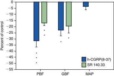
21. Berggreen E, Heyeraas KJ. Effect of the sensory neuropeptide antagonists h-CGRP(8–37) and SR 140.33 on pulpal and gingival blood flow in ferrets. Arch Oral Biol. 2000;45:537-542.
22. Berggreen E, Heyeraas KJ. Role of K+ATP channels, endothelin A receptors, and effect of angiotensin II on blood flow in oral tissues. J Dent Res. 2003;82:33-37.
23. Berggreen E, Heyeraas KJ. The role of sensory neuropeptides and nitric oxide on pulpal blood flow and tissue pressure in the ferret. J Dent Res. 1999;78:1535-1543.
24. Bernick S, Nedelman C. Effect of aging on the human pulp. J Endod. 1975;1:88.
25. Bevilacqua MP, et al. Interleukin-1 activation of vascular endothelium. Effects on procoagulant activity and leukocyte adhesion. Am J Pathol. 1985;121:394-403.
26. Bhussary BR. Modification of the dental pulp organ during development and aging. In: Finn SB, editor. Biology of the Dental Pulp Organ: a Symposium. Birmingham: University of Alabama Press, 1968.
27. Biesterfeld RC, Taintor JF, Marsh CL. The significance of alterations of pulpal respiration: a review of the literature. J Oral Pathol. 1979;8:129.
28. Bishop MA, Malhotra MP. An investigation of lymphatic vessels in the feline dental pulp. Am J Anat. 1990;187:247.
29. Bishop MA, Malhotra M, Yoshida S. Interodontoblastic collagen (von Korff fibers) and circumpulpal dentin formation: an ultrathin serial section study in the cat. Am J Anat. 1991;191:67.
30. Bishop MA, Yoshida S. A permeability barrier to lanthanum and the presence of collagen between odontoblasts in pig molars. J Anat. 1992;181:29.
31. Bletsa A, et al. Cytokine signalling in rat pulp interstitial fluid and transcapillary fluid exchange during lipopolysaccharide-induced acute inflammation. J Physiol. 2006;573(Pt 1):225-236.
32. Bongenhielm U, Haegerstrand A, Theodorsson E, Fried K. Effects of neuropeptides on growth of cultivated rat molar pulp fibroblasts. Regul Pept. 1995;60:2391-2398.
33. Bonucci E. Matrix vesicles: their role in calcification. In: Linde A, editor. Dentin and dentinogenesisis, Vol I. Boca Raton: CRC Press; 1984:135-154.
34. Borda E, et al. Nitric oxide synthase and PGE2 reciprocal interactions in rat dental pulp: cholinoceptor modulation. J Endod. 2007;33:142-147.
35. Botero TM, et al. TLR4 mediates LPS-induced VEGF expression in odontoblasts. J Endod. 2006;32:951-955.
36. Bowles WR, et al. beta 2-Adrenoceptor regulation of CGRP release from capsaicin-sensitive neurons. J Dent Res. 2003;82:308-311.
37. Brännström M. Communication between the oral cavity and the dental pulp associated with restorative treatment. Oper Dent. 1984;9:57.
38. Brännström M. The transmission and control of dentinal pain. In: Grossman LJ, editor. Mechanisms and control of pain. New York: Masson Publishing USA, 1979.
39. Brännström M, Aström A. A study of the mechanism of pain elicited from the dentin. J Dent Res. 1964;43:619.
40. Breschi L, Lopes M, Gobbi P, Mazzotti G, Falconi M, Perdigao J. Dentin proteoglycans: an immunocytochemical FEISEM study. J Biomed Mater Res. 2002;61:40.
41. Brown AC, Yankowitz D. Tooth pulp tissue pressure and hydraulic permeability. Circ Res. 1964;15:42-50.
42. Butler WT, D’Sousa RN, Bronckers AL, Happonen RP, Somerman MJ. Recent investigations on dentin specific proteins. Proc Finn Dent Soc. 1992;88(suppl 1):369.
43. Butler WT, Ritchie H. The nature and functional significance of dentin extracellular matrix proteins. Int J Dev Biol. 1995;39:213-222.
44. Byers MR. Dynamic plasticity of dental sensory nerve structure and cytochemistry. Arch Oral Biol. 1994;39(suppl):13S.
45. Byers MR. Neuropeptide immunoreactivity in dental sensory nerves: variations related to primary odontoblast function and survival. In: Shimono M, Takahashi K, editors. Dentin/Pulp Complex. Tokyo: Quintessence Publishing Co, 1996.
46. Byers MR, Chudler EH, Iadarola MJ. Chronic tooth pulp inflammation causes transient and persistent expression of Fos in dynorphin-rich regions of rat brainstem. Brain Res. 2000;861:191-207.
47. Byers MR, Närhi MVO. Dental injury models: experimental tools for understanding neuroinflammatory interactions and polymodal nociceptor functions. Crit Rev Oral Biol Med. 1999;10:4.
48. Byers MR, Närhi MVO. Nerve supply of the pulpodentin complex and response to injury. In: Hargreaves K, Goodis H, editors. Seltzer and Bender’s dental pulp. Chicago: Quintessence Publishing Co, 2002.
49. Byers MR, Narhi MV, Mecifi KB. Acute and chronic reactions of dental sensory nerve fibers to cavities and desiccation in rat molars. Anat Rec. 1988;221:872-883.
50. Byers MR, Schatteman GC, Bothwell MA. Multiple functions for NGF-receptor in developing, aging and injured rat teeth are suggested by epithelial, mesenchymal and neural immunoreactivity. Development. 1990;109:461.
51. Byers MR, Sugaya A. Odontoblast process in dentin revealed by fluorescent Di-I. J Histochem Cytochem. 1995;43:159.
52. Byers MR, Suzuki H, Maeda T. Dental neuroplasticity, neuro-pulpal interactions and nerve regeneration. Microsc Res Tech. 2003;60:503.
53. Byers MR, Taylor PE. Effect of sensory denervation on the response of rat molar pulp to exposure injury. J Dent Res. 1993;72:613.
54. Byers MR, Wheeler EF, Bothwell M. Altered expression of NGF and P75 NGF-receptor by fibroblasts of injured teeth precedes sensory nerve sprouting. Growth Factors. 1992;6:41-52.
55. Camps J, Pashley DH. In vivo sensitivity to air blasts and scratching of human root dentin. J Periodontol. 2003;74:1589.
56. Camps J, Salomon JP, Van Meerbeek B, Tay F, Pashley D. Dentin deformation after scratching with clinically-relevant forces. Arch Oral Biol. 2003;48:527.
57. Carter JM, Sorensen SE, Johnson RR, Teitelbaum RL, Levine MS. Punch shear testing of extracted vital and endodontically treated teeth. J Biomech. 1983;16:841.
58. Casasco A, et al. Immunohistochemical localization of endothelin-like immunoreactivity in human tooth germ and mature dental pulp. Anat Embryol (Berl). 1991;183:515-520.
59. Chaudhary P, Martenson ME, Baumann TK. Vanilloid receptor expression and capsaicin excitation of rat dental primary afferent neurons. J Dent Res. 2001;80:1518.
60. Chu SC, et al. Induction of vascular endothelial growth factor gene expression by proinflammatory cytokines in human pulp and gingival fibroblasts. J Endod. 2004;30:704-707.
61. Coffey CT, Ingram MJ, Bjöandal AM. Analysis of dentinal fluid. Oral Surg. 1970;30:835.
62. Costos CAS, Hebling J, Hanks CT. Current status of pulp capping with dentin adhesive systems: a review. Dent Mater. 2000;16:188.
63. Coure E. Ultrastructural changes during the life cycle of human odontoblasts. Arch Oral Biol. 1986;31:643.
64. Cox CF, Bogen G, Kopel HM, Ruby JP. Repair of pulpal injury by dental materials. Chap. 14. In: Hargreaves K, Goodis H, editors. Seltzer and Bender’s dental pulp. Chicago: Quintessence Publishing Co, 2002.
65. Cox CF, White KC, Ramus DL, Farmer JB, Snuggs HM. Reparative dentin: factors affecting its deposition. Quintessence Int. 1992;23:257.
66. Csillag M, Berggreen E, Fristad I, Haug SR, Bletsa A, Heyeraas KJ. Effect of electrical tooth stimulation on blood flow and immunocompetent cells in rat dental pulp after sympathectomy. Acta Odontol Scand. 2004;62:305-312.
67. Cuicchi B, Bouillaguet S, Holz J, Pashley D. Dentinal fluid dynamics in human teeth, in vivo. J Endod. 1995;21:191.
68. Cvek M, Granath L, Lundberg M. Failures and healing in endodontically treated non-vital anterior teeth with posttraumatically reduced pulpal lumen. Acta Odontol Scand. 1982;40:223-228.
69. Dahl E, Major IA. The fine structure of the vessels in the human dental pulp. Acta Odontol Scand. 1973;31:223-230.
70. Dahl T, Sabsay B, Veis A. Type I collagen-phosphophoryn interactions: specificity of the monomer-monomer binding. J Struct Biol. 1998;123:162.
71. Dia XF, ten Cate AR, Limeback H. The extent and distribution of intratubular collagen fibrils in human dentine. Arch Oral Biol. 1991;36:775.
72. Diamond J. The effect of injecting acetylcholine into normal and regenerating nerves. J Physiol (Lond). 1959;145:611.
73. Diamond RD, Stanley HR, Swerdlow H. Reparative dentin formation resulting from cavity preparation. J Prosthet Dent. 1966;16:1127.
74. Diogenes A, Akopian AN, Hargreaves KM. NGF up-regulates TRPA1: implications for orofacial pain. J Dent Res. 2007;86:550-555.
75. D’Souza RN, Bachman T, Baumgardner KR, Butler WT, Litz M. Characterization of cellular responses involved in reparative dentinogenesis in rat molars. J Dent Res. 1995;74:702.
76. Ebihara A, Tokita Y, Izawa T, Suda H. Pulpal blood flow assessed by laser Doppler flowmetry in a tooth with a horizontal root fracture. Oral Surg Oral Med Oral Path. 1996;81:229.
77. Eda S, Saito T. Electron microscopy of cells displaced into the dentinal tubules due to dry cavity preparation. J Oral Pathol. 1978;7:326.
78. Edwall L, Kindlová M. The effect of sympathetic nerve stimulation on the rate of disappearance of tracers from various oral tissues. Acta Odontol Scand. 1971;29:387.
79. Edwall B, et al. Neuropeptide Y (NPY) and sympathetic control of blood flow in oral mucosa and dental pulp in the cat. Acta Physiol Scand. 1985;125:253-264.
80. Eissmann HF, Radke RA. Postendodontic restoration. In Cohen S, Burns RC, editors: Pathways of the pulp, ed 4, St Louis: Mosby, 1987.
81. El-Backly RM, et al. Regeneration of dentine/pulp-like tissue using a dental pulp stem cell/poly(lactic-co-glycolic) acid scaffold construct in New Zealand white rabbits. Aust Endod J. 2008;34:52-67.
82. Embery G. Glycosaminoglycans of human dental pulp. J Biol Buccale. 1976;4:229-236.
83. Embery G, Hall R, Waddington R, Septier D, Goldberg M. Proteoglycans in dentinogenesis. Crit Rev Oral Biol Med. 2001;12:331.
84. England MC, Pellis EG, Michanowicz AE. Histopathologic study of the effect of pulpal disease upon nerve fibers of the human dental pulp. Oral Surg Oral Med Oral Pathol. 1974;38:783.
85. Engström C, Linde A, Persliden B. Acid hydrolases in the odontoblast-predentin region of dentinogenically active teeth. Scand J Dent Res. 1976;84:76.
86. Evans D, Reid T, Strang R, Stirrups D. A comparison of laser Doppler flowmetry with other methods of assessing vitality in traumatized anterior teeth. Endod Dent Traumatol. 1999;15:284.
87. Fearnhead RW. Innervation of dental tissues. In: Miles AEW, editor. Structural and chemical organization of the teeth, vol 1. New York: Academic Press; 1967.
88. Ferrara N. Vascular endothelial growth factor. Eur J Cancer. 1996;32A:2413-2422.
89. Ferrari M, Mason PN, Goracci C, Pashley DH, Tay FR. Collagen degradation in endodontically-treated teeth after clinical function. J Dent Res. 2004;88:414.
90. Finkelman RD, Mohan S, Jennings JC, Taylor AK, Jepsen S, Baylink DJ. Quantitation of growth factors IGF-1, SGF/IGF-11 and TGF-b in human dentin. J Bone Miner Res. 1990;5:717.
91. Firestone AR, Wheatley AM, Thüer UW. Measurement of blood perfusion in the dental pulp with laser Doppler flowmetry. Int J Microcirc Clin Exp. 1997;17:298.
92. Fish WE. An experimental investigation of enamel, dentine and the dental pulp. London: John Bale, Sons and Danielson; 1932.
93. Fisher AK. Respiratory variations within the normal dental pulp. J Dent Res. 1967;46:424.
94. Fisher AK, Schumacher ER, Robinson NR, Sharbondy GP. Effects of dental drugs and materials on the rate of oxygen consumption in bovine dental pulp. J Dent Res. 1957;36:447.
95. Fisher AK, Walters VE. Anaerobic glycolysis in bovine dental pulp. J Dent Res. 1968;47:717.
96. Fitzgerald M, Chiego DJ, Heys DR. Autoradiographic analysis of odontoblast replacement following pulp exposure in primate teeth. Arch Oral Biol. 1990;35:707.
97. Fogel HM, Marshall FJ, Pashley DH. Effects of distance of the pulp and thickness on the hydraulic conductance of human radicular dentin. J Dent Res. 1988;67:1381.
98. Fraser JR, et al. Uptake and degradation of hyaluronan in lymphatic tissue. Biochem J. 1988;256:153-158.
99. Fried K. Changes in pulp nerves with aging. Proc Finn Dent Soc. 1992;88(suppl 1):517.
100. Fried K, et al. Target finding of pain nerve fibers: neural growth mechanisms in the tooth pulp. Physiol Behav. 2007;92:40-45.
101. Fristad I, Heyeraas KJ, Kvinnsland I. Nerve fibres and cells immunoreactive to neurochemical markers in developing rat molars and supporting tissues. Arch Oral Biol. 1994;39:633-646.
102. Fristad I, Jacobsen EB, Kvinnsland IH. Coexpression of vasoactive intestinal polypeptide and substance P in reinnervating pulpal nerves and in trigeminal ganglion neurones after axotomy of the inferior alveolar nerve in the rat. Arch Oral Biol. 1998;43:183-189.
103. Fristad I, Kvinnsland IH, Jonsson R, Heyeraas KJ. Effect of intermittent long-lasting electrical tooth stimulation on pulpal blood flow and immunocompetent cells: a hemodynamic and immunohistochemical study in young rat molars. Exp Neurol. 1997;146:230-239.
104. Fuss Z, Trowbridge H, Bender IB, Rickoff B, Sorin S. Assessment of reliability of electrical and thermal pulp testing agents. J Endod. 1986;12:301.
105. Gani O, Visvisian C. Apical canal diameter in the first upper molar at various ages. J Endod. 1999;10:689.
106. Garant PR. The organization of microtubules within rat odontoblast processes revealed by perfusion fixation with glutaraldehyde. Arch Oral Biol. 1972;17:1047.
107. Garberoglio R, Brännström M. Scanning electron microscopic investigation of human dentinal tubules. Arch Oral Biol. 1976;21:355.
108. Gaucher C, Boukpessi T, Septier D, et al. Dentin noncollagenous matrix proteins in familiar hypophosphatemic rickets. Cells, Tissues, Organs. 2009;189:219-223.
109. George A, Bannon L, Sabsay B, et al. The carboxyl-terminal domain of phosphophoryn contains unique extended triplet amino acid repeat sequences forming ordered carboxyl-phosphate interaction ridges that may be essential in the biomineralization process. J Biol Chem. 1996;271:32869-32873.
110. George CH, Kendall JM, Evans WH. Intracellular trafficking pathways on assembly of connexins into tight junctions. J Biol Chem. 1999;274:8678.
111. Gibbs JL, Hargreaves KM. Neuropeptide Y Y1 receptor effects on pulpal nociceptors. J Dent Res. 2008;87:948-952.
112. Gilbert TM, Pashley DH, Anderson RW. Response of pulpal blood flow to intra-arterial infusion of endothelin. J Endod. 1992;18:228-231.
113. Gloe T, Pohl U. Laminin binding conveys mechanosensing in endothelial cells. News Physiol Sci. 2002;17:166.
114. Gold M. Tetrodotoxin-resistant Na currents and inflammatory hyperalgesia. Proc Natl Acad Sci U S A. 1999;96:7645.
115. Goldberg M, Lasfargues J-J. Dentin-pulpal complex revisited. J Dent. 1995;23:15.
116. Goldberg M, Six N, Decup F, Lasfargues JJ, Salih E, Tompkins K, et al. Bioactive molecules and the future of pulp therapy. Am J Dent. 2003;16:66.
117. Goldberg M, Takagi M. Dentine proteoglycans: composition, ultrastructure and functions. Histochem J. 1993;25:781.
118. Goldberg M, et al. Inflammatory and immunological aspects of dental pulp repair. Pharmacol Res. 2008;58:137-147.
119. Gotjamanos T. Cellular organization in the subodontoblastic zone of the dental pulp. II. Period and mode of development of the cell-rich layer in rat molar pulps. Arch Oral Biol. 1969;14:1011.
120. Gregg JM, Dixon AD. Somatotopic organization of the trigeminal ganglion. Arch Oral Biol. 1973;18:487.
121. Grossman ES, Austin JC. Scanning electron microscope observations on the tubule content of freeze-fractured peripheral vervet monkey dentine (Cercopithecus pygerythrus),. Arch Oral Biol. 1983;28:279.
122. Gunji T. Morphological research on the sensitivity of dentin. Arch Histol Jpn. 1982;45:45.
123. Guzy CE, Nicholls JI. In vitro comparison of intact endodontically-treated teeth with and without endo-post reinforcement. J Prosthet Dent. 1979;42:39.
124. Hahn C-L, Falkler WAJr, Siegel MA. A study of T cells and B cells in pulpal pathosis. J Endod. 1989;15:20.
125. Hahn C-L, Overton B. The effects of immunoglobulins on the convective permeability of human dentin in vivo. Arch Oral Biol. 1997;42:835.
126. Hals E, Tonder KJ. Elastic pseudoelastic tissue in arterioles of the human and dog dental pulp. Scand J Dent Res. 1981;89:218-227.
127. Hamersky PA, Weimer AD, Taintor JF. The effect of orthodontic force application on the pulpal tissue respiration rate in the human premolar. Am J Orthod. 1980;77:368.
128. Han SS. The fine structure of cells and intercellular substances of the dental pulp. In: Finn SB, editor. Biology of the dental pulp organ. Birmingham: University of Alabama Press; 1968:103.
129. Hargreaves KM. Pain mechanisms of the pulpodentin complex. In: Hargreaves KM, Goodis HE, editors. Seltzer and Bender’s dentin pulp. Chicago: Quintessence Publishing Co, 2002.
130. Hargreaves KM, Bowles WR, Jackson DL. Intrinsic regulation of CGRP release by dental pulp sympathetic fibers. J Dent Res. 2003;82:398-401.
131. Harris R, Griffin CJ. Fine structure of nerve endings in the human dental pulp. Arch Oral Biol. 1968;13:773.
132. Hartmann A, Azerad J, Boucher Y. Environmental effects on laser Doppler pulpal blood-flow measurements in man. Arch Oral Biol. 1996;41:333.
133. Hashioka K, et al. Relationship between clinical symptoms and enzyme-producing bacteria isolated from infected root canals. J Endod. 1994;20:75-77.
134. Hattler AB, Listgarten MA. Pulpal response to root planing in a rat model. J Endod. 1984;10:471.
135. Haug SR, Heyeraas KJ. Effects of sympathectomy on experimentally induced pulpal inflammation and periapical lesions in rats. Neuroscience. 2003;120:827-836.
136. Haug SR, Heyeraas KJ. Modulation of dental inflammation by the sympathetic nervous system. J Dent Res. 2006;85:488-495.
137. Helfer AR, Melwick S, Schilder H. Determination of the moisture content of vital and pulpless teeth. Oral Surg Oral Med Oral Pathol. 1972;34:661.
138. Hermanstyne TO, Markowitz K, Fan L, Gold MS. Mechanotransducers in rat pulpal afferents. J Dent Res. 2008;87:834-838.
139. Herr P, Holz J, Baume LJ. Mantle dentine in man: a quantitative study microradiographic study. J Biol Buccale. 1986;14:139.
140. Heyeraas KJ. Pulpal hemodynamics and interstitial fluid pressure: balance of transmicrovascular fluid transport. J Endod. 1989;15:468-472.
141. Heyeraas KJ, Berggreen E. Interstitial fluid pressure in normal and inflamed pulp. Crit Rev Oral Biol Med. 1999;10:328.
142. Heyeraas KJ, Jacobsen EB, Fristad I Vascular and immunoreactive nerve fiber reactions in the pulp after stimulation and denervation: proceedings of the International Conference on Dentin/Pulp Complex Shimono M Maeda T Suda H Takahashi K 1996 Quintessence Publishing Co Tokyo 162
143. Heyeraas KJ, Kvinnsland I. Tissue pressure and blood flow in pulpal inflammation. Proc Finn Dent Soc. 1992;88(Suppl 1):393-401.
144. Heyeraas KJ, et al. Effect of electrical tooth stimulation on blood flow, interstitial fluid pressure and substance P and CGRP-immunoreactive nerve fibers in the low compliant cat dental pulp. Microvasc Res. 1994;47:329-343.
145. Hikiji A, Yamamoto H, Sunakawa M, Suda H. Increased blood flow and nerve firing in the cat canine tooth in response to stimulation of the second premolar pulp. Arch Oral Biol. 2000;45:53.
146. Hildebrand C, et al. Teeth and tooth nerves. Prog Neurobiol. 1995;45:165-222.
147. Hirakawa S, Detmar M. New insights into the biology and pathology of the cutaneous lymphatic system. J Dermatol Sci. 2004;35:1-8.
148. Hirvonen T, Närhi M, Hakumäki M. The excitability of dog pulp nerves in relation to the condition of dentine surface. J Endod. 1984;10:294.
149. Holland GR. Morphological features of dentine and pulp related to dentine sensitivity. Arch Oral Biol. 1994;39(suppl):3S.
150. Holland GR. The extent of the odontoblast process in the cat. Am J Anat. 1976;121:133.
151. Holland GR. The odontoblast process: form and function. J Dent Res. 1985;64(special issue):499.
152. Howe CA, McKendry DJ. Effect of endodontic access preparation on resistance to crown-root fracture. J Am Dent Assoc. 1990;121:712.
153. Ibricevic H, et al. Identification of alpha 2 adrenoceptors in the blood vessels of the dental pulp. Int Endod J. 1991;24:279-289.
154. Ikeda H, Tokita Y, Suda H. Capsaicin-sensitive A fibers in cat tooth pulp. J Dent Res. 1997;76:1341.
155. Inoue H, Kurosaka Y, Abe K. Autonomic nerve endings in the odontoblast/predentin border and predentin of the canine teeth of dogs. J Endodon. 1992;18:149.
156. Isidor F, Odman P, Brondum K. Intermittent loading of teeth restored using prefabricated carbon fiber posts. Int J Prosthodont. 1996;9:131.
157. Itthagarum A, Tay FR. Self-contamination of deep dentin by dentinal fluid. Am J Dent. 2000;13:195.
158. Jacobsen EB, Heyeraas KJ. Pulp interstitial fluid pressure and blood flow after denervation and electrical tooth stimulation in the ferret. Arch Oral Biol. 1997;42:407-415.
159. Jernvall J, et al. Evidence for the role of the enamel knot as a control center in mammalian tooth cusp formation: non-dividing cells express growth stimulating Fgf-4 gene. Int J Dev Biol. 1994;38:463-469.
160. Johnsen DC, Harshbarger J, Rymer HD. Quantitative assessment of neural development in human premolars. Anat Rec. 1983;205:421.
161. Johnsen D, Johns S. Quantitation of nerve fibers in the primary and permanent canine and incisor teeth in man. Arch Oral Biol. 1978;23:825.
162. Johnson G, Brännström M. The sensitivity of dentin: changes in relation to conditions at exposed tubule apertures. Acta Odontol Scand. 1974;32:29.
163. Jones PA, Taintor JF, Adams AB. Comparative dental material cytotoxicity measured by depression of rat incisor pulp respiration. J Endod. 1979;5:48.
164. Jontell M, Okiji T, Dahlgren U, Bergenholtz G. Immune defense mechanisms of the dental pulp. Crit Rev Oral Biol Med. 1998;9:179.
165. Katoh Y, Yamaguchi R, Shinkai K, et al. Clinicopathological study on pulp-irritation of adhesive resinous materials (report 3). Direct capping effects on exposed pulp of Macaca fascicularis. Jpn J Conserv Dent. 1997;40:163.
166. Kayaoglu G, Orstavik D. Virulence factors of Enterococcus faecalis: relationship to endodontic disease. Crit Rev Oral Biol Med. 2004;15:308-320.
167. Kaye H, Herold RC. Structure of human dentine. I. Phase contrast, polarization, interference, and bright field microscopic observations on the lateral branch system. Arch Oral Biol. 1966;11:355.
168. Kelley KW, Bergenholtz G, Cox CF. The extent of the odontoblast process in rhesus monkeys (Macaca mulatta) as observed by scanning electron microscopy. Arch Oral Biol. 1981;26:893.
169. Kerezoudis NP, Olgart L, Edwall L. CGRP(8–37) reduces the duration but not the maximal increase of antidromic vasodilation in dental pulp and lip of the rat. Acta Physiol Scand. 1994;151:73-81.
170. Kerezoudis NP, Olgart L, Edwall L. Involvement of substance P but not nitric oxide or calcitonin gene-related peptide in neurogenic plasma extravasation in rat incisor pulp and lip. Arch Oral Biol. 1994;39:769-774.
171. Kerezoudis NP, et al. Activation of sympathetic nerves exerts an inhibitory influence on afferent nerve-induced vasodilation unrelated to vasoconstriction in rat dental pulp. Acta Physiol Scand. 1993;147:27-35.
172. Kettunen P, Thesleff I. Expression and function of FGFs-4, -8, and -9 suggest functional redundancy and repetitive use as epithelial signals during tooth morphogenesis. Dev Dyn. 1998;211:256-268.
173. Kettunen P, et al. Coordination of trigeminal axon navigation and patterning with tooth organ formation: epithelial-mesenchymal interactions, and epithelial Wnt4 and Tgfbeta1 regulate semaphorin 3a expression in the dental mesenchyme. Development. 2005;132:323-334.
174. Khullar SM, Fristad I, Brodin P, Kvinnsland IH. Upregulation of growth associated protein 43 expression and neuronal co-expression with neuropeptide Y following inferior alveolar nerve axotomy in the rat. J Peripher Nerv Syst. 1998;3:79-90.
175. Kim S, Edwall L, Trowbridge H, Chien S. Effects of local anesthetics on pulpal blood flow in dogs. J Dent Res. 1984;63:650.
176. Kim S, Schuessler G, Chien S. Measurement of blood flow in the dental pulp of dogs with the 133xenon washout method. Arch Oral Biol. 1983;28:501.
177. Kim S, Trowbridge HO, Dorscher-Kim JE. The influence of 5-hydroxytryptamine (serotonin) on blood flow in the dog pulp. J Dent Res. 1986;65:682-685.
178. Kim SK, et al. Antagonistic effect of D-myo-inositol-1,2,6-trisphosphate (PP56) on neuropeptide Y-induced vasoconstriction in the feline dental pulp. Arch Oral Biol. 1996;41:791-798.
179. Kim S, et al. Effects of selected inflammatory mediators on blood flow and vascular permeability in the dental pulp. Proc Finn Dent Soc. 1992;88(suppl 1):387-392.
180. Kim S, et al. Functional alterations in pulpal microcirculation in response to various dental procedures and materials. Proc Finn Dent Soc. 1992;88(suppl 1):65-71.
181. Kim S, Dorscher-Kim JE, Liu M. Microcirculation of the dental pulp and its autonomic control. Proc Finn Dent Soc. 1989;85:279-287.
182. Kimberly CL, Byers BR. Inflammation of rat molar pulp and periodontium causes increased calcitonin-gene-related peptide and axonal sprouting. Anat Rec. 1988;222:289.
183. Kinney JH, Balooch M, Marshall SJ, Marshall GWJr, Weihs TP. Hardness and Young’s modulus of human peritubular and intertubular dentin. Arch Oral Biol. 1996;41:9-13.
184. Kinney JH, Marshall SJ, Marshall GW. The mechanical properties of human dentin: a critical review and reevaluation of the dental literature. Crit Rev Oral Biol Med. 2003;14:13.
185. Kinney JH, Pople JA, Driessen CH, Breunig TM, Marshall GW, Marshall SJ. Intrafibrillar mineral may be absent in dentinogenesis imperfecta type II (D1–11). J Dent Res. 2001;80:1555.
186. Kollar EJ, Lumsden AG. Tooth morphogenesis: the role of the innervation during induction and pattern formation. J Biol Buccale. 1979;7:49-60.
187. Kontturi-Nähri V, Närhi M. Testing sensitive dentin in man. Int Endod J. 1993;26:4.
188. Kramer IRH. The distribution of blood vessels in the human dental pulp. In: Finn SB, editor. Biology of the dental pulp Organ. Birmingham: University of Alabama Press; 1968:361.
189. Kroeger DC, Gonzales F, Krivoy W. Transmembrane potentials of cultured mouse dental pulp cells. Proc Soc Exp Biol Med. 1961;108:134.
190. Kvinnsland IH, Luukko K, Fristad I, Kettunen P, Jackson DL, Fjeld K, et al. Glial cell line-derived neurotrophic factor (GDNF) from adult rat tooth serves a distinct population of large-sized trigeminal neurons. Eur J Neurosci. 2004;19:2089-2098.
191. Langeland K, Langeland LK. Histologic study of 155 impacted teeth. Odontol Tidskr. 1965;73:527.
192. Langeland K, Langeland LK. Pulp reactions to cavity and crown preparations. Aust Dent J. 1970;15:261.
193. Lantelme RL, Handleman SL, Herbison RJ. Dentin formation in periodontally diseased teeth. J Dent Res. 1976;55:48.
194. Laurent TC, et al. The catabolic fate of hyaluronic acid. Connect Tissue Res. 1986;15:33-41.
195. Lechner JH, Kalnitsky G. The presence of large amounts of type III collagen in bovine dental pulp and its significance with regard to the mechanism of dentinogenesis. Arch Oral Biol. 1981;26:265-273.
196. Lesot H, Osman M, Ruch JV. Immunofluorescent localization of collagens, fibronectin and laminin during terminal differentiation of odontoblasts. Dev Biol. 1981;82:371.
197. Lesot H, et al. Epigenetic signals during odontoblast differentiation. Adv Dent Res. 2001;15:8-13.
198. Levi-Montalcini R. The nerve growth factor: its mode of action on sensory and sympathetic nerve cells. Harvey Lect. 1966;60:217-259.
199. Lewinstein I, Grajower R. Root dentin hardness of endodontically treated teeth. J Endod. 1981;7:421.
200. Lilja J. Innervation of different parts of the predentin and dentin in a young human premolar. Acta Odontol Scand. 1979;37:339.
201. Lilja J, Noredenvall K-J, Brännström M. Dentin sensitivity, odontoblasts and nerves under desiccated or infected experimental cavities. Swed Dent J. 1982;6:93.
202. Linde A. The extracellular matrix of the dental pulp and dentin. J Dent Res. 1985;64(special issue):523.
203. Linde A. A study of the dental pulp glycosamino-glycans from permanent human teeth and rat and rabbit incisors. Arch Oral Biol. 1973;18:49-59.
204. Linde A, Goldberg M. Dentinogenesis. Crit Rev Oral Biol Med. 1993;4:679-728.
205. Linde A, Lundgren T. From serum to the mineral phase. The role of the odontoblast in calcium transport and mineral formation. Int J Dev Biol. 1995;39:213-222.
206. Liu L, Simon SA. Capsaicin, acid and heat-evoked currents in rat trigeminal ganglion neurons: relationship to functional VR1 receptors. Physiol Behav. 2000;69:363.
207. Lohinai Z, Szekely AD, Benedek P, Csillag A. Nitric oxide synthetase containing nerves in the cat and dog dental pulps and gingiva. Neurosci Lett. 1997;227:91.
208. Lohinai Z, et al. Evidence for the role of nitric oxide in the circulation of the dental pulp. J Dent Res. 1995;74:1501-1506.
209. Lumsden AG. The developing innervation of the lower jaw and its relation to the formation of tooth germs in mouse. In: TEETH; Form, function and evolution. New York: Columbia University Press; 1982:32-43.
210. Lundberg JM, Änggård A, Fahrenkrug J, Hökfelt T, Mutt V. Vasoactive intestinal polypeptide in cholinergic neurons of exocrine glands: functional significance of coexisting transmitters for vasodilation and secretion. Proc Natl Acad Sci U S A. 1980;77:1651-1655.
211. Lundberg JM, Fried G, Fahrenkrug J, Holmstedt B, Hökfelt T, Lagercrantz H, et al. Subcellular fractionation of cat submandibular gland: comparative studies on the distribution of acetylcholine and vasoactive intestinal polypeptide (VIP). Neuroscience. 1981;6:1001-1010.
212. Lundgren T, Nannmark U, Linde A. Calcium ion activity and pH in the odontoblast-predentin region: ion-selective microelectrode measurements. Calcif Tissue Int. 1992;50:134.
213. Luthman J, Luthman D, Hökfelt T. Occurrence and distribution of different neurochemical markers in the human dental pulp. Arch Oral Biol. 1992;37:193.
214. Luukko K, Kvinnsland IH, Kettunen P. Tissue interactions in the regulation of axon pathfinding during tooth morphogenesis. Dev Dyn. 2005;234:482-488.
215. Luukko K, et al. Identification of a novel putative signaling center, the tertiary enamel knot in the postnatal mouse molar tooth. Mech Dev. 2003;120:270-276.
216. Luukko K, et al. Secondary induction and the development of tooth nerve supply. Ann Anat. 2008;190:178-187.
217. Madison S, Whitsel E, Suarez-Roca H, Maixner W. Sensitizing effects of leukotriene B4 on intradentinal primary afferents. Pain. 1992;49:99.
218. Maeda T, Honma S, Takano Y. Dense innervation of radicular human dental pulp as revealed by immunocytochemistry for protein gene-product 9.5. Arch Oral Biol. 1994;39:563.
219. Maita E, Simpson MD, Tao L, Pashley DH. Fluid and protein flux across the pulpodentin complex of the dog, in vivo. Arch Oral Biol. 1991;36:103.
220. Maltos KL, et al. Vascular and cellular responses to pro-inflammatory stimuli in rat dental pulp. Arch Oral Biol. 2004;49:443-450.
221. Mangkornkarn C, Steiner JC. In vivo and in vitro glycosaminoglycans from human dental pulp. J Endod. 1992;18:327-331.
222. Marbach JJ, Raphael KG. Phantom tooth pain: a new look at an old dilemma. Pain Med. 2000;1:68-77.
223. Marfurt CF, Zaleski EM, Adams CE, Welther CL. Sympathetic nerve fibers in rat orofacial and cerebral tissues as revealed by the HRP-WGA tracing technique: a light and electron microscopic study. Brain Res. 1986;366:373-378.
224. Marion D, Jean A, Hamel H, Kerebel LM, Kerebel B. Scanning electron microscopic study of odontoblasts and circumferential dentin in a human tooth. Oral Surg Oral Med Oral Pathol. 1991;72:473.
225. Martin-de las Heras S, Valenzuela A, Overall CM. The matrix metalloproteinases gelatinase A in human dentine. Arch Oral Biol. 2000;45:757.
226. Martinez-Insua A, de Silva L, Rilo B, Santana U. Comparison of the fracture resistance of pulpless teeth restored with a cast post and core or carbon-fiber post with a composite core. J Prosthet Dent. 1998;80:527.
227. Matsuo S, Ichikawa H, Henderson TA, Silos-Santiago I, Barbacid M, Arends JJ, et al. trkA modulation of developing somatosensory neurons in oro-facial tissues: tooth pulp fibers are absent in trkA knockout mice. Neuroscience. 2001;105:747-760.
228. Matthews B, Andrew D. Microvascular architecture and exchange in teeth. Microcirculation. 1995;2:305-313.
229. Matthews B, Andrew D, Amess TR: The functional properties of intradental nerves: proceedings of the International Conference on Dentin/Pulp Complex, ed. by Shimono M.
230. Matthews B, Vongsavan N. Interactions between neural and hydrodynamic mechanisms in dentine and pulp. Arch Oral Biol. 1994;39(suppl 1):87S.
231. McGrath PA, Gracely RH, Dubner R, Heft MW. Non-pain and pain sensations evoked by tooth pulp stimulation. Pain. 1983;15:377-388.
232. Meyer MW, Path MG. Blood flow in the dental pulp of dogs determined by hydrogen polarography and radioactive microsphere methods. Arch Oral Biol. 1979;24:601.
233. Michelich V, Pashley DH, Whitford GM. Dentin permeability: a comparison of functional versus anatomical tubular radii. J Dent Res. 1978;57:1019.
234. Michelich VJ, Schuster GS, Pashley DH. Bacterial penetration of human dentin in vitro. J Dent Res. 1980;59:1398.
235. Mitsiadis TA, De Bari C, About I. Apoptosis in developmental and repair-related human tooth remodeling: a view from the inside. Exp Cell Res. 2008;314:869-877.
236. Mjör IA, Nordahl I. The density and branching of dentinal tubules in human teeth. Arch Oral Biol. 1996;41:401.
237. Moe K, et al. Development of the pioneer sympathetic innervation into the dental pulp of the mouse mandibular first molar. Arch Oral Biol. 2008;53:865-873.
238. Mohamed SS, Atkinson ME. A histological study of the innervation of developing mouse teeth. J Anat. 1983;136(Pt 4):735-749.
239. Mullaney TP, Howell RM, Petrich JD. Resistance of nerve fibers to pulpal necrosis. Oral Surg. 1970;30:690.
240. Murray PE, About I, Lumley PJ, Franquin JC, Remusat M, Smith AJ. Human odontoblast cell numbers after dental injury. J Dent. 2000;28:277.
241. Murray PE, Hafez AA, Windsor LJ, Smith AJ, Cox CF. Comparison of pulp responses following restoration of exposed and non-exposed cavities. J Dent. 2002;30:213.
242. Murray PE, Lumley PJ, Ross HF, Smith AJ. Tooth slice organ culture for cytotoxicity assessment of dental materials. Biomaterials. 2000;21:1711.
243. Naftel JP, et al. Course and composition of the nerves that supply the mandibular teeth of the rat. Anat Rec. 1999;256:433-447.
244. Nagaoka S, Miyazaki Y, Liu HJ, Iwamoto Y, Kitano M, Kawagoe M. Bacterial invasion into dentinal tubules of human vital and nonvital teeth. J Endod. 1995;21:70.
245. Nair PN, et al. Histological, ultrastructural and quantitative investigations on the response of healthy human pulps to experimental capping with mineral trioxide aggregate: a randomized controlled trial. Int Endod J. 2008;41:128-150.
246. Nakamura O, Gohda E, Ozawa M, et al. Immunohistochemical studies with a monoclonal antibody on the distribution of phosphophoryn in predentin and dentin. Calcif Tissue Int. 1985;37:491-500.
247. Nalla RK, Imberi V, Kinney JH, Staininec M, Marshall SJ, Richie RO. In vitro fatigue behavior of human dentin with implications for life predictions. J Biomed Mater Res. 2003;64A:10.
248. Nalla RK, Kinney JH, Marshall SJ, Richie RO. On the in vitro fatigue behavior of human dentin: effect of mean stress. J Dent Res. 2004;83:211.
249. Närhi M. Activation of dental pulp nerves of the cat and the dog with hydrostatic pressure. Proc Finn Dent Soc. 1978;74(suppl 5):1.
250. Närhi M, Jyväsjärvi E, Hirronen T. Activation of heat-sensitive nerve fibers in the dental pulp of the cat. Pain. 1982;14:317.
251. Närhi M, Jyväsjärvi E, Virtanen A, Huopaniemi T, Ngassapa D, Hirvonen T. Role of intradentinal A- and C-type nerve fibers in dental pain mechanisms. Proc Finn Dent Soc. 1992;88(suppl 1):507.
252. Närhi M, Virtanen A, Kuhta J, Huopaniemi T. Electrical stimulation of teeth with a pulp tester in the cat. Scand J Dent Res. 1979;87:32.
253. Närhi M, Yamamoto H, Ngassapa D. Function of intradental nociceptors in normal and inflamed teeth. In: Shimono M, Maeda T, Suda H, Takahashi K, editors. Dentin/pulp complex. Tokyo: Quintessence Publishing Co; 1996:136.
254. Närhi M, Yamamoto H, Ngassapa D, Hirvonen T. The neurophysiological basis and the role of inflammatory reactions in dentine hypersensitivity. Arch Oral Biol. 1994;39(suppl):23S.
255. Nawroth PP, Stern DM. Modulation of endothelial cell hemostatic properties by tumor necrosis factor. J Exp Med. 1986;163:740-745.
256. Ngassapa D, Närhi M, Hirvonen T. The effect of serotonin (5-HT) and calcitonin gene-related peptide (CGRP) on the function of intradental nerves in the dog. Proc Finn Dent Soc. 1992;88(suppl 1):143.
257. Nicol GD, Vasko MR. Unraveling the story of NGF-mediated sensitization of nociceptive sensory neurons: ON or OFF the Trks? Molecular interventions. Feb 2007;7:26-41.
258. Nishioka M, Shiiya T, Ueno K, Suda H. Tooth replantation in germ-free and conventional rats. Endod Dent Traumatol. 1998;14:163.
259. Nitzan DW, Michaeli Y, Weinreb M, Azaz B. The effect of aging on tooth morphology: a study on impacted teeth. Oral Surg Oral Med Oral Pathol. 1986;61:54.
260. O’Neil RG, Brown RC. The vanilloid receptor family of calcium-permeable channels: molecular integrators of microenvironmental stimuli. News Physiol Sci. 2003;18:226.
261. Ochoa JL, Torebjork E, Marchettini P, Sivak M. Mechanism of neuropathic pain: cumulative observations, new experiments, and further speculation. In: Fields HL, Dubner R, Cervero F, editors. Advances in pain research and therapy. New York: Raven Press; 1985:431.
262. Oehmke MJ, Knolle E, Oehmke H-J. Lymph drainage in the human dental pulp. Microsc Res Tech. 2003;62:187.
263. Ogawa K, Yamashita Y, Ichijo T, Fusayama T. The ultrastructure and hardness of the transparent layer of human carious dentin. J Dent Res. 1983;62:7.
264. Okiji T, Kawashima N, Kosaka T, Matsumoto A, Kobayashi C, Suda H. An immunohistochemical study of the distribution of immunocompetent cells, especially macrophages and Ia antigen-presenting cells of heterogeneous populations, in normal rat molar pulp. J Dent Res. 1992;71:1196.
265. Okiji T, et al. Involvement of arachidonic acid metabolites in increases in vascular permeability in experimental dental pulpal inflammation in the rat. Arch Oral Biol. 1989;34:523-528.
266. Olgart LM, Edwall L, Gazelius B. Involvement of afferent nerves in pulpal blood-flow reactions in response to clinical and experimental procedures in the cat. Arch Oral Biol. 1991;36:575-581.
267. Olgart LM, Edwall L, Gazelius B. Neurogenic mediators in control of pulpal blood flow. J Endod. 1989;15:409-412.
268. Olgart LM, Gazelius B, Brodin E, Nilsson G. Release of substance P-like immunoreactivity from the dental pulp. Acta Physiol Scand. 1977;101:510.
269. Olgart L, Kerezoudis NP. Nerve-pulp interactions. Arch Oral Biol. 1994;39(suppl):47S.
270. Orchardson R, Cadden SW. An update on the physiology of the dentine-pulp complex. Dent Update. 2001;28:200-206. 208–209, 2001
271. Orchardson R, Gillam DG. Managing dentin hypersensitivity. J Am Dent Assoc. 2006;137:990-998. quiz 1028–1029
272. Oxlund H, Manschot J, Viidik A. The role of elastin in the mechanical properties of skin. J Biomech. 1988;21:213-218.
273. Parsons RJ, McMaster PD. The effect of the pulse upon the formation and flow of lymph. J Exp Med. 1938;68:353-376.
274. Pashley DH. Dentin conditions and disease. In: Lazzari G, editor. CRC handbook of experimental dentistry. Boca Raton, FL: CRC Press; 1983:97.
275. Pashley DH. Dentin permeability and dentin sensitivity. Proc Finn Dent Soc. 1992;88(suppl 1):31.
276. Pashley DH. Dentin permeability: theory and practice. In: Spangberg L, editor. Experimental endodontics. Boca Raton, FL: CRC Press; 1990:19.
277. Pashley DH. Dynamics of the pulpodentin complex. Crit Rev Oral Biol Med. 1996;7:104.
278. Pashley DH. Potential treatment modalities for dentin hypersensitivity—in office products. In Addy M, Orchardson R, editors: Tooth wear and sensitivity. London: Martin-Dunitz Publishers; 2000.
279. Pashley DH, Matthews WG. The effects of outward forced convective flow on inward diffusion in human dentin in vitro. Arch Oral Biol. 1993;38:577.
280. Pashley DH, Pashley EL, Carvalho RM, Tay FR. Effects of dentin permeability on restorative dentistry. Dent Clin North Am. 2002;46:211.
281. Pashley DH, Tay FR, Yiu C, Hashimoto M, Breschi B, Carvalho RM, et al. Collagen degradation by host-derived enzymes during aging. J Dent Res. 2004;83:216.
282. Pashley DH, Zhang Y, Agee KA, Rouse CJ, Carvalho RM, Russell CM. Permeability of demineralized dentin to HEMA. Dent Mater. 2000;16:7.
283. Pimenta FJ, Sa AR, Gomez RS. Lymphangiogenesis in human dental pulp. Int Endod J. 2003;36:853-856.
284. Pissiotis E, Spängberg L. Dentin permeability to bacterial proteins in vitro. J Endod. 1994;20:118.
285. Poggi P, et al. Ultrastructural localization of elastin-like immunoreactivity in the extracellular matrix around human small lymphatic vessels. Lymphology. 1995;28:189-195.
286. Pohto P, Antila R. Innervation of blood vessels in the dental pulp. Int Dent J. 1972;22:228-239.
287. Prati C, Cervellati F, Sanasi V, Montebugnoli L. Treatment of cervical dentin hypersensitivity with resin adhesives: 4 week evaluation. Am J Dent. 2001;14:378.
288. Prescott RS, et al. In vivo generation of dental pulp-like tissue by using dental pulp stem cells, a collagen scaffold, and dentin matrix protein 1 after subcutaneous transplantation in mice. J Endod. 2008;34:421-426.
289. Qian XB, Naftel JP. Effects of neonatal exposure to anti-nerve growth factor on the number and size distribution of trigeminal neurones projecting to the molar dental pulp in rats. Arch Oral Biol. 1996;41:359-367.
290. Qin C, Baba O, Butler WT. Post-translational modifications of SIBLING proteins and their roles in osteogenesis and dentinogenesis. Crit Rev Oral Biol Med. 2004;15:126-136.
291. Rapp R, el-Labban NG, Kramer IR, Wood D. Ultrastructure of fenestrated capillaries in human dental pulps. Arch Oral Biol. 1977;22:317.
292. Reader A, Foreman DW. An ultrastructural qualitative investigation of human intradental innervation. J Endod. 1981;7:493.
293. Renton T, Yiangou Y, Plumpton C, Tate S, Bountra C, Anand P. Sodium channel Nav1.8 immunoreactivity in painful human dental pulp. BMC Oral Health. 2005;5:5.
294. Roberts-Clark D, Smith AJ. Angiogenic growth factors in human dentine matrix. Arch Oral Biol. 2000;42:1013.
295. Rodd HD, Boissonade FM. Comparative immunohistochemical analysis of the peptidergic innervation of human primary and permanent tooth pulp. Arch Oral Biol. 2002;47:375.
296. Rodd HD, Boissonade FM. Innervation of human tooth pulp in relation to caries and dentition type. J Dent Res. Jan 2001;80:389-393.
297. Rodd HD, Boissonade FM, Day PF. Pulpal status of hypomineralized permanent molars. Pediatr Dent. 2007;29:514-520.
298. Rutherford B. BMP-7 gene transfer into inflamed ferret-dental pulps. Eur J Oral Sci. 2001;109:422.
299. Rutherford B, Fitzgerald M. A new biological approach to vital pulp therapy. Crit Rev Oral Biol Med. 1995;6:218.
300. Rutherford RB, Spanberg L, Tucker M, Rueger D, Charette M. The time-course of the induction of reparative dentine formation in moneys by recombinant human osteogenic protein-1. Arch Oral Biol. 1994;39:833.
301. Ryan TJ, Mortimer PS, Jones RL. Lymphatics of the skin. Neglected but important. Int J Dermatol. 1986;25:411-419.
302. Sakamoto N, et al. Identification of hyaluronidase activity in rabbit dental pulp. J Dent Res. 1981;60:850-854.
303. Sakurai K, Okiji T, Suda H. Co-increase of nerve fibers and HLA-DR- and/or factor XIIIa-expressing dendritic cells in dentinal caries-affected regions of the human dental pulp: an immunohistochemical study. J Dent Res. 1999;78:1596.
304. Sasaki S. Studies on the respiration of the dog tooth germ. J Biochem (Tokyo). 1959;46:269.
305. Sasano T, Kuriwada S, Sanjo D. Arterial blood pressure regulation of pulpal blood flow as determined by laser Doppler. J Dent Res. 1989;68:791.
306. Sasano T, Kuriwada S, Shoji N, Sanjo D, Izumi H, Karita K. Axon reflex vasodilatation in cat dental pulp elicited by noxious stimulation of the gingiva. J Dent Res. 1994;73:1797.
307. Sasano T, Shoji N, Kuriwada S, Sanjo D, Izumi H, Karita K. Absence of parasympathetic vasodilatation in cat dental pulp. J Dent Res. 1995;74:1665-1670.
308. Schmid-Schonbein GW. Microlymphatics and lymph flow. Physiol Rev. 1990;70:987-1028.
309. Schüpbach P, Lutz F, Finger WT. Closing of dentin tubules by Gluma desensitizer. Eur J Oral Sci. 1997;105:414.
310. Scott JN, Weber DF. Microscopy of the junctional region between human coronal primary and secondary dentin. J Morphol. 1977;154:133.
311. Seltzer S, Bender IB, Ziontz M. The interrelationship of pulp and periodontal disease. Oral Surg. 1963;16:1474.
312. Senger DR, et al. Tumor cells secrete a vascular permeability factor that promotes accumulation of ascites fluid. Science. 1983;219:983-985.
313. Sessle BJ. Recent developments in pain research: central mechanisms of orofacial pain and its control. J Endod. 1986;12:435-444.
314. Sessle BJ. The neurobiology of facial and dental pain: present knowledge, future directions. J Dent Res. 1987;66:962.
315. Shortland PJ, Jacquin MF, De Maro JA, Kwan CL, Hu JW, Sessle BJ. Central projections of identified trigeminal primary afferents after molar pulp differentiation in adult rats. Somatosens Mot Res. 1995;12:227.
316. Shulman K, et al. Expression of vascular permeability factor (VPF/VEGF) is altered in many glomerular diseases. J Am Soc Nephrol. 1996;7:661-666.
317. Shuttleworth CA, Ward JL, Hirschmann PN. The presence of type III collagen in the developing tooth. Biochim Biophys Acta. 1978;535:348-355.
318. Sigal MJ, Pitaru S, Aubin JE, Ten Cate AR. A combined scanning electron microscopy and immunofluorescence study demonstrating that the odontoblast process extends to the dentinoenamel junction in human teeth. Anat Rec. 1984;210:453.
319. Smith AJ, Garde C, Cassidy N, Ruch JV, Lesot H. Solubilization of dentin extracellular matrix by calcium hydroxide. J Dent Res. 1995;74:829. (abstract)
320. Smith AJ, Sloan AJ, Matthews JB, Murray PE, Lumley P. Reparative processes in dentine and pulp. In: Addy M, Embery G, Edger WM, Orchardson R, editors. Tooth wear and sensitivity: clinical advances in restorative dentistry. Martin Dunitz Publishers, 2000.
321. Smith AJ, Tobias RS, Cassidy N, Plant CG, Browne RM, Begue-Kirn C, et al. Odontoblast stimulation in ferrets by dentine matrix components. Arch Oral Biol. 1994;39:13.
322. Souza PP, et al. Regulation of angiotensin II receptors levels during rat induced pulpitis. Regul Pept. 2007;140:27-31.
323. Stanley HR, White CL, McCray L. The rate of tertiary (reparative) dentin formation in the human tooth. Oral Surg. 1966;21:180.
324. Stenvik A, Iverson J, Mjör IA. Tissue pressure and histology of normal and inflamed tooth pulps in Macaque monkeys. Arch Oral Biol. 1972;17:1501.
325. Stern D, et al. An endothelial cell-dependent pathway of coagulation. Proc Natl Acad Sci U S A. 1985;82:2523-2527.
326. Sunakawa M, Tokita Y, Suda H. Pulsed Nd:YAG laser irradiation of the tooth pulp in the cat. II. Effect of scanning lasing. Lasers Surg Med. 2000;26:477-488.
327. Swift ML, Byers MR. Effects of aging responses of nerve fibers to pulpal inflammation in rat molars analyzed by quantitative immunohistochemistry. Arch Oral Biol. 1992;37:901.
328. Tagami J, Hosoda H, Burrow MF, Nakajima M. Effect of aging and caries on dentin permeability. Proc Finn Dent Soc. 1992;88(suppl 1):149.
329. Takahashi K, Kishi Y, Kim S. A scanning electron microscope study of the blood vessels of dog pulp using corrosion resin casts. J Endodon. 1982;8:131.
330. Tanaka T. The origin and localization of dentinal fluid in developing rat molar teeth studied with lanthanum as a tracer. Arch Oral Biol. 1980;25:153-162.
331. Telles PD, et al. Lipoteichoic acid up-regulates VEGF expression in macrophages and pulp cells. J Dent Res. 2003;82:466-470.
332. Thesleff I. The genetic basis of tooth development and dental defects. Am J Med Genet A. 2006;140:2530-2535.
333. Thomas HF. The extent of the odontoblast process in human dentin. J Dent Res. 1979;58(D):2207.
334. Thomas HF, Payne RC. The ultrastructure of dentinal tubules from erupted human premolar teeth. J Dent Res. 1983;62:532.
335. Thomas JJ, Stanley HR, Gilmore HW. Effects of gold foil condensation on human dental pulp. J Am Dent Assoc. 1969;78:788.
336. Tokita Y, Sunakawa M, Suda H. Pulsed ND: YAG laser irradiation of the tooth pulp in the cat. I. Effect of spot lasing. Lasers Surg Med. 2000;26:477.
337. Tönder KJ. Blood flow and vascular pressure in the dental pulp. Summary. Acta Odontol Scand. 1980;38:135-144.
338. Tönder KJ. Effect of vasodilating drugs on external carotid and pulpal blood flow in dogs: “stealing” of dental perfusion pressure. Acta Physiol Scand. 1976;97:75-87.
339. Tönder KJH, Kvininsland I. Micropuncture measurements of interstitial fluid pressure in normal and inflamed dental pulp in cats. J Endod. 1983;9:105.
340. Tönder KH, Naess G. Nervous control of blood flow in the dental pulp in dogs. Acta Physiol Scand. 1978;104:13-23.
341. Torebjörk HE, Hanin RG. Perceptual changes accompanying controlled preferential blocking of A and C fiber responses in intact human skin nerves. Exp Brain Res. 1973;16:321.
342. Torneck CD. Dentin-pulp complex. In: Ten Cate AR, editor. Oral histology: development, structure, and function. ed 5. St Louis: Mosby; 1998:150.
343. Torneck CD, Kwan CL, Hu JW. Inflammatory lesions of the tooth pulp induce changes in brainstem neurons of the rat trigeminal subnucleus oralis. J Dent Res. 1996;75:553.
344. Trantor IR, Messer HH, Birner R. The effects of neuropeptides (calcitonin-gene-related peptide and substance P) on cultured human pulpal cells. J Dent Res. 1995;74:1066.
345. Trowbridge HO. Pathogenesis of pulpitis resulting from dental caries. J Endod. 1981;7:52.
346. Trowbridge HO, Franks M, Korostoff E, Emling R. Sensory response to thermal stimulation in human teeth. J Endod. 1980;6:405.
347. Trowbridge HO, Shibata F. Mitotic activity in epithelial rests of Malassez. Periodontics. 1967;5:109.
348. Trowbridge HO, Silver DR. Review of current approaches to in-office management of tooth hypersensitivity. Dent Clin North Am. 1990;16:561.
349. Trowbridge HO, Stewart JCB, Shapiro IM Assessment of indurated, diffusely calcified human dental pulps. In Proceedings of the International Conference on Dentin/Pulp Complex 1996 Quintessence Publishing Co Tokyo 297
350. Turner DF. Immediate physiological response of odontoblasts. Proc Finn Dent Soc. 1992;88(suppl 1):55.
351. Turner D, Marfurt C, Sattelburg C. Demonstration of physiological barrier between pulpal odontoblasts and its perturbation following routine restorative procedures: a horseradish peroxidase tracing study in the rat. J Dent Res. 1989;68:1262.
352. Uddman R, et al. Occurrence of VIP nerves in mammalian dental pulps. Acta Odontol Scand. 1980;38:325-328.
353. Vaahtokari A, et al. The enamel knot as a signaling center in the developing mouse tooth. Mech Dev. 1996;54:39-43.
354. Vainio S, et al. Identification of BMP-4 as a signal mediating secondary induction between epithelial and mesenchymal tissues during early tooth development. Cell. 1993;75:45-58.
355. van Amerongen JP, Lemmens IG, Tonino GJ. The concentration, extractability and characterization of collagen in human dental pulp. Arch Oral Biol. 1983;28:339-345.
356. Van Hassel HJ. Physiology of the human dental pulp. Oral Surg Oral Med Oral Path. 1971;32:126.
357. Van Hassel HJ, Brown AC. Effect of temperature changes on intrapulpal pressure and hydraulic permeability in dogs. Arch Oral Biol. 1969;14:301-315.
358. Vashishth D, Tanner KE, Bonfield W. Experimental validation of a microcracking-based toughening mechanism for cortical bone. J Biomech. 2003;36:121.
359. Veis A. Mineral-matrix interactions in bone and dentin. J Bone Miner Res. 1993;Suppl 2:S493-S497.
360. Vickers ER, Cousins MJ. Neuropathic orofacial pain part 1–prevalence and pathophysiology. Aust Endod J. 2000;26:19-26.
361. Vongsavan N, Matthews B. Fluid flow through cat dentine in vivo. Arch Oral Biol. 1992;37:175-185.
362. Vongsavan N, Matthews B. The permeability of cat dentine in vivo and in vitro. Arch Oral Biol. 1991;36:641.
363. Vongsavan N, Matthews B. The relation between fluid flow through dentine and the discharge of intradental nerves. Arch Oral Biol. 1994;39(suppl):140S.
364. Vongsavan N, Matthews B. The vascularity of dental pulp in cats. J Dent Res. 1992;71:1913-1915.
365. Wakisaka S. Neuropeptides in the dental pulp: their distribution, origins and correlation. J Endod. 1990;16:67.
366. Wakisaka S, Ichikawa H, Akai M. Distribution and origins of peptide- and catecholamine-containing nerve fibres in the feline dental pulp and effects of cavity preparation on these nerve fibres. J Osaka Univ Dent Sch. 1986;26:17-28.
367. Wakisaka S, Sasaki Y, Ichikawa H, Matsuo S. Increase in c-fos-like immunoreactivity in the trigeminal nucleus complex after dental treatment. Proc Finn Dent Soc. 1992;88(suppl 1):551-555.
368. Wall PD. Alterations in the central nervous system after deafferentation: connectivity control. In: Bonica JJ, Lindblom U, Iggo A, editors. Advances in pain research and therapy, vol 5. New York: Raven Press; 1983:677.
369. Wang RZ, Weiner S. Strain-structure relations in human teeth using Moire fringes. J Biomech. 1998;31:135.
370. Warfvinge J, Dahlen G, Bergenholtz G. Dental pulp response to bacterial cell wall material. J Dent Res. 1985;64:1046.
371. Weber DF. Human dentine sclerosis: a microradiographic study. Arch Oral Biol. 1974;19:163.
372. Weber DK, Zaki AL. Scanning and transmission electron microscopy of tubular structure presumed to be human odontoblast processes. J Dent Res. 1986;65:982.
373. Weiger R, Axmann-Kremar D, Lost C. Prognosis of conventional root canal treatment reconsidered. Endodon Dent Traumatol. 1998;14:1.
374. Weiner S, Vies A, Beniash E, Arad T, Dillon JW, Sabsay B, et al. Peritubular dentin formation: crystal organization and the macromolecular constituents in human teeth. J Struct Biol. 1999;126:27.
375. Weinstock M, Leblond CP. Synthesis, migration and release of precursor collagen by odontoblasts as visualized by radioautography after 3H-proline administration. J Cell Biol. 1974;60:92.
376. Weinstock A, Weinstock M, Leblond CP. Autoradiographic detection of 3H-fucose incorporation into glycoprotein by odontoblasts and its deposition at the site of the calcification front in dentin. Calcif Tissue Res. 1972;8:181.
377. Wiig H, Aukland K, Tenstad O. Isolation of interstitial fluid from rat mammary tumors by a centrifugation method. Am J Physiol Heart Circ Physiol. 2003;284:H416-H424.
378. Wiig H, et al. The role of the extracellular matrix in tissue distribution of macromolecules in normal and pathological tissues: potential therapeutic consequences. Microcirculation. 2008;15:283-296.
379. Winter HF, Bishop JG, Dorman HL. Transmembrane potentials of odontoblasts. J Dent Res. 1963;42:594.
380. Woodnutt DA, Wager-Miller J, O’Neill PC, Bothwell M, Byers MR. Neurotrophin receptors and nerve growth factor are differentially expressed in adjacent nonneuronal cells of normal and injured tooth pulp. Cell Tissue Res. 2000;299:225-236.
381. Yamada T, Nakamura K, Iwaku M, Fusayama T. The extent of the odontoblast process in normal and carious human dentin. J Dent Res. 1983;62:798.
382. Yamaguchi M, Kojima T, Kanekawa M, Aihara N, Nogimura A, Kasai K. Neuropeptides stimulate production of interleukin-1 beta, interleukin-6, and tumor necrosis factor-alpha in human dental pulp cells. Inflamm Res. 2004;53:199-204.
383. Yamamura T. Differentiation of pulpal wound healing. J Dent Res. 1985;64(special issue):530.
384. Yang BH, Piao ZG, Kim Y-B. Activation of vanilloid receptor 1 (VR1) by eugenol. J Dent Res. 2003;82:781.
385. Yu CY, Boyd NM, Cringle SJ. An in vivo and in vitro comparison of the effects of vasoactive mediators on pulpal blood vessels in rat incisors. Arch Oral Biol. 2002;47:723-732.
386. Yu CY, Boyd NM, Cringle SJ, Alder VA, Yu DY. Oxygen distribution and consumption in rat lower incisor pulp. Arch Oral Biol. 2002;47:529.
387. Zerlotti E. Histochemical study of the connective tissue of the dental pulp. Arch Oral Biol. 1964;9:149.
388. Zhang J, Kawashima N, Suda H, Nakano Y, Takano Y, Azuma M. The existence of CD11c+ sentinel and F4/80+ interstitial dendritic cells in dental pulp and their dynamics and functional properties. Int Immunol. 2006;18:1375-1384.
Baker JH. Lymphatic vessels on human alveolar bone. Lymphology. 1982;15:1.
Brodin P, Linge L, Aars H. Instant assessment of pulpal blood flow after orthodontic force application. J Orofac Ortho. 1996;57:306.
Casasco A, Casasco M, Ciuffreda M, Springall DR, Calligaro A, Bianchi S, et al. Immunohistochemical evidence for the occurrence of endothelin in the vascular endothelium of normal and inflamed human dental pulp. J Dent Res. 1992;71:475.
Di Nardo Di Maio FDN, Lohinai Z, D’Arcangelo C, De Fazio PE, Speranza L, De Luttiis MA, et al. Nitric oxide synthase in healthy and inflamed human dental pulp. J Dent Res. 2004;83:312.
Foldi M. Tissue channels, prelymphatics and lymphatics. Experimentia. 1982;38:1120.
Heyeraas KJ. Vascular and immunoreactive nerve fibre reactions in the pulp after stimulation and denervation. In Proceedings of the International Conference on Dentin/Pulp Complex. Japan: Quintessence Publishing Co., Ltd; 1995.
Ikawa M, Fujiwara M, Horiuchi H, Shimauchi H. The effect of short-term tooth intrusion on human pulpal blood flow measured by laser Doppler flowmetry. Arch Oral Biol. 2001;46:781.
Karjalainen S, et al. Immunohistochemical localization of types I and III collagen and fibronectin in the dentine of carious human teeth. Arch Oral Biol. 1986;31:801-806.
Kettunen P, et al. Fgfr2b mediated epithelial-mesenchymal interactions coordinate tooth morphogenesis and dental trigeminal axon patterning. Mech Dev. 2007;124:868-883.
Kettunen P, et al. Regulering av utvikling av tannens form og sensorisk nerveforsyning er samordnet. Nor Tannlaegeforen Tid. 2009;119:162-166.
Kim S. Neurovascular interactions in the dental pulp in health and inflammation. J Endod. 1990;14:48.
Kim S, Liu M, Simchon S, Dorscher-Kim JE. Effects of selected inflammatory mediators in blood flow and vascular permeability in the dental pulp. Proc Finn Dent Soc. 1992;88(suppl 1):387.
Kinney JH, Balooch M, Marshall GW, Marshall SJ. A micromechanics model of the elastic properties of human dentine. Arch Oral Biol. 1999;44:813.
Kraintz L, Tyler CD, Ellis BR. Lymphatic drainage of teeth in dogs demonstrated by radioactive colloidal gold. J Dent Res. 1959;38:198.
Linde A, et al. Localization of fibronectin during dentinogenesis in rat incisor. Arch Oral Biol. 1982;27:1069-1073.
Maeda T, Suda H, Takahashi K 1996 Quintessence Publishing Co Tokyo p 146
Marchetti C, Piacentini C, Menghini P. Lymphatic vessels in inflamed human dental pulp. Bull Group Int Rech Sci Stomatol Odontol. 1990;33:155.
Matsumoto Y, Zhang B, Kato S. Lymphatic networks in the periodontal tissue and dental pulp as revealed by histochemical study. Microsc Res Tech. 2002;56:50.
Naftel JP, Qian XB, Bernanke JM. Effects of postnatal anti-nerve growth factor serum exposure on development of apical nerves of the rat molar. Brain Res Dev Brain Res. 1994;80:54-62.
Nakata K, et al. Anaerobic bacterial extracts influence production of matrix metalloproteinases and their inhibitors by human dental pulp cells. J Endod. 2000;26:410-413.
Orlowski WA. A potential for high collagen turnover in the molar pulp independent of eruption. J Dent Res. 1977;56:1488.
Panagakos FS, O’Boskey JFJr, Rodriguez E. Regulation of pulp cell matrix metalloproteinase production by cytokines and lipopolysaccharides. J Endod. 1996;22:358-361.
Ruch JV, Lesot H, Begue-Kirn C. Odontoblast differentiation. Int J Dev Biol. 1995;39:51-68.
Stanley HR, Pereira JC, Spiegel E, Broom C, Schultz M. The detection and prevalence of reactive and physiologic sclerotic dentin, reparative dentin and dead tracts beneath various types of dental lesions according to tooth surface and age. J Oral Pathol. 1983;12:257.
Stein TJ, Corcoran JF. Anatomy of the root apex and its histologic changes with age. Oral Surg Oral Med Oral Pathol. 1990;69:238.
Stevens A, Zuliani T, Olejnik C, Leroy H, Obriot H, Kerr-Conte J, et al. Human dental pulp stem cells differentiate into neural crest–derived melanocytes and have label-retaining and sphere-forming abilities. Stem Cells Dev. 2008;17:1175-1184.
Tamura M, Nagaoka S, Kawagoe M. Interleukin-1 alpha stimulates interstitial collagenase gene expression in human dental pulp fibroblast. J Endod. 1996;22:240-243.
Thesleff I, Vaahtokari A. The role of growth factors in determination and differentiation of the odontoblast cell lineage. Proc Finn Dent Soc. 1992;88(suppl 1):357.
Thomas GI, Speight PM. Cell adhesion molecules and oral cancer. Crit Rev Oral Biol Med. 2001;12:479.
Tjäderhane L, Palosaari H, Sulkala M, et al. The expression of matrix metalloproteinases (MMPs) in human odontoblasts. In: Ishikawa T, Takahashi K, Maeda T, Suda H, Shimono M, Inoue T, editors. Proceedings of the international conference on dentin/pulp complex. Chicago: Quintessence Publishing Co; 2002:45.
Todoki K. [Effect of vasoactive substances and electrical stimulation of the inferior alveolar nerve on blood flow in the dental pulp in dogs.]. Nippon Yakurigaku Zasshi. 1988;92:61-67.
Tönder KJ. The effects of variations in arterial blood pressure and baroreceptor reflexes on blood flow in dogs. Arch Oral Biol. 1975;20:345.
Topham RT, et al. Effects of epidermal growth factor on tooth differentiation and eruption. In: Davidovitch Z, editor. The biological mechanisms of tooth eruption and root resorption. Birmingham, AL: Ebsco Media, 1988.
Trowbridge HO. Intradental sensory units: physiological and clinical aspects. J Endod. 1985;11:489.
van Amerongen JP, Lemmens IG, Tonino GJ. Immunofluorescent localization and extractability of fibronectin in human dental pulp. Arch Oral Biol. 1984;29:93-99.
Walton RE, Langeland K. Migration of materials in the dental pulp of monkeys. J Endod. 1978;4:167.
Westrum LE, Canfield RC, Black RG. Transganglionic degeneration in the spinal trigeminal nucleus following removal of tooth pulps in adult cats. Brain Res. 1976;101:137.
Yu CY, Boyd NM, Cringle SJ. Agonist-induced vasoactive responses in isolated perfused porcine dental pulp arterioles. Arch Oral Biol. 2002;47:99-107.
Yu CY, Boyd NM, Cringle SJ, Su EN, Yu DY. Vasoactive response of isolated pulpal arterioles to endothelin-1. J Endod. 2004;30:149.
* The authors acknowledge the outstanding work of Drs. Henry Trowbridge, Syngcuk Kim, Hideaki Suda, David H. Pashley, and Fredrick R. Liewehr in previous editions of this text. The present chapter is built on their foundational work.
* References 44, 47, 73, 77, 192, and 241.
* A receptor cell is a non–nerve cell capable of exciting adjacent afferent nerve fibers. Synaptic junctions connect receptor cells to afferent nerves.
* To appreciate fully the dimensions of dentin tubules, understand that the diameter of the tubules (about 1 µm) is much smaller than that of red blood cells (about 7 µm). The thickness of coronal dentin is about 3 mm, so each tubule is 3000 µm long but only 1 µm in diameter. Thus each tubule is 3000 diameters long.
* A force of 44 cM (44 g) applied to an explorer having a tip 40 µm in diameter would produce a pressure of 2437 MPa on the dentin.55 This is far in excess of the compressive strength of dentin, listed as 245 MPa, as evidenced by the shallow grooves lined by smear layers created in dentin using this force.56
* The term dead tract refers to a group of dentinal tubules in which odontoblast processes are absent. Dead tracts are easily recognized in ground sections because the empty tubules refract transmitted light, and the tract appears black in contrast to the light color of normal dentin.