Pulpectomy and root canal filling procedures on primary teeth have been the subject of much controversy. Fear of damage to developing permanent tooth buds and a belief that the tortuous root canals of primary teeth could not be adequately negotiated, cleaned, shaped, and filled have led to the needless sacrifice of many pulpally involved primary teeth. Much has been written regarding potential damage to the developing permanent tooth bud from root canal fillings. The extraction of pulpally involved primary teeth and placement of space maintainers is an alternative to pulpectomy. However, there is no better space maintainer than the primary tooth. If a space maintainer is placed but adequate monitoring and preventive care is not achieved, further problems often occur.
For example, with a “band and loop” design of space maintainer, loose bands and poor oral hygiene increase the risk of dental caries and gingival inflammation. Prolonged retention of the appliance may cause deflection of the erupting permanent tooth, and premature loss of the band can result in loss of space, particularly if the patient delays returning for treatment.
It has been reported126 that minor hypoplasia is increased in permanent successor teeth after root canal treatment of the primary precursors. Others39 have reported no such increased effect and concluded that defects result from the infection existing before the pulpectomy and not the procedure itself. It is noteworthy that these studies are retrospective, involving erupted permanent teeth; findings should be viewed with caution.
Economics has been advanced as an argument against endodontic treatment of primary teeth, but it is not a reasonable argument when compared with the cost of space maintainers, including the required follow-up treatment. In fact, endodontic treatment is probably the less expensive alternative when the entire treatment sequence is considered.
Success of endodontic treatment on primary teeth is judged by the same criteria that are used for permanent teeth. The treated primary tooth must remain firmly attached and function without pain or infection. Radiographic signs of furcal and periapical infection should be resolved with a normal periodontal attachment. The primary tooth should resorb normally and in no way interfere with the formation or eruption of the permanent tooth.
Success rates ranging from 75% to 96% have been reported.13,123,155,327 The usual means of studying root canal filling on primary teeth have been clinical and radiographic. There exists a great need for histologic study in this area.
Early reports of endodontic treatment on primary teeth usually involved devitalization with arsenic in vital teeth and the use of creosote, formocresol, or paraformaldehyde pastes in nonvital teeth. The canals were filled with a variety of materials, usually consisting of zinc oxide and numerous additives.62,98,138,281
Rabinowitch234 published the first well-documented scientific report of endodontic procedures on primary teeth in 1953. A 13-year study of 1363 cases of partially or totally nonvital primary molars was reported. Only seven cases were failures; most patients were followed for 1 or 2 years clinically and with radiographs. Patients underwent multiple visits to achieve root canal fillings of ZOE and silver nitrate. Periapically involved teeth required an average of 7.7 visits to complete treatment, and teeth with no periapical involvement required an average of 5.5 visits. Rabinowitch listed internal resorption and gross pathologic external resorption as contraindications to primary root canal fillings.
Another well-documented study reported a success rate of 95% in vital and infected teeth using a filling material of thymol, cresol, iodoform, and zinc oxide.13 (See Bennett21 for a review of the techniques of partial and total pulpectomy.)
In a well-controlled clinical study of primary root canals using Oxpara paste as the filling material,155 five preexisting factors were reported to render the prognosis less favorable:
When teeth with these factors were eliminated, a clinical success rate of 96% was achieved. When all symptoms of residual infection were resolved before filling of the canals, the success rate improved.
Many primary teeth with pulpal involvement that has spread beyond the coronal pulp are candidates for root canal fillings, whether they are vital or nonvital. Box 23-8 lists the categories of teeth that are not good candidates for pulpectomy.
Internal resorption usually begins just inside the root canals near the furcation area. Because of the thinness of the roots of the primary teeth, once internal resorption has become visible on radiographs, there is invariably a perforation of the root by the resorption (see Fig. 23-13). The short furcal surface area of the primary teeth leads to rapid communication between the inflammatory process and the oral cavity through the periodontal attachment. The end result is loss of the periodontal attachment of the tooth and, ultimately, further resorption and loss of the tooth. Mechanical or carious perforations of the floor of the pulp chamber fail for the same reasons. It has been shown that root length is the most reliable criterion of root integrity, and at least 4 mm of root length is necessary for the primary tooth to be treatable.244
Access openings for endodontic treatment on primary or permanent anterior teeth have traditionally been through the lingual surface. This continues to be the surface of choice except for discolored maxillary primary incisors, in which it is recommended that the clinician use a facial approach followed by an acid-etched composite restoration to improve aesthetics (Fig. 23-20).170
FIG. 23-20 Primary anterior root canal treatment using a facial approach. A, Discolored primary central incisor with a necrotic pulp. B, Tooth during root canal cleansing. C, Root canal filling with zinc oxide eugenol (ZOE) has been completed. ZOE was removed to the cervical line, and a Dycal liner was placed over the dentin. Tooth has been acid etched. D, Composite resin has been bonded over the facial surface to achieve esthetics. E, Postrestorative radiograph showing completed procedures.
Access openings into the posterior primary root canals are essentially the same as those for the permanent teeth. Important differences between the primary and permanent teeth are the length of the crowns, the bulbous shape of the crowns, and the very thin dentinal walls of the pulpal floors and roots. The depth necessary to penetrate into the pulpal chamber is much less than that in the permanent teeth. Likewise, the distance from the occlusal surface to the pulpal floor of the pulp chamber is much less than in permanent teeth. In primary molars, care must be taken not to overinstrument the relatively thin pulpal floor, owing to the high risk of perforation (Fig. 23-21).
FIG. 23-21 Illustration to show the safe removal of the roof of a pulp chamber in a primary molar. A non-end cutting bur ensures that the relatively thin floor to the pulp chamber is not perforated inadvertently by rotary cutting instruments.
When the roof of the pulp chamber is breached and the pulp chamber identified, the entire roof should be removed. Because the crowns of the primary teeth are more bulbous, less extension toward the exterior of the tooth is necessary to uncover the openings of the root canals than in the permanent teeth.
As in permanent endodontic therapy, the main objective of the chemical and mechanical preparation of the primary tooth is débridement of the canals. Although an apical taper is desirable, it is not necessary to have an exact shape to the canals because obturation is achieved using a resorbable paste. Fig. 23-22 provides a schematic overview of the procedure.
FIG. 23-22 Illustration to show the stages of pulpectomy and root canal filling in a mandibular second primary molar. A, Extensive approximal caries. Note the irreversible inflammation present in coronal and radicular pulp. B, Following caries removal and unroofing of the pulp chamber, the coronal pulp is amputated. Irreversibly inflamed tissue will bleed profusely. A premeasured hand file is placed approximately 2 mm from the radiographic apex; canals are gently cleaned with minimal shaping. C, Irrigation with sodium hypochlorite or chlorhexidine digluconate solution should be undertaken during the cleaning phase. D, If root canals are not to be obturated at the same visit, they may be dressed with nonsetting calcium hydroxide, or canals can be left empty and the tooth restored with a small cotton wool pledget and an interim intracoronal restoration. E, At the subsequent visit, root canals can be obturated with a resorbable root-filling material such as zinc oxide eugenol (ZOE). This can be applied using various methods; shown here is the ZOE being tamped down the canal by the piston action of a cotton pledget held in tweezers. F, Following root canal filling, the tooth is restored definitively using a preformed metal (stainless steel) crown.
Because many of the pulpal ramifications cannot be reached mechanically, copious irrigation during cleaning and shaping must be maintained (see Chapter 9). Débridement of the primary root canal is more often accomplished by chemical means than by mechanical means.130 This statement should not be misinterpreted as a de-emphasis on the importance of thorough débridement and disinfection of the canal. Initially, RC-Prep (Premier Dental Products, Norristown, PA) may be used as the canals are negotiated. It will digest and emulsify pulp tissue and is an effective lubricant. Once a working length has been established, the use of NaOCl solution to dissolve organic debris can play an important part in removal of tissue from inaccessible areas of the root canal system (see Fig. 23-22, C).
If a primary incisor is intrinsically discolored following loss of pulp vitality, there may be an esthetic need to improve the tooth’s color. Bleaching is not advised in the primary dentition. The anatomy of the maxillary primary incisors is such that access may successfully be made from the facial surface. The only variation to the opening is more extension to the incisal edge than with the normal lingual access to give as straight an approach as possible into the root canal.
The root canal is filled with ZOE (see the following section); then the ZOE is carefully removed to near the cervical line. A liner of Dycal or Life is placed over the ZOE to serve as a barrier between the composite resin and the root canal filling. The liner is extended over the darkly stained lingual dentin to serve as an opaquer. The access opening and entire facial surface are acid etched and restored with composite resin (see Fig. 23-20, C-D).
After canal débridement, the canals are again copiously flushed with NaOCl and are then dried with sterile premeasured paper points. If the canals are dry and there is no exudate, obturation is performed in the same session. If the obturation cannot be done in the first appointment, a slurry paste of nonsetting Ca(OH)2 can be injected into the canals and the tooth restored with a well-sealing temporary restoration.
At a subsequent appointment, the rubber dam is placed and the canals reentered. As long as the patient is free of all signs and symptoms of inflammation, the canals are irrigated with NaOCl to remove the intracanal dressing and dried before obturation. If signs or symptoms of inflammation are present, the canals are recleaned and remedicated and the canal obturation delayed until a later time.
The ideal root canal filling material for primary teeth should:
No material currently available meets all these criteria. The filling materials most commonly used for primary pulp canals are ZOE paste, iodoform paste, and Ca(OH)2. These will be discussed briefly in the following section.
Most reports in the U.S. literature have advocated the use of ZOE as the filler, whereas other parts of the world have used iodoform-containing pastes.124,151 The antibacterial activity of ZOE has been shown to be greater than that of an iodoform-containing paste (KRI paste, Pharmachemic AG, Zurich, Switzerland), whereas its cytotoxicity in direct and indirect contact with cells is equal to and less than (respectively) than that of KRI paste. The filling material of choice in the United States is ZOE without a catalyst. The lack of a catalyst is necessary to allow adequate working time for filling the canals.
Several authors have reported the use of KRI paste, which is a mixture of iodoform, camphor, parachlorophenol, and menthol.243 It resorbs rapidly and has no undesirable effects on successor teeth when used as a pulp canal medicament in abscessed primary teeth. Further, KRI paste that extrudes into periapical tissue is rapidly replaced with normal tissue.124 Sometimes the material is also resorbed inside the root canal. A paste developed by Maisto has been used clinically for many years, and good results have been reported with its use.174,289 This paste has the same components as the KRI paste, with the addition of zinc oxide, thymol, and lanolin.
Several clinical and histopathologic investigations of Ca(OH)2 and iodoform mixture (Vitapex, Neo Dental Chemical Products Co, Tokyo) have been published.83,207 These authors found that this material is easy to apply, absorbs at a slightly faster rate than that of the roots, has no toxic effects on the permanent successor, and is radiopaque. For these reasons, one researcher167 considers the calcium hydroxide–iodoform mixture to be a nearly ideal primary tooth root canal obtundant. Another preparation with similar composition, Endoflas, is available in the United States (Sanlor Laboratories, A.A. 7523 Cali, Colombia, South America). The results of root canal treatments using Endoflas in a students’ clinic were similar to those observed with KRI paste.87
Obturation of the primary root canal is usually performed without a local anesthetic. This is preferable, if possible, so the patient’s response can be used to indicate proximity to the apical foramen. It is, however, sometimes necessary to anesthetize the gingiva with a drop of anesthetic solution to place the rubber dam clamp without pain.
The chosen obturation technique depends upon the material employed and accessibility of the canal to relevant instruments.
If using ZOE, it is mixed to a thick consistency and carried into the pulp chamber with a plastic instrument or on a Lentulo spiral. The material may be packed into the canals with pluggers or the Lentulo spiral. A cotton pellet held in cotton pliers and acting as a piston within the pulp chambers is quite effective in forcing the ZOE into the canals (see Fig. 23-22, E). The endodontic pressure syringe23,103 is also effective for placing the ZOE in root canals. However, in a study of apical seal and quality of filling evaluated on radiographs, no statistically significant differences were reported between the Lentulo spiral, pressure syringe, or plugger.56
When the root canal is filled with a resorbable paste such as KRI, Maisto, or Endoflas, a Lentulo spiral mounted in a low-speed handpiece can be used to introduce the material into the canal. When the canal is completely filled, the material is compressed with a cotton pellet. Excessive material is rapidly resorbed.
Vitapex is packed in a convenient sterile syringe and the paste injected into the canal with disposable plastic needles. This technique is particularly easy to use for primary incisors but less practical for the narrow canals of primary molars.210
Regardless of the method used to fill the canals, care should be taken to prevent extrusion of the material into the periapical tissues. It is reported that a significantly greater failure rate occurs with overfilling of ZOE than with filling just to the apex or slightly underfilling.39,123 The adequacy of the obturation is checked by radiographs (see Fig. 23-20, E; Fig. 23-23, A; Fig. 23-24, C).
FIG. 23-23 Pulpectomy and root canal filling with zinc oxide eugenol (ZOE) paste in a primary maxillary central incisor. A, Root canal has been slightly overfilled with extrusion of ZOE paste apically. B, Same patient showing newly erupted permanent incisors. Note there are no enamel defects present on the crown, despite overfill of the root canal of the predecessor. C, Radiograph almost 5 years after pulpectomy and root canal filling of predecessor. Note normal apical development and almost total absorption of ZOE remnants.
FIG. 23-24 Pulpectomy and root canal filling with zinc oxide eugenol (ZOE) in a maxillary second primary molar. A, Carious pulp exposure with a chronic abscess. Note furcal and periapical radiolucencies. B, Instruments in place establishing the working length. C, Root canal filled with ZOE. Note overfill and extrusion of the ZOE. D, At 
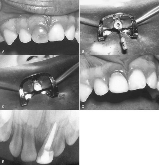
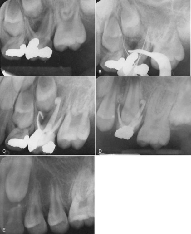
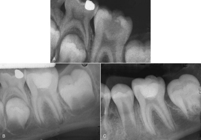
years after root canal treatment, primary tooth is near to exfoliation. E, One year later, premolar is erupted fully and all traces of ZOE have been resorbed.
In the event a small amount of the ZOE is inadvertently forced through the apical foramen, it is left alone (because the material is absorbable). It has been reported that defects on successor teeth have no relationship to length of the ZOE filling.39
When the canals are satisfactorily obturated, a fast-setting temporary cement is placed in the pulp chamber to seal over the root canal filling. The tooth may then be restored permanently. In primary molars, it is advisable to place a preformed (stainless steel) crown as the permanent restoration to ensure good coronal seal and prevent possible fracture of the tooth (see Fig. 23-22, F).
If a primary tooth requires pulpectomy and the permanent successor tooth is absent, the primary root canals are filled with gutta-percha and sealer in an attempt to retain the primary tooth long term (Fig. 23-25).
FIG. 23-25 Pulpectomy and root canal filling with gutta-percha in a retained mandibular primary second molar with no succedaneous permanent tooth. A, Carious exposure of the pulp. B, Because the permanent premolar is absent, root canals were filled with gutta-percha and sealer rather than just zinc oxide eugenol.
As previously stated, the rate of success after primary pulpectomy is high. However, these teeth should be periodically recalled to check for success of the treatment and intercept any problem associated with a failure. It has been reported39,283 that pulpectomized primary teeth may show delayed exfoliation. One study39 described a 20% incidence of crossbites or palatal eruption of permanent incisors after pulpectomy on primary incisors. In the posterior teeth, extraction was required in 22% of cases because of ectopic eruption of the premolars or difficulty in exfoliation of the treated primary molar.39 After normal physiologic resorption of the roots reaches the pulp chamber, the large amount of ZOE present may impair the resorptive process and lead to prolonged retention of the crown. Treatment usually consists of simply removing the crown and allowing the permanent tooth to complete its eruption.
Retention of ZOE in the tissues is a common sequela to primary pulpectomy. One long-term study reported that after loss of the tooth, 50% of cases had retained ZOE. Teeth filled short of the apices had significantly less retained filler, and in time, most showed complete absorption or reducing amounts. Retention of filler was not related to success and caused no pathosis.254 Therefore no attempt is made to remove retained filler from the tissues (see Fig. 23-23, A; Fig. 23-24, C).
While resorbing normally without interference from the eruption of the permanent tooth, the primary tooth should remain asymptomatic, firm in the alveolus, and free of pathosis. Traditionally, root treatments were considered successful when no pathologic resorption associated with bone rarefaction was present.95,124 If evidence of pathosis is detected, extraction and conventional space maintenance are recommended.
Investigators223 claim that most clinicians are prepared to accept pulp-treated primary teeth that have a limited degree of radiolucency or pathologic root resorption in the absence of clinical signs and symptoms. This is contingent on the assurance that the parent will contact the clinician if there is an acute problem, and the patient will return for review in 6 months. These criteria seem to be more suitable for pediatric dental practices and have been adopted clinically by Fuks et al87; they consider such teeth to be “successfully treated.”
It could be argued that mature permanent teeth can survive for a lifetime without the support of a vital pulp. For the immature permanent tooth, the future is less secure. Premature loss of a functioning pulp results in a fragile tooth with a compromised crown/root ratio, thin dentin walls, and a wide and often apically diverging root that presents significant endodontic and restorative challenges (Fig. 23-26). A central responsibility of all clinicians is therefore to safeguard pulp survival until dental development is complete.
FIG. 23-26 Premature loss of vital pulp functions in an immature maxillary permanent incisor. Development is arrested, leaving a fragile tooth with a compromised crown-root ratio, thin dentin walls, and a wide-open root end which presents endodontic and restorative challenges.
The procedures described in this section have much in common with those for primary teeth and focus on preserving all or part of the pulp in a functional condition. In addition to aspects of pulp protection, indirect pulp therapy, direct pulp capping and pulpotomy, attention will be given to the root canal treatment and restoration of nonvital, immature permanent teeth. The emerging potential of pulp regeneration and bioroot engineering will also be considered.
The pulps of young permanent teeth are at risk of breakdown following traumatic injuries (considered fully in Chapter 17), dental caries, and restorative dentistry. There is good evidence that residual dentin thickness is a key determinant of pulp survival after cavity preparation,190 and avoiding pulp exposure has been considered advantageous. The management of deep caries by partial and serial excavation has gained considerable support in recent years, reducing the risks of pulp exposure and harnessing the natural defenses of the pulp in laying down protective tertiary (reactionary) dentin.25 In the case of serial excavation, the need for a secondary excavation has recently been brought into question, while acknowledging the need for a tightly-sealing coronal restoration.241 Researchers continue to investigate the role of antimicrobial treatments, including ozone fumigation,229 photo-activated disinfection (PAD), and antimicrobial resins in sterilizing deep layers of affected dentin307 and creating the conditions for arrest and remineralization. Considerable interest has also focused on the active upregulation of reactionary dentinogenesis by applying bioactive agents such as the TGF-β family of molecules to the depths of cavity preparations.306 Therapeutic approaches present a hopeful future for pulp protection after deep caries, but significant challenges remain, not least in defining optimal agents and predictably delivering them to the pulp through the fluid-filled dentinal tubules of vital teeth.232 More basic and clinical research is needed before such agents can be widely adopted in practice.
There is, however, a substantial body of evidence to support the view that complete excavation is not necessary for the successful management of deep caries,299 and that indirect pulp therapy may be the most effective means of securing pulp health in asymptomatic teeth.
Even with the greatest clinical judgment and restorative skill, pulps are sometimes exposed during deep caries excavation. A recent survey has also shown that 62% of clinicians would remove all caries when presented with a case in which one would expect pulp exposure, whereas only 18% would partially excavate caries, and 21% would initiate root canal treatment.211 Direct pulp capping has generally not enjoyed predictable success in cariously exposed teeth. One study showed a 44.5% failure at 5 years and a disappointing 79.7% failure at 10 years.20 Attitudes may be changing, especially for the management of immature permanent teeth, where pulp diagnosis is notoriously difficult and the stakes for pulp preservation are high. Though much of the evidence is relatively short term and involves previously uninflamed pulps, MTA has established a strong track record for promoting reparative tertiary dentin bridge formation after direct pulp capping200 (see Fig. 23-5).
In a radically different approach to the management of deep caries, one of the authors (JC) has advocated complete caries excavation and direct capping of the exposure in the immature teeth of children (Fig. 23-27). Teeth with percussion sensitivity, swelling, or other obvious signs of pulpal necrosis are not considered good candidates, and the pulp tissue exposed during caries excavation must appear vital with no signs of degeneration or suppuration. In this procedure, the tooth is anesthetized and isolated with the rubber dam. All decay is removed with round carbide burs and copious water spray. No further pulpal tissue is removed except that occurring with the caries removal. The often profuse bleeding that occurs is controlled by lavaging the pulp with NaOCl, which is not only antimicrobial but appears to have no adverse effects on pulpal healing, odontoblastic cell formation, or dentinal bridging.3,43 The solution may be left in contact with the exposed pulp tissue for 10 to 15 minutes, refreshing with new solution every 3 to 4 minutes. Care must be taken in aspirating the excess NaOCl to prevent further hemorrhage, the aspirator tip being placed lateral to and never directly above the crown. Once hemorrhage is controlled, the tooth structure is cleansed with cotton pellets moistened with NaOCl, once again avoiding further pulp hemorrhage.
FIG. 23-27 Direct pulp capping in a symptomatic young permanent mandibular molar with an open apex. A, Extensive carious exposure in an immature molar with a history of spontaneous pain. B, After complete caries excavation and hemostasis, the pulp is overlaid with mineral trioxide aggregate and the tooth sealed with composite resin. C, Three-year recall shows completed root formation. Note lack of calcification within pulp chamber and canals.
The exposed pulp is then covered with a 0.5- to 1-mm thickness of MTA, which is gently teased against the exposed pulp tissue with a damp cotton pellet. Wisps of cotton are wet with sterile water and placed over the MTA so it is completely covered. The tooth is then provisionally sealed with a temporary cement such as Cavit to allow hardening of the MTA.
The patient is seen again within 12 to 48 hours, and the tooth is anesthetized and isolated with a rubber dam. Following removal of the Cavit and cotton, the MTA is examined to ensure that it is set hard, and the tooth is restored with a bonded composite restoration. If the MTA has failed to harden, the author has advocated washing out the uncured material and repeating the procedure after removing pulp tissue to canal orifice level. This approach has received further support from two studies. In the first,73 30 asymptomatic permanent molars were reviewed clinically and radiographically after caries excavation and direct pulp capping with MTA; 93% were successful at 24 months, with continued root development. In the second report, Bogen et al. (2008)27 described a series of 53 teeth diagnosed at the outset with deep caries and reversible pulpitis but with no periapical involvement. Caries was completely eliminated with the help of a caries-detector dye and magnification (Fig. 23-28, A-B), often resulting in multiple, large pulp exposures. Hemostasis was secured by bathing the pulp with 5.25% to 6% NaOCl for between 1 and 10 minutes (Fig. 23-28, B-D). One pulp which continued to bleed after this time was considered unsuitable for direct capping. After the application of 1.5 to 3 mm gray or white MTA (Fig. 23-28, E) and permanent bonded restorations at 5 to 10 days, teeth were reviewed for between 1 and 9 years (mean 3.94 years). The recall rate was 92.5%, and 97.96% of teeth were found to have favorable outcomes on the basis of radiographic appearances, subjective symptoms, and cold testing (Fig. 23-28, F). Of the 15 teeth which were immature at the time of treatment, 100% went on to complete root formation.27
FIG. 23-28 Radiographic and clinical sequence of mineral trioxide aggregate (MTA) direct pulp capping of a mandibular right molar in a 9-year-old female. A, Pretreatment radiograph showing initial deep caries and immature apices. B-D, Five-minute application of 5.25% sodium hypochlorite hemostasis, on two 1.5- to 2.0-mm exposures. E, Radiograph of molar with MTA, water-moistened cotton pellet, and unbonded Clearfil Photocore (Kuraray Medical, Okayama, Japan) provisional restoration after initial visit. F, Radiograph taken at the 5.5-year recall appointment showing permanent restoration and evidence of complete root formation. The tooth exhibited a normal response to cold testing.
(From Bogen G, Kim JS, Bakland LK: Direct pulp capping with mineral trioxide aggregate. An observational study. J Am Dent Assoc 139:305-315, 2008.
At the time of writing, a conservative general consensus probably holds with partial and serial caries excavation, avoiding pulp exposure and attempting to create an environment which will upregulate reactionary tertiary dentin responses.25,241 But evidence is growing that in correctly chosen cases with no indication of irreversible pulpitis or periapical change and where radical caries excavation is followed by confirmation of hemostasis, direct capping with MTA can enjoy remarkable levels of success in teeth with incomplete apices. Although generally restricted to asymptomatic teeth, one of the authors (JC) has noted success in both asymptomatic and symptomatic teeth, with resolution of pain and continued root development. This procedure may in effect be considered a very superficial form of mechanical and chemical pulpotomy. Because the loss of vital pulp function is so devastating in teeth with immature apices, it seems advisable to attempt conservative pulp-preserving procedures in deeply carious permanent teeth. If failure occurs, apexification or regenerative techniques can always be considered.
The pulpotomy procedure involves removing only part of the pulp, eliminating tissue that has inflammatory or degenerative changes and leaving intact the underlying healthy pulp tissue.15 This is then covered with a wound-dressing agent in an effort to promote healing at the amputation site. Traditionally, the term pulpotomy has implied removal of pulp tissue to the cervical line. However, the depth to which tissue is removed is determined by clinical judgment of how deeply the pulp is affected. Superficial amputation (partial pulpotomy) may allow better visualization of the working area but risks leaving damaged tissue that may go on to break down. In multirooted teeth, the procedure may be simplified by removing tissue to the orifices of the root canals.
Pulpotomy is an established technique for preserving vital pulp functions in immature teeth which have been subject to pulp-exposing trauma. A full account of the Cvek pulpotomy is provided in Chapter 17 (see Fig. 17-11). Briefly, studies55,113,114 have shown that inflammation is confined to the surface 2 to 3 mm of the pulp when traumatically exposed and left untreated for up to 168 hours. In experimental animals, the results were the same whether the crowns were fractured or ground off.55 Direct invasion of vital pulp tissue by bacteria did not occur, although the pulps were left exposed to saliva.
In their classic report, Cvek50 described a pulpotomy technique where only the superficial 2 to 3 mm of hyperplastic inflamed tissue was removed with a water-cooled high-speed diamond bur102 to place the wound in a healthy site. Hemostasis was then secured before capping with an appropriate material. If hemostasis could not be secured after several minutes of saline-moistened cotton pellet application, the preparation was checked carefully for residual superficial tags of bleeding tissue which had not been fully removed, or questions were asked about the condition of the underlying pulp. Persistent bleeding from an inflamed pulp usually indicates that the tissue should be resected at a deeper level to preserve a vital apical pulp stump (see Apexogenesis).
Following hemostasis with saline and a pulp cap of calcium hydroxide overlaid with a sealing coronal restoration, Cvek reported success in an impressive 94% to 96% of cases (Fig. 23-29). However, histologically better results have been shown more recently with MTA as the pulp-capping agent.1,140,198,200,228 MTA is thus recommended as the pulp-capping agent of choice in cases that do not extend deeply into the roots, where subsequent retrieval may present considerable challenge. Preference should be given to nonstaining white MTA products.
FIG. 23-29 Follow-up of a Cvek pulpotomy. A, Pretreatment radiograph of right maxillary central incisor after traumatic pulp exposure. B, Four months after Cvek pulpotomy and Ca(OH)2 dressing showing dentin bridge formation. C, Three-year recall shows continued root formation. In this case, the dentin bridge has not thickened. There is no evidence of uncontrolled mineralization within the pulp space.
The question then is whether such techniques can be applied to posterior teeth, and especially to those with symptomatic cariously-affected pulps. The answer has historically been no, except as a short-term, pain-relieving exercise. Studies involving cavity preparations into teeth that left areas of impacted food, debris, and bacteria in contact with pulp tissue resulted in inflammation extending from 1 to 9 mm into the pulp, with abscess and pus formation.55,113 The extent of inflammation in the pulp of a cariously exposed pulp is therefore difficult to judge, with the expectation that pulpotomy may be less predictable than posttraumatic exposure.
A technique termed partial pulpotomy for the management of vital carious pulp exposure on young permanent molars has been reported by several researchers.175,176,179,209 This approach has usually been reserved for teeth with little or no history of pain and in the absence of radiographic signs, percussion sensitivity, swelling, or mobility.
The procedure usually involves a 1- to 3-mm cutback of pulp tissue judged to be inflamed beneath the exposure site to reach underlying healthy tissue. After hemostasis, the exposure site has traditionally been covered with Ca(OH)2 and the tooth sealed with ZOE and a permanent restoration. Mejàre and Cvek179 followed 31 initially symptom-free teeth for a mean of 56 months (range 24 to 140 months) and found 93.5% healing. Others175 reported 91.4% success in 35 cases followed between 12 and 48 months. Exposures exceeding 2 mm and those in which bleeding could not be controlled within 1 to 2 minutes were excluded from this study.
The use of MTA as the capping agent has again met with success in the hands of the authors and others.261
Although some success has been reported,179 vital pulp therapy in permanent teeth with a history of pain has generally been contraindicated. Reports do, however, support this pulp-preserving approach even to symptomatic teeth.261 Of the six teeth with temporary pain, widened periodontal ligament space, and/or condensing osteitis managed in Mejàre and Cvek’s study,179 by Ca(OH)2 pulpotomy, 66.7% healed. More recently, one study33 undertook a similar Ca(OH)2 pulpotomy procedure on 26 permanent vital molars with carious pulp exposures and apical periodontitis. Observation between 16 and 72 months revealed that 24 (92.3%) were free from clinical symptoms, responded to sensitivity testing, showed evidence of hard-tissue barrier formation, resolution of periapical involvement, and the absence of intraradicular pathosis radiographically.
Partial pulpotomy was also found to be successful in a randomized controlled trial233 that compared Ca(OH)2 and MTA pulpotomies in the permanent molars (n = 64) of children. Mean follow-up was 34.8 months (range 25.5 to 45.6 months), and success was comparably favorable in both Ca(OH)2 (91%) and MTA (93%) groups.
A full coronal pulpotomy may be more predictable in the symptomatic case in which the depth of pulp inflammation is difficult to predict. Follow-up of MTA pulpotomies in 15 children with immature molars revealed no clinical or radiographic failures at 12 months, though four pulps had undergone calcific metamorphosis.65 In a 2006 clinical report,326 investigators described a series of 23 irreversibly pulpitic anterior and posterior teeth in children and adolescents, treated by full coronal pulpotomy, hemostasis for 1 minute with 6% NaOCl, and wound dressing with MTA. Follow-up between 6 and 53 months (mean 19.7 months) revealed that 79% had healed, 16% were healing, and only 5% had evidence of persistent disease. More research is needed, but the MTA pulpotomy seems to be gaining an evidence base as a reliable technique for preserving vital pulp functions in both asymptomatic and symptomatic teeth in children.326
This body of evidence suggests that young teeth, with their rich vascular supply, are good candidates for conservative pulp-preserving treatments. Again, the stakes are high, and efforts to secure vital pulp functions are to be promoted. Outcomes are probably more predictable in asymptomatic cases than symptomatic, and care should be taken preoperatively to distinguish between cases with symptoms of reversible and irreversible pulpitis. The correlation of clinical symptoms with histologic state is, however, poor.60
After pulp capping and pulpotomy, the patient should be seen periodically for 2 to 4 years to determine success. Although normal vitality tests (e.g., electrical and thermal sensitivity tests) are reliable after pulp capping, they are not usually helpful in the pulpotomy-treated tooth. Since histologic success cannot be determined, clinical success is judged by the absence of any clinical or radiographic signs of pathosis and the presence of continued root development in teeth with incompletely formed roots.
Controversy exists as to whether the pulp should be reentered after the completion of root development in the pulpotomy-treated tooth. Some researchers110,154 believe that pulp capping and pulpotomy procedures invariably lead to progressive calcification of the root canals. After successful root development, they advocate pulpectomy and root canal treatment before canals become obliterated and challenging to manage in the event of future endodontic infection. However, it has been the experience of the authors and others53,90,114 that with good case selection, a gentle technique in removing infected tissue and dentin chips, and care in avoiding the compaction of pulp dressing into underlying pulp tissue, calcification of the pulp is an infrequent sequela of pulpotomy (see Fig. 23-29, C; Fig. 23-30).
FIG. 23-30
Deep pulpotomy for apexogenesis. A, Immature maxillary central incisor 10 weeks after traumatic pulp exposure and a deep pulpotomy dressed with Ca(OH)2. Note early formation of a dentin bridge. B, Three-year recall showing the dentin bridge to have thickened and root formation to be complete; tooth remained asymptomatic and required no further treatment at that time. Note absence of uncontrolled mineralization within apical pulp space.
In a follow-up study of clinically successful pulpotomies,50 researchers removed the pulps 1 to 5 years later for restorative reasons and found the tissue to be histologically normal.53 They concluded that changes seen in the pulps do not present sufficient histologic evidence to support routine pulpectomy after pulpotomy in accidentally fractured teeth with pulp exposures. Thus, routine reentry to remove the pulp and place a root canal filling after completion of root development is contraindicated unless dictated by restorative considerations such as the necessity for a retentive post. Nevertheless, calcific obliteration, internal resorption, and pulp necrosis are potential sequelae of pulp-capping and pulpotomy procedures which should be screened for during recall appointments. Although unlikely, they are possible, and the patient should be clearly informed.
In posterior teeth, where surgical endodontics is difficult, the observation of continued calcification after root closure may justify reentry of the tooth for root canal treatment. In anterior teeth, if calcification of the canal has made conventional endodontics impossible, surgical endodontics may be performed with relative ease. Therefore in anterior teeth, routine endodontic therapy after completion of root development is contraindicated unless clinical signs and symptoms of pathosis are present or unless such therapy is necessary for restorative procedures (e.g., placement of a post for retention of a crown because of missing tooth structure).
Because of the historical track record of formocresol pulpotomy in primary teeth, interest developed in this technique for the management of young permanent teeth. Reports have demonstrated the potential for continued apical development after formocresol pulpotomy in young permanent teeth,77,191,256,315 and several authors have reported better results with the diluted rather than the full-strength form. However, they reported a high incidence of internal resorption, which increased in severity with longer periods of time.86,224
The formocresol procedure had appeal because there was a lower risk of pulp calcification, a recognized complication of Ca(OH)2 pulpotomy. After root completion, the tooth could easily be reentered, the pulp extirpated, and routine endodontic therapy performed. Contrary to these findings, one group of researchers14 has shown calcification of the canals by continuous apposition of dentin on the lateral walls, with equal frequency whether using Ca(OH)2 or formocresol. The only common denominator to this reaction was the presence of dentin chips that had accidentally been pushed into the radicular pulp tissue.
Although this treatment has been reported to be partly successful, it cannot be routinely recommended in an era of effective alternatives such as MTA.
Formocresol pulpotomy procedures can only be recommended as temporary treatments on permanent teeth with necrotic pulps, where clinical success after 3 years has been reported.301 The formocresol pulpotomy has particularly been performed in preference to extraction when routine endodontic treatment was not economically viable. This procedure may buy symptom-free time until funds become available to complete root canal treatment.
Apexogenesis is treatment designed to preserve vital pulp tissue in the apical part of a root canal in order to complete formation of the root apex (Fig. 23-31).111
FIG. 23-31 Schematic representation of apexogenesis, a treatment designed to preserve at least the apical portion of pulp tissue in a healthy condition in order to complete root formation. A, Following a deep pulpotomy and hemostasis, the radicular pulp is dressed and a sealing coronal restoration applied. B, Success is evidenced by continued root development (length and wall thickness) and formation of a calcific barrier in response to the wound dressing.
The clinical procedure is essentially a deep pulpotomy undertaken to preserve the formative capacity of the radicular pulp in immature teeth that have deep pulpal inflammation. Examples include carious exposures and some trauma cases in which treatment of the exposed pulp is delayed, and it becomes necessary to extend further into the canal to reach healthy tissue.
Deep resection of pulp tissue is usually undertaken in single-rooted anterior teeth, with a small endodontic spoon excavator or round, abrasive diamond bur (see Fig. 23-30). In posterior teeth, the use of endodontic files or reamers may be necessary if tissue is being amputated within the canals. Extension of the amputation site into the root canals is undertaken only when there is little confidence that more superficial pulp capping or pulpotomy will be successful and where there is a desperate desire to preserve pulp functions in teeth with wide-open, blunderbuss apices (Fig. 23-32).
FIG. 23-32 Apexogenesis following deep Ca(OH)2 pulpotomy on a mandibular permanent molar. A, Pretreatment radiograph showing extensive caries, incomplete root development, and possible periapical pathosis. The tooth was asymptomatic. B, One year after deep pulpotomy, extended several mm into canals, hemostasis and restoration. Root formation is progressing, and periapical tissues appear healthy. C, Two years later, root formation is complete.
Bleeding is usually controlled with saline-soaked cotton pellets or NaOCl. If hemostasis cannot be secured by conventional means, this may indicate that even the deep pulp is inflamed, and treatment will be compromised. Despite the risks, it is legitimate to attempt pulp-preserving treatment after controlling the hemorrhage with hemostatic chemicals such as aluminum chloride or ferric sulfate.
The pulp wound is then covered with a dressing material before securely restoring the crown. It is challenging to determine the status of pulp tissue residing deep in the root canal, and its capacity for survival is difficult to predict. Radiographic and clinical follow-up is mandatory, and if there is no evidence of continued root formation and calcific barrier formation in response to the dressing, apexification or a regenerative technique may be considered.
Because of the depth at which this procedure is performed, preference has usually been given to the use of Ca(OH)2 rather than MTA because, in the event of failure, this may facilitate reentry to the root canal to perform apexification or pulp regeneration. Also, if apexogenesis is successful and root-end formation is complete, the tooth could be reentered if desired for conventional root canal treatment. It is unclear if these concerns hold true in an era of microscopy and ultrasonic instrumentation.
Calcium hydroxide powder has usually been preferred over hard-setting products, carried into the canal with an amalgam carrier or gun system employed for MTA application. Small increments of Ca(OH)2 powder are carefully teased against the entire surface of the pulp stump with a rounded-end, plastic instrument, ideally with microscope control. Care must be taken not to pack the Ca(OH)2 into the pulp tissue because this causes greater inflammation and increases the chances of failure. Even if pulpotomy is successful, there is an increased risk that the remaining pulp tissue will mineralize around impacted particles of Ca(OH)2.308
Commercial nonsetting calcium hydroxide pastes may also be spun or injected to flow lightly over the pulp stump. Care must be taken to avoid trapping air bubbles when applying such products, and it is recommended that they be overlaid with a hard-setting calcium hydroxide or glass ionomer cement52 before meticulous cleaning of the cavity walls and restoration with a bonded composite resin restoration. Though little has been published on this topic, MTA probably does represent a viable alternative to calcium hydroxide, provided the challenges in its retrieval are recognized.
Apexification, or root-end closure, is the process whereby a nonvital, immature, permanent tooth which has lost the capacity for further root development is induced to form a calcified barrier at the root terminus (Fig. 23-33). This barrier forms a matrix against which root canal filling or restorative material can be compacted with length control.
FIG. 23-33 Schematic representation of apexification, a treatment to develop a hard barrier at an open root end. A, Immature permanent tooth with a nonvital pulp. B, Traditional approach, with the formation of a “calcified barrier” at the root end following repeated dressing over many months with Ca(OH)2. Canal is subsequently filled with gutta-percha and sealer before coronal restoration. C, Artificial apical barrier technique, where a 4- to 5-mm plug of mineral trioxide aggregate is placed at the root end. Canal space is subsequently restored with dual-curing composite resin, often accompanied by a fiber post to provide mechanical support.
Unlike the pulp capping, pulpotomy, and apexogenesis procedures described previously, apexification will at best result in closure of the root end and cannot be expected to cause further root development in terms of length or wall thickness. Apexification is thus regarded as a treatment of last resort in immature teeth which have lost pulp vitality. The growing body of recent evidence on pulp regeneration, even in infected, nonvital immature teeth, may also relegate this approach to history archives in the years to come.
For now, root-end closure techniques, both those involving the generation of a biologic calcific barrier and those involving artificial root-end closure with a material such as MTA, still have a place in practice and will be considered.
Before the introduction of conservative apical closure techniques, the usual approach to this problem was surgical. Although this could be successful, psychological and patient-management issues in patients who were usually young children offered many contraindications. Local dental considerations presented a further disincentive, including worsening the crown/root ratio if further root reduction was required to achieve a seal, and the intrinsic difficulties of sealing a fragile, incompletely formed apex with traditional materials. A more predictable and less traumatic approach was desirable.
Until recently, the most widely accepted technique has involved cleaning and filling the canal with a temporary paste, most commonly Ca(OH)2, which was replaced at intervals over several months, to stimulate the formation of an apical calcified barrier (see Fig. 23-33, A-B; Fig. 23-34).284
FIG. 23-34 Apexification with Ca(OH)2. A, Maxillary central incisor after several months of Ca(OH)2 medication. B, Following removal of the Ca(OH)2, a calcific barrier is apparent. C, Tooth following root filling with thermoplastic gutta-percha and sealer. Note extrusion of filling material through porosities in the calcific barrier. The root canal filling is dense, but it may be questioned how the gutta-percha and sealer have added to the structural integrity and reinforcement of this fragile tooth.
Diagnosis of pulpal necrosis in a tooth with an incompletely formed apex is often difficult, with the electronic pulp tester rarely providing meaningful data,95 and thermal tests often giving equivocal or false results in young children and traumatized teeth. The presence of acute or chronic pain, percussion sensitivity, mobility, coronal discoloration, or a discharging sinus may be helpful guides, whereas radiographic diagnosis can be complicated by the normal radiolucencies appearing at the apices of developing teeth. Comparison of root formation with contralateral teeth should always be considered.
If any doubt persists, it is usually wise to adopt a watch-and-wait approach before entering the immature tooth endodontically. Only when there is convincing evidence of pulp breakdown should the tooth be entered, and exposed dentin should in the interim be covered to reduce the risks of microbial entry to a potentially compromised pulp.
The extent of apical closure may be difficult to ascertain with plain radiographs. Three-dimensional imaging offers new opportunities to understand the complexity of root ends which may have different conformation in mesiodistal and faciolingual dimensions (see Fig. 23-14). In practical terms, the precise limit of apical opening that can be filled by conventional means is unclear.
Many materials have been reported to successfully stimulate apexification. The use of nonsetting Ca(OH)2 was first reported by Kaiser in 1964.141 The technique was popularized by the work of Frank,81 and since that time, Ca(OH)2 alone or in combination with other drugs became the most widely accepted material to promote apexification until the development of MTA and the potential for rapid, artificial barrier formation.
For historical perspective, Ca(OH)2 powder has been mixed with CMCP, metacresyl acetate, Cresanol (i.e., a mixture of CMCP and metacresyl acetate), physiologic saline, Ringer’s solution, distilled water, and anesthetic solution. All have been reported to stimulate apexification, but most reports in the U.S. literature34,108,285 have advocated mixing Ca(OH)2 with CMCP or Cresanol, whereas reports from other parts of the world52,178 showed the same success using distilled water or physiologic saline as the vehicle. The addition of 1:8 barium sulfate to Ca(OH)2 enhanced radiopacity with no apparent adverse effects on apexification.320 Comparable outcomes have been noted in humans and animals with tricalcium phosphate,41,149,246 collagen calcium phosphate,206 osteogenic protein-1,267 bone growth factors,300 and a number of other materials.320
The most important factors in achieving apexification seem to be thorough débridement of the root canal (to remove all necrotic pulp tissue and microbial infection) and sealing of the tooth (to prevent the ingress of bacteria and substrate67). Apexification does not occur when the apex of the tooth penetrates the cortical plate. To be successful, the apex must be completely within the confines of the cortical plates.
In the apexification technique, the canal is cleaned and disinfected in line with the principles defined in Chapter 9. The use of a rubber dam is mandatory, and resourcefulness may be needed to isolate partially erupted or damaged teeth in children.
The access opening may require some extension, especially in the anterior teeth, to accommodate the larger instruments necessary to clean the root canals, but care should be taken not to heavily instrument the already thin and relatively fragile walls of the root. Neither should operators deceive themselves that they are able to steer instruments against all walls of the canal system for complete débridement, particularly in apically diverging canals.
The length of the canal is established by radiographs, since the absence of an apical constriction may make electronic methods unreliable.128 A constant drying point, determined with paper points, may provide helpful additional information on length.18 Irrigation is central to the débridement of immature teeth, and with proper precautions, operators should not hesitate to benefit from the antimicrobial and tissue-solvent properties of NaOCl. Sonic, ultrasonic, and other vibratory devices capable of activating the irrigant within the canal may be advantageous,313 and benefit may also come from the use of small brushes of the sort that are designed for interproximal tooth brushing or the application of etchants to post channels. After thorough débridement, the canal is dried and medicated with a fluid Ca(OH)2 paste, carried into the canal with a Lentulo spiral or injected from a proprietary paste syringe. There is little evidence to commend any commercial Ca(OH)2 paste over another in this application. The tooth is then sealed coronally, and the patient is recalled at 3-monthly intervals to wash out the Ca(OH)2 paste and inspect clinically (with the aid of a gutta-percha or paper point or by direct visual inspection through the microscope) and radiographically for the development of a calcified barrier (see Fig. 23-34, B). Treatment typically extends over 9 to 24 months, with obvious demands on patient and parent compliance, while risking tooth embrittlement and cervical root fracture following long-term medication with calcium hydroxide.10,51,252
Histologic studies consistently report the absence of Hertwig’s epithelial root sheath, and normal root formation should never be anticipated. Instead, there appears to be a differentiation of adjacent connective tissue cells into specialized cells; there is also deposition of calcified tissue adjacent to the filling material. The calcified material that formed over the apical foramen was histologically identified as an osteoid (i.e., bonelike) or cementoid (i.e., cementum-like) material (Fig. 23-35).34,108,284 In a primate study, bridging of the root end with osteodentin was reported after vital pulpectomy and canal medication with Ca(OH)2/parachlorophenol paste. The material appeared to be distinct from but continuous with the cementum, dentin, and predentin at the root apex.61 The closure of the apex may be partial or complete but consistently has minute communications with the periapical tissues (see Fig. 23-33, B; Fig. 23-34, C; Fig. 23-35). For this reason, apexification stimulated by pastes must always be followed by obturation of the canal with a permanent root canal filling, traditionally of thermoplastic gutta-percha and sealer, though MTA would be a good contemporary alternative.
FIG. 23-35 Histologic section of a dog’s tooth after Ca(OH)2 apexification. A, Cementum-like mineralized tissue is closing the wide-open root end. Debris is apparent within the canal because of inadequate débridement before filling. B, Higher magnification showing cellular detail; periodontal ligament is free of inflammation. Root canal filling material was lost in histologic preparation. Note presence of tissue communication through the apical barrier (stain H&E).
Although the apexification technique with Ca(OH)2 has enjoyed considerable tooth-preserving success, the many disadvantages of this protracted treatment have justified a search for alternatives, such as artificial barrier techniques, with their potential for more rapid treatment, and regeneration techniques, with their potential for continued tooth development.
Investigators41 reported the use of tricalcium phosphate as an apical barrier in 1979. The material was packed into the apical 2 mm of the canal, against which gutta-percha was compacted. The treatment was completed in one appointment, and radiographic assessment confirmed successful apexification comparable to that achieved with Ca(OH)2. Calcium hydroxide powder has also been used successfully as an apical barrier against which to pack gutta-percha.264
The use of MTA as an apical barrier was first reported in 1996,300 and subsequent clinical investigations in animals267 and humans230,258,274 have established this as the standard, with biologic outcomes in terms of periapical healing and root-end closure at least comparable to those treated with Ca(OH)2.230 Simon et al. (2007)274 treated 57 teeth in patients with a mean age of 18 years (standard deviation 12 years). Of the 43 teeth with at least 12-month follow-up, healing had occurred in 81%. More recently, a series of 17 nonvital immature incisors in children with a mean age of 11.7 years was followed up for a mean of 12.5 months. This study reported 94.1% clinical success, with radiographic healing in 76.5% and a further 17.6% uncertain.258 This approach to the problem of root-end closure allows treatment to be completed on a short time scale, with advantages including improved patient compliance, reduced cost of clinical time, and the ability to securely restore the tooth at an earlier stage. The risk of tooth fracture after long-term Ca(OH)2 medication10,252 is also eliminated.
In the apexification technique, the canal is cleaned and disinfected as described for Ca(OH)2 apexification. Research158 has suggested that tissue pH may affect the hydration reaction and final physical properties of MTA, and it has become standard practice to medicate canals for at least 1 week with Ca(OH)2 to raise the acidic pH of the inflamed periapical tissues before permanently sealing.
FIG. 23-36 Mineral trioxide aggregate (MTA) apical barrier apexification procedure and restoration with bonded composite. A, Maxillary right central incisor with a large periapical lesion. Patient is undergoing orthodontic treatment. B, Following placement of MTA in the apical 4 mm of the canal. C, Six months later. The entire canal coronal to the MTA apical barrier is filled with bonded composite resin. Note that calcification has occurred periapically as orthodontic treatment has continued. D, Thirty months after treatment. The periapical lesion has healed.
FIG. 23-37 Apexification with mineral trioxide aggregate (MTA) barriers in a mandibular permanent molar. A, Periapical lesion reveals open apices in this pulpless molar. B, Apical plugs of MTA and remainder of the pulp space restored with bonded composite resin. C, Two years later, showing closure of the root ends with cementum.
When the tooth is free of signs and symptoms of infection, it is reisolated with a rubber dam and the Ca(OH)2 washed free, often with the help of ultrasonics and small brushes. After drying the canal with large, sometimes inverted, premeasured paper points, the canal is filled incrementally with MTA, delivered to the canal with a dedicated MTA carrier or deposited in small pellets from an amalgam gun. The material can then be worked up the canal with premeasured pluggers and set some 1 to 3 mm short of the root end, often with the help of sonic or ultrasonic energy to settle the material.329 Apical matrices have not generally been considered necessary to limit the flow of MTA, though impressive-looking results may be obtained (see Fig. 17-17) by packing the apical region with calcium sulphate prior to MTA application.304 An apical plug of 4- to 5-mm thickness is usually considered optimal (see Fig. 23-36, B).311 The adequacy of the apical plug is verified radiographically.
All excess MTA is removed from the canal walls by scrubbing with large, moistened paper points or brushes. Meticulous cleanup is important to allow optimal bonding of the subsequent composite resin restoration, which will extend deeply into the canal and offer internal reinforcement of the fragile root.
A very wet cotton pellet is placed in the canal to provide moisture for the setting reaction. The pellet should not be in contact with the MTA because fibers of the cotton will become impregnated into the material. Excess water in the access preparation is dried with cotton pellets and the opening sealed with a provisional restorative material such as Cavit. At a subsequent appointment, the tooth is reisolated and the hard set of the MTA verified with an endodontic file or probe. If for some reason the MTA has not hardened, the canal can be recleansed and the procedure repeated before final bonded restoration.
Immature teeth, and particularly those which are pulpless and have undergone apexification, are at high risk of fracture. Within 3 years of long-term Ca(OH)2 medication and root filling with gutta-percha, it has been reported51 that 28% to 77% of immature teeth suffered a cervical root fracture. The degree of dental development appeared to be a key variable. Clinically, it is the impression of the authors that rapid MTA plug techniques in combination with the internal placement of bonded composite resin appears to have virtually eliminated cervical root fractures. Many of the studies are laboratory based, using simulated rather than real immature teeth, but the use of contemporary dentinal bonding techniques has been shown to strengthen endodontically treated teeth to levels close to those of intact teeth.104,116,144 A 2004 study156 demonstrated significantly greater resistance to root fracture after placement of a 4-mm thick apical plug of MTA followed by an intracanal composite resin when compared with MTA followed by gutta-percha and sealer. Root reinforcement has also been reported to improve by the cementation of a metal post within the channel created by removal of a light-transmitting composite curing post.36 The potential of fiber-reinforced posts would also appear great,28 though little clinical evidence has yet been published on the treatment of immature teeth.
Alternative materials which have been suggested to bond and reinforce fragile roots include resin-modified glass ionomer cements100 and most recently, Resilon.294 The evidence on Resilon however, remains highly contentious,325 and the ideal of predictable bonding and tooth reinforcement throughout the length of the root canal system remains some way off.293
Bonding dual- or light-curing composite resin directly over the MTA plug, with no interposing layer of gutta-percha, has become an established clinical method (see Fig. 23-33, C; Fig. 23-36, C). Care should be taken to etch and bond the canal according to the manufacturer’s instructions, with proper wash-out of etchant and avoiding gross pooling of unfilled resin. Complete resin infiltration of dentin cannot be guaranteed in the depths of a root canal, and the potential exists that host-derived metalloproteinases liberated by acid etching may degrade resin-dentin bonds with time. Flushing the preparation after etching with a synthetic protease inhibitor such as 2% chlorhexidine may help to counter some of these adverse events.35
Clear light-transmitting posts (e.g., Luminex System, Dentatus USA, New York) have been developed to ensure complete bonding of deep increments of light-activated composite. This may be less of an issue with dual-curing materials.
In the Luminex technique, a light-curing composite resin is placed in the canal, building up 2 mm increments with care to avoid trapping air bubbles. The Luminex post is placed to the depth of the preparation and the composite cured by transmitting light through the post. After curing, the plastic post is trimmed to the cervical line, and the incisal opening is restored.
If a core is needed for crown placement, a Luminex post without serrations is used for curing the composite. Because the composite does not bond to the smooth post, it can be gently removed and a corresponding metal Dentatus post cemented into the space with a resin cement. A composite buildup for crown retention may then be completed.
A variety of commercial quartz and glass fiber post systems are available for use in such applications, ensuring the delivery of light deep into the canal system and offering the potential for internal reinforcement (see Fig. 23-33, C). The heads of fiber posts should always be covered with composite resin to prevent them from absorbing oral fluids and delaminating.
Until recently, there have been few treatment options for the nonvital, permanent immature tooth. On confirming pulp necrosis, efforts could be made to preserve the tooth by apexification, or it could be extracted within a broader orthodontic treatment plan. This view has recently been challenged by a number of ground-breaking reports in which nonvital immature teeth with clear evidence of pulp breakdown and periapical suppuration were encouraged to grow new pulps and complete root formation (length and wall thickness; see also Chapter 16).19,37,135,139,297 Success is dependent on the activity of a newly identified population of stem cells, the so-called stem cells from apical papilla (SCAP) (Fig. 23-38, A), a hidden treasure with enormous potential for tissue regeneration and bioroot engineering.127
FIG. 23-38 Schematic representation of pulp regeneration. A, Immature, nonvital permanent tooth, showing the location of the apical papilla with its rich collection of stem cells. B, Following medication of the canal with triantibiotic paste, the canal is overinstrumented to encourage bleeding up to the cervical level. Subsequent blood clot is overlaid with MTA and a sealing restoration and forms a scaffold for invasion by SCAP cells. C, Pulp regeneration is expected to allow continued root formation in a previously pulpless tooth. SCAP, Stem cells from apical papilla.
All immature teeth with open apices may be considered candidates for regenerative treatment, even if they have frank pulp-space infection, a discharging sinus, or have been previously root canal treated.
The currently recommended protocol (see Fig. 23-38)139,303 involves disinfecting the isolated canal system with little or no mechanical instrumentation and copious irrigation for 10 to 15 minutes with 5.25% NaOCl. The carefully dried canal is then medicated with a freshly mixed paste of ciprofloxacin, metronidazole, and minocycline in a macrogol ointment/propylene glycol base, which is sealed in place with a well-sealing provisional restoration. After 1 week, the tooth is carefully reopened under strict asepsis, and after washing out the antibiotic paste with NaOCl, the periapical tissues are gently instrumented to encourage bleeding into the canal. A blood clot is produced to the cemento-enamel junction, which will form a scaffold for pulp regeneration. The tooth is then sealed with several millimeters thickness of MTA before closing the crown with a provisional and later a resin-bonded composite restoration (see Fig. 23-38, B-C). Details of protocol vary from report to report, with some researchers demonstrating success with simpler protocols involving formocresol rather than antibiotic disinfection, and closure of the crown with glass ionomer cement.268 Procedures are likely to evolve rapidly in this most exciting area of practice. Teeth are kept under periodic clinical and radiographic review, with the suggestion303 that more traditional treatment may be considered if there are no signs of regeneration within 3 months. Clinical reports to date offer much promise, since there is little to be lost in attempting a treatment that could transform the prospects of a tooth which would otherwise require apexification. It is conceivable that Ca(OH)2 and even MTA apexification techniques may in the foreseeable future be consigned to the history books. Readers are cross-referred to Chapter 16, which discusses broader aspects of regenerative endodontics, and to the clinical Figures 16-5 to 16-8 Figure 16-5 Figure 16-6 Figure 16-7 Figure 16-8 which illustrate immature teeth managed in this way.
The potential of bioengineering is huge, and work continues to optimize scaffolds that may encourage revascularization of the pulp space, and to explore the options of seeding cell populations into the properly sterilized pulp spaces of immature teeth.189 Our call for action has never been louder as we seek effective, biologically based treatments for our pediatric patients.
1. Abedi HR, Torabinejad M, Pitt Ford TR, Bakland LK. The use of mineral trioxide aggregate cement (MTA) as a direct pulp-capping agent. J Endod. 1996;22:199. (abstract)
2. Aeinehchi M, Eslami B, Ghanhariha M, Saffar AS. Mineral trioxide aggregate (MTA) and calcium hydroxide as pulp-capping agents in human teeth: a preliminary report. Int Endod J. 2003;36:225.
3. Agamy HA, Bakry NS, Mounir MMF, Avery DR. Comparison of mineral trioxide aggregate and formocresol as pulp capping agents in pulpotomised primary teeth. Pediatr Dent. 2004;26:302.
4. Akimoto N, Momoi Y, Kohno A, et al. Biocompatibility of Clearfil linear bond 2 and Clearfil AP-X system nonexposed and exposed primate teeth. Quintessence Int. 1998;29:177.
5. Alacam A. Long-term effects of primary teeth pulpotomies with formocresol, glutaraldehyde-calcium hydroxide and glutaraldehyde-zinc oxide-eugenol on succedaneous teeth. J Periodontol. 1989;13:307.
6. Al-Zayer MA, Straffon LH, Feigal RJ, Welch KB. Indirect pulp treatment of primary posterior teeth: a retrospective study. Pediatr Dent. 2003;25:29.
7. American Academy of Pediatric Dentistry. Reference manual: clinical guideline on pulp therapy for primary and young permanent teeth. Pediatr Dent. 2003-2004;25:87.
8. Andreasen F. Transient apical breakdown and its relation to color and sensibility changes. Endod Dent Traumatol. 1986;2:9.
9. Andreasen J, Andreasen F, Andreasen L. Textbook and color atlas of traumatic injuries to the teeth, ed 4. Philadelphia: Wiley-Blackwell; 2008.
10. Andreasen JO, Farik B, Munksgaard EC. Long-term calcium hydroxide as a root canal dressing may increase risk of root fracture. Dent Traumatol. 2002;18:134.
11. Andreasen JO, Ravn JJ. Epidemiology of traumatic dental injuries to primary and permanent teeth in a Danish population sample. Int J Oral Surg. 1972;1:235.
12. Andreasen JO, Ravn JJ. The effect of traumatic injuries to primary teeth on their permanent successors: II–a clinical and radiographic follow-up study of 213 teeth. Scand J Dent Res. 1971;79:284.
13. Andrew P. The treatment of infected pulps in deciduous teeth. Br Dent J. 1955;98:122.
14. Aponte AJ, Hartsook JT, Crowley MC. Indirect pulp capping success verified. J Dent Child. 1966;33:164.
15. Armstrong RL, Patterson SS, Kafrawy AH, Feltman EM. Comparison of Dycal and formocresol pulpotomies in young permanent teeth in monkeys. Oral Surg. 1979;48:160.
16. Attala MN, Noujaim AA. Role of calcium hydroxide in the formation of reparative dentin. J Can Dent Assoc. 1969;35:267.
17. Avram DC, Pulver F. Pulpotomy medicaments for vital primary teeth: surveys to determine use and attitudes in pediatric dental practice and in dental schools throughout the world. J Dent Child. 1989;56:426.
18. Baggett FJ, Mackie IC, Worthington HV. An investigation into the measurement of the working length of immature incisor teeth requiring endodontic treatment in children. Brit Dent J. 1996;181:96.
19. Banchs F, Trope M. Revascularization of immature permanent teeth with apical periodontitis: a new treatment protocol? J Endod. 2004;30:196.
20. Barthel CR, Rosenkranz B, Leuenberg A, Roulet JF. Pulp capping of carious exposures: treatment outcome after 5 and 10 years: a retrospective study. J Endod. 2000;26:525.
21. Bennett CG. Pulpal management of deciduous teeth. Pract Dent Monogr. 1965;May-June:1.
22. Bergenholtz G. Evidence for bacterial causation of adverse pulpal responses in resin-based dental restorations. Crit Rev Oral Biol Med. 2000;11:467.
23. Berk H, Krakow AA. Endodontic treatment in primary teeth. In: Goldman HM, et al, editors. Current therapy in dentistry, vol 5. St Louis: Mosby; 1974.
24. Bevelander G, Benzer D. Morphology and incidence in secondary dentin in human teeth. J Am Dent Assoc. 1943;30:1079.
25. Björndal L. Indirect pulp therapy and stepwise excavation. J Endod. 2008;34(7S):S29.
26. Block RM, Lewis RD, Sheats JB, Fawley J. Cell-mediated immune response to dog pulp tissue altered by formocresol within the root canal. J Endod. 1977;3:424.
27. Bogen G, Kim JS, Bakland LK. Direct pulp capping with mineral trioxide aggregate. An observational study. J Amer Dent Assoc. 2008;139:305.
28. Bonfante G, Kaizer OB, Pegoraro LF, do Valle LA. Fracture strength of teeth with flared root canals restored with glass fibreposts. Int Dent J. 2007;57:153.
29. Borum MK, Andreasen JO. Sequelae of trauma to primary maxillary incisors: I–complications in the primary dentition. Endod Dent Traumatol. 1998;14:31.
30. Burnett S, Walker J. Comparison of ferric sulfate formocresol and a combination of ferric sulfate/formocresol in primary tooth vital pulpotomies: a retrospective radiographic survey. J Dent Child. 2002;69:44.
31. Byers M, Narchi M. The dental injury model: experimental tools for understanding neuro-inflammatory interactions and polymodal nociceptors functions. Crit Rev Oral Biol Med. 1999;10:4.
32. Caldwell RE, Freilich MM, Sandor GKB. Two radicular cysts associated with endodontically treated primary teeth: rationale for long-term follow-up. Ont Dent. 1999;76:29.
33. Caliskan MK. Pulpotomy of carious vital teeth with periapical involvement. Int Endod J. 1995;28:172.
34. Camp JH: Continued apical development of pulpless permanent teeth after endodontic therapy, master’s thesis, Bloomington, 1968, Indiana University School of Dentistry.
35. Carrilho MR, Geraldeli S, Tay F, et al. In vivo preservation of the hybrid layer by chlorhexidine. J Dent Res. 2007;86:529.
36. Carvahlo CAT, Valera MC, Oliveira LD, Camargo CHR. Structural resistance in immature teeth using root reinforcements in vitro. Dent Traumatol. 2005;21:155.
37. Chueh L-H, Huang G T-H. Immature teeth with periradicular pathosis or abscess undergoing apexogenesis: a paradigm shift. J Endod. 2006;32:1205.
38. Cohenca N, Karnis S, Rotstein I. Transient apical breakdown following tooth luxation. Dent Traumatol. 2003;19:289.
39. Coll JA, Sadrian R. Predicting pulpectomy success and its relationship to exfoliation and succedaneous dentition. Pediatr Dent. 1996;18:57.
40. Cotes O, Boj JR, Canalda C, Carreras M. Pulpal tissues reaction to formocresol vs. ferric sulfate in pulpotomized rat teeth. J Clin Ped Dent. 1997;21:247.
41. Coviello J, Brilliant JD. A preliminary clinical study of the use of tricalcium phosphate as an apical barrier. J Endod. 1979;5:6.
42. Cox CF, Suzuki S. Re-evaluating pulpal protection: calcium hydroxide liners vs cohesive hybridization. J Am Dent Assoc. 1994;125:823.
43. Cox CF, Hafez AA, Akimoto N, Otsuki M, Suzuki S, Tarim B. Biocompatibility of primer, adhesive and resin composite systems on non-exposed and exposed pulps of non-human primate teeth. Am J Dent. 1998;11:55. (special issue)
44. Cox CF, Keall CL, Keall HJ, Ostro E, Bergenholtz C. Biocompatibility of surface-sealed dental materials against exposed pulp. J Prosthet Dent. 1987;57:1.
45. Cox CF, Sübay RK, Suzuki S, Suzuki SH, Ostro E. Biocompatibility of various dental materials: pulp healing with a surface seal. Int J Periodont Restorative Dent. 1996;16:241.
46. Cox CF, Bergenholtz G, Fitzgerald M, et al. Capping of the dental pulp mechanically exposed to the oral microflora: a 5-week observation of wound healing in the monkey. J Oral Pathol. 1982;11:327.
47. Cox CF, Bergenholtz G, Heys DR, Syed SA, Fitzgerald M, Heys RJ. Pulp-capping of dental pulp mechanically exposed to oral microflora: a 1- to 2-year observation of wound healing in the monkey. J Oral Pathol. 1985;14:156.
48. Cox CF, Sübay RK, Ostro E, Suzuki S, Suzuki SH. Tunnel defects in dentin bridges: their formation following direct pulp capping. Oper Dent. 1996;21:4.
49. Croll TP, Pascon EA, Langeland K. Traumatically injured primary incisors: a clinical and histological study. J Dent Child. 1987;54:401.
50. Cvek M. A clinical report on partial pulpotomy and capping with calcium hydroxide in permanent incisors with complicated crown fractures. J Endod. 1978;4:232.
51. Cvek M. Prognosis of luxated non-vital maxillary incisors treated with calcium hydroxide and filled with gutta-percha. A retrospective clinical study. Endod Dent Traumatol. 1992;8:45.
52. Cvek M. Treatment of non-vital permanent incisors with calcium hydroxide. Odontol Revy. 1972;23:27.
53. Cvek M, Lundberg M. Histological appearance of pulps after exposure by a crown fracture, partial pulpotomy, and clinical diagnosis of healing. J Endod. 1983;9:8.
54. Cvek M, Granath L, Cleaton-Jones P, Austin J. Hard tissue barrier formation in pulpotomized monkey teeth capped with cyanoacrylate or calcium hydroxide for 10 and 60 minutes. J Dent Res. 1987;66:1166.
55. Cvek M, Cleaton-Jones PE, Austin JC, Andreasen JO. Pulp reactions to exposure after experimental crown fracture or grinding in adult monkey. J Endod. 1982;8:391.
56. Dandashi MB, Nazif MM, Zullo T, Elliott MA, Schneider LG, Czonstkowsky M. An in vitro comparison of three endodontic techniques for primary incisors. Pediatr Dent. 1993;15:254.
57. Davis MJ, Myers R, Switkes MD. Glutaraldehyde: an alternative to formocresol for vital pulp therapy. J Dent Child. 1982;49:176.
58. Dean J, Avery D, McDonald R. McDonald and Avery’s dentistry for the child and adolescent, ed 10. St. Louis: Mosby; 2011.
59. Dominguez MS, Witherspoon DE, Gutmann JL, Opperman LA. Histological and scanning electron microscopy assessment of various vital pulp-therapy materials. J Endod. 2003;29:324.
60. Dummer PMH, Hicks R, Huws D. Clinical signs and symptoms in pulp disease. Int Endod J. 1980;13:27.
61. Dylewski JJ. Apical closure of non-vital teeth. Oral Surg. 1971;32:82.
62. Easlick KA. Operative procedures in management of deciduous molars. Int J Orthod. 1934;20:585.
63. Eidelman E, Holan G, Fuks AB. Mineral trioxide aggregate vs. formocresol in pulpotomized primary molars: a preliminary report. Pediatr Dent. 2001;23:15.
64. Elliott RD, Roberts MW, Burkes J, Phillips C. Evaluation of the carbon dioxide laser on vital human primary pulp tissue. Pediatr Dent. 1999;21:327.
65. El-Meligy OA, Avery DR. Comparison on mineral trioxide aggregate and calcium hydroxide as pulpotomy agents in young permanent teeth (apexogenesis). Pediatr Dent. 2006;28:399.
66. Emmerson C, et al. Pulpal changes following formocresol applications on rat molars and human primary teeth. J South Calif Dent Assoc. 1959;27:309.
67. England MC, Best E. Non induced apical closure in immature roots of dogs’ teeth. J Endod. 1977;3:411.
68. Evans D, Reid J, Strang R, Stirrups D. A comparison of laser Doppler flowmetry with other methods of assessing the vitality of traumatized anterior teeth. Endod Dent Traumatol. 1999;15:284.
69. Fairbourn DR, Charbeneau GT, Loesche WJ. Effect of improved Dycal and I.R.M. on bacteria in deep carious lesions. J Am Dent Assoc. 1980;100:547.
70. Falster CA, Araujo FB, Straffon LH, Nor JE. Indirect pulp treatment: in vivo outcomes of an adhesive resin system vs calcium hydroxide for protection of the dentin-pulp complex. Pediatr Dent. 2002;24:241.
71. Faraco IMJr, Holland R. Response of the pulp of dogs to capping with mineral trioxide aggregate or a calcium hydroxide cement. Dent Traumatol. 2001;17:163.
72. Farooq NS, Coll JA, Kuwabara A. Success rates of formocresol pulpotomy and indirect pulp therapy in the treatment of deep dentinal caries in primary teeth. Pediatr Dent. 2000;22:278.
73. Farsi N, Alamoundi N, Balto K, Al Mushayat A. Clinical assessment of mineral trioxide aggregate (MTA) as direct pulp capping in young permanent teeth. J Clin Pediatr Dent. 2006;31:72.
74. Fayle SA, Welbury RR, Roberts JF. British society of paediatric dentistry: a policy document on management of caries in the primary dentition. Int J Pediatr Dent. 2001;11:153.
75. Fei AL, Udin RD, Johnson R. A clinical study of ferric sulfate as a pulpotomy agent in primary teeth. Pediatr Dent. 1991;13:327.
76. Feigal RJ, Messer HH. A critical look at glutaraldehyde. Pediatr Dent. 1990;12:69.
77. Feltman EM: A comparison of the formocresol pulpotomy techniques and dycal pulpotomy technique in young permanent teeth, master’s thesis, Bloomington, 1972, School of Dentistry, Indiana University.
78. Finn SB, et al, editors. Clinical Pedodontics, ed 3, Philadelphia: WB Saunders, 1967.
79. Fischer DE. Tissue management: a new solution to an old problem. Gen Dent. 1987;35:178-182.
80. Fishman SA, Udin RD, Good DL, Rodef F. Success of electrofulguration pulpotomies covered by zinc oxide and eugenol. Pediatr Dent. 1996;18:385.
81. Frank AL. Therapy for the divergent pulpless tooth by continued apical formation. J Am Dent Assoc. 1966;72:87.
82. Frankel SN. Pulp therapy in pedodontics. Oral Surg. 1972;34:293.
83. Fuchino T. Clinical and histopathological studies of pulpectomy in deciduous teeth. Shikwa Gakuho. 1980;80:971.
84. Fuks AB. Current concepts in vital primary pulp therapy. Eur J Paediatr Dent. 2002;3:115.
85. Fuks AB, Bimstein EC. Clinical evaluation of diluted formocresol pulpotomies in primary teeth of school children. Pediatr Dent. 1981;3:321.
86. Fuks AB, Bimstein E, Bruchimn A. Radiographic and histologic evaluation of the effect of two concentrations of formocresol on pulpotomized primary and young permanent teeth in monkeys. Pediatr Dent. 1983;5:9.
87. Fuks AB, Eidelman E, Pauker N. Root fillings with Endoflas in primary teeth: a retrospective study. J Clin Pediatr Dent. 2002;27:41.
88. Fuks AB, Holan G, Davis JM, Eidelman E. Ferric sulfate versus diluted formocresol in pulpotomized primary molars: long term follow-up. Pediatr Dent. 1997;19:327.
89. Fuks AB, Bimstein E, Guelmann M, Klein H. Assessment of a 2 percent buffered glutaraldehyde solution in pulpotomized primary teeth of school children. J Dent Child. 1990;57:371.
90. Fuks AB, Chosack A, Klein H, Eidelman E. Partial pulpotomy as a treatment alternative for exposed pulps in crown-fractured permanent incisors. Endod Dent Traumatol. 1987;3:100.
91. Fulling HJ, Andreasen JO. Influence of maturation status and tooth type of permanent teeth upon electrometric and thermal pulp testing procedures. Scand J Dent Res. 1976;84:286.
92. Fulling HJ, Andreasen JO. Influence of splints and temporary crowns upon electric and thermal pulp-testing procedures. Scand J Dent Res. 1976;84:291.
93. Fulton R, Ranly DM. An autoradiographic study of formocresol pulpotomies in rat molars using 3H-formaldehyde. J Endod. 1979;5:71.
94. Fuss Z, Trowbridge H, Bender IB, Rickoff B. Assessment of reliability of electrical and thermal pulp testing agents. J Endod. 1986;12:301.
95. Garcia-Godoy F. Evaluation of an iodoform paste in root canal therapy for infected primary teeth. J Dent Child. 1987;54:30.
96. Garcia-Godoy F, Novakovic DP, Carvajal IN. Pulpal response to different application times of formocresol. J Periodontol. 1982;6:176.
97. Garcia-Godoy F, Ranly D. Clinical evaluation of pulpotomies with ZOE as the vehicle for glutaraldehyde. Pediatr Dent. 1987;9:144.
98. Gerlach E. Root canal therapeutics in deciduous teeth. Dent Surv. 1932;8:68.
99. Glendor V. On dental trauma in children and adolescents: incidence, risk, treatment, time and cost. Swed Dent J. 2000;140(Suppl):1.
100. Goldberg F, Kaplan A, Roitman M, Manfre S, Picca M. Reinforcing effect of a resin glass ionomer in the restoration of immature roots in vitro. Dent Traumatol. 2002;18:70.
101. Goldmacher VS, Thilly WG. Formaldehyde is mutagenic for cultured human cells. Mutat Res. 1983;116:417.
102. Granath LE, Hagman G. Experimental pulpotomy in human bicuspids with reference to cutting technique. Acta Odontol Scand. 1971;29:155.
103. Greenberg M. Filling root canals of deciduous teeth by an injection technique. Dent Digest. 1964;67:574.
104. Guelmann M, Brookmyer KL, Villalta P, Garcia-Godoy F. Microleakage of restorative techniques for pulpotomised primary molars. ASDC J Dent Child. 2004;71:209.
105. Guthrie TJ, McDonald RE, Mitchell DF. Dental hemogram. J Dent Res. 1965;44:678.
106. Gwinnett AJ, Tay FR. Early and intermediate time response of the dental pulp to an acid etch technique in vivo. Am J Dent. 1998;11:S35. (special issue)
107. Hadeer AA, Niveen SB, Maha MFM, Avery DR. Comparison of mineral trioxide aggregate and formocresol as pulp-capping agents in pulpotomized primary teeth. Pediatr Dent. 2004;26:302.
108. Ham JW, Patterson SS, Mitchell DF. Induced apical closure of immature pulpless teeth in monkeys. Oral Surg. 1972;33:438.
109. Hamaguchi F, Tsutsui T. Assessment of genotoxicity of dental antiseptics: ability of phenol, guaiacol, p-phenolsulfonic acid, sodium hypochlorite, p-chlorophenol, m-cresol or formaldehyde to induce unscheduled DNA synthesis in cultured Syrian hamster embryo cells. Jpn J Pharmacol. 2000;83:273.
110. Hargreaves K. Seltzer and Bender’s the dental pulp, ed 2. Chicago: Quintessence; 2010.
111. Heasman P, McCracken G. Harty’s dental dictionary, 3rd Edition. London: Churchill Livingstone Elsevier; 2007.
112. Hebling J, Giro EMA, deSouza Costa CA. Biocompatibility of an adhesive system applied to exposed human dental pulp. J Endod. 1999;25:676.
113. Heide S. Pulp reactions to exposure for 4, 24 and 168 hours. J Dent Res. 1980;59:1910.
114. Heide S, Kerekes K. Delayed partial pulpotomy in permanent incisors of monkeys. Int Endod J. 1986;19:78.
115. Hermann BW. Dentinobliteran der Wurzelkanalc nach der Behandlung mit Kalzium. Zahnärztl Rundschau. 1930;39:888.
116. Hernandez R, Bader S, Boston D, Trope M. Resistance to fracture of endodontically treated premolars restored with new generation dentin bonding systems. Int Endod J. 1994;27:281.
117. Hibbard ED, Ireland RL. Morphology of the root canals of the primary molar teeth. J Dent Child. 1957;24:250.
118. Hill S, Berry CW, Seale NS, Kaga M. Comparison of antimicrobial and cytotoxic effects of glutaraldehyde and formocresol. Oral Surg Oral Med Oral Pathol. 1991;71:89.
119. Hobson P. Treatment of deciduous teeth. Part 2. Clinical investigation. Br Dent J. 1970;128:275.
120. Holan G. Development of clinical and radiographic signs associated with dark discolored primary incisors following traumatic injuries: a prospective controlled study. Dent Traumatol. 2004;20:276.
121. Holan G. Tube like mineralizations in the dental pulp of traumatized primary incisors. Endod Dent Traumatol. 1988;14:279.
122. Holan G, Eidelman E, Fuks AB. Long-term evaluation of pulpotomy in primary molars using mineral trioxide aggregate or formocresol as dressing materials. Pediatr Dent. 2005;27:129.
123. Holan G, Fuks AB. A comparison of pulpectomies using ZOE and KRI paste in primary molars: a retrospective study. Pediatr Dent. 1993;15:403.
124. Holan G, Fuks AB. Root canal treatment with ZOE and KRI paste in primary molars: a retrospective study. Pediatr Dent. 1993;15:403.
125. Holan G, Fuks AB. The diagnostic value of coronal dark-gray discoloration in primary teeth following traumatic injuries. Pediatr Dent. 1996;18:224.
126. Holan G, Topf J, Fuks AB. Effect of root canal infection and treatment of traumatized primary incisors on their permanent successors. Endod Dent Traumatol. 1992;8:12.
127. Huang G T-J, Sonoyama W, Liu Y, Liu H, Wang S, Shi S. The hidden treasure in apical papilla: the potential role in pulp/dentin regeneration and bioroot engineering. J Endod. 2008;34:645.
128. Hülsmann M, Pieper K. Use of an electronic apex locator in the treatment of teeth with incomplete root formation. Endod Dent Traumatol. 1989;5:238.
129. Ibricevic H, Al-Jame Q. Ferric sulfate as pulpotomy agent in primary teeth: twenty month clinical follow up. J Clin Pediatr Dent. 2000;24(4):269.
130. Ingle JI, Bakland LK, Baumgartner JC. Ingle’s endodontics, ed 6. New York: BC Decker; 2008.
131. Innes NP, Evans JP, Stirrups DR. The Hall technique; a randomized controlled clinical trial of a novel method of managing carious primary molars in general dental practice: acceptability of the technique and outcomes at 23 months. BMC Oral Health. 2007;7:18.
132. Innes NP, Stirrups DR, Evans DJ, Hall N, Leggate M. A novel technique using preformed metal crowns for managing carious primary molars in general practice–a retrospective analysis. Br Dent J. 2006;200:451.
133. International Agency for Research on Cancer. IARC classifies formaldehyde as carcinogenic to humans. Press release no. 153, June 2004
134. Ireland RL. Secondary dentin formation in deciduous teeth. J Am Dent Assoc. 1941;28:1626.
135. Iwaya S, Ikawa M, Kubota M. Revascularization of an immature permanent tooth with apical periodontitis and sinus tract. Dent Traumatol. 2001;17:185.
136. Jacobsen I. Criteria for diagnosis of pulpal necrosis in traumatized permanent incisors. Scand J Dent Res. 1980;88:306.
137. Jeng HW, Feigal RJ, Messer HH. Comparison of the cytotoxicity of formocresol, formaldehyde, cresol, and glutaraldehyde using human pulp fibroblast cultures. Pediatr Dent. 1987;9:295.
138. Jordon ME. Operative dentistry for children. New York: Dental Items of Interest Publishing Co; 1925.
139. Jung I-L, Lee S-J, Hargreaves KM. Biologically based treatment of immature permanent teeth with pulpal necrosis: a case series. J Endod. 2008;34:876.
140. Junn DJ, McMillan P, Bakland LK, Torabinejad M. Quantitative assessment of dentin bridge formation following pulp-capping with mineral trioxide aggregate (MTA). J Endod. 1998;24:278. (abstract)
141. Kaiser JH: Management of wide-open canals with calcium hydroxide. Paper presented at the meeting of the American Association of Endodontics, Washington, DC, April 17, 1964. Cited by Steiner JC, Dow PR, Cathey GM: Inducing root end closure of nonvital permanent teeth. J Dent Child 35:47, 1968.
142. Kakehashi S, Stanley HR, Fitzgerald RT. The effects of surgical exposures of dental pulps in germ-free and conventional laboratory rats. Oral Surg. 1965;20:340.
143. Karp WB, Korb P, Pashley D. The oxidation of glutaraldehyde by rat tissues. Pediatr Dent. 1987;9:301.
144. Katebzadeh N, Dalton BC, Trope M. Strengthening immature teeth during and after apexification. J Endod. 1998;24:256.
145. Kato S, Fusayama T. Recalcification of artificially decalcified dentin in vivo. J Dent Res. 1970;49:1060.
146. Kennedy DB, El-Kafrawy AH, Mitchell DF, Roche JR. Formocresol pulpotomy in teeth of dogs with induced pulpal and periapical pathosis. J Dent Child. 1973;40:44.
147. King JB, Crawford JJ, Lindahl RL. Indirect pulp capping: a bacteriologic study of deep carious dentine in human teeth. Oral Surg. 1965;20:663.
148. Klein H. Pulp responses to electric pulp stimulator in the developing permanent anterior dentition. J Dent Child. 1978;45:199.
149. Koenigs JF, Heller AL, Brilliant JD, Melfi RC, Driskell TD. Induced apical closure of permanent teeth in adult primates using a resorbable form of tricalcium phosphate ceramic. J Endod. 1975;1:102.
150. Kopel HM, Bernick S, Zacrhrisson E, DeRomero SA. The effects of glutaraldehyde on primary pulp tissue following coronal amputation: an in vivo histologic study. J Dent Child. 1980;47:425.
151. Kubota K, Golden BE, Penugonda B. Root canal filling materials for primary teeth: a review of the literature. J Dent Child. 1992;59:225.
152. Landau MJ, Johnsen DC. Pulpal responses to ferric sulfate in monkeys. J Dent Res. 1988;167:215. Special issue
153. Langeland K. Management of the inflamed pulp associated with deep carious lesion. J Endod. 1981;7:169.
154. Langeland K, Dowden WE, Tronstad L, Langeland LK. Human pulp changes of iatrogenic origin. Oral Surg. 1971;32:943.
155. Laurence RP. A method of root canal therapy for primary teeth, master’s thesis. Atlanta: School of Dentistry, Emory University; 1966.
156. Lawley GR, Schindler WG, Walker WAIII, Kolodrubetl D. Evaluation of ultrasonically placed MTA and fracture resistance intracanal composite resin in a model of apexification. J Endod. 2004;30:167.
157. Laws AJ. Pulpotomy by electro-coagulation. N Z Dent J. 1957;53:68.
158. Lee YL, Lee BS, Lin FH, Yun Lin A, Lan WH, Lin CP. Effects of physiological environments on the hydration behavior of mineral trioxide aggregate. Biomaterials. 2004;25:787.
159. Lekka M, Hume WR, Wolinsky LE. Comparison between formaldehyde and glutaraldehyde diffusion through the root tissues of pulpotomy-treated teeth. J Pedod. 1984;8:185.
160. Lemon RR, Steele PJ, Jeansonne BG. Ferric sulfate hemostasis: effect on osseous wound healing. I. Left in situ for maximum exposure. J Endod. 1993;19:170.
161. Liu J, Chen LR, Chao SY. Laser pulpotomy of primary teeth. J Pediatr Dent. 1999;21:128.
162. Lloyd JM, Scale NS, Wilson CFG. The effects of various concentrations and lengths of application of glutaraldehyde on monkey pulp tissue. Pediatr Dent. 1988;10:115.
163. Loh A, O’Hoy P, Tran X, Charles R, Hughes A, Kubo K, Messer LB. Evidence-based assessment: evaluation of the formocresol versus ferric sulfate primary molar pulpotomy. Pediatr Dent. 2004;26:401.
164. Loos PJ, Han SS. An enzyme histochemical study of the effect of various concentrations of formocresol on connective tissues. Oral Surg. 1971;31:571.
165. Loos PJ, Straffon LH, Han SS. Biological effects of formocresol. J Dent Child. 1973;40:193.
166. Loyola-Rodriguez JP, Garcia-Godoy F, Lindquist R. Growth inhibition of glass ionomer cements on mutans streptococci. Pediatr Dent. 1994;16:346.
167. Machida Y. Root canal therapy in deciduous teeth. Jpn Dent Assoc J. 1983;36:796.
168. Mack ES: Personal communication, 1967.
169. Mack RB, Dean JA. Electrosurgical pulpotomy: a retrospective human study. ASDC J Dent Child. 1993;60:107.
170. Mack RB, Halterman CW. Labial pulpectomy access followed by esthetic composite resin restoration for nonvital maxillary deciduous incisors. J Am Dent Assoc. 1980;100:374.
171. Magloire H, Joffre A, Bleicher F. An in vitro model of human dental pulp repair. J Dent Res. 1996;75:1971.
172. Mansukhani N. Pulpal reactions to formocresol, master’s thesis. Urbana: College of Dentistry, University of Illinois; 1959.
173. Marsh PD. Dental plaque as a microbial biofilm. Caries Res. 2004;38:204.
174. Mass E, Zilberman U. Endodontic treatment of infected primary teeth, using Maisto’s paste. J Dent Child. 1989;56:117.
175. Mass E, Zilberman U. Clinical and radiographic evaluation of partial pulpotomy in carious exposures of permanent molars. Pediatr Dent. 1993;15:257.
176. Mass E, Zilberman U, Fuks AB. Partial pulpotomy: another treatment option for cariously exposed permanent molars. J Dent Child. 1995;62:342.
177. Massler M, Mansukhani H. Effects of formocresol on the dental pulp. J Dent Child. 1959;26:277.
178. Matsumiya S, Susuki A, Takuma S. Atlas of clinical pathology. Tokyo: Tokyo Dental College Press, 1962.
179. Mejàre I, Cvek M. Partial pulpotomy in young permanent teeth with deep carious lesions. Endod Dent Traumatol. 1993;9:238.
180. Messagne J. The transforming growth factor-family. Ann Rev Cell Biol. 1990;6:597.
181. Messer LB, Cline JT, Korf NW. Long-term effects of primary molar pulpotomies on succedaneous bicuspids. J Dent Res. 1980;59:116.
182. Milnes AR. Persuasive evidence that formocresol use in pediatric dentistry is safe. J Can Dent Assoc. 2006;72:247.
183. Milsom KM, Tickle M, Blinkhorn AS. Dental pain and dental treatment of young children attending the general dental service. Br Dent J. 2002;192:280.
184. Mjör IA. Reaction patterns in human teeth. Boca Raton, FL: CRC Press; 1983.
185. Morawa AP, et al. Clinical evaluation of pulpotomies using dilute formocresol. J Dent Child. 1975;42:360.
186. Morawa AP. Clinical studies of human primary teeth following dilute formocresol pulpotomies. J Dent Res. 1974;53:269. (abstract)
187. Mulder GR, van Amerongen WE, Vingerling PA. Consequences of endodontic treatment of primary teeth. II. A clinical investigation into the influence of formocresol pulpotomy on the permanent successor. J Dent Child. 1987;54:35.
188. Murray PE, About I, Lumley PJ, Franquin JC, Remmusat M, Smith AJ. Cavity remaining dentin thickness and pulpal activity. Am J Dent. 2002;15:41.
189. Murray PE, Garcia-Godoy F, Hargreaves KM. Regenerative endodontics: a review of current status and a call for action. J Endod. 2007;33:377.
190. Murray PE, Smith AJ, Garcia-Godoy F, Lumley PJ. Comparison of operative procedure variables on pulp viability in an ex vivo model. Int Endod J. 2008;41:389.
191. Myers DR: Effects of formocresol on pulps of cariously exposed permanent molars, master’s thesis, 1972, College of Dentistry, University of Tennessee, Memphis, TN.
192. Myers DR, Pashley DH, Whitford GM, Sobel RE, McKinney RV. Acute toxicity of high doses of systemically administered formocresol in dogs. Pediatr Dent. 1981;3:37.
193. Myers DR, Shoaf HK, Dirksen TR, Pashley DH, Whitford GM, Reynolds KE. Distribution of 14C-formaldehyde after pulpotomy with formocresol. J Am Dent Assoc. 1978;96:805.
194. Myers DR, Battenhouse MR, Barenie JT, McKinney RV, Singh B. Histopathology of furcation lesions associated with pulp degeneration in primary molars. Pediatr Dent. 1987;9:279.
195. Myers DR, Durham LC, Hanes CM, Barenie JT, McKinney RV. Histopathology of radiolucent furcation lesions associated with pulpotomy-treated primary molars. Pediatr Dent. 1988;10:291.
196. Myers DR, Pashley DH, Lake FT, Burnham D, Kalathoor S, Waters R. Systemic absorption of 14C-glutaraldehyde from glutaraldehyde-treated pulpotomy sites. Pediatr Dent. 1986;8:134.
197. Myers DR, Pashley DH, Whitford GM, McKinney RV. Tissue changes induced by the absorption of formocresol from pulpotomy sites in dogs. Pediatr Dent. 1983;5:6.
198. Myers K, Kaminski E, Lautenschlater E. The effects of mineral trioxide aggregate on the dog pulp. J Endod. 1996;22:198.
199. Nadin G, Goel BR, Yeung CA, Gleny AM: Pulp treatment for extensive decay in primary teeth. Cochrane Database Syst Rev 2003 (1) CD003220.
200. Nair PN, Duncan HF, Pitt Ford TR, Luder HU. Histological, ultrastructural and quantitative investigations on the response of healthy human pulps to experimental capping with mineral trioxide aggregate: a randomized controlled trial. Int Endod J. 2008;41:128.
201. Nakabayashi N. Importance of mini-dumbbell specimen to access tensile strength of restored dentine: historical background and the future perspective in dentistry. J Dent. 2004;32:431.
202. Nakabayashi N, Kojima K, Masuhara E. The promotion of adhesion by the infiltration of monomers into tooth substrates. J Biomed Mater Res. 1982;16:265.
203. Nakashima M, Nagasawa H, Yamada Y, Reddi AH. Regulatory roll of transforming growth factor-β, bone morphogenetic protein-2, and protein-4 on gene expression of extracellular matrix proteins and differentiation on dental pulp cells. Dev Biol. 1994;162:18.
204. Nanci A, editor. Ten Cate’s oral histology, development, structure and function, ed 7, St Louis: Mosby, 2008.
205. Nelson S, Ash M. Wheeler’s dental anatomy: physiology and occlusion, ed 9. St. Louis: Saunders; 2010.
206. Nevins A, Finkelstein F, Laporta R, Borden BG. Induction of hard tissue into pulpless open-apex teeth using collagen-calcium phosphate gel. J Endod. 1978;4:76.
207. Nishino M. Clinico-roentgenographical study of iodoform-calcium hydroxide root canal filling material Vitapex in deciduous teeth. Jpn J Pedod. 1980;18:20.
208. Nocentini S, Moreno G, Coppey J. Survival, DNA synthesis and ribosomal RNA transcription in monkey kidney cells treated by formaldehyde. Mutat Res. 1980;70:231.
209. Nosrat IV, Nosrat CA. Reparative hard tissue formation following calcium hydroxide application after partial pulpotomy in cariously exposed pulps of permanent teeth. Int Endod J. 1998;31:221.
210. Nurko C, Ranly DM, Garcia-Godoy F, Lakshmyya KN. Resorption of a calcium hydroxide/iodoform paste (Vitapex) in root canal therapy for primary teeth: a case report. Pediatr Dent. 2000;22:517.
211. Oen KT, Thompson VP, Vena D, Cauldfield PW, Curro F, Dasanayake A, Ship JA, Limblad A. Attitudes and expectations of treating deep caries: a PEARL Network survey. Gen Dent. 2007;55:197.
212. O’Kane S, Ferguson MWJ. Transforming growth factor βs and wound healing. Int J Biochem Cell Biol. 1997;29:63.
213. Olmez A, Oztas N, Basak F, Sabuncuoglu B. A histopathologic study of direct pulp-capping with adhesive resins. Oral Surg Oral Med Oral Pathol Oral Radiol Endod. 1998;86:98.
214. Olsson H, Petersson K, Rohlin M. Formation of a hard tissue barrier after pulp cappings in humans. A systematic review. Int Endod J. 2006;39:429.
215. Orban BJ, editor. Oral histology and embryology, ed 4, St Louis: Mosby, 1957.
216. Oringer MJ. Electrosurgery in dentistry, ed 2. Philadelphia: WB Saunders; 1975.
217. Ørstavik D, Holgslo JK. Mutagenicity of endodontic sealers. Biomats. 1985;6:129.
218. Pameijer CH, Stanley HR. The disastrous effects of the “total etch” technique in vital pulp capping in primates. Am J Dent. 1998;11:S45.
219. Papagiannoulis L. Clinical studies on ferric sulfate as a pulpotomy medicament in primary teeth. Eur J Paediatr Dent. 2002;3:126.
220. Pashley EL, Myers DR, Pashley DH, Whitford GM. Systemic distribution of 14C-formaldehyde from formocresol-treated pulpotomy sites. J Dent Res. 1980;59:603.
221. Patel S, Dawood A, Ford TP, Whaites E. The potential applications of cone beam computed tomography in the management of endodontic problems. Int Endod J. 2007;40:818.
222. Paterson RC, Watts A. Further studies on the exposed germ-free dental pulp. Int Endod J. 1987;20:112.
223. Payne RG, Kenny DJ, Johnston DH, Judd PL. Two-year outcome study of zinc oxide-eugenol root canal treatment for vital primary teeth. J Can Dent Assoc. 1993;59:528.
224. Peron LC, Burkes EJ, Gregory WB. Vital pulpotomy utilizing variable concentrations of paraformaldehyde in rhesus monkeys. J Dent Res. 1976;55:B129. (abstract 269)
225. Physical and Theoretical Chemistry Laboratory. Oxford University http://physchem.ox.ac.uk/MSDS/CR/cresol.html, 2006. (Accessed 15th August 2007.)
226. Pinkham JR, Cassamassimo P, Fields HW, et al, editors. Pediatric dentistry: infancy through adolescence, ed 4, St. Louis: Saunders, 2005.
227. Pisanti S, Sciaky I. Origin of calcium in the repair wall after pulp exposure in the dog. J Dent Res. 1964;43:641.
228. Pitt Ford TR, Torabinejad M, Abedi HR, Bakland LK, Kariyawasam SP. Mineral trioxide aggregate as a pulp-capping material. J Am Dent Assoc. 1996;127:1491.
229. Polydorou O, Pelz K, Hahn P. Antimicrobial effect of an ozone device and its comparison with two dentin-bonding systems. Eur J Oral Sci. 2006;114:349.
230. Pradham DP, Chawla HS, Gauba K, Goyal A. Comparative evaluation of endodontic management of teeth with unformed apices with mineral trioxide aggregate and calcium hydroxide. J Dent Child. 2006;73:79.
231. Primosch RE, Glomb TA, Jerrell RG. Primary tooth pulp therapy as taught in predoctoral pediatric dental programs in the United States. Pediatr Dent. 1997;19:118.
232. Puapichartdumrong P, Ikeda H, Suda H. Influence of the pulpal components in human dentine permeability in vitro. Int Endod J. 2005;38:152.
233. Qudeimat MA, Barrieshi-Nussair KM, Owais AI. Calcium hydroxide vs mineral trioxide aggregates for partial pulpotomy of permanent molars with deep caries. Eur Arch Paediatr Dent. 2007;8:99.
234. Rabinowitch BZ. Pulp management in primary teeth. Oral Surg. 1953;6:542.
235. Ranly DM. Glutaraldehyde purity and stability: implications for preparation, storage, and use as a pulpotomy agent. Pediatr Dent. 1984;6:83.
236. Ranly DM. Pulpotomy therapy in primary teeth: new modalities for old rationales. Pediatr Dent. 1994;16:403.
237. Ranly DM, Amstutz L, Horn D. Subcellular localization of glutaraldehyde. Endod Dent Traumatol. 1990;6:251.
238. Ranly DM, Garcia-Godoy F, Horn D. Time, concentration, and pH parameters for the use of glutaraldehyde as a pulpotomy agent: an in vivo study. Pediatr Dent. 1987;9:199.
239. Ranly DM, Horn D. Distribution, metabolism, and excretin of (14C)glutaraldehyde. J Endod. 1990;16:135.
240. Ranly DM, Horn D, Zislis T. The effect of alternatives to formocresol on antigenicity of protein. J Dent Res. 1985;64:1225.
241. Ricketts DNJ, Kidd EAM, Innes N, Clarkson J: Complete or ultraconservative removal of decayed tissue in unfilled teeth. Cochrane Database Syst Rev 3: CD003808, 2006.
242. Rickman GA, Elbadrawy HE. Effect of premature loss of primary incisors on speech. Pediatr Dent. 1985;7:119.
243. Rifkin A. The root canal treatment of abscessed primary teeth—a three to four year follow-up. J Dent Child. 1982;49:428.
244. Rimondini L, Baroni C. Morphologic criteria for root canal treatment of primary molars undergoing resorption. Endod Dent Traumatol. 1995;11:136.
245. Ritwik P, Cuisia ZV, Dahir P, Musselman RJ: MTA pulpotomies in the primary molars of children: results after 3 years. Presented at the IAPD meeting in New Orleans, October 2003.
246. Roberts SCJr, Brilliant JD. Tricalcium phosphate as an adjunct to apical closure in pulpless permanent teeth. J Endod. 1975;1:263.
247. Robertson A, Lundgren T, Andreasen JO, Dietz W, Hoyer I, Noren JG. Pulp calcifications in traumatized primary incisors: a morphologic and inductive analysis study. Eur J Oral Sci. 1997;105:196.
248. Rodd HD, Boissonade FM. Immunocytochemical investigation of immune cells within human primary and permanent tooth pulp. Int J Paediatr Dent. 2006;16:2.
249. Rodd HD, Boissonade FM. Substance P exposure in human tooth pulp in relation to caries and pain experience. Eur J Oral Sci. 2000;108:476.
250. Rodd HD, Waterhouse PJ, Fuks AB, Fayle SA Moffat M. Pulp therapy for primary molars. UK national clinical guidelines in paediatric dentistry. Int J Paediatr Dent. 2006;16(suppl 1):15.
251. Rolling I, Poulsen S. Formocresol pulpotomy of primary teeth and occurrence of enamel defects on the permanent successors. Acta Odontol Scand. 1978;36:243.
252. Rosenberg B, Murray PE, Namerow K. The effect of calcium hydroxide root filling on dentin fracture strength. Dent Traumatol. 2007;23:26.
253. Ruemping DR, Morton THJr, Anderson MW. Electrosurgical pulpotomy in primates—a comparison with formocresol pulpotomy. Pediatr Dent. 1983;5:14.
254. Sadrian R, Coll JA. A long-term follow-up on the retention of zinc oxide eugenol filler after primary tooth pulpectomy. Pediatr Dent. 1993;15:249.
255. Salako N, Joseph B, Ritwik P, Salonen J, John P, Junaid TA. Comparison of bioactive glass, mineral trioxide aggregate, ferric sulfate, and formocresol as pulpotomy agents in rat molar. Dent Traumatol. 2003;19:314.
256. Sanchez ZMC. Effects of formocresol on pulp-capped and pulpotomized permanent teeth of rhesus monkeys, master’s thesis. Ann Arbor: University of Michigan; 1971.
257. Sarkar NK, Saunderi B, Moiseyevai R, Berzins DW, Kawashima I. Interaction of mineral trioxide aggregate (MTA) with a synthetic tissue fluid. J Dent Res. 2002;81:A-391. (abstract #3155)
258. Sarris S, Tahmassebi JF, Duggal MS, Cross IA. A clinical evaluation of mineral trioxide aggregate for root-end closure of non-vital immature permanent incisors in children–a pilot study. Dent Traumatol. 2007;24:79.
259. Savage NW, Adkins KF, Weir AV, Grundy GE. A histological study of cystic lesions following pulp therapy in deciduous molars. J Oral Pathol. 1986;15:209.
260. Sciaky I, Pisanti S. Localization of calcium placed over amputated pulps in dogs’ teeth. J Dent Res. 1960;39:1128.
261. Schmitt D, Lee J, Bogen G. Multifaceted use of ProRoot MTA root canal repair material. Pediatr Dent. 2001;23:326.
262. Schroder UL. Effect of an extra-pulpal blood clot on healing following experimental pulpotomy and capping with calcium hydroxide. Odontol Revy. 1973;24:257.
263. Schröder V, Wennberg E, Granath LE, Moller H. Traumatized primary incisors: follow-up program based on frequency of periapical osteitis related to tooth color. Swed Dent J. 1997;1:95.
264. Schumacher JW, Rutledge RE. An alternative to apexification. J Endod. 1993;19:529.
265. Seale NS, Glickman GN. Contemporary perspectives on vital pulp therapy: views from the endodontists and pediatric dentists. Pediatr Dent. 2008;30:261.
266. Selliseth N. The significance of traumatized primary incisors on the development and eruption of permanent teeth. Trans Eur Orthod Soc. 1970;46:443.
267. Shabahang S, Torabinejad M, Boyne PP, Adebi H, McMillan P. A comparative study of root-end induction using osteogenic protein-I, calcium hydroxide, and mineral trioxide aggregate in dogs. J Endod. 1999;25:1.
268. Shah N, Logani A, Bhaskar U, Aggarwal V. Efficacy of revascularization to induce apexification/apexogenesis infected, nonvital immature teeth: a pilot clinical study. J Endod. 2008;34:919.
269. Shaw DW, Sheller B, Barrus BD, Morton THJr. Electrosurgical pulpotomy—a 6-month study in primates. J Endod. 1987;13:500.
270. Sheller B, Morton THJr. Electrosurgical pulpotomy: a pilot study in humans. J Endod. 1987;13:69.
271. Shulman ER, Melver FF, Burkes EJJr. Comparison of electrosurgery and formocresol as pulpotomy techniques in monkey primary teeth. Pediatr Dent. 1987;9:189.
272. Sigurdsson A. Pulpal diagnosis. Endod Topics. 2003;5:12.
273. Simon M, van Mullem PJ, Lamers AC. Formocresol: no allergic effect after root canal disinfection in non-presensitized guinea pigs. J Endod. 1982;8:269.
274. Simon S, Rilliard F, Berdal A, Machtou P. The use of mineral trioxide aggregate in one-visit apexification treatment: a prospective study. Int Endod J. 2007;40:186.
275. Smith AJ, Cassidy M, Perry H, Begue-Kirn C, Ruch JV, Lesot H. Reactionary dentinogenesis. Int J Dev Biol. 1995;39:273.
276. Smith NL, Seale NS, Nunn ME. Ferric sulfate pulpotomy in primary molars: a retrospective study. Pediatr Dent. 2000;22:192.
277. Snuggs HM, Cox CF, Powell CF, White KC. Pulp healing and dentinal bridge formation in an acidic environment. Quintessence Int. 1993;24:501.
278. Srinivasan V, Patchett CL, Waterhouse PJ. Is there life after Buckley’s formocresol? Part 1. A narrative review of alternative interventions and materials. Int J Paed Dent. 2006;16:117.
279. Stanley HR, Lundy T. Dycal therapy for pulp exposure. Oral Surg. 1972;34:818.
280. Stanley HR, Pameijer CH. Pulp capping with a new visible-light-curing calcium hydroxide composition (Prisma VLC Dycal). Oper Dent. 1985;10:156.
281. Stanton WG. The non-vital deciduous tooth. Int J Orthod. 1935;21:181.
282. Starkey PE. Management of deep caries of pulpally involved teeth in children. In: Goldman HM, et al, editors. Current therapy in dentistry, vol 3. St Louis: Mosby; 1968.
283. Starkey PE. Treatment of pulpally involved primary molars. In: McDonald RE, et al, editors. Current therapy in dentistry, vol 7. St Louis: Mosby; 1980.
284. Steiner JC, Dow PR, Cathey GM. Inducing root end closure of nonvital permanent teeth. J Dent Child. 1968;35:47.
285. Steiner JC, Van Hassel HJ. Experimental root apexification in primates. Oral Surg. 1971;31:409.
286. Straffon LH, Han SS. Effects of varying concentrations of formocresol on RNA synthesis of connective tissue in sponge implants. Oral Surg. 1970;29:915.
287. Sweet CA. Procedure for the treatment of exposed and pulpless deciduous teeth. J Am Dent Assoc. 1930;17:1150.
288. Swenberg JA, Kerns WD, Mitchell RI, Gralla EJ, Pavkov KL. Induction of squamous cell carcinomas of the rat nasal cavity by inhalational exposure to formaldehyde vapor. Cancer Res. 1980;40:3398.
289. Tagger E, Sarnat H. Root canal therapy of infected primary teeth. Acta Odontol Pediatr. 1984;5:63.
290. Tagger E, Tagger M, Sarnat H. Pulpal reaction for glutaraldehyde and paraformaldehyde pulpotomy dressings in monkey primary teeth. Endod Dent Traumatol. 1986;2:237.
291. Tagger M, Tagger E. Pulp capping in monkeys with Reolite and Life, two calcium hydroxide bases with different pH. J Endod. 1985;11:394.
292. Tatsumi T, Inokoshi S, Yamada T, Hosada H. Remineralization of etched dentin. J Prosthet Dent. 1992;67:617.
293. Tay FR, Pashley DH. Monoblocks in root canals: a hypothetical or tangible goal. J Endod. 2007;33:391.
294. Teixeira FB, Teixeira ECN, Thompson JY, Trope M. Fracture resistance of roots endodontically treated with a new resin filling material. J Am Dent Assoc. 2004;135:646.
295. Teplitsky PE. Formocresol pulpotomies on posterior permanent teeth. J Can Dent Assoc. 1984;50:623.
296. Teuscher GW, Zander HA. A preliminary report on pulpotomy. Northwest Univ Dent Res Grad Q Bull. 1938;39:4.
297. Thibodeau B, Trope M. Pulp revascularization of a necrotic infected immature permanent tooth: case report and review of the literature. Pediatr Dent. 2007;29:47.
298. Thoden van Velzen SK, Feltkamp-Vroom TM. Immunologic consequences of formaldehyde fixation of autologous tissue implants. J Endod. 1977;3:179.
299. Thompson V, Craig RG, Curro FA, Green WS, Ship JA. Treatment of deep carious lesions by complete excavation or partial removal. A critical review. J Am Dent Assoc. 2008;139:705.
300. Tittle KW, Farley J, Linkhardt M, Torabinejad M. Apical closure induction using bone growth factors and mineral trioxide aggregate. J Endod. 1996;22:198. (abstract #41)
301. Trask PA. Formocresol pulpotomy on (young) permanent teeth. J Am Dent Assoc. 1972;85:1316.
302. Tronstad L. Reaction of the exposed pulp to Dycal treatment. Oral Surg. 1974;38:945.
303. Trope M. Regenerative potential of dental pulp. J Endod. 2008;34(7 Suppl):S13.
304. Trope M. Treatment of immature teeth with non-vital pulps and apical periodontitis. Endod Topics. 2006;14:51.
305. Turner C, Courts FJ, Stanley HR. A histological comparison of direct pulp capping agents in primary canines. J Dent Child. 1987;54:423.
306. Tziafas D. The future role of a molecular approach to pulp-dentinal regeneration. Caries Res. 2004;38:314.
307. Tziafas D, Koliniotou-Koumpia E, Tziafa C, Papadimitriou S. Effects of a new antibacterial adhesive on the repair capacity of the pulp-dentine complex in infected teeth. Int Endod J. 2007;40:58.
308. Tziafas D, Molyvdas I. The tissue reaction after capping of dog teeth with calcium hydroxide experimentally crammed into the pulp space. Oral Surg Oral Med Oral Pathol. 1988;65:604.
309. Tziafas D, Smith AJ, Lesot H. Designing new treatment strategies in vital pulp therapy. J Dent. 2000;28:77.
310. Tziafas D, Pantelidou O, Alvanou A, Belibasakis G, Papadimitriou S. The dentinogenic effect of mineral trioxide aggregate (MTA) in short-term capping experiments. Int Endod J. 2002;35:245.
311. Valois CR, Costa EDJr. Influence of the thickness of mineral trioxide aggregate on scaling ability of root-end fillings in vitro. Oral Surg Oral Med Oral Pathol Oral Radiol Endod. 2004;97:108.
312. van Amerongen WE, Mulder GR, Vingerling PA. Consequences of endodontic treatment in primary teeth. I. A clinical and radiographic investigation into the influence of the formocresol pulpotomy on the life-span of primary molars. J Dent Child. 1986;53:364.
313. van der Sluis LWM, versluis M, Wu MK, Wesselink PR. Passive ultrasonic irrigation of the root canal: a review of the literature. Int Endod J. 2007;40:415.
314. van Mullen PJ, Simon M, Lamers AC. Formocresol: a root canal disinfectant provoking allergic skin reactions in presensitized guinea pigs. J Endod. 1983;9:25.
315. Venham LL: Pulpal responses to variations in the formocresol pulpotomy technique: a histologic study, master’s thesis, Columbus, 1967, College of Dentistry, Ohio State University.
316. Vij R, Coll JA, Sheldon P, Farooq NS. Caries control and other variables associated with success of primary molar vital pulp therapy. Pediatr Dent. 2004;26:214.
317. WATCH: Working group on action to control chemicals The carcinogenicity of formaldehyde committee paper 2005/6 http://www.hse.gov.uk/aboutus/hsc/iacs/acts/watch/130105/p6.pdf/ Accessed 10th June 2007
318. Waterhouse PJ. ‘New age’ pulp therapy–personal thoughts on a hot debate. Pediatr Dent. 2008;30:247.
319. Waterhouse PJ, Nunn JH, Whitworth JM. Prostaglandin E2 and treatment outcome in pulp therapy of primary molars with carious exposures. Int J Pediatr Dent. 2002;12:116.
320. Webber RT. Apexogenesis versus apexification. Dent Clin North Am. 1984;28:669.
321. Wemes JC, Purdell-Lewis D, Jongebloed W, Vaalburg W. Diffusion of carbon-14-labeled formocresol and glutaraldehyde in tooth structures. Oral Surg. 1982;54:341.
322. Wemes JC, Jansen HW, Purdell-Lewis D, Boering G. Histologic evaluation of the effect of formocresol and glutaraldehyde on the periapical tissues after endodontic treatment. Oral Surg. 1982;54:329.
323. White KC, Cox CF, Kanka J3rd, Dixon DL, Farmer JB, Snuggs HM. Pulpal response to adhesive resin systems applied to acid-etched vital dentin: damp versus dry primer application. Quintessence Int. 1994;25:259.
324. Wilkins FJ, Macleod HD. Formaldehyde induced DNA-protein crosslinks in Escherichia coli. Mutat Res. 1976;36:11.
325. Wilkinson KL, Beeson TJ, Kirkpatrick TC. Fracture resistance of simulated immature teeth filled with Resilon, gutta-percha, or composite. J Endod. 2007;33:480.
326. Witherspoon DE, Small JC, Harris GZ. Mineral trioxide aggregate pulpotomies: a case series outcomes assessment. J Am Dent Assoc. 2006;137:610.
327. Yacobi R, Kenny DJ, Judd PL, Johnston DH. Evolving primary pulp therapy techniques. J Am Dent Assoc. 1991;122:83.
328. Yanpiset K, Vongsavan N, Siggurdsson A, Trope M. Efficacy of laser Doppler flowmetry for the diagnosis of revascularization of reimplanted immature dog teeth. Dent Traumatol. 2001;17:63.
329. Yeung P, Liewehr FR, Moon PC. A quantitative comparison of the fill density of MTA produced by two placement techniques. J Endod. 2006;32:456.
330. Zander HA. Reaction of the pulp to calcium hydroxide. J Dent Res. 1939;18:373.
331. Zurcher E. The Anatomy of the root canals of the teeth of the deciduous dentition and of the first permanent molars. New York: William Wood & Co; 1925.