A lumbar puncture is the definitive diagnostic test. The fluid pressure is measured and samples are obtained for culture, Gram stain, blood cell count, and determination of glucose and protein content. The findings are usually diagnostic. Culture and sensitivity are needed to identify the causative organism. Spinal fluid pressure is usually elevated, but interpretation is often difficult when the child is crying. Sedation with fentanyl and midazolam can alleviate the child’s pain and fear associated with this procedure (see Atraumatic Care box). If there is evidence or suspicion of increased ICP (papilledema, focal neurologic deficits, coma, presence of a CSF shunt, history of hydrocephalus), a CT scan of the head may be warranted before the procedure (Tunkel, Hartman, Kaplan, et al, 2004).
The patient generally has an elevated white blood cell count, often predominantly polymorphonuclear leukocytes. The glucose level is reduced, generally in proportion to the duration and severity of the infection. The relationship between the CSF glucose and serum glucose levels is important in evaluating the glucose content of CSF; therefore a serum glucose sample is drawn approximately one half hour before the lumbar puncture. Protein concentration is usually increased.
A blood culture is advisable for all children suspected of having meningitis and occasionally will be positive when CSF culture is negative. Nose and throat cultures may provide helpful information in some cases.
Acute bacterial meningitis is a medical emergency that requires early recognition and immediate therapy to prevent death and avoid residual disabilities. The initial therapeutic management includes:
The child is isolated from other children, usually in an intensive care unit for close observation. An IV infusion is started to facilitate administration of antimicrobial agents, fluids, antiepileptic drugs, and blood, if needed. The child is placed on a cardiac monitor and in respiratory isolation.
Drugs: Until the causative organism is identified, the choice of antibiotic is based on the known sensitivity of the organism most likely to be the infective agent. After identification of the organism, antimicrobial agents are adjusted accordingly.
Signs of gastrointestinal hemorrhage or secondary infection may complicate steroid administration. Antibiotic treatment with cephalosporins demonstrates superiority for promptly sterilizing the CSF and reducing the incidence of severe hearing impairment.
Nonspecific Measures: Maintaining hydration is a prime concern, and the patient’s condition determines whether IV fluids are needed and the type and amount of fluid. The optimum hydration involves correction of any fluid deficits followed by fluid restriction as ordered to prevent cerebral edema. Cerebral edema and electrolyte disturbances are associated with poor neurologic outcome after bacterial meningitis (Bonthius and Karacay, 2002). Children with bacterial meningitis must be monitored for signs of increased ICP. If needed, measures to decrease ICP are implemented (see p. 1520).
Complications are treated appropriately, such as aspiration of subdural effusion in infants and treatment for disseminated intravascular coagulation syndrome. Shock is managed by restoration of circulating blood volume and maintenance of electrolyte balance. Seizures can occur during the first few days of treatment. These are controlled with the appropriate antiepileptic drug. Hearing loss is not uncommon. The patient should undergo auditory evaluation 6 months after the illness has resolved.
Lumbar puncture is carried out as needed to determine the effectiveness of therapy. The patient is evaluated neurologically during the convalescent period.
Prognosis: Ten percent to 15% of cases of bacterial meningitis are fatal (Centers for Disease Control and Prevention, 2000). The child’s age, duration of illness before antibiotic therapy, rapidity of diagnosis after onset, type of organism, prolonged or complicated seizures, low CSF glucose concentration, and adequacy of therapy are important in the prognosis of bacterial meningitis (see Research Focus box). Bacterial meningitis can result in brain damage, hearing loss, or learning disability (Centers for Disease Control and Prevention, 2000; Prober, 2009).
The sequelae of bacterial meningitis occur most often when the disease occurs in the first 2 months of life and least often in children with meningococcal meningitis. The residual deficits in infants are primarily a result of communicating hydrocephalus and the greater effects of cerebritis on the immature brain. In older children the residual effects are related to the inflammatory process itself or result from vasculitis associated with the disease. Bacterial meningitis continues to cause substantial morbidity in infants and children. The mortality rate and incidence of poor neurologic outcome is highest in patients with pneumococcal meningitis (Prober, 2009).
Hearing impairment is the most common sequela of this disease. Evaluation of CN VIII is needed for at least a 6-month follow-up period to assess for possible hearing loss.
Prevention: Vaccines are available for types A, C, Y, and W-135 meningococci and H. influenzae type b. Routine meningococcal polysaccharide vaccination of children is licensed for use only in children 2 years and older (Pichichero, 2005). Routine vaccinations for H. influenzae type b are recommended for all children beginning at 2 months of age. (See Immunizations, Chapter 12.) Pneumococcal conjugate vaccine is now recommended for all children beginning at 2 months of age (Centers for Disease Control and Prevention, 2009).
Nurses should take the necessary precautions to protect themselves and others from possible infection. Teach parents the proper procedures and supervise them in their application.
Nursing Care Plan—The Child with Bacterial Meningitis
Keep the room as quiet as possible, and keep environmental stimuli at a minimum, since most children with meningitis are sensitive to noise, bright lights, and other external stimuli. Most children are more comfortable without a pillow and with the head of the bed slightly elevated. A side-lying position is more often assumed because of nuchal rigidity. The nurse should avoid actions that cause pain or increase discomfort, such as lifting the child’s head. Evaluating the child for pain and implementing appropriate relief measures are important during the initial 24 to 72 hours. Acetaminophen with codeine is often used. Measures are used to ensure safety because the child is often restless and subject to seizures.
The nursing care of the child with meningitis is determined by the child’s symptoms and treatment (see Box 37-6). Observation of vital signs, neurologic signs, LOC, urinary output, and other pertinent data is carried out at frequent intervals. The child who is unconscious is managed as described previously (see pp. 1519-1524), and all children are observed carefully for signs of the complications just described, especially increased ICP, shock, or respiratory distress. Frequent assessment of the open fontanels is needed in the infant because subdural effusions and obstructive hydrocephalus can develop as a complication of meningitis.
Administration of fluids and nourishment is determined by the child’s status. The child with dulled sensorium is usually given nothing by mouth. Other children are allowed clear liquids initially and, if tolerated, progress to a diet suitable for their age. Careful monitoring and recording of intake and output are needed to determine deviations that might indicate impending shock or increasing fluid accumulation, such as cerebral edema or subdural effusion.
One of the most difficult problems in the nursing care of children with meningitis is maintaining IV infusion for the length of time needed to provide adequate antimicrobial therapy (usually 10 days). Because continuous IV fluids are usually not necessary, an intermittent infusion device is used. In some cases children who are recovering uneventfully are sent home with the device, and the parents are taught IV drug administration.
Family Support: The sudden nature of the illness makes emotional support of the child and parents extremely important (see Family-Centered Care box). Parents are upset and concerned about their child’s condition and often feel guilty for not having suspected the seriousness of the illness sooner. They need much reassurance that the natural onset of meningitis is sudden and that they acted responsibly in seeking medical assistance when they did. The nurse encourages the parents to openly discuss their feelings to minimize blame and guilt. The nurse also keeps them informed of the child’s progress and of all procedures, results, and treatments. In the event that the child’s condition worsens, they need the same psychologic care as other parents facing the possible death of their child. (See Chapter 23.)
Many different viruses cause aseptic meningitis. The onset may be abrupt or gradual. The initial manifestations are headache, fever, malaise, and gastrointestinal symptoms. Signs of meningeal irritation develop 1 or 2 days after the onset of illness. Onset is more insidious in infants and toddlers. Signs and symptoms are vague and are often thought to be associated with a minor illness.
Diagnosis is based on clinical features and CSF findings. Table 37-4 lists variations in CSF values in bacterial and viral meningitis. It is important to differentiate this self-limiting disorder from the more serious forms of meningitis.
TABLE 37-4
VARIATION OF CEREBROSPINAL FLUID ANALYSIS IN BACTERIAL AND VIRAL MENINGITIS
MANIFESTATIONS | BACTERIAL* | VIRAL |
White blood cell count | Elevated; increased polys | Slightly elevated; increased lymphs |
Protein content | Elevated | Normal or slightly increased |
Glucose content | Decreased | Normal |
Gram stain; bacteria culture | Positive | Turbid or cloudy |
Color | Clear or slightly cloudy | Negative |
Treatment is primarily symptomatic, such as acetaminophen for headache and muscle pain, maintenance of hydration, and positioning for comfort. Until a definitive diagnosis is made, antimicrobial agents may be administered and isolation enforced as a precaution against the possibility that the disease might be of bacterial origin. Nursing care is similar to the care of the child with bacterial meningitis.
Tuberculous meningitis must be considered, especially in persons traveling or living in, and in immigrants from, developing countries. The advent of drug-resistant tuberculosis may lead to infections in an increasing number of children. Tuberculous meningitis is more likely to be disseminated (including CNS involvement) in very young or immunosuppressed children.
Ischemic infarction can occur with tuberculous meningitis. The most common clinical findings are meningeal signs, fever, alteration of consciousness, CN involvement, seizures, and focal neurologic deficit.
Early diagnosis of tuberculous meningitis in the child can significantly reduce the disability caused by hydrocephalus, a common complication of this type of meningitis. Nursing care is similar to the care of the child with bacterial meningitis and involves administration of medications, support of the child, control of pain, and neurologic monitoring.
Intracerebral abscesses form when pyogenic organisms gain access to neural tissue by way of the bloodstream from foci of infection or from direct inoculation of organisms from meningitis, penetrating trauma, or surgical procedures. Chronic ear infection, mastoiditis, sinusitis, and cyanotic congenital heart disease are the most common predisposing factors for children with brain abscesses. Meningitis and ventriculitis are dominant causes in infants. The majority (70%) of brain abscesses are caused by a single organism (Haslam, 2009). The most common pyogenic organisms include staphylococci, streptococci, and Proteus organisms. However, many children with brain abscesses have no discernible source of infection.
The most common sites of intracerebral abscesses are the temporal and frontal lobes between the gray and white matter. Early signs of the disease are vague; however, the most common symptom is a severe headache. Other symptoms may include vomiting, lethargy, fever, seizures, and progression to coma. Specific neurologic signs are related to the area invaded by the infectious process and, as this area enlarges, resemble those produced by an intracranial tumor. Cerebellar abscesses produce signs and symptoms associated with any posterior fossa mass. (See Brain Tumors, Chapter 36.) Because mortality rates from brain abscesses may exceed 20%, a prompt diagnosis is critical. Successful management consists of surgical drainage and antibiotic therapy. Surgical drainage is necessary if medical therapy does not resolve the abscess. Where possible, the source of the infection is eradicated. Children may experience epilepsy as a long-term complication.
Encephalitis is an inflammatory process of the CNS that is caused by a variety of organisms, including bacteria, spirochetes, fungi, protozoa, helminths, and viruses. Most infections are associated with viruses, and this discussion is limited to those agents.
Encephalitis can occur as a result of (1) direct invasion of the CNS by a virus or (2) postinfectious involvement of the CNS after a viral disease. Often the specific type of encephalitis may not be identified. The cause of more than half the cases reported in the United States is unknown. The majority of cases of known etiology are associated with the childhood diseases of measles, mumps, varicella, and rubella and, less often, with the enteroviruses, herpesviruses, and West Nile virus.
Herpes simplex encephalitis is an uncommon disease, but 30% of cases involve children. The initial clinical findings are nonspecific (fever, altered mental status), but most cases evolve to demonstrate focal neurologic signs and symptoms. Children may experience focal seizures. The CSF is abnormal in most cases. Because of a rise in the number of children with herpes simplex encephalitis, suspected cases require prompt attention, especially because the diagnosis can be difficult. CSF polymerase chain reaction (PCR) testing can confirm the clinical diagnosis rapidly. The early use of IV acyclovir reduces mortality and morbidity. Empiric therapy with acyclovir is given before precise virologic diagnosis has been established.
The multiplicity of causes of viral encephalitis makes diagnosis difficult. Most are those involved with arthropod vectors (togaviruses and bunyaviruses) and those associated with hemorrhagic fevers (arenaviruses, filoviruses, and hantaviruses). In the United States the vector reservoir for most agents pathogenic for humans is the mosquito (St. Louis or West Nile encephalitis); therefore most cases of encephalitis appear during the hot summer months and subside during the autumn.
The clinical features of encephalitis are similar regardless of the agent involved. Manifestations can range from a mild benign form that resembles aseptic meningitis, lasts a few days, and is followed by rapid and complete recovery, to a rapidly progressing encephalitis with severe CNS involvement. The onset may be sudden or may be gradual with malaise, fever, headache, dizziness, apathy, nuchal rigidity, nausea and vomiting, ataxia, tremors, hyperactivity, and speech difficulties (Box 37-7). In severe cases the patient has high fever, stupor, seizures, disorientation, spasticity, and coma that may proceed to death. Ocular palsies and paralysis also may occur.
The diagnosis is made on the basis of clinical findings and, where possible, identification of the specific virus. Early in the course of encephalitis, CT scan results may be normal. Later, hemorrhagic areas in the frontotemporal region may be seen. Togaviruses (some of which were formerly labeled arboviruses) are rarely detected in the blood or spinal fluid, but viruses of herpes, mumps, measles, and enteroviruses may be found in the CSF. Serologic testing may be required. The first blood sample should be drawn as soon as possible after onset, with the second sample drawn 2 or 3 weeks later.
Patients suspected of having encephalitis are hospitalized promptly for observation. Treatment is primarily supportive and includes conscientious nursing care, control of cerebral manifestations, and adequate nutrition and hydration, with observations and management as for other cerebral disorders. Viral encephalitis can cause devastating neurologic injury. Cerebral hyperemia occurs in severe viral encephalitis, and ICP monitoring to reduce the pressure may be needed (Prober, 2009). Follow-up care with periodic reevaluation and rehabilitation is important for patients who develop residual effects of the disease.
The prognosis for the child with encephalitis depends on the child’s age, the type of organism, and residual neurologic damage. Very young children (<2 years of age) may exhibit increased neurologic disability, including learning difficulties and epilepsy.
Nursing care of the child with encephalitis is the same as for any unconscious child and for the child with meningitis. Additional nursing interventions include observation for deterioration in consciousness. Isolation of the child is not necessary; however, follow good hand-washing technique. A main focus of nursing management is the control of rapidly rising ICP. Neurologic monitoring, administration of medications, and support of the child and parents are the major aspects of care.
Rabies is an acute infection of the nervous system caused by a virus that is almost invariably fatal if left untreated. It is transmitted to humans by the saliva of an infected mammal and is introduced through a bite or skin abrasion. After entry into a new host, the virus multiplies in muscle cells and is spread through neural pathways without stimulating a protective host immune response.
Approximately 88% of rabies cases come from wild animals and 12% from domestic animals. Carnivorous wild animals (skunks, raccoons, and bats) are the animals most often infected with rabies and the cause of most indigenous cases of human rabies in the United States (Centers for Disease Control and Prevention, 2006). The likelihood of human exposure to a rabid domestic animal has decreased greatly.
The circumstances of a biting incident are important. An unprovoked attack is more likely than a provoked attack to indicate a rabid animal. Bites inflicted on a child attempting to feed or handle an apparently healthy animal can generally be regarded as provoked. Any child bitten by a wild animal is assumed to be exposed to rabies.
Although rabies is common among wildlife species, human rabies is rarely acquired. Modern-day prophylaxis is nearly 100% successful. The highest incidence occurs in children under age 15 years. The incubation period usually ranges from 1 to 3 months but may be as short as 10 days or as long as 8 months. Only 10% to 15% of persons bitten develop the disease, but once symptoms are present, rabies progresses to a fatal outcome. In the United States, human fatalities associated with rabies occur in people who fail to seek medical attention, usually because they are unaware of their exposure.
The disease is characterized by a period of nonspecific flulike symptoms including general malaise, anorexia, fever, and sore throat, followed by a phase of excitement that features hypersensitivity and increased reaction to external stimuli, seizures, fluctuating consciousness, hypersalivation, and choking (Box 37-8). Attempts at swallowing may cause spasms of respiratory muscles so severe that they produce apnea, cyanosis, and anoxia—the characteristics from which the term hydrophobia was derived.
Diagnosis is made on the basis of history and clinical features.
Treatment is of little avail once symptoms appear, but the long incubation period allows time for the induction of active and passive immunity before the onset of illness. Two types of immunizing products are available for use in humans: (1) the inactivated rabies vaccines, which induce an active immune response; and (2) the globulins, which contain preformed antibodies. The two types of products should be used concurrently for rabies postexposure treatment when prophylaxis is indicated.
The current therapy for a rabid animal bite consists of thorough cleansing of the wound and passive immunization with human rabies immunoglobulin as soon as possible after exposure to provide rapid, short-term passive immunity (Centers for Disease Control and Prevention, 2008).
Postexposure active immunity is conferred by administration of the human diploid cell rabies vaccine. The first intramuscular injection of the vaccine is given at the same time as the immunoglobulin (day 0) and is followed by injections at 3, 7, 14, and 28 days after the first dose (Centers for Disease Control and Prevention, 2008). The World Health Organization recommends an additional dose in 90 days. Before antirabies prophylaxis is initiated, consult the local or state health department.
Parents and children are frightened by the urgency and seriousness of the situation. They need anticipatory guidance for the therapy and support and reassurance regarding the efficacy of the preventive measures for this dreaded disease. The vaccine is well tolerated by children, although they need preparation for the series of injections. Mass immunization is unnecessary and unlikely to be implemented. In areas in which rabies is rare, the schedule given is sufficient. However, certain circumstances may warrant preexposure vaccination, such as when a child is being taken to an area of the world where rabies in stray dogs is still a problem.
RS is a disorder defined as a metabolic encephalopathy associated with other characteristic organ involvement. It is characterized by fever, profoundly impaired consciousness, and disordered hepatic function.
The etiology of RS is not well understood, but most cases follow a common viral illness, typically influenza or varicella. RS is a condition characterized pathologically by cerebral edema and fatty changes of the liver. The onset of RS is notable for profuse vomiting and varying degrees of neurologic impairment, including personality changes and deterioration in consciousness (Pugliese, Beltramo, and Torre, 2008). The cause of RS is a mitochondrial insult induced by different viruses, drugs, exogenous toxins, and genetic factors. Elevated serum ammonia levels tend to correlate with the clinical manifestations and prognosis.
Definitive diagnosis is established by liver biopsy. The staging criteria for RS are based on liver dysfunction and on neurologic signs that range from lethargy to coma. As a result of improved diagnostic techniques, children who in the past would have been diagnosed with RS are now diagnosed with other illnesses such as viral or inborn metabolic errors affecting organic acid, ammonia, and carbohydrate metabolism. Cases of unrecognized, drug-induced encephalopathy by antiemetics given to children during viral illnesses have symptoms similar to those of RS.
The potential association between aspirin therapy for the treatment of fever in children with varicella or influenza and the development of RS precludes its use in these patients. However, by the time the FDA required aspirin product labeling in 1986, most of the decline in RS incidence had already occurred.
The most important aspect of successful management of the child with RS is early diagnosis and aggressive therapy. Cerebral edema with increased ICP represents the most immediate threat to life. Recovery from RS is rapid and usually without sequelae if the diagnosis was made and therapy implemented early. In about one third of patients, RS causes death or long-term neurologic sequelae (Pugliese, Beltramo, and Torre, 2008).
Care and observations are implemented as for any child with an altered state of consciousness (see p. 1512) and increasing ICP. Accurate and frequent monitoring of intake and output is essential for adjusting fluid volumes to prevent both dehydration and cerebral edema. Because of related liver dysfunction, monitor laboratory studies to determine impaired coagulation, such as prolonged bleeding time.
Keep the parents of children with RS informed of the child’s progress and explain diagnostic procedures and therapeutic management. They also need concerned and sympathetic support.*
Documented routine human immunodeficiency virus (HIV) testing and counseling for all pregnant women in the United States is recommended. Consent is obtained before testing. The use of zidovudine (AZT) by HIV-infected pregnant women significantly reduces the chance that the mother will pass the virus on to her infant.
HIV infection is acquired through direct exposure to blood, semen, or vaginal fluid or via breast milk. The majority of pediatric HIV cases worldwide are acquired vertically from an infected mother. HIV deoxyribonuclease polymerase chain reaction can identify HIV infection in more than 90% of infected newborns at 1 month of age. In the United States, antiretroviral therapy is recommended for all HIV-infected infants less than 1 year of age.
Children with HIV infection can develop neurologic manifestations, including progressive multifocal encephalopathy, microcephaly, epilepsy, peripheral neuropathy, and developmental delay or regression. Changes on CT examination, including generalized brain atrophy, and bilateral calcifications of the basal ganglia, may be seen (see Research Focus box).
Seizures are caused by excessive and disorderly neuronal discharges in the brain. The manifestation of seizures depends on the region of the brain in which they originate and may include unconsciousness or altered consciousness; involuntary movements; and changes in perception, behaviors, sensations, and posture. Seizures are the most common treatable neurologic disorder in children and can occur with a wide variety of conditions involving the CNS. The Nursing Care Guidelines box provides seizure terminology.
Epilepsy is a condition characterized by two or more unprovoked seizures and can be caused by a variety of pathologic processes in the brain. Seizures are a symptom of an underlying disease process. A single seizure event should not be classified as epilepsy and is generally not treated with long-term antiepileptic drugs. Some seizures may result from an acute medical or neurologic illness and cease once the illness is treated. In other cases, children may have a single seizure without the cause ever being known.
Critical Thinking Exercise—Seizures
Once it is determined that the child has had a seizure, it is important to classify the seizure, according to the International Classification of Epileptic Seizures, and assign it to the appropriate epilepsy syndrome, according to the International Classification of Epilepsies and Epileptic Syndromes. Optimum treatment and prognosis require an accurate diagnosis and a determination of the cause whenever possible.
Seizures in children have many different causes. Seizures are classified not only according to type, but also according to etiology. The International League Against Epilepsy guidelines classify seizures as acute symptomatic, remote symptomatic, cryptogenic, or idiopathic (Commission on Epidemiology and Prognosis of International League Against Epilepsy, 1993). Acute symptomatic seizures are associated with an acute insult such as head trauma or meningitis. Remote symptomatic seizures are those without an immediate cause but with an identifiable prior brain injury such as major head trauma, meningitis or encephalitis, hypoxia, stroke, or a static encephalopathy such as cognitive impairment or cerebral palsy. Cryptogenic seizures are those occurring with no clear cause. Idiopathic seizures are genetic in origin. Box 37-9 presents a partial list of causative factors.
Epilepsy and seizures affect about 2.3 million Americans. At least 8% of the general population experience one or more seizures in a lifetime. Approximately 1% develop epilepsy, that is, recurring seizures. Epilepsy affects people of all ages, but particularly the very young and the elderly. The onset of epilepsy in children is highest during the first few months of life. The causative factors associated with childhood seizures are often related to the child’s age. In very young infants the most common causes are birth injuries (e.g., intracranial trauma, hemorrhage, or anoxia, and congenital defects of the brain). Acute infections are a common cause of seizures in late infancy and early childhood but become an uncommon cause in middle childhood. In children older than 3 years, the most common cause is idiopathic epilepsy.
Regardless of the etiologic factor or type of seizure, the basic mechanism is the same. Abnormal electrical discharges (1) may arise from central areas in the brain that affect consciousness; (2) may be restricted to one area of the cerebral cortex, producing manifestations characteristic of that particular anatomic focus; or (3) may begin in a localized area of the cortex and spread to other portions of the brain; if sufficiently extensive, this produces generalized seizure activity.
Seizure activity is caused by spontaneous electrical discharges initiated by a group of hyperexcitable cells referred to as the epileptogenic focus. As evidenced on EEG tracings, these cells display increased electric excitability but may remain quiescent over time while discharging intermittently. Normally these discharges are restrained from spreading beyond the focal area by normal inhibitory mechanisms.
In response to physiologic stimuli, such as cellular dehydration, severe hypoglycemia, electrolyte imbalance, sleep deprivation, emotional stress, and endocrine changes, these hyperexcitable cells activate normal cells in surrounding areas and in distant, synaptically related cells. A generalized seizure develops when the neuronal excitation from the epileptogenic focus spreads to the brainstem, particularly the midbrain and reticular formation. These centers within the brainstem, known as the centrencephalic system, are responsible for the spread of the epileptic potentials. The discharges can originate spontaneously in the centrencephalic system or be triggered by a focal area in the cortex. On the basis of these characteristic neuronal discharges (as recorded by the EEG), seizures are designated as partial, generalized, and unclassified epileptic seizures (Menkes and Sankar, 2000). In a large proportion of children focal seizures spread to other areas, ultimately becoming generalized with loss of consciousness.
There are many different types of seizures, and each has unique clinical manifestations. Seizures are classified into three major categories: (1) partial seizures, which have a local onset and involve a relatively small location in the brain; (2) generalized seizures, which involve both hemispheres of the brain and are without local onset; and (3) unclassified epileptic seizures (Box 37-10).
Animation—Seizure, Generalized
Partial Seizures: Partial seizures may arise from any area of the cerebral cortex, but the frontal, temporal, and parietal lobes are most often affected and are characterized by localized motor symptoms; somatosensory, psychic, or autonomic symptoms; or a combination of these. The abnormal EEG discharges remain unilateral and are evident as focal spikes or sharp waves. Partial seizures are subdivided into three types:
Simple partial seizures—Elementary or simple symptoms and no alteration of consciousness (also called an aura; see discussion under Complex Partial Seizures, p. 1548). These are caused by a focal cortical discharge that results in clinical manifestations related to the area of cerebral involvement, without impairment of consciousness. Simple partial seizures may consist of motor, sensory, autonomic, or psychic symptoms.
Complex partial seizures—Complex symptoms and impairment of consciousness.
Simple or complex seizures secondarily generalized—Simple or complex partial seizures that evolve into generalized seizures, usually a tonic-clonic event.
Partial seizures exhibit manifestations related to where they occur in the brain. A clear description of the seizure (ictal state) by an eyewitness is a valuable aid in localizing the brain area involved. The initial event may provide the best clue for assessing the type of seizure and its localization. Correctly localizing the area of the brain involved during a seizure event is crucial for diagnostic and therapeutic reasons, since many antiepileptic drugs are specific for each type of seizure.
In addition to the initial event, the circumstances that precipitated the episode are important. Identifying and eliminating triggering factors may be the only treatment needed (see p. 1559). The postictal state (the period following a seizure) may be varied. The child may be drowsy, be uncoordinated, have transient aphasia or confusion, and display some sensory or motor impairment. Document neurologic changes. Weakness, hypotonia, or inactivity of a body part may indicate an epileptogenic focus in the corresponding contralateral cortical region.
Simple partial seizures with motor signs originate from the primary motor cortex, located in the temporal lobe, which is the area of the brain that controls muscle movement. They are the most frequent type of simple partial seizure. The simplest form of simple partial seizures with motor signs is clonus, the rhythmic alternating contraction and relaxation of muscle groups.
Eye movements provide clues to the focus or origin of the seizure. Discharge in the cortex of one hemisphere tends to cause the eyes to deviate to the opposite side. Bilateral discharges tend to cause the eyes to move upward or straight ahead. When the child’s eyes are closed during the seizure episode, a gentle attempt to open them may provide valuable information.
Simple partial seizures with sensory symptoms are usually described as numbness, tingling, or pins and needles. This may be the only symptom of a seizure, or it may spread to involve an adjacent sensory cortex or motor cortex. Auditory seizures may manifest as humming, buzzing, or hissing. Visual seizures typically manifest as flashes of light or colors.
Simple partial seizures with autonomic symptoms may consist of feelings of epigastric rising, flushing or pallor, sweating, or pupil dilation.
Simple partial seizures with psychic symptoms may include speech arrest or vocalizations, the sensation that an experience has occurred before (déjà vu), fear, displeasure, anger, or irritability. The affective symptoms associated with partial seizures last only a few minutes and are unprovoked.
The main feature of complex partial seizures is impairment of consciousness. During the period of impaired consciousness the child may look vacant, dazed, or frightened and be unable to respond when spoken to or to follow instructions. Complex partial seizures are the most difficult to diagnose and control. These are the most common type of seizures. Complex partial seizures are observed more often in children from 3 years of age through adolescence. These seizures may begin with an aura—a simple partial seizure that is usually a sensation or sensory phenomenon that reflects the complicated connections and integrative functions of that area of the brain. The most common sensation is a strange feeling at the bottom of the stomach that rises toward the throat. This feeling may be accompanied by odd or unpleasant odors or taste, complex auditory or visual hallucinations, or ill-defined feelings of elevation or strangeness (e.g., déjà vu). Small children may emit a cry as a manifestation of an aura. Strong feelings of fear and anxiety and a disturbed sense of time can be associated with an aura. The aura is part of the seizure event and is associated with EEG changes.
Another feature of a complex partial seizure may be automatisms (repetitive involuntary activities without purpose, carried out in a dreamy state). The predominant observations may be oropharyngeal activities such as lip smacking, chewing, drooling, or swallowing; ambulatory activities such as wandering or running; and verbal manifestations such as repeating words (“please, please,” “help, help,” or “oh, oh”). These automatisms may be exhibited by antisocial behaviors, such as removing clothes in public or attempting to open the door of a moving car. The child may begin walking or running and may unknowingly run out into traffic or into obstacles. It is important to realize that the child’s consciousness is impaired and that these actions are not deliberate. It is sometimes difficult to determine whether such behavior is related to the seizure activity or to a behavioral deviation. If the behavior results from seizure activity, all attempts to control such behavior with counseling or behavior plans are ineffective. The child may suddenly cease activity, appear dazed, stare into space, become confused or apathetic, become limp or stiff, or display some form of posturing. The term psychomotor seizure was formerly used because of the frequent association of psychic symptoms and motor automatisms with complex partial seizures.
If the seizure involves areas of the brain that control motor function, the child exhibits movements such as jerking of the hands and arms. Complex partial seizures generally last only a few minutes. After the seizure the postictal period occurs, with signs of confusion and lack of recollection of the ictal period. Depending on the brain area involved during the episode, the child may sleep for time. (See Table 37-5 for a comparison of simple partial, complex partial, and absence seizures.)
Partial Seizures That Generalize:
Simple or complex partial seizures may spread and become generalized, usually into a tonic-clonic seizure. In such cases the partial seizure is considered the primary seizure event, and the generalized seizure is considered the secondary one. Thus it would be stated that the tonic-clonic seizure was not generalized at the onset but was a partial seizure that secondarily generalized.
Generalized Seizures: Generalized seizures without a focal onset appear to arise in the reticular formation, and the clinical observations indicate that the initial involvement is from both hemispheres. Loss of consciousness and impairment of motor function occur from the outset. Unlike partial seizures that become generalized, there is no aura. Seizures occur at any time, day or night, and the interval between events may be minutes, hours, weeks, or even years.
The generalized tonic-clonic seizure, formerly known as grand mal, is the most dramatic of all seizure manifestations of childhood (Wiederholt, 2000). The seizure usually occurs without warning and consists of two distinct phases: tonic and clonic.
In the tonic phase the person rolls the eyes upward and immediately loses consciousness. If standing, the child falls to the ground. The musculature stiffens in a generalized and symmetric tonic contraction of the entire body. The arms usually flex, and the legs, head, and neck extend. The mouth snaps shut and the tongue may be bitten. The thoracic and abdominal muscles contract and sometimes produce a “tonic cry” as air is forced over the vocal cords. Parents often misinterpret this as an expression of pain. The average tonic phase lasts 10 to 30 seconds, during which the child is apneic and may become cyanotic. Autonomic phenomena that may be observed include increased blood pressure, increased heart rate, flushing, and increased salivation (Browne and Holmes, 2004).
In the clonic phase the tonic rigidity is replaced by intense jerking movements as the trunk and extremities undergo rhythmic contraction and relaxation. During this time the child cannot control oral secretions and may be incontinent of urine and feces. As the seizure ends, the movements become less intense and occur at less frequent intervals until they cease entirely. The average clonic phase lasts 30 to 50 seconds.
In the postictal phase the child may remain semiconscious and difficult to arouse. The average duration of the postictal phase is from 1 to 15 minutes (Browne and Holmes, 2004). The child may remain confused or sleep for several hours. He or she may have mild impairment of fine motor movements. The child may have visual and speech difficulties and may vomit or complain of headache. On awakening, he or she is fully conscious but usually feels tired and may complain of sore muscles and a headache. The child has no recollection of the event.
Absence seizures, formerly called petit mal or lapses, are generalized seizures. They have a sudden onset and are characterized by a brief loss of consciousness, a blank stare, and automatisms. Absence seizures are divided into typical and atypical. These seizures almost always first appear during childhood. In most instances the onset occurs between 5 and 12 years of age, and they often stop spontaneously in the teenage years (Browne and Holmes, 2004).
The onset of typical absence seizures is abrupt, with the child suddenly experiencing 20 or more events daily. Characteristically the brief loss of consciousness appears without warning and usually lasts approximately 5 to 10 seconds. The child has a motionless blank stare, which may be confused with inattentiveness or daydreaming. Slight loss of muscle tone may cause the child to drop objects, but he or she seldom falls. There may be automatisms such as lip smacking, twitching of the eyelids or face, or fumbling with the clothes. The sudden arrest of activity and consciousness is not accompanied by incontinence, and the child will not remember the episode. There is no postictal sleepiness, but the child may be momentarily confused. Atypical absence seizures have a less abrupt onset than typical absence seizures, and there is a greater loss of tone. Atypical absence seizures, unlike typical absence seizures, may last several minutes.
Hyperventilation is a potent precipitator of absence seizures. Photic stimulation may also precipitate absence seizures but is less likely to do so than hyperventilation (Browne and Holmes, 2004). If the child is involved in a group activity, such as classroom reading or discussion, he or she may need help to catch up with the group after the seizure. Frequent episodes can result in slowed intellectual processes and deterioration in schoolwork and behavior. This is often the first indication of the problem. It is important that the absence seizure be distinguished from daydreaming, attention deficit hyperactivity disorder, and complex partial seizures.
Atonic seizures are manifested as a sudden, momentary loss of muscle tone. The onset is usually between 2 and 5 years of age. During a mild seizure the child may simply experience several sudden brief head drops. During a more severe episode the child suddenly falls to the ground (generally face down), loses consciousness briefly, and after a few seconds gets up as if nothing happened. Because of the sudden loss of tone, the child is unable to break the fall by putting out a hand, and so suffers injuries to the head, face, or shoulder. Therefore, if a child has frequent atonic seizures, he or she should wear a helmet with a face guard to prevent injury to the face and teeth.
Myoclonic seizures are characterized by sudden, brief contractions of a muscle or group of muscles. The seizures may involve only the face and trunk or one or more extremities. They may occur singly or repetitively. The seizures may or may not be symmetric. Myoclonic seizures often occur in combination with other seizure types. Myoclonic seizures should not be confused with myoclonic jerks that can occur normally in the course of falling asleep.
The myoclonic seizure can be confused with the exaggerated startle reflex but may be distinguished by placing one’s palm against the back of the child’s head. If it is possible to push the child’s head forward, this indicates an exaggerated startle reflex. In the case of a myoclonic seizure, the child’s head resists attempts to bring the head forward.
Tonic seizures are characterized by a sudden onset of increased tone. The child falls if standing. The child may cry out because of contraction of the respiratory and abdominal muscles. Tonic seizures are longer than myoclonic seizures, with an average duration of 10 seconds. Postictal confusion, tiredness, and headache are common. Tonic seizures are uncommon and typically begin between 1 and 7 years of age (Browne and Holmes, 2004).
Clonic seizures are characterized by loss of consciousness and decreased tone followed by jerking movements of the extremities. These movements may be more predominant in one extremity. The duration is typically from 1 to several minutes and may be followed by a rapid recovery or may have a prolonged period of postictal confusion (Browne and Holmes, 2004).
Unclassified Epileptic Seizures: Unclassified epileptic seizures are seizures that lack sufficient information to classify. In addition to the seizures listed in the International Classification of Epileptic Seizures, several types of epileptic syndromes display a group of signs and symptoms that collectively characterize or indicate a particular condition (Commission on Classification and Terminology of International League Against Epilepsy, 1985). Several syndromes associated with epilepsy occur in infants and children. Two of these are West syndrome and Lennox-Gastaut syndrome (LGS).
Infantile spasms are a rare disorder that has an onset within the first 6 to 8 months of life. The underlying cause of infantile spasms is often not found. The pathophysiology is poorly understood. Nearly all children with infantile spasms have some degree of cognitive impairment (Shields, 2000).
This disorder is also known as massive spasms, salaam seizures, flexion spasms, jackknife seizures, massive myoclonic jerks, or infantile myoclonic spasms. It is twice as common in boys as in girls. There are three types of seizures in infantile spasms: flexor, extensor, and mixed flexor-extensor. Flexor spasms consist of brief contractions of the neck, trunk, arms, and legs. The arms may either adduct or abduct with the arms flexed at the elbow. Extensor spasms consist predominantly of extensor contractions resulting in abrupt extension of the neck and trunk with extensor adduction or abduction of the arms and legs. Eye deviation or nystagmus often occurs with infantile spasms. Infantile spasms may occur as a single event or in clusters with as many as 150 seizures within a cluster. The infant often cries or is irritable during or after a cluster of spasms.
Adrenocorticotropic hormone (ACTH) is used to treat infantile spasms, but it is associated with significant adverse effects (e.g., immunosuppression, hypertension) (Haberlandt, Weger, Sigl, et al, 2010). Vigabatrin (Sabril) is also for infantile spasms and refractory complex partial seizures but is associated with irreversible visual field deficits (Riikonen, 2010; Willmore, Abelson, Ben-Menachem, et al, 2009). Vigabatrin acts to increase levels of the neurotransmitter γ-aminobutyric acid and thus decreases seizure activity. A Cochrane review of 11 randomized controlled studies found no single treatment proved to be more efficacious than any other in the treatment of infantile spasms, except for vigabatrin in the treatment of infantile spasms in tuberous sclerosis (Hancock and Osborne, 2003).
Many children who have infantile spasms eventually develop LGS. LGS is diagnosed on the evidence of mixed seizure types (atonic, myoclonic, tonic, and atypical absence), slow mental development, poor response to treatment, and typical EEG changes (diffuse slow spike waves at 1.5 to 2.0 Hz while awake or burst of fast rhythms [10 Hz] while asleep). Onset of LGS is between 1 and 7 years of age, after which it is far less common. Children with LGS typically have multiple seizures daily. Tonic seizures are the most common seizure type in this syndrome. There are many causes of LGS; about one third are idiopathic and two thirds are symptomatic. In addition to cognitive impairments, many of these children develop other problems, including hyperactivity, aggression, or autistic features (van Rijckevorsel, 2008).
Treatment is difficult, and most cases do not respond to therapy. Drugs are chosen according to the types of seizures. Valproic acid (valproate) continues to be the drug of choice for LGS. Other drugs include benzodiazepines, especially clonazepam and nitrazepam. Felbamate has been an effective treatment for LGS, but it is associated with a considerable risk of aplastic anemia and hepatotoxicity (Yoon and Jagoda, 2000). Lamotrigine may also be beneficial. In addition, the ketogenic diet may be efficacious for some of these children. ACTH and steroids may be beneficial, but because of the potential for significant side effects they are infrequently used.
The prognosis is typically poor (van Rijckevorsel, 2008). Additional family support is often required to maintain the child at home.
Establishing a diagnosis is critical for establishing a prognosis and planning the proper treatment. The process of diagnosis in a child suspected of having epilepsy includes (1) determining whether epilepsy or seizures exist and not an alternative diagnosis; and (2) defining the underlying cause, if possible. The assessment and diagnosis rely heavily on a thorough history, skilled observation, and several diagnostic tests.
It is especially important to differentiate epilepsy from other brief alterations in consciousness or behavior. Clinical entities that mimic seizures include migraine headaches, toxic effects of drugs, syncope (fainting), breath-holding spells in infants and young children, movement disorders (tics, tremor, chorea), prolonged Q-T syndrome, sleep disturbances (sleepwalking, night terrors), psychogenic seizures, rage attacks, and transient ischemic attacks (rare in children) (Browne and Holmes, 2004). Cocaine intoxication should be considered in the differential diagnosis of new-onset seizure activity in newborn infants.
It is unusual to observe the child during a seizure; therefore obtain a complete, accurate, and detailed history from a reliable and knowledgeable informant. The history involves prenatal, perinatal, and neonatal periods, including any episodes of infection, apnea, colic, or poor feeding and any previous accidents or serious illnesses.
The history of the seizure should be equally detailed, including the type of seizure or description of the child’s behavior during the event, the age at onset, and the time at which the seizure occurs (e.g., early morning, before meals, while awake, or during sleep). Any factors that may have precipitated the seizure are important, including fever, infection, head trauma, anxiety, fatigue, sleep deprivation, menstrual cycle, alcohol, and activity (e.g., hyperventilation or exposure to strong stimuli such as bright flashing light or loud noises). If the child can describe any sensory phenomena, record them. Also record the duration and progression of the seizure (if any) and the postictal feelings and behavior (e.g., confusion, inability to speak, amnesia, headache, and sleep). It is important to determine whether more than one seizure type exists. It is often more informative to ask parents to mime the seizure rather than rely on their oral description. Miming often reveals features, such as head turning, that would otherwise go unrecognized. Some seizures are overlooked by parents. For example, some parents may not identify brief head nods or brief single jerks as seizures unless specifically asked whether their child has these symptoms. The family history should include whether other family members have had a seizure, cognitive impairments, cerebral palsy, or other neurologic disorders. A family history can offer clues to paroxysmal disorders such as migraine headaches, breath-holding spells, febrile seizures, or neurologic diseases.
A complete physical and neurologic examination, including developmental assessment of language, learning, behavior, and motor abilities, may provide clues to the etiology of the seizures. A number of laboratory and neuroimaging tests may be ordered depending on the child’s age, whether this is a new-onset seizure, the characteristics of the seizure, and the history. Laboratory studies that may prove to be of value include a venous lead level if the history warrants or white blood cell count (for signs of infection). Blood glucose may give evidence of hypoglycemic episodes, and serum electrolytes, blood urea nitrogen, calcium, serum amino acids, lactate, ammonia, and urine organic acids may indicate metabolic disturbances. Blood for chromosomal analysis may also be done if a genetic etiology is suspected. A toxic screen may be done if alcohol or drug abuse or withdrawal is suspected. Lumbar puncture can confirm a suspected diagnosis of meningitis. CT may be done to detect a cerebral hemorrhage, infarctions, and gross malformations. MRI provides greater anatomic detail and is used to detect developmental malformations, tumors, and cortical dysplasias (Kuzniecky, 2001).
The EEG is obtained for all children with seizures and is the most useful tool for evaluating a seizure disorder. The EEG confirms the presence of abnormal electrical discharges and provides information on the seizure type and the focus. The EEG is carried out under varying conditions—with the child asleep, awake, awake with provocative stimulation (flashing lights, noise), and hyperventilating. Stimulation may elicit abnormal electrical activity, which is recorded on the EEG. Various seizure types produce characteristic EEG patterns: high-voltage spike discharges are seen in tonic-clonic seizures, with abnormal patterns in the intervals between seizures; a three-per-second spike and wave pattern is observed in an absence seizure; and absence of electrical activity in an area suggests a large lesion, such as an abscess or subdural collection of fluid.
A normal EEG does not rule out seizures because the EEG is only a surface recording and only represents approximately 1 hour of time and, therefore, may show normal interictal activity. If there is concern about whether a child has seizures or the seizure type cannot be determined, then a long-term video EEG may be done to record the child during wakefulness and sleep. The full body image is recorded on video, with selected EEG channels displayed on the same screen for simultaneous recording and viewing. EEG monitoring is also available in digital EEG and digital video imaging, which allows for greater selection of EEG channels and is available in both routine and long-term EEGs. Polygraph equipment may also be used to monitor physiologic data such as respiratory effort, eye movements, heart rate, and systemic blood pressure. These techniques can be used concurrently and are especially valuable in differentiating epileptic activity from paroxysmal behavior or nonepileptic motor events.
The goal of treatment of seizure disorders is to control the seizures or to reduce their frequency and severity, discover and correct the cause when possible, and help the child live as normal a life as possible. If the seizure activity is a manifestation of an infectious, traumatic, or metabolic process, the seizure therapy is instituted as part of the general therapeutic regimen. Management of epilepsy has four treatment options: drug therapy, the ketogenic diet, vagus nerve stimulation, and epilepsy surgery.
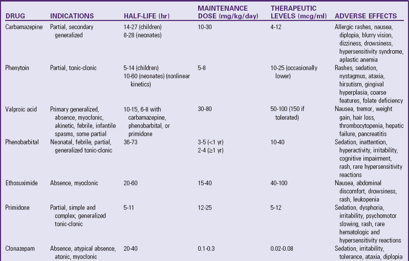
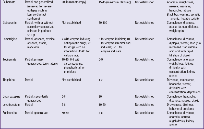
Drug Therapy: It is known that persons predisposed to epilepsy have seizures when their basal level of neuronal excitability exceeds a critical point; no event occurs if the excitability is maintained below this threshold. The administration of antiepileptic drugs serves to raise this threshold and prevent seizures. Consequently, the primary therapy for seizure disorders is the administration of the appropriate antiepileptic drug or combination of drugs in a dosage that provides the desired effect without causing undesirable side effects or toxic reactions. Antiepileptic drugs exert their effect primarily by reducing the responsiveness of normal neurons to the sudden, high-frequency nerve impulses that arise in the epileptogenic focus. Thus the seizure is effectively suppressed; however, the abnormal brain waves may or may not be altered. Complete control can be achieved in 80% of children with epilepsy (Curatolo, Moavero, Lo Castro, et al, 2009). Table 37-6 outlines the drugs used for control of seizures.
TABLE 37-6
COMMON ANTIEPILEPTIC MEDICATIONS
Modified from Browne TR, Holmes GL: Handbook of epilepsy, Philadelphia, 2004, Lippincott Williams & Wilkins.
Therapy begins with a single drug known to be effective and have the lowest toxicity, that is, the safest side effect profile for the child’s particular type of seizure. The dosage is gradually increased until the seizures are controlled or the child develops side effects. If the drug is effective but does not sufficiently control the seizures, a second drug is added in gradually increasing doses. Once seizures are controlled, the first drug may be tapered to reduce the potential adverse effects of polytherapy. However, this decision is individualized for each child (Browne and Holmes, 2004). Monotherapy remains the treatment method of choice for epilepsy, but polypharmacy may be a viable alternative for children who cannot attain seizure control with only one agent (Leppik, 2000).
If complete seizure control is maintained on an anticonvulsant drug for 2 years, it is safe for patients with no risk factors to discontinue the drug. Risk factors include age greater than 12 years at onset, history of neonatal seizures, numerous seizures before control is achieved, and presence of a neurologic dysfunction (i.e., motor handicap or cognitive impairment). Up to 25% of children whose medications are discontinued experience seizure recurrence. Recurrence occurs most frequently within 6 months of discontinuation (Johnston, 2009).
When seizure medications are discontinued, the dose is decreased gradually over several weeks. Sudden withdrawal of a drug is not recommended because it can cause an increase in the number and severity of seizures.
Complications of Drug Therapy:
The side effects of continued use of antiepileptic medications are sometimes distressing to the child and the family. Most side effects are transient and dose related, but drug reactions warrant immediate attention. Dose-related side effects such as dizziness, headache, ataxia, and sleepiness often disappear over time or when drug dosages are reduced. Drug reactions require clinical evaluation and may require monitoring of serum drug levels. Combination therapy, such as with barbiturates and carbamazepine, can potentiate drug levels. Knowledge of drug-to-drug interactions, including other medications such as antibiotics, is critical in caring for the child with epilepsy. Knowledge of potential adverse effects is also imperative. Severe, potentially life-threatening side effects can occur with specific antiepileptic medications. For example, carbamazepine, phenytoin, and lamotrigine may cause a severe, life-threatening rash. Valproic acid may cause liver toxicity, particularly in a child less than 2 years of age. To avoid possible complications of tissue damage and difficulties with administration of IV phenytoin, fosphenytoin should be used. Therefore critical thinking and careful monitoring are necessary in providing optimum care to the child with epilepsy.
Chronic treatment with phenytoin may cause gum hypertrophy. Surgical removal of the excess tissue may be needed in severe cases. Enlargement of the tonsillar and adenoidal tissue can cause partial airway obstruction, which produces snoring during sleep. Chronic treatment with antiepileptic medications has been associated with decreased bone mineral density that does not correlate with serum vitamin D levels. However, no guidelines have been established for monitoring bone mineral density in individuals on antiepileptic medications (Farhat, Yamout, Mikati, et al, 2002).
Ketogenic Diet: The ketogenic diet is a high-fat, low-carbohydrate, and adequate-protein diet (Kossoff, Zupec-Kania, and Rho, 2009). Consumption of such a diet forces the body to shift from using glucose as the primary energy source to using fat, and the individual develops a state of ketosis. The diet is rigorous. All foods and liquids the child consumes must be carefully weighed and measured. The diet is deficient in vitamins and minerals; therefore vitamin supplements are necessary. Potential side effects of the diet are constipation, weight loss, lethargy, and kidney stones. There are reports of increased blood lipids in children on the ketogenic diet; the long-term effects of which are unknown (Levy and Cooper, 2003) (see Research Focus box).
Vagus Nerve Stimulation: Vagus nerve stimulation uses an implantable device that reduces seizures in individuals who have not had effective control with drug therapy. It is currently indicated as adjunctive therapy in patients 12 years and older with partial onset seizures (with or without secondary generalization). A programmable signal generator is implanted subcutaneously in the chest. Electrodes tunneled underneath the skin deliver electrical impulses to the left vagus nerve (CN X). The device is programmed noninvasively to deliver a precise pattern of stimulation to the left vagus nerve. The patient or caregiver can activate the device using a magnet at the onset of a seizure. Studies show a median reduction in seizures of 35% to 45% after 1 year of therapy (Saillet, Langlois, Feddersen, et al, 2009).
Surgical Therapy: When seizures are caused by a hematoma, tumor, or other cerebral lesion, surgical removal is the treatment. Surgery is reserved for children who suffer from incapacitating, refractory seizures. Refractory seizures are usually defined as the persistence of seizures despite adequate trials of three antiepileptic medications, alone or in combination (Browne and Holmes, 2004).
There are several types of surgical interventions. In resective surgery the focal area of the seizure activity is excised with the expectation that the surgery will not produce serious deficits or increase existing deficits. The use of intraoperative electrocorticography helps localize anatomic areas of focal seizure onset, guide the extent of surgery, map cortical anatomy, and predict epilepsy surgical outcome (Gallentine and Mikati, 2009). Surgical excision of the epileptogenic focus may not eliminate the need for drug therapy. A hemispherectomy to remove all or most of one hemisphere is typically used in patients who have severe epilepsy and who already have hemiparesis or nonfunctional hand use. Patients with Rasmussen syndrome or Sturge-Weber syndrome may benefit from this procedure. Corpus callosotomy involves the separation of the connections between the two hemispheres of the brain and is used in some generalized seizures. In multiple subpial transection, horizontal fibers of the motor cortex are divided to reduce seizures, whereas the vertical fibers are spared to allow for function (Browne and Holmes, 2004).
Status Epilepticus: Status epilepticus is a continuous seizure that lasts more than 30 minutes or a series of seizures from which the child does not regain a premorbid level of consciousness (Shorvon and Walker, 2005; Treiman and Walker, 2006). No consensus has been reached on the duration of seizure required to qualify as status epilepticus (Chen and Wasterlain, 2006). Direct the initial treatment toward support and maintenance of vital functions, that is, the ABCs of life support, administration of oxygen, and gaining of IV access, immediately followed by IV administration of antiepileptic agents.
Rectal diazepam is a simple, effective, and safe treatment for home or prehospital management (Pellock and Shinnar, 2005). It is available in a prefilled rectal gel syringe (Diastat) for easy administration. Rectal diazepam is not associated with respiratory depression when used as recommended (Pellock and Shinnar, 2005). Midazolam has been given successfully by intranasal route for treatment of acute epileptic seizures (Fisgin, Gurer, Senbil, et al, 2000; Kutlu, Yakinci, Dogrul, et al, 2000; Scheepers, Scheepers, Clarke, et al, 2000). Intranasal midazolam not only is safe and effective for stopping seizures, but also is easier to administer than rectal diazepam (Harbord, Kyrkou, Kyrkou, et al, 2004).
For in-hospital management of status epilepticus, IV diazepam or lorazepam (Ativan) is the first-line drug of choice (Browne and Holmes, 2004). Lorazepam may be replacing IV diazepam as the drug of choice. It has a longer duration of action and causes less respiratory depression in children over 2 years of age. Concurrent IV loading with fosphenytoin is usually necessary for sustained control of seizures. Valproic acid has also been effective in status epilepticus when given rectally or intravenously (Yamamoto and Yim, 2000). The child must be closely monitored during administration to detect early alterations in vital signs that may indicate impending respiratory depression (see Evidence-Based Practice box). When diazepam is ineffective, fosphenytoin or phenobarbital is given intravenously as the next line of treatment. This combination of therapy places the child at high risk for apnea, and therefore respiratory support is generally necessary.
Children who continue to have seizures despite the above drug treatment may be given anesthetizing doses of midazolam, propofol, or pentobarbital (Morrison, Gibbons, and Whitehouse, 2006; van Gestel, Blusse van Oud-Alblas, Malingre, et al, 2005). Sodium valproate has also been studied for control of refractory status epilepticus in children (Mehta, Singhi, and Singhi, 2007). In this situation, continuous EEG monitoring is typically done to detect and treat electrographic seizures.
Nursing care of a child with status epilepticus includes, in addition to the ABCs of life support, monitoring blood pressure and body temperature. During the first 30 to 45 minutes of the seizure the blood pressure may be elevated. Thereafter the blood pressure typically returns to normal but may be decreased, depending on the medications being administered for seizure control. Hyperthermia requiring treatment may occur as a result of increased motor activity.
Status epilepticus is a medical emergency that requires immediate intervention to prevent possible brain injury or death. As imperative as halting the tonic-clonic movement is correct diagnosis of the underlying problem. The outcome is related to the etiology and duration of the status epilepticus.
Most children who experience a second seizure will experience additional seizures. Therefore a history of two seizures is sufficient to diagnose epilepsy (Shinnar, Berg, O’Dell, et al, 2000). Epidemiologic studies using population- or community-based cohorts show that the etiology and specific epilepsy syndromes are the most important factors affecting prognosis. Children who have cognitive impairments or cerebral palsy are at the highest risk for developing epilepsy. Seizures remit in more than two thirds of children with childhood onset of seizure. Mortality is increased in children with epilepsy; those with neurologic abnormalities or seizures that are refractory to treatment are at the highest risk (Browne and Holmes, 2004).
Convulsive status epilepticus is convincingly related to serious morbidity and mortality and is often associated with a severe neurologic abnormality, uncontrolled seizure disorder, or concurrent serious illness or infection. In one study, 79% of children who suffered status epilepticus had neurologic abnormalities. The highest morbidity was in patients with a nonidiopathic, nonfebrile cause (Barnard and Wirrell, 1999).
An important nursing responsibility is to observe the seizure episode and accurately document the events. Record and note any alterations in behavior preceding the seizure and the characteristics of the episode, such as sensory-hallucinatory phenomena (e.g., an aura), motor effects (e.g., eye movements, muscular contractions), alterations in consciousness, and postictal state (Box 37-11). Describe only what is observed, rather than trying to label a seizure type. Note the time that the seizure began and the duration of the seizure.
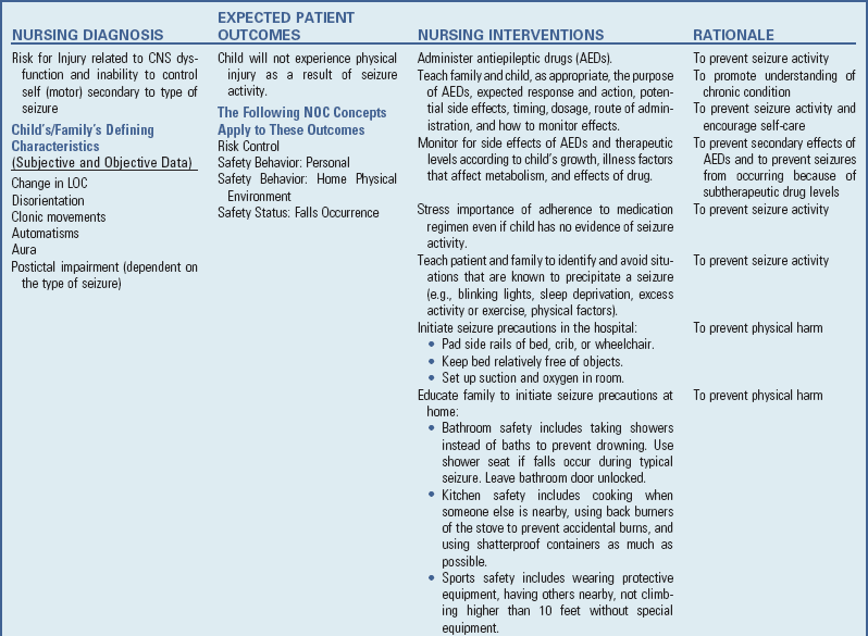
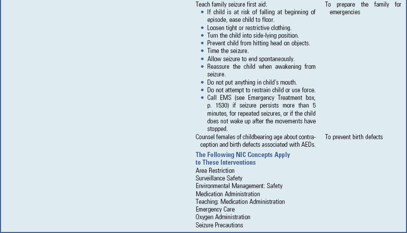
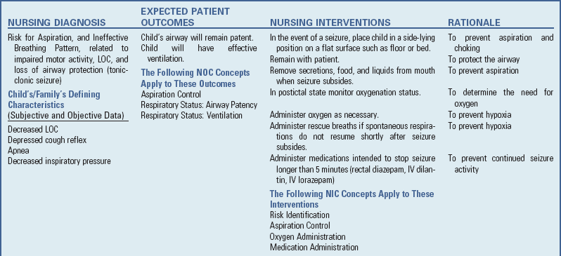
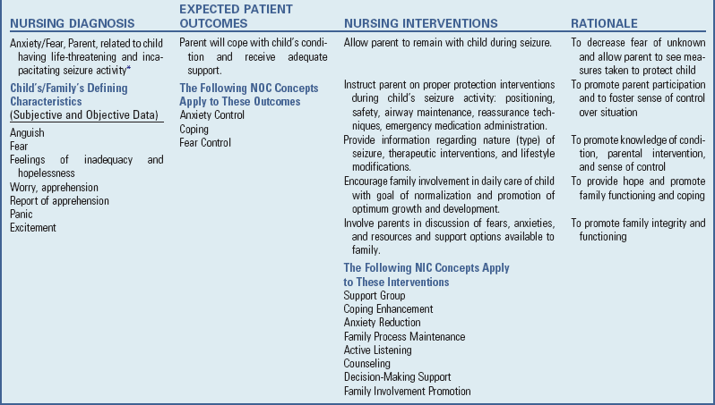
Nursing Care Plan—The Child with Seizure Disorder
Generalized seizures and other types with clear manifestations are easy to detect, but absence seizures may present more difficulties. They are easily misinterpreted as inattention. Any unusual behavior, even seemingly inconsequential, such as a momentary interruption of activity, staring, or mental blankness, should be described. The more detailed these descriptions, the more valuable they are for assessment (see Nursing Care Plan).
The child must be protected from injury during the seizure. Nursing observations made during the event provide valuable information for diagnosis and management of the disorder (see Emergency Treatment box).
It is impossible to halt a seizure once it has begun, and no attempt should be made to do so. The nurse must remain calm, stay with the child, and prevent the child from sustaining any harm during the seizure. If possible, isolate the child from the view of others by closing a door or pulling screens. A seizure can be upsetting to the child, other visitors, and their families. If other persons are present, reassure them that everything is being done for the child. After the seizure, they can be given a simple explanation about the event as needed.
If the nurse is able to reach the child in time, a child who is standing or seated in a chair (including a wheelchair) is eased to the floor immediately. During (and sometimes after) the tonic-clonic seizure, the swallowing reflex is lost, salivation increases, and the tongue is hypotonic. Therefore the child is at risk for aspiration and airway occlusion. Placing the child on the side facilitates drainage and helps maintain a patent airway. Suctioning of the oral cavity and posterior oropharynx may be necessary. Take vital signs, and allow the child to rest if at school or away from home. When feasible, the child is integrated into the environment as soon as possible. Sending a child with a chronic seizure disorder home from school is not necessary unless requested by the parents.
Seizure precautions are required for children who are known to have seizures or who are under observation for seizures. The extent of these measures depends on the type and frequency of the seizure (Box 37-12).
Long-Term Care: Care of the child with epilepsy involves physical care and instruction regarding the importance of the drug therapy and, probably more significant, the problems related to the emotional aspects of the disorder. Few diseases generate as much anxiety among relatives as epilepsy. Fears and misconceptions about the disease and its treatment are common. For many, it represents the archetype of severe hereditary affliction. Direct nursing care toward educating the child and family about epilepsy and helping them develop strategies to cope with the psychologic and sociologic problems related to epilepsy.
Physical Aspects: Children with epilepsy are prescribed antiepileptic medications, which are administered at regular intervals to maintain adequate levels in the blood. The nurse can help the parents plan the administration of the medication at convenient times to disrupt the family routine as little as possible. The dosage schedule is based on the drug’s half-life (the time required to reduce to one-half the amount of unchanged drug that is in the body) and the child’s age. Drugs with longer half-lives are given less frequently, and a missed dose will have less of a negative effect than with a drug with a short half-life. The younger child may need a more frequent dosing schedule because of more rapid metabolism. The aim is to simplify the medication routine as much as possible and incorporate it into the parents’ and child’s daily activities. This also increases the likelihood of compliance. The most convenient times for administration seem to be with meals or at bedtime.
It is important to impress on the family the necessity of giving the antiepileptic medication regularly and for as long as required. In general, antiepileptic medications are continued until the child has been seizure free for 2 years (Johnston, 2009). The medication is then slowly tapered over a period of weeks to avoid the possibility of precipitating a seizure. It is sometimes easy to skip doses or omit them for a variety of reasons, especially when the child is free of seizures most of the time. This is particularly so when the child is older and assumes responsibility for his or her medication. The seizure threshold may be lowered during any illness, but particularly with fever. Therefore parents should be aware that if their child has an illness, he or she is at increased risk for seizures. Parents should contact their health professional if their child misses medications during an illness because of vomiting.
Educate the child and parents about the possible adverse reactions to the medications used to treat seizures. Parents should understand the common side effects and be encouraged to report their observations to their health care provider. Parents should understand that the child needs periodic physical assessment and laboratory studies. Possible adverse effects on the hematopoietic system, liver, and kidneys may be reflected in symptoms such as fever, sore throat, enlarged lymph nodes, jaundice, and bleeding (e.g., easy bruising, petechiae, ecchymoses, epistaxis). A common factor in status epilepticus is inadequate blood levels of antiepileptic drugs.
Parents need to be aware of possible behavioral changes associated with some antiepileptic medications. Changes in personality, indifference to school activities and family, hyperactivity, or even psychotic behavior may sometimes be observed. If so, the parents should contact their health care provider. The potential effects of antiepileptic drugs on learning and behavior should also be considered. Progressive intellectual deterioration in a child with epilepsy requires investigation of the present medication plus the role of the underlying cerebral pathologic condition.
Although children with epilepsy are at increased risk for injury, few limitations should be placed on activities. The degree to which activities are restricted is individualized for each child and depends on the type, frequency, and severity of the seizures; the child’s response to therapy; and the length of time the seizures have been controlled. Children are encouraged to engage in most normal activities; participation in competitive sports is determined on an individual basis. With encouragement, most older children can accept the restrictions placed on activities. To avoid accentuating differences when possible, only the essential restrictions are placed on children regarding sports and peer activity; these restrictions are approached in a positive way in terms of what the child can do rather than what he or she cannot do. Parents sometimes limit the child’s activities more than necessary, which may lead to impaired self-esteem.
To prevent head injuries, children should wear appropriate safety devices, such as helmets, and should avoid activities involving heights. Although bike riding is safe for most children, children with frequent seizures in whom impairment of consciousness occurs should avoid bike riding. Skating, roller-blading, and skateboarding should be restricted only in children with frequent seizures. Helmets must be worn while participating in these activities.
Children with epilepsy are at higher risk for submersion injury than children without epilepsy. Young children should never be left alone in the bathtub, even for a few seconds. Older children and adolescents should be encouraged to use a shower and reminded not to lock the bathroom door when showering. They should never swim unsupervised.
All children should avoid open fires, hot stoves and ovens, and dangerous machinery. Parents must assume that a seizure could occur at any time and should not allow a child to be in a situation where a seizure could be deadly.
Because the child is encouraged to attend school, camp, and other normal activities, the school nurse and teacher should be made aware of the child’s condition and therapy. They can help ensure regularity of medication administration and provision of any special care the child might need. Teachers, child care providers, camp counselors, youth organization leaders, coaches, and other adults who assume responsibility for children should be instructed regarding care of the child during a seizure so that they can act in a calm manner for the child’s welfare and influence the attitude of the child’s peers.*
Triggering Factors: Careful and detailed documentation of seizures over time may indicate a pattern. In such cases the nurse or responsible adult may intervene to identify the triggering factors and alter the environment to prevent or decrease seizure frequency. Often the necessary changes are simple but can make an enormous difference in the lives of the child and family (see Critical Thinking Exercise).
The most common factors that may trigger seizures in children include emotional stress, sleep deprivation, fatigue, fever, and illness (Frucht, Quigg, Schwaner, et al, 2000; Nakken, Solaas, Kjeldsen, et al, 2005). Other precipitating factors include sleep, flickering lights, menstrual cycle, alcohol, heat, hyperventilation, and fasting (Frucht, Quigg, Schwaner, et al, 2000). Some individuals have pattern-sensitive epilepsy, that is, seizures precipitated by changes in dark-light patterns, such as those that occur with a flash on a camera, automobile headlights, reflections of light on snow or water, or rotating blades on a fan (see Research Focus box).
Family Support: Parental attitudes and management of a child with a seizure disorder vary. Whether the seizures result from illness, injury, or unknown cause, the parents may feel guilt, anxiety, and even humiliation. They want to know if the seizures will affect their child’s mental capacities. Many persons erroneously associate epilepsy with mental deficiency. Seizures do commonly accompany other manifestations of severe brain damage from disease or injury, but children with seizures, like any population of healthy children, display a wide range of intelligence.
Parents also wonder how the illness will affect their child’s future. The answer to this question depends on the cause of the seizures and other comorbid conditions. In many cases parents can be reassured that the illness will not shorten their child’s life and that their child can attend school, marry, and elect to have children. The child may need vocational guidance, and the parents need to become familiar with the laws in their state regarding any limitations imposed on people with epilepsy. For children who are severely impaired, the family needs to become familiar with local early childhood programs. The nurse should emphasize that the seizures can be controlled or greatly reduced in the large majority of affected children. Parents need reassurance that less stigma is attached to the condition than in the past.
Encourage a healthy attitude toward the child and the condition, and help the parents feel competent in their ability to meet their responsibilities to their child. The child should be reared in the same manner as any normal child, with natural concern tempered by understanding of the need not to overprotect. Many parents refrain from correcting or punishing their child, especially if the child has had a seizure after being disciplined. The child must not be made to feel different in any way. Encourage parents to be honest and open about the disorder with their child and with others. Some parents try to conceal the nature of their child’s illness because of their belief that the disorder is shameful or a disgrace to the family.
Educational materials and support groups may prove beneficial for families. The Epilepsy Foundation* is a national organization that works for the welfare of persons with epilepsy and their families; helps with employment and legal problems; and provides education to patients, families, and communities.
The Child with Epilepsy: The child who is provided the security of a loving family, rewards and punishments no different from those of other children, and support in acquiring self-esteem is more likely to have a positive attitude toward the condition. Children derive their self-concept and self-esteem from observations of others’ reactions to them and from their own perceptions of their capabilities. The suddenness and unpredictability of the seizures and the reactions of others further influence their feelings. When others consider children to be different, inferior, or objects of ridicule, the children come to view themselves in the same light.
Children with epilepsy need to learn about their condition and how medication contributes to their prolonged well-being. As soon as they are old enough, children should assume responsibility for taking their own medication and should carry medical identification with pertinent information about their condition. Planning activities with children and emphasizing those in which they can engage, rather than those which are restricted, help them succeed and gain satisfaction in their achievements. They should be offered opportunities and encouraged to exercise judgment in their daily lives.
The adolescent period may prove to be a trying time for the child with epilepsy. Limits imposed on the young person’s activities at a time when freedom and independence are desired may bring the disability into sharp focus. For example, all U.S. states have a defined seizure-free period before a driver’s license can be obtained.
Epilepsy should not be a severe impairment to most youngsters. The nurse can help provide positive outcomes for the child and family by assuming the role of patient advocate, helping educate the public about the condition, working to make opportunities available to persons with the disorder, and lobbying for legislation that recognizes the needs of individuals with seizure disorders.
Febrile seizure is “a seizure in association with a febrile illness in the absence of a central nervous system infection or acute electrolyte imbalance in children older than 1 month of age without prior afebrile seizures” (Ostergaard, 2009). Febrile seizures are one of the most common neurologic conditions of childhood, affecting approximately 3% of children. Most febrile seizures occur after 6 months of age and usually before age 3 years, with the average age of onset between 18 and 22 months. They are unusual after 5 years of age. Boys are affected about twice as often as girls, and there appears to be an increased familial susceptibility.
The cause of febrile seizures is still uncertain. Both animal and human studies demonstrate an age-specific susceptibility to seizures induced by fever and that it is the peak temperature that is important, not the rapidity of the temperature elevation (Ostergaard, 2009). The temperature usually exceeds 38.8° C (101.8° F), and the seizure occurs during the temperature rise rather than after a prolonged elevation. Sometimes it constitutes the dramatic beginning of an illness, often an upper respiratory tract or gastrointestinal infection.
Most febrile seizures have stopped by the time the child is taken to a medical facility. However, if the seizure continues, treatment consists of controlling the seizure with IV or rectal diazepam and reducing the temperature with acetaminophen. Antiepileptic prophylaxis is not indicated. Parental education and emotional support are important interventions. Parents need reassurance regarding the benign nature of febrile seizures. Several large studies show no difference in intelligence, behavior, or academic performance in children with febrile seizures compared with either population or sibling controls (Verity, Greenword, and Golding, 1998). Parents also need education on how to protect the child from harm and observe exactly what happens to the child during the event. Attempts to lower the temperature will not prevent a seizure. Tepid sponge baths are not recommended for several reasons: they are ineffective in significantly lowering the temperature, the shivering effect further increases metabolic output, and cooling causes discomfort to the child.
Long-term antiepileptic therapy is usually not required for children with simple febrile seizures. Antipyretic therapy during febrile illness offers symptomatic relief for fever-associated symptoms but appears to be ineffective in preventing a seizure (Ostergaard, 2009).
Headaches are a common complaint of children and are associated with different pathologic conditions, including extracranial disease, intracranial disease, vascular abnormalities, psychogenic disorders, or a combination of the above (Table 37-7). Headaches are classified according to the International Headache Society classification system (Headache Classification Subcommittee of the International Headache Society, 2004).
It is important to determine the pattern of the headache—single acute episode, paroxysmal, acute and recurrent, chronic and progressive, chronic nonprogressive, or mixed. Other assessment information includes the presence of seizures, ataxia, lethargy, weakness, nausea or vomiting, or any personality changes. Factors related to early development and past illnesses and a family history of headaches may also be pertinent. A “headache diary,” which includes time of onset and termination of headaches, intensity, associated events, and actions taken and their effects, can be helpful for the patient and practitioner.
Clues to etiology may be found in the family history, including information about the home or social situation (e.g., divorce, separation, alcoholism, school avoidance). Box 37-13 lists specific questions that often elicit needed information. Thorough physical and neurologic examinations are performed, and further diagnostic tests (e.g., CT, MRI, or EEG) are ordered if indicated.
Tension headaches are common in children. They are typically frontal, and the pain is described as a pressing tightness; it is nonthrobbing in character and is not typically accompanied by nausea or vomiting. Simple analgesics, including acetaminophen and ibuprofen, are usually the most effective pharmacologic intervention. Biofeedback and relaxation techniques may be useful nonpharmacologic interventions in children with recurrent tension headaches (Haslam, 2009; Rowley, 2005).
Migraine headaches occur in children as well as adults. Migraine headaches can have their onset in very young children, including infants, although this is uncommon. The onset tends to be earlier in boys than girls, with a male prevalence until age 10 to 14 years of age after which time more females are affected (Bigal, Lipton, Winner, et al, 2007).
The exact pathophysiology of migraines continues to be researched. It is postulated that migraine headaches have a genetic and multifactorial etiology. However, no consistent genetic etiology for migraines has been established except for familial hemiplegic migraine, which has autosomal dominant inheritance (Ducros, Tournier-Lasserve, and Bousser, 2002). Previously, it was thought that migraine headaches were caused by dilatation of cerebral blood vessels. However, this is no longer thought to be correct. Although vasodilation appears to play a role in the throbbing pain of migraine, this is likely a secondary symptom. Rather, the primary cause of migraine headaches is now thought to be a neuronal dysfunction that leads to a sequence of changes in blood flow to specific regions of the brain and the release of various polypeptides that cause pain and vasodilation of cranial vessels (Bolay, Reuter, Dunn, et al, 2002).
Revisions in the classification of migraine headaches introduced some new terms and eliminated, renamed, or reclassified others. Migraine headaches are now classified as migraine without aura and migraine with aura; the latter category includes the following subtypes: auras and prodrome, familial hemiplegic migraine, sporadic hemiplegic migraine, and basilar-type migraine (Box 37-14) (Headache Classification Subcommittee of the International Headache Society, 2004).
The headaches are paroxysmal. The symptoms of migraine headaches vary depending on age. Typical symptoms include nausea, vomiting, and abdominal pain, which are relieved by sleep. Toddlers may be seen with episodic pallor, decreased activity, and vomiting. The onset of a migraine headache in a young child is typically in the afternoon and may be bifrontal, temporal, and bilateral or unilateral. Children may vomit repetitively during a migraine headache. A family history of migraine is elicited in 70% of children with migraine; 5% of all children who have migraines experience a headache before age 15 years. Before the onset of puberty, migraines are more common in boys; this trend reverses after puberty.
Migraine headaches are managed with general measures (education, a headache diary to identify and eliminate precipitating factors, and documented response to treatment), abortive treatment, and prophylactic treatment. At the onset of the headache, the child should rest or sleep in a quiet, dark room when feasible. Migraine therapy, if administered early in the course of the headache, may provide rapid relief. Acetaminophen or ibuprofen is often effective if given early.
The outlook for a child with migraine is good, but the child and parents should be informed that predisposition to the headaches may be lifelong. Severe headaches can adversely affect the child’s routine activities of daily living, including family relations and school.
• The CNS is composed of the brain and spinal cord.
• Gait abnormalities that may indicate cerebral dysfunction include ataxia, spastic paraplegic gait, hemiplegic gait, cerebellar gait, and extrapyramidal gait.
• LOC is the most important indicator of neurologic health; altered levels include sleep, confusion, delirium, and comatose states.
• Complete neurologic examination includes LOC; gait, motor, sensory, CN, and reflex testing; and vital signs.
• Nursing care of the unconscious child focuses on respiratory management, neurologic assessment, increased ICP monitoring, adequate nutrition and hydration, drug therapy, promotion of elimination, maintenance of hygiene, positioning and exercise, stimulation, and family support.
• Primary head injury involves events that occur at the time of trauma, including fractured skull, contusions, intracranial hematoma, and diffuse injury. Secondary complications include hypoxic brain damage, increased ICP, infection, cerebral edema, and posttraumatic syndromes.
• The young child’s response to head injury is different because of a larger head size in proportion to body; larger blood volume to the brain; small subdural spaces; and thinner, softer brain tissue.
• Fractures resulting from head injuries can be classified as linear, depressed, compound, basilar, and diastatic.
• Problems resulting from submersion injuries are caused by hypoxia and include asphyxiation, aspiration, and hypothermia.
• Nursing care of the child with meningitis includes administration of antibiotics; vital signs monitoring; IV therapy; and promotion of fluid, nutritional status, and family support.
• Routine immunization of infants against H. influenzae type b and S. pneumoniae infection has reduced the incidence of bacterial meningitis.
• Encephalitis may result from direct invasion of the CNS by a virus or from postinfectious involvement of the CNS after viral illness.
• A seizure is a symptom of underlying pathologic condition and may be manifested by sensory-hallucinatory phenomena, motor effects, sensorimotor effects, or impaired or loss of consciousness.
• Partial seizures are categorized as simple (without associated impairment of consciousness) or complex; both types may become generalized.
• Generalized seizures are categorized as tonic-clonic, absence, atonic, or myoclonic.
• Long-term care of the child with epilepsy involves teaching caregivers appropriate interventions during a seizure, emphasizing the importance of antiepileptic therapy, giving practical advice regarding drug administration and scheduling, and helping the child and family cope with diagnosis.
• Febrile seizures are the most common type of childhood seizure. The most important nursing intervention is to reassure parents of their benign nature and educate parents regarding protection of their child and meaningful observation during the event.
• A child’s complaint of headache requires a thorough history and physical examination with a neurologic assessment. Stress is the most common cause of recurrent headache in children.
1. Yes. Thomas’s behavior 2 weeks after a fall, consisting of enuresis, crying episodes, early morning vomiting, decreased appetite, and a desire to be held, are all signs of increased intracranial pressure (ICP).
2. a. Preschoolers have increased curiosity and are unaware of safety measures.
b A fall predisposes a preschooler to develop posttraumatic insidious brain injury even after an initial negative CT scan.
c Behavioral changes 2 weeks after a fall may be signs of ICP because of postconcussion syndrome.
3. The nurse should immediately notify the medical provider because of the worsening postconcussion symptoms and signs of increased ICP, then obtain vital signs and assess Thomas’s neurologic status.
4. Yes. The preschooler’s signs of ICP approximately 2 weeks after head trauma (fall) support the nurse’s actions.
1. Yes. Jane is a teenager with previously well-controlled seizures who is now having frequent seizures. However, until further assessment is performed, the exact reason is not known.
2. a. Seizures may change during brain maturation and either become easier or more difficult to control.
b Further evaluation is needed in any child whose seizures have been well controlled and then increase in frequency. The child and family should be advised to seek medical reevaluation.
c Seizures have many triggering factors. Jane’s history is suggestive of several, including sleep deprivation, fatigue, and stress. Further assessment is required to elicit the possibility of others, including poor medication compliance, relation to menses, and alcohol intake.
3. Nursing care should focus on educating Jane and her family on the avoidance of triggering factors for seizures and maintaining healthy lifestyle habits (e.g., getting adequate sleep, taking medications as prescribed, avoiding alcohol).
4. Yes, the history and physical assessment support these conclusions. Additionally, knowledge of triggering factors for seizures supports this conclusion. It is not within the scope of nursing practice to change the antiepileptic medication or dosage.
American Academy of Pediatrics, Committee on Infectious Diseases. Pickering L, ed. Red book: report of the Committee on Infectious Diseases, ed 27, Elk Grove Village, Ill: The Academy, 2006.
American Heart Association, Part 10.3 drowning. Circulation 2005;112:IV133–IV135. available at http://circ.ahajournals.org/cgi/content/full/112/24_suppl/IV-133 [(accessed May 17, 2009)].
Anderson, VA, Catroppa, C, Haritou, F, et al. Identifying factors contributing to child and family outcome 30 months after traumatic brain injury in children. J Neurol Neurosurg Psychiatry. 2005;76:401–408.
Anderson, VA, Catroppa, C, Haritou, F, et al. Predictors of acute child and family outcome following traumatic brain injury in children. Pediatr Neurosurg. 2001;34:138–148.
Baandrup, L, Jensen, R. Chronic post-traumatic headache: a clinical analysis in relation to the International Headache Classification 2nd edition. Cephalalgia. 2005;25:132.
Barnard, C, Wirrell, E. Does status epilepticus cause developmental deterioration and development of epilepsy? J Child Neurol. 1999;14(12):787–794.
Bechtel, K, Stoessel, K, Leventahal, JM, et al. Characteristics that distinguish accidental from abusive injury in hospitalized young children with head trauma. Pediatrics. 2004;114(1):165–168.
Bigal, ME, Lipton, RB, Winner, P, et al. Migraine in adolescents: association with socioeconomic status and family history. Neurology. 2007;69(1):16–25.
Blinman, TA, Houseknecht, E, Snyder, C, et al. Postconcussive symptoms in hospitalized pediatric patients after mild traumatic brain injury. J Pediatr Surg. 2009;44(6):1223–1228.
Bolay, H, Reuter, U, Dunn, AK, et al. Intrinsic brain activity triggers trigeminal meningeal afferents in a migraine model. Nature Med. 2002;8(2):136–142.
Bonthius, DJ, Karacay, B. Meningitis and encephalitis in children: an update. Neurol Clin. 2002;20(4):1013–1038.
Browne, TR, Holmes, GL. Handbook of epilepsy, ed 3. Philadelphia: Lippincott Williams & Wilkins; 2004.
Case, ME. Accidental traumatic head injury in infants and young children. Brain Pathol. 2008;18(40):583–589.
Castiglia, PT. Shaken baby syndrome. J Pediatr Health Care. 2001;15:78–80.
Centers for Disease Control and Prevention. General Recommendation on Immunization. available at www.cdc.gov/vaccines/pubs/ACIP-list.htm, 2009. [(accessed June 13, 2009)].
Centers for Disease Control and Prevention. Human rabies prevention: United States, 2008. MMWR Early Release. 2008;57:1–27.
Centers for Disease Control and Prevention. U.S. Rabies Surveillance Data. available at www.cdc.gov/rabies/epidemiology.html, 2006. [(accessed June 13, 2009)].
Centers for Disease Control and Prevention. Meningococcal disease. available at www.cdc.gov/ncidod/dbmd/diseaseinfo/meningococcal_t.htm, 2000. [(accessed May 2, 2010)].
Chavez-Bureno, S, McCracken, GH, Jr. Bacterial meningitis in children. Pediatr Clin North Am. 2005;52(3):795–810.
Chen, JWY, Wasterlain, CG. Status epilepticus: pathophysiology and management in adults. Lancet Neurol. 2006;5:246–256.
Chiaretti, A, DeBenedictis, R, Polidori, G, et al. Early post-traumatic seizures in children with head injury. Childs Nerv Syst. 2000;16(12):862–866.
Cohen, RH, Matter, KC, Sinclair, SA, et al. Unintentional pediatric submersion-injury–related hospitalizations in the United States, 2003. Inj Prev. 2008;14(2):131–135.
Commission on Classification and Terminology of International League Against Epilepsy. Proposal for classification of epilepsies and epileptic syndromes. Epilepsia. 1985;26(3):268–278.
Commission on Epidemiology and Prognosis of International League Against Epilepsy. Guidelines for epidemiologic studies on epilepsy. Epilepsia. 1993;34(4):592–596.
Curatolo, P, Moavero, R, Lo Castro, A, et al. Pharmacotherapy of idiopathic generalized epilepsies. Exp Opin Pharmacother. 2009;10(1):5–17.
de Louvois, J, Halket, S, Harvey, D. Neonatal meningitis in England and Wales: sequelae at 5 years of age. Eur J Pediatr. 2005;164(12):730–734.
DeVita, MA. The death watch: certifying death using cardiac criteria. Prog Transplant. 2001;11(1):58–66.
DiCarlo, JV, Frankel, LR. Scoring systems and predictors of mortality. In Behrman RE, Kliegman RM, Jenson HB, eds.: Nelson textbook of pediatrics, ed 18, Philadelphia: Elsevier, 2009.
Ducros, A, Tournier-Lasserve, E, Bousser, MG. The genetics of migraine. Lancet Neurol. 2002;1(5):285–293.
El Bashir, H, Laundy, M, Booy, R. Diagnosis and treatment of bacterial meningitis. Arch Dis Child. 2003;88(7):814–819.
Ewing-Cobbs, L, Prasad, JR, Kramer, L, et al. Late intellectual and academic outcomes following traumatic brain injury sustained during early childhood. J Neurosurg. 2006;105(4 Suppl Pediatr):287–296.
Farhat, G, Yamout, B, Mikati, MA, et al. Effect of antiepileptic drugs on bone density in ambulatory patients. Neurology. 2002;58:1348–1353.
Feigin, RD, Perlman, E. Bacterial meningitis beyond the neonatal period. In Feigin RD, Cherry JD, eds.: Textbook of pediatric infectious diseases, ed 5, Philadelphia: Saunders, 2004.
Fisgin, T, Gurer, Y, Senbil, N, et al. Nasal midazolam effects on childhood acute seizures. J Child Neurol. 2000;15(12):833–835.
Frucht, MM, Quigg, M, Schwaner, C, et al. Distribution of seizure precipitants among epilepsy syndromes. Epilepsia. 2000;41(12):1543–1549.
Gallentine, WB, Mikati, MA. Intraoperative electrocorticography and cortical stimulation in children. J Clin Neurophysiol. 2009;26(2):95–108.
Greenberg, RA, Bolte, RG, Schunk, JE. Infant carrier-related falls: an unrecognized danger. Pediatr Emerg Care. 2009;25(2):66–68.
Greenes, DS, Schutzman, SA. Clinical significance of scalp abnormalities in asymptomatic head-injured infants. Pediatr Emerg Care. 2001;17(2):88–92.
Haberlandt, E, Weger, C, Sigl, SB, et al. Adrenocorticotropic hormone versus pulsatile dexamethasone in the treatment of infantile epilepsy syndromes. Pediatr Neurol. 2010;42(1):21–27.
Hancock E, Osborne J: Treatment of infantile spasms, Cochrane Database Syst Rev (3):CD001770.DOI: 10.1002/14651858.CD001770, 2003.
Harbord, JG, Kyrkou, NE, Kyrkou, MR, et al. Use of intranasal midazolam to treat acute seizures in paediatric community settings. J Pediatr Child Health. 2004;40(9-10):556–558.
Haslam, RH. Brain abscess. In Behrman RE, Kliegman RM, Jenson HB, eds.: Nelson textbook of pediatrics, ed 18, Philadelphia: Elsevier, 2009.
Headache Classification Subcommittee of the International Headache Society. The International Classification of Headache Disorders, ed 2. Cephalalgia. 2004;24(Suppl 1):9–160.
Hoyt, CS. Brain injury and the eye. Eye. 2007;21(10):1285–1289.
Hung, KL, Liao, HT, Huang, JS. Rational management of simple depressed skull fractures in infants. J Neurosurg. 2005;103(1 Suppl):69–72.
Idris, AH, Berg, RA, Bierens, J, et al. Recommended guidelines for uniform reporting of data from drowning: the “Ustein style,”. Circulation. 2003;108(20):2565–2574.
Johnston, MV. Seizures in childhood. In Behrman RE, Kliegman R, Jenson HB, eds.: Nelson textbook of pediatrics, ed 18, Philadelphia: Elsevier, 2009.
Kallas, HJ. Drowning and submersion injury. In Behrman RE, Kliegman R, Jenson B, eds.: Nelson textbook of pediatrics, ed 18, Philadelphia: Elsevier, 2009.
Kaplan, SL, Mason, EO, Jr., Wald, ER, et al. Decrease of invasive pneumococcal infections in children among eight children’s hospitals in the United States after the introduction of 7-valent pneumococcal conjugate vaccine. Pediatrics. 2004;113(3 Pt 1):443–449.
Kline, N. Neurologic manifestations of HIV-infection (HIV nursing curriculum). Houston: Baylor College of Medicine; 2001.
Kossoff, EH, Zupec-Kania, BA, Rho, HM. Ketogenic diets: an update for child neurologists. J Child Neurol. 2009;24(8):979–988.
Kutlu, NO, Yakinci, C, Dogrul, M, et al. Intranasal midazolam for prolonged convulsive seizures. Brain Dev. 2000;22(6):359–361.
Kuzniecky, RI. Neuroimaging in pediatric epilepsy. In: Pellock JM, Dodson WE, Bourgeois BFD, eds. Pediatric epilepsy: diagnosis and therapy. New York: Demos, 2001.
Langlois, JA, Rutland-Brown, W, Thomas, KE. The incidence of traumatic brain injury among children in the United States. J Head Trauma Rehabil. 2005;20(3):229–238.
Lee, LK. Controversies in the sequelae of pediatric mild traumatic brain injury. Pediatr Emerg Care. 2007;23(8):580–583.
Leppik, IE. Monotherapy and polypharmacy. Neurology. 2000;55(Suppl 3):525–529.
Levy R, Cooper P: Ketogenic diet for epilepsy, Cochrane Database Syst Rev (3):CD001903. DOI: 10.1002/14651858.CD001903, 2003.
Lewis, D, Ashwal, S, Hershey, A, et al. Practice parameter: Pharmacological treatment of migraine headache in children and adolescents. Neurology. 2004;63:2215–2224.
Ljungman, G, Kreuger, A, Andreasson, S, et al. Midazolam nasal spray reduces procedural anxiety in children. Pediatrics. 2000;105(1 Pt 1):73–78.
Lloyd, CJ, Alredy, T, Lloyd, JC. Intranasal midazolam as an alternative to general anaesthesia in the management of children with oral and maxillofacial trauma. Br J Oral Maxillofac Surg. 2000;38(6):593–595.
Lo, WD, Lee, J, Rusin, J, et al. Intracranial hemorrhages in children: an evolving spectrum. Arch Neurol. 2008;65(12):1629–1633.
Marcin, JP, Pollack, MM. Triage scoring systems, severity of illness measures, and mortality prediction models in pediatric trauma. Crit Care Med. 2002;30(11 Suppl):S457–S467.
Marcoux, KK. Management of increased intracranial pressure in the critically ill child with an acute neurological injury. AACN Clin Issues. 2005;16(2):212–231.
Marsh, N, Whitehead, G. Skull fracture during infancy: a 5 year follow-up. J Clin Experiment Neuropsychol. 2005;27:352–366.
Mathur, M, Petersen, L, Stadtler, M, et al. Variability in pediatric brain death determination and documentation in Southern California. Pediatrics. 2008;121:988–993.
McCabe, CF, Donahue, SP. Prognostic indicators for vision and mortality in shaken baby syndrome. Arch Ophthalmol. 2000;118(3):373–377.
Mehta, V, Singhi, P, Singhi, S. Intravenous sodium valproate versus diazepam infusion for the control of refractory status epilepticus in children: a randomized controlled trial. J Child Neurol. 2007;22(10):1191–1197.
Menkes, JH, Sankar, R. Paroxysmal disorders. In: Menkes JH, Sarnat HB, eds. s: Child neurology. Philadelphia: Lippincott Williams & Wilkins, 2000.
Morrison, G, Gibbons, E, Whitehouse, WP. High-dose midazolam therapy for refractory status epilepticus in children. Intensive Care Med. 2006;32(12):2070–2076.
Myhre, S, Grogaard, JB, Dyb, GA, et al. Traumatic head injury in infants and toddlers. Acta Paediatr. 2007;96(8):1159–1163.
Nakken, KO, Solaas, MH, Kjeldsen, MJ, et al. Which seizure-precipitating factors do patients with epilepsy most frequently report? Epilepsy Behav. 2005;6(1):85–89.
Nigrovic, LE, Kuppermann, N, Malley, R. Children with bacterial meningitis presenting to the emergency department during the pneumococcal conjugate vaccine era. Acad Emerg Med. 2008;15(6):522–528.
Ong, I, Selladurai, BM, Dhillon, MK, et al. The prognostic value of the Glasgow Coma Scale: hypoxia and computerized tomography in outcome prediction of pediatric head injury. Pediatr Neurosurg. 1996;24(6):285–291.
Ostergaard, JR. Febrile seizures. Acta Paediatr. 2009;98(5):771–773.
Paniak, D, Reynolds, S, Phillips, K, et al. Patient complaints within 1 month of mild traumatic brain injury: a controlled study. Arch Clin Neuropsychol. 2002;17(4):319–334.
Papa, L, Hoelle, R, Idris, A. Systematic review of definitions for drowning incidents. Resuscitation. 2005;65(3):255–264.
Pearson, DA, McGrath, NM, Nozyce, M, et al. Predicting HIV disease progression in children using measures of neuropsychological and neurological functioning, Pediatric AIDS Clinical Trials 152 Study Team. Pediatrics. 2000;106(6):E76.
Pellock, JM, Shinnar, S. Respiratory adverse events associated with diazepam rectal gel. Neurology. 2005;64(10):1768–1770.
Pichichero, M. Meningococcal immunization update: a new conjugated vaccine. Consultant for Pediatricians. 2005;4(6):1–2.
Prober, CG. Central nervous system infections. In Behrman RE, Kliegman RM, Jenson HTS, et al, eds.: Nelson textbook of pediatrics, ed 18, Philadelphia: Elsevier, 2009.
Proctor, MB. Neurosurgical aspects of nonaccidental trauma in children. In: Loftus G, ed. Neurological surgery principles and practice. Philadelphia: Lippincott Williams & Wilkins, 2003.
Pugliese, A, Beltramo, T, Torre, D. Reye’s and Reye’s-like syndromes. Cell Biochem Funct. 2008;26(7):741–746.
Radhakrishnan, K, St Louis, EK, Johnson, JA, et al. Pattern-sensitive epilepsy: electroclinical characteristics, natural history, and delineation of the epileptic syndrome. Epilepsia. 2005;46(1):48–58.
Report of Special Task Force. Guidelines for the determination of brain death in children. Pediatrics. 1987;80(2):298–300.
Riikonen, RS. Favourable prognostic factors with infantile spasms. Eur J Paediatr Neurol. 2010;14(1):13–18.
Rivara, FP, Grossman, D. Injury control. In Behrman RE, Kliegman RM, Jenson HTS, eds.: Nelson textbook of pediatrics, ed 18, Philadelphia: Elsevier, 2009.
Rocchi, G, Caroli, E, Raco, A, et al. Traumatic epidural hematoma in children. J Child Neurol. 2005;20(7):569–572.
Rowley, SM. Headaches in children and adolescents. A blueprint for pharmacologic and nonpharmacologic approaches. Adv Nurse Pract. 2005;13(2):31–43.
Saillet, S, Langlois, M, Feddersen, B, et al. Manipulation the epileptic brain using stimulation: a review of experimental and clinical studies. Epileptic Disord. 2009;11(2):100–112.
Salomez, F, Vincent, JL. Drowning: a review of epidemiology, pathophysiology, treatment and prevention. Resuscitation. 2004;63(3):261–268.
Scheepers, M, Scheepers, B, Clarke, M, et al. Is intranasal midazolam an effective rescue medication in adolescents and adults with severe epilepsy? Seizure. 2000;9(6):417–422.
Shields, WD. Catastrophic epilepsy in childhood. Epilepsia. 2000;41(Suppl 2):52–56.
Shinnar, S, Berg, AT, O’Dell, C, et al. Predictors of multiple seizures in a cohort of children prospectively followed from the time of their first unprovoked seizure. Ann Neurol. 2000;48(2):140–147.
Shoja, MM, Tubbs, RS, Malekian, A, et al. Video game epilepsy in the twentieth century: a review. Childs Nerv Syst. 2007;23(3):265–267.
Shorvon, S, Walker, M. Status epilepticus in idiopathic generalized epilepsy. Epilepsia. 2005;46(Suppl 9):73–79.
Treiman, DM, Walker, MC. Treatment of seizure emergencies: convulsive and non-convulsive status epilepticus. Epilepsy Res. 2006;68S:S77–S82.
Tunkel, AR, Hartman, BJ, Kaplan, SL, et al. Practice guidelines for the management of bacterial meningitis. Clin Infect Dis. 2004;39(9):1267–1284.
van Gestel, P, Blusse van Oud-Alblas, HJ, Malingre, M, et al. Propofol and thiopental for refractory status epilepticus in children. Neurology. 2005;65(4):591–592.
van Rijckevorsel, K. Treatment of Lennox-Gestaut syndrome: overview and recent findings. Neuropsychiatr Dis Treat. 2008;4(6):1001–1019.
Verity, CM, Greenword, R, Golding, J. Longterm intellectual and behavioral outcomes of children with febrile convulsions. N Engl J Med. 1998;338(24):1723–1728.
Vignes, JR, Jeelani, NU, Jeelani, A, et al. Growing skull fracture after minor closed-head injury, J. Pediatrics. 2007;151(3):316–318.
Wetzell, RC. Anesthesia and perioperative care. In Behrman RE, Kliegman RM, Jenson HB, eds.: Nelson textbook of pediatrics, ed 18, Philadelphia: Elsevier, 2009.
White, JR, Farukhi, Z, Bull, C, et al. Predictors of outcome in severely head-injured children. Crit Care Med. 2001;29(7):534–540.
Wiederholt, WC. Neurology for non-neurologists, ed 4. Philadelphia: Saunders; 2000.
Willmore, LJ, Abelson, MB, Ben-Menachem, E, et al. Vigabatrin: 2008 update. Epilepsia. 2009;50(2):163–173.
Woods, C. Neisseria meningitidis. In Behrman RE, Kliegman RM, Jenson HB, eds.: Nelson textbook of pediatrics, ed 18, Philadelphia: Elsevier, 2009.
Yamamoto, LG, Yim, GK. The role of intravenous valproic acid in status epilepticus. Pediatr Emerg Care. 2000;16(4):296–298.
Yeates, KO, Taylor, HG, Rusin, J, et al. Longitudinal trajectories of postconcussive symptoms in children with mild traumatic brain injuries and their relationship to acute clinical status. Pediatrics. 2009;123:735–743.
Yogev, R, Chadwick, EG. Acquired immunodeficiency syndrome (human immunodeficiency virus). In Behrman RE, Kliegman RM, Jenson HTS, eds.: Nelson textbook of pediatrics, ed 18, Philadelphia: Elsevier, 2009.
Yogev, R, Guzman-Cottrill, J. Bacterial meningitis in children: critical review of current concepts. Drugs. 2005;65(8):1097–1112.
Yoon, U, Jagoda, A. New antiepileptic drugs and preparations. Emerg Med Clin North Am. 2000;18(4):755–765.
Zeltzer, LK, Krell, H. Pediatric pain management. In Behrman RE, Kliegman RM, Jenson HTS, eds.: Nelson textbook of pediatrics, ed 18, Philadelphia: Elsevier, 2009.
*1301 Pennsylvania Ave., NW, Suite 1000, Washington, DC 20004-1707; 202-662-0600; www.safekids.org.
*1608 Spring Hill Road, Suite 110, Vienna, VA 22182; 703-761-0750; fax: 703-761-0755; www.biausa.org.
*National Reye’s Syndrome Foundation, PO Box 829, Bryan, OH 43506; 800-233-7393; e-mail: nrsf@reyessyndrome.org; www.reyessyndrome.org.
*Excellent resources are Santilli N, Dodson WE, Walton AV: Students with seizures: a manual for school nurses, Landover, Md, 1991, Epilepsy Foundation; and Schachter SC, Montouris GD, Pellock JM: The brainstorms family: epilepsy on our terms—stories by children with seizures and their parents, Philadelphia, 1996, Lippincott Williams and Wilkins.
*8301 Professional Place, Landover, MD 20785-7223; 800-332-1000; www.efa.org. In Canada: Epilepsy Canada, 2255B Queen St. E., Suite 336, Toronto, Ontario, Canada M4E 1G3; 877-734-0873; www.epilepsy.ca.