Microanatomy of the skin
Introduction
The skin is one of the largest organs in the body, having a surface area of 1.8 m2 and making up about 16% of body weight. It has many functions, the most important of which is as a barrier to protect the body from noxious external factors and to keep the internal systems intact.
Skin is composed of three layers: the epidermis, the dermis and the subcutis (Fig. 1).
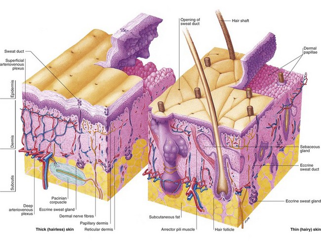
The diagram shows a comparison between thick, hairless skin (plantar and planar) and thinner, hirsute skin.
Epidermis
The epidermis is a stratified squamous epithelium that is about 0.1 mm thick, although the thickness is greater (0.8–1.4 mm) on the palms and soles. Its prime function is to act as a protective barrier. The main cells of the epidermis are keratinocytes, which produce the protein keratin. Keratinocytes are squamous cells functionally similar to all other structural epithelial cells as found in the airways and gastrointestinal tract. The four layers of the epidermis (Fig. 2) represent the stages of maturation of keratin by keratinocytes (p. 6).
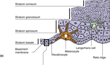
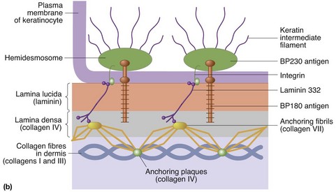
Fig. 2 Cross-sectional anatomy of the epidermis.
(a) Layers of the epidermis and other structures. (b) Detailed view of the basement membrane zone at the dermoepidermal junction. Components are arranged in three layers. The lamina lucida is traversed by filaments connecting the basal cells with the lamina densa, from which anchoring fibrils extend into the papillary dermis. These laminae are the sites of cleavage in certain bullous disorders (p. 84).
Basal cell layer (stratum basale)
The basal cell layer of the epidermis is composed mostly of keratinocytes, which are either dividing or non-dividing. The cells contain keratin tonofibrils (p. 6) and are secured to the basement membrane (see Fig. 2) by hemidesmosomes. Melanocytes make up 5–10% of the basal cell population. These cells synthesize melanin (p. 8) and transfer it via dendritic processes to neighbouring keratinocytes.
Melanocytes are most numerous on the face and other exposed sites, and are of neural crest origin. Merkel cells are also found, albeit infrequently, in the basal cell layer. These cells are closely associated with terminal filaments of cutaneous nerves and seem to have a role in sensation. Their cytoplasm contains neuropeptide granules, as well as neurofilaments and keratin. Basal keratinocytes synthesize antimicrobial peptides, important in defence against bacteria.
Prickle cell layer (stratum spinosum)
Daughter basal cells migrate upwards to form this layer of polyhedral cells, which are interconnected by desmosomes (the ‘prickles’ seen at light microscope level). Keratin tonofibrils form a supportive mesh in the cytoplasm of these cells. Langerhans cells are mostly found in this layer; these dendritic, immunologically active cells are described fully on page 10.
Embryology of the skin
The epidermis (ectoderm) begins to develop at 4 weeks of life, and, by 7 weeks, flat cells overlying the basal layer form the periderm (which is eventually cast off). Nails start to take shape at 10 weeks. The dermis (mesoderm) develops at 11 weeks, and, by 12 weeks, indented basal buds of the epidermis form the hair bulbs, with dermal papillae supplying vessels and nerves. Fingerprint ridges are determined by 17 weeks’ gestation. Maturation of the epidermis into a fully functional protective barrier continues throughout gestation and, because of this, pre-term infants are now frequently cared for with plastic occlusion to replicate this function.
Granular cell layer (stratum granulosum)
Cells become flattened and lose their nuclei in the granular cell layer. Keratohyalin granules are seen in the cytoplasm together with membrane-coating granules (which expel their lipid contents into the intercellular spaces).
Horny layer (stratum corneum)
The end result of keratinocyte maturation can be found in the horny layer, which is composed of sheets of overlapping polyhedral cornified cells with no nuclei (corneocytes). The layer is several cells thick on the palms and soles, but less thick elsewhere. The corneocyte cell envelope is broadened, and the cytoplasm is replaced by keratin tonofibrils in a matrix formed from the keratohyalin granules. Cells are stuck together by lipid glue that is partly derived from membrane-coating granules.
Dermis
The dermis is defined as a tough supportive connective tissue matrix, containing specialized structures, found immediately below and intimately connected with the epidermis. It varies in thickness, being thin (0.6 mm) on the eyelids and thicker (3 mm or more) on the back, palms and soles. The papillary dermis – the thin upper layer of the dermis – lies below and interdigitates with the epidermal rete ridges. It is composed of loosely interwoven collagen. Coarser and horizontally running bundles of collagen are found in the deeper and thicker reticular dermis.
Collagen fibres make up 70% of the dermis and impart a toughness and strength to the structure. Elastin fibres are loosely arranged in all directions in the dermis and provide elasticity to the skin. They are numerous near hair follicles and sweat glands, and less so in the papillary dermis. The ground substance of the dermis is a semisolid matrix of glycosaminoglycans (GAGs), which allows dermal structures some movement (p. 9).
The dermis contains fibroblasts (which synthesize collagen, elastin, other connective tissue and GAGs), dermal dendritic cells, mast cells, macrophages and lymphocytes.
Subcutaneous layer
The subcutis consists of loose connective tissue and fat (up to 3 cm thick on the abdomen).
Microanatomy
 The skin constitutes 16% of body weight, with a surface area of 1.8 m2.
Structure and thickness vary with site.
The epidermis is the outer covering, mainly composed of keratinocytes arranged in four layers, namely stratum corneum, stratum granulosum, stratum spinosum and stratum basale.
The epidermis also contains melanocytes and Langerhans cells.
The thickness of the epidermis varies from 0.1 mm to 0.8–1.4 mm on the palms and soles.
The dermis is supportive connective tissue, mainly collagen, elastin and glycosaminoglycans. The thickness varies between 0.6 mm (e.g. eyelids) and 3 mm (e.g. back and soles).
The dermis contains fibroblasts that synthesize the collagen, elastic fibres and glycosaminoglycans. Dermal dendritic cells are also found together with other immunocompetent cells.