Skin changes in internal conditions
Skin signs are seen with many internal disorders and are not uncommonly their presenting feature. The astute dermatologist can recognize undiagnosed systemic disease.
Skin signs of endocrine and metabolic disease
Almost all endocrine diseases (and several metabolic defects) have cutaneous signs that depend on the over- or underproduction of a hormone or metabolite (Table 1).
Table 1 Skin signs of endocrine and metabolic disorders
| Disorders | Skin signs |
|---|---|
| Diabetes mellitus | Necrobiosis lipoidica, granuloma annulare, xanthomas, Candida albicans infection, ‘dermopathy’, neuropathic ulcers |
| Thyrotoxicosis | Pink soft skin, hyperhidrosis, alopecia, pigmentation, vitiligo, onycholysis, clubbing, pretibial myxoedema, palmar erythema |
| Myxoedema | Alopecia (including eyebrows), coarse hair, dry puffy yellowish skin (e.g. hands, face), asteatotic eczema, xanthomas |
| Addison’s disease | Pigmentation (p. 75), vitiligo, loss of axillary and pubic hair |
| Cushing’s disease | Pigmentation, hirsutism, striae, acne, obesity, buffalo ‘hump’ |
| Acromegaly | Thickened moist greasy skin, pigmentation, skin tags |
| Phenylketonuria | Fair hair and skin, atopic eczema (p. 36), photosensitivity |
| Hyperlipidaemia | Xanthomas (tuberous, tendinous, eruptive, plane), xanthelasma |
| Cutaneous porphyrias | Photosensitivity, blistering, skin fragility, atrophic scarring, thickening of skin, hypertrichosis, pigmentation (p. 46) |
Diabetes mellitus
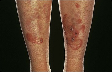
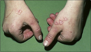
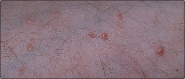
Candida albicans or bacterial infection is more common with untreated or poorly controlled diabetes. The neuropathy or arteriopathy of diabetes may result in ulcers on the feet (p. 73), and an associated secondary hyperlipidaemia can produce eruptive xanthomas (see Fig. 4). Diabetic dermopathy describes depressed pigmented scars on the shins, associated with diabetic microangiopathy. Necrobiosis lipoidica (Fig. 1), characterized by shiny atrophic yellowish–red plaques on the shins, was associated with diabetes in 65% of cases in one series, although others find a much lower figure. It affects less than 1% of all diabetics. Histologically, degenerate dermal collagen is seen with epithelioid cells and giant cells. The condition is chronic and may ulcerate. It is unresponsive to treatment. In contrast, granuloma annulare – recognized as palpable annular lesions on the hands, feet or face (Fig. 2) – is only rarely associated with diabetes and usually fades in 2 years. It must be differentiated from tinea corporis.
Thyroid disease
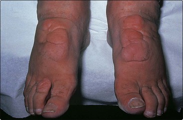
Both over- and underproduction of thyroxine result in skin and hair changes (see Table 1). Pretibial myxoedema (Fig. 3), seen in 1–10% of patients with hyperthyroidism, presents on the shins as raised erythematous plaques due to the deposition of mucin in the dermis. Topical steroids may be of benefit.
Flushing
Flushing may be physiological, drug/food induced or associated with thyrotoxicosis, but rarely with carcinoid syndrome, mastocytosis and phaeochromocytoma.
Hyperlipidaemia
Both primary (genetic metabolic defects) and secondary (associated with diabetes, hypothyroidism or the nephrotic syndrome) lipid abnormalities may produce a variety of xanthomatous deposits. These may be:
 eruptive: red–yellow papules on shoulders and buttocks (Fig. 4)
tendinous: subcutaneous nodules; hand, foot or Achilles tendons
Xanthelasma, seen as yellowish plaques on the eyelids, are not always due to a lipid abnormality. Treatment of xanthomas is usually aimed at underlying hyperlipidaemia.
Skin signs of nutritional and other internal disorders
Skin changes are common with nutritional deficiency and are not infrequent with gastrointestinal, hepatic and renal disease.
Nutritional deficiency
Protein malnutrition results in retarded growth, wasted muscles, oedema and skin changes of altered pigmentation, desquamation and ulcers with, in black Africans, dry and pale-brown/red hair. Vitamin C deficiency (scurvy) and niacin deficiency (pellagra) produce distinct lesions. In Europe, scurvy is mainly seen in elderly men who do not eat fresh fruit or vegetables. Deficiencies of other B vitamins and of iron also produce cutaneous changes (Table 2). Acrodermatitis enteropathica is a rare inherited defect of zinc absorption seen in weaned infants and cured by zinc supplements.
Table 2 Skin signs of nutritional and internal disorders
| Disorder | Skin signs |
|---|---|
| Protein malnutrition | Pigmentation, dry skin, oedema, pale-brown/orange hair |
| Iron deficiency | Alopecia, koilonychia, itching, angular cheilitis |
| Scurvy | Perifollicular purpura, bleeding gums, woody oedema |
| Pellagra | Light-exposed dermatitis and pigmentation |
| Acrodermatitis enteropathica | Perianal/perioral red scaly pustular eruption in infants, failure to thrive, diarrhoea, poor wound healing |
| Malabsorption | Dry itchy skin, ichthyosis, eczema, oedema |
| Liver disease | Pruritus, jaundice, spider naevi, palmar erythema, white nails, pigmentation, xanthomas, porphyria cutanea tarda, zinc deficiency, striae, gynaecomastia, lichen planus (p. 40) |
| Renal failure | Pruritus, pigmentation, white/red nails, dry skin with fine scaling |
| Pancreatic disease | Panniculitis, thrombophlebitis, glucagonoma syndrome |
| Crohn’s disease | Perianal abscesses, sinuses, fistulae, erythema nodosum, Sweet’s disease, necrotizing vasculitis, aphthous stomatitis, glossitis |
| Ulcerative colitis | Pyoderma gangrenosum, erythema nodosum, Sweet’s disease |
| Sarcoidosis | Nodules, plaques, erythema nodosum, dactylitis, lupus pernio, scar granulomas, small papules, nail involvement |
Gastrointestinal disease
Malabsorption and its deficiency states have accompanying skin problems that include dryness, eczema, ichthyosis, pigmentation and defects of the hair and nails. Some gut disorders show specific skin changes (see Table 2). Coeliac disease is associated with an eczema (in addition to the link of dermatitis herpetiformis with gluten enteropathy, p. 79). and both Crohn’s disease and ulcerative colitis induce various eruptions. Peutz–Jeghers syndrome (p. 75) and pseudoxanthoma elasticum (p. 93) affect both the skin and the gut. Bowel bypass surgery induces a vesiculopustular eruption.
Other internal disorders
Hepatic and renal diseases often produce troublesome itching and pigmentation. Lesions may also be related to the underlying disease process, e.g. primary biliary cirrhosis (associated with systemic sclerosis) or vasculitis.
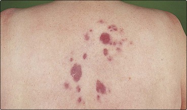
Sarcoidosis, a disorder of unknown aetiology in which granulomas commonly develop in the lung, lymph nodes, bone and nervous tissue, affects the skin in a third of cases. Cutaneous changes are variable and include brownish–red papules (typically on the face), nodules, plaques (on the limbs and shoulders, Fig. 5) and scar involvement. Lupus pernio is a particular pattern of sarcoidosis that appears as dusky-red infiltrated plaques on the nose or, occasionally, the fingers. Erythema nodosum may also result. Topical steroids have little effect. Resistant lesions may improve with intralesional steroid injection, but oral prednisolone or methotrexate is sometimes prescribed, particularly when there is progressive internal disease.
Skin changes in pregnancy
Skin changes are common in pregnancy. Pigmentation generally increases (p. 74), melanocytic naevi become more prominent, and spider naevi and abdominal striae develop. Telogen effluvium may occur in the postpartum period (p. 66). Pruritus and an urticated papular eruption (p. 76) are not uncommon, although pemphigoid gestationis (p. 78) is rare. The effect on common dermatoses is variable and unpredictable: psoriasis tends to improve, but eczema may get worse.
Skin changes in internal conditions
Endocrine and metabolic disorders, nutritional deficiencies and malabsorption are frequently associated with skin changes.
Flushing may be physiological or induced by food, drugs or conditions such as thyrotoxicosis and carcinoid syndrome.
Hyperlipidaemias are associated with a variety of xanthomata and xanthelasma, although the latter can occur with normal lipid levels.
Liver and kidney failure, in particular, are complicated by pruritus and pigmentation. Treatment is often difficult.
Inflammatory bowel disease and sarcoidosis have specific skin manifestations, often granulomatous or with cellular infiltration.
Pregnancy can be associated with increased pigmentation (e.g. melasma), an urticated papular eruption and, rarely, with the blistering eruption pemphigoid gestationis.