Urticaria and angioedema
Urticaria (hives) is a common eruption characterized by transient, usually pruritic, wheals due to acute dermal oedema from extravascular leakage of plasma. Angioedema signifies a larger area of oedema involving the dermis and subcutis. A classification is shown in Table 1.
Table 1 Classification of urticaria and angioedema
| Allergic (IgE mediated) mast cell degranulation | SystemicSkin contact | Food, drugs, latex (aerosols)Animal saliva, pollen, latex |
| Non-allergic (non-IgE mediated) mast cell degranulation | Chronic ordinaryPhysicalPharmacological | No cause identifiable (commonest subtype)Dermographism, cholinergic, cold, solar, heat, delayed pressureAspirin, opiates, non-steroidal drugs, food additives, ACE inhibitors (p. 87) |
| Autoimmune disease | Systemic lupus erythematosus (p. 80), thyroid antibodies, anti-IgE receptor antibodies, urticarial vasculitis (p. 77) | |
| Genetic | C1 esterase inhibitor deficiency (p. 77), mastocytosis (p. 116) | |
| Other | Infection, paraneoplastic, skin contact (nettle sting) |
Aetiopathogenesis
Urticaria is mediated through immune (allergic) or non-immune mechanisms. Lesions result from the release from mast cells of biologically active substances, particularly histamine, which produce vasodilatation and increased vascular permeability. Several pathways are recognized:
 IgE-mediated (type I) hypersensitivity (p. 11) is the best understood mechanism; antigen cross-links immunoglobulin (Ig) E molecules on the surface of mast cells, resulting in degranulation with release of vasoactive agents.
Complement activation can produce dermal oedema, as in hereditary angioedema or urticaria associated with circulating immune complexes.
Direct release of histamine from mast cells, in a non-immune manner, is caused by some drugs, e.g. opiates and contrast media.
Blocking of the prostaglandin pathway from arachidonic acid, by some drugs such as aspirin and non-steroidal anti-inflammatory agents, promotes urticaria by accumulating vasoactive leukotrienes.
Anti-IgE receptor (anti-FcεR1) antibodies have been shown to be present in 40% of cases of ‘chronic’ urticaria, which is capable of inducing autoimmune mast cell degranulation.
Clinical presentation
Three-quarters of cases of urticaria fall into the ‘chronic idiopathic’ or acute categories. Another 20% are due to dermographism, cholinergic urticaria or physical factors. Other causes are rare.
Chronic idiopathic urticaria
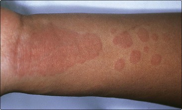
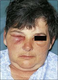
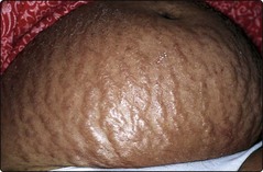
Itchy pink wheals appear as papules or plaques anywhere on the skin surface (Fig. 1). Typically, they last for less than 24 h and disappear without a trace. Wheals may be round, annular or polycyclic, and vary in diameter from a few millimetres to several centimetres. Their number ranges from a few to many appearing each day, depending on the severity of the condition. Angioedema, usually with swelling of the tongue or lips, may occur (Fig. 2). Pharmacological agents often act as provoking factors, but normally no underlying cause is found. The condition resolves spontaneously within 6 months in 50% of cases, although a minority are troubled for years.
Acute urticaria
The sudden onset of urticaria or angioedema may be due to an IgE-mediated type I reaction. The patient can often identify the offending allergen. Commonly, it is a food (e.g. egg, fish or peanuts), a drug (e.g. an antibiotic) or contact with latex (p. 125). Sometimes no cause is found.
Physical urticarias
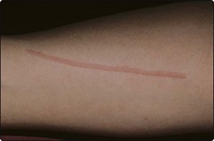
Cold, heat, sun exposure, pressure and even water can all induce urticaria at the stimulated site. Dermographism, found in 5% of normal people, describes whealing induced by firm stroking of the skin (Fig. 3). In a few individuals, it is exaggerated and symptomatic. The wheals in cholinergic urticaria are small, intensely itchy papules that appear in response to sweating, as induced by exercise, heat, emotion or spicy food. The eruption lasts for a few minutes to an hour.
C1 esterase inhibitor deficiency
Hereditary angioedema is a rare and potentially fatal autosomal dominant condition with angioedema symptoms only (no urticaria). It usually presents in childhood, with episodes of angioedema often with vomiting and abdominal pain. A deficiency of C1 esterase inhibitor allows complement activation (e.g. caused by trauma) to go unchecked, with an accumulation of vasoactive mediators. Individuals will have low levels of C4 during attacks. Acute attacks are treated with intravenous infusion of C1 esterase inhibitor concentrate. Novel synthetic peptide blockers of bradykinin B2 receptors or kallikrein can also be utilized in acute attacks. Acquired deficiency of C1 esterase inhibitor will present later in life with identical features to the inherited form, except genetic testing will be negative.
Urticarial vasculitis
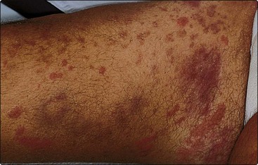
Urticarial vasculitis often has an acute onset with widespread urticarial lesions that are unusual as they persist for more than 24 h and fade leaving purpura (Fig. 4). Systemic abnormalities and low complement levels may be found.
Differential diagnosis
Urticaria is usually differentiated from other dermatoses, although pemphigoid (p. 78) or dermatitis herpetiformis (p. 79) occasionally present with an urticarial eruption. Toxic erythema and erythema multiforme (p. 82) may be urticated at first but, when the lesions persist for over 48 h, urticaria can be excluded. Facial erysipelas sometimes resembles angioedema but has a sharper margin and the patient is unwell with a fever.
Investigation
Underlying causes or provoking factors are better revealed by a careful history and examination than by laboratory tests. However, a full blood count, liver function tests, antinuclear antibody test, erythrocyte sedimentation rate (ESR) and urinalysis are often done to exclude systemic conditions (Table 1). Dermographism is demonstrated by firmly stroking the skin, and cold urticaria induced by holding an ice cube on the arm for up to 20 min. If C1 esterase inhibitor deficiency is suspected, C4 levels can be used as a screening test prior to C1 esterase inhibitor level assay.
Management
Any underlying cause should be eliminated. Provoking factors, e.g. aspirin ingestion or swimming (for those with cold urticaria), are to be avoided. Desensitization may be possible for some physical urticarias; for example, individuals with cold urticaria can build up tolerance by gradual immersion of progressively more of the body in cold water. However, the mainstay of treatment is with antihistamines.
Antihistamines
Histamine type 1 receptor blockers (H1 blockers) are usually effective. Non-sedative antihistamines, such as cetirizine 10 mg daily, fexofenadine 180 mg once daily, desloratadine 5 mg once daily or acrivastine 8 mg three times daily, are now preferred unless the sedative qualities of the older preparations are desired. The addition of an H2 blocker has only a small summative effect, if any.
Corticosteroids
Systemic steroids are very occasionally used to control severe acute urticaria or angioedema and urticarial vasculitis, but are not indicated for chronic urticaria.
Adrenaline (epinephrine)
Acute airway obstruction or anaphylactic shock is treated with adrenaline as an intramuscular injection (500 micrograms; 0.5 mL adrenaline injection 1 in 1000), repeated 5 min later if necessary. An antihistamine such as chlorphenamine (Piriton), 10–20 mg given by slow intravenous injection, is a useful adjunct. Intravenous steroids are often given, although their onset of action is delayed by several hours.
Diet
Salicylates in food aggravate chronic urticaria in up to a third of cases, and dietary azo dyes and benzoic acid preservatives produce an exacerbation in 10%. Diets low in these compounds are tried if routine measures are ineffective.
Urticaria
Urticaria is a common eruption of transient pruritic wheals that typically clear within 1 day.
Associated dermal oedema is usually the result of mast cell degranulation and the release of vasoactive amines.
No cause is usually found, but urticaria may result from histamine-releasing IgG autoantibodies, IgE-mediated allergy, physical stimuli, the pharmacological effect of drugs or food additives, or complement deficiencies.
Causative or provoking factors should be eliminated, and non-sedating antihistamines prescribed.
Systemic steroids are rarely used in treatment. Aspirin is avoided. Intramuscular adrenaline is given for an anaphylactic reaction.