Dental X-Ray Film
After completion of this chapter, the student will be able to do the following:
• Define the key words associated with dental x-ray film
• Describe in detail film composition and latent image formation
• List and describe the different types of x-ray film used in dentistry
• Define intraoral film and describe intraoral film packaging
• Identify the types and sizes of intraoral film available
• Discuss the differences between intraoral film and extraoral film
• Describe the difference between screen and nonscreen films
The dental radiographer must have a working knowledge of the dental x-ray film. The film used in dental radiography is a type of photographic film that has been adapted for dental use. An image is produced on dental x-ray film when it is exposed to radiation that has passed through teeth and adjacent structures. To avoid film-related errors that result in increased patient exposure to x-radiation, the dental radiographer must understand the composition of the x-ray film and latent image formation. In addition, the dental radiographer must be familiar with the types of film used in dental radiography as well as film storage and protection.
The purpose of this chapter is to define film composition; to detail latent image formation; to describe the types of intraoral, extraoral, and duplicating film used in dental radiography; and to discuss film storage and protection.
Dental X-Ray Film Composition and Latent Image
In dental radiography, after the x-ray beam passes through teeth and adjacent structures, it reaches the x-ray film. The dental x-ray film serves as a recording medium, or image receptor; the term image refers to a picture or likeness of an object, and the term receptor refers to something that responds to a stimulus. Images are recorded on the dental x-ray film when the film is exposed to a stimulus—specifically, energy in the form of x-radiation or light. To understand how these images result, an understanding of film composition and latent image formation is necessary.
Film Composition
The x-ray film used in dentistry has four basic components: (1) a film base, (2) an adhesive layer, (3) film emulsion, and (4) a protective layer (Figure 7-1).
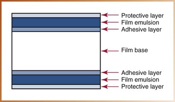
FIGURE 7-1 Schematic diagram showing construction of a typical dental x-ray film. The film emulsion is coated on both the top and the bottom surface of the polyethylene base; this allows for reduced exposure time and therefore a “faster” film. (From Miles DA, Van Dis ML, Razmus TF: Basic principles of oral and maxillofacial radiology, Philadelphia, 1992, Saunders.)
Film Base
The film base is a flexible piece of polyester plastic 0.2 mm in thickness that is constructed to withstand heat, moisture, and chemical exposure. The film base is transparent and exhibits a slight blue tint that is used to emphasize contrast and enhance image quality. The primary purpose of the film base is to provide a stable support for the delicate emulsion. The base also provides strength.
Adhesive Layer
The adhesive layer is a thin layer of adhesive material that covers both sides of the film base. The adhesive layer is added to the film base before the emulsion is applied and serves to attach the emulsion to the base.
Film Emulsion
The film emulsion is a coating attached to both sides of the film base by the adhesive layer to give the film greater sensitivity to x-radiation. The emulsion is a homogeneous mixture of gelatin and silver halide crystals.
Gelatin: The gelatin is used to suspend and evenly disperse millions of microscopic silver halide crystals over the film base. During film processing, the gelatin absorbs the processing solutions and allows the chemicals to react with the silver halide crystals.
Halide Crystals: A halide is a chemical compound that is sensitive to radiation or light. The halides used in dental x-ray film are made up of the element silver plus a halogen (bromine or iodine). Silver bromide (AgBr) and silver iodide (AgI) are two types of silver halide crystals found in the film emulsion; the typical emulsion is 80% to 99% silver bromide and 1% to 10% silver iodide. The silver halide crystals absorb radiation during x-ray exposure and store energy from the radiation (Figure 7-2).

Latent Image Formation
Silver halide crystals absorb x-radiation during x-ray exposure and store the energy from the radiation. Depending on the density of the objects in the area exposed, silver halide crystals contain various levels of stored energy. For example, the silver halide crystals on the film that are positioned behind an amalgam restoration receive almost no radiation. Amalgam is dense and absorbs the x-ray energy. As a result, the silver halide crystals are not energized. In contrast, the silver halide crystals that correspond to air space (no density) receive more radiation and are highly energized.
The stored energy within the silver halide crystals forms a pattern and creates an invisible image within the emulsion on the exposed film. This pattern of stored energy on the exposed film cannot be seen and is referred to as a latent image. The latent image remains invisible within the emulsion until it undergoes chemical processing procedures. When the exposed film with latent image is processed, a visible image results (see Chapter 9).
How does the stored energy of the silver halide crystals result in a latent image? When the x-ray photons hit the surface of the film emulsion, some silver bromide crystals are exposed and energized, while other crystals are not exposed. The silver bromide crystals exposed to x-ray photons are ionized, and the silver and bromine atoms are separated. Irregularities in the lattice structure of the exposed crystal, known as sensitivity specks, attract the silver atoms (Figure 7-3). These aggregates of neutral silver atoms are known as latent image centers (Figure 7-4). Collectively, the crystals with aggregates of silver at the latent image centers become the latent image on the film.
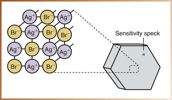
FIGURE 7-3 Each crystal in the radiographic emulsion consists of silver (Ag+) and primarily bromide (Br−) ions. Irregularities in the lattice structure form the sensitivity speck. (From Miles DA, Van Dis ML, Razmus TF: Basic principles of oral and maxillofacial radiology, Philadelphia, 1992, Saunders.)
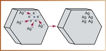
FIGURE 7-4 The sensitivity speck tends to attract free electrons (e−), which, in turn, attract positively charged silver ions (Ag). The aggregate of neutral silver atoms (Ag+) makes up the latent image center in the crystal. (From Bird DL, Robinson DS: Modern dental assisting, ed 9, Philadelphia, 2008, Saunders.)
Types of Dental X-Ray Film
Three types of x-ray film may be used in dental radiography: (1) intraoral film, (2) extraoral film, and (3) duplicating film.
Intraoral Film
An intraoral film, as defined in Chapter 6, is a film that is placed inside the mouth during x-ray exposure. An intraoral film is used to examine teeth and supporting structures.
Intraoral Film Packaging
Each intraoral film is packaged to protect it from light and moisture; the film and its surrounding packaging are referred to as a film packet. In dentistry, the terms “film packet” and “film” are often used interchangeably. Intraoral film packets are typically available in quantities of 25, 100, or 150 films per container. Film packets are packaged in convenient plastic trays or cardboard boxes that can be recycled (Figure 7-5). Boxes of intraoral film are labeled with the type of film, film speed, film size, number of films per individual packet, total number of films enclosed, and the film expiration date.
FIGURE 7-5 Intraoral film packets in a cardboard box that can be recycled. (Courtesy Carestream Health, Inc., Rochester, NY.)
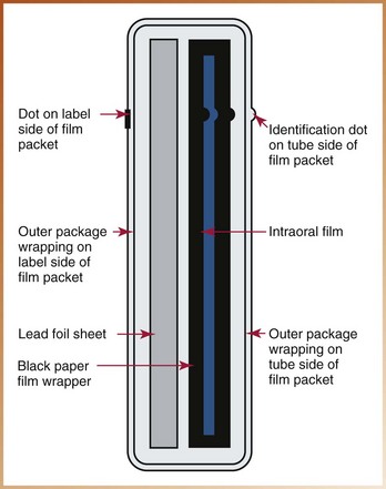
An intraoral x-ray film packet is made up of four separate items: (1) x-ray film, (2) paper film wrapper, (3) lead foil sheet, and (4) outer film wrapping (Figure 7-6).
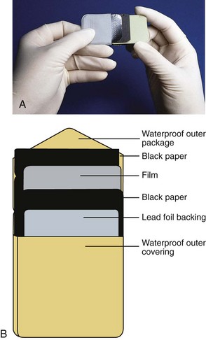
FIGURE 7-6 A, Back of an opened dental film packet. B, Diagram of A. (From Bird DL, Robinson DS: Modern dental assisting, ed 10, St Louis, 2012, Sounders.)
X-Ray Film: The intraoral x-ray film is a double-emulsion film (emulsion on both sides). Double-emulsion film is used instead of single-emulsion film (emulsion on one side) because it requires less radiation exposure to produce an image. A film packet may contain one film (one-film packet) or two films (two-film packet). A two-film packet produces two identical radiographs with the same amount of exposure necessary to produce a single radiograph. The two-film packet is used when a duplicate record of a radiographic examination is needed (e.g., for insurance claims, patient referrals, etc.).
A small, raised bump known as the identification dot is located in one corner of the intraoral x-ray film (Figure 7-7). The raised bump is used to determine film orientation. After the film is processed, the raised identification dot is used to distinguish between the left and right sides of the patient. The dot is significant in film mounting and interpretation (see Chapter 27).
Paper Film Wrapper: The paper film wrapper within the film packet is a protective sheet that covers the film and shields the film from light.
Lead Foil Sheet: The lead foil sheet is a single piece of lead foil within the film packet that is located behind the film wrapped in protective paper. The thin lead foil sheet is positioned behind the film to shield the film from backscattered (secondary) radiation that results in film fog.
The manufacturer-placed embossed pattern on the lead foil sheet is visible on a processed radiograph if the film packet is inadvertently positioned in the mouth backward and then exposed (Figure 7-8).
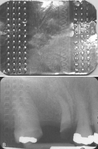
FIGURE 7-8 A, The lead foil insert in this packet has a raised diamond pattern across both ends. B, Radiograph showing the raised diamond pattern from the lead backing when the film is positioned backward in the mouth. (From Bird DL, Robinson DS: Modern dental assisting, ed 10, St Louis, 2012, Saunders.)
Outer Package Wrapping: The outer package wrapping is a soft-vinyl or paper wrapper that hermetically seals the film packet, protective paper, and lead foil sheet. This outer wrapper serves to protect the film from exposure to light and saliva.
The outer wrapper of the film packet has two sides: (1) the tube side and (2) the label side.
Tube Side: The tube side is solid white and has a raised bump in one corner that corresponds to the identification dot on the x-ray film. When placed in the mouth, the white side (tube side) of the film packet must face the teeth and the tubehead.
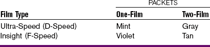
Label Side: The label side of the film packet has a flap used to open the film packet and remove the film before processing. The label side is color-coded to identify films outside of the plastic packaging container; color codes are used to distinguish one-film and two-film packets and film speeds (Table 7-1). When placed in the mouth, the color-coded side (label side) of the packet must face the tongue. It may be easier to remember that “the white side of the film faces the white teeth.”
The following information is printed on the label side of the film packet (Figure 7-9):
Intraoral Film Types
Three types of intraoral films are available: (1) periapical, (2) bite-wing, and (3) occlusal.
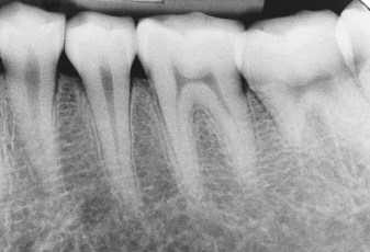
Periapical Film: The periapical film is used to examine the entire tooth (crown and root) and supporting bone (Figure 7-10). The term periapical is derived from the Greek root peri, meaning “around,” and the Latin word apex, referring to the terminal end of a tooth root. As the term suggests, this type of film shows the tip of the tooth root and surrounding structures as well as the crown.
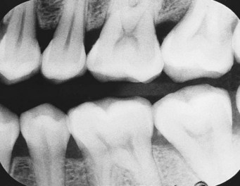
Bite-Wing Film: The bite-wing film is used to examine the crowns of both maxillary and mandibular teeth on one film (Figure 7-11). The bite-wing film is particularly useful in examining interproximal, or adjacent, tooth surfaces. The bite-wing film has a “wing,” or a tab, attached to the tube side of the film (Figure 7-12). The patient “bites” on the wing to stabilize the film. Bite-wing films may be purchased with tabs attached to the film or may be constructed from a periapical film and bite-wing loop.
Occlusal Film: The occlusal film is used for examination of large areas of the maxilla or the mandible (Figure 7-13). The occlusal film is so named because the patient “occludes,” or bites on, the entire film. The occlusal film is larger than periapical or bite-wing films.
Intraoral Film Sizes
The intraoral film is manufactured in five sizes to accommodate the varying mouth sizes of children, adolescents, and adults; the larger the number, the larger is the size of the film. Different sizes of film are used with periapical, bite-wing, and occlusal exposures (Figure 7-14).
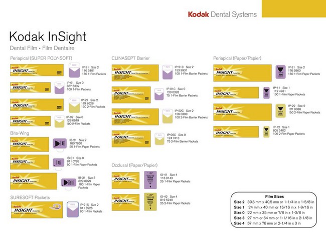
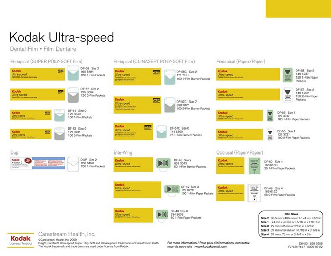
FIGURE 7-14 Film sizes for periapical, bite-wing, and occlusal films. (Courtesy Carestream Health, Inc., Rochester, NY.)
Periapical Film: Three sizes (0, 1, and 2) of the periapical film are available.
• The size 0 periapical film is the smallest intraoral film available and is used for very small children.
• The size 1 periapical film is used primarily to examine the anterior teeth in adults.
• The size 2 periapical film, also known as the standard film, is used to examine the anterior and posterior teeth in adults.
Bite-Wing Film: Three sizes (0, 2, and 3) of the bite-wing film are available. With the exception of the size 3 film, the size and shape of the bite-wing film are identical to the size and shape of the periapical film.
• The size 0 bite-wing film is used to examine the posterior teeth in small children.
• The size 2 bite-wing film is used to examine the posterior teeth in adults. This is the most frequently used bite-wing film.
• The size 3 film is longer and narrower than the standard size 2 film and is used only for bite-wing images. This bite-wing film shows all the posterior teeth on one side of the arch in one radiograph.
Intraoral Film Speed
Film speed refers to the amount of radiation required to produce a radiograph of standard density. Film speed, or sensitivity, is determined by the following:
Film speed determines how much radiation and how much exposure time are necessary to produce an image on a film. For example, a fast film requires less radiation exposure because the film responds more quickly; a fast film responds more quickly because the silver halide crystals in the emulsion are larger. The larger the crystals, the faster is the film speed.
An alphabetical classification system is used to identify film speed. X-ray films are given speed ratings ranging from A speed (the slowest) to F speed (the fastest). Only the D-speed film and the F-speed film are used for intraoral radiography; the E-speed film has been discontinued by Kodak. The American Dental Association (ADA) and the American Academy of Oral and Maxillofacial Radiology (AAOMR) currently recommend the use of the F-speed film. The F-speed film requires 60% of the exposure time of the D-speed film and has comparable image contrast and resolution. Use of the F-speed film results in less radiation exposure of the patient. The F-speed film is a faster film than the D-speed film because of the larger crystals and the increased amount of silver bromide in the emulsion. Current F-speed films not only reduce radiation dose to the patient but also provide stable contrast characteristics under various processing conditions.
The speed of a film is clearly indicated on the label side of the intraoral film packet as well as on the outside of the film box or container.
Extraoral Film
An extraoral film, as described in Chapter 6, is placed outside the mouth during x-ray exposure. Extraoral films are used to examine large areas of the skull or jaws. Common extraoral films include panoramic and cephalometric films. A panoramic film shows a panoramic (wide) view of the maxilla and the mandible and surrounding structures on a single radiograph (Figure 7-15). A cephalometric film exhibits the bony and soft tissue areas of the facial profile (Figure 7-16).
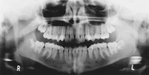
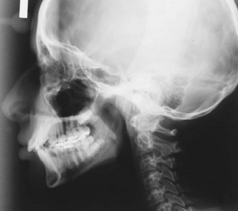
FIGURE 7-16 A cephalometric image. (From Bird DL, Robinson DS: Modern dental assisting, ed 10, St Louis, 2012, Saunders.)
Extraoral Film Packaging
Unlike intraoral films, extraoral films are designed for use outside the mouth and therefore are not enclosed in moisture-proof packets. Extraoral films used in dental radiography are available in 5 × 7–inch and 8 × 10–inch sizes as well as in the panoramic 5 × 12–inch and 6 × 12–inch sizes. Extraoral films are boxed in quantities of 50 or 100. Some manufacturers separate each piece with protective paper. Labels on the boxes of extraoral films contain information, including the type of film, film size, total number of films enclosed, and expiration date (Figure 7-17).
Extraoral Film Types
Two types of film may be used in extraoral radiography: (1) screen film and (2) nonscreen film.
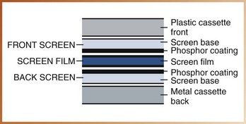
Screen Film: The majority of extraoral films are screen films. A screen film is a film that requires the use of a screen for exposure (see later discussion). A screen film is placed between two special intensifying screens in a cassette (Figure 7-18). When the cassette is exposed to x-rays, the screens convert the x-ray energy into light, which, in turn, exposes the screen film. The screen film is sensitive to fluorescent light rather than to direct exposure to x-radiation.
Films used in a screen–film combination are sensitive to specific colors of fluorescent light. Some screen films are sensitive to blue light (Kodak X-Omat and Ektamat films), whereas others are sensitive to green light (Kodak Ortho and T-Mat films). Blue-sensitive film must be paired with screens that produce blue light, and green-sensitive film must be paired with screens that produce green light. Properly matched film–screen combinations are imperative to obtain high-quality images and to minimize exposure of the patient.
Nonscreen Film: A nonscreen film is an extraoral film that does not require the use of screens for exposure. A nonscreen extraoral film is exposed directly to x-rays; the emulsion is sensitive to direct x-ray exposure rather than to fluorescent light. A nonscreen extraoral film requires more exposure time than does a screen film and is not recommended for use in dental radiography.
Extraoral Film Equipment
In extraoral radiography, screen films are used in combination with two special equipment items: (1) intensifying screens and (2) cassettes.
Intensifying Screens: An intensifying screen is a device that transfers x-ray energy into visible light; the visible light, in turn, exposes the screen film. These screens intensify the effect of x-rays on the film. With the use of intensifying screens, less radiation is required to expose a screen film, and the patient is exposed to less radiation.
In extraoral radiography, a screen film is sandwiched between two intensifying screens of matching size and is secured in a cassette (Figure 7-19). An intensifying screen is a smooth plastic sheet coated with minute fluorescent crystals known as phosphors. When exposed to x-rays, the phosphors fluoresce and emit visible light in the blue or green spectrum; the emitted light then exposes the film (Figure 7-20). As discussed in Chapter 2, one of the properties of x-rays is that they cause certain materials (e.g., phosphors) to fluoresce.
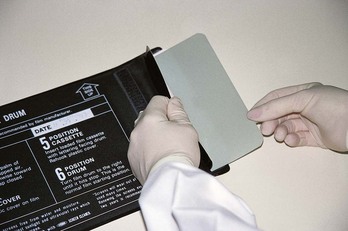
FIGURE 7-19 Cassette in open position, showing front and back intensifying screens and a piece of film. (From Bird DL, Robinson DS: Modern dental assisting, ed 10, St Louis, 2012, Saunders.)
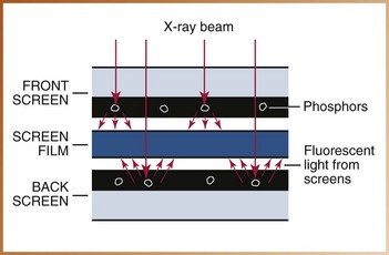
FIGURE 7-20 Phosphors in the intensifying screen emit visible light when hit by x-ray photons. Multiple visible light photons then strike and expose the film.
Conventional calcium tungstate screens have phosphors that emit blue light. The newer rare earth screens have phosphors that are not commonly found in the earth (thus “rare earth”) and emit green light. Rare earth intensifying screens are more efficient at converting x-rays into light than are calcium tungstate intensifying screens. As a result, rare earth screens require less x-ray exposure than do calcium tungstate screens and are considered faster. The use of rare earth screens means less exposure of the patient to x-radiation. Rare earth intensifying screens (Kodak Lanex Regular and Medium screens) are designed for use with green-sensitive films (Kodak Ortho and T-Mat films), whereas conventional screens (Kodak X-Omatic Regular screens) are used with blue-sensitive films (Kodak X-Omat and Ektamat films).
Cassette: A cassette is a special device that is used to hold the extraoral film and the intensifying screens. Cassettes are available in a variety of sizes that correspond to film and screen sizes. A cassette may be flexible or rigid; most cassettes are rigid, although the panoramic cassette may be flexible (Figure 7-21).
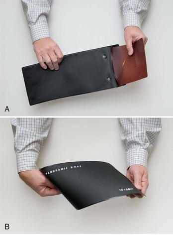

FIGURE 7-21 A and B, Close-up views of flexible 5 × 12–inch panoramic cassette. (Courtesy Instrumentarium Dental, Inc., Milwaukee, WI.) C, Rigid panoramic cassette.
A rigid cassette is more expensive but usually lasts longer than does a flexible cassette. A rigid cassette also better protects screens from damage. The film fits the rigid cassette exactly and cannot be loaded incorrectly. To load the flexible cassette properly, however, the film must be placed between the two screens and pushed to the end of the cassette.
Both rigid and flexible cassettes must be “light-tight” not only to protect the extraoral film from exposure but also to hold the intensifying screens in perfect contact with the extraoral film. Contact between the screen and the film is critical; lack of contact between screen and film results in loss of image sharpness.
A rigid cassette has a front cover and a back cover. The front cover is placed in such a way that it faces the tubehead and is usually constructed of plastic to permit the x-ray beam to pass through. The back cover is constructed of heavy metal and serves to reduce scatter radiation. Intensifying screens are installed inside the front and back covers of the cassette. The film is positioned between the two intensifying screens. Each screen exposes one side of the film.
The cassette must be marked to orient the finished radiograph; a metal letter “L” is attached to the front cover of the cassette to indicate the patient’s left side, and a metal letter “R” indicates the patient’s right side.
Duplicating Film
A duplicate radiograph is one that is identical to the original x-ray film. In dentistry, duplicate radiographs are used for patient referrals to specialists, for insurance claims, and as teaching aids. A special film, or duplicating film, is required to make a duplicate radiograph.
Description
In dental radiography, a duplicating film is a type of photographic film used to make an identical copy of an intraoral or extraoral radiograph. Unlike intraoral and extraoral films, the duplicating film is used only in a darkroom setting and is not exposed to x-rays.
When examined in the darkroom under safe light conditions, duplicating film has an emulsion on one side only. The emulsion side of the film appears dull, whereas the side without the emulsion appears shiny. The emulsion side of the film must contact the radiograph during the duplication process. (Chapter 9 describes the equipment necessary for film duplication and the duplication process.)
Film Storage and Protection
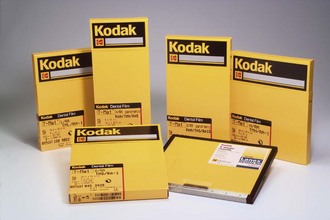
Films are adversely affected by heat, humidity, and radiation. To prevent film fog (see Chapter 9), unexposed, unprocessed films must be kept in a cool, dry place. The optimum temperature for film storage ranges from 50° F to 70° F, and the optimum relative humidity level ranges from 30% to 50%. Films must be stored in areas that are adequately shielded from sources of radiation and should not be stored in areas where patients are exposed to x-radiation. Lead-lined or radiation-resistant film dispensers and storage boxes are ideal to prevent film fog.
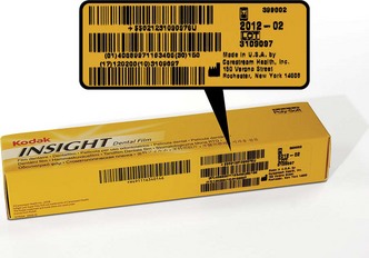
All dental x-ray films have limited shelf life. Each box or container of films is clearly labeled with an expiration date (Figure 7-22). Films must be used before the labeled expiration date. The “first-in, first-out” rule of thumb should be applied to film use; the oldest films in stock should always be used before new films.
Summary
• The dental x-ray film is an image receptor that has four basic components: (1) a film base, (2) an adhesive layer, (3) film emulsion, and (4) a protective layer.
• An image is recorded on the dental x-ray film when the film is exposed to x-radiation.
• The silver halide crystals in the film emulsion absorb the x-radiation during x-ray exposure and store the energy from the radiation. The stored energy forms an invisible pattern on the emulsion and is known as the latent image.
• When the exposed film with the latent image undergoes chemical processing, a visible image results.
• Three types of film are used in dental radiography: (1) intraoral film, (2) extraoral film, and (3) duplicating film.
• The intraoral film is placed inside the mouth and is then exposed; the extraoral film is placed outside the mouth and is then exposed; and the duplicating film is used to make a copy of a dental radiograph.
• An intraoral film packet is made up of four separate items: (1) x-ray film, (2) paper film wrapper, (3) lead foil sheet, and (4) outer package wrapping.
• Intraoral films are manufactured in five sizes (0, 1, 2, 3, 4); the larger the number, the larger is the size of the film.
• Intraoral films are available in two speeds: (1) D speed and (2) F speed. The F-speed film reduces patient exposure to radiation by 60% compared with the D speed film, with no loss of image contrast or quality.
• Extraoral films are typically screen films and require the use of intensifying screens and a cassette for exposure.
• Intensifying screens transform x-ray energy into visible light, which, in turn, exposes the screen film.
• The use of intensifying screens requires less radiation to expose a screen film and results in less radiation exposure of the patient.
• The duplicating film is a special type of photographic film used to make an identical copy of an intraoral or extraoral radiograph.
• The duplicating film is used in a darkroom setting and is not exposed to x-radiation.
• Films are adversely affected by heat, humidity, and radiation and must be stored away from sources of radiation at 50° to 70° F and with a relative humidity level of 30% to 50%.
• Dental films should always be used before the expiration date printed on the label.
Frommer, HH, Savage-Stabulas, JJ, Image formation. Radiology for the dental professional, ed 8, St Louis, Mosby, 2005.
Frommer, HH, Savage-Stabulas, JJ, Image receptors. Radiology for the dental professional, ed 8, St Louis, Mosby, 2005.
Johnson, ON, Thomson, EM, Dental x-ray films. Essentials of dental radiography for dental assistants and hygienists, ed 8, Pearson, Upper Saddle River, NJ, 2007.
Ludlow, JB, et al. Characteristics of Kodak Insight, an F-speed intraoral film. Oral Surg Oral Med Oral Pathol Oral Radiol Endod. 2001;91(1):120–129.
Miles, DA, Van Dis, ML, Jensen, CW, Ferretti, A, Film processing and quality assurance. Radiographic imaging for the dental team, ed 4, St Louis, Saunders, 2009.
White, SC, Pharoah, MJ, X-ray film, intensifying screens, and grids. Oral radiology: Principles and interpretation, ed 6, St Louis, Mosby, 2009.
Fill in the Blank
1. The component of an x-ray film described as “a thin transparent coating that is placed over the emulsion” is termed:
________________________________________________________________
________________________________________________________________
2. The component of the x-ray film described as “a flexible piece of plastic that withstands heat, moisture, and chemical heat” is termed:
________________________________________________________________
________________________________________________________________
3. The chemical compounds that change when exposed to radiation or light are termed:
________________________________________________________________
________________________________________________________________
4. The invisible pattern of stored energy on the exposed film is termed:
Identification
For questions 5 to 11, identify the items indicated on the intraoral film packet illustrated in Figure 7-23.
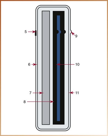
FIGURE 7-23
Multiple Choice
____12. Dental x-ray film that is placed inside the mouth and used to examine the teeth and supporting structures is termed:
____13. The identification dot on the intraoral film is significant because:
a. the dot indicates the patient’s right or left side
b. the dot determines film orientation
____14. One advantage of a film with an emulsion coating on both sides (double-emulsion film) is that:
a. the film requires less radiation exposure to make an image
b. the image produced is less distorted
____15. The purpose of a lead foil sheet in the film packet is:
a. to protect the film from primary radiation
b. to protect the film from saliva
c. to protect the film from backscattered radiation
d. to distinguish between the patient’s right and left sides
____16. Which of the following is not found on the label side of the film packet?
____17. Which of the following film sizes is known as the standard film?
____18. Which of the following is the largest intraoral film size?
____19. The film characteristic that is “the amount of radiation needed to produce a radiograph of standard density” is:
____20. The speed of a film is determined by the size of the silver halide crystals in the emulsion. Identify the true statement:
a. The larger the crystals, the faster is the film speed.
b. The larger the crystals, the slower is the film speed.
____21. A film that is placed outside the mouth during x-ray exposure is termed a(n):
____22. A screen film is more sensitive to fluorescent light than to direct exposure to x-rays.
____23. Nonscreen extraoral film is commonly used in extraoral radiography.
____24. The device that transfers x-ray energy into visible light is termed a:
____25. The intensifying screen that emits green light and must be used with green-sensitive film is a:
____26. The device used to hold the extraoral film and intensifying screens is termed a:
____27. Which of the following statements is true?
a. Cassettes are available in sizes that correspond to film and screen sizes.
b. A flexible cassette is more expensive than is a rigid cassette.
c. Film can be loaded incorrectly in the rigid cassette.
d. Film cannot be loaded incorrectly in the flexible cassette.
____28. Which of the following results if the intensifying screens are not in perfect contact with the screen film?
____29. Which of the following statements about the duplicating film is false?
a. It is not exposed to x-rays.
b. It is used in the darkroom.
____30. Identify the ideal temperature and humidity levels for film storage: