Dental X-Ray Equipment
After completion of this chapter, the student will be able to do the following:
• Define the key words associated with dental x-ray equipment
• Discuss the regulation of dental x-ray machines at the federal, state, and local levels
• Recognize dental x-ray machines used for intraoral and extraoral films
• Identify the component parts of the dental x-ray machine
• Describe the purpose and use of dental x-ray film holders and devices
• Identify commonly used dental x-ray film holders and devices
The dental radiographer must be familiar with dental x-ray equipment and dental x-ray film holders and beam alignment devices. With each passing year, digital imaging is becoming more prevalent and widely used. Because the majority of dental offices still use conventional radiography methods, the information presented in this section of the textbook is predominantly devoted to dental film and processing equipment. The purpose of this chapter is to introduce the dental radiographer to a variety of intraoral and extraoral dental x-ray machines, to detail the component parts of x-ray machines, and to describe the more common dental x-ray film holders and beam alignment devices.
Dental X-Ray Machines
A variety of intraoral and extraoral dental x-ray machines are available for diagnostic purposes. Dental x-ray machines vary in both design and operation. The dental radiographer must have a clear understanding of the operating procedures for the specific equipment that is used in the dental office to avoid improper exposure of patients and dental personnel.
Performance Standards
Before 1974, no federal standards existed for the manufacture of dental x-ray machines. All dental x-ray machines manufactured after 1974 must, however, meet specific federal guidelines regulating diagnostic equipment performance standards. The federal government regulates the manufacture and installation of dental x-ray equipment. State and local governments regulate how dental x-ray equipment is used and dictate codes that pertain to the use of x-radiation. Depending on state and local radiation safety codes, dental equipment must be inspected and monitored periodically. A fee is typically charged for such an inspection.
Types of Machines
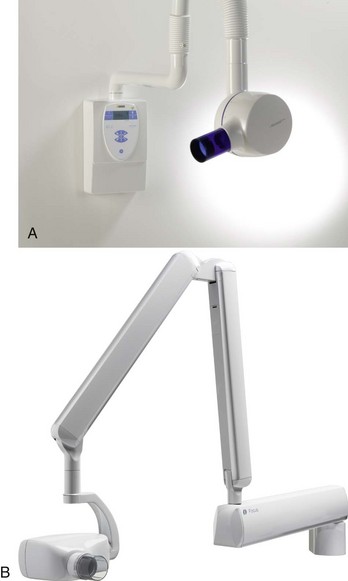
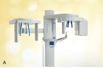
Dental x-ray machines may be used to expose intraoral receptors or extraoral receptors. Some machines are used only for intraoral exposures (Figure 6-1), whereas others are limited to extraoral exposures (Figure 6-2). A variety of dental x-ray machines are available from different manufacturers. (See Chapter 25 for examples of digital radiography units.)

Component Parts
As detailed in Chapter 2, the typical intraoral dental x-ray machine features three component parts: (1) tubehead, (2) extension arm, and (3) control panel.
Tubehead
The tubehead, or tube housing, contains the x-ray tube that produces dental x-rays (Figure 6-3). Extending from the tubehead opening is the position-indicating device (PID), or cone. The PID may be circular or rectangular in shape and restricts the size of the x-ray beam.

Extension Arm
The extension arm suspends the x-ray tubehead, houses the electrical wires, and allows for movement and positioning of the tubehead.
Control Panel
The control panel, which allows the dental radiographer to regulate the x-ray beam, is plugged into an electrical outlet and appears as a console or cabinet. A control panel may be mounted on a floor pedestal, a wall support, or a remote wall location outside the dental operatory. A single control panel may be used to operate more than one x-ray unit located in adjacent rooms. The control panel consists of (1) an on-off switch and indicator light, (2) an exposure button and exposure light, (3) a control device for time, and (4) with some units, control devices for kilovoltage peak and milliamperage (Figure 6-4).
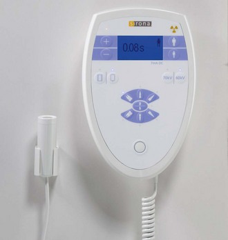
On-Off Switch: The on-off switch must be placed in the “on” position to operate the dental x-ray equipment. An indicator light is illuminated when the equipment is turned on.
Exposure Button: The exposure button activates the machine to produce x-rays. The dental radiographer must firmly depress the exposure button until the preset exposure time is completed. As a visible sign that x-rays are being produced, an exposure light on the control panel is illuminated during x-ray exposure. In addition, a beep sounds during x-ray exposure as an audible signal that x-rays are being produced. The exposure light turns off and the beep stops when the x-ray exposure is completed.
Control Devices: The control devices that regulate the x-ray beam include the timer and the kilovoltage peak (kVp) and milliamperage (mA) selectors. The timer determines the length of exposure time in seconds or impulses. The kVp and mA selectors permit the dental radiographer to adjust and set the correct kilovoltage peak and milliamperes. Some dental x-ray units have preset programmable settings for the various anatomic areas of the maxilla and the mandible or for different sizes of patients, thus eliminating the need to set the individual controls of kVp, mA, and time.
Dental X-Ray Film Holders and Beam Alignment Devices
A film holder is a device used to hold and align intraoral dental x-ray films in the mouth. Film holders eliminate the need for the patient to stabilize the film. With certain intraoral techniques (e.g., paralleling technique) the use of a film-holding device is required. Specific intraoral techniques and film-holding devices are discussed in Chapters 17, 18, and 19.
A beam alignment device is used to help the dental radiographer position the PID in relation to the tooth and the film. For use in conjunction with a beam alignment device, a collimating device, which is a metal plate with an opening, can be used to restrict the size of the beam.
Types of Film Holders
Intraoral film holders are commercially available from a number of manufacturers. The simplest film holder is a disposable Styrofoam bite-block with a backing plate and a slot for film retention; examples include XCP Bite-Block and Stabe Bite-Block (Rinn Corporation) (Figure 6-5). Molded-plastic devices that can be sterilized are also available, including EEZEE-Grip, formerly named Snap-A-Ray. EEZEE-Grip is a double-ended instrument that holds the film between two serrated plastic grips that can be locked in place (Figure 6-6). Other film-holding products include EndoRay and Uni-bite (Figure 6-7).
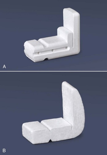
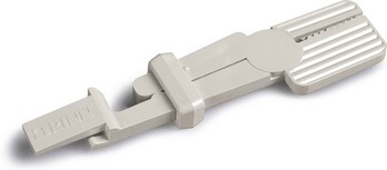
FIGURE 6-6 Snap-A-Ray film holder. The film can be positioned for anterior areas and most posterior areas. (Courtesy Dentsply Rinn, Elgin, IL.)
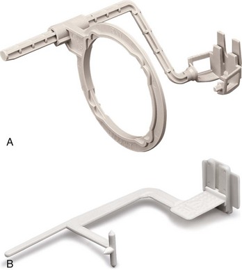
FIGURE 6-7 A, The EndoRay film holder is used during root canal procedures. It fits around rubber dam clamps and allows space for files to protrude from the tooth. B, The Uni-bite is a universal film holder that can be used with the bite-wing technique or the long-cone paralleling technique for film exposure. (Courtesy Rinn Corporation, Elgin, IL.)
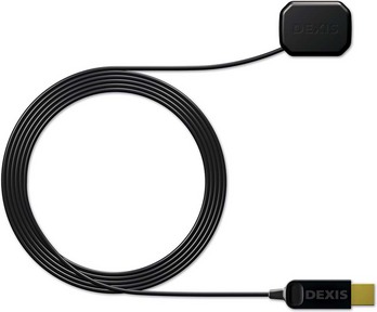
In digital radiography, a sensor is held in place by a bite-block attachment or by devices that aim the beam and sensor accurately. Beam alignment devices must be used to stabilize and secure the sensor (Figure 6-8).
Types of Beam Alignment Devices
Beam alignment devices and collimating devices, which are available from a number of manufacturers, are used to indicate the PID position in relation to the tooth and film. Metal beam alignment and collimating devices include the Precision film holders (Masel Orthodontics), which feature four metal collimating shields and film-holding devices that restrict the size of the x-ray beam to the size of the film (Figure 6-9).
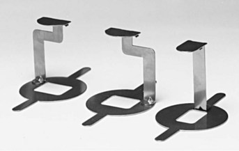
FIGURE 6-9 Precision film holders restrict the size of the x-ray beam to the size of the film. (Courtesy Masel Orthodontics.)
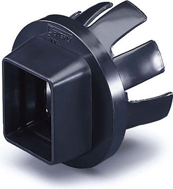
The XCP and BAI beam alignment devices (Rinn) feature plastic bite-blocks, plastic aiming rings, and metal indicator arms (Figure 6-10). To reduce the amount of radiation a patient receives, a snap-on universal collimator can be added to the plastic XCP and BAI rings (Figure 6-11).
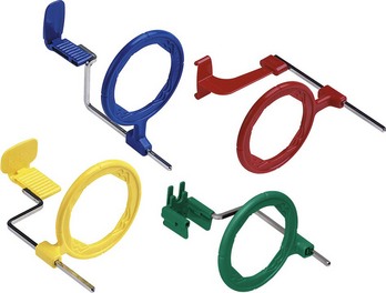
FIGURE 6-10 XCP instruments. The blue instruments are used in the anterior region, the yellow instruments are used in the posterior region, the red instruments are used in the bite-wing technique, and the torquoise instruments are used for endodontic procedures. (Courtesy Dentsply Rinn Corporation, Elgin, IL.)
Summary
• The dental radiographer must be familiar with how x-ray equipment, film holders, and beam alignment devices are used in dentistry.
• The typical intraoral dental x-ray machine consists of three component parts: (1) tubehead, (2) extension arm, and (3) control panel.
• A film holder is used to stabilize an intraoral film.
• A beam alignment device helps the dental radiographer position the position-indicating device in relation to the tooth and the film.
• A collimating device is used with the beam alignment device to further restrict the size of the x-ray beam.
Frommer, HH, Savage-Stabulas, JJ, Operator protection. Radiology for the dental professional, ed 9, St Louis, Mosby, 2011.
Frommer, HH, Savage-Stabulas, JJ, Patient protection. Radiology for the dental professional, ed 9, St Louis, Mosby, 2011.
Johnson, ON, Thomson, EM, The periapical examination. Essentials of dental radiography for dental assistants and hygienists, ed 8, Pearson, Upper Saddle River, NJ, 2007.
Multiple Choice
____1. No federal standards existed for dental x-ray machines manufactured before the year:
____2. Dental receptors placed inside the mouth are termed:
____3. The component part of the dental x-ray machine that contains the x-ray tube is termed the:
____4. The component part of the dental x-ray machine that allows movement and positioning of the tubehead is termed the:
____5. The dental radiographer can regulate the x-ray beam (kilovoltage peak, milliamperage, time) through the use of the:
____6. An instrument that is used to help the dental radiographer position the PID in relation to the tooth and receptor is the:
____7. A device that is used to stabilize an intraoral receptor is a:
____8. A metal instrument that is used to restrict the size of the x-ray beam to the size of an intraoral receptor is the: