Chapter 3 Skeletal system
The skeletal system is the ‘framework’ upon which the body is built – it provides support, protection and enables the animal to move (Fig. 3.1). The joints are considered to be an integral part of the skeleton. The skeletal system is made of the specialised connective tissues, bone and cartilage.
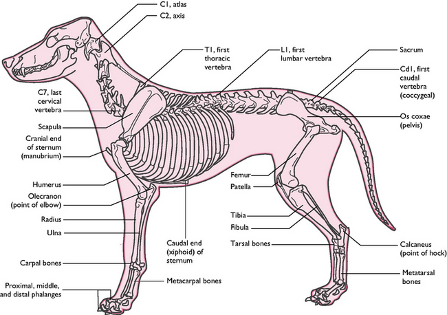
Fig. 3.1 The skeleton of the dog showing the major bones.
(With permission from Colville T, Bassett JM 2001 Clinical anatomy and physiology for veterinary technicians. Mosby, St Louis, MO, p 102.)
The functions of the skeletal system are:
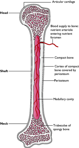
Bone structure and function
Bone shape
Bones can be categorised according to their shape:
Some specialised types of bone are:
Development of bone
The process by which bone is formed is called ossification and there are two types: intramembranous and endochondral ossification. The cells responsible for laying down new bone are called osteoblasts; the cells that destroy or remodel bone are called osteoclasts.
Endochondral ossification
The process of endochondral ossification (Fig. 3.3) is as follows:
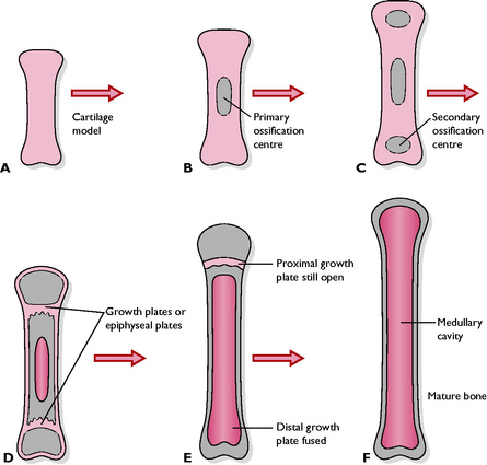
Fig. 3.3 Stages of endochondral ossification. A A cartilage model of the bone exists in the embryo. B Ossification begins from the primary ossification centre in the shaft (diaphysis). C Secondary ossification centres appear in the ends of the bone (epiphyses). D Ossification continues in primary and secondary centres; osteoclasts start to break down bone in the shaft, creating the marrow cavity. E The first growth plate fuses and growth is now only possible at the proximal growth plate; the medullary cavity extends into the epiphysis. F The proximal growth plate fuses and bone growth ceases.
Rickets is a disease of young growing animals caused by a nutritional deficiency of vitamin D or phosphorus. The bones fail to calcify and become bowed, and the joints appear swollen because of enlargement of the epiphyses. Any animal kept permanently inside is at risk of developing rickets because vitamin D is formed by the action of ultraviolet light on the skin.
The skeleton
The skeleton (Fig. 3.1) can be divided into three parts.
When studying the anatomy of the skeletal system it is helpful to understand the terms that are used to describe the various projections, passages and depressions that are found on and within bones.
The axial skeleton
The skull
The bones of the head include the skull, nasal chambers, mandible or lower jaw and hyoid apparatus. The functions of the skull are:
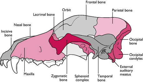
Cranium
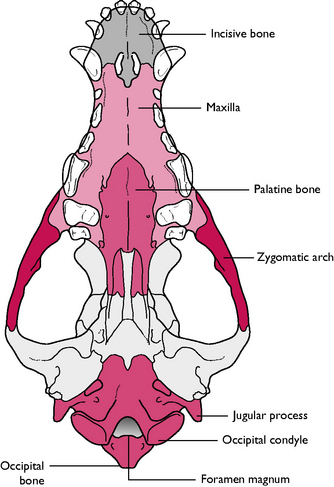
The caudal part of the skull that provides the bony ‘case’ in which the brain sits is called the cranium (Figs 3.4, 3.5). The bones of the cranium include:
Nasal chambers
The most rostral part of the skull carries the nasal chamber, the sides of which are formed by the maxilla and the roof by the nasal bone. The nasal chamber is divided lengthways into two by a cartilaginous plate called the nasal septum. Each of the chambers is filled with delicate scrolls of bone called the nasal turbinates or conchae. These are covered in ciliated mucous epithelium (see Ch. 8). At the back of the nasal chamber, forming a boundary between the nasal and cranial cavities, is the ethmoid bone. In the centre of this bone is the cribriform plate – a sieve-like area perforated by numerous foramina through which the olfactory nerves pass from the nasal mucosa to the olfactory bulbs of the brain (see Ch. 5).
The roof of the mouth is called the hard palate and is formed from three bones on the ventral aspect of the skull: the incisive bone or premaxilla, part of the maxilla and the palatine. The incisive bone is the most rostral and carries the incisor teeth (Fig. 3.5).
Many of the bones of the skull are joined together by fibrous joints called sutures (p 41). Sutures are firm and immovable joints but allow for expansion of the skull in a growing animal.
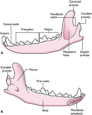
Mandible
The mandible or lower jaw is comprised of two halves or dentaries, joined together at the chin by a cartilaginous joint called the mandibular symphysis. Each half is divided into a horizontal part, the body, and a vertical part, the ramus (Fig. 3.6). The body carries the sockets or alveoli for the teeth of the lower jaw. The ramus articulates with the rest of the skull at the temporomandibular joint via a projection called the condylar process. A rounded coronoid process, which projects from the ramus into the temporal fossa, is the point to which the temporalis muscle attaches (Fig. 3.6). There is a depression on the lateral surface of the ramus, the masseteric fossa, in which the masseter muscle lies.
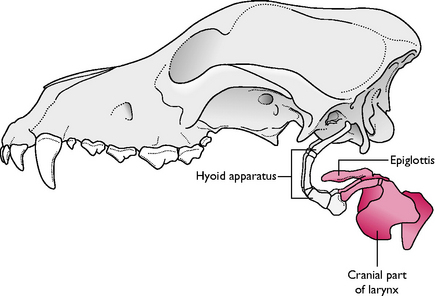
Hyoid apparatus
The hyoid apparatus lies in the intermandibular space and consists of a number of fine bones and cartilages joined together in an arrangement that resembles a trapeze (Fig. 3.7). The hyoid apparatus is the means by which the larynx and tongue are suspended from the skull. The apparatus articulates with the temporal region of the skull in a cartilaginous joint.
Skull shapes
The shape of the skull varies between species. In the domestic cat the skull is much more rounded or ‘apple-shaped’ than it is in the dog, and there is little difference between the various cat breeds. In the dog, although the basic anatomy remains the same, the overall appearance differs greatly between the different breeds (Fig. 3.8).
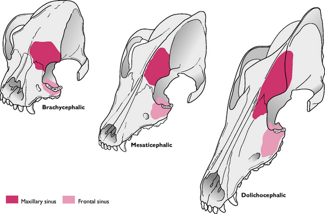
Fig. 3.8 The three basic shapes of the dog skull seen in common breeds. Also shows the positions of the maxillary and frontal sinuses.
Three morphological forms of dog skull are recognised:
The shape of the skull, particularly in brachycephalic breeds such as the bulldog, can lead to a variety of clinical conditions, e.g. snoring respiration, difficulty in breathing. The relative size of the head can also cause difficulties with whelping. Breeds with a mesaticephalic shape of skull are much less likely to exhibit these conditions.
The vertebrae
The vertebral column is comprised of a number of bones arranged in a series along the midline of the body and extending from the base of the skull to the tip of the tail. The vertebrae are divided into regions depending upon their position in the body:
Each species has a characteristic number of vertebrae within each region, which is written as a formula. In the dog and cat this formula is: C7, T13, L7, S3, Cd20–23 (Fig. 3.1).
The functions of the vertebral column are:
Basic plan of a vertebra
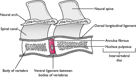
The vertebrae within all regions of the vertebral column are basically similar in structure, although each region shows slight differences related to function. A typical vertebra (Fig. 3.9) consists of a roughly cylindrical ventral body with a convex cranial end and a concave caudal end. This arrangement enables the bodies to fit together, creating a flexible rod. The body is topped by an arch, called the vertebral or neural arch, which forms a tunnel-like vertebral foramen through which the spinal cord passes. When linked together, the vertebral foramina constitute the spinal canal.
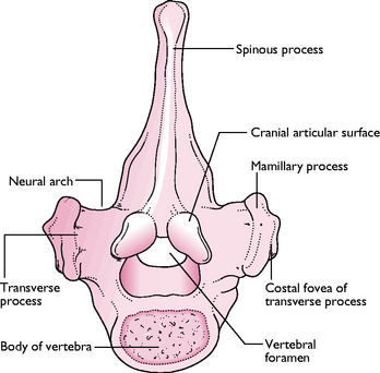
The neural arch has a dorsal projection called the spinous process or neural spine, which varies in height and size from one region of the vertebral column to another. On either side of the vertebra there are laterally projecting processes, the transverse processes, which also vary in shape and size between regions. The transverse processes divide the muscles of the vertebral column into dorsal or epaxial and ventral or hypaxial divisions (see muscles of the trunk, p 48).
On the cranial and caudal edges of the arch of each vertebra there are cranial and caudal articular processes. These form a synovial joint with those of the adjacent vertebrae, creating a flexible rod running in the midline of the body. There are also a number of other processes, which are sites for muscle attachment. Lying between the bodies of each pair of vertebrae is a fibrocartilaginous intervertebral disc, which acts as a ‘shock absorber’, preventing damage to the spinal cord (Fig. 3.10). The intervertebral disc is composed of a tough fibrous connective tissue outer area, called the annulus fibrosus, and a core of gelatinous material called the nucleus pulposus.
The vertebrae are linked by ligaments that run between them and they articulate with one another by two types of joint:
In the case of a ‘slipped disc’, the annulus fibrosus ruptures and the pulpy centre of the disc protrudes outward. This puts pressure either on the spinal cord or on the associated nerves leaving the cord, causing the animal to show symptoms ranging from pain to paralysis due to the loss of motor function.
Regional variations
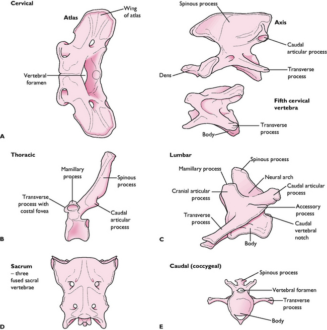
Fig. 3.11 Variations in the shape of vertebrae. A Cervical. B Thoracic. C Lumbar. D Sacral (the three sacral vertebrae are fused into one bone). E Caudal/coccygeal.
The synovial joint between the atlas (C1) and the occipital condyles of the skull allows nodding movements of the head, and the synovial joint between the atlas and axis (C1–C2) allows a pivotal movement so that the head can turn in all directions.
The ribs and sternum
The ribs form the walls of the bony thoracic cage that protects the organs of the chest. There are 13 pairs of ribs in the dog and cat, which articulate with the thoracic vertebrae (Fig. 3.12). A rib is a flat bone consisting of compact bone on the outside packed with cancellous bone on the inside. Each rib has a bony dorsal part and a cartilaginous ventral part – the costal cartilage. The most dorsal part of the bony rib has two projections: the head, which articulates with the costal fovea of the vertebra, and the tubercle or neck, which articulates with the transverse fovea of the appropriate thoracic vertebra.
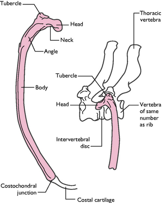
Fig. 3.12 Structure of the canine rib. A Caudal view. B Lateral view showing articulation with a thoracic vertebra.
(With permission from Colville T, Bassett JM 2001 Clinical anatomy and physiology for veterinary technicians. Mosby, St Louis, MO, p 112.)
During parturition, under the influence of the hormone relaxin, the sacroiliac ligament relaxes and softens so that the pelvis can stretch, enabling the fetuses to pass out through the birth canal.
The costal cartilage articulates with the sternum, either directly or indirectly. The first eight pairs of ribs attach directly to the sternum and are called the sternal ribs. The ribs from pairs 9–12 are called asternal or ‘false’ ribs, and they attach via their costal cartilages to the adjacent rib, forming the costal arch. The last ribs (pair 13) have no attachment at their cartilaginous ends, which lie free in the abdominal muscle – this pair are called the ‘floating’ ribs. The space between each successive pair of ribs is called the intercostal space and is filled by the intercostal muscles of the trunk (Fig. 3.13).
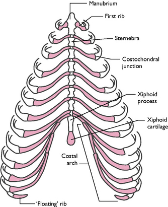
Fig. 3.13 The rib cage of the dog, showing sternal, asternal and ‘floating’ ribs.
(With permission from Colville T, Bassett JM 2001 Clinical anatomy and physiology for veterinary technicians. Mosby, St Louis, MO, p 112.)
The sternum forms the floor of the thoracic cage (Fig. 3.13) and is composed of eight bones, the sternebrae, and the intersternebral cartilages. The most cranial sternebra is the manubrium, which projects in front of the first pair of ribs and forms part of the cranial thoracic inlet. Sternebrae 2–7 are short cylindrical bones. The last sternebra is longer and dorsoventrally flattened and is called the xiphoid process. Attached to the xiphoid process and projecting caudally is a flap of cartilage called the xiphoid cartilage. The linea alba attaches to this. Between each pair of sternebrae are cartilaginous discs called the intersternebral cartilages.
The appendicular skeleton
The appendicular skeleton is composed of the pectoral (or fore) limb and the pelvic (or hind) limb and the shoulder and pelvic girdles that attach these to the body. The forelimb has no bony connection to the trunk, only being attached by muscles. This absorbs the ‘shock’ at the point when the limb takes the animal’s weight in four-legged animals or running quadrupeds. This differs from primates, which generally walk on their hind legs and so have evolved a pectoral girdle with a clavicle. However, the hindlimb does have a bony articulation in the pelvic girdle, which forms the platform for the muscles that provide the propulsive force as the animal is running.
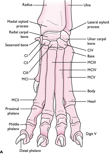
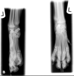
Bones of the forelimb
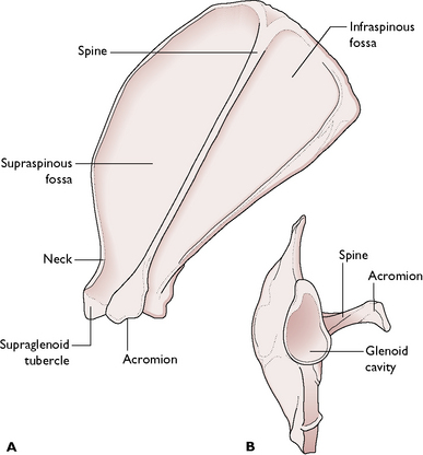
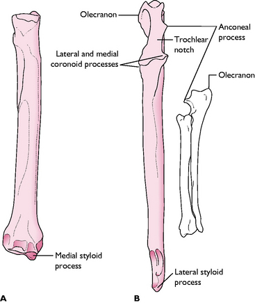
The bones of the forelimb (Fig. 3.1) are:
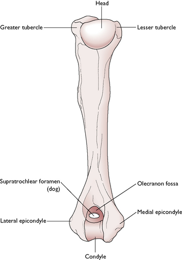
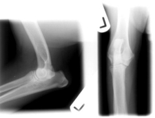
The shaft of the humerus has a slight twist on it. If the humerus is broken, the resulting fracture is often in the form of a spiral and may affect the radial nerve that runs within the spiral causing temporary or permanent radial nerve paralysis. This is indicated by the animal ‘knuckling’ over on its lower forelimb.
Elbow dysplasia is a common condition of the heavier breeds of dog, e.g. Newfoundland, St Bernard, Rottweiler, Basset hound. It encompasses a number of developmental conditions, such as an ununited anconeal process and detached olecranon process, which result in instability of the elbow joint, leading to osteoarthritis. The disease can be inherited and there is a BVA/KC scheme to identify affected individuals and to help breeders select the most suitable dogs for breeding.
Bones of the hindlimb
The bones of the hindlimb (Fig. 3.1) are:
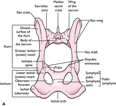
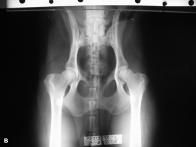
Fig. 3.19 A Bones and bony features of the dog pelvis. B Ventrodorsal view of the canine pelvis.
(Courtesy of Richard Aspinall.)
Hip dysplasia is an inherited condition that affects a range of larger breeds of dog, e.g. Labradors, Retrievers, German Shepherds. It is caused by malformation of the femoral head and/or a shallow or malformed acetabulum, resulting in subluxation of the hip joint leading to osteoarthritis. There is a BVA/KC scheme to help identify affected dogs and to advise breeders on the choice of breeding stock.
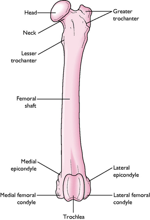
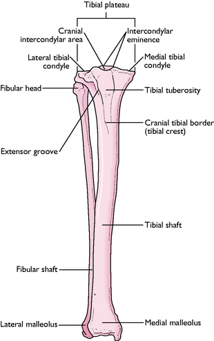
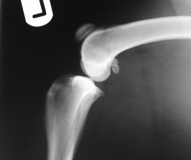
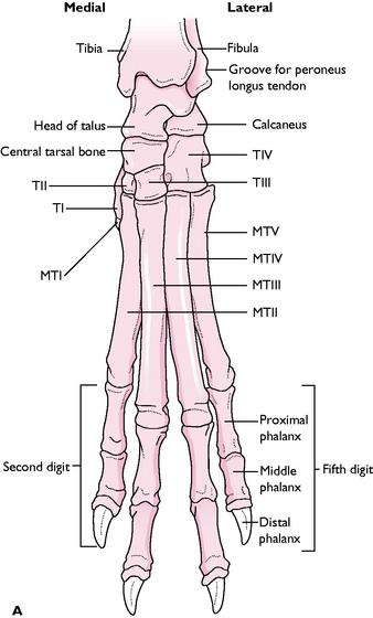
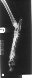
Fig. 3.23 A The dog tarsus or hindpaw. TI, TII, etc. = tarsal bones 1, 2, etc. MT = metatarsal bones. B Lateral and dorsoplantar views of the canine tarsus and distal hindlimb.
(Courtesy of Panagiotis Mantis.)
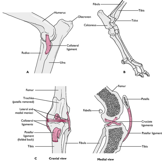
Fig. 3.25 A The elbow joint, lateral view. B The tarsus or hock joint, lateral view. C The stifle joint, cranial and medial views.
In some small breeds of dog, e.g. Yorkshire Terrier, the patella may slip out of place, causing extreme pain and difficulty in extending the stifle joint. This is an inherited condition and is due to malpositioning of the tibial crest or too shallow a trochlear groove on the distal end of the femur.
Splanchnic skeleton
This is composed of the splanchnic bones. A splanchnic bone is a bone that develops in soft tissue and is unattached to the rest of the skeleton. The only example of a splanchnic bone in the dog and cat is the bone of the penis, the os penis. The urethra lies in the urethral groove, on the ventral surface of the os penis in the dog. In the cat the urethral groove is on the dorsal surface of the os penis, because of the different orientation of the penis (see Ch. 11).
The cow has a splanchnic bone in its heart, called the os cordis, while birds have splanchnic bones forming a rim around the eye to provide strength to the large eyeball (see Ch. 13).
Joints
When one bone connects to another they form an articulation, also known as an arthrosis or joint. Joints allow variable degrees of movement and can be categorised into one of three groups:
Fibrous joints
Fibrous joints are immovable joints and the bones forming them are united by dense fibrous connective tissue, e.g. in the skull fibrous joints unite the majority of the component bones and are called sutures. The teeth are attached to the bony sockets in the jaw bone by fibrous joints.
Fibrous joints are also classed as synarthroses, i.e. a type of joint that permits little or no movement. Some cartilaginous joints also fall into this category.
Cartilaginous joints
Cartilaginous joints allow limited movement or no movement at all and are united by cartilage, e.g. the pubic symphysis connecting the two hip bones and the mandibular symphysis joining the two halves of the mandible. Both these joints are also classed as synarthroses.
Some cartilaginous joint may also be classed as amphiarthroses, which allow some degree of movement between the bones, e.g. between the bodies of the vertebrae, allowing for limited flexibility of the spinal column.
Synovial joints
Synovial joints or diarthroses allow a wide range of movement. In synovial joints, the bones are separated by a space filled with synovial fluid known as the joint cavity (Fig. 3.24). A joint capsule surrounds the whole joint; the outer layer consists of fibrous tissue, which serves as protection, and the joint cavity is lined by the synovial membrane, which secretes synovial fluid. This lubricates the joint and provides nutrition for the hyaline articular cartilage covering the ends of the bone. Synovial fluid is a straw-coloured viscous fluid that may be present in quite large quantities in large joints, especially in animals that have a lot of exercise.
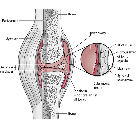
Some synovial joints may have additional stabilisation from thickened ligaments within the fibres of the joint capsule. These are most commonly found on either side of the joint, where they are called collateral ligaments. However, other synovial joints have stabilising ligaments attached to the articulating bones within the joint – these are known as intracapsular ligaments and examples include the cruciate ligaments within the stifle joint (Fig. 3.25).
A few synovial joints possess one or more intraarticular fibrocartilaginous discs or menisci within the joint cavity. These are found in the stifle joint – it has two crescent shaped menisci – and in the temporomandibular joint between the mandible and the skull. These structures help to increase the range of movement of the joint and act as ‘shock absorbers’, reducing wear and tear.
Synovial joints allow considerable freedom of movement between the articulating bones, the extent of which depends upon the type of synovial joint. The movement allowed by a synovial joint may be in a single plane only, or in multiple planes.
The range of movements that are possible in synovial joints is:
Synovial joints can be further classified into subcategories based upon the types of movement that they allow (Table 3.1).
Table 3.1 Properties of different synovial joint types
| Type of synovial joint’ | Description | Example of joint |
|---|---|---|
| Plane/gliding | Allows sliding of one bony surface over the other | Joints between the rows of carpal and tarsal bones |
| Hinge | Allows movement in one plane only, i.e. flexion and extension | Elbow; stifle |
| Pivot | Consists of a peg sitting within a ring; allows rotation | Atlantoaxial joint (between C1 and C2) |
| Condylar | Consists of a convex surface (condyles) that sits in a corresponding concave surface; allows movement in two planes (flexion, extension and overextension) | Hock or (tarsus) |
| Ball and socket | Consists of a rounded end or ball, sitting within a socket or cup; allows a great range of movement | Hip; shoulder |