SHEEP KEDS

The sheep ked, Melophagus ovinus, is a wingless fly approximately 6 to 7 mm in diameter with a ticklike appearance. Sheep keds have a worldwide distribution. They primarily parasitize sheep and occasionally goats that are kept under poor management conditions.152,175

The entire life cycle is spent on the host. Females live 4 to 5 months, laying 10 to 15 larvae individually that are cemented to the wool or hair. The larvae pupate in 12 hours, and the adult emerges 3 weeks later. Adults feed on blood and do not survive longer than a few days off the host. Transmission is by direct contact, and infestation is more common in the winter months.152,153,175

Clinical signs of infestation include pruritus with subsequent self-trauma, wool stains from the flies’ fecal material, and in severely parasitized animals, anemia. Multiple firm nodules (cockles) develop as a result of repeated puncture of the skin as the keds feed.152,153 Infestation results in economic loss from a reduction in dressed carcass weights of lambs, reduced clean dry weight of fleece, wool staining, and reduced value of sheep skins because of nodular defects.152,175,176 Diagnosis is based on demonstration of the parasite.

Therapy involves shearing all sheep in the affected flock, followed by two topical applications of malathion, diazinon, or coumaphos at 14- to 21-day intervals to kill emerging adults.152 Because the larvae are attached to the wool or hair some distance above the skin surface, many larvae and pupae are removed by shearing.153,176 Recently, imidacloprid was found to be effective in killing the parasite.177 All new animals should be isolated and treated before introduction to prevent reinfestation. A control program includes annual treatment of the entire flock.

  Page 1327 

CUTANEOUS HABRONEMIASIS (EQUINE SUMMER SORE)

Habronemiasis (“summer sore”) is a granulomatous disease caused by the deposition of Habronema microstoma, Habronema muscae, or Draschia megastoma larvae by flies at the site of wounds or natural body moisture (sheath, eyes).178,179 The adult parasites normally reside in the stomach, where they cause little tissue reaction, with the exception of D. megastoma, which produces gastric nodules of varying sizes near the margo plicatus. Females are viviparous, and larvae are passed in the feces, where they are then ingested by the larvae of flies acting as intermediate hosts. H. muscae and D. megastoma develop in the house fly, Musca domestica, and H. microstoma develops in the stable fly, Stomoxys calcitrans.159 The L3 larvae are then deposited around the horse’s mouth and swallowed to pass to the stomach, where they mature to adults. Cutaneous lesions occur when the larvae are deposited in damaged skin or areas of natural body moisture. The larvae cannot penetrate normal healthy skin.33 In these aberrant locations, they are unable to mature to adults, and the resulting proliferative lesions are thought to represent a hypersensitivity reaction to the dead or dying larvae. In temperate parts of the world, habronemiasis is a seasonal disease, occurring in warm, wet weather.

Arabians, gray horses, and horses with a dilute hair coat seemingly have a predilection for this disease. The medial canthus of the eye, male genitalia, nictitating membrane (“third eyelid”), and distal extremities are the most common parts of the body affected.178 Lesions consist of ulcers and nodules. Diagnosis is based on clinical signs, history, and the presence of calcified concretions (sulfur granules) and is confirmed by biopsy. The nodular lesions may take on the gross appearance of neoplasia. Alternatively, the larvae may infest a preexisting tumor, especially squamous cell carcinoma of the male genitalia, and a biopsy is crucial for accurate diagnosis. Characteristic histopathologic findings include granulation tissue with a diffuse infiltration of eosinophils and an ulcerative epithelial surface. The granulation tissue usually contains focal areas of coagulation necrosis surrounded by a dense eosinophilic infiltrate. Cross sections of larvae can often be identified within some of the necrotic foci.178 Rarely, larvae are found in scrapings from the lesions.159 Larvae are not always found in the biopsy sections178; in these cases, if neoplasia has been ruled out, presumptive treatment for habronemiasis should be performed. Recently, a PCR assay for diagnosing habronemiasis has been developed; this may hold promise in the future for those cases wherein the larvae are not found on biopsy.179a

Treatment in the past has been either corticosteroids or organophosphates, topical or systemic. Ivermectin (0.3 mg/kg PO) has been shown to be effective and is considered the treatment of choice by many clinicians. Moxidectin (0.4 mg/kg PO) may also be used. Systemic corticosteroids (e.g., prednisolone, 1 mg/kg once daily for 10 to 14 days, then tapered over 2 weeks) or intralesional or topical corticosteroids often are used because of the hypersensitivity-reaction nature of the disease process. In severe cases, surgical removal or debulking of the lesion should be considered.179

Habronemiasis has been seen in horses routinely given ivermectin as part of their deworming program.179 Prompt removal and disposal of manure and soiled bedding are important to eliminate vector breeding habitats. Insect repellents should be applied to affected horses. Face guards (fly masks) will also prevent infection. In general, the prognosis for resolution of individual lesions is good if the therapeutic goals are achieved.

TUMORS AND CYSTS

SQUAMOUS CELL CARCINOMA

STEPHEN D. WHITE

Definition

Squamous cell carcinomas (SCCs) are tumors composed of squamous epithelial cells. They occur in all domestic species and are the most common bovine ocular tumor (see Chapter 39) and the second most common tumor recognized in the horse.180 Although their gross appearance may vary, these tumors are usually slightly raised, broad based, and white to pink and have a cobbled or cauliflower-like surface. SCCs frequently occur on the penis and sheath of aged stallions and geldings (see Chapter 43). They also occur on the lips, nose, eyelids, eyes, and ears of horses. SCC often accompanies cutaneous papillomas of the udder and teats in Saanen milk goats, as well as in female Angora goats in South Africa. The ears, base of the horns, and prepuce may also be affected.181 SCC is reported as being the most common cancer of the ear of sheep; an outbreak primarily affecting the eyelids in 15% to 18 % of a sheep population has been reported.182 Diagnosis is by biopsy.

The treatment of choice is wide surgical excision.183,184 Solar elastosis (aggregates of thick, wavy, interwoven elastic fibers, mixed with areas of degenerated collagen), when seen histologically with SCC, may lend a more favorable prognosis after complete surgical removal of lesions.185 Other treatment modalities reported as successful include cryosurgery, radiofrequency hyperthermia, and radiation therapy.186 More recently, topical application of 5-fluorouracil (5-FU) and intralesional cisplatin were shown to be effective in horses with SCC.187-190 Piroxicam was successful in the long-term control of SCC with metastases in one horse.191

EQUINE SARCOID

ALAIN P. THÉON

Epidemiology and Pathogenesis

The equine sarcoid was first reported in 1936 by Jackson.192 It is the most common tumor in Equidae (horses, mules, and donkeys) worldwide and accounts for more than 50% of cutaneous tumors.

Sarcoids are locally aggressive, nonregressing, fibroblastic tumors of the dermis and subcutis with a variable proliferative epithelial component. This dual epidermal-dermal involvement is the hallmark of the sarcoid and explains its multiple and evolving clinical appearance. The epidermal component of sarcoid shares features with benign papilloma, and the fibroblastic component is consistent with low-grade fibrosarcoma.193 These characteristics can lead to an incorrect histologic diagnosis of fibrosarcoma or nerve sheath tumors when the skin is not included in the tissue analyzed.194

Despite numerous clinical reports on sarcoids, the clinical and epidemiologic data are still puzzling. The inconsistent and sometimes conflicting data do not support a gender, breed, age, or genetic predisposition or any anatomic predilection.195 These data reflect in part the frequent lack of histologic confirmation of the disease in reported studies and may represent regional or national variations in the clinical characteristics of sarcoids.

Epidemiologic evidence of a causative agent comes from the fact that age is not a risk factor. Unlike spontaneous tumors, affected horses are likely to have synchronous or metachronous lesions,195 and sarcoid outbreaks have been reported.196 It is widely accepted that bovine papillomavirus (BPV) types 1 and 2 are associated with the pathogenesis of sarcoid disease. The presence of viral genetic material is apparently necessary because it is always found in sarcoids, although it is not sufficient to produce the disease. The lesions and surrounding normal cutaneous margins contain detectable sequences of the viral genome, including E5, E6, and occasionally E2.197,198 Both the epithelial and the fibroblastic component of the tumor contains viral genetic sequences.199 However, the infecting viral genome sequences are nonproductive for infectious virions, and viral particles have never been detected. Transmission studies of sarcoids have been unsuccessful. Inoculation of sarcoid cell—free extract does not induce warts in cows or sarcoids in unaffected horses.194 Experimental infection with BPV induces fibropapillomas clinically similar to sarcoids that regress spontaneously with production of antibodies to the virus.200

  Page 1328 

The mode of transmission of sarcoid disease is unknown. It does not occur by casual contact with contaminated fomites or affected horses201 or by BPV-infected insect bites.202 The mechanisms of cellular oncogenesis are similar to those of other transforming papillomaviruses. Oncogenesis is associated with genomic integration and expression of the two transforming genes E6 and E7, which respectively bind to cell cycle control gene products, the Rb and p53 proteins.203

Biologic Behavior

Clinically, many sarcoids behave initially as benign lesions because they are slow growing, giving the false impression that the lesion is inactive. In addition, sarcoids are frequently classified as biologically “benign” tumors because they do not metastasize to distant organs, and regional metastasis is rare and usually associated with unsuccessful treatments.204 However, the equine sarcoid is not a benign tumor because it is a progressive disease that invades and destroys surrounding normal tissue, does not regress spontaneously, and recurs predictably after surgical removal.194,203,205 Early lesions are restricted to the dermis and epidermis, but advanced lesions typically invade the subcutis and may extend through fascia into deeper muscular structures. Considering sarcoids as benign tumors leads to inappropriate management early in the course of the disease and thus to multiple recurrences, resulting in unsatisfactory cosmetic and functional outcomes as well as unnecessary expenses to control the disease.

Clinical Findings and Diagnosis

Sarcoids are recognized as having different clinical manifestations, including occult, verrucous, and nodular clinical types194 (Fig. 40-14). In addition, approximately one third of affected horses have multiple sarcoids at different clinical stages on diagnosis.195 The different types represent stages of tumor progression and reflect the relative predominance of the dermal or epidermal component. Lesions with a predominant epidermal component include occult and verrucous sarcoids. Occult sarcoids appear as an almost-circular area of alopecia with a gray, scaly surface. Verrucous sarcoids may be sessile (flat variety) or pedunculated (warty variety); the skin is thickened with a dry rough surface, with partial or total alopecia. Nodular sarcoids, also called fibroblastic sarcoids, range in appearance from a dermal or subcutaneous nodule to a large, exophytic mass with a skin surface that eventually ulcerates. The nodular type is locally invasive, destroying adjacent tissues, and may ultimately infiltrate lymphatic vessels and nerve sheaths and disseminate to form regional metastases.204 Mixed forms represent a transition from one type to another. Other classifications have been proposed, including up to six clinical types with four subtypes.206 The multiplicity of recognized clinical forms confirms the equivocal and progressive nature of sarcoid disease. A classification, however, must be simple and reflect the biology of the disease to be clinically relevant and have prognostic value. As a result, tumors should be categorized as indolent sarcoids, including occult and verrucous types, or invasive nodular sarcoids. Invasive sarcoids may result from a sarcomatous progression of an indolent form or may appear de novo.

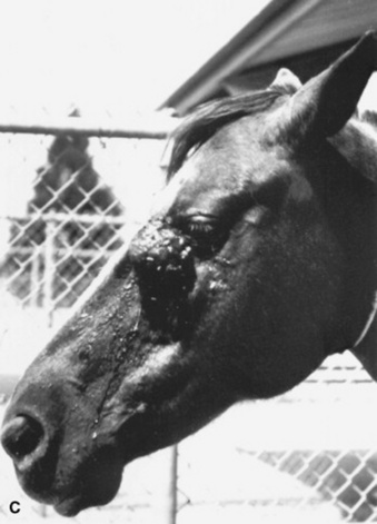
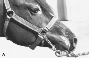
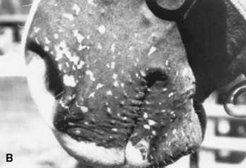
image image image

Fig 40-14 Equine sarcoids. A, Small occult sarcoid. B, Multiple verrucous sarcoids. C, Fibroblastic sarcoid.

The polymorphic appearance of sarcoids, the lack of consensus on clinical classification and anatomic distribution, and the long list of differential diagnoses make the clinical diagnosis of sarcoids unreliable. The diagnosis and treatment of sarcoids require a biopsy because they are often overdiagnosed clinically. In a review of 681 horses referred to the Oncology Clinic at the Veterinary Medical Teaching Hospital of the UCD School of Veterinary Medicine for evaluation and treatment of sarcoids from January 1995 to June 2004, 345 cases were presented with a clinical diagnosis of sarcoid without a biopsy, and 31% of these were found to have nonneoplastic dermatologic conditions.207 After ruling out a nonneoplastic skin condition, any clinically suspicious lesion should be biopsied.208 As with any tumor, early recognition and treatment of a small lesion are always associated with a better prognosis. It is important to keep in mind that any large and invasive sarcoid associated with a poor prognosis was, earlier in its evolution, an apparently inactive small lesion that was not recognized or was deliberately neglected. There is no contraindication for biopsy of a suspicious skin lesion as long as definitive treatment is instituted immediately after the diagnosis is made. Clinicians should not be concerned about performing a biopsy of a suspected sarcoid because effective treatments are available. As with any trauma, however, a biopsy can increase proliferation of a previously slow-growing tumor and may accelerate tumor progression. As a result, a biopsy should not be recommended if the owner is not willing to pursue treatment, if needed, because the process of biopsy without subsequent treatment has the same effect on overall prognosis as an unsuccessful attempt at treatment.

Therapy

Because tumor size and previous unsuccessful treatment attempts are the most important prognostic factors,205,207 early and complete surgical resection is the mainstay of treatment of sarcoids.208 Failure to eliminate the disease results in regrowth of a tumor that is histologically and biologically more aggressive and requires wider excision than the primary lesion. After surgery, it is critical to submit the resected specimen for histopathologic examination to determine the status of the surgical margins. As a rule, grossly or histologically incomplete resection (i.e., positive or close pathologic margins, <5 mm) must be followed by a re-resection, if possible, or by effective adjuvant treatment. For noninvasive sarcoids resected with pathologic margins greater than 5 mm and invasive sarcoids with margins greater than 1 cm, the risk of recurrence is low, and no further treatment except observation at regular intervals for at least 1 year is recommended. For invasive sarcoids resected with surgical margins between 5 and 10 mm, the risk of recurrence is high, and adjuvant treatment may be recommended, particularly when tumor recurrence may be difficult to manage because of unfavorable anatomic location. Access to molecular techniques (PCR and TaqMan PCR) designed to detect BPV E5 or E6 in pathologic margins will help assess the risk of recurrence and determine the need for adjuvant treatment.209

  Page 1329 

Among the nonsurgical treatments, interstitial radiation therapy and intralesional chemotherapy with cisplatin have well-documented efficacy against biopsy-confirmed sarcoids of all clinical types in any location.210,211 Compared with radiation therapy, intratumoral chemotherapy with cisplatin is as effective, but without long-term side effects, and it does not require a special license. Although several antineoplastic drugs212-215 and drug formulations205,207,213,214,216 for intralesional chemotherapy have been evaluated, a crystal suspension of cisplatin in a sesame seed oil and Sorbitan monooleate (Span 80) emulsion has been shown most effective. (Native protein [i.e., the patient’s own serum] preparations are proscribed because of their strong binding to cisplatin.217) When all stages and clinical types were combined, the cure rate after treatment with intratumoral cisplatin therapy used alone or as an adjuvant to surgery has been reported to be as high as 96%.207 Treatment includes a series of four intralesional doses of cisplatin (∼1 mg/cm3 tissue) given at 2-week intervals. Generic cisplatin is widely available and inexpensive. Treatments are done on an outpatient basis, and the methods of administration and safety precautions have been described.214

Nonspecific immunotherapy using bacille Calmette-Guérin (BCG) cell wall derivatives* administered intralesionally211 has been shown to be effective only for periocular sarcoids.205,218,219 The number of treatments depends on the rate of tumor regression, and the treatment schedule is dependent on normal tissue toxicity; most sarcoids require 2 to 9 treatments over several weeks.205

Encouraging results have been reported with imiquimod, a biologic response modifier, used topically. The ointment is recommended for the treatment of viral warts and basal cell carcinomas in people. It is applied as a thin layer to the tumor surface three times a week on nonconsecutive days, up to 32 weeks until resolution.220 The reported 60% response rate suggests that topical imiquimod may be a therapeutic option for specific equine sarcoids. However, the data must be confirmed in a larger series of horses with sarcoids and after adequate long-term follow-up.

  Page 1330 

Topical applications of escharotics with zinc chloride—based caustic ointments* or 5-FU cream are used empirically for treatment of sarcoids. Unfortunately, no scientific data have been published documenting treatment efficacy against actual equine sarcoids. As a result, no information on treatment protocol, efficacy, prognosis, or toxicity is available.

MASTOCYTOSIS

STEPHEN D. WHITE

In horses, mastocytosis (mast cell tumors, mastocytomas) occur in animals 1 to 18 years old (average, 9 years), with no breed predilection. A predilection for males has been proposed but is not always sustained.221,222 In addition, multiple mast cell tumors resembling urticaria pigmentosa of humans may occur in newborn foals; these spontaneously appear and regress.222,223

Equine mastocytosis is usually solitary and occurs most often on the head and trunk. Lesions are 0.5 to 20 cm in diameter, well to poorly circumscribed, firm to fluctuant, dermal or subcutaneous, and may or may not be alopecic, ulcerated, and hyperpigmented. Lesions on the legs tend to be very firm and immovable.

Histology may vary from sheets of mast cells with few eosinophils (presumably early lesions) to sections showing both sheets of mast cells with numerous eosinophils and collagen degranulation. Ultrastructural features are similar to those noted in mastocytomas of other species.224

Clinically, most mast cell tumors in horses do not recur after being excised (22 of 25 in one study).224 In one anecdotal case of metastasis from a tumor on the muzzle to regional lymph nodes, the tumor and the nodes were removed, and the horse was clinically sound 3 years later. There is some debate as to whether equine mastocytomas are benign neoplasias or focal dysplasias of mast cells.

Mastocytomas are uncommon in cattle but typically occur in young calves.225,226 One report describes a cutaneous mast cell tumor in a kid goat.227 Although equine mastocytomas are almost always benign, mastocytosis in cattle may be malignant or benign and therefore carry a more guarded prognosis.

MELANOMA

STEPHEN D. WHITE

Equine Melanomas

Melanomas occur in all domestic animals, but of the large domesticated species, they are most important in the horse. Excessive exposure to sunlight has not been definitively proven to predispose horses to the development of melanoma. A disturbance in melanin metabolism associated with graying has been hypothesized to stimulate formation of new melanoblasts or to stimulate their activity, resulting in focal areas of overproduction in the dermis and epidermis, with subsequent tumor formation.228 A higher incidence is observed in the Arabian, Lipizzaner, and Percheron breeds, probably because gray coat color occurs more often in these breeds. There is no gender predilection.

Melanocytic skin tumors of horses traditionally have been described in aging gray horses, in typical locations: the ventral tail, perineum, external genitalia, lip, udder, and periocular and parotid gland regions. These tumors have been the subject of several classification schemes in attempting to correlate histopathologic appearance with clinical behavior (i.e., benign or malignant). One study distinguished three basic types of melanocytic skin tumors, as discussed next.229

Melanocytic nevi (melanocytoma) occur in the superficial dermis or at the epidermal-dermal junction and frequently have epithelial involvement, with nests of relatively large, mildly to moderately pleomorphic cells showing variable cytoplasmic pigmentation and occasional mitoses. More than 70% of these occur in horses less than 6 years of age and may occur in horses of any color (not just gray). Most of these tumors occurred in atypical locations. Of 28 melanocytic nevi, only one became invasive; the rest exhibited benign behavior.

Dermal melanomas are found in the deep dermis and are composed of small, homogenous, indistinct tumor cells, either round or dendritic, with no mitoses. (If there are multiple, confluent dermal melanomas, this is referred to as dermal melanomatosis). About 80% of these tumors are in horses older than 6 years229 or between 5 and 15 years230 and are much more common in gray horses. Most of these tumors occurred in typical locations. Of 14 cases available for follow-up in one study,229 eight had malignant behavior, as demonstrated by metastases.

In another study, clinicopathologic characteristics of cutaneous melanomas occurring in 83 Camargue-type gray-skinned horses showed that the tumors occurred most frequently underneath the tail (93.9%) and at high rates in the perianal region (43.0%), the lips (33.0%), and the eyelids (24.0%), but rarely in the vulva (3.8%).231 Microscopic examination indicated that these tumors were composed mostly of melanocytes and numerous melanophages, and that these cells manifested a remarkable cellular atypia. Early stages of the tumors occurred in close association with apocrine sweat glands, but not at the dermal-epidermal junction.

A clinical study was conducted on 296 gray horses of the Lipizzaner breed.232 Of the 296 horses, dermal melanomas were present in 148 horses (50%), 68 of which were older than 15 years; 51 of these were melanoma bearing. In 75.6% of cases, melanotic tumors were detected underneath the tail. None of the affected individuals had any severe clinical effect or was handicapped in performance. The authors concluded that in contrast to melanomas in solid-colored horses characterized by early metastases, melanomas in gray horses showed less malignancy. Affected individuals often had encapsulated nodules or structures similar to human blue nevi. This finding at least partially reflects confusion in terminology between true malignant melanomas and dermal melanomas.

Anaplastic malignant melanomas are composed of sheets of extremely pleomorphic epithelioid cells with poor pigmentation and many mitoses. These are usually seen in horses older than 20 years of age and occur in horses of any color. Metastasis usually occurs first to the regional lymph nodes, then to the lungs, spleen, and liver. Hematogenous spread may also occur. Metastatic growths may be larger than the primary lesions and softer in consistency.

In regard to treatment, one study reported good success with excising dermal melanomatosis from the perineal, perianal, perirectal, or ventral tail regions.233 In a study of three horses, cimetidine (2.5 mg/kg PO q8h) was shown to decrease the number and size of melanoma growth.234 However, a more recent study of 10 horses found that cimetidine had no consistent effects on either the number of tumors or the tumor surface area over the 16 weeks of treatment at 5 mg/kg PO q12h.235 Another recent article noted a cure rate of 81% for melanomas treated with intratumoral injections of cisplatin.207

  Page 1331 

Bovine Melanomas

Melanomas represented a large proportion of the cutaneous neoplasms of cattle in an older study.236 The majority are benign, well-differentiated tumors, subcutaneous in location, and without site predilection. Dark-haired cattle are predisposed, particularly the Aberdeen Angus breed. There is no gender predilection.236 Melanomas usually occur in young cattle and are occasionally recognized as congenital lesions.237

Caprine and Ovine Melanomas

Melanomas have been observed only rarely in sheep and goats.236-239 A survey of 800,000 slaughtered goats revealed only five melanomas.237 The most common site for melanomas in the goat is the perineal region,29 although there is a case report of a malignant melanoma occurring in the coronary band region.238 Melanomas in Angora goats histologically resemble the corresponding tumors in humans.239

CUTANEOUS LYMPHOSARCOMA

STEPHEN D. WHITE

Equine Lymphoma

Cutaneous lymphoma has occasionally been reported in horses.240-247 Both T-cell and B-cell forms have been reported. Lesions present as nodules, either cutaneous or subcutaneous. Diagnosis is made by biopsy and, ideally, immunohistochemistry to determine cell type.246,247 In one horse, progesterone receptors were demonstrated on the lymphoma (B) cells, and the lesions regressed after removal of an estrogen-secreting ovarian tumor.243 This horse also had a history of partial regression of its tumor after administration of a synthetic progestin, altrenogest (0.044 mg/kg PO once daily for 10 days). Another horse demonstrated reduction in tumor size after administration of another synthetic progestagen, megestrol acetate (0.2 mg/kg PO once daily for 8 days) as well as a local injection of 20 mg betamethasone into a mass.244 Clearly, treatment is far from standardized, but the progesterone drugs may offer a reasonable treatment modality.

Lymphosarcoma is discussed in Chapter 37.

Bovine Lymphoma

Cattle occasionally are affected by cutaneous lymphoma. The presentation is multifocal intracutaneous nodules, often accompanied by alopecia. Lymphadenopathy, leukocytosis, and lymphocytosis are often present, and internal organs may be affected.248

Ovine Lymphoma

There are rare reports of cutaneous lymphoma in sheep.249,250

CYSTS

STEPHEN D. WHITE

Cutaneous cysts are benign lesions characterized by an epithelial wall with keratinous contents. Cutaneous cysts are subdivided into several types on the basis of their histopathologic features.

Epidermal Cysts

Among large animals, epidermal cysts have been reported in horses,251 cattle,252 and sheep.253 These cysts may be more properly called follicular cysts because the epithelial lining is probably most often derived from the follicular epithelium rather than the epidermis. The cysts can be found anywhere on the body, single or multiple, congenital or acquired, and generally range in size from 0.2 to 3 cm in diameter. The cysts are covered by intact epithelium and generally do not attach to the overlying epidermis. Microscopically, epidermal cysts consist of a wall of stratified squamous epithelium surrounding a keratin-filled lumen. Epidermal appendages are not associated with the cyst wall, a feature that distinguishes epidermal from dermoid cysts.

Epidermal cysts are thought to originate from occlusion of a hair follicle or by traumatic implantation of the epidermis. A tentative diagnosis may be made by performing fine-needle aspiration of a lesion and obtaining a fluid that is clear to brownish in color. Aspiration of the contents may temporarily decrease the size of the cyst, but it typically refills. Definitive diagnosis is made by excisional biopsy, which is curative. Epidermal cysts are benign lesions, although painful inflammatory responses and ulceration may result if the cyst is ruptured, with extrusion of contents into the adjacent dermis and subcutis.

Dermoid Cysts

Dermoid cysts are very similar clinically to epidermal cysts but are much less common. Among large animals, they have been identified in horses,251 goats,254 and cattle.255 In cattle, dermoid cysts may be congenital, have been reported to be as large as 10 cm (4 inches) in diameter, and are said to develop most frequently over the cranial area of the thorax255 and in the pharyngeal region. In horses, dermoid cysts may be single or multiple and are observed most frequently along the dorsal midline between the withers and the croup. Dermoid cysts are believed to result from displacement of embryonic cells into the subcutaneous tissue. They can be distinguished histologically from epidermal cysts by the presence of epidermal appendages within the wall of the cyst and by a lumen that often contains hair and secretions from sebaceous and sweat glands in addition to keratin. As with epidermal cysts, surgical excision is diagnostic and curative.

Dentigerous Cysts

Dentigerous cysts are a congenital defect recognized in horses and are believed to be the result of an abnormality of the first branchial cleft.256 Clinically, a unilateral saclike swelling that contains embryonic teeth is seen at the base of the ear. The lesion may be firmly attached to the concheal cartilage or temporal bone. Dentigerous cysts tend to fistulate. Treatment consists of surgical excision.256 Dentigerous cysts have also been reported in sheep and are suspected of being related to a nutritional deficiency of copper.257

Wattle Cysts

Wattle cysts are found in goats and usually are present at the base of the wattle. Nubians and Nubian crossbreeds may be predisposed to developing these cysts. The cysts are congenital, but they may not be apparent until the animal is several months old. Tentative diagnosis is based on aspiration of clear fluid, which will temporarily decrease the size of the cyst. Surgical excision is diagnostic and curative. Histologic examination reveals a cyst wall composed of one or two layers of cuboidal to columnar epithelial cells. The cyst cavity contains homogenous, amorphous basophilic substances.257

  Page 1332 

FROSTBITE

STEPHEN D. WHITE

Frostbite is an uncommon injury among healthy, well-nourished animals, with all species being susceptible. It is caused by prolonged exposure to subfreezing temperatures. The length of time needed to cause frostbite depends on the ambient temperature, area of the body exposed, and health status. The areas most often affected by cold injury include the ears, tail, teats, scrotum, and distal legs.

Frozen tissue must be handled gently and thawed rapidly in warm water 38° C to 44° C (100.2° F to 111.1° F) as soon as possible after it is known that refreezing can be prevented. Tissue damage is greatly increased if thawing and subsequent refreezing occur (freeze-thaw-freeze-thaw syndrome). Tissue thawing is painful, and analgesics should be administered. Slow thawing is less painful than rapid thawing but results in much greater tissue damage. Frozen tissue should not be massaged during warming. Damaged areas are best left exposed during the healing process rather than covered with occlusive dressings, and premature debridement should be avoided because more tissue may be viable than first apparent. The animal should be given good supportive care and may need to be restrained to prevent self-mutilation. Management practices should be changed to prevent recurrence. Tissues previously damaged by freezing are more susceptible to cold injury when reexposed to subnormal temperatures.7,258

SKIN DISORDERS OF UNKNOWN OR GENETIC ORIGIN

STEPHEN D. WHITE

EQUINE SEBORRHEA

Several possibly related equine skin diseases characterized by scaling and crust formation are referred to as “seborrhea.” Contrary to claims in the older literature, there is little evidence that seborrhea is related to excess sebum production by the sebaceous glands. Most types of seborrhea are probably diseases of abnormal cornification (development of the stratum corneum). Further, most cases reported as “generalized seborrhea” in the horse were probably pemphigus foliaceus, equine sarcoidosis (chronic granulomatous disease), or some other immune-mediated or autoimmune disease. Mane and tail seborrhea is typified by moderate to heavy scaling, with minimal or no pruritus. Some horses have considerable alopecia of the tail.

Cannon keratosis is a common equine skin condition. It involves the cranial surface of the rear cannon bone region and rarely the forelimbs. The lesions consist of areas of scaling and crusting with varying degrees of alopecia. There is no pruritus or other signs of inflammation. Cannon keratosis occurs in both males and mares, so there is no basis for the theory that the condition is caused by urine splatter (“stud crud”).33

LINEAR KERATOSIS AND LINEAR ALOPECIA

Linear keratosis and linear alopecia are rare equine dermatoses of unknown cause. There is one report in a cow.259 The lesions do not follow blood or lymphatic vessels, nerves, or dermatomes. Because both conditions coexist in some horses, it has been suggested that they are variations of the same abnormality, although this is difficult to justify histopathologically. Both conditions have been seen in a wide variety of breeds, but quarter horses appear to be predisposed. Most horses develop lesions between 6 months and 5 years of age.

Linear alopecia is characterized by the gradual development of annular areas of alopecia, usually in a linear, vertically oriented configuration. One or more linear areas may be present. The lesions are usually 2 to 10 mm wide by a few centimeters to over 1 m in length and occur on the neck, shoulder, and lateral thorax. Mild surface scale and crust may be present. The lesions are neither painful nor pruritic. Affected horses are typically otherwise healthy.

Linear keratosis is characterized by the gradual, asymptomatic occurrence of one or more unilateral, linear, vertically oriented bands of hyperkeratotic papules that progress to marked hyperkeratosis and alopecia. The lesions vary from 0.25 to 3.5 cm in width by 5 to 70 cm in length and occur most often over the neck, shoulder, and lateral thorax. Lesions have also been reported to involve the legs, hip, and pectoral region. Again, affected horses are typically otherwise healthy.

These disorders are visually distinctive. Histopathologic findings in linear alopecia include early lymphocytic, infiltrative, mural folliculitis and later granulomatous, infiltrative, mural folliculitis. The mural infiltrate is often directed at the middle area (isthmus) of the follicle. Sebaceous glands may be involved in some cases, and complete follicular destruction and permanent alopecia are seen in severe chronic lesions. Histopathologic findings in linear keratosis include irregular to papillated epidermal hyperplasia and marked compact orthokeratotic hyperkeratosis.260 I have seen one horse that had lesions grossly diagnosed as linear keratosis, whereas the histopathology had features of both conditions.

Neither condition is known to undergo spontaneous resolution. Owners should be advised of the potential hereditary nature of these disorders. Linear alopecia has been anecdotally reported to respond to topical or systemic glucocorticoids, but recurrence is likely. Response to therapy is more likely to be seen in early lesions, when complete destruction of hair follicles has not occurred. Linear keratosis responds poorly to treatment. Topical keratolytic and keratoplastic agents, such as sulfur/salicylic acid—containing shampoos or 50% propylene glycol, can reduce the hyperkeratosis but must be continued for life. I have had some success using tacrolimus (Protopic, Astellas Pharma US, 0.1% ointment), a drug similar to cyclosporine but better absorbed through the skin, once or twice daily. Because neither condition is symptomatic, observation without treatment may be an acceptable approach.

Interestingly, a report exists of a familial incidence of linear epidermal nevi in Belgian horses.261

ALBINISM

Complete and partial albinism occurs in cattle, sheep, and horses. It is a genetic defect (probably autosomal recessive) in melanin synthesis, resulting in white skin, white hair, pink eyes, and photophobia. In horses, albinism must be distinguished from lethal white syndrome, which is primarily a problem in Paint horses (especially, but not exclusively, in overo breedings). The defective gene has also been found in American miniature horses, half-Arabians, thoroughbreds, and cropout quarter horses (foals born to registered quarter horses that have too much white to register as with the AQHA).262 The Veterinary Genetics Laboratory at UCD offers a diagnostic test to determine carrier status.*

  Page 1333 

The lethality of albinism in horses comes from the association with intestinal aganglionosis. The foals die shortly after birth. Because some white foals are not affected, euthanasia should be performed only after signs of intestinal malfunction occur. This disease is similar to Hirschsprung disease in humans and is linked to a mutation in the endothelin-B receptor gene.263

JUVENILE ARABIAN LEUKODERMA (ARABIAN FADING SYNDROME, PINKY SYNDROME, HEREDITARY VITILIGO)

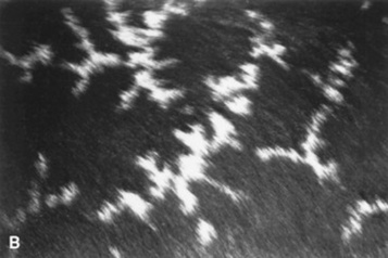
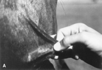
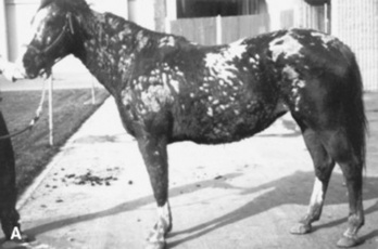
Loss of melanin in the skin (depigmentation) occurs in young Arabian horses 6 months to 2 years of age (Fig. 40-15,A). The areas most frequently affected are periocular tissues, muzzle, genitalia, anus, perineum, inguinal region, and undersurface of the base of the tail. Depigmentation may persist, repigment, or wax and wane. The condition is probably hereditary.33

image image

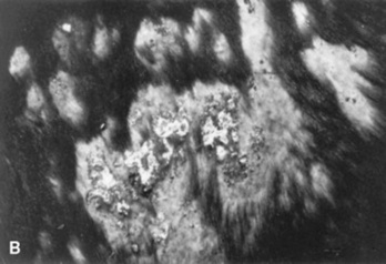
Fig 40-15 A, Juvenile Arabian leukoderma. B, Vitiligo in a horse. (Courtesy Dr. Anthony Stannard.)

VITILIGO

This condition is best defined as “idiopathic depigmentation.” Typically, no trauma has occurred to produce the loss of pigment, and other skin structures are not affected (i.e., no scarring or alopecia) (Fig. 40-15,B). There is no known treatment for vitiligo.

RETICULATED AND HYPERESTHETIC LEUKOTRICHIA

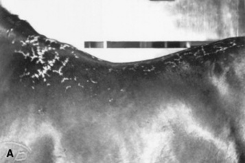
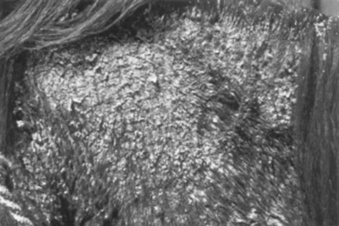
Reticulated leukotrichia occurs mainly in quarter horses, usually as yearlings, and occasionally in other breeds. The lesions occur on the dorsal midline and consist initially of linear crusts arranged in a characteristic cross-hatch pattern (Fig. 40-16). The crusts shed, alopecia occurs, and white hair grows in permanently. There is leukotrichia without leukoderma (depigmented skin). Histologically, these lesions resemble an epidermal form of erythema multiforme, resulting in individual keratinocyte necrosis. Hyperesthetic leukotrichia is a similar disease both clinically and histologically, except the crusts are extremely painful to the touch. Within a few weeks, white hairs appear in the affected areas. The crusts resolve, and the pain subsides in 1 to 3 months, but the leukotrichia persists. Several cases have been linked to recent rhinopneumonitis vaccination. These diseases may rarely recur, and there is no known effective treatment.33

image image

Fig 40-16 Reticulated leukotrichia in a yearling quarter horse. (Courtesy Dr. Anthony Stannard.)

HEREDITARY EQUINE REGIONAL DERMAL ASTHENIA (HYPERELASTOSIS CUTIS)

Hereditary equine regional dermal asthenia (HERDA) occurs early in life in horses. Most affected horses are quarter horses, but registered Paint horses and Appaloosas with quarter horse lineage have developed this disease.264,265 Many of the quarter horses are from high-quality cutting lines. The disease (or similar condition) has also been reported in a crossbred Arabian mare, a thoroughbred gelding, a Hanoverian foal, and a Hafflinger horse.7,266-268 Rarely, a similar disease has been noted in cattle, termed dermatosporaxis; this is caused by mutations in the procollagen I N-proteinase gene.268,269

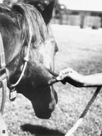
The working hypothesis for HERDA in horses is a defect in the structure or healing process of the collagen fibers in the middle to deep dermis. Typically, these areas are over the back and sides of the neck (Fig. 40-17). The skin in these areas may be easily torn or stretched and often develops seromas and hematomas (“blisters” filled with either serum or blood). Healing is usually adequate but often leaves rather unsightly scars. Diagnosis is often based on the clinical signs alone; histologic findings are sometimes subtle, but “clumped” or poorly organized collagen fibers below the level of the hair follicles may be seen. A zone of middermal to deep dermal separation has been reported in two horses and is present in some biopsy samples.265,270 “Poorly oriented” collagen fibers are sometimes seen on electron microscopy.

image image

Fig 40-17 Hereditary equine regional dermal asthenia (HERDA, hyperelastosis cutis) in a horse showing hyperextensible skin. (Courtesy Dr. Anthony Stannard.)

  Page 1334 

This condition is almost certainly present at birth, but HERDA is often not noticed until about 2 years of age, when horses start being trained with tack and saddle, and the resulting friction and trauma induce the typical lesions. As with many genetic diseases, no effective treatment or cure exists; some of these horses have been maintained as “pasture pets.”

This disease follows an autosomal recessive mode of inheritance, so in order for the foal to be affected, both the sire and the dam must carry the gene, and if they were bred again, there would be approximately a 25% chance that the next foal would also be affected.271 Recently, a genetic marker was determined for this disease.271a The Veterinary Genetics Laboratory at the University of California, Davis offers a diagnostic test to determine carrier or affected status (http://www.vgl.ucdavis.edu/service/horse/index.html). Both carriers and clinically affected horses with HERDA should be removed from breeding programs.

EPIDERMOLYSIS BULLOSA

Epidermolysis bullosa (EB) includes a number of diseases typified in humans by the common finding of blister formation after minor trauma. Most forms are congenital and apparent soon after birth. In animals and humans, subsets of EB are classified by the histologic location of the blister or cleft. These subtypes (and respective cleft location) are termed EB simplex (basal cell layer of epidermis), junctional EB (intralamina lucida or basal cell layer), and dystrophic EB (sublamina densa).

Junctional EB has been reported in Belgian foals of both genders, in other breeds, and in a donkey.272-275 Lesions are usually noted within 3 days of birth and include multiple asymmetric skin erosions and ulcers, often encrusted. Lesions may be especially prominent around the coronary bands (causing the hoof to crack and slough) and on the oral, anal, and genital mucosa. Histology and ultrastructural findings indicate a cleft in the intralamina lucida of the basement membrane zone. This is presumably caused by a defect in the anchoring filaments that connect the basement membrane to filaments in the superficial dermis.273 A laminin-5 defect has been demonstrated in Belgians and in two French draft breeds, Trait Breton and Trait Comtois; the mutation is a cytosine insertion in exon 10 of the LAMC2 gene.276-278 Because of this knowledge, the Veterinary Genetics Laboratory at UCD offers a diagnostic test to determine carrier status in Belgian draft horses and related breeds.*

Clinical presentation and the age of the foal are highly suggestive of EB diagnosis. Histology and ideally electron microscopy are required to confirm the diagnosis. There is no known treatment, and affected horses, as well as the sires and dams of affected horses, should not be bred; the mode of inheritance is autosomal recessive.

  Page 1335 

This disease differs from epitheliogenesis imperfecta (see following discussion) in that large areas of the skin are not at first devoid of epidermis, but rather lose their skin because of the fibril defect.

EPITHELIOGENESIS IMPERFECTA (APLASIA CUTIS)

Epitheliogenesis imperfecta is a rare inherited congenital discontinuity of squamous epithelium. It is thought to be an autosomal recessive trait and has been reported in several breeds. Lesions are most common on the limbs, head, and tongue. Hooves may slough in severe cases. Clinical presentation is usually diagnostic.279 In moderately to severely affected animals, the disease is fatal within a few days; the foals die of septicemia or other developmental abnormalities. Mildly affected areas may heal by scar formation. More recent reports suggest that some of these horses (saddlebreds) may have a condition similar to the junctional epidermolysis bullosa in Belgian foals previously noted.280-282

EOSINOPHILIC GRANULOMA (NODULAR NECROBIOSIS, COLLAGENOLYTIC GRANULOMA)

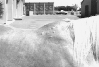
Eosinophilic granuloma is the most common nontumor nodular disease in the author’s practice. In most cases the etiology is unknown, although a hypersensitivity reaction to insect bites has been suggested. There is no apparent age, breed, or gender predilection. The disease often begins in warmer months. Lesions up to 5 cm (2 inches) in diameter may be single or multiple and most often affect the neck, withers, and back (Fig 40-18). Skin biopsy reveals multifocal areas of abnormal-staining collagen surrounded by granulomatous inflammation containing eosinophils, lymphocytes, and histiocytes.33 This is not thought to be caused by “degeneration,” but rather by degranulation of eosinophils and the coating of normal collagen fibers with the degranulated material.283

image

Fig 40-18 Eosinophilic granuloma (nodular necrobiosis), with dermal nodules on the withers of a horse. (Courtesy Dr. Anthony Stannard.)

Development of equine eosinophilic granuloma has been noted in areas of previous injections using standard silicone-coated, stainless steel hypodermic needles.284 The reaction may occur at sites of intravenous as well as intramuscular injections. The lesions consist of nonpainful, cool, raised papules or nodules 0.25 to 1 cm in diameter at sites of previous injection. The nodule appears 24 to 48 hours after the injection, and the subsequent eosinophilic granuloma can persist for months to years. Affected horses do not develop a lesion at the site of injection if nondisposable, noncoated needles are used. The use of the noncoated needles is recommended for any horse that develops injection-site collagenolytic granulomas.

Horses with solitary or a few lesions may be treated by surgical excision (not in the saddle area) or glucocorticoid injections under the lesions. Triamcinolone acetonide (3 to 5 mg per lesion) is effective. It has been recommended that no more than 20 mg triamcinolone acetonide be administered at once to any horse because any more of this drug may cause laminitis.33 Horses with multiple lesions may be treated with oral prednisolone at 1 mg/kg once daily for 2 to 3 weeks. Multiple, small (<1 cm) lesions may be indicative of insect bite hypersensitivity.

CUTANEOUS AMYLOIDOSIS

Cutaneous amyloidosis is an uncommon nodular dermatosis of unknown etiology. There is no age, breed, or gender predilection. The condition appears to be a primary form of amyloidosis, in that concurrent inflammatory processes are not present, and amyloid (a fibrillar protein substance derived from immunoglobulins) deposition is usually restricted to the skin and occasionally the regional lymphatics, regional lymph nodes, and upper respiratory mucosa.7,33 One case was documented as being caused by lambda—light chain deposition from an extramedullary plasmacytoma.285 Another horse showed amyloidosis concurrent with lymphoma.286

Papules, nodules, and plaques on the head, neck, shoulders, thorax, or inguinal areas are firm, 0.5 to 10 cm in diameter, nonpainful, and nonpruritic. The overlying skin is normal, although it may scale or crust in severe cases (Fig. 40-19). The course of cutaneous amyloidosis is progressive and prolonged. Megestrol acetate may be an effective treatment (0.2 mg/kg PO q24h until lesions resolve). Because certain forms of primary amyloidosis in humans have a genetic basis, it may not be advisable to use affected horses for breeding until more is learned about the genetics of the equine disorder.

image image

Fig 40-19 A and B, Cutaneous amyloidosis in a horse. (Courtesy Dr. Anthony Stannard.)

  Page 1336 

EQUINE SARCOIDOSIS (GENERALIZED GRANULOMATOUS DISEASE)

Equine sarcoidosis is characterized by skin lesions and usually concurrent systemic involvement. It is not a neoplastic disease and should not be confused with equine sarcoids.

The disease is infrequently encountered, and there is no known breed predilection. Geldings may be at increased risk.287 The skin lesions typically are generalized scaling and crusting associated with varying degrees of alopecia (Fig. 40-20). Occasionally, the disease is focal or multifocal in distribution. Rarely, the skin lesions consist of nodules or large, tumor-like masses. The different types of skin lesions may coexist. In addition to skin lesions, the most frequent presenting complaints are weight loss, decreased appetite, and a persistent low-grade fever. Lung involvement is manifested by exercise intolerance, increased resting respiratory rate, and mild dyspnea.288-290 Although one report showed positive titers to Borrelia burgdorferi in three of four horses with equine sarcoidosis,291 a more recent report was unable to demonstrate the presence, using PCR and histologic stains, of any causative organism.287

image

Fig 40-20 Equine sarcoidosis (generalized granulomatous disease) in a horse. In addition to the scaling and crusting skin lesions, the horse has systemic involvement. (Courtesy Dr. Anthony Stannard.)

Diagnosis is by histopathology and ruling out infectious etiologies. The major histologic change is the presence of noncaseating granulomas consisting of aggregates of epithelioid cells and multinucleated giant cells. Neutrophils, lymphocytes, and plasma cells are present in small numbers. In the skin the granulomas tend to be located in the superficial portion of the dermis. Because of the small number of horses studied and the variability of clinical signs, response to therapy has not been well documented. A recent abstract reported that five of six horses did well and lived longer than 1 year with prednisolone treatment.292 Another extensive review showed that some horses did well on corticosteroid therapy (usually at doses approximating those used to treat pemphigus and other autoimmune skin diseases); gastrointestinal involvement was a sign of poorer prognosis.287

PHOTOSENSITIZATION

Photosensitivity is an abnormal reaction of the skin when exposed to light. Photosensitivity in the horse is usually caused by a photodynamic agent in or on the skin that absorbs or transfers energy from light and transfers it to body cells. The activating light is generally in the ultraviolet A (UVA) range (320 to 400 nm). Melanin in the skin screens UV light, thereby limiting photosensitivity reactions to the white and light-colored areas of the body.293

Photodynamic agents may be drugs, topical medications, foodstuffs, contactants, or excessive phylloerythrin from liver disease. Examples of plants containing photodynamic agents follow:

Common Name Scientific Name Agent
St. John’s wort Hypericum perforatum Hypericin
Buckwheat Polygonum fagopyrum Fagopyrin
Perennial ryegrass Lolium perenne Perloline
Whiteheads Spheosciadium capitellatum Unknown

In addition, I have sometimes seen alfalfa or clover induce a photosensitivity reaction; these plants have been suspected to contain either an ingested or contact photodynamic agent. Recent reports describe photosensitivities apparently induced by the ingestion of gluten in horses, and of cocoa shells in calves.294,294a

As horses ingest plants containing chlorophyll, this molecule is degraded by bacteria in the intestine into the porphyrin, phylloerythrin. Some phylloerythrin is normally absorbed into the portal circulation, removed by the liver, and excreted by the bile. In liver disease the hepatic excretion of phylloerythrin is decreased, leading to excessive levels in the peripheral circulation and eventually in the skin, causing photosensitivity in approximately 25% of horses with liver dysfunction.295-297 One of the more common reasons for liver disease in the United States is ingestion of the following plants containing pyrrolizidine alkaloids:

Common Name Scientific Name
Common groundsel Senecio vulgaris
Ragwort, stinking Willie Senecio jacobea
Tarweed Amsinckia intermedia
Rattleweed Crotalaria species
Salvation Jane Echium lycopsis

In addition, consumption of alsike clover (Trifolium hybridum) may induce hepatic dysfunction (with histologic lesions distinct from those caused by pyrrolizidine alkaloids), leading to photosensitization signs (alsike clover poisoning, dew poisoning). Cases seem to be correlated to years with heavy rainfall and the ingestion of the plant blossoms.298

Ingested plants and feed should be noted and collected for future analysis. Environmental considerations include the type of pasture (or other material) with which the animal is in contact, the amount of time the animal is exposed to sunlight, any seasonality of the condition, and any other horses involved. A seasonal incidence would tend to negate liver disease; multihorse involvement should arouse suspicion of an ingested or contact photosensitizer. A thorough history of recent drug therapy should be obtained.

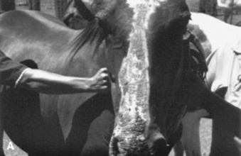
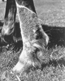
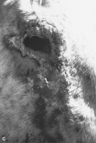
Physical examination usually reveals lesions limited to the hairless, white, or lightly pigmented areas of the skin. The involved skin is erythematous, swollen, and painful. The lesions may progress to serum exudation, thickening, fissuring, and in severe cases, necrosis and sloughing (Fig. 40-21).

image image image

Fig 40-21 Photosensitization. A, White blaze of a horse is selectively affected. B, White stocking of a horse is selectively affected. C, Area around eye where hair is less dense is most severely affected.

Therapy is related to etiology. Chronic liver disease carries a guarded prognosis. Removal of feedstuffs containing photodynamic agents, or removal of the horse from a pasture with plants containing these agents, will usually result in full recovery if the horse is also kept out of the sun for 1 to 2 weeks. Corticosteroids are helpful in controlling inflammation and pruritus. Oral prednisolone at 1 mg/kg daily for 1 week, then halving the daily dosage for a second week, is an effective regimen.

  Page 1337 

CHRONIC PROGRESSIVE LYMPHEDEMA

Chronic progressive lymphedema is the term for a condition seen in Shires, Clydesdales, and Belgians. It is characterized by progressive swelling, hyperkeratosis (thickening), and fibrosis (hardening) of the skin on the lower legs. This chronic progressive disease starts at an early age, progresses throughout the life of the horse, and often ends in disfigurement and disability of the limbs. Inevitably, this condition leads to the horse’s premature death. In the Belgian draft horse, it has reduced the average life expectancy of a stallion from 20 to only 6 years.

The pathologic changes and clinical signs closely resemble a condition known in humans as chronic lymphedema, or elephantiasis nostras verrucosa. The lower leg swelling is caused by abnormal functioning of the lymphatic system in the skin, which results in chronic lymphedema (swelling), fibrosis, a compromised immune system, and subsequent secondary infections of the skin. Based on preliminary research, it appears that a similar pathogenic mechanism is involved in the disease that affects these specific draft horse breeds.

The clinical signs of this disease are highly variable. The earliest lesions are characterized by skin thickening and crusting; both are often visible only after clipping the long feathering. Secondary infections develop very easily in these horses’ legs and usually consist of either chorioptic mange or bacterial infections. Both dark skin and white skin on the lower legs are equally affected. These lesions are consistent with pastern dermatitis, certainly seen in other breeds. In Shires, Clydesdales, and Belgians, however, these lesions do not respond well to therapy.

  Page 1338 

As the disease progresses, one or two thick skin folds and sometimes multiple small, well-demarcated ulcerations develop, predominantly in the rear of the pastern region. The ulcerations are covered with adherent crusts. Manual removal of the crusts or even movement during exercise results in bleeding. These small sores may seem to respond initially to various topical medications, but often reverse course, only to progress in severity and multiply in number. Small lesions tend to coalesce into larger and more intractable (resistant to cure) areas of skin ulceration. Over time, the lesions extend up the leg, often affecting the skin as high as the knees or hocks. These lesions are, at the very least, irritating to the horses and at times can be quite painful. Severely affected individuals often exhibit generalized swelling in all four legs.

This condition therefore is primarily a lymph system disease, and the pastern dermatitis in these draft horses is secondary to the body’s inability to supply fluids properly and oxygenate the skin of the lower leg. The lymphatics break down over time, and the protein-rich fluid leaks into the tissues of the lower leg, which results in fibrosis of the tissues under the skin and thickening of the skin itself. The tissue fibrosis leads to even more blockage of fluid within the legs, inhibiting circulatory flow. This results in neovascularization, a process by which the body develops new blood vessels in a futile attempt to provide oxygen to its tissues.

Researchers suspect that a deficiency or abnormality in the connective tissue component known as elastin is the underlying factor and perhaps the cause of the lymphatic degeneration in these horses.299,300 In affected animals the lymph vessels and deep tissues of the skin do not have sufficient amounts or the proper configuration of elastin. The lack of this critical tissue element apparently instigates the progression of disease and the chronic progression of clinical signs. A recent report documents high levels of antielastin antibodies in affected horses.301

As the condition becomes more chronic, the lower leg enlargement becomes permanent, and the swelling is firm on palpation. More of the thick skin folds and large, poorly defined, firm nodules develop. The nodules may become quite large and often are described as golf ball or even baseball in size. Both skin folds and nodules first develop in the back of the pastern area. With progression, they may extend and encircle the entire lower leg. The nodules become a mechanical problem because they interfere with free movement and frequently are injured during exercise. This disease often progresses to include massive secondary infections that produce copious amounts of foul-smelling exudates, generalized illness, debilitation, and even death.300,301

In a recent report of possibly the same condition in several draft breeds, the authors found a perivascular dermatitis dominated by T lymphocytes with an increase in major histocompatibility complex (MHC) class II–positive, dendritic-like cells. Immunohistochemical labeling for cytokeratins CK5/6(4), CK10, and CK14 indicated a change in their expression pattern. This correlated with the degree of epidermal hyperplasia, indicating abnormal differentiation of keratinocytes. There was a statistically significant correlation between the severity of skin lesions and several other factors, including increasing age, increasing cannon circumference, prominence of anatomic structures (e.g., fetlock tufts of hairs, ergots, chestnuts), and bulges in the fetlock region.302

REFERENCES

1 Vandenabeele SI, White SD, Kass P, et al. Pemphigus foliaceus in the horse: 20 cases. Vet Dermatol. 2004;15:381.

2 Zabel S, Mueller RS, Fieseler KV, et al. Review of 15 cases of pemphigus foliaceus in horses and a survey of the literature. Vet Rec. 2005;157:505.

3 Pappalardo E, Abramo F, Noli C. Pemphigus foliaceus in a goat. Vet Dermatol. 2002;13:331.

4 Valdez RA, Gelberg HB, Morin DE, et al. Use of corticosteroids and aurothioglucose in a pygmy goat with pemphigus foliaceus. J Am Vet Med Assoc. 1995;207:761.

5 Bourdeau P, Baudry J. [Pemphigus-type bullus dermatosis assocciated with pregnancy in a female donkey]. Informations Dermatologiques Vétérinaries. 2005;11:19.

6 White SD, Carlotti DN, Pin D, et al. Putative drug-induced pemphigus foliaceus in four dogs. Vet Dermatol. 2002;13:195.

7 Scott DW, Miller WHJr. Equine dermatology. St Louis: Saunders-Elsevier, 2003.

8 Scott DW. Marked acantholysis associated with dermatophytosis due to Trichophyton equinum in two horses. Vet Dermatol. 1994;5:105.

9 Scott DW, Smith MC, Smith CA, et al. Pemphigus foliaceus in a goat. Agric Pract. 1984;5(4):38.

10 Scott DW, Walton DK, Smith CA. Pitfalls in immunofluorescence testing in dermatology. III, Pemphigus-like antibodies in the horse and direct immunofluorescence testing in equine dermatophilosis. Cornell Vet. 1984;74:305.

11 Peroni DL, Stanley S, Kollias-Baker C, et al. Prednisone per os is likely to have limited efficacy in horses. Equine Vet J. 2002;34:283.

12 Humber KA, Beech J, Cudd TA, et al. Azathioprine for treatment of immune-mediated thrombocytopenia in two horses. J Am Vet Med Assoc. 1991;199:591.

13 McGurrin MK, Arroyo LG, Bienzle D. Flow cytometric detection of platelet-bound antibody in three horses with immune-mediated thrombocytopenia. J Am Vet Med Assoc. 2004;224:83.

14 White SD, Rosychuk RA, Outerbridge CA, et al. Thiopurine methyltransferase in red blood cells of dogs, cats, and horses. J Vet Intern Med. 2000;14:499.

15 White SD, Maxwell LK, Szabo N, et al. Pharmacokinetics of azathioprine following single-dose intravenous and oral administration and effects of azathioprine following chronic oral administration in horses. Am J Vet Res. 2005;66:1578.

16 Olivry T, Borrillo AK, Xu L, et al. Equine bullous pemphigoid IgG autoantibodies target linear epitopes in the NC16A ectodomain of collagen XVII (BP180, BPAG2). Vet Immunol Immunopathol. 2000;73:45.

17 Rees CA. Response to immunotherapy in six related horses with urticaria secondary to atopy. J Am Vet Med Assoc. 2001;218:753.

18 Lorch G, Hillier A, Kwochka KW, et al. Comparison of immediate intradermal test reactivity with serum IgE quantitation by use of a radioallergosorbent test and two ELISA in horses with and without atopy. J Am Vet Med Assoc. 2001;218:1314.

19 Lorch G, Hillier A, Kwochka KW, et al. Results of intradermal tests in horses without atopy and horses with chronic obstructive pulmonary disease. Am J Vet Res. 2001;62:389.

20 Lorch G, Hillier A, Kwochka KW, et al. Results of intradermal tests in horses without atopy and horses with atopic dermatitis or recurrent urticaria. Am J Vet Res. 2001;62:1051.

21 Jose-Cunilleras E, Kohn CW, Hillier A, et al. Intradermal testing in healthy horses and horses with chronic obstructive pulmonary disease, recurrent urticaria, or allergic dermatitis. J Am Vet Med Assoc. 2001;219:1115.

22 Wong DM, Buechner-Maxwell VA, Manning TO. Equine skin: structure, immunologic function, and methods of diagnosisng disease. Compend Cont Educ (Pract Vet). 2005;27:463.

23 Morris DO, Lindborg S. Determination of “irritant” threshold concentrations for intradermal testing with allergenic insect extracts in normal horses. Vet Dermatol. 2003;14:31.

24 Wong DM, Buechner-Maxwell VA, Manning TO, et al. Evaluation of the precision of intradermal injection of control substances for intradermal testing in clinically normal horses. Am J Vet Res. 2005;66:1341.

25 White SD. Advances in equine serologic and intradermal allergy testing. Clin Tech Equine Pract. 2005;4:311.

26 Tallarico NJ, Tallarico CM. Results of intradermal allergy testing and treatment by hyposensitization of 64 horses with chronic obstructive pulmonary disease, urticaria, headshaking, and/or reactive airway disease. J Vet Allergy Clin Immunol. 1998;6:25.

27 Evans AG. Recurrent urticaria due to inhaled allergens. In Robinson NE, editor: Current therapy in equine medicine, ed 2, Philadelphia: Saunders, 1986.

28 White SD. Equine bacterial and fungal diseases: a diagnostic and therapeutic update. Clin Tech Equine Pract. 2005;4:302.

29 Scott DW. Large animal dermatology. Philadelphia: Saunders, 1988.

30 Campbell SG. Milk allergy, an autoallergic disease of cattle. Cornell Vet. 1970;60:684.

31 Campbell SG. The milk proteins responsible for milk allergy, an autoallergic disease of cattle. J Allergy Clin Immunol. 1971;48:230.

32 Marshall C. Erythema multiforme in two horses. J S Afr Vet Assoc. 1991;62:133.

33 Stannard AA. Stannard’s illustrated equine dermatology. Vet Dermatol. 2000;11:163.

34 Affolter VK, Von Tscharner C. Cutaneous drug reactions: a retrospective study of histopathological changes and their correlation with the clinical disease. Vet Dermatol. 1993;4:79.

34 Yeruham I, Perl S, Elad D. Nine cases of idiopathic toxic epidermal necrolysis in cattle in Israel. Zentralbl Veterinarmed B. 1999;46:493.

35 Risberg AI, Webb CB, Cooley AJ, et al. Leucocytoclastic vasculitis associated with Staphylococcus intermedius in the pastern of a horse. Vet Rec. 2005;156:740.

36 De Geest J, Muylle E, Deprez P. An outbreak of cutaneous vasculitis in newborn calves. Vlaams Diergeneeskundig Tijdschrift. 1990;59:7.

37 Ihrke PJ. Contact dermatitis. In: Robinson NE, editor. Current therapy in equine medicine. Philadelphia: Saunders, 1983.

38 Scott DW, Manning TO. Equine folliculitis and furunculosis. Equine Pract. 1980;2:11.

39 Scanlan CM, Garrett PD, Geifer DB. Dermatophilus congolensis infections of cattle and sheep. Compend Cont Educ (Pract Vet). 1984;6:S4.

40 Scott DW, Manning TO. Caprine dermatology. I. Normal skin and bacterial and fungal disorders. Compend Cont Educ (Pract Vet). 1984;6:S190.

41 Outerbridge CA, Ihrke PJ. Folliculitis: staphylococcal pyoderma, dermatophilosis, dermatophytosis. In: Robinson NE, editor. Current therapy in equine medicine 5. St Louis: Saunders-Elsevier, 2003.

42 Shimizu A, Kawano J, Ozaki J, et al. Characteristics of Staphylococcus aureus isolated from lesions of horses. J Vet Med Sci. 1991;53:601.

43 Chiers K, Decostere A, Devriese L, et al. Bacteriological and mycological findings and in vitro antibiotic sensitivity of pathogenic staphylococci in equine skin infections. Vet Rec. 2003;152:138.

44 Inokuma H, Kanaya N, Fujii K, et al. Equine pyoderma associated with malnutrition and unhygienic conditions due to neglect in a herd. J Vet Med Sci. 2003;65:527.

45 Heffner KA, White SD, Frevert CW, et al. Corynebacterium folliculitis in a horse. J Am Vet Med Assoc. 1988;193:89.

46 Yasuda R, Kawano J, Onda H, et al. Methicillin-resistant coagulase-negative staphylococci isolated from healthy horses in Japan. Am J Vet Res. 2000;61:1451.

47 Busscher JF, van Duijkeren E, Sloet van Oldruitenborg-Oosertbaan MM. The prevalence of methicillin-resistant staphylococci in healthy horses in the Netherlands. Vet Microbiol. 2006;113:131.

48 Weese JS, Rousseau J, Traub-Dargatz JL, et al. Community-associated methicillin-resistant Staphylococcus aureus in horses and humans who work with horses. J Am Vet Med Assoc. 2005;226:580.

49 Cuny C, Kuemmerle J, Stanek C, et al. Emergence of MRSA infections in horses in a veterinary hospital: strain characterisation and comparison with MRSA from humans. Eur Surveill. 2006;11:44.

50 Weese JS, Caldwell F, Willey BM, et al. An outbreak of methicillin-resistant Staphylococcus aureus skin infections resulting from horse to human transmission in a veterinary hospital. Vet Microbiol. 2006;114:160.

51 Peck KE, Matthews NS, Taylor TS, et al. Pharmacokinetics of sulfamethoxazole and trimethoprim in donkeys, mules, and horses. Am J Vet Res. 2002;63:349.

52 Egerbacher M, Edinger J, Tschulenk W. Effects of enrofloxacin and ciprofloxacin hydrochloride on canine and equine chondrocytes in culture. Am J Vet Res. 2001;62:704.

53 Epstein K, Cohen N, Boothe D, et al. Pharmacokinetics, stability, and retrospective analysis of use of an oral gel formulation of the bovine injectable enrofloxacin in horses. Vet Ther. 2004;5:155.

54 Dunnett M, Richardson DW, Lees P. Detection of enrofloxacin and its metabolite ciprofloxacin in equine hair. Res Vet Sci. 2004;77:143.

55 Orsini JA, Snooks-Parsons C, Stine L, et al. Vancomycin for the treatment of methicillin-resistant staphylococcal and enterococcal infections in 15 horses. Can J Vet Res. 2005;69:278.

56 Yeruham I, Perl S, Elad D, Avidar Y. A generalized staphylococcal scalded skin—like disease in lambs. Zentralbl Veterinarmed B. 1999;46:635.

57 Yeruham I, Hadani A, Elad D, et al. Staphylococcal furunculosis in sheep severely infested by psoroptic mange. Aust Vet J. 2002;80:349.

58 Oliveira AM, MacKellar A, Hume L, et al. Immune responses to Staphylococcus aureus and Psoroptes ovis in sheep infected with P. ovis—the sheep scab mite. Vet Immunol Immunopathol. 2006;113:64.

59 Markel MD, Wheat JD, Jang SS. Cellulitis associated with coagulase-positive staphylococci in racehorses: nine cases (1975-1984). J Am Vet Med Assoc. 1986;189:1600.

60 Biberstein EL, Jang SS, Hirsh DC. Species distribution of coagulase-positive staphylococci in animals. J Clin Microbiol. 1984;19:610.

61 Devriese LA, Nzuambe D, Godard C. Identification and characteristics of staphylococci isolated from lesions and normal skin of horses. Vet Microbiol. 1985;10:269.

61 Adam EN, Southwood LL. Primary and secondary limb cellulitis in horses: 44 cases (2000-2006). J Am Vet Med Assoc. 2007;231:1696.

62 Spier SJ, Leutenegger CM, Carroll SP, et al. Use of a real-time polymerase chain reaction—based fluorogenic 5′ nuclease assay to evaluate insect vectors of Corynebacterium pseudotuberculosis infections in horses. Am J Vet Res. 2004;65:829.

63 Aleman M, Spier SJ, Wilson WD, et al. Corynebacterium pseudotuberculosis infection in horses: 538 cases (1982-1993). J Am Vet Med Assoc. 1996;209:804.

64 Farstvedt EG, Hendrickson DA, Dickenson CE, et al. Treatment of suppurative facial cellulitis and panniculitis caused by Corynebacterium pseudotuberculosis in two horses. J Am Vet Med Assoc. 2004;224:1139.

65 Read DH, Walker RL. Papillomatous digital dermatitis (footwarts) in California dairy cattle: clinical and gross pathologic findings. J Vet Diagn Invest. 1998;10:67.

66 Rodriguez-Lainz A, Hird DW, Walker RL, et al. Papillomatous digital dermatitis in 458 dairies. J Am Vet Med Assoc. 1996;209:1464.

67 Blowey RW, Sharp MW. Digital dermatitis in dairy cattle. Vet Rec. 1988;122:505.

68 Laven RA, Logue DN. Treatment strategies for digital dermatitis for the UK. Vet J. 2006;171:79.

69 Rodriguez-Lainz A, Melendez-Retamal P, Hird DW, et al. Farm- and host-level risk factors for papillomatous digital dermatitis in Chilean dairy cattle. Prev Vet Med. 1999;42:87.

70 Holzhauer M, Hardenberg C, Bartels CJ, et al. Herd- and cow-level prevalence of digital dermatitis in the Netherlands and associated risk factors. J Dairy Sci. 2006;89:580.

71 Rodriguez-Lainz A, Hird DW, Carpenter TE, et al. Case-control study of papillomatous digital dermatitis in southern California dairy farms. Prev Vet Med. 1996;28:117.

72 Wells SJ, Garber LP, Wagner BA. Papillomatous digital dermatitis and associated risk factors in US dairy herds. Prev Vet Med. 1999;38:11.

73 Demirkan I, Murray RD, Carter SD. Skin diseases of the bovine digit associated with lameness. Vet Bull. 2000;70:149.

74 Demirkan I, Carter SD, Murray RD, et al. The frequent detection of a treponeme in bovine digital dermatitis by immunocytochemistry and polymerase-chain-reaction. Vet Microbiol. 1998;60:285.

75 Demirkan I, Walker RL, Murray RD, et al. Serological evidence of spirochaetal infections associated with digital dermatitis in dairy cattle. Vet J. 1999;157:69.

76 Walker RL, Read DH, Loretz KJ, et al. Spirochetes isolated from dairy cattle with papillomatous digital dermatitis and interdigital dermatitis. Vet Microbiol. 1995;47:343.

77 Döpfer D, Koopmans A, Meijer FA, et al. Histological and bacteriological evaluation of digital dermatitis in cattle, with special reference to spirochaetes and Campylobacter faecalis. Vet Rec. 1997;140:620.

78 Walker RL, Read DH, Loretz KJ, et al. Humoral response of dairy cattle to spirochetes isolated from papillomatous digital dermatitis lesions. Am J Vet Res. 1997;58:744.

79 Shibahara T, Ohya T, Ishii R, et al. Concurrent spirochaetal infections of the feet and colon of cattle in Japan. Aust Vet J. 2002;80:497.

80 Berry SL, Read DH, Walker RL. Topical treatment with oxytetracycline or lincomycin HCl for papillomatous digital dermatitis: gross and histological evaluation. In: Proceedings of the 10th International Symposium on Lameness in Ruminants. Luzern: Switzerland; 1998:291.

81 Hernandez J, Shearer JK, Elliott JB. Comparison of topical application of oxytetracycline and four nonantibiotic solutions for treatment of papillomatous digital dermatitis in dairy cows. J Am Vet Med Assoc. 1999;214:688.

82 Britt JS, Carson MC, von Bredow JD, et al. Antibiotic residues in milk samples obtained from cows after treatment for papillomatous digital dermatitis. J Am Vet Med Assoc. 1999;215:833.

83 Nuss K. Footbaths: the solution to digital dermatitis? Vet J. 2006;171:11.

84 Ertze RA, Read DH, Hird DW, et al. Field evaluation of prophylactic and therapeutic effects of a vaccine against (papillomatous) digital dermatitis in dairy cattle on two California dairies. Bovine Pract. 2006;40:76.

85 Jelinek F, Tachezy R. Cutaneous papillomatosis in cattle. J Comp Pathol. 2005;132:70.

86 Uzal FA, Latorraca A, Ghoddusi M, et al. An apparent outbreak of cutaneous papillomatosis in merino sheep in Patagonia, Argentina. Vet Res Commun. 2000;24:197.

87 Manni V, Roperto F, Di Guardo G, et al. Presence of papillomavirus-like DNA sequences in cutaneous fibropapillomas of the goat udder. Vet Microbiol. 1998;61:1.

88 Ghim SJ, Rector A, Delius H, et al. Equine papillomavirus type 1: complete nucleotide sequence and characterization of recombinant virus-like particles composed of the EcPV-1 L1 major capsid protein. Biochem Biophys Res Commun. 2004;324:1108.

89 Chambers G, Ellsmore VA, O’Brien PM, et al. Sequence variants of bovine papillomavirus E5 detected in equine sarcoids. Virus Res. 2003;96:141.

90 Ogawa T, Tomita Y, Okada M, et al. Broad-spectrum detection of papillomaviruses in bovine teat papillomas and healthy teat skin. J Gen Virol. 2004;85:2191.

91 Rebhun WC. Interdigital papillomatosis in dairy cattle. J Am Vet Med Assoc. 1980;177:437.

92 White KS, Fuji RN, Valentine BA, et al. Equine congenital papilloma: pathological findings and results of papillomavirus immunohistochemistry in five cases. Vet Dermatol. 2004;15:240.

93 Davis CL, Kemper HE. Warts in goats. J Am Vet Med Assoc. 1936;88:175.

94 Theilen G, Wheeldon EB, East N, et al. Goat papillomatosis. Am J Vet Res. 1985;46:2519.

95 Kirnbauer R, Chandrachud LM, O’Neil BW, et al. Virus-like particles of bovine papillomavirus type 4 in prophylactic and therapeutic immunization. Virology. 1996;219:37.

96 Jarrett WF, O’Neil BW, Gaukroger JM, et al. Studies on vaccination against papillomaviruses: a comparison of purified virus, tumour extract and transformed cells in prophylactic vaccination. Vet Rec. 1990;126:449.

97 Barthold SW, Olson C, Larson LL. Precipitin response of cattle to commercial wart vaccine. Am J Vet Res. 1976;37:449.

98 Fairley RA, Haines DM. The electron microscopic and immunohistochemical demonstration of a papilloma virus in equine aural plaques. Vet Pathol. 1992;29:79.

99 Gibbs EP. Viral diseases of the skin of bovine teat and udder. Vet Clin North Am Large Anim Pract. 1984;6:187.

100 Letchworth GJ3rd, LaDue R. Bovine herpes mammillitis in two New York dairy herds. J Am Vet Med Assoc. 1982;180:902.

101 Janett F, Stauber N, Schraner E, et al. [Bovine herpes mammillitis: clinical symptoms and serologic course]. Schweiz Arch Tierheilkd. 2000;142:375.

102 Rao TV, Bandyopadhyay SK. A comprehensive review of goat pox and sheep pox and their diagnosis. Anim Health Res Rev. 2000;1:127.

103 Bhanuprakash V, Indrani BK, Hosamani M, et al. The current status of sheep pox disease. Comp Immunol Microbiol Infect Dis. 2006;29:27.

104 Kane J, Padhye AA, Ajello L. Microsporum equinum in North America. J Clin Microbiol. 1982;16:943.

105 Roman C, Massai L, Gianni C, et al. Case reports. six cases of infection due toTrichophyton verrucosum, Mycoses. 2001;44:334.

106 Scott DW. Marked acantholysis associated with dermatophytosis due to Trichophyton equinum in two horses. Vet Dermatol. 1994;5:105.

107 Gudding R, Lund A. Immunoprophylaxis of bovine dermatophytosis. Can Vet J. 1995;36:302.

108 Pier AC, Zancanella PJ. Immunization of horses against dermatophytosis caused by Trichophyton equinum. Equine Pract. 1993;15:23.

109 Milan R, Alois R, Josef C, et al. Recombinant protein and DNA vaccines derived from hsp60 Trichophyton mentagrophytes control the clinical course of trichophytosis in bovine species and guinea-pigs. Mycoses. 2004;47:407.

110 Welsh RD. Sporotrichosis. J Am Vet Med Assoc. 2003;223:1123.

111 Irizarry-Rovira AR, Kaufman L, Christian JA, et al. Diagnosis of sporotrichosis in a donkey using direct fluorescein-labeled antibody testing. J Vet Diagn Invest. 2000;12:180.

112 Rosser EJJr. Sporotrichosis. In: Robinson NE, editor. Current therapy in equine medicine 5. St Louis: Saunders-Elsevier, 2003.

113 Kohler LM, Monteiro PC, Hahn RC, et al. In vitro susceptibilities of isolates of Sporothrix schenckii to itraconazole and terbinafine. J Clin Microbiol. 2004;42:4319.

114 Valentine BA, Taylor GH, Stone JK, et al. Equine cutaneous fungal granuloma: a study of 44 lesions from 34 horses. Vet Dermatol. 2006;17:266.

115 Perez RC, Luis-Leon JJ, Vivas JL, et al. Epizootic cutaneous pythiosis in beef calves. Vet Microbiol. 2005;109:121.

116 Tabosa IM, Riet-Correa F, Nobre VM, et al. Outbreaks of pythiosis in two flocks of sheep in northeastern Brazil. Vet Pathol. 2004;41:412.

117 Grooters AM, Whittington A, Lopez MK, et al. Evaluation of microbial culture techniques for the isolation of Pythium insidiosum from equine tissues. J Vet Diagn Invest. 2002;14:288.

118 Hubert JD, Grooters AM. Treatment of equine pythiosis. Compend Contin Educ (Pract Vet). 2002;24:812.

119 Mendoza L, Mandy W, Glass R. An improved Pythium insidiosum–vaccine formulation with enhanced immunotherapeutic properties in horses and dogs with pythiosis. Vaccine. 2003;21:2797.

120 Butler JF. Lice affecting livestock. In: Williams RE, Hall RD, Broce AB, editors. Livestock entomology. New York: Wiley-Interscience, 1985.

121 Loomis EC. Common ectoparasites and their control. In: Robinson NE, editor. Current therapy in equine medicine 1, vol 6. Philadelphia: Saunders; 1983:529.

122 Bergvall K. Advances in acquisition, identification, and treatment of equine ectoparasites. Clin Tech Equine Pract. 2005;4:296.

123 Bourdeau P. Mites and ticks. In: Robinson NR, editor. Current therapy in equine medicine 5. St Louis: Saunders-Elsevier, 2003.

124 Zahler M, Essig A, Gothe R, et al. Genetic evidence suggests that Psoroptes isolates of different phenotypes, hosts and geographic origins are conspecific. Int J Parasitol. 1998;28:1713.

125 Meintjes T, Fourie LJ, Horak IG. Host preference of the sheep scab mite, Psoroptes ovis. J S Afr Vet Assoc. 2002;73:135.

126 Stromberg PC, Fisher WF, Guillot FS, et al. Systematic pathologic responses in experimental Psoroptes ovis infestation in Hereford calves. Am J Vet Res. 1986;47:1326.

127 Oliveira A, MacKellar A, Hume L, et al. Immune responses to Staphylococcus aureus and Psoroptes ovis in sheep infected with P. ovis–the sheep scab mite. Vet Immunol Immunopathol. 2006;113:64.

128 Cadiergues MC, Laguerre C, Roques M, et al. Evaluation of the bioequivalence of two formulations of deltamethrin for treatment of sheep with psoroptic mange. Am J Vet Res. 2004;65:151.

129 Rehbein S, Visser M, Winter R, et al. Productivity effects of bovine mange and control with ivermectin. Vet Parasitol. 2003;114:267.

130 Losson BJ. Mange in large animals. In Kahn CM, Line S, editors: The Merck veterinary manual, ed 9, Rahway, NJ: Merck, 2005.

131 Essig A, Rinder H, Gothe R, et al. Genetic differentiation of mites of the genus Chorioptes (Acari: Psoroptidae). Exp Appl Acarol. 1999;23:309.

132 Rehbein S, Visser M, Winter R. Chorioptic mange in dairy cattle: treatment with eprinomectin pour-on. Parasitol Res. 2005;98:21.

133 Rehbein S, Visser M, Winter R, et al. Productivity effects of bovine mange and control with ivermectin. Vet Parasitol. 2003;114:267.

133 Osman SA, Hanafy A, Amer SE. Clinical and therapeutic studies on mange in horses. Vet Parasitol. 2006;141:191.

134 Warnick LD, Nydam D, Maciel A, et al. Udder cleft dermatitis and sarcoptic mange in a dairy herd. J Am Vet Med Assoc. 2002;221:273.

135 Matthes HF. Investigations of pathogenesis of cattle demodicosis: sites of predilection, habitat and dynamics of demodectic nodules. Vet Parasitol. 1994;53:283.

136 Brugger M, Braun U. [Demodicosis in a Toggenburg goat]. Schweiz Arch Tierheilkd. 2000;142:639.

137 Auer DE, Seawright AA, Pollitt CC, et al. Illness in horses following spraying with amitraz. Aust Vet J. 1984;61:257.

138 Strabel D, Schweizer G, Gansohr B, et al. [The use of avermectins in two goats with demodicosis]. Schweiz Arch Tierheilkd. 2003;145:585.

139 Littlewood JD. Incidence of recurrent seasonal pruritus (“sweet itch”) in British and German shire horses. Vet Rec. 1998;142:66.

140 Wilson AD, Harwood LJ, Bjornsdottir S, et al. Detection of IgG and IgE serum antibodies to Culicoides salivary gland antigens in horses with insect dermal hypersensitivity (sweet itch). Equine Vet J. 2001;33:707.

141 Hellberg W, Wilson AD, Mellor P, et al. Equine insect bite hypersensitivity: immunoblot analysis of IgE and IgG subclass responses to Culicoides nubeculosus salivary gland extract. Vet Immunol Immunopathol. 2006;113:99.

142 Van der Haegen A, Griot-Wenk M, Welle M, et al. Immunoglobulin-E-bearing cells in skin biopsies of horses with insect bite hypersensitivity. Equine Vet J. 2001;33:699.

143 McKelvie J, Foster AP, Hamblin AS, et al. Culicoides antigen extract stimulates equine blood mononuclear (BMN) cell proliferation and the release of eosinophil adherence-inducing factor(s). Res Vet Sci. 2001;70:115.

144 McKelvie J, Foster AP, Cunningham FM, et al. Characterisation of lymphocyte subpopulations in the skin and circulation of horses with sweet itch (Culicoides hypersensitivity). Equine Vet J. 1999;31:466.

145 Kurotaki T, Narayama K, Arai Y, et al. Langerhans cells within the follicular epithelium and the intradermal sweat duct in equine insect hypersensitivity “Kasen,”. J Vet Med Sci. 2002;64:539.

146 Kurotaki T, Narayama K, Oyamada T, et al. Ultrastructural study of Langerhans cells in equine insect hypersensitivity “Kasen,”. J Vet Med Sci. 2000;62:1021.

147 Kurotaki T, Narayama K, Oyamada T, et al. The kinetics of Langerhans cells in equine insect hypersensitivity “Kasen,”. J Vet Med Sci. 2000;62:561.

148 Kurotaki T, Narayama K, Oyamada T, et al. Immunopathological study on equine insect hypersensitivity (“Kasen”) in Japan. J Comp Pathol. 1994;110:145.

149 Kolm-Stark G, Wagner R. Intradermal skin testing in Icelandic horses in Austria. Equine Vet J. 2002;34:405.

150 Anderson GS, Belton P, Jahren E, et al. Immunotherapy trial for horses in British Columbia with Culicoides hypersensitivity. J Med Entomol. 1996;33:458.

151 Broce AB, et al. Myiasis-producing flies. In: Williams RE, Hall RD, Broce AB, et al, editors. Livestock entomology. New York: Wiley-Interscience, 1985.

152 Radostits OM, Blood DC, Gray CC. Veterinary medicine, ed 8. London: Baillière Tindall, 1994.

153 Hendrix CM. Flies. In Kahn CM, Line S, editors: The Merck veterinary manual, ed 9, Rahway, NJ: Merck, 2005.

154 Lima WS, Malacco MA, Bordin EL, et al. Evaluation of the prophylactic effect and curative efficacy of fipronil 1% pour on (Topline) on post-castration scrotal myiasis caused by Cochliomyia hominivorax in cattle. Vet Parasitol. 2004;125:373.

155 Moya-Borja GE, Muniz RA, Umehara O, et al. Protective efficacy of doramectin and ivermectin, against. Cochliomyia hominivorax, Vet Parasitol. 1997;72:101.

156 Fadok VA. Parasitic skin diseases of large animals. Vet Clin North Am. 1984;6:3.

157 Marques SM, Scroferneker ML. Onchocerca cervicalis in horses from southern Brazil. Trop Anim Health Prod. 2004;36:633.

158 Stannard AA, Cello RM. Onchocerca cervicalis infection in horses from the western United States. Am J Vet Res. 1975;36:1029.

159 Klei TR. Helminths of the skin. In Kahn CM, Line S, editors: The Merck veterinary manual, ed 9, Rahway, NJ: Merck, 2005.

160 Cello RM. Ocular onchocerciasis in the horse. Equine Vet J. 1971;3:148.

161 Foil CS. Cutaneous onchocerciasis. In: Robinson NE, editor. Current therapy in equine medicine 2. Philadelphia: Saunders, 1987.

162 Schmidt GM, Coley SC, Leid RW. Onchocerca cervicalis in horses: dermal histopathology. Acta Trop. 1985;42:55.

163 French DD, Klei TM, Foil CS, et al. Efficacy of ivermectin in paste and injectable formulations against microfilariae of Onchocerca cervicalis and resolution of associated dermatitis in horses. Am J Vet Res. 1988;49:1550.

164 Mancebo OA, Verdi JH, Bulman GM. Comparative efficacy of moxidectin 2% equine oral gel and ivermectin 2% equine oral paste against Onchocerca cervicalis (Railliet and Henry, 1910) microfilariae in horses with naturally acquired infections in Formosa (Argentina). Vet Parasitol. 1997;73:243.

165 Njongmeta LM, Nfon CK, Gilbert J, et al. Cattle protected from onchocerciasis by ivermectin are highly susceptible to infection after drug withdrawal. Int J Parasitol. 2004;34:1069.

166 Beytut E, Akca A, Bain O. Teat onchocercosis in cows with reference to prevalence, species involved and pathology. Res Vet Sci. 2005;78:45.

167 Kral F, Schwartzman RM. Veterinary and comparative dermatology. Philadelphia: Lippincott, 1964;328.

168 Johnson SJ, Toleman MA. Prevalence of stephanofilariasis in young Bos indicus cattle in northern Australia. Vet Parasitol. 1988;29:333.

169 Rosser EJJr. Parasitic dermatoses. In: Howard JL, editor. Current veterinary therapy 3: food animal practice. Philadelphia: Saunders, 1993.

170 Watrelot-Virieux D, Pin D. Chronic eosinophilic dermatitis in the scrotal area associated with stephanofilariasis infestation of charolais bull in France. J Vet Med B Infect Dis Vet Public Health. 2006;53:150.

171 LIoyd JE. Cattle grubs. In Kahn CM, Line S, editors: The Merck veterinary manual, ed 9, Rahway, NJ: Merck, 2005.

172 Lloyd JE. Flies, lice and grubs. In: Howard JL, Smith RA, editors. Current veterinary therapy 4: food animal practice. Philadelphia: Saunders, 1993.

173 Otranto D, Boulard C, Giangaspero A, et al. Serodiagnosis of goat warble fly infestation by Przhevalskiana silenus with a commercial ELISA kit. Vet Rec. 1999;144:726.

174 Campbell WC, Benz GW. Ivermectin: a review of efficacy and safety. J Vet Pharmacol Ther. 1984;7:1.

175 Manning TO, Scott DW. Caprine dermatology. III. Parasitic, allergic, hormonal and neoplastic disorders. Compend Cont Educ (Pract Vet). 1985;7:S437.

176 Arthropod pests of sheep. In: Williams RE, Hall RD, Broce AB, Lloyd JE, editors. Livestock entomology. New York: Wiley-Interscience, 1985.

177 Mehlhorn H, D’Haese J, Mencke N, et al. In vivo and in vitro effects of imidacloprid on sheep keds (Melophagus ovinus): a light and electron microscopic study. Parasitol Res. 2001;87:331.

178 Pusterla N, Watson JL, Wilson WD, et al. Cutaneous and ocular habronemiasis in horses: 63 cases (1988-2002). J Am Vet Med Assoc. 2003;222:978.

179 Rees CA, Craig TM. Equine cutaneous habronemiasis. In: Robinson NE, editor. Current therapy in equine medicine 5. Philadelphia: Saunders-Elsevier, 2003.

179 Traversa D, Iorio R, Petrizzi L, et al. Molecular diagnosis of equid summer sores. Vet Parasitol. 2007;150:116.

180 Valentine BA. Survey of equine cutaneous neoplasia in the Pacific Northwest. J Vet Diagn Invest. 2006;18:123.

181 Baipoledi EK. A case of caprine perineal squamous cell carcinoma in Botswana. J S Afr Vet Assoc. 2001;72:165.

182 Mendez A, Perez J, Ruiz-Villamor E, et al. Clinicopathological study of an outbreak of squamous cell carcinoma in sheep. Vet Rec. 1997;141:597.

183 Mair TS, Walmsley JP, Phillips TJ. Surgical treatment of 45 horses affected by squamous cell carcinoma of the penis and prepuce. Equine Vet J. 2000;32:406.

184 McCauley CT, Hawkins JF, Adams SB, et al. Use of a carbon dioxide laser for surgical management of cutaneous masses in horses: 32 cases (1993-2000). J Am Vet Med Assoc. 2002;220:1192.

185 Campbell GA, Gross TL, Adams R, et al. Solar elastosis with squamous cell carcinoma in two horses. Vet Pathol. 1987;24:463.

186 Mosunic CB, Moore PA, Carmicheal KP, et al. Effects of treatment with and without adjuvant radiation therapy on recurrence of ocular and adnexal squamous cell carcinoma in horses: 157 cases (1985-2002). J Am Vet Med Assoc. 2004;225:1733.

187 Fortier LA, Mac Harg MA. Topical use of 5-fluorouracil for treatment of squamous cell carcinoma of the external genitalia of horses: 11 cases (1988-1992). J Am Vet Med Assoc. 1994;205:1183.

188 Paterson S. Treatment of superficial ulcerative squamous cell carcinoma in three horses with topical 5-fluorouracil. Vet Rec. 1997;141:626.

189 Théon AP, Pascoe JR, Madigan JE, et al. Comparison of intratumoral administration of cisplatin versus bleomycin for treatment of periocular squamous cell carcinomas in horses. Am J Vet Res. 1997;58:431.

190 Théon AP, Pascoe JR, Galuppo LD, et al. Comparison of perioperative versus postoperative intratumoral administration of cisplatin for treatment of cutaneous sarcoids and squamous cell carcinomas in horses. J Am Vet Med Assoc. 1999;215:1655.

191 Moore AS, Beam SL, Rassnick KM, et al. Long-term control of mucocutaneous squamous cell carcinoma and metastases in a horse using piroxicam. Equine Vet J. 2003;35:715.

192 Jackson C. The incidence and pathology of tumours of domesticated animals in South Africa. Onderstepoort J Vet Sci. 1936;6:378.

193 Tarwid JN, Fretz PB, Clark EG. Equine sarcoids: a study with emphasis on pathologic diagnosis. Comp Cont Educ (Pract Vet). 1985;7:S293.

194 Ragland W, Keown G, Spencer G. Equine sarcoid. Equine Vet J. 2, 1970.

195 Marti E, Lazary S, Antczak DF, et al. Report of the First International Workshop on Equine Sarcoid. Equine Vet J. 1993;25:397.

196 Ragland WL, Keown GH, Gorham JR. An epizootic of equine sarcoid. Nature. 1966;210:1399.

197 Nixon C, Chambers G, Ellsmore V, et al. Expression of cell cycle associated proteins cyclin A, CDK-2, p27kip1 and p53 in equine sarcoids. Cancer Lett. 2005;221:237.

198 Carr EA, Théon AP, Madewell BR, et al. Expression of a transforming gene (E5) of bovine papillomavirus in sarcoids obtained from horses. Am J Vet Res. 2001;62:1212.

199 Martens A, De Moor A, Ducatelle R, et al. PCR detection of bovine papilloma virus DNA in superficial swabs and scrapings from equine sarcoids. Vet J. 2001;161:280.

200 Ragland WL, Spencer GR. Attempts to relate bovine papilloma virus to the cause of equine sarcoid: Equidae inoculated intradermally with bovine papilloma virus. Am J Vet Res. 1969;30:743.

201 Bogaert L, Martens A, De Baere C, et al. Detection of bovine papillomavirus DNA on the normal skin and in the habitual surroundings of horses with and without equine sarcoids. Res Vet Sci. 2005;79:253.

202 Kemp-Symonds JG. The detection and sequencing of bovine papillomavirus type 1 and 2 DNA from Musca autumnalis (Diptera: Muscidae) face flies infesting sarcoid-affected horses [MSc Thesis]. London: Royal Veterinary College, 2000.

203 Nasir L, Reid SWJ. Bovine papillomaviruses and equine sarcoids. In Papillomavirus research: from natural history to vaccines and beyond. Wymondham, UK: Caister Academic; 2006.

204 Knottenbelt D, Edwards S, Daniel E. Diagnosis and treatment of the equine sarcoid. In Practice. 1995;17:123.

205 Knottenbelt DC, Kelly DF. The diagnosis and treatment of periorbital sarcoid in the horse: 445 cases from 1974 to 1999. Vet Ophthalmol. 2000;3:169.

206 Knottenbelt DC. A suggested clinical classification for the equine sarcoid. Clin Tech Equine Pract. 2005;4:278.

207 Théon AP, Wilson WD, Magdesian KG, et al. Long-term outcome associated with intratumoral chemotherapy with cisplatin for cutaneous tumors in equidae. J Am Vet Med Assoc. 2007;230:1506. 573 cases (1995-2004)

208 Théon AP. Oncology. In Higgins A, Snyder J, editors: Equine manual, ed 2, Philadelphia: Saunders-Elsevier, 2005.

209 Martens A, De Moor A, Demeulemeester J, et al. Polymerase chain reaction analysis of the surgical margins of equine sarcoids for bovine papilloma virus DNA. Vet Surg. 2001;30:460.

210 Théon AP. Radiation therapy in the horse. Vet Clin North Am Equine Pract. 1998;14:673.

211 Théon AP. Intralesional and topical chemotherapy and immunotherapy. Vet Clin North Am Equine Pract. 1998;14:659.

212 Boure L, Krawiecki JM, Thoulon F. Trial treatment of sarcoids in horses with intratumoral injections of bleomycin. Point Veterinaire. 1991;23:199.

213 Orenberg EK, Luck EE, Brown DM, et al. Implant delivery system: intralesional delivery of chemotherapeutic agents for treatment of spontaneous skin tumors in veterinary patients. Clin Dermatol. 1991;9:561.

214 Théon AP. Cisplatin treatment for cutaneous tumors. In: Robinson NE, editor. Current therapy in equine medicine 4. Philadelphia: Saunders, 1997.

215 Stewart AA, Rush B, Davis E. The efficacy of intratumoural 5-fluorouracil for the treatment of equine sarcoids. Aust Vet J. 2006;84:101.

216 Spoormakers TJ, Klein WR, Weeren PR, et al. Treatment of equine sarcoids with cisplatin in arachid oil: a useable alternative? Tijdschr Diergeneeskd. 2002;127:350.

217 Howe-Grant ME, Lippard SJ. Aqueous platinum (II) chemistry; binding to biological molecules. In: Sigel H, editor. Metal ions in biological systems. New York: Marcel Dekker, 1980.

218 Klein WR, Bras GE, Misdorp W, et al. Equine sarcoid: BCG immunotherapy compared to cryosurgery in a prospective randomised clinical trial. Cancer Immunol Immunother. 1986;21:133.

219 Owen RA, Jagger DW. Clinical observations on the use of BCG cell wall fraction for treatment of periocular and other equine sarcoids. Vet Rec. 1987;120:548.

220 Nogueira SA, Torres SM, Malone ED, et al. Efficacy of imiquimod 5% cream in the treatment of equine sarcoids: a pilot study. Vet Dermatol. 2006;17:259.

221 Altera K, Clark L. Equine cutaneous mastocytosis. Pathol Vet. 1970;7:43.

222 Cheville NF, Prasse KW, van der Maaten M, et al. Generalized equine cutaneous mastocytosis. Vet Pathol. 1972;9:394.

223 Prasse KW, Lundvall RL, Cheville NF. Generalized mastocytosis in a foal, resembling urticaria pigmentosa of man. J Am Vet Med Assoc. 1975;166:68.

224 McEntee MF. Equine cutaneous mastocytomas: morphology, biological behavior and evolution of the lesion. J Comp Pathol. 1991;104:171.

225 Head KW. Cutaneous mast cell tumors in the dog, cat and ox. Br J Dermatol. 1958;70:389.

226 Smith BI, Phillips LA. Congenital mastocytomas in a Holstein calf. Can Vet J. 2001;42:635.

227 Allison N, Fritz DL. Cutaneous mast cell tumour in a kid goat. Vet Rec. 2001;149:560.

228 Theilen GH, Madwell BR. Veterinary cancer medicine, ed 2. Philadelphia: Lea & Febiger, 1987.

229 Valentine BA. Equine melanocytic tumors: a retrospective study of 53 horses (1988-1991). J Vet Int Med. 1995;9:291.

230 Fleury C, Berard F, Leblond A, et al. The study of cutaneous melanomas in Camargue-type gray-skinned horses (2): epidemiological survey. Pigment Cell Res. 2000;13:47.

231 Fleury C, Berard F, Balme B, et al. The study of cutaneous melanomas in Camargue-type gray-skinned horses (1): clinical-pathological characterization. Pigment Cell Res. 2000;13:39.

232 Seltenhammer MH, Simhofer H, Scherzer S, et al. Equine melanoma in a population of 296 grey Lipizzaner horses. Equine Vet J. 2003;35:153.

233 Rowe EL, Sullins KE. Excision as treatment of dermal melanomatosis in horses: 11 cases (1994-2000). J Am Vet Med Assoc. 2004;225:94.

234 Goetz TE, Ogilvie GK, Keegan KG, et al. Cimetidine for treatment of melanomas in three horses. J Am Vet Med Assoc. 1990;196:449.

235 Bowers JR, Huntington PJ, Slocombe RF. Efficacy of cimetidine for therapy of skin tumours of horses: 10 cases. Aust Equine Vet. 1994;12:30.

236 Jackson C. The incidence and pathology of tumors of domesticated animals in South Africa: a study of the Onderstepoort collection of neoplasms with special reference to their histopathology. Onderstepoort J Vet Sci Anim Indust. 1936;6:1.

237 Bradley PJ, Magaki G. Types of tumors found by federal meat inspectors in an eight-year survey: epizootiology of cancer in animals. Ann NY Acad Sci. 1963;108:872.

238 Sockett DC, Knight AP, Johnson LW. Malignant melanoma in a goat. J Am Vet Med Assoc. 1984;185:907.

239 Parsons PG, Takahashi H, Candy J, et al. Histopathology of melanocytic lesions in goats and establishment of a melanoma cell line: a potential model for human melanoma. Pigment Cell Res. 1990;3:297.

240 Detilleux PG, Cheville NF, Sheahan BJ. Ultrastructure and lectin histochemistry of equine cutaneous histiolymphocytic lymphosarcomas. Vet Pathol. 1989;26:409.

241 Gollagher RD, Ziola B, Chelack BJ, et al. Immunotherapy of equine cutaneous lymphoma using low-dose cyclophosphamide and autologous tumor cells infected with vaccinia virus. Can Vet J. 1993;34:371.

242 Gerard MP, Healy LN, Bowman KF, et al. Cutaneous lymphoma with extensive periarticular involvement in a horse. J Am Vet Med Assoc. 1998;213:391.

243 Henson KL, Alleman AR, Cutler TJ, et al. Regression of subcutaneous lymphoma following removal of an ovarian granulosa—theca cell tumor in a horse. J Am Vet Med Assoc. 1998;212:1419.

244 Littlewood JD, Whitwell KE, Day MJ. Equine cutaneous lymphoma: a case report. Vet Dermatol. 1995;6:105.

245 Potter K, Anez D. Mycosis fungoides in a horse. J Am Vet Med Assoc. 1998;212:550.

246 Kelley LC, Mahaffey EA. Equine malignant lymphomas: morphologic and immunohistochemical classification. Vet Pathol. 1998;35:241.

247 Epstein V, Hodge D. Cutaneous lymphosarcoma in a stallion. Aust Vet J. 2005;83:609.

248 Schweizer G, Hilbe M, Braun U. Clinical, haematological, immunohistochemical and pathological findings in 10 cattle with cutaneous lymphoma. Vet Rec. 2003;153:525.

249 Da Silva DL. Attempted treatment of cutaneous lymphomatosis in a ram. J S Afr Vet Assoc. 2002;73:90.

250 Scott PR, Rhind S, Dun K. Cutaneous lymphoma in a Scottish blackface ram. Vet Rec. 1997;141:473.

251 Hillyer LL, Jackson AP, Quinn GC. Epidermal (infundibular) and dermoid cysts in the dorsal midline of a three-year-old thoroughbred-cross gelding. Vet Dermatol. 2003;14:205.

252 Oz HH, Williams MD, Memon MA. Epidermal inclusion cysts in a cow. J Am Vet Med Assoc. 1985;187:504.

253 Lloyd LC. The aetiology of cysts in the skin of some families of merino sheep in Australia. J Pathol Bacteriol. 1964;88:219.

254 Gamlem BS, Crawford TB. Dermoid cysts in identical locations in a doe and her kid. Vet Med Small Anim Clin. 1974;72:616.

255 Adams SB, Horstman L, Hoerr FJ. Periocular dermoid cyst in a calf. J Am Vet Med Assoc. 1983;182:1255.

256 Neiberle CP. Textbook of special pathological anatomy of domestic animals. New York: Pergamon, 1967.

257 Fubini SL, Campbell SG. External lumps on sheep and goats. Vet Clin North Am Large Anim Pract. 1983;5:457.

258 Scott DW. Environmental skin diseases. In: Howard JL, editor. Current veterinary therapy 3: food animal practice. Philadelphia: Saunders, 1993.

259 Deprez P, De Cock H, Sustronck B, et al. A case of bovine linear keratosis. Vet Dermatol. 1995;6:45.

260 Scott DW. Linear alopecia and linear keratosis. In: Robinson NE, editor. Robinson’s current therapy in equine medicine 5. St Louis: Saunders-Elsevier, 2003.

261 Paradis M, Rossier Y, Rosseel G. Linear epidermal nevi in a family of Belgian horses. Equine Pract. 1993;15:10.

262 Schott HCII, Petersen AD. Cutaneous markers of disorders affecting young horses. Clin Tech Equine Pract. 2005;4:314.

263 Metallinos DL, Bowling AT, Rine J. A missense mutation in the endothelin-B receptor gene is associated with lethal white foal syndrome: an equine version of Hirschsprung disease. Mamm Genome. 1998;9:426.

264 Hardy MH, Fisher KR, Vrablic OE, et al. An inherited connective tissue disease in the horse. Lab Invest. 1988;59:253.

265 White SD, Affolter V, Bannasch DL, et al. Hereditary equine regional dermal asthenia (HERDA; “hyperelastosis cutis”) in 50 horses: clinical, histologic and immunohistologic findings. Vet Dermatol. 2004;15:207.

266 Solomons B. Equine cutis hyperelastica. Equine Vet J. 1984;16:541.

267 Gunson DE, Halliwell RE, Minor RR. Dermal collagen degradation and phagocytosis: occurrence in a horse with hyperextensible fragile skin. Arch Dermatol. 1984;120:599.

268 Witzig P, Suter M, Wild P, et al. Dermatosparaxis in a foal and a cow: a rare disease? Schweiz Arch Tierheilk. 1984;126:589.

269 Colige A, Sieron AL, Li SW, et al. Human Ehlers-Danlos syndrome type VII C and bovine dermatosparaxis are caused by mutations in the procollagen I N-proteinase gene. Am J Hum Genet. 1999;65:308.

270 Brounts SH, Rashmir-Raven AM, Black SS. Zonal dermal separation: a distinctive histopathological lesion associated with hyperelastosis cutis in a quarter horse. Vet Dermatol. 2001;12:219.

271 Tryon RC, White SD, Famula TR, et al. Inheritance of hereditary equine regional dermal asthenia (HERDA) in the American quarter horse. Am J Vet Res. 2005;66:437.

271 Tryon RC, White SD, Bannasch DL. Homozygosity mapping approach identifies a missense mutation in equine cyclophilin B (PPIB) associated with HERDA in the American Quarter Horse. Genomics. 2007;90:93.

272 Frame SR, Harrington DD, Fessler J, et al. Hereditary junctional mechanobulluous disease in a foal. J Am Vet Med Assoc. 1988;193:1420.

273 Johnson GC, Kohn CW, Johnson CW, et al. Ultrastructure of junctional epidermolysis bullosa in Belgian foals. J Comp Pathol. 1988;99:331.

274 Kohn CW, Johnson GC, Garry F, et al. Mechanobullous disease in two Belgian foals. Equine Vet J. 1989;21:297.

275 Sloet van Oldruitenborgh-Oostrbaan M, Boord M. Equine Dermatology Workshop. In: Thoday KL, Foil CS, Bond R, editors. Advances in Veterinary Dermatology, vol 4. Oxford, England: Blackwell Science; 2002.

276 Linder KE, Olivry T, Yager JA, et al. Mechanobullous disease of Belgian foals resembles lethal (Herlitz) junctional epidermolysis bullosa of humans and is associated with failure of laminin-5 assembly. Vet Dermatol. 2000;11(suppl 1):24.

277 Spirito F, Charlesworth A, Linder K, et al. Animal models for skin blistering conditions: absence of laminin 5 causes hereditary junctional mechanobullous disease in the Belgian horse. J Invest Dermatol. 2002;119:684.

278 Milenkovic D, Chaffaux S, Taourit S, et al. A mutation in the LAMC2 gene causes the Herlitz junctional epidermolysis bullosa (H-JEB) in two French draft horse breeds. Genet Sel Evol. 2003;35:249.

279 Crowell WA, Stephenson C, Gosser HS. Epitheliogenesis imperfecta in a foal. J Am Vet Med Assoc. 1976;168:56.

280 Dubielzig RR, Wilson JW, Beck KA, et al. Dental dysplasia and epitheliogenesis imperfecta in a foal. Vet Pathol. 1986;23:325.

281 Lieto LD, Swerczek TW, Cothran EG. Equine epitheliogenesis imperfecta in two American saddlebred foals is a lamina lucida defect. Vet Pathol. 2002;39:576.

282 Lieto LD, Cothran EG. The epitheliogenesis imperfecta locus maps to equine chromosome 8 in American saddlebred horses. Cytogenet Genome Res. 2003;102:207.

283 Fernandez CJ, Scott DW, Erb HN. Staining abnormalities of dermal collagen in eosinophil- or neutrophil-rich inflammatory dermatoses of horses and cats as demonstrated with Masson’s trichrome stain. Vet Dermatol. 2000;11:43.

284 Slovis NM, Watson JL, Affolter VK, et al. Injection site eosinophilic granulomas and collagenolysis in 3 horses. J Vet Intern Med. 1999;13:606.

285 Linke RP, Geisel O, Mann K. Equine cutaneous amyloidosis derived from an immunoglobulin lambda-light chain: immunohistochemical, immunochemical and chemical results. Biol Chem Hoppe Seyler. 1991;372:835.

286 Gliatto JM, Alroy J. Cutaneous amyloidosis in a horse with lymphoma. Vet Rec. 1995;137:68.

287 Spiegel IB, White SD, Foley J, et al. Retrospective study of cutaneous equine sarcoidosis and potential underlying infectious aetiologies. Vet Dermatol. 2006;17:51.

288 Heath SE, Bell RJ, Clark EG, et al. Idiopathic granulomatous disease involving the skin in a horse. J Am Vet Med Assoc. 1990;197:1033.

289 Axon JE, Robinson P, Lucas J. Generalised granulomatous disease in a horse. Aust Vet J. 2004;82:48.

290 Sellers RS, Toribio RE, Blomme EA. Idiopathic systemic granulomatous disease and macrophage expression of PTHrP in a miniature pony. J Comp Pathol. 2001;125:214.

291 Rose JF, Littlewood J, Smith K. A series of four cases of generalized granulomatous disease in the horse. In: Abstracts of the Third World Congress of Veterinary Dermatology. Edinburgh; 1996:93.

292 Lowenstein C, Bettany SV, Mueller RS. A retrospective study of equine sarcoidosis. Vet Dermatol. 2004;15(suppl):67. (abstract)

293 White SD. Photosensitivity. In: Robinson NE, editor. Current veterinary therapy in equine medicine 2. Philadelphia: Saunders, 1987.

294 Yeruham I, Avidar Y, Perl S. An apparently gluten-induced photosensitivity in horses. Vet Hum Toxicol. 1999;41:386.

294 Yeruham I, Avidar Y, Perl S. Photosensitivity in feedlot calves apparently related to cocoa shells. Vet Hum Toxicol. 2003;45:249.

295 Tennant B, Evans Cd, Schwartz LW, et al. Equine hepatic insufficiency. Vet Clin North Am Large Anim Pract. 1973;3:279.

296 Ford EJ, Gopinath C. The excretion of phylloerythrin and bilirubin by the horse. Res Vet Sci. 1974;16:186.

297 Petersen AD, Schott HCII. Cutaneous markers of disorders affecting adult horses. Clin Tech Equine Pract. 2005;4:324.

298 Colón JL, Jackson CA, Del Piero F. Hepatic dysfunction and photodermatitis secondary to alsike clover poisoning. Compend Contin Educ (Pract Vet). 1996;18:1022.

299 De Cock HE, Affolter VK, Wisner ER, et al. Progressive swelling, hyperkeratosis, and fibrosis of distal limbs in Clydesdales, Shires, and Belgian draft horses, suggestive of primary lymphedema. Lymphat Res Biol. 2003;1:191.

300 De Cock HE, Affolter VK, Farver TB, et al. Measurement of skin desmosine as an indicator of altered cutaneous elastin in draft horses with chronic progressive lymphedema. Lymphat Res Biol. 2006;4:67.

301 van Brantegem L, de Cock HE, Affolter VK, et al. Antibodies to elastin peptides in sera of Belgian Draught horses with chronic progressive lymphoedema. Equine Vet J. 2007;39:418.

302 Geburek F, Ohnesorge B, Deegen E, et al. Alterations of epidermal proliferation and cytokeratin expression in skin biopsies from heavy draught horses with chronic pastern dermatitis. Vet Dermatol. 2005;16:373.
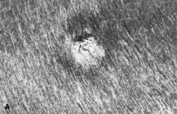
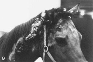
* Portions of this chapter are adapted from previous editions, which were written by Anne G. Evans, DVM.

* Platinum Performance, Inc; Buellton, Calif; Dr. W. Rosenkrantz, personal communication, 2004.

* Organic iodide powder, Neogen Corp, Lexington, KY, or EDDI 20 Gr. Dextrose base, Vedco, 5503 Corporate Dr, St. Joseph, MO.

* Nomagen, Fort Dodge Laboratories, Fort Dodge, Iowa.

Imiquimod 5% cream; Aldara, 3M Pharmaceuticals, Minneapolis, MN.

* Indian Mud, Original Cream Company, Magnolia, AR; XXTERRA, Larson Laboratories, Fort Collins, Colo; Animex, NIES, Las Vegas.

Efudex (5% fluorouracil), Hoffmann La Roche, Nutley, NJ.

* www.vgl.ucdavis.edu/service/horse/index.html.

* www.vgl.ucdavis.edu/service/horse/index.html.