Orthopedic Injuries

Before loading and transport to the hospital, orthopedic injuries should be appropriately stabilized with bandages, splints, or casts as described later in this chapter. Horses with fractures or breakdown injuries are best loaded onto a trailer by ramp, or the trailer can be positioned so that the step is level with the ground, such as backed against a curb, a dirt mound, or into a ditch. This allows the horse to be loaded and unloaded without having to step up into or down out of a trailer while trying to protect the injured limb. The horse should be hauled in a slant load or two-horse trailer with partitions and a chest and rear bar to allow it to lean and redistribute its weight off the affected limb.

image TECHNICIAN NOTE

Horses with fractures or breakdown injuries are best loaded onto a trailer by ramp or the trailer can be positioned so that the step is level with the ground, such as backed against a curb, a dirt mound, or into a ditch.

Because it is more difficult for the horse to maintain its balance when stopping suddenly than when accelerating, horses with forelimb fractures should be transported facing the back of the trailer. Horses with hind limb fractures can be transported facing forward in the normal direction.

Recumbent Horses

The greatest difficulty in transporting recumbent horses is the loading process. This is also the time of greatest danger because horses that thrash or are seizing pose the risk of kicking the veterinarian or the assistants. For this reason, recumbent horses should be heavily sedated or anesthetized before loading. Once sedated, the horse should be fitted with head protection and all four limbs wrapped with thick quilts and leg wraps. Recumbent horses are best hauled in open stock trailers or horse trailers in which the middle partitions have been removed. If possible, the trailer should be positioned on lower ground than the patient to allow gravity to assist the loading process. Soft cotton ropes can be attached to the limbs, tail, and halter to allow the horse to be manually pulled onto the trailer. Alternatively the horse can be positioned on a plywood or rubber mat in which rope has been attached to the front corners (Figure 33-78). The ropes can then be passed through the front of the trailer and attached to a truck, tractor, or winch system to allow the horse to be slowly pulled into the trailer. Specially designed air mattresses are available to provide cushioning for the patient during transport. In their absence, the patient can be surrounded by hay bales to prevent excess repositioning during transport.

image

FIGURE 33-78 Recumbent horse ready for transport

Foals

Foals should be hauled in a separate compartment from their dam and with an assistant. Young foals tend to lie down during transport, but may be injured while attempting to stand while the vehicle is moving. For this reason, foals should be hauled with an attendant or sedated.

image TECHNICIAN NOTE

Foals should be hauled in a separate compartment from their dam and with an assistant.

Specialized Purpose-Built Equine Ambulances

Many specialty practices provide purpose-built ambulance service for ill or injured horses (Figure 33-79). These ambulances are manned by personnel trained in the rescue, care, and hauling of sick or injured patients. There are a variety of horse trailers that are converted to provide specific functions to ease the loading and hauling of patients. Some trailers are designed to allow front and rear loading and unloading to prevent patients from having to back on or off of the trailer. Other equine ambulances are equipped with hydraulic systems that allow the rear of the ambulance to be lowered to the ground to allow the horse to load without stepping up into the ambulance. The inner walls of these trailers are padded and in some cases are also equipped with a hydraulic system to allow repositioning of the wall once the horse is loaded. Slings are available to minimize weight bearing on fractured limbs in some ambulances, and winch systems with rubber palates and air mattresses are available to assist in loading and hauling patients that are recumbent.

image

FIGURE 33-79 Purpose-built equine ambulance

PATIENT ASSESSMENT

A presumptive diagnosis may be made based on the history and signalment of the patient. For example, a broodmare within 60 days of foaling that has acute severe abdominal pain is likely to be suffering from a large colon volvulus that will require immediate surgery.

The severity of the situation often results in the initiation of treatment without a complete physical examination. Triage is the assignment or priority order to projects on the basis of where resources are best used. This means to focus on the immediate needs of the patient. For instance, if the patient exhibits signs of severe dehydration and shock, an IV catheter should be placed and fluid therapy initiated before a thorough examination or definitive treatment is initiated.

The patient assessment should begin as the patient is unloaded from the trailer. Severe hemorrhage from a laceration should prompt the examiner to assess heart rate and mucous membrane color before the administration of sedation because even appropriate doses of sedation in a patient with acute blood loss can result in recumbency or even death. A patient with a history of abdominal pain may have abrasions along the head and face suggesting severe pain. This may dictate the location where the examination and initial treatment is performed. Horses that repeatedly lie down and thrash as a result of pain should not be placed in a stock because of the potential for orthopedic trauma. These patients may be best examined in a large stall with exits in both the front and rear of the stall to allow personnel to escape if necessary or in the middle of a large open examination room. If at a referral hospital, patients suffering from severe abdominal pain may be moved directly into the surgical induction area for examination and initial treatment.

The animal's mental status can also be evaluated shortly after unloading. A young thoroughbred colt would not be expected to be docile when unloaded into a new environment. This may suggest depression associated with dehydration or other systemic disease.

Measurement of the animal's temperature, pulse, and respiration, and their mucous membrane color and CRT, should be collected at a minimum. Heart rate is an indication of the level of pain and also provides an estimation of hydration or the level of blood loss. Respiration rate may suggest a compromise or disease affecting the respiratory system or may be an indicator of infectious disease. Examination of mucous membrane color (Figure 33-80), moisture, and CRT gives a rapid assessment of peripheral perfusion and the delivery of oxygen to the tissues. Pale mucous membranes that are tacky with a CRT greater than 3 seconds are consistent with a patient that is dehydrated. Brick red mucous membranes with a normal or rapid CRT may indicate endotoxemia or sepsis. Mucous membranes that are purple are frequently observed in patients with severe abdominal disease that are dehydrated, endotoxemic, and have poor peripheral perfusion or hypoxemia. Finally, mucous membranes that are pale or ashen may suggest severe blood loss or the failure to adequately deliver oxygen to the tissues.

image

FIGURE 33-80 Example of normal light pink, moist equine mucous membranes

image TECHNICIAN NOTE

Minimal data that should be collected from all patients seen on an emergency basis include temperature, pulse, and respiration, mucous membrane color, and CRT.

SPECIFIC EQUINE EMERGENCIES

ABDOMINAL PAIN (COLIC)

Abdominal pain, otherwise known as colic, is one of the most common emergencies encountered in equine practice. A large percentage of horses examined for colic will respond and their signs resolve with minimal or no specifically directed therapy. Gas or spasmodic colic is usually mild, and the administration of analgesics (flunixin meglumine) and laxatives (mineral oil) result in rapid resolution of signs of pain. More important are the small percentage of horses that require intensive medical therapy or surgical intervention to correct potentially life-threatening causes of abdominal pain. Early recognition and aggressive therapy is essential to the survival of these patients. In practices that treat more serious cases of abdominal pain, the veterinary technicians need to have a basic knowledge of the common conditions that occur in the practice area. For instance, coastal Bermuda grass hay is a common component of the equine diet in the Southeastern United States. This feed has been intimately associated with the development of mechanical obstruction of the gastrointestinal tract. This condition often requires intensive care or abdominal surgery. In addition, certain areas of the country are prone to specific conditions as a result of the predominant population of horses. In areas with a high concentration of broodmares, surgical colic—such as large colon displacement or volvulus—are frequent occurrences, particularly in mares within 60 days of foaling. Without adequate triage and surgery, theses patients will almost certainly die from these conditions. The typical examination of a horse with abdominal pain will include a general physical examination (assessment of the severity of pain, temperature, pulse, and respiration, assessment of mucous membranes and gastrointestinal motility), nasogastric intubation, abdominal palpation per rectum, abdominocentesis, complete blood count (CBC) and biochemistry profile, and abdominal ultrasound examination. In specialized practices, in areas where colic associated with enterolithiasis (large stonelike masses that form in the colon and mechanically obstruct the passage of ingesta) is common or in the examination of foals with abdominal pain, abdominal radiographs may also be performed.

General Physical Examination

Minimal materials are required to perform a good physical examination. Essentially, good observation skills, a thermometer, and a stethoscope are all that are required. Initially a presumptive diagnosis can be made based on the history and signalment of the patient. Much information can also be gained from the observation of the patient at a distance. The degree of pain can be assessed by observing patient behavior. The severity of gastrointestinal disease is often directly correlated to the severity of abdominal pain. Mild pain is recognized as intermittent pawing, stretching out, flank watching, splashing water in buckets, and frequent attempts to urinate. These horses often show considerable relief with light sedation or mild exercise, such as walking.

Horses with moderate pain will also exhibit these signs, but also have an elevated heart rate (greater than 40 beats per minute), intermittent attempts to lie down and stand up, occasional rolling, restlessness, and anxiety. These horses will often remain comfortable for short periods of time (45 to 60 minutes) following sedation with an α2-agonist, such as xylazine or detomidine, with or without an opioid, such as butorphanol. Severe abdominal pain is often manifested as continuous pawing and kicking at the abdomen; repeated, sometimes violent, attempts to lie down; rolling; and frenzied behavior. Sedation may have minimal effect on this degree of pain often lasting as short as 5 to 10 minutes. The presence of severe pain can also be suspected in horses that have evidence of trauma, such as abrasions over the head, face, eyes, and boney prominences of the body or patients that are covered with an unusual amount of dirt, dust, or mud suggestive of repeated recumbency and thrashing.

Abdominal distention may be noted at a distance. If suspected, the owner may be questioned regarding the normal contour of the patient's abdomen.

If possible, the patient's temperature, pulse, respiration should be taken and recorded, and an estimation of the horse's gastrointestinal motility should be performed and noted in the medical record before the administration of sedation. Drugs administered for sedation will lower the horse's pulse and respiratory rate either by direct suppression or improvement in the level of pain. The horse's temperature must be taken before rectal examination. Air introduced into the rectum by the examiner's arm will artificially lower the body temperature measured per rectum.

image TECHNICIAN NOTE

Temperature, pulse, and respiration should be taken, and an estimation of the horse's gastrointestinal motility should be made before the administration of sedation.

Gastrointestinal motility can be estimated by auscultation of the abdomen with a stethoscope beginning in the paralumbar fossa and proceeding along the caudal edge of the costal margin toward the xyphoid. This should be performed on both sides of the abdomen. Sounds typically heard may be the result of either progressive large intestinal motility (borborygmi) or mixing of ingesta. The two sounds are indistinguishable, but provide a means of assessing gastrointestinal health. Normal large intestinal motility is perceived as prolonged fluid rushing that occurs every 2 to 4 minutes. In addition, high-pitched tinkling sounds can be ausculted in the right paralumbar fossa every 2 to 4 minutes and are routinely associated with cecal motility. Progressive small intestinal motility cannot be determined by abdominal auscultation. It must also be remembered that many factors, including the last time the horse has eaten, the horse's degree of anxiety, and previous administration of medication, may significantly alter gastrointestinal motility. Gastrointestinal motility can be significantly decreased by the administration of sedation. However, the complete absence of gastrointestinal sound in a patient with abdominal pain is always significant. This information is usually recorded in the record by abdominal quadrant (upper and lower right and left) as absent, decreased, normal, or increased. The abnormal accumulation of gas can also be determined by simultaneous auscultation and percussion. This is generally performed over the right paralumbar fossa. The head of the stethoscope is held in place, and the abdomen is percussed by briskly and repeatedly thumping the body while listening for a “ping.” This is a high-pitched sound similar to a basketball bouncing on a concrete surface. The presence of a ping often denotes an abnormal accumulation of gas in a hollow viscus.

The horse's level of hydration can be assessed by pinching the skin over the neck, shoulder, or upper eyelid and observing for prolonged skin tent. Care must be taken in assessing hydration in overly fat and thin horses since skin tent is partly the result of subcutaneous fat accumulation. Fat horses may be significantly dehydrated with a near normal skin tent as a result of a large amount of subcutaneous fat. Thin horses with normal hydration may have a prolonged skin tent as a result of little subcutaneous fat. For this reason, the author generally recommends that the skin over the upper eyelid be used to assess hydration. Mucous membrane color, moisture, and CRT are also assessed (Figure 33-81). All physical examination findings should be immediately recorded in the medical record. This should include the time of the horse's arrival and the time and dose of any pain medications administered. The perceived time between the administration of sedation or pain medication may be different than the actual time.

image

FIGURE 33-81 Brick red mucous membranes in a horse suffering from severe diarrhea, colic, and endotoxemia

Nasogastric Intubation

Passage of a nasogastric tube is a procedure performed by a veterinarian that is both diagnostic and therapeutic. Often the presence of abdominal pain is the result of accumulation of gas and fluid in the small intestine resulting from either a functional or mechanical obstruction of flow. Over time this ingesta may back up into the stomach, causing severe distention contributing to the abdominal pain. This excess fluid is termed gastric reflux. Elimination of gastric reflux through a tube reduces the distention, improves the patient's comfort, and may prevent rupture of the stomach, which is a universally fatal event. This procedure should be performed in all patients examined for abdominal pain, regardless of the severity of pain or cause. Nasogastric tubes are available in a variety of sizes ranging from ¼ to ¾ inches in diameter (Table 33-3) and are manufactured from silicone, polyvinyl chloride (PVC), or polyurethane.

TABLE 33-3

Relative Sizes of Nasogastric Tubes Used in Different Sized Equine Patients

image

The size of the tube is determined by the size of the patient. In general, the largest tube that can be passed through the ventral meatus of the nose into the esophagus should be used. For foals where commercially available nasogastric tubes may be too large, a stallion urethral catheter or in miniature foals a red rubber catheter is a reasonable alternative. The patient is generally lightly sedated and restrained with a nose twitch. The end of the tube is lubricated with a water-soluble lubricant and passed through the nose down the esophagus and into the stomach. Once in the stomach, it is often necessary to fill the tube with water to create a siphon effect to allow excess fluid to be removed. This procedure may be repeated several times in a row, especially if the stomach contains a large amount of feed material and is often repeated at regular timed intervals to assess response to therapy and control pain. The net volume, appearance, and odor should be recorded in the medical record. Normal stomach content is usually light green and is often foamy, and typically, less than 4 L is obtained. Net volumes (total volume collected − volume instilled to create a siphon) should always be recorded in the record. The presence of spontaneous reflux, large volumes greater than 4 L, foul-smelling fluid, and discolored (yellow, orange, or bloody) fluid are all significant and should be recorded. Following removal of gastric reflux, the tube may be removed, temporarily maintained until the completion of the examination to allow administration of oral fluids and laxatives, or fixed in place using nonelastic tape to allow repeat removal of accumulated reflux or administration of medication (refer to Chapter 31, Figure 31-17).

image TECHNICIAN NOTE

Normal gastric contents is typically low volume (less than 4 L), light green in color, and foamy.

Bleeding from the nose is a frequent complication to nasogastric intubation and may cause the owner great concern. This is usually the result of trauma to the ethmoid turbinate and nasal passage and is not of serious consequence. The nose should be loosely covered by a towel to prevent blood from being blown by the horse over equipment, the holder or handler, or the environment. Bleeding generally stops within minutes of removing the tube.

Abdominal Palpation per Rectum

Rectal palpation is a diagnostic procedure performed in patients suffering from severe, recurrent, or persistent abdominal pain. This technique allows the veterinarian to identify the organ system involved, the approximate location of abdominal viscera, to evaluate the abdominal contents, to assess abnormalities in the wall of the bowel, and to localize pain. A rectal examination should only be performed in a horse that is adequately restrained, and proper lubrication is critical to prevent potentially life-threatening complications, such as rectal tears or injury to the examiner. If possible, the horse should be restrained in a standing stock. Light sedation is recommended, and a nose twitch should be applied. A shoulder-length sleeve is used to protect the examiner's hand and arm from fecal contamination, and copious quantities of water-soluble lubricant (Figure 33-82) should be available. In horses that strain during the examination, a caudal epidural can be administered, or 50 to 60 ml of 2% lidocaine can be mixed with an equivalent amount of lubricant and infused into the rectum using a dose syringe to provide a local anesthetic effect. Immediately following the examination, the rectal sleeve should be examined for evidence of blood. Following the examination, all feces and lubricant should be removed from the tail and perineal area with a moistened cloth or warm water.

image

FIGURE 33-82 Shoulder-length sleeve and water-soluble lubricant for rectal palpation

Abdominocentesis

Abdominocentesis, otherwise known as a “belly tap,” is the surgical puncture of the abdominal cavity for the purpose of obtaining abdominal fluid (Figure 33-83). This technique provides information regarding the health of the peritoneal cavity and may reveal horses with devitalized intestine that require surgery or may identify horses suffering from rupture of a viscus. This technique requires absolute aseptic preparation. A 10-cm × 10-cm area of the most dependent part of the abdomen, usually centered 3 to 5 cm caudal to the xyphoid and 3 to 5 cm to the right of midline, is clipped. The area is cleaned of dirt, debris, and dander using soap and water or alcohol followed by a final scrub using aseptic technique. Two to 5 ml of 2% lidocaine can be infused into the skin and subcutaneous tissues at the proposed site for centesis. A variety of instruments may be used to collect abdominal fluid, depending on the veterinarian's preference (Figure 33-84), including an 18-gauge × 1½-inch needle, 18-gauge × 3½-inch spinal needle, sterile teat cannula, or sterile bitch catheter. Once the instrument has entered the abdominal cavity, fluid is collected into an ethylenediaminetetraacetic acid (EDTA) tube (purple top) and red-top tube. The fluid is grossly examined for color and clarity. Additional information may be obtained by measuring TP by refractometer and submission to the laboratory for cell count, cytology, Gram stain, and culture. Normal peritoneal fluid values are found in Box 33-2.

BOX 33-2   Normal Peritoneal Fluid Values

Normal Peritoneal Fluid

Gross appearance

Clear and straw colored or yellow

Cellularity

Adult WBC ≤10,000 cells/μl

Foal WBC ≤1500 cells/μl

TP ≤2.5 g/dl

image

FIGURE 33-83 Surgical puncture of the abdomen (abdominocentesis) for the collection of peritoneal fluid

image

FIGURE 33-84 Common instruments used to perform abdominocentesis. From left to right: 18-guage × 1.5-inch needle, 20-guage × 3.5-inch needle, sterile teat cannula, and sterile bitch catheter

Clinical Pathology

At a minimum, a PCV and TP measured by refractometer should be measured at the time of presentation. This minimal database is easily obtainable, provides rapid results, and is a good estimation of hydration. In addition, repeated PCV and TP measurements provide a means of assessing improvement in hydration status and may be used by the veterinarian to determine prognosis. Although CBCs and biochemistry profiles do not provide specific diagnostic information regarding the cause of abdominal pain, alterations in normal may direct antibiotic therapy, fluid and electrolyte therapy, and response to treatment and may give an indication of prognosis. Biochemistry profiles may also provide information indicating sources of pain other than the gastrointestinal tract. For instance, horses suffering from rhabdomyolysis (tying-up) may have signs similar to colic. Significant elevation of creatine kinase (CK) and aspartate transaminase (AST) on a biochemistry profile may alter the diagnostic and therapeutic plan in these horses (see Chapter 16).

Abdominal Ultrasound

This is a valuable tool for the evaluation of abdominal pain in horses. It is a safe, noninvasive diagnostic technique that allows direct evaluation of the abdominal viscera and evaluation of nongastrointestinal structures. Removal of the hair over the abdomen is not necessary to perform abdominal ultrasound. The body surface is thoroughly wet with isopropyl alcohol and a 2.5- to 10.0-MHz probe is used to evaluate the intraabdominal structures.

image TECHNICIAN NOTE

Ultrasound is a safe, noninvasive diagnostic technique that allows direct evaluation of the abdominal viscera and nongastrointestinal structures, such as spleen, liver, and uterus.

Specific information obtainable by ultrasound includes visceral distention, wall thickness of the abdominal viscera, and motility of specific portions of the gastrointestinal tract. Specific causes for abdominal pain, such as nephrosplenic entrapment, intussusception, and diaphragmatic hernia, can be reliably diagnosed by ultrasound examination. Ultrasound examination also allows the veterinarian to locate fluid for abdominocentesis; identification of intraabdominal pathologic conditions, such as an intraabdominal abscess; assessment of fetal viability; and evaluation of intraabdominal organs, such as the liver and spleen.

Abdominal Radiology

Radiology of the equine abdomen has limited value because of the horse's large size and poor tissue resolution, high radiation exposure to personnel, and decreased life of radiographic equipment. Its usefulness improves in areas of the country where enterolithiasis is a common problem. Additionally, radiology may be useful as a diagnostic tool on foals and in small horses, such as American miniature horses, to assess gastric, small intestinal, and large intestinal distention. Contrast radiology is useful for the diagnosis of detailed gastric emptying and meconium impaction in foals.

Abdominal Exploratory

Abdominal pain that is the result of disruption of the blood supply to the intestine (strangulation), malpositioning of segments of the intestine (displacement), or mechanical obstruction of the bowel require abdominal surgery for correction (Figure 33-85). The five most common indications for abdominal surgery in the horse are: (1) an abnormal rectal examination, (2) large quantities of gastric reflux, (3) an abnormal abdominocentesis, (4) uncontrollable abdominal pain, and (5) systemic deterioration or a patient that fails to improve even with aggressive and appropriate medical therapy. Once the decision has been made to pursue surgical correction, the patient will require presurgical preparation to minimize the anesthetic time and to ensure the best chance for a positive outcome.

image

FIGURE 33-85 Horse in which a portion of the small intestine has lost its blood supply

If the patient has not received an IV catheter, one should be placed at this time to allow the administration of perioperative antibiotics and anesthetic-induction drugs. If the patient has not received a tetanus vaccination in the last 6 months or if the vaccination history is unknown, a tetanus toxoid should be administered before surgery. Perioperative antibiotics are administered no more that 30 to 60 minutes

CASE PRESENTATION 33-1

A 10-year-old quarter horse mare acutely developed signs of colic: inappetence, pawing, and rolling. The concerned owner called the local equine veterinarian to come out to the farm. On a physical examination, the veterinarian noted depression; pale pink mucous membranes; CRT = 2 seconds; temperature = 99° F; tachycardia (heart rate = 60 bpm); tachypnea (respiratory rate = 40 breaths/min); and dry, scant manure in the stall. On auscultation of the abdomen, borborygmal sounds were decreased (0 or 1) in each of the four quadrants. A rectal examination revealed a large amount of doughy fecal matter in the large colon. The veterinarian passed a nasogastric tube and obtained no refl ux, so he administered 4 gal of warm water and ½ gal of mineral oil. He also gave 500 mg fl unixin meglumine IV for pain. The veterinarian diagnosed an impaction in the large colon and referred the patient to a local equine hospital for additional treatment.

At the referral hospital, the experienced veterinary team performed their usual tasks. The veterinary technician passed a nasogastric tube. The veterinarian spoke with the owner briefl y to obtain the patient's history and performed a physical examination with similar fi ndings to the local veterinarian. Since no refl ux was obtained, the veterinary technician administered 6 L of warm water and ½ gal of mineral oil via the nasogastric tube. A second veterinary technician performed a sterile prep, placed a 14-gauge catheter in the right jugular vein, and started 20 L of crystalloid fl uids (Normosol R). The veterinarian performed a rectal examination and confi rmed the diagnosis of impaction in the large colon. Since the horse was mildly uncomfortable and continued to paw, the veterinarian asked the veterinary technician to administer 150 mg xylazine and 4 mg butorphanol IV. The veterinary technician performed a venipuncture and submitted samples for venous blood gas, CBC, and chemistry profile. The abnormal results included: increased PCV and total protein, and decreases in levels of sodium and chloride.

The horse was placed in the ICU and monitored frequently for comfort by the veterinarian and veterinary technicians. Within a few hours, she appeared more comfortable and began to pass small amounts of dry manure. The veterinary technicians continued to pass a nasogastric tube every 6 hours and administer 6 L of warm water and 1/2 gallon of mineral oil. By the next morning, the veterinarian repeated a rectal examination, and the impaction was smaller in size. The owner called and said he found that the automatic water machine in the horse's stall was broken. She did not have access to water for at least 24 hours, which caused this impaction.

The mare continued to improve with 2 days of treatment at the hospital, and the impaction resolved. Small amounts of hay and soaked feed were offered, and the horse ate readily. She showed no more signs of colic and was discharged to a grateful owner. The veterinary team was happy to have helped another patient with their effi cient and effective team approach.

before the induction of anesthesia. This will ensure that adequate plasma levels of antibiotics will have been reached by the time the incision has been made and will be maintained for the duration of the surgery. The choice of antibiotic is at the discretion of the surgeon; however, the broad-spectrum combination of an aminoglycoside antibiotic, such as gentamicin, and crystalloid penicillin, such as potassium penicillin, administered IV is most common. Potassium penicillin should not be administered within 15 minutes of induction because of the effect of high levels of IV potassium on the heart and blood pressure.

image TECHNICIAN NOTE

Perioperative antibiotics should be given IV 30 to 60 minutes before the induction of anesthesia.

If it is safe to do so, the ventral abdomen should be clipped from the xyphoid caudally to the inguinal area and the clipped area cleaned with soap and water or alcohol to remove all gross contamination. Once the patient is anesthetized, any remaining hair should be removed, and a final rough scrub applied to remove any further debris. A final aseptic scrub using chlorhexidine or Betadine scrub and rinsed with alcohol is applied before surgery.

ORTHOPEDIC INJURIES

Orthopedic injuries are a common occurrence in horses. The large size, temperament, use, and speed of the horse can result in catastrophic life-threatening injury. There are injuries that are common to specific disciplines, such as condylar fractures and disruption of the suspensory apparatus in thoroughbred racehorses or first or second phalanx fractures in Western performance horses. These injuries occur with regular frequency and are directly related to the horse's occupation. There are other orthopedic injuries that occur without obvious knowledge of their cause. Horses that are pastured are frequently found at the time of morning feeding with fractures, laceration, and serious injury that can arise from kicks by other horses, missteps during exercise, or obstacles in the environment. Regardless of the cause of an orthopedic injury, first aid and stabilization of the fractured or injured limb before transport to the hospital are some of the most important determinants of a successful outcome or repair. Assuming the horse's systemic condition has been appropriately assessed and stabilized, the goals of adequate temporary external fixation of a fractured limb include the elimination of patient pain and anxiety; to allow at least partial weight bearing on the affected limb; minimize further damage to the surrounding soft tissue, including muscle, blood, and nervous supply; and to prevent rubbing and eburnation of the bone ends that may affect reduction and definitive repair.

image TECHNICIAN NOTE

Temporary external coaptation of a fractured limb reduces the patient's pain and anxiety, allows partial weight bearing on the affected limb, protects the soft tissue from further trauma, and prevents eburnation of the fractured bone ends.

External fixation of an unstable limb should occur before any further diagnostic techniques or movement and transport of any injured patient in which surgical repair is to be considered. For the purpose of fracture stabilization, the front and hind limbs are divided into four regions (Figure 33-86), and specific recommendations for these regions have been published.

image

FIGURE 33-86 Anatomic division of the horse for emergency external coaptation

Forelimb Injuries

Ground to Distal Metacarpus: Injuries that commonly occur from the ground to the distal metacarpus that require emergency external coaptation include unstable fractures of the first and second phalanges, luxation of the fetlock joint, fractures of the distal metacarpus, laceration of the flexor tendons, disruption of the suspensory apparatus by fracture through both proximal sesamoid bones, and displaced condylar fractures. With the exception of condylar fractures, these injuries result in hyperextension of the fetlock joint either because of loss of the slinglike support of the fetlock by the suspensory apparatus or a change in the location of the primary bending force of the distal limb from the fetlock joint to the fracture. Hyperextension of the fetlock can result in damage to the nerves and blood vessels of the distal limb as a result of tearing or thrombosis of the palmar digital vessel. Therefore the goal of emergency coaptation is to prevent hyperextension of the fetlock. This can be accomplished by splinting the limb with the dorsal surfaces of the bones of the phalanges and metacarpus in axial alignment. There are several means in which this can be accomplished. The simplest is to apply a lightly padded bandage with a dorsally applied splint. The most common splint material for this purpose is PVC pipe cut to size. A bandage with approximately 1 cm of compressed padding is applied from the coronary band to the carpus. The splint is applied to the dorsal surface of the limb and extends from the proximal metacarpus to the tip of the hoof and is fixed in place with either 3-inch nonelastic tape or a more rigid alternative would be fiberglass cast tape. A wooden wedge can be incorporated beneath the heels to assist in maintaining the alignment of the bony column.

Alternatively, rigid stability can be provided by application of a distal limb cast. In a quiet, lightly sedated horse, elevation of the front foot off the ground results in natural alignment of the bony column. This is accomplished by an assistant grasping the limb behind the carpus and pulling the carpus up and forward in front of the horse (Figure 33-87). By this method, a distal limb cast can be applied to the standing horse. This technique requires that the horse stand still balancing on three limbs at least until the cast is applied and hardened, which can take from 30 to 60 minutes, depending on the material used and the skill of the veterinarian. Therefore this may not be appropriate to attempt in all horses. In these cases, short-term general anesthesia may be required to apply the cast correctly.

image

FIGURE 33-87 Proper elevation of the forelimb to allow standing application of a distal limb cast on the front leg

Correct cast application is vitally important to an overall successful outcome. Casts that are applied too tight or that have folds or ridges adjacent to the skin will result in limb ischemia or cast sores that may be as or more severe than the original injury. The needed material and equipment to apply a cast should be accumulated and organized before anesthetizing the patient or attempting cast application (Box 33-3).

BOX 33-3   Material and Equipment Necessary to Apply a Half-Limb Cast

Cast Equipment and Material

Stainless steel or other appropriately clean bucket

Water—the temperature specified by the manufacturer of the cast tape

Examination gloves or gloves provided by the manufacturer to properly apply cast material

Cast synthetic stockinette

Cast felt

Bandage scissors

1-inch white tape

Custom Support Foam or other appropriate cast padding

Various cast tape size, prim1208arily 4 or 5 in for adults, 3 in for foals

Polymethylmethacrylate (acrylic)

If possible, the shoe should be removed, the hoof appropriately trimmed, all debris and loose sole and frog removed, and the periople rasped. If the patient will not tolerate adequate preparation of the hoof, the cast can be applied directly over the hoof and shoe. To apply the cast, the limb is initially covered by the appropriately sized double layer of synthetic stockinette. This material should fit snug but not tight and should not sag. Felt cast padding of approximately 1 inch width should be applied circumferentially around the limb at the top of the cast and over any bony prominences to prevent rubbing. The stockinette is then covered by a ½-inch layer of cast padding or a porous, water-curable polyurethane foam bandage material, such as 3M's Custom Support Foam. Custom Support Foam is resilient, porous, produces a close anatomic fit, and has the advantage of adherence to the fiberglass-resin cast tape that will be applied, thus forming a single unit with the cast material. This decreases the risk of movement and the development of cast sores. Padding material of any type should not be applied too thick. If cast material is too thick, it will become compressed with weight bearing, and the cast will slip, potentially resulting in serious cast sores.

Plaster of Paris cast material has all but been replaced by polyurethane-impregnated fiberglass cast tape in cast application because of its superior strength and light weight. A wedge-shaped block of wood or roll of cast material positioned beneath the heels provides additional support and helps to maintain alignment while the cast is applied. The manufacture's recommendations for water temperature and time of submersion of rolls of cast material should be read before application.

This first layer of cast material should be applied without tension directly over the foam padding with no creases or folds to minimize cast sores. Subsequent layers should be applied with increasing tension. Additional strength can be gained by fashioning a splint by layering a roll of cast material in alternating directions to the length of the cast. This can be applied to the dorsal surface of the cast and fixed in place with additional rolls of cast material. For an adult horse 4 to 7, 4- to 5-inch rolls of cast material are used. The cast material is overlapped by no more than one half of its width during application. Once the cast tape has been applied, the bottom of the cast can be protected from excess wear by the application of polymethyl methacrylate (PMMA). The surface of the PMMA should be made rough by compressing it with a rasp to prevent slipping.

An added benefit to the application of a cast for temporary fracture stabilization is that radiographs may be taken and the fracture assessed through the cast (Figure 33-88).

image

FIGURE 33-88 Example of radiographs of a previously repaired fracture taken through the cast. Note that the radiopaque cast material allows the fracture and implants to be visualized through the cast

Another alternative is the application of a manufactured purpose-built splint to the distal limb. The Kimzey Leg Saver is a good example of a purpose-built splint specifically designed to align the boney column of the distal limb (Figure 33-89). These can be applied over a light padded bandage and can be used to support both fractures of the distal limb and disruption of the suspensory apparatus.

image

FIGURE 33-89 Kimzey Leg Saver splint applied to the distal limb of a horse with a fracture

image TECHNICIAN NOTE

The Kimzey Leg Saver is a commercially available distal limb splint designed for temporary stabilization of fractures of the limb below the distal one third of the metacarpus or metatarsus.

Distal Metacarpus to Distal Radius: The most common injury to this region of the limb is fracture of the third metacarpal (cannon) bone. Fractures in this region are severely unstable and require fixation of both the joint above and below the fracture to provide stability and shared weight bearing. A Robert Jones bandage with two splints at 90-degree angles provides excellent stability to these fractures. It has the additional advantages of controlling hemorrhage and minimizing the development of and hastening the resolution of swelling associated with edema. The Robert Jones bandage is composed of multiple layers of sheet cotton or similar material compressed by gauze. The final diameter of the completed bandage should approximate three times the diameter of the normal limb. For fractures of the forelimb, splints consisting of PVC pipe cut to extend the full length of the limb or wood should be positioned caudally and laterally and fixed with nonelastic tape or cast tape.

Distal Radius to Elbow: Anatomically the flexor and extensor tendons of the forelimb act as abductors of the limb once disruption of the boney column of the radius occurs. Therefore these fractures are at high risk for penetrating the skin on the medial aspect of the limb as a result of the absence of soft tissue covering the radius at this location. Therefore external coaptation of these fractures not only must provide axial stability, it must also prevent abduction of the limb. This is accomplished by the application of a Robert Jones bandage as previously described; however, in addition to the caudal splint, a lateral splint that extends to the top of the scapula and is secured to the scapula area by tape or other suitable bandage material that encircles the axilla, thorax, and withers should be applied (Figure 33-90). The lateral splint is commonly composed of 2 × 4-inch lumber, and elastic tape is employed to fix it to the scapula to prevent abduction.

image

FIGURE 33-90 Full limb Robert Jones bandage with a caudal and lateral splint extending to the withers for transport of a patient with a suspected distal radial fracture

Olecranon Fractures: Complete fractures of the olecranon result in the disruption of the horse's triceps apparatus. The triceps muscle serves to extend the elbow. The ulna, which is the distal extremity of the olecranon, is at least partially fixed to the caudal aspect of the radius and acts as a lever against the pull of the triceps muscle and is necessary for the horse to fix the carpus in extension. Therefore horses with complete fractures of the olecranon lose this lever and are unable to fix the carpus in extension. This results in the classic “dropped elbow” appearance and buckling or flexion of the carpus (Figure 33-91). Although this is not a weight-bearing bone and typically responds well to surgical fixation, loss of the ability to extend the carpus results in a great deal of anxiety for the horse and makes ambulation on three legs difficult. Patients with olecranon fractures should be treated with a stacked bandage extending the entire length of the limb and a dorsally applied splint extending at least the length of the carpus. This essentially allows the limb to act as a support post, and the horse quickly adapts to the rigid limb by using the muscles that extend the shoulder to advance the limb while walking. This type of splint is also useful in patients with radial nerve paralysis and those with fractures of the distal humerus and scapula that also lose the function of the triceps apparatus. In these cases, the splint should not extend proximally above the level of the fracture.

image

FIGURE 33-91 Dropped elbow appearance of a horse with a complete olecranon fracture

image TECHNICIAN NOTE

Horses with complete olecranon fractures are unable to extend the carpus. A full-limb bandage with a dorsally applied splint spanning the length of the carpus allows these patients to stand and ambulate comfortably.

Proximal to the Elbow: Fractures of this region are surrounded by a heavy muscle mass and are unable to be appropriately externally stabilized. Horses with fractures of the distal humerus that have lost their triceps apparatus function will benefit from a light full-limb bandage and cranially or caudally applied splint to fix the carpus in extension.

Hind Limb Injuries

Ground to Distal Metatarsus: Fractures in this area are similar to those described for the forelimb. The goal is alignment of the dorsal cortices of the bone. However, this is more difficult in the hind limb than the forelimb. To stabilize these fractures, a plantar splint is applied extending from the solar surface of the foot to the proximal metacarpal bone. Specialized splints, such as the Kimzey Leg Savers, are also valuable to support these fractures. Kimzey Welding Works Inc. also manufactures a hind limb leg saver splint that more closely matches the anatomy of the hind limb specifically for these fractures.

Distal Metatarsus to Tarsus: These fractures are also treated similar to fractures of the same region in the forelimb with a few exceptions. First, the thickness of the bandage applied to the hind limb is reduced compared with the forelimb. Second, a plantar splint can be applied using the calcaneus as the proximal extent of the splint. The external splint should extend only the distance of the most proximal extent of the calcaneus.

Tarsus to the Stifle: The response to contraction of the muscles surrounding the tibia is similar to those of the radius. Therefore fractures of the tibia result in abduction of the limb and a high potential for penetration of the skin covering the vulnerable medial aspect of the tibia. These fractures are stabilized with a Robert Jones bandage and a caudally applied splint extending to the length of the calcaneus and a lateral splint that extends proximally to the hip. A straight or angled splint following the contour of the hock can be fashioned and fixed in place using nonelastic tape.

Proximal to Stifle: The musculature proximal to the stifle is sufficient to stabilize fractures of the femur. It should be noted that complete fractures of the femur can result in significant subcutaneous and intramuscular accumulation of hemorrhage and severe swelling over the thigh. A compressive bandage should be applied to the distal limb to prevent excess swelling secondary to edema and gravitation of fluid distally from the fracture.

image TECHNICIAN NOTE

Fractures proximal to the elbow or stifle are unable to be appropriately stabilized using external bandages, splints, or casts. The large muscle mass surrounding the bones in this area provides adequate temporary stabilization.

SOFT TISSUE TRAUMA

Lacerations and puncture wounds occur frequently to the head, body, and limbs of the horse, but are most commonly observed on the distal limb. These injuries are usually the result of sharp lacerations from contact with loose sheet metal, barbed wire, and protruding nails, sawlike lacerations that occur when a limb becomes entangled in smooth wire, or blunt force trauma from kicks or high-speed impact with inanimate objects. Because of the limited soft tissue covering of the distal limb penetration of synovial structures, such as joints and tendon sheaths, or disruption of the neurovascular supply to the limb is a frequent potentially life-threatening complication. The initial care of soft tissue injuries has a profound influence on outcome of the injury.

Initial Assessment

The most important aspect of the initial assessment of the patient is the control of hemorrhage. It is often difficult to determine the exact amount of time that has passed since the initial injury and the owner finding the horse. Significant blood loss may have occurred during this time. The horse's environment makes accurate assessment of the volume of blood lost difficult. In some instances, the owner may report significant hemorrhage before presentation of the horse. In this instance, the owner can be instructed over the telephone in methods to control hemorrhage. In extreme circumstances, hemorrhage can be controlled by wrapping the distal limb with clean towels that are tightly compressed with Vet Wrap, elastic tape, or even duct tape. These crude bandages may become heavily soaked with blood, but they should not be removed until the patient has been transported to the hospital or the veterinarian has arrived at the farm. An unstable clot is likely to have formed under the bandage at least slowing the hemorrhage. Removing the bandage may disrupt the clot resulting in the recurrence of severe bleeding.

If there is active hemorrhage at the time of initial examination, this may be controlled by the application of a Robert Jones bandage or a tightly compressed stacked bandage. If there are exposed bleeding vessels, the examining veterinarian may elect to place a hemostat across the lacerated vessels followed by a ligature to control hemorrhage. The use of tourniquets to control hemorrhage is not advised. The ischemia that results from the application of a tourniquet will result in severe discomfort to the patient. In addition, tissue ischemia may result in the loss of vital soft tissue and the development of large defects that take an extended time to heal.

If severe hemorrhage is not obvious at the time of presentation, a brief physical examination should be performed before administering sedation to ensure that the patient is not exhibiting signs of hypovolemia associated with blood loss. Common signs of anemia as a result of blood loss include depression and lethargy, tachycardia, tachypnea, cool extremities, and pale mucous membranes. If these signs are noted, a PCV and TP should be measured and supportive care, such as IV fluids or blood transfusion, should be initiated before treatment of the wound.

Initial Wound Treatment

Once the patient has been stabilized, initial wound care revolves around decreasing further contamination, eliminating necrotic tissue, and removing foreign material from the wound. This is vitally important in wounds that are in close proximity to joints or tendon sheaths. The patient may be sedated and either regional anesthesia, such as nerve blocks, or local infiltration of the wound with lidocaine may be useful in controlling pain. Contamination of synovial structures may result in the development of septic arthritis or tenosynovitis (Figure 33-92). The hair surrounding all wounds should be removed with clippers or a razor. Before clipping, the wound should be covered with sterile moistened gauze or sterile water-soluble lubricant, such as K-Y Jelly, to prevent hair from entering the wound.

image

FIGURE 33-92 Laceration located directly over the fetlock joint with the potential for joint contamination and septic arthritis

The area clipped is dependent on the location of the wound. For wounds that are distant from synovial structures or those on the body or head, only the area immediately surrounding the wound needs to be clipped. For wounds that are suspected of involving a joint or tendon sheath, the limb should be clipped circumferentially and 5 to 10 cm proximal and distal to the wound to allow centesis of the synovial structure at a location distant from the wound. Once clipped, the skin surrounding the wound should be cleansed with an antiseptic soap and rinsed with saline.

image TECHNICIAN NOTE

Before clipping, wounds should be covered with sterile water-soluble lubricant to prevent hair from adhering to the wound.

Once the surrounding skin has been cleansed, the wound may be gently scrubbed using sterile gloves and an antiseptic soap and rinsed with saline. Wounds may be initially irrigated with saline by syringe and 18-gauge needle or by making multiple holes in the cap of a saline bottle, turning the bottle upside down, and applying pressure to the outside of the bottle to produce a forceful stream. For superficial wounds that do not involve synovial structures, debris can be removed from the skin and wound surface with antiseptic soap and rinsed with water. Excess fluid pressure should not be applied to the wound because this may force bacteria and debris deeper into the tissue.

Diagnostic Imaging

Any wound involving the distal limb, over a boney prominence, or that is chronic and nonhealing should be evaluated either by radiographs or ultrasound at the time of injury. Diagnostic imaging allows the early diagnosis of fractures (Figure 33-93) and foreign bodies (Figure 33-94) and provides a means for further evaluation of the development of sequestrum (Figure 33-95).

image

FIGURE 33-93 Splint bone fracture secondary to wound over plantar surface of the metatarsus

image

FIGURE 33-94 Intraarticular metallic foreign body associated with a fetlock wound

image

FIGURE 33-95 Sequestrum as a result of a wound over the proximal metatarsus

image TECHNICIAN NOTE

Any wound involving the distal limb, located over a boney prominence, or that is chronic and nonhealing should be evaluated either by radiographs or ultrasound.

Blunt traumatic wounds that are severe enough to disrupt the skin are severe enough to result in fracture of the small bones of the distal limb, such as the splint bones.

Ultrasound is useful in the evaluation of wounds for the detection of foreign bodies of nonbone or metallic origin. Wood, plant material, glass, and other nonbone or metal foreign bodies cannot always be detected by radiology. These structures can often be observed by ultrasound as bright hyperechoic structures that produce acoustic shadowing (Figure 33-96).

image

FIGURE 33-96 Characteristic ultrasonographic appearance of a foreign body with the bright hyperechoic line delineating the foreign material and acoustic shadowing

In addition, radiology allows early assessment of the bone for comparison in 3 to 4 weeks for the development of a sequestrum, a nonviable segment of bone that may develop as a result of trauma to the periosteum on the surface of the bone. This “dead” boney material also acts as a foreign body that will impede wound healing.

Specific Wound Therapy

Once the wound has been cleansed, the involvement of synovial structures determined, the extent of the wound fully assessed, and diagnostic imaging performed, definitive therapy can proceed. Wound therapy may be as straightforward as suturing the wound and the application of a bandage to as complicated as surgery, arthroscopic joint lavage, and the application of a cast. The treatment to be employed and the material needed will be determined by the veterinarian. However, in most instances, sedation; local anesthetic drugs, such as lidocaine; a surgery pack containing a scalpel handle, hemostats, thumb forceps, needle drivers, and scissors; and tetanus prophylaxis are the minimal materials required.

RESPIRATORY EMERGENCIES

Respiratory emergencies can be the result of an acute disease process or trauma, acute exacerbation, decompensation or progression of an existing condition, or the respiratory manifestation of a systemic disease. Respiratory distress occurs when there is inadequate delivery of oxygen from the lungs to the tissue and may be the result of an inability of oxygen to reach the lungs (obstructive disease), an inability of the lungs to expand adequately (restrictive disease), failure of oxygen to diffuse across the respiratory membrane at the alveolus, failure of oxygen to be transported in the blood to the peripheral tissues (anemia), or the inability of the tissue to extract or use delivered oxygen.

Common signs of respiratory distress in horses include rapid breathing (tachypnea); flaring nostrils; exaggerated thoracic or abdominal excursions; shrill, grating, or whistling inspiratory or expiratory noise also termed stridor; nasal discharge; or bluish-purple discoloration of the mucous membrane also known as cyanosis. Horses in respiratory distress are frequently anxious, assume an abnormal posture characterized by abduction of the elbows and extension of the head and neck, may be reluctant to move, and may exhibit signs of thoracic pain.

image TECHNICIAN NOTE

Common signs of respiratory distress include tachypnea, flaring nostrils, exaggerated abdominal or thoracic excursion, inspiratory or expiratory stridor, nasal discharge, and cyanosis.

It must be kept in mind that horses in severe respiratory distress may be hypoxic resulting in severe anxiety, panic, uncontrollable behavior, and sudden violent death. These horses pose a significant danger to the handler, veterinarian, and staff and should be handled with extreme caution. It is recommended that clients not be allowed to restrain horses exhibiting signs of severe respiratory distress, and for their own protection, they should be moved a safe distance away from the horse in case the patient becomes panicked or violent.

Upper Respiratory Tract Emergencies

Equine respiratory emergencies can be divided into conditions affecting the upper or the lower respiratory tract. Diseases affecting the upper respiratory tract are typically the result of obstruction of airflow to the lungs and are recognized most commonly by the presence of inspiratory stridor without abnormal lung sounds. These are likely the most common forms of respiratory emergencies in horses. This is due in part to the anatomy of the equine upper respiratory tract. The soft palate of the horse extends caudally as far as and ventral to the epiglottis dividing the oral and nasal pharyngeal areas completely except during swallowing (Figure 33-97). Because of this, horses are obligate nasal breathers and are unable to overcome decreased airway diameter or obstruction by opening the mouth as are dogs or humans. Nasopharyngeal foreign body or cicatrix (scars), severe facial swelling, and facial deformity resulting from acute fractures or space-occupying masses of the nasal passages or pharynx (Figure 33-98) cause respiratory distress as a result of this anatomic arrangement. In addition, an anatomic structure unique to the horse, the guttural pouch, or common anatomic structures in close proximity to the pharynx and larynx, such as retropharyngeal lymph nodes if enlarged, may impinge on these structures significantly decreasing upper airway diameter (Figure 33-99). Laryngeal paralysis may also be a potential cause of upper airway obstruction. Bilateral laryngeal paralysis in horses may be the result of ingestion of toxic plants, lead poisoning, or hyperkalemic periodic paralysis (HYPP). In addition, laryngospasm immediately following anesthetic recovery is a potentially life-threatening complication to prolonged anesthesia or poor positioning during surgery (extreme extension of the head and neck) causing upper airway obstruction.

image

FIGURE 33-97 Normal endoscopic appearance of the upper respiratory tract

image

FIGURE 33-98 Subepiglottic cyst acting as a space-occupying mass causing respiratory distress

image

FIGURE 33-99 Infection of the guttural pouch causing narrowing of the upper airway as a result of compression on the trachea

image TECHNICIAN NOTE

Diseases affecting the upper respiratory tract are typically the result of obstruction of airflow to the lungs and are recognized most commonly by the presence of inspiratory stridor without abnormal lung sounds.

Lower Respiratory Tract Emergencies

Conditions affecting the lower respiratory tract are divided into obstructive disease, restrictive disease, or diseases causing pulmonary edema. Obstructive diseases are characterized by the presence of expiratory stridor and abnormal lung sounds, including crackles and wheezes. Examples of these diseases include recurrent airway obstruction (ROA) or “heaves,” septic bronchitis or alveolitis, and lungworm infection. These conditions are the result of accumulation of inflammatory fluid in the small airways and reflex bronchospasm.

Restrictive disease prevents the lungs from expanding normally. These conditions are commonly associated with expiratory stridor and the absence of lung sounds on thoracic auscultation. Pneumothorax is an uncommon form of restrictive respiratory disease that usually accompanies severe thoracic trauma or penetration of the chest wall. In this condition, free air accumulates in the thoracic cavity causing collapse of the lung. Lung sounds in this case are absent dorsally over the thoracic wall. It is usually unilateral; because the mediastinum of the horse is incomplete, pneumothorax can be bilateral. A more common restrictive respiratory disease in horses is the presence of fluid in one or both sides of the thoracic cavity known as pleural effusion. Pleural effusion also results in mechanical compression and failure of the lung to expand adequately. In this case, lung sounds are absent ventrally, and there may be accentuation of the heart sounds throughout the thorax. The most common cause of pleural effusion in the horse is infectious pleural pneumonia. This condition is frequently accompanied by signs of systemic disease, including fever, weight loss, and ventral edema.

Conditions causing pulmonary edema including acute respiratory distress syndrome and nonrespiratory systemic conditions, such as endotoxemia, heart failure, and smoke inhalation, are associated with both inspiratory and expiratory stridor and abnormal lung sounds. In horses with pulmonary edema, fluid accumulates in the small airways and alveoli and prevents gas exchange across the alveolar membrane. In addition to stridor and abnormal lung sounds, these horses may also have frothy tracheal exudates and frothy nasal discharge.

Management of Respiratory Emergencies

The most important goals in the management of respiratory emergencies are: (1) establish a patent airway, (2) administration of oxygen, and (3) treat the inciting cause.

In patients with acute upper respiratory tract obstruction, reestablishing a patent airway may be all that is needed to ameliorate the clinical signs. This can be accomplished in a number of ways all of which may be dangerous in an adult horse that is panicked as a result of respiratory distress. If the obstruction is affecting the nasopharyngeal region, such as a space-occupying mass or postanesthetic laryngospasm, standing placement of a nasotracheal tube may be an effective measure. It is vital that the nasotracheal tube extends through the arytenoid cartilages and past (distal to) the site of respiratory obstruction. The size of the tube to be used is dependent on the size of the patient, and generally, cuffed tubes are not necessary for temporary use. If possible, light sedation and topical anesthetic agents, such as Cetacaine, applied to the mucous membrane of the nasal passages will facilitate tube placement. Tubes ranging in size from 14 to 22 mm in internal diameter will fit most horses 200 to 450 kg in size.

The tube is lubricated with a water-soluble lubricant, introduced through the nares into the ventral meatus and advanced to the pharynx. This distance can be estimated before placement by measuring the distance from the opening of the nares to the medial canthus of the eye. Once in the pharynx, the head and neck are extended and the tube is advanced while simultaneously rotated one-quarter turn. Cessation of abnormal respiratory signs, unrestricted movement of the tube, and movement of air through the tube are good indications of proper tube placement.

image TECHNICIAN NOTE

For nasotracheal intubation, endotracheal tubes ranging in size from 14 to 22 mm in internal diameter will fit most horses 200 to 450 kg in size.

If the tube is inadvertently passed into the esophagus, it may be palpable as a second tubular structure in the neck, there may be resistance to movement of the tube, and no air will pass through the tube. If this occurs, the tube can be withdrawn and the procedure repeated until proper placement is verified. The tube can be temporarily fixed in position with a tape butterfly sutured to the nares or circumferential application around the tube and muzzle of nonelastic tape. The use of nasotracheal tubes is contraindicated in cases of nasopharyngeal or tracheal foreign bodies or with severe deviation of the nasal passages.

Temporary tracheotomy can be an effective means of relieving signs of respiratory distress caused by upper airway obstruction. It cannot be understated concerning the potential danger to the veterinarian and staff when performing this procedure in a patient suffering from severe respiratory distress. All precautions should be taken to ensure the safety of all personnel involved in the procedure. This technique can be performed as an elective or emergency procedure either standing or under general anesthesia. If performed standing, the patient can be lightly sedated before performing the tracheotomy. The most desirable location for the tracheotomy is the junction of the upper and middle one third of the trachea; however, the location should always be distal to the obstruction. If possible, the surgical site should be clipped and aseptically prepared, and local anesthesia is provided by infiltration of the skin and subcutaneous tissue with 15 to 20 ml of lidocaine or mepivacaine.

image TECHNICIAN NOTE

Temporary tracheotomy is an effective means of relieving signs of respiratory distress caused by upper airway obstruction.

Under circumstances of severe respiratory distress and impending death, the tracheotomy can be performed as a lifesaving procedure without typical aseptic preparation or analgesia. Under these circumstances, patients often tolerate the procedure and are greatly relieved immediately following incision into the trachea. Minimal surgical instruments are needed to perform this procedure. A scalpel blade will suffice in times of extreme emergency. However, a small surgical pack containing a scalpel handle, scissors, mosquito hemostats, thumb forceps, and needle driver is helpful in case of hemorrhage and to partially close the incision, if desired.

image TECHNICIAN NOTE

Under circumstances of severe respiratory distress as a result of upper respiratory tract obstruction and impending death, a tracheotomy can be performed as a lifesaving procedure without typical aseptic preparation or analgesia.

A 10-cm incision is made parallel to and directly over the trachea and the overlying cutaneous colli and sternothyrohyoideus muscles. Once the trachea is visualized, a perpendicular incision is made through the annular ligament between the tracheal rings into the tracheal lumen. This incision should not extend more than one third of the circumferential diameter of the trachea. If necessary, a portion of up to three tracheal rings can be removed to facilitate placement of a temporary tracheotomy tube. There are several different sizes and styles of silicone or stainless steel tubes, cuffed and uncuffed tubes, and methods of fixing the tubes in place (Figure 33-100). The size of tube is dependent on the size of the patient, and the style is at the discretion of the veterinarian performing the procedure. If the tube is to remain in place for some period, daily care and cleaning will be necessary and should be carried out according to the manufacture's directions. Once removed, reusable tubes should be cleaned and sterilized between patients.

image

FIGURE 33-100 A cuffed Bivona tracheotomy tube used to temporarily manage an upper airway obstruction in a horse

In patients that are frenzied and unable to be restrained because of upper airway obstruction, a patent airway can be established by orotracheal intubation. This should be considered a last resort because at least a short-acting anesthesia will be required for the tube to be passed through the oral cavity into the trachea. Standard IV anesthetic-induction techniques using xylazine and ketamine with or without diazepam are commonly used (see Chapter 27). These have the benefit of a relatively rapid onset of sedation and anesthesia and a short duration. The disadvantages to orotracheal intubation include the potential danger of anesthetic induction in a patient already suffering from severe respiratory distress.

Anesthetic drugs suppress the respiratory centers in the brain; therefore some form of oxygen delivery should be available preferably in the form of mechanical ventilation or a demand valve. Secondly, this technique is only a temporary solution to upper respiratory obstruction and should be maintained only as long as necessary until some other definitive therapy, such as removal of a foreign body or temporary tracheotomy, can be used. The same principles are followed for orotracheal intubation as were previously described for nasotracheal intubation. The patient is in lateral recumbency with the head and neck extended. The tube is passed through a speculum to prevent damage to the tube to the level of the pharynx. Once at the pharynx, the tube is advanced while simultaneously rotating one-quarter turn. If correctly passed, the tube should be movable within the trachea without resistance.

Oxygen Administration

Arterial partial pressure of oxygen measured by blood gas analysis less than 60 mm Hg stimulates respiratory distress and decreased oxygen saturation. Therefore oxygen supplementation is beneficial in any patient suffering from respiratory distress that is not associated with the failure of tissues to extract or use oxygen. Oxygen supplementation has its greatest effect in patients with lower respiratory tract disease. In humans suffering from conditions caused by hypoventilation or impaired gas exchange across the alveolar membrane, breathing 100% oxygen results in up to a fivefold increase in the amount of oxygen moved into the alveolus compared with patients breathing room air. This is due to an increased oxygen pressure gradient between the alveolus and blood.

In patients suffering from blood loss or other conditions causing anemia and decreased delivery of oxygen to the tissue, modest benefit is obtained from oxygen supplementation. This is because available hemoglobin is already completely saturated in these patients. The benefit is obtained from increased levels of dissolved oxygen in the blood. Although this may produce minimal increases in tissue oxygenation, this increase may be the difference between life and death while other definitive therapies are organized.

The equipment required to deliver oxygen to the lungs includes an oxygen source, usually a compressed gas cylinder, a pressure regulator; flowmeter; humidifier; and oxygen delivery tubing. Two types of cylinders are commonly used: an E cylinder that contains approximately 700 L of compressed oxygen at 2200 psi (Figure 33-101) or an H cylinder that holds approximately 7000 L compressed oxygen at 2200 psi (Figure 33-102). Attached downstream from the cylinder is a pressure gauge. Because the content of the cylinder is gaseous, the reading on the gauge is directly proportional to the contents of the cylinder. A pressure regulator, also sometimes referred to as a pressure reducing valve, reduces the pressure coming from the cylinder (approximately 2000 psi in a full cylinder) to a safer worker pressure of approximately 50 psi. Additionally a flowmeter capable of delivering 15 L/min oxygen is required for controlled delivery of oxygen to the patient. Finally, some means of humidification, most commonly a bubble humidifier, is attached to the oxygen delivery tubing (Figure 33-103).

image

FIGURE 33-101 Example of an E type of compressed gas cylinder

image

FIGURE 33-102 Example of an H type of compressed gas cylinder

image

FIGURE 33-103 Regulator, flowmeter, and bubble humidifier

Oxygen can be delivered to the lungs by insufflations, demand valve, or mechanical ventilation. Oxygen insufflations is a simple and effective means of increasing the inspired oxygen content in both horses recovering from general anesthesia, or standing and awake horses. When recovering from anesthesia, oxygen can be delivered from the delivery tube through a nasotracheal, orotracheal, or tracheotomy tube. In a conscious patient, nasal insufflation is accomplished through a soft rubber catheter with multiple small fenestrations at its end. The tip of the catheter is positioned at the nasopharynx, the distance to which can be approximated as the length from the opening of the nares to the medial canthus of the eye. Oxygen delivery tubing can be taped to a stiff wire with a hook at the end to allow it to be fixed to the halter and curve into the nostril, or the catheter can be sutured at the nares and the delivery tube taped to the muzzle to secure it in place (Figure 33-104). Oxygen flow rates vary depending on the size of the horse with adults requiring at least 15 L/min for effective therapy. Foals and miniature horses typically require 5 L/min, and proportionally higher rates are necessary for ponies, weanlings, and juvenile horses.

image

FIGURE 33-104 Oxygen insufflation through a tracheotomy tube in a horse with postanesthetic laryngeal spasm

image TECHNICIAN NOTE

Flow rates for oxygen supplementation vary and are dependent on the size of the patient. Adults require flows of at least 15 L/min, foals and miniatures 5 L/min, and proportionally higher rates are needed for ponies, weanlings, and juveniles.

Demand valves are small, relatively inexpensive ($200 to $500), portable, and effective means of delivering oxygen to patients during short anesthetic procedures, for patients in respiratory arrest, or in patients requiring assistance with breathing (Figure 33-105). They are attached either directly to a nasotracheal, endotracheal, or tracheotomy tube or may require an adaptor. High oxygen flow (200 L/min) human demand valves including the Hudson demand valve and the Elder CPR/demand valve have been adapted for use in equine patients. One company is manufacturing a demand valve for specific use in equine practice. This valve can be purchased complete with different lengths of oxygen supply line and several sizes of adaptors for use with varying sizes of tubes.

image

FIGURE 33-105 Oxygen demand valve to provide positive pressure ventilation

Demand valves can be set to deliver oxygen when initiated by the patient's own inspiratory efforts (negative inspiratory pressure), or they can be triggered manually to deliver oxygen for a predetermined time or pressure. Exhalation is passive through the valve; however, as a result of the size of the valve, it may be restrictive to expiration. In this case, the valve should be removed following completion of inspiration to allow unrestricted expiration.

Thoracocentesis

Lower respiratory tract conditions may improve significantly following establishment of a patent airway and supplemental oxygen. However, these cases often require additional therapy to improve the horse's ability to expand the lung and the ability of oxygen to reach the alveolus and cross the alveolar membrane into the systemic circulation.

Horses suffering from restrictive diseases, such as pleural effusion or pneumothorax, are treated by thoracocentesis. This is an emergency, lifesaving procedure involving the puncture of the thoracic wall with a needle, catheter, or large trocar for the purpose of removing air or fluid from the thoracic cavity. This procedure must be carried out under the strictest of aseptic conditions and only by a trained veterinarian.

For the removal of pleural fluid, the location of thoracocentesis is best determined by ultrasound examination of the thorax. In the absence of an ultrasound, the extent of filling of the thoracic cavity can be determined by simultaneous auscultation and percussion of the thorax. The horse is sedated, and once the location is identified, a 5- to 10-cm square area is clipped and aseptically prepared. The skin, subcutaneous tissue, and muscle between the ribs is infiltrated with 10 to 20 ml of Carbocaine, and a stab incision or if using a chest tube a 5- to10-mm incision is made using a #15 scalpel blade. The incision should be positioned near the cranial aspect of the rib to prevent damage to the accompanying artery, vein, and nerve.

image TECHNICIAN NOTE

For the removal of pleural fluid, the location of thoracocentesis is best determined by ultrasound examination of the thorax.

Temporary evacuation of fluid can be accomplished using a teat cannula or IV catheter passed through a stab incision. Prolonged fluid evacuation is accomplished through a temporary chest tube (Argyle chest tube) fixed in position using a Chinese finger trap suture pattern (Figure 33-106). Chest tubes may be occluded with a sterile syringe and drained intermittently, or they may be allowed to drain continuously. For continuous drainage, a one-way valve must be placed on the end of the chest tube to prevent air from being aspirated into the chest. This can be accomplished using a latex condom with the end removed with a scissors and taped to the end of the tube or a commercially manufactured one-way valve (Heimlich Valve) inserted into the end of the tube (Figure 33-107). Chest tubes should be examined multiple times per day for patency and contamination.

image

FIGURE 33-106 Argyle chest tube for the removal of pleural fluid

image

FIGURE 33-107 Example of a one-way Heimlich valve to prevent aspiration of air and debris into the chest tube

Pneumothorax is also treated by thoracocentesis to reestablish negative intrathoracic pressure. If the pneumothorax is the result of a laceration to the chest wall, the laceration should be sealed by a sterile bandage or definitively repaired. An area extending from the twelfth to the fifteenth intercostal spaces, approximately 10 cm wide and just below the muscles that make up the back, is clipped and aseptically prepared. Pneumothorax is relieved by inserting a 14-guage needle or chest tube through the skin, subcutaneous tissue, and muscle into the thoracic cavity. A three-way stopcock and syringe or mechanical suction device can be used to remove free air from the thoracic cavity. Reexpansion of the collapsed lung results in rapid resolution of the signs of respiratory distress in these cases. Continuous monitoring of treated horses is required to recognize signs of reoccurrence of pneumothorax or reaccumulation of pleural fluid that may require additional treatment.

MANAGEMENT OF EQUINE EMERGENCIES

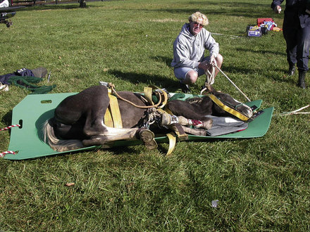
INTRAVENOUS FLUID THERAPY

Horses coming to the hospital for emergency treatment frequently are suffering from varying degrees of dehydration and circulatory (hypovolemic) shock. Dehydration is defined as the excessive loss of total body water. In many cases, electrolyte abnormalities accompany this loss of water and contribute to the clinical signs exhibited by the patient. When dehydration is mild, compensatory mechanisms, including peripheral vasoconstriction, movement of fluid from the interstitial to the intravascular space, increased heart rate and contractility, and retention of water and sodium by the kidney, serve to increase the circulating plasma volume, maintain cardiac output, and therefore tissue perfusion. In these cases, there are often only subtle signs of dehydration. In other cases, these compensatory mechanisms are overwhelmed, or the loss of fluids and electrolytes is continuous leading to dehydration and hypovolemic shock. With shock there is a decreased cardiac output leading to decreased tissue perfusion and hypoxia at the cellular level. Under conditions of hypoxia, cellular energy stores are rapidly depleted leading to derangement in the normal cellular metabolic pathways. If untreated, organ dysfunction ensues progressing from the least perfused organs, the gastrointestinal tract, kidneys, and liver, to the organs demanding the greatest perfusion, such as the heart and brain. Eventually the perfusion of the brain and heart fall to critical levels, compensatory mechanisms fail, and complete circulatory collapse leads to death of the patient.

Clinical Signs of Dehydration

The percent dehydration is a subjective estimate of the percentage of body mass lost in fluid. Subjective estimations of hydration are not highly accurate, but they serve as a starting point for treatment that requires frequent reassessment. In horses, signs of dehydration are usually not obvious until the horse is at least 5% dehydrated. At this level, signs of dehydration are mild and may include depression, tachycardia, and decreased pulse quality. As dehydration worsens, additional signs of dehydration may include delayed recovery of skin tenting, tacky mucous membranes, prolonged CRT (greater than 3 seconds), and poor jugular distensibility.

image TECHNICIAN NOTE

Subjective estimations of hydration are not highly accurate, but they serve as a starting point for treatment that requires frequent reassessment.

A more accurate clinical assessment of dehydration is the change in body mass measured by weight before, during, and after fluid therapy. Although this is a highly accurate measure of hydration, it is impractical in most practice settings. In addition to physical examination findings, hematology findings consistent with a presumptive diagnosis of dehydration include an elevated PCV and plasma TP concentration. This is a simple test that can be performed quickly with equipment available in most private practices and can be used to measure response to fluid therapy. In addition, results of a biochemistry profile that are consistent with dehydration include an elevated creatinine and BUN level. Elevation in BUN and creatinine are collectively termed azotemia and may be the result of dehydration (prerenal azotemia), kidney failure (renal azotemia), and urethral obstruction (postrenal azotemia).

Catheter Location

The location of venous access is determined by the goals of therapy. If the catheter is placed to deliver large quantities of IV fluids, blood or blood products, or if fluids of high osmolarity, such as parenteral nutrition solutions, are to be delivered through the catheter, a larger peripheral vein, such as the jugular vein, or central venous access is recommended. This allows for a larger-size catheter to be placed and as a result of the laminar flow characteristics of these veins decreases the likelihood of thrombophlebitis that occurs when fluids of high osmolarity are administered IV. Administration of antibiotics can be accomplished via jugular catheterization; however, there may be instances in which catheterization of the jugular vein is not desirable. In those cases, catheterization of the accessory cephalic, saphenous, or lateral thoracic veins are reasonable alternatives.

Catheter Selection

IV catheters have become a mainstay in large animal emergency nursing. Advances in emergency and critical care of large animals have resulted in the need for short- and long-term venous access for the delivery of large volumes of IV fluids, medications, such as sedation and analgesics, antibiotics, antiinflammatory drugs, and unfortunately, on occasion euthanasia solutions. In addition, the more frequent use of blood, blood products, and parenteral nutrition has resulted in the need for specialty catheters and delivery systems to minimize catheter-related complications. There are now a wide variety of choices for catheter materials, size, mechanisms for introduction into the vein, and lumen number available. Initially, human catheters were adapted to veterinary use; however, since the emergence of companies, such as Mila International, basic and specialty catheters are now manufactured specifically for veterinary use. Factors that must be considered when selecting the size and type of catheter to be used include the size of the patient, vein to be catheterized, the condition of the patient at presentation, the fluid or medication to be administered through the catheter, the desired rate of administration, expected duration of catheterization, and what is readily available at the time of emergency.

Commercially available IV catheters for large animal use range in size from 10 to 25 gauge and lengths from 1 to 6 inches. The gauge of a catheter is a measurement of its outside diameter. Smaller-gauge catheters have larger outside diameters, which correspond to a larger inside diameter and a higher rate of flow. The size used is dictated by the size of the patient and the treatment to be administered. Large-diameter catheters (10 to 14 gauge × 5.25 inches) are most commonly used for the administration of IV fluids and medication to adult horses. Foals, ponies, and miniature horses are typically catheterized with smaller 16- to 18-gauge catheters ranging from 3.5 to 5.25 inches in length. In addition to size, fluids with higher viscosity, such as blood or plasma, are more easily administered through large-diameter catheters.

Catheters with a large internal diameter permit more rapid administration of fluid, but have the disadvantage of being more damaging to the vein. Catheters of small diameter, 18 to 25 gauge, are reserved for special uses, such as arterial blood pressure monitoring, where they are placed in small peripheral arteries, such as the facial, transverse facial, or metatarsal artery.

Catheter material also affects the choice of catheters. Commonly used materials for manufacture of catheters include polypropylene, PVC, polytetrafluoroethylene (Teflon), nylon, silicone rubber (Silastic), polyurethane, and polyether block amide (Pebax). Different materials result in catheters of different internal diameters, lengths, and stiffness. The differences in material characteristics and size of catheters results in differences in their potential to damage the vessel wall and form a thrombus (thrombogenicity). Traditionally, large-diameter catheters have been manufactured using polypropylene and Teflon. These catheters are stiff with large outside diameters and typically contact the vessel wall over their entire length. This contact damages the vascular endothelial lining exposing collagen and initiates the clotting cascade. This can result in the formation of a blood clot (thrombus) or fibrin sheaths that extend the length of the catheter. Contamination of this thrombus with bacteria can cause the development of inflammation or infection of the thrombus and vein termed thrombophlebitis. Catheters manufactured from silicone (Silastic), polyurethane, or Pebax have the advantage of being softer than Teflon or polypropylene and tend to float in the lumen of the vein. This decreases the thrombogenic potential of the catheter. A disadvantage of Silastic catheters is their porosity that predisposed them to bacterial adherence. For this reason, Silastic catheters must be placed under strict aseptic conditions. As a general rule, large-diameter catheters made of polypropylene or Teflon should only be used for short duration (12 to 24 hours) and are inappropriate for use in small patients, such as foals. Catheters manufactured from Silastic, polyurethane, or Pebax can be maintained as long as 2 to 3 weeks depending on the manufacturer's recommendations.

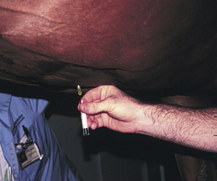
The process of introduction of the catheter into the vein is also variable depending on the size and material the catheter is made from. Short-term Teflon and polypropylene catheters and 14- and 16-gauge polyurethane catheters are most commonly purchased as over-the-needle catheters (Figure 33-108). These have the advantage of simple insertion into the vein with minimal training necessary and low cost. Silastic catheters and certain styles of polyurethane catheters used in foals are of an over-the-wire design. For these catheters, a flexible wire is inserted into the vein before catheterization. The wire acts as a guide for the flexible catheter to follow to ensure proper placement without kinking (Figure 33-109). These catheters are less thrombogenic and have fewer catheter-related complications, such as kinking or breaking. Their disadvantages include expense, the need for added training and practice for correct insertion, and they require absolute sterile preparation for administration. Peel-away introducers are also available that allow catheter placement through a preplaced introducer. After passage of the catheter through the introducer, it is split into two halves for easy removal from the vein.

image

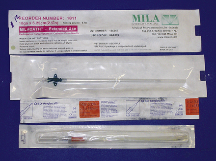
FIGURE 33-108 Example of over-the-needle catheters

image

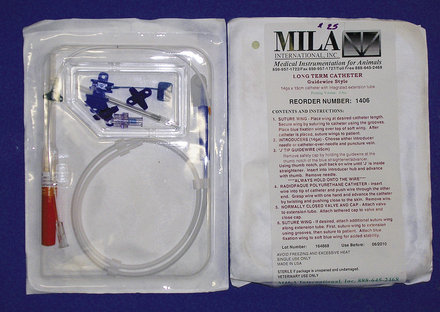
FIGURE 33-109 Example of an over-the-wire catheter

Specialized catheters for the simultaneous administration of IV fluid, total parenteral nutrition (TPN), blood products, and medication are also available. These catheters come in double- or triple-lumen configurations with over-the-wire or peel-away introducers for ease of insertion. Multiport catheters must maintain continuous flow of fluids through each lumen. Stagnation of fluid flow enhances proliferation of bacteria that may have gained access to the ports. Double- or triple-lumen catheters used when additional ports are no longer needed should be replaced with a single-lumen catheter.

In emergency situations, adult horses frequently arrive at the hospital seriously dehydrated and in shock. These patients require large volumes of fluid to be administered in a short period of time. These horses are best treated by insertion of a large catheter, such as a 10-gauge Teflon catheter for initial fluid volume replacement. Because of their size and the material used to manufacture these catheters, they cause significant damage to the vein and predispose the horse to catheter-related complications. Therefore they should be used for only short periods of time (12 to 24 hours) and removed once the fluid deficit has been replaced. An alternative plan is to place multiple catheters. This may be accomplished in several ways. First, a 10-gauge catheter can be placed alone and replaced with a smaller 14- to 16-gauge catheter once the initial volume deficit has been replaced. This is frequently accomplished by passing a guidewire through the original catheter, removing the 10-gauge catheter by passing it over the guidewire, and replacing it with a smaller catheter placed over the wire. This technique has also been demonstrated to be successful at treating catheter-associated infection without jeopardizing additional veins. Alternatively a 10-gauge catheter may be positioned opposite to a smaller catheter and removed once fluid deficits are corrected. Finally, multiple smaller catheters, usually 14- to 16-gauge, may be placed in multiple veins or stacked in the same vein if large-diameter catheters are not available (Figure 33-110). Fourteen- to 16-gauge catheters are most commonly employed once fluid deficits have been replaced and are used primarily for continued maintenance fluids and the administration of other therapeutic agents, such as antibiotics (Figure 33-111).

image

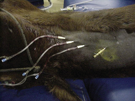
FIGURE 33-110 IV catheters stacked in a single vein for rapid delivery of large volumes of IV fluids

image

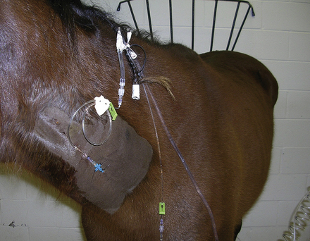
FIGURE 33-111 IV fluid administration through a 14-gauge catheter

Catheter Maintenance

Regardless of the type of catheter and the duration of use, the catheter and surrounding skin should be examined at least two times per day for evidence of heat surrounding the insertion site, swelling around the catheter or a cordlike swelling of the catheterized vein, pain on palpation, or drainage around the catheter insertion site. These may be signs of inflammation or infection of the subcutaneous tissue surrounding the catheter or thrombophlebitis. Catheters with a continuous flow of fluids do not require regular flushing unless the fluids are discontinued. Catheters that are not removed following discontinuation of fluids, those used for the administration of IV medication, and unused lumens of multilumen catheters should be flushed with heparinized saline solutions two to four times per day. Flushing ensures that the catheter remains patent and prevents stagnant flow in the unused catheter lumen and bacterial colonization. If signs of subcutaneous infection around the catheter or thrombophlebitis arise, the catheter should be removed immediately and the catheter tip aseptically collected and submitted for bacterial culture and sensitivity.

image TECHNICIAN NOTE

IV catheters and the surrounding skin should be examined at least two times per day for evidence of heat, swelling around the catheter or a cordlike swelling of the catheterized vein, pain on palpation, or drainage from the catheter insertion site.

Fluid Therapy Plan

Fluid therapy can be performed by the administration of oral or IV fluids in the horse. In most emergency situations, fluid administration is for the purpose of replacing the extracellular fluid lost to gastrointestinal disease; therefore oral administration of fluid for replacement is often contraindicated. IV fluids are employed to correct acute dehydration and to maintain the circulating plasma volume in the face of ongoing fluid loss (diarrhea, gastric reflux, ileus, etc.). Three basic questions must be answered when designing a fluid therapy plan:

1. What type of fluid is to be administered?

2. How much fluid will be administered?

3. At what rate will the fluid be administered?

In most cases, isotonic crystalloid fluids are used for the initial replacement of fluid in horses with dehydration and hypovolemic shock. Two broad classifications of replacement fluids are available for use in horses: normal saline (0.9% sodium chloride) and balanced polyionic electrolyte solutions (LRS, Normasol-R, and Plasma-Lyte). Normal saline is much higher in sodium and chloride than plasma, but contains no other electrolytes. Its use is primarily as a replacement solution in conditions where plasma sodium levels are less than 125 mEq/L or in disease conditions where potassium-free solutions are desired, such as HYPP, urinary bladder rupture in foals, or renal failure.

The electrolyte composition of most commercially available balanced electrolyte fluids for horses is essentially the same as that found in plasma. The use of balanced electrolyte solutions is to replace the fluid volume and electrolytes lost from the extracellular space acutely. Once the fluid to be administered is decided, additional electrolytes can be added to the stock solution to correct specific electrolyte deficiencies and to correct acid-base deficits. Potassium, calcium, and magnesium are frequently supplemented intravenously in horses that are unable to ingest food or water because of gastrointestinal disease. These electrolytes are rapidly depleted in patients without oral intake of food or water and are essential for normal cardiac and smooth muscle function. If electrolyte solutions or other medications are added to stock solutions of fluid, the bags should be clearly labeled with the additive, the concentration, amount added, and the date the fluid was mixed.

Bicarbonate is supplemented in patients suffering from severe metabolic acidosis, most commonly foals. Dehydration leads to decreased tissue perfusion and a change from aerobic to anaerobic metabolism at the cellular level. The end product of anaerobic metabolism is lactic acid that causes a decrease in plasma pH and lactic acidemia. In most cases, the administration of IV fluid rapidly improves tissue perfusion, and lactic acidemia is corrected without specific therapy. In patients where plasma pH is less than 7.2, bicarbonate supplementation is required. The dose of bicarbonate to be replaced is calculated as:

image

0.6 represents an estimation of volume of total body fluid. This number is different for foals and adults. For foals, 0.6 is the correct estimation because a greater percentage of body mass is made up of water. In adults needing bicarbonate supplementation, 0.3 is used as an estimate of volume of total body fluid.

Sodium bicarbonate solutions are mixed in sterile water or saline for administration. Bicarbonate cannot be administered with fluids containing calcium. Mixing of bicarbonate with calcium-containing fluid will result in the formation of an insoluble precipitate. Patients receiving IV bicarbonate must have normal respiratory function for excess carbon dioxide to be expired or acidemia will worsen. One half of the calculated dose is replaced over 4 to 6 hours with the remainder administered over 12 to 24 hours.

Once the type of fluid to be administered is determined, the volume to be administered is calculated. When developing an initial fluid therapy plan, three components must be accounted for to adequately correct and maintain hydration:

1. The volume to be replaced

2. The volume required for maintenance

3. The volume of continued loss

To determine the volume of fluid to be replaced, an estimation of the percent dehydration must be made. Dehydration is a subjective estimation that requires frequent reevaluation to determine if dehydration is being corrected. The simplest estimation is based on a scale of mild, moderate, and severe dehydration (Table 33-4).

TABLE 33-4

Estimation of the Percentage Dehydration in Horses

Severity of Dehydration % Dehydration Clinical Signs
Mild 5-6 Normal mucous membranes, normal to slightly prolonged skin tent, CRT 1-2 sec
Moderate 7-9 Tacky mucous membranes, mildly prolonged skin tent, CRT 2-4 sec
Severe >9 Dry mucous membranes, prolonged skin tent, CRT >4 sec

The percent dehydration is multiplied by the estimated body weight of the patient to determine the replacement fluid volume.

Maintenance fluid needs are relatively straightforward. The average adult horse requires 50 to 60 ml/kg/24-hr period. Foals have a higher total body water percentage and therefore a higher maintenance requirement usually 100 ml/kg/24 hr.

Continued losses can be roughly estimated or measured directly, such as volume of gastric reflux collected over a 24-hour period. Estimation of 1 to 4 L is not uncommon, or continued loss can be estimated as a multiplication of maintenance (1.5 to two times maintenance).

These volumes are added together to give an estimation of the volume of fluid to be administered to a patient over a 24-hour period. Usually the replacement volume is administered over 4 to 6 hours with the remainder over the next 18 to 24 hours. A typical fluid therapy plane is illustrated in Box 33-4 for a 500-kg horse. This example will be used for the remainder of this chapter.

BOX 33-4   A Typical Fluid Therapy Plan for a 500-kg Horse That is 8% Dehydrated

500-kg horse  
Estimated dehydration: 8%  
1. Rehydration:  
 % estimated dehydration × weight in kg  
 0.08 × 500 kg = 40 L 40 L
2. Maintenance:  
 Adult: 50 ml/kg/day  
 50 ml/kg/24 hr × 500 kg = 25,000 ml = 25 L 25 L/24 hr
3. Continued loss:  
 Estimate volume: 2-5 L/hr  
 2 L/hr × 12 hr = 24 L 24 L
 OR  
 Multiple of maintenance 1.5-3 × maintenance  
 2 × 25 L = 50 L/24 hr = 25 L/12 hr ___
  Total 89 L

Frequent reassessment of the patient's physical condition and monitoring of PCV, TP, BUN, creatinine, and electrolytes are necessary to adjust the volume and rate of fluid administration and any additives.

Rate of Administration

Although the rate of fluid delivery can be determined by simple calculation, the volume of fluid delivered to equine patients makes estimating an accurate rate difficult. Most horses seen in the hospital on an emergency basis are initially administered a “shock” dose of fluids equivalent to 60 to 90 ml/kg/hr. Fluid rate is usually calculated as drops per second. As can be seen in the example in Box 33-5, the number of calculated drops per second cannot be accurately measured and is essentially equal to a fluid rate that is a constant wide open stream. In these cases, the volume of fluid delivered should be estimated by the graduations marked on the fluid bag. The volume should be recorded in the medical record every 1 to 2 hours and the rate adjusted as needed to deliver the desired amount of fluid.

BOX 33-5   Calculation for Delivery Rate of Replacement Fluid

Initial Fluid Delivery Rate

Example 1 60 ml/hr (shock dose)

500 kg × 60 ml/kg/hr = 30,000 ml/hr (30 L/hr)

30,000 ml × 10 drops/ml = 300,000 drops/hr

image

image

Example 2 Replacement volume of 40 L to be replaced in 6 hr

40 L/6 hr = 6.7 L/hr

6.7 L × 1000 ml/L = 6700 ml/hr

6700 ml/hr × 10 drops/ml = 67,000 drops/hr

image

image

Once the initial fluid deficit has been replaced, the fluid rate should be slowed to meet the requirements for maintenance and continued loss. This rate can be calculated as in Box 33-6.

BOX 33-6   Fluid Rate for Maintenance and Continued Losses

Maintenance and Continued Loss Fluid Rate

49 L/24 hr = 2 L/hr

2 L × 1000 ml/L = 2000 ml

2000 ml/hr ×10 drops = 20,000 drops/hr

image

image

This can be counted as 55 drops/10 sec.

Also note that this approximates two times maintenance as described previously.

Hypertonic Saline Solution

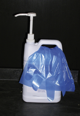
Hypertonic saline solutions used in equine veterinary medicine consist of a 7.2% solution of sodium chloride usually packaged in 1-L bottles. Hypertonic saline has an almost immediate, though short-lived, effect on the circulating plasma by drawing fluid from the interstitial and intracellular space into the vasculature. Plasma volume expansion results in increased cardiac output, improved blood pressure, better oxygen delivery to and better oxygen use by the tissues. Hypertonic saline is administered at a dose of 4 ml/kg over 5 to 10 minutes. It rapidly expands circulating plasma volume; however, its effects last for approximately 60 minutes.

image TECHNICIAN NOTE

Hypertonic saline is administered at a dose of 4 ml/kg over 5 to 10 minutes.

Colloids

Hydroxyethyl starches, also known as synthetic colloids, are high-molecular-weight molecules that function similar to the natural colloid albumin in the circulatory system. When administered intravenously to horses suffering from dehydration and hypovolemic shock, they exert oncotic pressure that serves to maintain plasma volume and peripheral perfusion. The advantages of synthetic colloids over albumin include lower antigenicity, and because of their larger molecular weight, they tend to remain in the circulation for a longer period of time. In conditions where albumin is lost from the circulation as a result of increased capillary permeability, IV administration of albumin will only result in further redistribution of albumin to the interstitial space. Synthetic colloid will remain in the intravascular space longer, exerting its oncotic effect for up to 120 hours. Hetastarch is the most commonly used synthetic colloid in the United States. It is administered at a dose of 10 ml/kg of a 6% solution. Disadvantages of hetastarch include expense, the association of bleeding disorders with its use, and the inability to measure the oncotic effect of colloids by refractometer.

BLOOD AND BLOOD PRODUCTS

Blood loss and anemia in horses is caused by one of three mechanisms: whole blood loss, erythrocyte destruction, or the failure to produce erythrocytes. Whole blood loss is the most common cause for anemia in horses and presents most frequently as an emergency. Significant blood loss in horses is most commonly associated with wounds involving the neurovascular supply of the distal limb, middle uterine artery rupture in mares, guttural pouch mycosis, hemolytic anemias, and neonatal isoerythrolysis in foals.

On average, blood constitutes approximately 8% of the horse's body weight. This means that an average 500-kg horse has a blood volume of approximately 40 L. This estimate varies depending on the age, breed, and use of the horse and can range from 7% to 15%. The horse can lose approximately 20% of its total circulating blood volume without demonstrating clinical signs. When acute blood loss occurs, the immediate response by the body is to ensure the delivery of oxygen to the vital organs, such as the heart and brain. Hemorrhage of greater than one third of the circulating blood volume can result in irreversible shock and death. This is the result of inadequate oxygen delivery to the tissues. Therefore the goal of treatment of acute anemia or blood loss is to maintain appropriate oxygen delivery to the tissue.

image TECHNICIAN NOTE

On average, blood constitutes approximately 8% of the horse's body weight that is equivalent to 40 L in a 500-kg horse.

Oxygen delivery to the tissue is affected by two primary mechanisms: (1) cardiac output and (2) oxygen content of the blood reaching the tissues. The oxygen content of the blood is dependent on hemoglobin concentration and is directly related to the RBC mass. Whole blood loss affects oxygen delivery not only by the loss of erythrocytes and hemoglobin, but also the loss of plasma. This decreased plasma volume causes a decrease in the amount of blood pumped by each contraction of the heart (stroke volume) and therefore the total amount of blood pumped by the heart to the tissues (cardiac output). The loss of RBCs and hemoglobin decreases the oxygen-carrying capacity of the blood.

In response to blood loss, the body has several compensatory mechanisms to maintain cardiac output and oxygen delivery. Immediate compensatory responses include peripheral vasoconstriction, splenic contraction (the spleen can maintain up to 30% of the circulating erythrocyte mass), and a shift of fluid from the interstitial space to the intravascular space.

The decision to perform a blood transfusion is often difficult and should not be taken lightly. Blood transfusion is an invasive procedure requiring the catheterization and collection of blood from the donor potentially creating a situation of acute blood loss and the administration of the collected blood to the recipient. This process is time and labor intensive and may add significant cost to the therapy. In addition, undesirable transfusion reactions that range in severity from mild urticaria to acute anaphylaxis and death have been reported in horses. Compensatory mechanisms and the loss of equivalent concentrations of fluid, cells, and protein with whole blood loss can make the decision to perform a blood transfusion based on hematologic values potentially inaccurate. Hematologic measurements, such as PCV and TP concentrations, may take up to 24 hours after the initial blood loss to change significantly. Therefore the decision to perform a blood transfusion should always be based on the nature of the illness or injury, a history of known large volume blood loss, clinical signs, and hematologic measurements (Box 33-7).

BOX 33-7   General Recommendations for Blood Transfusion

PCV <20% following acute hemorrhage

PCV <12%-14% following chronic hemorrhage

Clinical signs consistent with hypovolemia following acute hemorrhage

The clinical signs exhibited by the horse following acute hemorrhage are the result of a combination of hypovolemia and decreased erythrocyte mass. These signs are consistently observed in horses with anemia regardless of the underlying cause and can include tachycardia, tachypnea, lethargy, inappetence, depression, colic, sweating, cold extremities, and pale mucous membranes.

Clinicopathologic changes are dependent on the time between presentation and changes in signs because of the previously described compensatory mechanisms. In a recent review of 31 horses treated by blood transfusion, only 11 of 18 (61%) horses with acute blood loss had PCV or hemoglobin levels less than the normal reference range. When present, clinicopathologic abnormalities may include decreased PCV and TP, increased creatinine and BUN levels, and decreased oxygen saturation.

image TECHNICIAN NOTE

Clinical signs consistent with blood loss in horses regardless of the underlying cause can include tachycardia, tachypnea, lethargy, inappetence, depression, colic, sweating, cold extremities, and pale mucous membranes.

Blood Transfusion

Transfusion of whole blood to the anemic patient has been demonstrated to result in a significant improvement in PCV, creatinine, and oxygen saturation and a significant improvement in clinical signs. An organized well-thought-out plan must be established and personnel trained to proceed through the process in a systematic fashion. This will minimize the cost, materials used, and time needed to safely collect the necessary volume of blood from the donor and promptly transfuse the blood to the patient.

Blood Donors: A minimum of two blood donors should be available at all times. These may be horses housed at the practice or client-owned horses in which special arrangements have been made for their use as donors. Unlike humans that have three blood types (A, B, and O), horses have seven different blood types represented by capital letters A, C, D, P, K, Q, and U and up to 30 factors in each type designated by lower case letters. Also unlike humans, there is no universal equine blood donor. Identification of the most appropriate donor should be made well in advance of the need for transfusion. Blood can be collected from potential donors and sent to a number of veterinary laboratories for typing (Box 33-8).

BOX 33-8   Blood Typing Laboratories

Equine Blood Typing Research Laboratory

University of Kentucky

Department of Veterinary Science

Lexington, KY 40546

859-257-3022

Serology Laboratory

University of California

Davis, CA 95616

530-752-9284

Stormont Laboratory

1237 E. Beamer St. Suite D

Woodland, CA 95776

530-661-3078

Mann Equitest, Inc.

335 Laird Rd. Unit 4

Guelph, Ontario N1H 6J3

Canada

519-836-2400

The most desirable donors are geldings with no history of receiving a blood transfusion and Aa and Oa negative because these are the most immunogenic erythrocyte antigens. Donors should have a complete well-maintained health record and should receive regularly scheduled vaccinations, deworming, an annual enzyme immunoassay (EIA) (Coggins) test, and have routine dental and foot care performed.

Larger blood donors are desirable to allow a greater volume of blood to be collected. Although 450 kg is a minimum weight, horses greater than 550 kg are more desirable. Mares and horses with a history of previous transfusion have a greater risk for antierythrocyte antibodies (Box 33-9).

BOX 33-9   Desirable Blood Donor Characteristics

>450 kg

Gelding

Free of blood-borne diseases

In good health

No previous transfusion or pregnancy if a mare

Aa, Oa, and hemolysin negative

PCV >35 and TP >6.0

A PCV and TP should be performed on all donor horses before blood collection. Donors with PCV less than 35 or a TP of less than 6 should not be used. A healthy donor 500 kg in body weight with a PCV of 35% to 40% can safely donate up to 8 L (20% of blood volume) of blood every 30 days.

image TECHNICIAN NOTE

At the time of blood collection, donor horses should have a PCV of at least 35 and a TP of at least 6.0 mg/dl.

Blood Collection: A blood collection kit containing all necessary supplies can be organized and stored for ready use. Technical staff should be adequately trained in the process of blood collection and the process rehearsed to ensure that it is carried out efficiently.

Blood can be collected into a number of containers, including sterilized glass jars, commercial blood collection bottles containing acid-citrate-dextrose anticoagulant (ACD bottle), specially designed bags for blood collection, and gas-sterilized fluid bags. The container determines the type of anticoagulant used. ACD bottles have the advantage of a vacuum that allows rapid collection of blood; however, the disadvantage when used for blood collection in horses is their size, damage to the erythrocytes from impact on the bottle, inactivation of platelets by the glass, and risk of breaking the glass (Figure 33-112). ACD bottles also require additional tubing for the collection process. Commercially available plastic blood bags for blood collection in horses have the advantage of preservation of platelet function, less erythrocyte damage, the ability to collect large volumes in a single container, and no risk of breakage (Figure 33-113). Their disadvantage is that collection is by gravity flow and can be time consuming.

image

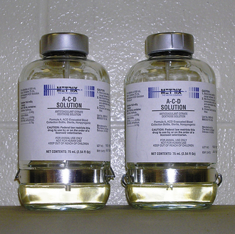
FIGURE 33-112 ACD glass bottles for the collection of blood

image

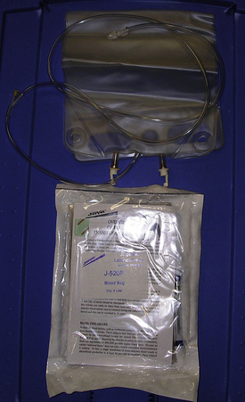
FIGURE 33-113 Commercially available blood collection bags for horses

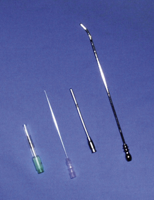
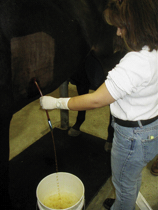
A crew composed of three individuals is recommended for safe, efficient blood collection. One person handles the horse, one manages the catheter, and one controls the blood collection device. The area over the donor's jugular vein is aseptically prepared and 2 to 5 ml of local anesthetic are locally infused over the proposed catheter or needle site. Blood can be collected safely through a larger needle (14-gauge × 1.5-inch), a specialized blood collection trochar (8- to 11-gauge × 75-mm), or IV catheter (14-gauge × 5.5-inch). Venous access should be directed in the opposite direction of normal blood flow to speed collection. The vein may need to be occluded continuously to distend the vein and speed collection. The blood must be collected into the chosen collection device under strict asepsis.

Standard ACD bottles contain enough ACD to collect 450 ml of blood. The amount of ACD in bags is dependent on the volume of the bag. When using a sterilized glass bottle, enough ACD should be added to the bottle to produce a ratio of 9 parts blood: 1 part ACD (2000 ml total volume = 200 ml ACD: 1800 ml whole blood). Although not commonly used as an anticoagulant for blood transfusions, in an emergency situation heparin can be added to sterile glass bottles for immediate collection and administration. When using heparin as an anticoagulant, 625 IU heparin is used per 50 ml of blood collected, and the blood should be administered immediately. The bottle or bag should be gently swirled or rocked during the collection to ensure proper mixing of the blood and anticoagulant. If collecting blood into bottles, once the bottle is full, the tubing should be clamped near the bottle and the needle removed from the stopper. To maintain strict asepsis, needles should be changed in between bottles before resuming collection. The tubing attached to bags should be stripped of residual blood, tied in three knots, or clamped and cut below the clamp. Following blood collection, donors from whom 20% of the blood volume has been removed should receive 5 to 20 L crystalloid fluids to prevent signs consistent with hypovolemia. They should also receive 1 to 3 lb of complete feed and free-choice water. The catheter site should be examined two times per day for signs of heat, pain, or swelling for the next 5 to 7 days.

Crossmatching: Crossmatching is an in vitro screening test used to assist in the detection of incompatibilities between donor and recipient blood. The incompatibilities are the result of antibodies present in the serum of the donor or recipient to the other's erythrocytes. Incompatibilities are recognized by clumping (agglutination) of erythrocytes following mixing of donor and recipient erythrocytes and plasma or hemolysis after the addition of rabbit complement to this mixture. The two components of this screening test are the major and minor crossmatch. The major is highly recommended by most authors and involves the mixing of recipient plasma or serum with donor erythrocytes. A minor crossmatch is considered optional in situations where the donor has no known exposure to the blood of other individuals and consists of mixing donor plasma or serum with recipient erythrocytes. Box 33-10 provides step-by-step instructions for performing major and minor crossmatching.

BOX 33-10   Crossmatch Procedure

Materials

1. EDTA anticoagulated blood and serum form recipient and donor

2. Pipette and tips accurate up to 0.05-0.1 ml

3. 37° C water bath or incubator

4. Microscope

Procedure

1. Into appropriately labeled donor and recipient tubes pipette 2-4 drops of whole blood from each donor and recipient and fill to within 1 cm of the top with normal saline.

2. Cover the tubes with parafilm (wax paper) film and mix thoroughly.

3. In a tabletop centrifuge, centrifuge for 1 min at maximum speed and decant the saline supernatant. There will be some residual saline in the bottom of the tube.

4. Gently strum the bottom of the tube to resuspend the pellet, fill again with saline, and repeat steps 2 and 3.

5. Gently resuspend the cells in the saline at the bottom of the tube and add approximately 4.7 ml of additional saline. The residual volume is approximately 0.3 ml. Addition of 4.7 ml will result in a solution of erythrocytes of approximately 6%.

6. Label 1 tube autocontrol. For each donor, label two tubes major/donor ID and minor/donor ID.

7. To each tube labeled major, add 0.1 ml recipient serum, and then add 0.05 ml of the 6% donor cell suspension to the corresponding major/donor ID tube.

8. To each tube labeled minor, add 0.1 ml of donor serum from the appropriate donor, then add 0.5 ml recipient 6% cell suspension.

9. To the tube labeled autocontrol, add 0.1 ml recipient serum and 0.05 ml recipient cell suspension.

10. Mix all tubes well and centrifuge at maximum speed for 30 seconds.

11. Gently shake the cells to slowly loosen them from the pellet and observe grossly for agglutination or hemolysis (red supernatant).

12. Centrifuge tube again 30 seconds at maximum speed and repeat step 11. Place a drop of suspension on a microscope slide and observe at 10× power for agglutination.

Although highly recommended, the limitations of equine crossmatching must be understood. False-positive and false-negative reactions are commonly observed as a result of spontaneous rouleaux formation, which must be distinguished from agglutination. Rouleau is described as the microscopic appearance of linear stacks of erythrocytes similar to stacks of coins. Agglutination is the irregular spherical clumping of erythrocytes observed under the microscope. Agglutination can be confirmed by mixing a small quantity of blood with normal saline. If present, agglutination will persist after mixing, and rouleaux will disperse. In addition, various amounts of agglutination are present in all crossmatch reactions, and it is difficult to determine at what level agglutination is considered incompatible. Therefore incompatibilities may only be recognized with large amounts of agglutination. Finally the most serious transfusion reactions occur as a result of erythrocyte hemolysis rather than agglutination. Lysis is only observed following the addition of complement, a procedure that is impractical in most practice settings and usually only performed in blood typing laboratories. Therefore crossmatching has its greatest value when reaction between donor and recipient blood is severe (large amounts of agglutination) indicating an obvious incompatibility. Transfusion reaction may still occur even after blood has been determined to be compatible by crossmatching. In most cases, naturally occurring antibodies to equine erythrocytes are uncommon, and a transfusion can be safely administered without crossmatching. Blood should initially be administered slowly and the recipient observed for clinical signs of a transfusion reaction and the transfusion slowed or discontinued if signs increase in severity or persist.

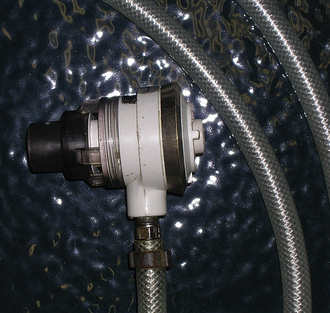
Administration: The recipient should have an IV catheter placed aseptically. Infusion should be performed only through a specialized blood delivery infusion set with an in-line filter (Figure 33-114). Some authors recommend replacing the infusion set after every 4 L of blood administered. Although seldom are crystalloid fluids and blood administered simultaneously, if this is practiced, normal saline (0.9% NaCl) is the only crystalloid fluid that can be safely administered with blood. Most replacement fluids, such as LRS, contain calcium, which can initiate blood clotting, and hypotonic fluids, such as dextrose, can cause hemolysis.

image

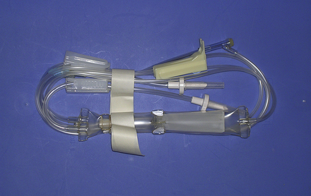
FIGURE 33-114 Blood delivery set with an in-line filter

The volume of blood to be transfused can be estimated by two methods:

image

This calculation is based on a normal blood volume in the adult horse of 72 ml/kg and of 151 ml/kg in the neonate.

By this calculation, an average 500-kg adult with a PCV of 15 and a desired PCV of 25 would require 10.28 L.

image

An alternative is simply to calculate 10 to 20 ml of blood/kg body weight

image

A recent retrospective study in horses demonstrated a 4% increase in PCV and significant improvement in clinical signs of horses with acute blood loss after the administration of 15 ml/kg whole blood.

The administration of blood should be closely monitored for signs of transfusion reactions. There are many published recommendations regarding the frequency of monitoring. The author has used a rate of 5 ml/kg/hr as an initial infusion rate with monitoring of the recipient's temperature, pulse, and respiration every 5 minutes for the first 15 minutes. If no signs of a transfusion reaction develop, the rate is increased to 10 to 25 ml/kg/hr while observing the patient continuously and monitoring and recording temperature, pulse, and respiration every 30 minutes. Signs of a transfusion reaction are variable and may include urticaria, dyspnea, tachypnea, tachycardia, fever, restlessness, muscle fasciculations, sudden recumbency, anaphylactic shock, and death. Depending on the severity of the signs, if observed, the transfusion should be slowed or discontinued.

Plasma Transfusion

Plasma is the cell-free portion of blood. Its constituents include colloids, such as albumin and other small protein; electrolytes; immunoglobulin (antibodies); antibacterial protein, such as complement; and clotting factors. The indications for plasma administration in horses are numerous and may include hypoproteinemia or hypoalbuminemia, failure of passive transfer, septicemia or endotoxemia, DIC, warfarin toxicity, and the treatment and prevention of specific disease for which antitoxic and passive immunization plasma have been developed. Although equine plasma readily separates from erythrocytes without the aid or expensive plasmapheresis equipment, collection of this plasma is both labor and time consuming and must be performed under the strictest of aseptic conditions. The collection of quantities large enough to treat most equine patients cannot be accomplished economically in most practice settings. Equine plasma is available commercially from several manufacturers worldwide (Box 33-11). These companies maintain herds of horses that are specifically for the production of various highly specific plasma products (Box 33-12) that can be shipped and stored frozen for extended periods of time.

BOX 33-11   Sources of Equine Plasma

image

BOX 33-12   Indications for Types of Equine Plasma

Normal

Replacement of albumin, complement clotting factors, and immunoglobulin in adults and foals

Hyper/Polyimmune

Failure of passive transfer in neonatal foals

E. coli J5/Salmonella

Treatment of septicemia and endotoxemia in foals and adults

Rhodococcus equi

Passive immunity and prophylaxis before exposure to R. equi

Clostridium botulinum Type B Antitoxin

Prophylaxis following exposure to C. botulinum by ingestion and antitoxin in clinically affected patients

West Nile Virus

Passive immunity and treatment of clinical cases

Streptococcus equi

Passive immunity and treatment of clinical cases

Plasma requires thawing before administration. The manufacturer's directions for storage, thawing, and administration of specific plasma products should be consulted before administration. Routinely, plasma is stored at −18° C (the same temperature as a standard refrigerator freezer) and thawed immediately before administration. The most convenient method for thawing is to place the bag in water that is approximately 40° C (approximately 104° F). The plasma should be mixed periodically and cool water replaced until approximately body temperature. Thawing in water that is too hot should be avoided because this can result in the destruction of plasma protein and immunoglobulin. Plasma that is thawed to room temperature or above should not be refrozen for later use. Administration should always be performed through a filtered blood delivery set.

image TECHNICIAN NOTE

Commercially available FP should be thawed by placing the bag in water that is approximately 104° F.

The volume of plasma to be administered is dependent on the manufacture's recommendation for treatment and prevention of the specific condition for which the plasma is administered or the calculated need based on hypoproteinemia. Plasma transfusion is indicated in horses in which the plasma TP is less than 4 g/L or albumin concentrations are less than 2 g/L. In horses that are hypoproteinemic, the volume of plasma to be administered can be estimated by the following formula:

image

This calculation is based on an estimated plasma volume in the adult horse of 48 ml/kg, 95 ml/kg in the neonate, 62 ml/kg in foals from 1 to 4 weeks of age, and 53 ml/kg in foals from 4 to 12 weeks of age.

Following plasma transfusion, the observed increase in plasma albumin concentration is less than usually expected as a result of redistribution of the albumin to the extravascular space. The volume necessary to significantly increase the TP and albumin concentrations of the blood are large. A minimum of 6 L of normal plasma is required to increase the recipient's albumin concentration 1 to 2 g/L. Although the concentrations do not change appreciably, a dramatic clinical improvement is often observed.

Plasma administration is similar to whole blood and should be performed through a blood delivery set with an in-line filter, and the patient should be carefully observed for signs of transfusion reaction and the transfusion slowed or discontinued if signs are observed.

Blood Substitutes

Oxyglobin (Biopure Corp., Cambridge, Mass.) is an ultrapurified, polymerized bovine hemoglobin in a modified LRS solution that contains13.8 g/dl of hemoglobin. Oxyglobin has been approved for use in dogs and has been used extralabel in foals with neonatal isoerythrolysis as a hemoglobin replacement. This solution increases the oxygen-carrying capacity in anemic patients. Oxyglobin has been used in horses at a dose range between 10 to 30 ml/kg delivered at a rate of 10 to 20 ml/kg/hr. The large hemoglobin molecule also acts as a colloid solution. It is a cell-free hemoglobin solution that lacks erythrocytes and erythrocyte antigens. Therefore crossmatching is not necessary before administration. Other advantages of oxyglobin include a long shelf life (36 months) and no preadministration preparation (off-the-shelf administration) and no need for specialized administration sets.

In limited use in horses, oxyglobin has been used in the treatment of a miniature horse with chronic ovarian bleeding and in foals with NI as a means of controlling clinical signs while blood is collected and processed. Disadvantages of oxyglobin include too high of cost to be used in adult horses, a short plasma half-life thus requiring additional blood products be administered, and evidence that suggests oxyglobin may decrease cardiac output.

CASE PRESENTATION 33-2

History and Signalment

A 12-year-old quarter horse gelding came to the hospital with a 2-day history of moderate to severe abdominal pain. A presumptive diagnosis of a small colon impaction was made by the referring veterinarian, and he was treated with IV fluids and flunixin meglumine for pain. There has been no response to therapy.

Initial Physical Examination

On presentation, the horse was mildly depressed and exhibited intermittent episodes of mild to moderate abdominal pain.

His temperature was 99, pulse 48, and respiration of 16 breaths per minute.

Mucous membranes were pale pink and tacky, and his CRT was 3 seconds. The skin over the eyelids remained persistently tented for 5 to 6 seconds.

He was determined to be moderately (7% to 8%) dehydrated based on physical examination.

This was confirmed by the results of a CBC and chemistry profile that revealed an elevated PCV (42) and TP (7) and what was suspected to be a prerenal azotemia (creatinine: 2.5, normal: 1.4 to 1.7).

His weight was estimated to be approximately 1000 lb (450 kg).

There was no gastric reflux, and a rectal examination revealed an extensive small colon impaction.

Initial Fluid Therapy

1. Replacement Fluids

(450 kg) (0.08) = 36 L to be administered over 4 hours

36 L/4 hr = 9 L/hr

9 L = 9000 ml

9000 ml/60 min/hr = 150 ml/min

150 ml/min/60 sec/min = 2.5 ml/sec

2.5 ml/sec × 10 drops/ml = 25 drops/sec, too fast to count; therefore the rate can only be monitored using the graduations on the fluid bag and adjusting the rate as needed.

2. Maintenance and continued loss

(450 kg) (50 ml/kg/24 hr) = 22,500 ml or 22.5 L/24 hr

22.5 L/24 hr = 937 ml/hr

937 ml/hr/60 min/hr = 15.6 ml/min

15.6 ml × 10 drops/ml = 156 drops/min

156 drops/min/60 sec/min = 2.5 drops/sec or 25 drops/10 sec

Continued loss was considered small and estimated to be 1.5 times maintenance.

15.6 ml/min × 1.5 = 23.4 ml/min

23.4 ml/min × 10 drops/ml = 234 drops/min

234 drops/min/60 sec/min = 3.9 or roughly 4 drops/sec

3. Continued therapy

    As time went on, the horse experienced more pain. Approximately 10 L of gastric reflux was collected, he began to exhibit abdominal distention, and his PCV increased to 48.

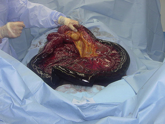
    The abnormal rectal examination, gastric reflux, and systemic deterioration were indications for abdominal exploratory surgery.

    The horse was anesthetized with a ventral midline celiotomy, and abdominal exploratory was performed and the small colon impaction relieved.

4. Anesthetic recovery

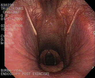
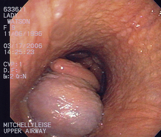
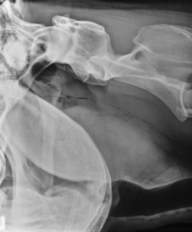
    The horse stood unassisted approximately 45 minutes after removal from gas anesthesia. Within 5 minutes of standing, he began to exhibit signs of discomfort initially thought to be abdominal pain. These signs progressed to flaring of the nostrils, exaggerated inspiratory efforts, inspiratory stridor, and severe anxiety.

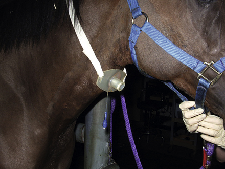
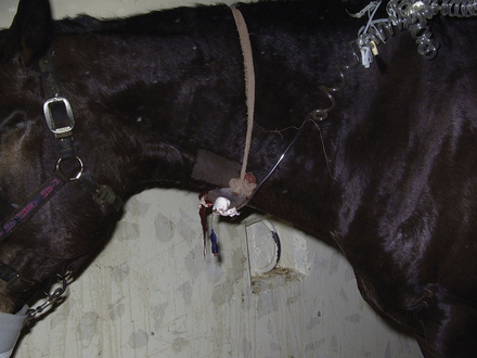
    A standing emergency tracheotomy was performed, and a temporary tracheotomy tube was positioned and fixed in place with gauze. Oxygen insufflation through the tracheotomy tube was initiated at 15 L/min, and the respiratory signs resolved within minutes. Oxygen insufflation was discontinued after 1 hour. The presumptive diagnosis was acute upper respiratory obstruction resulting from postanesthetic laryngeal spasm.

5. Postoperative care

    The horse remained on IV fluids for 48 hours following surgery. Food was introduced at 24 hours and gradually increased to free-choice hay over 5 days. The tracheotomy tube was intermittently occluded to determine if the laryngeal spasm persisted. The horse was able to breathe normally after 5 days. The tube was removed, and the horse was discharged 7 days after surgery.

RECOMMENDED READINGS

Auer J.A., Stick J.A., eds. Equine surgery, ed 3, St Louis: Saunders, 2006.

Bramlage, L.R. Current concepts of first aid and transport of the equine fracture patient. Comp Cont Educ Pract Vet. 1983;5:S564.

Durham, A.E. Blood and plasma transfusion in the horse. Equine Vet Educ. 1996;8(1):8–12.

Gonzales, G.L. How to establish an equine blood donor protocol. AAEP Proc. 2001;47:262–265.

Mason, D.E., et al. Respiratory emergencies in the adult horse. Vet Clin North Am: Equine Pract. 1994;10(3):685–701.

Seahorn, T.L., Cornick-Seahorn, J. Fluid therapy in horses with gastrointestinal disease. Vet Clin North Am: Equine Pract. 2003;19(3):665–679.

Slovis, N.M. How to approach whole blood transfusion in horses. AAEP Proc. 2001;47:266–269.

Wilson, D.A. Principles of early wound management. Vet Clin North Am: Equine Pract. 2005;21(1):45–62.


Equine Demand Valve, J D Medical Distributing Co., Inc., 1923 West Peoria Avenue, Phoenix, AZ 85029, 602-997-1758, www.jdmedical.com.