Osteochondrosis is the generic term used to describe a group of syndromes that share the common pathology of idiopathic bone disease (Ekman & Carlson 1998). The pathogenesis of osteochondritis is unclear, but all presentations are characterised by an interruption of normal enchondral ossification, together with a greater or lesser degree of focal death of the local trabeculated bone (Caselli et al 1998). A number of examples of osteochondritis affecting specific bone sites are named eponymously to the physician who first reported the disease. The onset of osteochondritis generally occurs during childhood, especially during times of rapid growth, but the full effects of the resultant joint and bone damage may not become apparent, and problematic, until adulthood. Osteochondritis may give rise to focal bone deformation during the healing phase of the disease, where the forces imposed by body mass impinge on the areas of abnormal ossification or diseased bone, causing a change in the local bone architecture. The altered shape of the involved bone sites may give rise to pathologies later in life.
Osteochondritis may be classified in relation to the anatomical location of the enchondral ossification defect (Griffin 1994):
Alternatively, osteochondritis may be classified by the effects brought about by local forces on the area of diseased enchondral bone:
The true aetiology of the various presentations of osteochondritis is unknown, but they have been linked to hereditary factors, local trauma, nutritional factors and local ischaemia within the affected area of bone (Ekman & Carlson 1998, Walsh & Dorgan 1988).
Plain radiographs are usually used to diagnose osteochondritis, but minor or early-stage lesions may be overlooked as they are difficult to visualise on a plain radiograph. Lesions are identified readily using bone scan, computerised axial tomography scan and magnetic resonance imaging (Bohndorf 1998).
The differential diagnosis for all the osteochondritides can include osteomyelitis, bone tumours and fractures (Caselli et al 1998).
The treatment of all presentations of osteochondritis focuses on rest and immobilisation to allow the affected bone to heal with no or only minimal distortion, together with the use of painkillers (such as NSAIDs), as necessary, during the acute or early presentations of the disease.
Rest and immobilisation of the affected bone is essential:
Early initiation of treatment is especially important where the involved osteochondritic bone forms part of a joint, in order to reduce the likelihood of later osteoarthritic changes, pain and dysfunction of the affected joint.
The format, intensity and duration of the regimen of rest is tailored to the presenting problem, and can be achieved by a variety of means, such as rigid splinting, soft splinting and strapping, and other means to reduce weight bearing, and traction and compressive forces on the involved bone.
CASE STUDY 4.3 PRESENTATION IN AN ADULT
OSTEOCHONDRITIS DISSICANS:
A 54-year-old woman presented with a 10-month history of pain in the right foot. She worked as a radiography assistant in a local hospital. She recalled that on 16 December of the previous year, whilst walking along a corridor from the x-ray department to one of the wards to deliver a patient’s radiograph, she experienced a sudden and severe shooting pain in the right forefoot. The pain located to the base of the second toe, and continued throughout that day, and she was unable to walk without limping heavily on the right foot. That night, she applied ice to the foot, and self-medicated with ibuprofen. By the next day, the pain had not reduced at all. There was some mild local swelling in the area of the right second metatarsophalangeal joint (MTPJ). The patient reported that a radiograph, taken within 24 hours of the incident, failed to show any bone lesion. It was assumed by the GP that she had suffered some form of foot strain. The swelling cleared within 3 weeks, and the severe pain gradually subsided over the next 3 months, although the right second toe continued to be very painful on movement at the second MTPJ.
She was referred to the podiatrist in September of the following year. On examination, it was noted that her feet showed the typical forefoot deformities of moderate hallux limitus, with 45° of available passive dorsiflexion at the first MTPJ. Passive sagittal plane movement of the right second toe was much reduced, with crepitus and pain at the right second MTPJ. There was a palpable dorsiplantar thickening of the right forefoot in the area of the second MTPJ. Plain radiographs showed marked degenerative changes at the head of the right second metatarsal and the base of the associated proximal phalanx, with flattening of the metatarsal dome, osteophytosis at the head of the right second metatarsal and base of the associated proximal phalanx, and local bone sclerosis.
On the basis of the history and the September radiograph the patient was diagnosed as showing arthritic degeneration of the right second MTPJ. It was presumed that the patient may have suffered an osteochondritis dissicans the previous December, secondary to an overload phenomenon imposed by the loss of movement at the first MTPJ, and possibly in association with a degree of age-related osteoporosis. The bone lesion at the second metatarsal head did not show up on the radiograph taken within 24 hours of the original incident, as the radiograph was taken at too early a stage in the pathology for it to be visible on plain radiography. Subsequent degenerative changes had developed over the ensuing months, causing osteoarthritis at the right second MTPJ.
The right forefoot problem was treated by conservative therapy, initially with defective clinical plantar padding, then with a bespoke cushioned orthotic and the use of a rocker-soled shoe. This reduced her forefoot pain to what the patient felt was an acceptable level. As the patient did not wish to contemplate forefoot surgery, she was not referred to an orthopaedic or podiatric surgeon.
This form of osteochondritis affects the metatarsal head(s). In four out of five cases the patient is a girl aged between 12 and 15 years (Griffin 1994, Manusov et al 1996b).
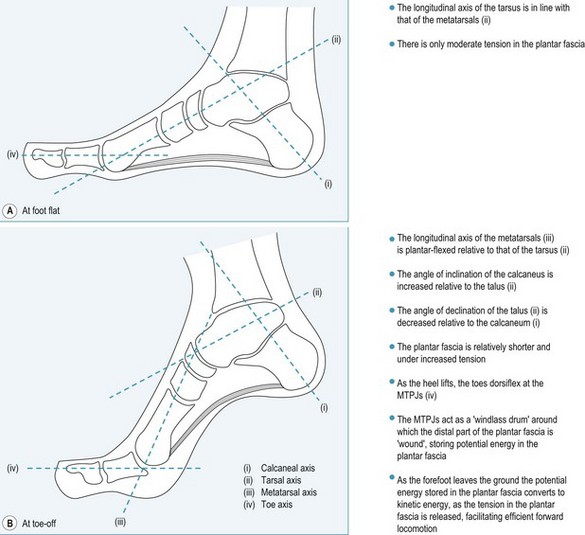
Freiberg’s disease may present uni- or bilaterally, as a focus of ischaemia and bone necrosis within the head of a metatarsal, leading to collapse of both the articular surface and the underlying area of subchrondral bone – the so-called eggshell fracture. The head of the second metatarsal is affected in almost 70% of cases, and the third metatarsal head in almost 30% of cases, but any of the metatarsal heads may be subject to infraction (Griffin 1994). The predominance of osteochondritis affecting the second metatarsal head is thought to be due to the greater length of the second metatarsal in relation to the first and third metatarsals, and its resultant susceptibility to local trauma during the gait cycle, especially at toe-off. It is thought that the blood supply to the epiphyseal plate is interrupted by repeated microtraumata, causing an area of avascular necrosis within the metatarsal head (Manusov et al 1996b).
The patient presents with increasing pain and associated swelling, and possible bruising on the dorsum of the foot, overlying the affected metatarsal head. Gait is affected and the patient may limp (Caselli et al 1998). The pain worsens on weight bearing and with activity. The range of motion at the affected MTPJ is decreased, particularly when active and/or weight-bearing dorsiflexion of the toes is attempted. Pain and crepitus is likely to be elicited on passive movement of the affected joint.
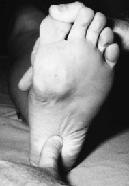
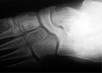
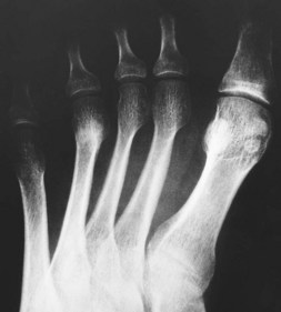
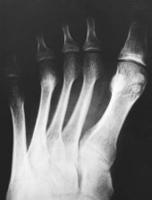
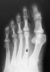
In the early stages of the disease plain radiographs may show little apparent bone involvement. In the later stages, as healing progresses, new bone growth in the affected area can be seen. The classical radiographic presentation in the adult who has undergone an episode of Freiberg’s infraction as a teenager, or an untreated Freiberg’s disease in a younger person, is of a flattened metatarsal head, with associated signs of osteoarthritic changes, such as bone sclerosis and osteophyte formation, occurring at the metatarsal head (Fig. 4.15). The base of the associated proximal phalanx may also show degenerative changes. The differential diagnoses of an acute presentation in a young person should exclude a march fracture, rheumatoid arthritis, intermetatarsal bursitis and other overuse injuries that may arise in relation to sports or dancing.
The principal focus of the treatment of Freiberg’s disease is to reduce or eliminate weight-bearing forces to the affected bone area during the 6 weeks of the acute phase of the disease. This is achieved by imposing a regimen of non-weight bearing on the affected foot, with rest and painkillers as necessary. Non-weight bearing during the acute phase of the disease is necessary to ensure that retrograde pressure from the base of the proximal phalanx on the affected metatarsal head at toe-off is minimised, allowing bone healing to proceed without loss of the normal architecture of the affected metatarsal head. Non-weight bearing on the affected foot is achieved by the use of crutches, together with soft splintage of the foot, or rigid splintage of the lower limb and foot.
Freiberg’s disease may cause permanent alteration to or loss of the normal dome shape of the affected metatarsal head, even in cases where the disease has been identified in its early stages. The long-term treatment of these cases includes:
CASE STUDY 4.4 ADOLESCENT PRESENTATION
FREIBERG’S INFRACTION:
A 12-year-old girl presented with a history of recent pain in her left forefoot. She related the onset of her symptoms to a sponsored walk that she had completed 3 weeks previously. She was otherwise in good health, with no marked foot deformity or swelling. She had not experienced any similar event in the past.
On examination the focus of pain was located to the second metatarsophalangeal joint (MTPJ) and the pain was exacerbated by both passive and active extension of the second toe, although there was no crepitus on movement of the second MTPJ. She was unable to stand on tiptoe on the left foot. The differential diagnosis included Freiberg’s infraction of the head of the second metatarsal or stress fracture of the second metatarsal. Plain radiographs of the left foot excluded a stress fracture (as there was no indication of bone callous formation, which should have showed 3 weeks after the onset of symptoms) but the radiograph did not point to a definite diagnosis of Freiberg’s disease.
The patient was treated symptomatically with orthoses designed to reduce plantar pressures at the painful second MTPJ area. These reduced her pain to a tolerable level. She was advised to rest the foot, and to wear stiff-soled shoes, to reduce movement at the MTPJs. Her condition was monitored. At 6 months after her initial visit a second radiographic examination was requested. The second radiograph showed pathological changes at the left second metatarsal head. The normal domed shape had been lost, and the head of the second metatarsal was flattened and ‘squared off’, and local bone sclerosis was noted.
After 18 months of palliative care the patient’s symptoms subsided. There is an increased risk of this patient developing premature osteoarthritis at the left second MTPJ in later life.
This form of osteochondritis affects the ossification centre of the navicular bone. Classically, Kohler’s disease of the navicular affects young boys aged between 2 and 9 years. There is a male/female ratio of 4 : 1.
It is thought that repetitive minor trauma to the navicular bone causes patchy ossification (Manusov et al 1996b).
The patient presents with, or the parent notes, pain and possibly swelling in the medial–plantar area of the instep, with focal tenderness in the area of the navicular. Small children may become reluctant to run around or play as normal.
Initially, little shows on radiography, but radiographs from patients who have had Kohler’s disease of the navicular in earlier childhood typically show anteroposterior narrowing (wafering) of the navicular, with increased bone density and loss of normal trabeculation in later life. In this age group, there are few other disease processes that give rise to these presenting features.
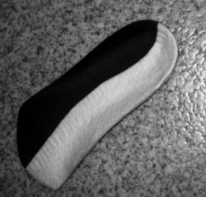
Some cases of Kohler’s disease are self-resolving. Others require the use of soft splintage, such as semicompressed felt valgus filler pads, or supportive orthoses and appropriate footwear to maintain the architecture of the longitudinal arch of the foot during healing. Paracetamol (e.g. Calpol™) may be used to control pain in young children. Surgery is not usually indicated.
Osteochondritis dissicans of the talus more commonly affects the talar head, although it may also affect the trochlear surface. The incidence is reported as 1 in 5000 (Griffin 1994), and it is more common amongst sports people such as skaters, runners and gymnasts (Manusov et al 1996a). There is often a history of recent trauma or severe inversion ankle sprain (Kaeding & Whitehead 1998), or the patient may recall only a relatively minor injury, such as slipping or stumbling.
The bone lesion is classified in four stages of presentation:
The patient presents with a painful, swollen ankle. The location of the pain and the swelling can aid the diagnosis of the exact site of the bone lesion. For example, osteochondritis dissicans of the talar head gives rise to focal pain and swelling in the medial area of the midtarsal joint, whereas osteochondritis dissicans of the trochlear surface of the talus will cause pain and swelling that relates to the anterior aspect of the ankle. However, if a loose body has formed within the ankle or talonavicular joints, the site of the pain and swelling is far less constant, and the patient describes episodes of acute focal pain and swelling that change location from day to day as the fragment of bone moves around within the joint cavity. Patients often report that the ankle ‘locks’ or ‘catches’, and that the ankle and subtalar joint area is unstable on weight bearing.
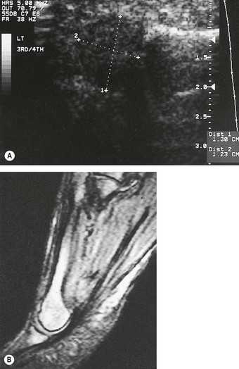
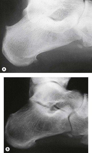
Radiographs may not identify the damaged area, especially in the early stages. Special views may need to be requested to visualise all aspects of the affected bone. Computerised tomography scans and magnetic resonance imaging are much more sensitive than radiography in the early diagnosis of osteochondritis dissicans of the ankle–subtalar–midtarsal joint complex.
The treatment of stages I and II includes rest and immobilisation for 6 weeks. This is achieved by the use of a below-knee plaster of Paris cast, an Aircast® boot, or an ankle–foot orthosis, together with the use of crutches to aid in walking. As healing progresses, the use of a firm walking boot, rather than a normal shoe, is of benefit, as it restricts movement of the rearfoot complex, yet allows relatively normal ambulation.
Stages III and IV may require surgical removal of the loose bone fragment, via arthroscopy, or internal fixation to reattach the detached bone fragment.
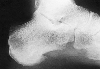
Sever’s disease is perhaps the most common cause of heel pain in children, aged between 7 and 12 years (Griffin 1994, Manusov et al 1996a).
Heel pain in Sever’s disease occurs as the result of an overuse syndrome, and is not a true avascular necrosis. Although it is commonly referred to as an osteochondritis, it is more accurately classified as a traction apophysitis.
The calcaneus has two centres of ossification: the primary centre, which is within the body of the calcaneum; and the secondary centre, which is within the posterior area of the bone. These centres of ossification unite when the bone attains maturity (becomes fully ossified) at about 12 years of age. Traction forces from the Achilles tendon on immature bone can induce epiphyseal distraction, (between the body and the posterior leaflet of the calcaneus) due to its insertion onto the middle third of the posterior surface of the calcaneum. Repetitive contractions of the muscles of the posterior compartment (gastrocnemius and soleus) may also predispose to microfractures at the calcaneal epiphyseal plate. A similar pathology is noted at the insertion of the patellar tendon into the tibial tubercle in Osgood Schlatter’s disease (Madden & Mellion 1996).
The condition may be unilateral or bilateral. The patient presents with pain and tenderness that localises to the posterior aspect of the calcaneus. Pain is maximal after vigorous or impact exercise, such as running or gymnastics. There may be an associated warmth and oedema at the posterior heel area, and wearing shoes with a close heel counter aggravates symptoms. It affects boys more often than girls, and onset may link with growth spurts, especially in physically active or overweight children (Madden & Mellion 1996). Many patients will also show biomechanical compensation for structural foot anomalies that tend to reduce shock absorption at heel strike and expose the heel to abnormal ground reaction forces (Madden & Mellion 1996).
Diagnosis in the early stages of the disease is based on the history and the presenting symptoms. Diagnostic signs include: increased pain on passive dorsiflexion of the foot at the ankle, and exacerbation of symptoms when standing on tiptoe (a positive Sever’s sign) (Madden & Mellion 1996). Radiographs of the calcaneus appear normal in the early stages of the disease process. As the disorder progresses, the apophyseal area shows sclerosis and fragmentation, although this finding is not necessarily diagnostic as this picture can also been seen in asymptomatic patients (Kaeding & Whitehead 1998).
The differential diagnosis should exclude a duck-bill fracture of the posterior leaflet of the calcaneum, Achilles tendon pathologies and deep retrocalcaneal bursitis.
Many authors believe Sever’s disease to be self-limiting, as it tends to resolve spontaneously when calcaneal ossification is completed, usually at about 12 years of age. However, the use of physical therapies such as PRICE (pain control, rest, ice, compression and elevation) is beneficial in the acute phase of the disease. When symptoms are severe, a non-weight-bearing regimen, such as a below-knee cast for 6 weeks, or an Aircast® boot, and crutches, together with a course of NSAIDs is indicated. When the presenting symptoms are mild, the use of a figure-of-eight bandage to the affected rearfoot with a heel raise in the shoe is useful.
A heel raise should continue to be used after the acute phase has passed, in order to reduce the pull of the Achilles tendon on the posterior surface of the calcaneum. This will also minimise further trauma to the epiphyseal plate during the healing phase. Night splints, such as those used for the treatment of plantar fasciitis (Powell et al 1998) are also of benefit. After healing is complete, a regimen of rehabilitation exercises to stretch the Achilles tendon will be needed to prevent or reduce a tendency to ankle equinus, and a programme of stretching exercises and ice massage should continue to be used after strenuous activity. The patient should undergo a full biomechanical evaluation, and orthoses should be prescribed to correct any noted faults. The routine use of well-fitting trainers with good shock absorption should be recommended.
Iselin’s disease is a traction apophysitis that affects the base of the fifth metatarsal, at the insertion of peroneus brevis tendon (Lehman et al 1986). The proximal apophysis of the fifth metatarsal usually does not ossify fully until approximately 16 years of age. Overuse of the peroneus brevis tendon prior to full ossification causes inflammation, swelling and tenderness, bruising and pain at the dorsolateral area at the base of the fifth metatarsal (the styloid process). Diagnosis is confirmed by the presenting symptoms, together with increased local pain when the foot is everted against resistance. The differential diagnosis should exclude a stress fracture of the styloid process, a Jones fracture or an avulsion fracture of the styloid process. Symptoms usually respond to a short period of rest (Griffin 1994). In less responsive cases, the foot should be strapped into eversion, to minimise the pull of the peroneus brevis tendon, until the symptoms subside.
The distal ends of the tibia and fibular (forming the medial and lateral malleoli, respectively), the talus and the calcaneus, their interposed joints (the ankle joint and the subtalar joint) and the associated and overlying soft tissues (tendons, ligaments, retinaculae, adventitious and congenital bursae, deep and superficial fascia, the skin and subcuticular structures) form the rearfoot. Pathology and pain in the rearfoot may arise from problems with any of these structures. Thus rearfoot pathology can be classified by the tissue type involved in the pathological process (bone, joint, soft tissues), in conjunction with the location of the pathology (plantar, medial, lateral, or posterior).
Posterior heel pain may be caused by a number of soft tissue pathologies (e.g.: blister formation, infalmmation of the deep or supericial retrocalcaneal bursae, inflammation or rupture I partial rupture of the Achilles tendon), bone pathology (e.g.: Haglund’s deformity, posterior heel spur, Sever’s disease, ‘duck-bill’ fracture) or local trauma.
The Achilles tendon inserts into the middle third of the posterior facet of the calcaneus. A bursa readily forms within the soft tissues superficial to the insertion of the Achilles tendon, in response to stress at the central–lateral area of the posterior surface of the heel. This adventitious bursa is known as the superficial retrocalcaneal bursa. Inflammation of the adventitious bursa (superficial retrocalcaneal bursitis) is associated with rearfoot motion in association with compensated rearfoot varus, mobile pes cavus, or in cases where there is an increase in the angle of inclination of the calcaneus. Eversion of the calcaneus (frontal plane motion) occurs during compensation for these pathologies, and the foot rotates about the longitudinal axis of the midtarsal joint. The frontal plane rotation allows the calcaneus to evert, but imposes shear stresses within the soft tissues overlying central–lateral area of the posterior aspect of the heel, as these tissues move relative to the inner surface of the shoe. The continuous compensatory eversion of the heel predisposes to inflammation of the bursa. If the condition is of long standing, there may also be local hypertrophy of the underlying area of the posterior aspect of the calcaneus. The bony prominence thus formed is known as Haglund’s deformity, or a ‘pump bump’. The shear stresses affecting the overlying soft tissues increase in direct proportion to the size of the bony prominence, and the bursitis can become chronic. If the overlying skin is breached, such as in a broken chilblain or a ruptured blister, the bursa may become infected.
The condition usually affects adolescent females, who present with pain in the posterior aspect of the heel, especially when wearing, or having worn a particular pair of shoes, such as high heels or a style with a marked heel counter. The patient may report seasonal fluctuations, as the problem may flare up if there is local perniosis (chilblain) affecting this area of the heel, when it is termed ‘winter heel’. On examination, there will be an inflamed, fluctuant and very tender area at the central–lateral aspect of the posterior surface of the heel, with a palpable hypertrophy of the superficial fascia, and underlying bone in long-standing cases. The patient is often unable to wear a normal shoe, and the pain of the condition causes her to walk with a limp.
The diagnosis is usually made by reference to the presenting signs and symptoms. Oblique-lateral radiographs of the rearfoot will also demonstrate any additional bone formation at the central–lateral heel area. The differential diagnosis should exclude other causes of local soft tissue inflammation, such as chilblain or blisters.
Initially, a conservative approach is indicated:
Surgery may be required to reduce the underlying bony prominence. A more laterally placed bony prominence can be reduced, but this becomes more difficult if the area of bone hypertrophy involves the attachment of the Achilles tendon. In those cases, a closing wedge osteotomy may be used to reduce the angle of inclination of the calcaneum.
The deep retrocalcaneal bursa is a true anatomical bursa. It is located within the soft tissues at the posterior aspect of the heel, lying between the upper third of the posterior aspect of the calcaneus and the inner aspect of the Achilles tendon, immediately superior to its insertion into the middle third of the posterior aspect of the calcaneus. The bursa is a horseshoe-shaped structure, and in the adult is approximately 2 cm long, 1 cm wide and 0.5 cm deep (Frey et al 1992). Its inferior surface lies on the Achilles fat pad at the posterosuperior aspect of the proximal calcaneus, its anterior surface at the upper third of the posterior surface of the calcaneus, and its posterior surface against the deep aspect of the Achilles tendon.
Pathological changes (deep retrocalcaneal bursitis) can occur in response to local mechanical irritation, such as where the upper edge of a shoe with a marked heel counter traumatises the soft tissues at the superior area of the posterior surface of the heel. The inflamed bursa shows marked thickening and oedema of its walls. The volume of the contained synovial fluid increases, but its viscosity decreases. Deep retrocalcaneal bursitis may also be a feature of inflammatory joint disease, such as rheumatoid disease and seronegative arthropathies. In these cases the area of the calcaneus that receives the insertion of the Achilles tendon may show bone erosions.
The patient presents with diffuse pain that locates to the posterior aspect of the heel. The pain is exacerbated by active dorsiflexion of the foot, such as when walking upstairs as the inflamed bursa is compressed further, between the overlying Achilles tendon and the underlying calcaneus. The visible signs of inflammation are not obvious, although there may be some local warmth and swelling. The inflamed bursa is usually palpable as a tender, bi-lobed, fluctuant area of soft tissue, to either side of the Achilles tendon at a point just superior to its insertion into the posterior surface of the calcaneus. Palpation of the inflamed tissue may show a fluid ‘thrill’.
The diagnosis is made from the presenting signs and symptoms. The bursa can be identified by bursography (the injection of a radio-opaque dye into the swollen bursa).
The differential diagnosis should exclude:
The underlying cause of the bursitis must be established, and in cases where there is evidence of underlying inflammatory joint disease the patient should be referred to a rheumatologist. Conservative therapy focuses on resting the traumatised area. This includes a review of the footwear. The use of a trainer, manufactured with a cut-out at the upper margin of the heel counter is very useful. A simple heel raise, such as a 7 mm semicompressed felt plantar heel pad, placed inside the shoe will prevent or reduce trauma from the upper edge of the heel counter and allow the bursal inflammation to subside. Severe cases may require aspiration of the bursa, and injection of a corticosteroid.
Achilles tendonitis is a painful and sometimes debilitating inflammation of the Achilles tendon occurring at a point just superior to its insertion into the calcaneum. It is thought to affect up to 20% of runners, and is especially common in athletes who are poorly conditioned, or in recreational joggers who have not trained properly. Participation in activities that involve sudden stops and starts and repetitive jumping (e.g. squash, basketball, netball, football, tennis, fencing and dancing) also increases the risk of developing the condition. The condition is aggravated by the use of high-heel-tab trainers, or by wearing high-heeled shoes with a marked heel counter.
Microtears within the distal part of the Achilles tendon (or heel cord) and resultant local inflammation may be precipitated by a sudden increase in activity levels, training on poor surfaces, or using inappropriate footwear. Achilles peritendonitis may be precipitated by a single incident, or can develop gradually as a general overuse phenomenon. It may also affect women who adopt flat-heeled shoes after habitually wearing high heels, and, much more rarely, may present as a later complication of congenital pes valgus. In both these examples, the undue pull on the Achilles tendon causes a reflex spasm of the posterior calf muscles, with increased tension and the development of microtears within the heel cord.
There are three presentations associated with inflammation of the Achilles tendon: peritendonitis (also known as paratendinitis), tendinosis and peritendonitis with tendinosis.
The patient presents with pain in the distal part of the Achilles tendon that localises to a point approximately 5 cm superior to the point of insertion at the calcaneum. Characteristically the pain develops during strenuous activity and eases with rest. In the later stages of the problem, the pain may be severe and unrelenting even at rest, and prevent normal walking. The soft tissues overlying the affected area may be hot and swollen. If the patient has associated tendinosis, there will be a palpable and tender fusiform thickening along the tendon, or nodule formation at a point approximately 2–5 cm superior to the insertion of the tendon into the calcaneus. Generally the condition has a gradual onset, with mild pain that worsens with continued activity. Patients may also complain of pain on first weight bearing after a period of rest, such as when they first stand up in the morning. Repeated activity in cases of Achilles tendonitis causes an increase in local inflammation and can result in later partial or complete rupture of the tendon. Patients who are prone to this condition often show excessive compensatory pronation, as pronation tends to reduce Achilles tendon traction.
The characteristic pain of Achilles tendonitis can be induced by plantar flexion of the ankle against resistance. This effect occurs as the patient begins to stand up from a sitting position, or by going up on tiptoe. In severe cases the patient will be unable to take the full body weight when asked to stand on the affected leg. These results, together with the presenting symptoms and case history facilitate the diagnosis. Severe cases will show inflammation and even tears with the tendon on magnetic resonance imaging. Radiographs of the tendon (a tenogram) will show local inflammation and deterioration of the tendon quality.
The differential diagnosis should exclude Haglund’s deformity, deep retrocalcaneal bursitis, inflammatory arthritides and partial or total rupture of the Achilles tendon. Patients with marked fatty nodules along the length of the Achilles tendon should undergo blood tests to exclude raised blood cholesterol levels (Citkowitz 2004).
People, especially those in middle age, who intend to embark on a jogging or running programme should be reviewed for appropriate orthotic therapy, and undergo a programme of stretching exercises before carrying out sporting activity in order to reduce the likelihood of developing Achilles tendonitis. In the earliest stages of presentation, a simple heel raise to minimise the pull on the Achilles tendon, together with advice on training and prestretching may be sufficient to resolve an incipient Achilles tendonitis. Of course, it will be necessary to rest from strenuous sports activity until all symptoms have cleared, and to undergo a programme of gradual rehabilitation when sports are resumed. Attention should be paid to the footwear, in particular the shape of the heel counter, to reduce the likelihood of further trauma. More advanced cases, where there is a degree of tendon degeneration, require more active intervention. Steroid injections are contraindicated because of the danger of causing further tendon deterioration, and subsequent rupture. Physical therapies such as the use of pain control, rest, ice and strapping (PRICE) in the acute stages, and a course of twice-weekly ultrasound and contrast footbaths to reduce inflammation are beneficial. Heavy-load, eccentric calf muscle training is very effective (Alfredson et al 1998), but immobilisation in a below-knee cast or Aircast® may be necessary to allow tendon healing, followed by a rehabilitation programme of intensive stretching and exercises, as an essential adjunct to treatment to allow the patient to get back to full weight bearing. After the acute injury has resolved the patient should undergo biomechanical evaluation and any identified faults should be corrected by the prescription of functional orthoses.
The Achilles tendon is prone to rupture at a point 2–5 cm superior to its point of insertion on the posterior surface of the calcaneus.
The Achilles tendon may rupture as the result of an isolated severe traumatic event, or as the culmination of repeated episodes of peritendonitis. The tendon rupture in either case may be partial or total.
Patients with either acute or chronic Achilles tendon rupture present ultimately with a similar set of clinical signs. These include a palpable painful swelling overlying and surrounding the distal part of the tendon, a discontinuity along the length of the tendon just superior to its insertion (Fig. 4.16), and inability to stand on tiptoe or plantar flex the foot against resistance, or bear weight normally on the affected foot. In an acute presentation of Achilles tendon rupture, the patient gives a characteristic history of a severe pain and a sudden sensation of something ‘snapping’ at the back of the heel, usually occurring during an episode of strenuous or sports activity. They experience severe localised pain, usually fall over, and are unable to use that foot to walk normally, as they will have lost the ability to plantar flex the foot against ground resistance. Sometimes they think that someone must have kicked them in the back of their heel. In a chronic presentation, the onset of symptoms is more gradual, as an increasingly severe Achilles tendonitis, leading to eventual partial or total rupture.
Figure 4.16 Achilles tendon rupture. This male patient, aged 74 years, developed a rupture of the Achilles tendon several months after experiencing pain at the posterior area of his right heel/lower leg. He was diagnosed as having type 2 diabetes mellitus at age 68 years, with associated chronic hypertension, hypercholesterolaemia, peripheral sensory neuropathy, chronic venous insufficiency and mild obesity. In view of his overall medical condition, the vascular status of the lower limb, and the chronicity of the Achilles tendon injury, the orthopaedic surgeon recommended that surgical repair of the tendon was not in the patient’s best interest. The case was managed initially by the use of an Aircast™ walking boot for 6 weeks, followed by prescription orthoses worn in walking boots.
In cases of partial Achilles tendon rupture the affected limb must be immobilised in a below-knee cast or Aircast® boot for several weeks to allow the tendon to heal. The patient will need to follow a programme of rehabilitation once the cast is removed and tendon repair is complete in order to regain full strength in the posterior calf muscles, reduce the chance of re-rupture, and prevent the development of ankle equinus due to any contraction of the posterior soft tissue structures that may have occurred during the healing period. More severe cases of partial rupture are corrected surgically.
Cases of full rupture of the Achilles tendon may be corrected surgically. The tendon ends are resutured together, sometimes including the additional use of donor tendon, such as a plantaris tendon graft. The foot is immobilised in a cast for 6–8 weeks, in a plantar-flexed position, and the patient not allowed to weight bear until tendon healing is complete. Wound healing in Achilles tendon repairs can be prolonged. In cases of total Achilles tendon rupture, immobilisation without surgery, even for several months, is not usually successful, and the risk of re-rupture is high in those few cases that do achieve a degree of repair solely by immobilisation.
CASE STUDY 4.5 RUPTURE OF THE ACHILLES TENDON SUBSEQUENT TO CHRONIC ACHILLES TENDONITIS
A 38-year-old man was referred for orthotic therapy subsequent to undergoing surgical repair of the Achilles tendon 6 months earlier. He was in good general health, and on no regular medication. He smoked 20 cigarettes a day. He had been employed as a firefighter since he was 20 years old, but was currently on long-term sick leave due to the Achilles tendon problem.
He presented the following history. At age 17 years he had a cartilage injury to his left knee, which was repaired surgically. He had suffered a recurrent problem with his left Achilles tendon since injuring it when he was 21 years old, whilst playing football. He did not take the advice he was given then to rest the injury, and felt that this had caused him to have a nagging problem ever since. Six months ago, he had slipped down a flight of stairs whilst answering a fire call and snapped the left Achilles tendon. The tendon was repaired, using a peroneus (fibularis) brevis tendon graft. The left limb was put in a below-knee cast for 6 weeks, then an Aircast for a further 6 weeks. He followed an extensive programme of rehabilitation and physiotherapy for a further 3 months, at the end of which he was referred to a podiatrist for orthotic therapy.
He reported the following symptoms: a constant dull ache in his left knee, a painful nodule in the lateral area of the left calf, lower back (lumbar/sacral) discomfort, and a tendency for his left foot to slip when walking on uneven surfaces.
On examination it was noted that the patient had bilateral pes cavus. There was marked contracture of the left Achilles tendon and inversion of the left heel. There was a surgical scar along the lateral aspect of the left foot from the base of the fifth metatarsal to the heel, and another surgical scar along the posterior aspect of the lower left leg extending from the posterior aspect of the heel to the inferior lateral–posterior calf area, with a tender nodule at its proximal end (the stump of the harvested peroneus brevis tendon). There were areas of marked plantar and lateral hyperkeratosis at the left fifth metatarsophalangeal joint area. There was no loss of continuity of the Achilles tendon, but there was some local thickening 3–5 cm superior to its attachment into the calcaneus. The left leg was 1 cm shorter than the right, and the left calf 3 cm narrower than the right. His gait was laboured. The left foot showed an early heel lift and remained inverted throughout gait. He was unable to stand fully on tiptoe on his left foot, and unable to maintain what toe stance he could demonstrate for longer than a few seconds. Muscle power (eversion and plantar flexion) of the left foot was markedly reduced.
His presenting problems related to the injuries he had suffered: the shortening of the left Achilles tendon, as the result of the surgery, caused the early heel lift during gait, and reduced active plantar flexion. The harvesting of the peroneus brevis had reduced his ability to evert the foot. His left foot, therefore, remained inverted throughout gait, causing him to feel unsteady on uneven ground. The loss of shock absorption, due to the loss of midstance pronation, was the cause of his left knee and lower back pain. Casts were made of both feet, to which intrinsically posted orthoses were made. He has now returned to work as a firefighter.
Pain of the plantar aspect of the heel may be caused by a number of pathologies, which include: heel pain syndrome, plantar fascia rupture, fat-pad atrophy, stress fractures of the calcaneus, proximal plantar fasciitis, distal plantar fasciitis, plantar fibromatosis, tendonitis of the flexor hallucis longus tendon, tumour of the calcaneum, and nerve entrapment or injury (Pfeffer et al 1999).
CASE STUDY 4.6 RUPTURE OF THE PLANTAR FASCIA FOLLOWING HYDROCORTISONE INJECTION
A 35-year-old woman in good general health presented with severe, chronic plantar heel pain syndrome of the left foot, for which she had been prescribed functional orthoses. The heel pain was debilitating, and although the orthoses did not fully eradicate the pain their regular use reduced the pain to a tolerable level. Her foot pain affected her quality of life, and she was frequently depressed by its unremitting nature. At the same time as having podiatric treatment, the patient underwent a course of hydrocortisone injections into the left heel. The injections were administered by the patient’s GP. As she was stepping up a kerbstone shortly after the third injection, she experienced severe pain and a sensation as if something in the sole of her left foot had snapped. The pain subsided over the next few days, but the patient was aware that she was no longer able to walk in her normal brisk manner.
The next time she attended for podiatric treatment it was noted that the longitudinal profile of the left foot was visibly flatter than that of the right. She was unable to go up onto tiptoe on her left foot. The plantar fascia did not tighten, and there was no alteration in the profile of the medial longitudinal arch when the hallux was dorsiflexed passively at the first metatarsophalangeal joint. The patient was diagnosed as having suffered a rupture of the plantar fascia. Her foot pain did not return, although the function of her left foot was altered. Rupture of the plantar fascia following corticosteroid injection has been reported in the literature (Acevedo & Beskin 1998, Rolf et al 1997, Sellman 1994).
The most common cause of plantar heel pain is inflammation of the proximal portion of the plantar fascia. It is a common condition that may occur at any time of life, although it tends predominantly to present in the age range 40–60 years, affecting males and females equally. Both feet are affected in 15% of patients. Heel pain can be considered as an overuse syndrome, as there is no single clear aetiology of the condition. Common predisposing factors include a history of increased activity, prolonged standing, excess foot pronation, and recent weight gain or obesity (a body mass index >25) (Aquino & Payne 2001, Hill 1989).
The soft tissues that comprise the plantar aspect of the heel are made up of the glabrous skin that overlies the fibrofatty heel pad, deep to which lies the plantar fascia. The skin and fat pad of the heel are highly specialised to accommodate friction and shock. The plantar heel pad is formed as a honeycombed, interattached meshwork of fibroelastic septa that enclose subcutaneous fat. The deepest parts of the septa merge with the deep fibrous structures that overlie the plantar aspect of the heel. The superficial area of the septa blends with the superficial fascia. This construction allows the absorption of ground reaction forces up to twice body weight, such as occur at heel strike (Singh et al 1997). The heel pad is approximately 18 mm thick in the adult, but often slightly thicker in males (Prichasuk 1994), and tends to atrophy with age, peripheral arterial disease and in cases of rheumatoid disease.
The plantar fascia is also known as the plantar aponeurosis, and is a tough, multilayered inelastic fibrous sheet made up of type I collagen. It is triangular in shape (Fig. 4.17) with the apex directed proximally. The apex originates from the medial calcaneal tuberosity where its deeper fibres merge with the calcaneal periosteum. The more superficial fibres of the plantar fascia merge with the distal Achilles tendon, the fibres of which extend distally to cover the inferior aspect of the proximal calcaneus (Fig. 4.17A) and (Fig. 4.17B). From its point of origin, the plantar aponeurosis fans out distally, becoming gradually broader and thinner, and inserts into the plantar plate on the plantar aspect of the metatarsal heads.
Figure 4.17 (A) Plantar view of the plantar fascia; (B) sagittal section of the foot to show the plantar fascia.
In the area of the MTPJs the plantar fascia separates into five distinct tissue bands. Each of the five bands further subdivides into superficial and deep parts:
Prolonged and continuing traction on the plantar fascia results in inflammation, swelling and pain, especially at its origin at the medial calcaneal tuberosity.
It is thought that the repetitive excessive tensile forces created by walking or standing cause microtears in the fascia, leading to acute and eventual chronic inflammation. If the inflammation affects the entire plantar fascia, it is referred to as a plantar fasciitis; however, if it is isolated to the heel alone, it is called heel pain. Involved tissues undergo changes that typify chronic inflammation. These include collagen necrosis, angiofibroblastic hyperplasia (i.e. overgrowth of local fibrous tissue and blood vessels), chondroid metaplasia (i.e. their transformation into a cartilage-like tissue) and eventual calcification, especially at the origin. In some cases, the first branch of the lateral plantar nerve, known as Baxter’s nerve, may become entrapped within the fibrous tissue, causing symptoms of distal sensory neuropathy.
Where the inflammation also affects the periosteum of the medial plantar calcaneal tubercle, an enthesopathy occurs (the insertion of ligamentous or fascial tissues into a bone surface is termed an ‘enthesis’). The pull of both the origin of the flexor digitorum brevis muscle and the proximal part of the plantar aponeurosis on the periosteum of the calcaneus acts as a stimulus to new bone formation at their points of insertion, resulting in the formation of a plantar calcaneal or heel spur. A heel spur is usually very painful during its formation, but later may become asymptomatic, as once formed it effectively reduces the tractional forces applied to the medial calcaneal tubercle by the plantar aponeurosis. The presence or absence of a heel spur is not diagnostic of heel pain, as 16% of people with heel spurs do not have heel pain, and 50% of people with heel pain do not have a heel spur (Crawford & Snaith 1996).
The patient reports a gradual and increasing pain at the medial–central heel area, which often radiates into the medial longitudinal arch. There is not usually a history of local trauma. The pain, which is usually described as sharp or severe, is worse on weight bearing, especially on first standing in the morning. Typically, the pain subsides gradually after the patient has walked about for a while, but tends to re-establish towards the end of the working day, and recur during the day after periods of rest or non-weight bearing. It is thought that the paradox of pain apparently increased by rest is due to the combined effects of accumulated inflammatory oedema and the sudden traction applied to the inflamed plantar tissues by weight bearing (Wapner & Sharkey 1991). The gradual reduction in the initial pain after walking about is thought to relate to the dispersion of the oedema by the ‘massage’ effect of walking.
On physical examination, the patient often has tightness of the Achilles tendon, due to the close anatomical relationship of the heel cord with the plantar fascia. Pain is elicited by the examiner applying traction to the plantar fascia by passively extending or dorsiflexing the toes at the MTPJ with one hand whilst palpating along the length of the now tightened medial band of the plantar fascia with the thumb of the other hand from distal to proximal. The examiner’s thumb comes to directly palpate the origin of the plantar aponeurosis at the medial plantar calcaneal tubercle (Fig. 4.19). Characteristically, the patient will grimace or verbally indicate pain as the thumb pressure is applied to the point of insertion of the plantar fascia at the medial–plantar heel area, and will also usually describe a sensation of pain as the thumb is moved down the taut plantar fascia.
The characteristic pattern of pain, tenderness in the medial longitudinal arch and the response to direct compression of the tissues at the origin of the plantar aponeurosis in a sedentary, overweight patient is largely diagnostic. The diagnosis can be confirmed by using ultrasonography and radioisotope bone scans to demonstrate inflammation and early new bone formation at the medial calcaneal tubercle. The presence of a heel spur on plain lateral radiographs is not necessarily diagnostic, as asymptomatic heel spur is noted in 10% of people aged over 50 years (Banadda et al 1992) and can be considered as a normal variant when the spur is small and well defined. Large, ill-defined or fluffy heel spurs are seen in conjunction with plantar enthesopathy, especially in cases with seronegative rheumatoid disease (Fig. 4.20).
Figure 4.20 Lateral radiograph of a large, irregular and painful plantar heel spur in a patient with psoriatic arthropathy.
The differential diagnoses should consider:
A subcalcaneal bursitis, also known as policeman’s heel, has been described as a cause of plantar heel pain. There is little anatomical evidence of a congenital or acquired bursa in the superficial plantar heel tissues (Lapidus & Guidotti 1965), and it seems likely that the inflammatory symptoms associated with plantar heel pain syndrome (see above) have been confused with the apparent symptoms of an inflamed bursa.
Entrapment of the medial calcaneal nerve (Beito et al 1989) or the first branch of the lateral plantar nerve (Schon et al 1993) has been identified as a cause of recalcitrant heel pain. Pain associated with the nerve entrapment is located medially, and is sharp and electrical in nature, and may radiate proximally into the lower leg. Excessive foot pronation is thought to cause repetitive microtraumata, chronic fibrosis of the nerve and resultant paraesthesia. In extreme cases, the nerve may require surgical decompression or excision to achieve pain resolution.
Proximal compression of the S1 nerve may cause a referred pain in the distribution of the medial plantar nerve (Schon et al 1993). Referred back pain should be considered in the diagnosis of patients with a history of lower-back problems who present with heel pain.
The treatment of plantar heel pain involves isolating the cause of the problem, measures to reduce the painful symptoms, and actions to resolve, where possible, the underlying mechanical or other pathologies. A variety of conservative measures will allow four out of five cases to make a full recovery within 6–9 months of onset of the symptoms, with the podiatric intervention providing palliative care to reduce mechanical stresses on the heel during the painful period. However, the recalcitrant one-fifth of plantar heel pain cases presents a considerable challenge to successfully treatment (Wolgin et al 1994).
Podiatric palliation includes the provision of:
Tarsal tunnel syndrome is a relatively uncommon entrapment neuropathy affecting the posterior tibial nerve at the point where it passes behind and below the medial malleolus, between the distal end of the tibia and the flexor retinaculum, so that the tibial nerve is entrapped within the fibro-osseous tunnel formed by the calcaneus and the lancinate ligament. The condition is analogous to carpal tunnel syndrome in the hand, and is often associated with inflammatory arthropathies, such as rheumatoid arthritis.
Synovitis of the tibialis posterior tendon, often in association with underlying rheumatoid disease, causes a local chronic inflammation and swelling of the synovial sheath of the tendon as it passes beneath the sustentaculum tali. The resultant local fibrosis causes the tibial nerve to become tethered within the fibro-osseous tunnel formed by the flexor retinaculum of the ankle and the medial malleolus (Cimino 1990). Either direct pressure on the nerve from the fibrous swelling and/or the indirect pressure of rearfoot pronation causes a relative local neuroischaemia, and resultant neural dysfunction, such as sensory neuropathy. The affected area of nerve shows fibrous thickening (Hadjari Hollis et al 2008).
The patient experiences a burning pain and paraesthesia in the sole of the foot and toes in the distribution of the distal part of the tibial nerve. The patient often reports paraesthesia affecting the whole of the plantar surface, with maximal symptoms at the medial longitudinal arch and the metatarsal heads area. Alternatively, the patient may describe paraesthesia, including tingling, pins and needles, and cramp-like feelings in the medial longitudinal arch area. Symptoms are exacerbated by weight bearing, and are often worse at night. Percussion over the inferior–medial aspect of the heel at the site of the entrapment, or sustained direct digital pressure to the nerve in the area of the medial malleolus will reproduce the symptoms (a positive Tinel’s sign). Long-standing cases may also show frank plantar and digital sensory neuropathy with weakness of intrinsic muscles subserved by the affected nerve.
Treatment of tarsal tunnel syndrome focuses on therapies that reduce the unpleasant and painful symptoms. Clinical padding, such as valgus filler pads or medial heel wedges either affixed to the foot or an insole, helps reduce nerve compression or stretching. Excess rearfoot pronation should be controlled as far as possible by the use of moulded cushioned orthoses, housed within appropriate bespoke shoes, to minimise local pressure at the medial aspect of the heel. The inflammation occurring as the result of the rheumatoid process should be controlled by the relevant drug therapies, including NSAIDs together with disease-modifying antirheumatic drugs (DMARDs) as prescribed by the general practitioner or rheumatologist, but many patients with rheumatoid or other inflammatory arthropathies who develop tarsal tunnel syndrome are already on high doses of NSAIDs, to control other aspects of their disease. Surgical treatment including the decompression of the nerve by freeing it from the surrounding fibrotic tissues, with excision of local fibrous tissue, is indicated in cases that do not respond to conservative measures (Takakura et al 1991).
Tibialis posterior tendon dysfunction is a cause of medial heel pathology that is often overlooked and misdiagnosed, especially in the early stages of its presentation (Wassef & Mikhail 2008). It is a disabling condition, arising as the direct result of the loss of function of the posterior tibial tendon. Chronic inflammation leads to degeneration and elongation of the tendon, with the formation of interstitial tears, attenuation and eventual tendon rupture. The condition, if left untreated, leads to increased misalignment of the rearfoot and midfoot, with calcaneal eversion, plantar flexion of the talus, subluxation of the talonavicular joint and unilateral flat foot.
The tibialis posterior muscle is active during the stance phase of gait: it fires shortly after heel strike and ceases to contract shortly after heel lift. Its belly originates deep within the posterior compartment of the lower limb. Its tendon courses down the lower limb to the posterior aspect of the medial malleolus, where it lies anterior to the tendon of the flexor digitorum longus, the posterior tibial neurovascular bundle (containing the posterior tibial artery, vein and nerve) and the tendon of the flexor hallucis longus. All these structures are restrained by the overlying flexor retinaculum at the medial malleolus. The tibialis posterior tendon runs in a groove behind and below the medial malleolus to split into three slips at the medial aspect of the tarsus. The anterior slip inserts into the tuberosity of the navicular bone; the middle slip continues into the plantar tarsal area to insert into the plantar aspects of the cuneiforms, the cuboid and the bases of the second, third, and fourth metatarsals; and the posterior slip has a band-like insertion into the anterior aspect of the inferior calcaneonavicular (spring) ligament. The vector followed by the tibialis posterior tendon at its point of entry to the foot is changed as it passes behind the medial malleolus which thus acts as a pulley block. This, in conjunction with the position of its points of insertion, allows supination of the rearfoot and midfoot during weight bearing, whilst stabilising the arched construction of the midfoot.
The primary actions of the tibialis posterior muscle are to bring about supination of the subtalar joint, and adduction of the forefoot about the oblique axis of the midtarsal joint:
Thus the tibialis posterior muscle acts as a prime stabiliser against rearfoot valgus and forefoot abduction, and is antagonistic to the action of the peroneal muscles, especially to the action of the peroneus brevis.
The cause of tibialis posterior dysfunction is unclear, but a number of factors are associated with the development of the condition. These include:
The presentation of tibialis posterior dysfunction may be classified into four stages:
Alternatively, the pathology of tibialis posterior dysfunction may be classified by the duration and severity of the presenting signs and symptoms:
Approximately 50% of cases present with a history of local trauma, such as a forced eversion of the rearfoot. The typical sufferer of tibialis posterior dysfunction is a female aged over 40 years, although the condition may also affect younger athletes.
The integrity of the tibialis posterior tendon can be assessed by palpating the tendon whilst the patient actively plantar flexes and adducts the foot and the examiner applies an abductory force to the forefoot. It is important to determine the exact site of the injury within the tendon, and it is important to compare the problematic foot to the asymptomatic foot. Direct pressure along the course of the tendon will elicit pain, and active inversion of the foot against resistance will show reduced tibialis posterior muscle power. If a partial tendon rupture has occurred, a distinct defect along the integrity of the tendon can be palpated. If the tendon has fully ruptured, the tendon cannot be palpated along its normal course, and the patient is unable to invert the foot against resistance. A partial or complete rupture of the tendon, secondary to trauma, is accompanied by distinct pain at the navicular tuberosity. Overuse injuries and tendon degeneration present with pain just distal to the medial malleolus.
Magnetic resonance imaging is the most useful method of imaging tendons around the ankle, and it is highly sensitive and specific for the detection of a tendon rupture. Other diagnostic tests include bone scans and injection of radio-opaque material into the tendon/tendon sheath. The early diagnosis is not enhanced by a plain radiograph, although views of the foot will show the extent of structural changes in Stage 3 presentations. A standard weight-bearing anterior–posterior view shows an increase in the angle between the longitudinal axis of the talus and the longitudinal axis of the calcaneus, with an anterior break in the cyma line, abduction of the forefoot and displacement of the second metatarsal. The long axis of the forefoot no longer bisects the rearfoot angle. The normal linear relationship of the talus, navicular, medial cuneiform and the first metatarsal is seen to be lost when the foot is viewed on a lateral radiograph. As the condition progresses, osteoarthritic changes may become evident at the first MTPJ, secondary to the development of hallux limitus.
The differential diagnoses should exclude bone anomalies such as os naviculare (os tibiale externum) syndrome, os trigonum syndrome, navicular avulsion, stress fracture of the navicular; osteochondritis or avascular necrosis of the head of the talus or the navicular, fracture of the medial malleolus, subtalar tarsal coalition and medial sinus tarsitis. Soft tissue anomalies such as strain of the deltoid ligament, medial ankle capsulitis and synovitis, tarsal tunnel syndrome, strain of the flexor hallucis longus or flexor digitorum longus, and retrocalcaneal bursitis should be excluded. Other causes of unilateral flat foot, such as true or apparent leg-length inequality and tarsal coalition, should also be considered in the differential diagnosis.
The most appropriate treatment for tibialis posterior dysfunction is dependent on the stage or phase of the pathology, the presenting symptoms and the severity of the pain reported by the patient. Treatment should always be implemented rapidly and aggressively to prevent further deterioration of the tendon. If the patient is seen in the early stages, conservative methods that focus on reduction of inflammation, joint stabilisation and pain control are indicated for up to 8 weeks. In advanced cases or recalcitrant mild cases, surgical repair of the tendon together with joint fixation may be the options of choice.
Conservative therapies of the condition include a course of NSAIDs for cases where the presenting pain is associated with tendon inflammation (tenosynovitis), a course of ultrasound therapy, together with strapping or taping of the rearfoot into inversion to reduce tension on the tibialis posterior tendon. Soft temporary orthoses, such as valgus filler pads or whole-foot medial padding (‘cobra pad’, Fig. 4.21), are used to invert the rearfoot. Bespoke rigid antipronatory orthoses are prescribed to allow the tibialis posterior muscle to function more effectively by addressing an underlying pathomechanical defect, controlling subtalar joint movement (using a medial heel skive), reducing strain on the tendon, and controlling forefoot abduction (using a lateral flange). A programme of remedial exercises is indicated to strengthen the tibialis posterior muscle. More severe cases may require immobilisation of the foot in an inverted position in a below knee-cast for several weeks. Steroid injections are not recommended as they tend to increase the likelihood of rupture in the already weakened tendon.
Surgical treatments are indicated for Stage 2, or subacute phase, presentations that do not respond to 8 weeks of conservative therapies, or for Stages 3 and 4, or chronic phase, presentations. Procedures for recalcitrant cases with mild tenosynovitis but without overt damage/fraying of the tendon include peritendinous release, synovectomy and tendon debridement. Synovectomy, insertion reattachment or transfer of the flexor digitorum longus is indicated for more severe cases that are characterised by tendon lengthening. Severe cases, with full rupture and fibrosis of the tibialis posterior tendon are treated by transfer of the flexor digitorum longus, shortening of the spring ligament and the talonavicular capsule, and surgical enlargement of the osseous groove below the medial malleolus. Late-stage cases with pain in the lateral rearfoot can be treated by arthrodesis of the rearfoot joints, such as calcaneotalar fusion, subtalar arthrodesis, talonavicular fusion, or combined talonavicular–calcaneocuboid arthrodesis.
The outcome of corrective surgery for tibialis posterior dysfunction is not always straightforward. The procedure requires a long period of recovery and recuperation, with a regimen of postoperative rehabilitation and exercise. The degree of postoperative correction of the plano-valgus deformity is difficult to predict with precision, although the patient can expect an increase in stability during stance. Rearfoot degenerative joint disease is likely in the long term due to the loss of normal joint alignment caused by the arthrodeses, and a reconstructed tendon may undergo further attenuation after corrective surgery, with recurrence of the preoperative symptoms.
Tarsal coalition is an autosomal-dominant, congenital condition, in which two or more bones in the midfoot and/or hindfoot are conjoined due to a failure of development of the intervening joint (Wang et al 2008). Symptomatic tarsal coalition has an incidence of approximately 1% within the general population, but as it is estimated that up to 75% of cases of tarsal coalition are asymptomatic, the true incidence of tarsal coalition may be nearer to 4% (Leonard 1974).
The most common presentations of tarsal coalitions include fusion of the calcaneus with another rearfoot or midfoot bone. These include coalition of the calcaneus and the talus (talocalcaneal coalition), the cuboid (calcaneocuboid coalition) or the navicular (calcaneonavicular coalition) (Fig. 4.22). The condition is normally asymptomatic in early childhood. It becomes symptomatic later in childhood or during adolescence, when the cartilaginous anlage undergoes ossification, and the flexibility between the conjoined bones is lost. Presentations of tarsal coalition in adulthood have been reported.
The normal motion of the subtalar joint during weight bearing involves both rotation and gliding. During the stance phase of gait, the axis of the subtalar joint rotates from a position of 4° external valgus to 6° internal varus to compensate for the internal rotation of the tibia. The loss of or reduction in internal rotation at the subtalar joint causes loss of the height of the medial longitudinal arch, flattening of the foot and fixed calcaneal eversion (a valgus heel). The plantar aspects of the midtarsal joints widen, and their dorsal aspects narrow. The navicular tends to override the head of the talus when the foot is in a position of maximum dorsiflexion, causing traction and elongation of both the deep midtarsal and spring ligaments and the talonavicular joint capsule. The overall flattening of the longitudinal profile of the foot leads to adaptive shortening of the peroneal tendons, with reactive spasm of the peroneal muscles – the so-called ‘peroneal spastic flatfoot’. Prolonged subtalar joint restriction of motion may lead to long-term degeneration of the posterior facet or arthrosis of the subtalar joint.
The patient with a tarsal coalition presents with a flat foot and marked restriction of available hind- and midfoot eversion. There is associated pain in the foot and lateral compartment of the leg that is usually relieved by rest and aggravated by activity. The patient may also complain of recurrent eversion ankle sprains.
The varying forms of tarsal coalitions ossify at different ages, and thus the age of onset of symptoms is an indicator of the site of the coalition. For example: talonavicular coalitions begin to ossify, and thus become symptomatic, in children aged 3–5 years; calcaneonavicular coalitions begin to ossify in children aged 8–12 years; and talocalcaneal unions become symptomatic during adolescence, most commonly between the ages of 12 and 16 years. The loss of subtalar joint motion and the increasing valgus position of the hindfoot become more apparent as ossification progresses, leading to the development of pes planus. Coalitions involving the middle facet of the subtalar joint are associated with the greatest loss of midtarsal joint motion and are the most likely to cause marked valgus deformity of the foot.
CASE STUDY 4.7 ADULT PRESENTATION OF CALCANEONAVICULAR FUSION
A 21-year-old female university student was referred for advice on the management of a swollen right heel. She was in excellent health, and had just completed the final examinations for an honours degree.
She reported that the medial area of her right ankle/heel became swollen 3 months ago, after running a half marathon. She did not recall noticing this before, and was concerned that she kept traumatising the swollen inner ankle area of the right foot, causing small local grazes.
On examination, it was noted that the right foot was markedly pronated, with a pronounced medial tarsal ‘bulge’. Movement was normal at both ankle joints, but there was no available inversion or eversion at the right subtalar joint, and that on the left was reduced. Pain could not be elicited by palpation, or by passive or active movement of the rearfoot complex. There were no signs of tibialis posterior dysfunction. Plain radiographs demonstrated calcaneonavicular fusions of both feet, that on the right being more marked than that on the left.
This was an unusually late presentation of tarsal coalition, and atypical in that, although both feet showed abnormal bone formation, only that of the right foot was symptomatic, although not painful. It was presumed that the local skin trauma was caused by the inner border of the overpronated right foot being scraped by the left shoe during the swing phase when walking and running.
Tarsal coalitions are most readily identified by computerised axial tomography or magnetic resonance imaging scans. Plain radiographs require special views to visualise tarsal coalitions. Calcaneonavicular coalition is suggested by the presence of an elongated anterior calcaneal process, and a talocalcaneal coalition is best visualised on a Harris–Beath (also known as an axial or ‘ski-jump’) view of the hindfoot.
The examiner can evaluate a loss of subtalar joint movement: In the normal weight-bearing foot the tibia and patella externally rotate and the foot supinates when the medial border is raised, the lateral border maintained in contact with the floor, and the heel maintained in the neutral position. When this examination technique is carried out on patients with limited or decreased subtalar joint motion, the amount of available external rotation of the tibia is decreased and the patella does not rotate externally. A loss of subtalar joint motion can also be demonstrated by the absence of or reduction in normal hindfoot inversion and a lack of increase in the height of the medial longitudinal arch when the patient attempts to stand on tiptoe on the affected foot.
The differential diagnoses should exclude bone tumours, rheumatological disease and fractures about the subtalar joint. Tarsal coalitions have been shown to be a feature of other foot deformities, such as talipes equinovarus, and are associated with fibular hemimelia (a congenital abnormality characterised by total or partial absence of the distal half of the limb), Nievergelt–Pearlman syndrome (a rare, autosomal-dominant, inherited bone disease that affects males more than females, characterised by upper limb and hand deformities, symmetric dysplasia of the lower limbs, with genu valgum, clubfoot and deformed great toes, and crura rhomboidei) and Apert’s syndrome (acrocephalosyndactylia – a rare developmental condition with characteristic craniofacial and limb anomalies including craniosynostosis (premature fusion of the skull bones), midface underdevelopment, and gross syndacylisation of the fingers and toes).
The early treatment of a non-ossified tarsal coalition includes immobilisation of the foot in rearfoot neutral by the use of an ankle–foot orthosis or ankle callipers to restrict movement of the affected joints and minimise pain. Surgery is indicated after full ossification of the coalition is complete. The procedure of choice is determined by the degree and location of the coalition, and includes resection of the bone bar, or fixation and arthrodesis of the affected degenerate and painful joints.
Midfoot pain may arise in conjunction with a pathology that affects the proximal plantar area or as a distinct entity.
Plantar fibromatosis describes a condition in which the patient tends to develop fibromae or fibrous nodules within the plantar fascia. Males tend to be more commonly affected than females, and the condition presents most frequently in the fifth and sixth decades.
The true pathology of plantar fibromatosis is unclear. Adherent fibrous nodules or fibromae develop, usually within the superficial parts of the plantar fascia, and the associated fascial tissues undergo a degree of contraction. The condition follows a similar pathology to Dupuytren’s contracture of the palmar aspect of the hand. Plantar fibromatosis is less common, and generally less disabling than Dupuytren’s contracture, as weight-bearing forces prevent the plantar tissues from undergoing the same degree of contraction as the palmar tissues.
Patients present with single or multiple, painful or asymptomatic nodules, or discrete firm, fluctuant swellings within the soft tissues overlying the plantar aspect of one or both feet.
The diagnosis is made from the clinical signs and symptoms. Patients presenting with plantar fibromatosis may also have a history of Dupuytren’s contracture affecting the hands, and there is a weak association with alcohol abuse.
Asymptomatic cases do not require treatment. Painful presentations may be treated with accommodative orthoses to reduce pressure over the tender nodules. More problematic cases can be treated by an injection of hydrocortisone into the nodule. Surgery may be indicated to excise multiple painful or large nodules, but fibromata tend to recur.
Osteoarthritic changes may occur at the joints of the midfoot, leading to local pain and the formation of exostoses.
Degenerative changes occur at the first metatarsal–medial cuneiform and the cuneionavicular joints in feet with pes cavus and pes valgus:
The patient presents with a pain in the dorsal midfoot area that radiates to the plantar aspect of the medial longitudinal arch. There is a palpable thickening of the bony architecture of the first metatarsal–medial cuneiform joint and the cuneionavicular joint, with the development of exostoses at the joint margins (Fig. 4.23). There is a loss of or reduction in the normal gliding movement within the joint, and possible crepitus. The overlying dorsal tissues may become inflamed, and a dorsal adventitious bursa may develop over these joints due to shoe pressure. In extreme cases, the bursa may even perforate through the overlying dorsal skin. In cases of pes valgus or mobile pes cavus the area of plantar skin overlying the exostoses may develop hyperkeratosis, or even ulcerate, especially in patients with sensory neuropathy or those on parenteral steroid therapy.
The condition can be confirmed, and the extent of the exostoses and joint degeneration visualised, by plain lateral radiographs of the foot.
The differential diagnosis should exclude midtarsal Charcot arthropathy, such as the neuroarthropathy that arises with diabetes mellitus, other causes of distal sensory neuropathy and midfoot fractures.
Clinical pads may be used to reduce pressure on painful areas. These include cavitied or holed semicompressed felt ovals applied to the dorsal or plantar areas of the midfoot, and fleece ovals, which are used to reduce sheer stress to a dorsal bursa. Shoe-style advice is essential, as the patient should obtain a style that will both accommodate the altered foot shape and reduce trauma to the painful area. Temporary or permanent orthoses can be made to control or reduce the effects of the underlying biomechanical anomaly. The exostoses can be excised surgically, with or without fixation of the affected arthritic joint.
Plantar fasciitis (foot strain) is a relatively common foot problem that causes mild to moderate, or moderate to severe pain in the plantar area of the medial longitudinal arch.
The pathology of plantar fasciitis resembles that of heel-pain syndrome. Biomechanical anomalies that predispose to excess tension along the medial column of the foot, together with obesity and prolonged standing predispose to chronic inflammation within the medial band of the plantar fascia.
The patient presents with a history of a gradual increase in discomfort or pain radiating along the medial band of the plantar fascia. The pain is worse on first weight bearing in the morning, or after periods of prolonged sitting or rest, and may radiate proximally to the central area of the heel, or distally to the MTPJs. The pain may become intractable and constant, especially by the end of the day. Pain can be induced by asking the patient to stand on tiptoe, or by the examiner palpating the medial band of the plantar fascia with the thumb whilst dorsiflexing the hallux at the first MTPJs. The medial band of the plantar fascia can be easily palpated as a tight, tender cord. Inflammation is not usually obvious. One or both feet may be affected, and sufferers may become quite debilitated, depressed and disabled by the chronic and intractable nature of their foot pain.
The diagnosis is made on the basis of the presenting signs and symptoms.
The differential diagnosis should exclude heel-pain syndrome, plantar fibromatosis, march fracture and tibialis posterior dysfunction.
Treatment of chronic foot strain is not straightforward, as the patient may have had the condition for several months before seeking treatment. The patient should be advised that they have a chronic soft tissue injury that has probably been present for some considerable time, and is caused by the movements and tensions that occur within the foot during standing and walking. Absolute rest of the foot is impossible to achieve in an ambulant patient, and thus there is no one ‘quick fix’ treatment for their problem. The recovery period may be protracted, and may not necessarily follow a smooth path to resolution. Thus the patient is likely to have ‘good’ and ‘bad’ days. The practitioner is advised to encourage the patient to use combinations of the therapeutic measures listed below, rather than to rely on one type of therapy alone, and to keep on with the treatment for as long as it takes to resolve the symptoms. Empirically, it appears that most cases will respond to a multitherapy approach and the condition will become tolerable within 1–2 months and fully resolve in 6–9 months.
‘Metatarsalgia’ is a generic term used to denote dorsal or plantar, deep or superficial pain in the area of the metatarsals, and thus is a symptom that is associated with a number of forefoot pathologies. Metatarsalgia arises with pathological changes in any forefoot tissues (bones and joints, ligaments, tendons, fascia, the skin and subcuticular tissues, the vasculature, and also in nerve tissue), either as a local problem or as a referred pain from more proximal sites. Other factors that may contribute to metatarsalgia are associated with excessive forefoot forces during weight bearing, and include obesity, pregnancy or occupational factors. Unsuitable or ill-fitting footwear that impedes foot function is implicated as a predisposing or aggravating factor of metatarsalgia. Thus, too short or narrow shoes, shoes with high heels, overlarge shoes, shoes with an incorrect heel-to-ball length, thin-soled and worn-out shoes all contribute to the development of metatarsalgia. Old age or the systemic illnesses that predispose to atrophy or distal displacement of the plantar fat pad also predispose to forefoot pain.
The nature of the underlying problem that has resulted in metatarsalgia must be established in order to fully diagnose and treat the forefoot pain. The diagnosis of metatarsalgia is dependent on gaining a full history of the presenting complaint, together with the examination of the patient, which is itself backed up by the results of diagnostic tests.
From the presenting symptoms and the patient examination, the clinician will be able to reach a working diagnosis. This hypothesis should be confirmed, where possible, by diagnostic tests, including radiographs or other forms of diagnostic investigations such as magnetic resonance imaging or computerised axial tomography scans, bone scan, ultrasound scan and blood tests. The combination of the history, the examination and the results of the diagnostic tests will allow the clinician to form the final diagnosis, and allow him or her to formulate a management plan, so that the presenting problem is treated in the most appropriate manner.
‘Greater metatarsalgia’ is a term that may be used to describe pain that relates to first-ray pathology, and ‘lesser metatarsalgia’ for pain relating to pathology in the area of the second to fifth rays. Alternatively, metatarsalgia may be classified as functional or non-functional.
Functional metatarsalgia is the term used to describe the forefoot pain that arises from the pathomechanical stresses, such as compression, shearing and tensile stress arising within the lower limb, the whole foot, the rearfoot or the forefoot, during both stance and gait. Pathologies associated with metatarsalgia are often ascribed to one particular form of abnormal stress, but which, in reality, develop as the result of combined stresses. For example, concurrent abnormal compression and shearing induces the formation of hyperkeratotic plantar lesions: the patient with a fixed plantar-flexed metatarsal may present with diffuse, asymptomatic, shear-stress-related plantar callosity overlying a compression-related deep painful corn.
Functional metatarsalgia may arise due to anomalies of the forefoot bone structure, such as the relative length of the metatarsals, a localised metatarsal equinus or iatrogenic structural change.
Figure 4.24 Congentially short 4th metatarsal in male aged 38 years (A), and the resultant plantar hyperkeratotic transfer lesions (B), affecting the skin over the head of the 3rd and 5th metatarsals.
Persistent intermittent abnormal compression of the plantar tissues results in contusion and inflammation of soft tissues, and predisposes to the formation of focal, nucleated plantar keratoses. Abnormal compression forces at the plantar fibrofatty pad of the forefoot are associated with:
Persistent abnormal shear stress predisposes to the development of plaques of diffuse plantar hyperkeratosis of skin overlying the MTPJs, and formation of adventitious bursae especially beneath the -first MTPJ. Abnormal shear stress primarily arises as the result of hypermobility. The hypermobility may affect the whole foot, such as is seen in generalised whole-body hypermobility. But it is also associated with biomechanical anomalies such as mobile pes cavus and fully compensated rearfoot varus. Associated excessive compensatory pronation at the subtalar and midtarsal joints causes a reduction in the height of the medial longitudinal arch, with a resultant relative lengthening of the foot on weight bearing and abnormal shear stress of plantar soft tissues. The forefoot is stabilised, in part, by the transverse metatarsal ligament and the tone of the transverse head of the adductor hallucis muscle, which together control the transverse spread of the metatarsal heads when under load. The first and fifth metatarsals are unable to resist metatarsal splaying in the hypermobile foot, with the development of metatarsus primus varus and metatarsus qinque valgus, a resultant increase in the width of the forefoot and associated hallux abducto valgus and digitus quintus varus deformities. In these cases the medial aspect of the first MTPJ and the lateral aspect of the fifth MTPJ are subjected to shoe-related shear stress, with resultant bursa formation at these sites. Over-large or slip-on shoes also predispose to shear stresses at the skin surface, with resultant development of painful diffuse plantar keratoses at the forefoot.
Abnormal tensile stresses occur as the result of chronic strain on fascial tissues. Examples of metatarsalgia associated with abnormal tensile foot stresses include chronic foot strain, and strain on the transverse intermetatarsal ligaments with associated fatigue of intrinsic musculature due to splaying of the metatarsals, such as is seen in pes cavus and hallux abducto valgus.
The successful management of functional metatarsalgia depends on the identification and elimination, or mitigation, of the underlying pathomechanical cause of pain. In cases where metatarsalgia is symptomatic of a forefoot (rather than hindfoot) malfunction, the management focuses on the promotion of maximum function of the lesser toes and their MTPJs, by the use of clinical padding, the use of correctly fitting shoes, the use of functional orthoses and surgery.
This is a term used to describe painful forefoot conditions that do not primarily relate to biomechanical problems within the lower limb and foot. Examples would include Freiberg’s disease and Morton’s neuroma. However, as the foot is a dynamic, weight-bearing structure, it is very difficult to isolate presentations of non-functional metatarsalgia from the influence of biomechanical and weight-bearing factors. Thus the remainder of this review of metatarsalgia groups the presentations of forefoot pain in relation to the tissues involved.
Metatarsalgia can be considered within three broad causative categories:
Focal hyperkeratoses (also known as intractable plantar keratoses [IPK]), tyloma, helloma vasculare and helloma neurovasculare) are deep-seated fibrous corns that form within the skin overlying the plantar aspects of the lesser metatarsal heads. They may incorporate a vascular or neurological element and are frequently underlain by fibrous tissue formation. They are difficult to remove in a pain-free manner without the use of local analgesia.
Focal hyperkeratoses are noted in association with:
The patient presents with a persistent painful hyperkeratotic lesion that usually locates within skin overlying a specific metatarsal head. The lesion requires frequent reduction to control the localised pain it causes, but tends to reform within a very short time after apparent full removal. Total enucleation of the central keratin plug is not always possible, as the patient may be unwilling or unable to accept local anaesthesia or tolerate the pain caused by debridement, especially in cases where a neural or vascular element is included at the deepest part of the hyperkeratotic plug. The long-standing nature of the lesion is characterised by chronic inflammation, which leads to the gradual replacement of that area of the normal fat pad with tough fibrous tissue, deep to the lesion. The resulting replacement of an area of the softer fibrofatty plantar pad with tough fibrous tissue reduces local tissue resilience and exacerbates the problem. Plantar cushioning is further reduced if the lesion is associated with a hammer-toe deformity within the same ray, or clawing or retraction of the lesser toes, which causes the plantar fibrofatty pad to be drawn distally away from the MTPJs and towards the plantar webbing area, as soft tissues are more firmly attached to the plantar basal area of the proximal phalanx than to the metatarsals. The volume and function of the tissues overlying the plantar aspects of the MTPJs is reduced, so that the plantar aspects of the metatarsal heads appear to be prominent and are easily palpated. The residual plantar fibrofatty pad is of reduced thickness, and unable to resist the compression imposed by the combination of body weight and ground reaction forces. Atrophy of the plantar fibrofatty pad is a feature of the elderly or rheumatoid foot, and is noted in patients with peripheral vascular disease.
The association with prominence of a particular metatarsal head can be established on an anteroposterior radiograph of the forefoot, where the location of the skin lesion is marked by affixing small metal marker, such as a small circle or wire, to the skin over the hyperkeratotic focus.
The conservative treatment of focal plantar hyperkeratosis does not always lead to resolution, and all approaches are indicated to ensure best results. Treatment options include:
Weakness in the wall of a synovial tendon sheath or a joint capsule predisposes to the formation of a local fluctuant swelling, often at the dorsum of the foot, especially at the point where the tendon sheath emerges from the overlying retinaculae.
The tendon sheath or joint capsule herniates, to form a thin-walled, cyst-like structure that lies within subcuticular structures. The cyst is continuous with the lining of the tendon sheath or joint capsule, and is filled with synovial fluid. The cyst may present as forefoot pain, due to the tension within the cyst, or due to local pressure from footwear. Where the cyst forms as a herniation of the distal interphalangeal joint, it can cause mucoid cyst formation with distortion of the local nail matrix, with resultant deformation of the visible nail plate.
The ganglion shows translumination when the beam from a bright torch is shone through it. The swelling usually has an easily palpable margin, delineating the limits of the lesion. The examiner can detect a ‘fluid thrill’ within the lesion. The examiner places two fingers on the ganglion and applies pressure with the tip of one of the fingers. The second finger detects the change in fluid pressure as the ganglion fluid moves in response to the applied pressure.
The most appropriate treatment is determined by the severity of the presenting problem, and can range from no active treatment through to lesion excision or bone surgery. The range of treatments include:
Capsulitis is the term used to describe inflammation of a joint capsule. Capsulitis frequently arises secondary to osteoarthritis and rheumatoid arthritis, or as the result of ongoing, low-grade joint trauma.
The articular synovium associated with joint ligaments and local tendon sheaths becomes engorged, swollen and softened by the inflammatory process. Where capsulitis is associated with rheumatoid arthritis, the toes may sublux at the MTPJs. Where the process is induced by trauma, such as stubbing the great toe leading to capsulitis of the first MTPJ, the clinical picture is one of acute hallux flexus (see below).
The condition can be treated by:
Gout is a crystal deposition disease that predisposes to arthropathy in susceptible subjects.
Insoluble uric acid in blood is converted by enzymic action into soluble urea. Raised uric acid levels predispose to the deposition of crystalline sodium urate in peripheral and cooler soft tissues, causing severe local pain and inflammation. The long-term accumulations of sodium urate within soft and joint tissues form gouty tophi (singular: tophus). Gout has a predilection to affect the first MTPJ, although tophi may arise in any area of superficial soft tissue, including the nail bed and the outer margin of the pinna of the ear. The disease tends to affect males, over 30 years of age, but postmenopausal women are also predisposed to gout.
Urate crystal deposition within soft tissues acts as a mediator of acute inflammation. Typically, the patient wakes in the middle of the night with severe pain in the first MTPJ. The affected area shows severe acute inflammation: it is very swollen and shiny, very red and hot, and far too tender to touch. Gradually, over the subsequent days, the inflammation gradually subsides, and the area becomes much less swollen, red and painful, and the involved area of skin may peel, reflecting the severity of the earlier acute inflammatory process. The inflammation may totally subside or may persist as a chronic condition. Repeated episodes of gout or prolonged chronic inflammation predispose to ongoing osteroarthritic and degenerative changes within the affected joint. The skin overlying the tophus may perforate or ulcerate and reveal an accumulation of bright, white urate crystals within the affected soft tissues. Where gouty tophi form in subungual tissues, the nail shows onycholysis and subungual breakdown. The nail matrix may be distorted by the long-term inflammation leading to dystrophy and thickening (onychauxis) of the nail plate.
The classic presentation of gout arthropathy and tophus formation is almost self-diagnostic. Radiographs of affected joints show characteristic ‘punched out’ erosions at the joint margins. In the acute phase of the disease, blood urate levels and the erythrocyte sedimentation rate will be raised, but these tend to fall to near-normal values between episodes. Polarised-light microscopy of aspirated joint fluid shows birefringent crystals.
The treatment of gout includes the use of drugs to control pain and inflammation, and agents to reduce blood urate levels.
Adventitious bursae form over bony prominences in relation to local shear stresses. In the long term they may become fibrous, distended, inflamed, infected or even calcified. The skin overlying the bursa may be prone to chilling. Patients with forefoot deformities, such as hallux abducto valgus, are prone to develop adventitious bursae over the medial exostosis at the first metatarsal head, and patients with rheumatoid arthritis tend to develop very large and distended bursae over the bunion joint and the plantar aspects of the MTPJs (see below).
Patients with rheumatoid arthritis are prone to develop large adventitious bursae within the plantar tissues overlying the MTPJs, and fibrinoid nodules over bony prominences (see Ch. 8).
The autoimmune inflammation that characterises rheumatoid arthritis is associated with bursa and nodule formation. Bilateral symmetrical multilobed, distended, fluid-filled soft tissue swellings form over the plantar aspects of the MTPJs, and fibrinoid deposits arise in areas of low-grade trauma, such as at the posterior aspect of the heel or the outer surface of the elbow.
These lesions are prone to flare-up, and become very swollen and acutely tender. The patient with distended plantar bursae describes a sensation of walking on ‘hot coals’. The plantar skin overlying the bursae may be covered with thin callus. Both rheumatoid bursae and nodules are subject to tissue breakdown, and secondary infection may occur, due to the decreased tissue viability that is a feature of rheumatoid arthritis, and the range of drugs that are used to control pain and inflammation. Disease modifying anti-rheumatic drug (DMARD) and NSAID regimens both tend to cause thinning of the skin and tissue breakdown.
The aim of treatment is to minimise trauma to the painful area, particularly during a time of flare-up.
The plantar plate is a tough, rectangular, fibrocartilaginous structure that overlies the plantar aspects of the MTPJs. It is formed from the distal part of the plantar aponeurosis and the plantar aspects of the capsules of the MTPJs. It has a weak origin at the plantar aspects of the necks of the metatarsals and a strong insertion into the plantar aspect of the base of the proximal phalanges. Its function is to reduce load at the plantar aspects of the metatarsal heads, stabilise the digit in association with the collateral ligaments and the intrinsic and extrinsic muscles, and guide the line of pull of the tendons that insert into the digits, such as the tendons of the lumbrical and flexor digitorum longus muscles. The plantar plate acts as an attachment for the distal part of the plantar fascia and has a role in the windlass mechanism (Hicks 1954), resisting hyperextension of the MTPJ. The loss of the integrity of the plantar plate is implicated in the genesis of hammer toe. When rupture of the plantar plate occurs at the first MTPJ the condition is termed ‘turf toe’, and commonly affects young men who engage in vigorous sports (see Ch. 13).
The plantar plate is subject to severe extension forces at toe-off. Any weakness at an origin of the plantar plate predisposes to the development of chronic digital hyperextension at the associated MTPJ, with subsequent attenuation (thinning), or even rupture, of the plantar plate. As a result, the MTPJ becomes unstable, and spontaneous joint dislocation may occur, especially at the second MTPJ. In some cases the rupture presents as a central tear in the joint capsule, allowing synovial fluid to leak out from the joint space into the surrounding tissues, where it acts as a mediator of acute inflammation. In cases where the affected toe deviates laterally, the tear is likely to have occurred in the collateral ligament.
Rupture or attenuation of the plantar plate at the second to fifth MTPJs occurs more frequently in women. It is thought that both the increase in the weight-bearing load at the forefoot and the forced hyperextension at the MTPJs imposed by wearing high-heeled shoes predisposes to plantar plate dysfunction. The condition has also been associated with a long second metatarsal, a short first metatarsal, inflammatory arthropathies, diabetes mellitus, age-related degeneration of the joint tissues and biomechanical anomalies. Athletes are also prone to this condition, as repetitive hyperextension of the MTPJ induces elongation and attenuation of the plantar aponeurosis and joint capsule.
The second MTPJ is most commonly affected by plantar plate rupture. Patients commonly present with chronic focal pain underlying the second MTPJ. The patient may report a history of tripping, and complain of an awareness of a ‘lump’ or a ‘bruised feeling’ on the plantar aspect of the second MTPJ. There may be associated mild swelling to the plantar and dorsal aspects of the joint. Commonly, patients note gradual sagittal (hammering) and/or transverse plane (lateral drift) deformity of the second toe.
The presentation of a plantar plate rupture may vary from a relatively mild local metatarsalgia to an exuberant and acutely painful inflammation with synovitis of the affected MTPJ and associated flexor tendon, with marked functional disability. More commonly, the patient presents with a progressive subluxation or frank dislocation of the second toe, and idiopathic inflammation about the associated MTPJ. The patient usually reports a history of sudden onset of foot pain whilst walking that locates to the base of the toe and the associated MTPJ. The disruption of the proximal phalanx retaining mechanism allows alteration in the alignment of the pull of the flexor tendons, with subsequent and progressive hammering and transverse deviation of the digit.
The clinical diagnosis is made from the history and presenting symptoms. A positive vertical stress test allows the affected toe to be elevated in a dorsal direction at the MTPJ, when the associated metatarsal is stabilised. Translocation of the digit by more than 2 mm in relation to the associated metatarsal head is indicative of rupture of the plantar plate. The tear within the plantar plate and the resultant leak of synovial fluid out of the joint space can be visualised on a radiograph when a radio-opaque dye is injected into the affected joint space (arthrography). The lesion shows on a magnetic resonance imaging scan as an increase in signal intensity in and around the plantar plate (Fig. 4.25), with discontinuity of the integrity of the plate, synovitis of the MTPJ and flexor tendon sheath, and persistent hyperextension of the proximal phalanx.
Figure 4.25 Magnetic resonance image of the second MTPJ, showing loss of integrity of the plantar plate.
The differential diagnosis should exclude synovitis in association with rheumatological disease, traumatic subluxation of the toe, osteochondritis of the metatarsal head (Freiberg’s disease), stress fracture and plantar digital neuroma.
The treatment of plantar plate rupture can be approached conservatively or surgically.
Conservative treatments of plantar plate rupture are designed to ease local pain, rest the painful area, and minimise digital deformity whilst inflammation subsides and tissue healing occurs.
Surgery is indicated for cases that fail to respond to conservative therapies. Recommended procedures include osseous correction of the deformed digit together with flexor-to-extensor tendon transfer to restore the correct alignment of the toe and MTPJ, with surgical repair of the tear in the plantar plate or collateral ligament.
Patients with painful neuropathy or hyperalgesia experience radiating burning, sharp or shooting pains in the distribution of the affected nerve. Their symptoms are usually exacerbated by movement. Tinel’s sign (i.e. distal tingling and paraesthesia) or Valleix’ sign (i.e. proximal tingling and paraesthesia) may be noted when the affected nerve trunk is percussed or palpated. Patients with abnormal sensory function will have altered nerve conduction rates on testing. Painful neuropathy or hyperaesthesia may be a feature of diabetic sensory neuropathy, and often precedes severe loss of peripheral sensation.
Morton’s neuroma, also known as plantar digital neuroma, causes spasmodic neurological pain in the forefoot. It is a common condition, often affecting middle-aged women. Durlacher first described the painful syndrome in 1845, although it is named after TG Morton who wrote on the condition in 1876. In 1883, Hoadley was the first person to identify a nerve lesion at a symptomatic area. Many clinicians have theorised on the true nature and cause of the pain of plantar digital neuroma: some theories are feasible, but others are anatomically incorrect. Plantar digital neuroma affects women more often than men, and the most likely age band of onset is 40–60 years. Sufferers show a tendency to the upper limit of the normal body mass index (BMI). The BMI is calculated as the body weight in kilograms divided by the square of the height in meters. The normal BMI range is 20–25. Patients with plantar digital neuroma tend to the upper levels of the normal BMI (i.e. a BMI of 25 or greater). Neuroma can affect either foot, but bilateral presentation is less common, as is the occurrence of more than one lesion in the same foot. The neuroma lies approximately 5–10 mm deep to the plantar skin, just proximal to the point of division forming the plantar digital nerves proper. Plantar digital neuroma arises as an entrapment pathology affecting an intermetatarsal nerve, causing focal thickening of the nerve at the point where it divides to form the plantar digital nerves. Approximately 90% of cases affect either the second–third or third–fourth web spaces, and only 10% affect the first–second or fourth–fifth interspaces.
Branches of the tibial nerve give rise to the lateral and medial plantar nerves, which subdivide to form paired plantar digital nerves proper. The lateral plantar nerve gives off digital branches that supply both sides of the fifth toe and the lateral side of the fourth toe. The medial plantar nerve gives off digital branches that supply both sides of the first, second and third toes, and the medial side of the fourth toe. There is often a communicating branch of the lateral plantar nerve that anastomoses with the medial plantar nerve, and also supplies the third web space. Other communicating branches anastomose with nerves deeper within the foot, and thereby tether the nerve in the third–fourth web space (Fig. 4.26). The metatarsal heads are separated by bursae, which lie superior (dorsal) to the transverse plantar ligament. The neurovascular bundles and lumbrical muscles lie within the intermetatarsal spaces, deep (plantar) to the transverse metatarsal ligament (Fig. 4.27).
Factors that predispose to the development of plantar digital neuroma include nerve compression and tension, especially in the third–fourth interspace, the distal extension of the intermetatarsal bursa at toe-off, transient nerve ischaemia and biomechanical factors.
There are a number of factors that predispose to compression and tension of the plantar digital nerves. These include:
The intermetatarsal bursae lie between adjacent metatarsal heads (Fig. 4.27). The distal margins of the intermetatarsal bursae project just distally to the associated MTPJ and the distal limit of the transverse intermetatarsal ligament, to lie between the bases of the proximal phalanges. At toe-off the bursal fluid tends to move into the more distal parts of the bursae. This effect is implicated in the pathogenesis of plantar digital neuroma:
The vasa nervosum (the artery serving the affected nerve) and the digital artery (which lies within the same neurovascular bundle as the affected nerve) are subject to intermittent compression during gait, by the distal distension of the intermetatarsal bursa, causing a local transient ischaemia, and acute ischaemic pain.
Empirical observation indicates that a majority of cases with plantar digital neuroma present with inversion of the forefoot when the foot is non-weight bearing. This is compensated at toe-off by sagittal plane movement between the second–third or third–fourth metatarsals. That is, the lateral column of the foot moves dorsally, and the medial column moves relatively in a plantar direction when the forefoot comes under load, leading to stretching of the intermetatarsal tissues, chronic inflammation and the development of local fibrosis around the plantar nerve at the point where it divides to form the plantar digital nerves. The fibrous tissue ensheaths the nerve, exacerbating the condition.
The affected plantar digital nerve shows a fusiform thickening of the perineural tissues, and hypertrophy of the plantar digital nerves – a neuroma. The neuroma is white to yellow in colour, and shiny. Microscopically, there is juxtaneural fibrosis of the nerve that is continuous with the intermetatarsal bursa, intraneural fibrosis and collagen deposition, subperineural hyalinised nodules (Renault’s bodies), fibrosis of endoneural blood vessels (endarteritis), demyelination of the involved nerve fibres and axonal loss. These pathological changes to the plantar digital nerve confirm the diagnosis of Morton’s neuroma, and indicate that the lesion forms as the result of degenerative processes, consistent with an entrapment neuropathy.
The patient typically reports a sudden, sharp, shock-like, burning, paroxysmal, spasmodic debilitating pain at the plantar aspect of the second–third or third–fourth intermetatarsal webbing area. The pain is often triggered by prolonged standing, walking or running, and may also occur during rest. The pain may be acute and disabling. Patients often report urgent need to remove the shoe and massage the foot to try to reduce the pain. Pain can be induced or aggravated by wearing tight or narrow shoes, thin-soled shoes or high-heeled shoes. Pain can also occur spontaneously and during the night. The pain often radiates into the toes that lie to either side of the affected web space, to the dorsum of foot and/or into the lower part of the back of the leg. The patient often describes a sensation of walking on a ‘lump’ or ‘pebble’ in the vicinity of the focal point of the pain at the plantar webbing, and has disturbed sensation, such as paraesthesia, tingling and numbness in the toes to either side of the affected web space. Sometimes the pain of plantar digital neuroma may present as an isolated apical pain. There may be a palpable thickening and oedema of the affected webbing area, which can be detected by palpating the dorsal–plantar dimension of the webbing tissues and comparing this with the same site on the other foot or the adjacent web space. Local oedema may cause the toes at the affected web space to diverge slightly, especially on weight bearing (the sunray sign).
CASE STUDY 4.8 MORTON’S NEUROMA
The patient was a 47-year-old woman, who was in good general health and worked as a home help. She presented with acute pain at the tip of the left fourth toe. The pain had been present for approximately 6 months, beginning gradually, but recently increasing to almost intolerable levels. She was otherwise in good health, although inclined to asthma and eczema.
On examination, the left fourth and fifth toes were in a marked varus position, with a large Durlacher’s corn deep in the lateral sulcus of the left fourth toe. Enucleation of the lesion gave almost instantaneous relief. However, as the pain re-established within 2 months, the outer segment of the left fourth nail was later excised under local anaesthetic, and the exposed pocket of matrix phenolised to prevent nail regrowth. Healing was uneventful, and the left fourth toe was pain free.
However, 1 year later, the patient returned, complaining of exactly the same apical pain in the left fourth toe. The pain was worse when wearing court shoes, and was described as like ‘toothache’ in its sudden and unpredictable onset. There was no sign of nail regrowth, or hyperkeratotic lesion. However, thumb pressure to the left thirrd/fourth plantar webbing whilst simultaneously applying lateral compression across the forefoot (Mulder’s test, Fig. 4.28) triggered the pain spasm. She was diagnosed with plantar digital (Morton’s) neuroma.
Figure 4.28 Mulder’s test: Apply pressure to the third and fourth web spaces, using the thumb, to elicit pain. The forefoot should simultaneously be compressed laterally to test for a palpable, painful ‘Mulder’s’ click.
A temporary insole with a medial heel/instep/forefoot (cobra) pad was made, and this gave some considerable, but short-lived, relief of the symptoms. Finally, the patient underwent day-case surgery, and a large neuroma was excised from the left third/fourth interspace via a web splitting incision. The area healed well, and the patient has been symptom-free in the 10 years since.
This was an unusual presentation of Morton’s neuroma, as the typical signs and symptoms were not obvious, and the treatments of the primary lesions gave good, but only temporary, relief. As the pain of the diagnosed neuroma was no different to that of either the Durlacher’s corn and nail trauma, it was assumed that the neuroma was the underlying cause of the forefoot pathology.
The diagnosis of plantar digital neuroma is generally made on the basis of the highly characteristic presenting symptoms, together with elicitation of the pain by lateral compression of the metatarsal heads whilst simultaneously applying direct pressure in a dorsal–proximal direction with the thumb at the affected web space (Mulder’s test) (Fig. 4.28). The patient will also characteristically show a degree of paraesthesia or hypoaesthesia in the associated interdigital cleft, in comparison to the unaffected interdigital areas (Fig. 4.29). Unambiguous loss of sensation at the affected interdigital space and adjacent toes can be taken as strong supportive evidence of a positive diagnosis of plantar digital neuroma. The patient with plantar digital neuroma or neuritis will experience a full reduction of pain, even when the action that normally triggers the pain is undertaken, by the injection of local anaesthetic, such as 2 ml of 1% lignocaine plain solution, into the area of the neuroma via the interdigital space. The nerve lesion is not visible on radiography, unless it is very large, when the adjacent metatarsal heads may show divergence. The lesion can be well visualised on magnetic resonance imaging, and high-resolution ultrasound imaging is a very useful indicator of the presence of plantar digital neuroma, showing the lesion as a hypoechoic mass, varying in density from the surrounding tissue and oriented parallel to the long axis of the metatarsals (Fig. 4.30A and B). Nerve conduction tests are not always conclusive in the diagnosis of Morton’s neuroma.
Figure 4.29 The affected interdigital space should be tested for loss of sensation using a sharp instrument e.g. a neurotip.
Figure 4.30 (A) Ultrasound scan of the third–fourth intermetatarsal space revealing a large neuroma. (B) An MRI scan of the same site, however, failed to identify the neuroma.
(Photographs printed with kind permission of Miss A. Wilson, The London Foot Hospital.)
The differential diagnosis of Morton’s neuroma or plantar digital neuritis should exclude forefoot pain arising as the result of: lumbar radiculopathy, tarsal tunnel syndrome, metatarsal or stress fracture, Freiberg’s infraction, peripheral neuritis or neuropathy, intermetatarsal bursitis, arthritides, metatarsal and soft tissue tumours, rupture of the plantar plate and MTPJ capsulitis.
The treatment of plantar digital neuroma should address all aspects of the presenting problem, as well as the associated predisposing factors.
Stress fractures are hairline cracks that develop in bone as the result of the repeated application of low-level forces – such as the forces arising during prolonged exercise, or unaccustomed activity. Stress fracture of a metatarsal is given as an example of this process, and the principles of the recognition and treatment of stress fractures elsewhere in the foot are discussed.
Bone fractures occur either as the result of high-level trauma, such as the sudden major exchange of energy that happens in an impact injury, or bone can develop a fracture as the result of the application of repeated minor trauma. This principle is illustrated by cutting a paper clip or a piece of wire. Considerable force, such as that applied by wire cutters, is needed to cut the wire in a single action, but wire can be easily broken if it is repeatedly bent and straightened with the fingers. The accumulated effect of the repeated movements breaks a paper clip after only four or five bend–straighten actions. If other bone pathologies coexist, such as osteoporosis in conjunction with the menopause or steroid therapy, disuse osteoporosis in conjunction with persistent foot pain or distal ischaemia, local Paget’s disease or bone tumour, the likelihood of stress fracture increases. General overall levels of fitness or disease states may also predispose to the development of stress fracture.
The diagnosis of a stress fracture is made from the history and the presenting signs and symptoms. These include an insidious onset of pain or aching that is increased by activity, together with local swelling with or without bruising. The patient may limp. There may be generalised foot or local oedema. The clinician will be able to palpate an isolated, excessively tender point in relation to the underlying bone. In cases of metatarsal fracture, pressure applied under the forefoot induces pain, as will the use of ultrasound over the fracture site, or attempting to move the opposing sides of the fracture in relation to one another.
Early-stage stress fractures do not usually show on plain radiographs, although a technetium-90 radioisotope bone scan will show an area of increased uptake of contrast medium, or ‘hot spot’, at the fracture site, early in the case. From weeks 3–4 onwards the fracture shows as a dark line surrounded by an area of diffuse bone callus on plain radiograph (Fig. 4.31). The fracture line may follow an oblique, transverse, longitudinal or spiral course through the bone tissue. Once healing is complete, the bone callus resorbs, but the cortical bone may remain permanently thickened around the site of the earlier fracture.
The typical stress fracture in the foot affects the second, third or fourth metatarsal shaft, and is termed a ‘march’ fracture. The fault develops within the shaft of the metatarsal, at the point one-third from the head and two-thirds from the base, but stress fractures of the first and fifth metatarsals tend to affect the more proximal part of the bone shaft. Classically, the lesion develops at the medial aspect of the affected metatarsal. Usually only one metatarsal per foot is affected at any one time. The patient rarely gives a history of an exciting incident or trauma, although he or she may have undergone a period of extra exercise or standing, or have experienced weight gain in the weeks prior to the problem developing.
The patient complains of pain and aching in the metatarsal area after, and sometimes during, exercise and when walking or standing, and may have noticed some dorsal swelling and bruising at the time of onset of the pain. Symptoms are often quite mild, so the patient may not seek help at first. The swelling and bruising tend to subside over the subsequent 2–3 weeks, but the pain persists, especially on direct palpation of the fracture site. Wearing certain shoe styles, such as high heels, exacerbates the symptoms, as the increased tension they induce in the plantar fascia causes potential movement in the metatarsal shaft at the fracture site. For the same biomechanical reason, pain is induced when the patient stands on tiptoe – due to the bowing effect of the tightened plantar fascia on the metatarsals.
The diagnosis is suggested by the history, and the presenting signs and symptoms, such as residual swelling and/or bruising at the dorsum of the foot.
The fracture rarely shows on a plain radiograph in its early stages, but after 3–4 weeks the bone repair subsequent to the fracture shows as an increase in the diameter of the bone shaft where diffuse bone callus is forming (Fig. 4.3). The fracture line shows as a radiolucent (black) area traversing the cortex at one aspect of the shaft. Seldom does a stress fracture cause a full trans-shaft fracture. A stress fractures is visible from its earliest stages on bone scan, due to the increased take up of contrast medium in the acute inflammation that characterises the fracture site.
The differential diagnoses of a march fracture should exclude metatarsal osteochondritis, bone tumour, Charcot neuroarthropathy and bone infection (osteomyelitis).
March fractures, as all bone fracture within the lower limb and foot, require immobilisation and protection from weight bearing and ground reaction forces to promote and allow uncomplicated bone healing.
In the majority of cases, healing of a march fracture is uncomplicated, and seldom requires complex interventions, such as internal fixation. However, the predisposing factor(s) that induced the initial bone lesion must be identified and eradicated to ensure good healing and to prevent future recurrence. For example, a 50-year-old woman with a BMI of 29 who is currently employed full time as a traffic warden, but is also working night shifts stacking shelves in a supermarket to earn extra money for a holiday, might develop a march fracture. The problem may be related entirely to her extra workload, but her obesity and age cannot be ignored as contributing factors to the bone pathology. Thus, in addition to the regimen of rest and immobilisation outlined above, the patient should be counselled to seek the advice of her general practitioner about possible menopausal or postmenopausal osteoporosis and help with weight loss. She should also consider whether her increased workload is detrimental to her general health.
Other bones in the foot may be prone to develop stress fracture.
Sesamoids are small bones that occur within tendons, especially at points where the tendon crosses a joint. The sesamoid reinforces the tendon, and facilitates its movement across the joint as the deep aspect of the sesamoid is covered with cartilage and articulates with the other bones of the joint complex. Sesamoid bones are classified as constant or variant. Constant sesamoids are found in almost all humans. Examples of constant sesamoids include the patella that forms part of the knee joint complex, and the paired sesamoids that form part of the first MTPJ complex. Variant sesamoids, and bi- or tripartite sesamoids within the foot occur far less often, and their incidence and presentation varies from person to person.
All sesamoids are subject to all bone pathologies. These include subluxation, dislocation, fractures of all types, osteochondritis, chondromalacia, osteoarthritis and osteoarthrosis, osteomyelitis, bone tumour and associated bursitis.
There are paired, constant sesamoids that form part of the first MTPJ. These are each approximately the size of a grapefruit pip and lie within the tendon of the flexor hallucis brevis muscle to articulate at their deep aspects with the medial (tibial) and lateral (fibular) grooves on the plantar aspect of the first metatarsal head. Their function is to form part of the first MTPJ and contribute a channel on the plantar aspect of the first MTPJ through which the tendon of the flexor hallucis longus muscle courses as it passes to its insertion at the plantar aspect of the distal phalanx of the great toe. The sesamoids reinforce the flexor hallucis brevis tendon, increase the relative depth of the first metatarsal head so that tension within the flexor hallucis longus tendon is maintained, even at toe-off, and increase the functional length of the lever arm of the lower limb at the first MTPJ. Thus the paired sesamoids of the first MTPJ stabilise the joint and increase foot function during gait. The medial sesamoid tends to be larger than the lateral one, but either may present in a mono- or multi- (bi-, tri-, or quadri-) partite form. Bipartite sesamoids are encountered twice as often as multipartite sesamoids, and whilst up to one-third of medial sesamoids are noted as bipartite, only 2% of lateral sesamoids show bipartitism.
Metatarsalgia in the area of the plantar aspect of the first MTPJ may relate to sesamoid pathology.
CASE STUDY 4.9 SESAMOID PATHOLOGY DUE TO OSTEOCHONDRITIS OF THE LATERAL SESAMOID
A 31-year-old woman presented with pain in the first metatarsophalangeal joint of the right foot. The area had been symptomatic for just over 1 year and the pain was becoming progressively worse. The patient was otherwise in good health, and normally a very active individual. She used to play netball for an amateur league team but since the onset of the right forefoot problem had had to give up sport altogether.
The patient presented with fixed pes cavus, with rearfoot inversion and a plantar-flexed first ray. Movement and direct palpation at the right first metatarsophalangeal joint (MTPJ) area was warm, puffy and very painful, with an exquisitely tender point at the lateral plantar aspect of the first MTPJ. A dorsiplantar weight-bearing radiograph showed osteopenia of the lateral sesamoid and a healing stress fracture of the fourth metatarsal.
A diagnosis of avascular necrosis of the lateral sesamoid was made. It was presumed that the metatarsal fracture had developed as stress fracture due to lateral forefoot overload and the patient’s inability to load the medial area of the right forefoot, secondary to the sesamoid problem.
The patient presented with fixed pes cavus, with rearfoot inversion and a plantarflexed first ray (partial forefoot valgus). Movement and direct palpation at the first metatarsophalangeal joint (MTPJ) area was warm, puffy and very painful, with an exquisitely tender point at the lateral plantar aspect of the first MPTJ. A dorsiplantar weight-bearing radiograph showed osteopenia of the lateral sesamoid shown in figure, indicating a continuing pathology. As her pain was so debilitating and unrelenting she was referred to a podiatric surgeon, who excised the lateral sesamoid, to good outcome.
The normal relationship of the sesamoids to the plantar aspect of the first metatarsal head is lost in hallux valgus (see hallux valgus, above), with the result that the lateral sesamoid comes to lie in the first intermetatarsal space, and the medial sesamoid articulates with the lateral plantar groove of the first metatarsal head. As a result of this, the relative depth of the first metatarsal head is reduced by approximately 30% and normal first MTPJ function and stability is compromised in hallux abducto valgus. The loss of the normal orientation of the medial sesamoid increases the opportunity of the foot to pronate, rather than supinate, at toe-off, and efficient propulsion at toe-off is compromised by the overall reduction in joint stability.
Chondromalacia, or dystrophy of sesamoid articular cartilage (at the deep aspect of the sesamoid), is a relatively common cause of sesamoid pathology. It tends to affect the medial sesamoid more often than the lateral one, especially in cases where the medial sesamoid shows bipartitism. The patient presents with a nagging pain or ache that localises to the plantar aspect of the medial sesamoid, especially on weight bearing or during exercise. The condition can be treated by the application of deflective clinical padding or by the manufacture of orthoses that elevate the distal part of the first metatarsal. Surgical removal of the medial sesamoid, or planing the plantar aspect of the sesamoid whilst leaving the deep cartilaginous articular surface intact is advocated for intractable cases that do not respond to conservative measures (see Ch. 13).
Sesamoiditis, or ongoing inflammation and pain around the sesamoid, is a common condition that typically affects physically active young girls, such as dancers and gymnasts. Any activity, even walking, that places constant and repetitive force on the head of the first metatarsal may cause sesamoiditis. Any pathology that damages the sesamoid, such as stress fractures of the medial sesamoid, can also induce sesamoiditis (Kliman et al 1983).
The patient complains of aching, tenderness and swelling in the plantar area of the first MTPJ, especially towards the medial–plantar angle of the joint. The pain increases insidiously, but may cause constant throbbing. Alternatively, movement, especially active and forced dorsiflexion of the great toe, may trigger or exacerbate the pathology. The soft tissues surrounding the affected sesamoid become tender and inflamed. The majority of cases do not show bruising or redness, but pain and swelling limits normal dorsiflexion and plantar flexion at the first MTPJ, so that walking is difficult, and the patient may limp.
The clinical picture of sesamoiditis, which is a history of the gradual onset of and increasing pain at the plantar aspect of the medial–plantar border of the first MTPJ area, with local swelling and a decreased range of joint movement in a young otherwise healthy and active young person, is indicative of medial sesamoiditis.
Minor presentations of sesamoiditis respond to a regimen of rest. This includes minimal weight bearing, cessation of the provocative activity and the use of deflective clinical padding to reduce pressure at the painful plantar–medial area. ‘Fan’ strapping (see Ch. 16) or soft splintage of the first MTPJ to restrict joint movement is also of help, as is a course of NSAIDs to help reduce local pain and swelling. More severe cases may require fixed immobilisation in a below-knee walking cast, together with intra-articular injection of corticosteroid. Once symptoms have subsided, the patient should undergo a full biomechanical assessment to determine the underlying cause of sesamoiditis. Predisposing causes include a plantarflexed first metatarsal (partial forefoot valgus), pes cavus and excessive rearfoot or midfoot pronation. Appropriate prescription orthoses should be made to control or minimise the predisposing factors, to prevent recurrence of the pathology.
A fracture of one or both sesamoids at the first MTPJ will show symptoms that mimic sesamoiditis, such as pain on the plantar aspect of the first MTPJ and the medial forefoot area, with swelling and limited sagittal plane movement of the first MTPJ. However, the patient with a sesamoid fracture usually presents a history of traumatic injury to the plantar first MTPJ area, such as landing heavily following a fall from a height, with the onset of severe pain that relates to the time of injury. Following the sesamoid fracture, the forefoot becomes very tender, swollen and bruised, with focal pain at the first MTPJ. The fracture is confirmed by a plain radiograph, although a naturally bipartite sesamoid can mimic the appearance of a fractured sesamoid. In these cases, a bone scan is indicated.
The fractured sesamoid is treated by rest and full immobilisation for 6–8 weeks in a below-knee cast to restrict first MTPJ movement. Non-union of the fracture may require the affected sesamoid to be excised, with resultant loss of aspects of normal first MTPJ function.
Freiberg’s infraction, or osteochondritis of the metatarsal head, causes metatarsalgia most commonly in the area of the second MTPJ area. It may present as a primary problem in teenagers, or as a secondary degenerative arthrosis in later life. See the section earlier in this chapter on Freiberg’s disease.
Metatarsalgia may develop as the result of forefoot or metatarsal surgery. Complications arising after metatarsal surgery can include delayed union, malunion, non-union and the development of a pseudoarthrosis, recurrence of the original metatarsal problem, or the development of plantar hyperkeratotic ‘transfer’ lesions over adjacent MTPJs. Where the integrity of the MTPJ is lost as the result of surgery, such as a Keller’s arthroplasty, used to correct hallux abducto valgus, the patient may develop a ‘floating’ digit. Other postsurgical complications can include the overexuberant formation of bone callus, with loss of the normal bone architecture; displacement of the metatarsal head and loss of MTPJ integrity; aseptic necrosis of the capital fragment of the metatarsal following osteotomy; and osteomyelitis (bone infection).
Any generalised pathology that affects the locomotor system is likely to give rise to pain in the foot, and thus can be a cause of metatarsalgia. Diseases of the locomotor system fall into two broad categories:
The text in the following section gives only a summary of the main features of these diseases. For greater detail the reader is advised to consult a textbook on general medicine.
Osteoarthritis (see Ch. 8) causes joint degeneration. It presents more commonly in the older population. Although statistically 1 in 10 people in the UK have osteoarthritis and half of all sufferers are over 60 years old. Osteoarthritis presents as a non-symmetrical monoarthritis, in which cartilage erodes and thins, joint space narrows, and osteophytes form at the margins of the affected joints. Osteoarthritis affects primarily the weight-bearing joints, such as the hip, the knee and the first MTPJ, but it also affects joints that have been damaged by an earlier pathology, such as osteochondritis, or affected by adverse pathomechanical influences such as hallux abducto valgus and metatarsus primus elevatus. Crystal deposition disease, such as gout, predisposes the affected joint to develop later osteoarthritis.
Rheumatoid arthritis is caused by an autoimmune-mediated inflammation that results in vasculitis in connective tissues. It characteristically causes hypertrophy of the synovium of the smaller joints of the hands and feet. The rheumatoid arthritis process leads to marked destruction of joint and periarticular tissues, with resultant marked and typical hand and foot deformities. Deformities include lateral deviation and subluxation of digital, and metacarpophalangeal joints and MTPJs, with associated muscle wasting and tendon rupture. Joint effects are noted first and more frequently in the feet, with radiologically evident erosions showing initially at the fifth metatarsal head, with subsequent third, fourth, second then first MTPJ involvement (Renton 1991). Early manifestations of rheumatoid arthritis in the feet include tenderness and swelling of the MTPJs due to synovitis and joint effusions. Characteristically, periarticular swelling causes the toes to diverge – the ‘daylight’ or ‘sunray’ sign. Persistent synovial disease leads to joint erosion, destruction and subluxation, with distal displacement of the fibrofatty pad and the formation of large painful plantar bursae that are prone to tissue breakdown and ulceration. Local bone becomes porotic, but joint ankylosis may also occur. Patients may require forefoot surgery to reduce the deformation and achieve pain relief. Procedures include forefoot arthroplasty such as Fowler’s procedure.
Extra-articular features of rheumatoid arthritis include the formation of rheumatoid or fibrinoid nodules on the extensor surfaces of the limb, tenosynovitis, and characteristic plantar bursae overlying the plantar aspects of the MTPJs. Subjects may develop neuropathy, such as glove and stocking sensory loss, and motor weakness with a positive Tinel’s sign in the affected dermatome of the tibial nerve. Vasculitis of the skin, where acute inflammation of small blood vessels is provoked by the deposition and accumulation of immune complexes, leads to tissue breakdown and ulceration, especially over distorted joints or in association with the plantar bursae. Joint surfaces become eroded and joint spaces widen.
CASE STUDY 4.10 HEEL SPUR IN ASSOCIATION WITH SERONEGATIVE RHEUMATOID DISEASE
A 27-year-old man, who was referred by his GP, had a 2-year history of lower back and hip pain and increasing heel pain, for which he had been prescribed ibuprofen. The patient reported that he thought he was otherwise in good general health, although he had had to give up playing amateur football each week since the onset of his foot and back problems. He worked as a gemstone polisher, which required him to stand all day long on a concrete floor. He was concerned that he had had to take several episodes of sick leave in the past year, because of the increasing severity of the back and heel pain. In the course of the past month, the heel pain had become severe and prevented him from wearing any shoe other than a trainer with a thick cushioned insole. His feet were now a source of considerable daily pain, no matter which type of shoes or cushioned insoles he wore. He also reported that he was finding it increasingly difficult to bend down to cut his toenails. He was currently on sick leave.
On examination: both feet were of normal shape, but with a generalised marked lack of joint mobility. There were no areas of hyperkeratosis, and all pulses in the feet were easily palpable. There was a gross swelling at the posterior aspect of the calcaneum at the insertion of the Achilles tendon (left worse than right), which was extremely tender to touch, and pain was induced by palpating the central plantar heel pad. Plain lateral radiographs showed marked irregularity of the posterior and inferior areas of the calcanei (see figure). Blood tests showed a raised erythrocyte sedimentation rate (ESR) (a normal ESR is <20; a raised ESR indicates a non-specific generalised inflammation).
The patient was made and issued with moulded cushioned insoles, and an appointment was made for him to receive ongoing nail care by the podiatry assistant. A letter to his GP requested an early referral to a rheumatologist, as his symptoms, gender, age and history suggested that he might have ankylosing spondylitis. This diagnosis was confirmed by the rheumatologist following further blood tests. The ankylosing spondylitis had caused posterior and inferior heel spur formation and sacroiliac enthesiopathy. The patient was prescribed a course of disease-modifying antirheumatic drugs (sulfasalazine), and radiographs taken some months later showed a degree of disease remission and resultant calcaneal remodelling (see figure).
Ankylosing spondylitis is a rheumatological disease that typically affects young men of the HLA B27 tissue type. It causes progressive stiffening of the spine, with calcification of ligaments and tendons at their insertions into bone. It predisposes to plantar and posterior heel spur formation (Fig. 4.32 and case study 4.10).
Connective tissue disorders, such as systemic lupus erythematosus, systemic sclerosis, polymyalgia rheumatica and dermatomyositis, are all characterised by a generalised immune-complex-mediated vasculitis, which can affect all body tissues. Vasculitis at specific body sites causes a range of presenting symptoms. These include rheumatoid-arthritis-like arthropathy, a characteristic facial ‘rash’ known as a butterfly rash, ischaemia of the finger and toe pulps with apical ulceration, Raynaud’s phenomenon and severe chilling, levedo reticularis, calcinosis, skin and epithelial sclerosis, telangectasia, polymyositis and muscular weakness, and sensory and kidney dysfunction. Vasculitis of medium-sized vessels characterises polyarteritis nodosa.
Arthritic joints, especially in those seen with severe rheumatoid disease, are prone to develop staphylococcal infection. Other generalised infections, such as tuberculosis (which itself is predisposed by alcoholism), diabetes mellitus or other chronic debilitating disease states, gonorrhoea, salmonella, rubella, and Lyme’s disease, may all cause transitory arthritis. Joints affected by transitory arthritis may become subject to later osteoarthritis. Reiter’s syndrome, which is often triggered by a gastrointestinal infection, affects males of the HLA B27 tissue type. It gives rise to heel and forefoot pain, and is characterised by Achilles tendonitis, plantar fasciitis and heel spur formation. Associated systemic symptoms include eye and urinary tract infections.
Crohn’s disease, chronic active hepatitis, Whipple’s disease and psoriasis can all cause forefoot pain. Psoriasis causes dactylitis – the isolated acute inflammation and swelling of a toe. Psoriasis may also cause arthritis mutilans, which is a form of seronegative arthritis that resembles the most severe joint effects of rheumatoid arthritis. Arthropathy may be a feature of many other diseases. For example, Charcot neuroarthropathy is a feature of long-standing diabetes mellitus and also of late-stage untreated syphilis. Small-joint arthritis can be a presenting feature of hypothyroidism. Acromegaly and joint pain are features of hyperpituitarism. The formation of large bone spurs within the foot area is a feature of Forrestier’s disease (characterised as showing four or more large anterior osteophytes within vertebral disc spaces in the absence of the disc-space narrowing). Avascular necrosis within joints can be associated with sickle-cell disease, thalassaemia and hypercholesterolaemia. Behçet’s syndrome can cause rheumatoid-type arthritis. Generalised hypermobility and foot strain is a feature of Ehlers–Danlos syndrome.
The development of fibrous nodules on the plantar fascial structures is reviewed in the section on plantar fibromatosis (see earlier in this chapter).
This can cause forefoot and generalised foot pain.
Myalgia or muscle pain of a muscle unit or group is characterised by muscular stiffness and pain on movement. Myalgia may occur as a feature of repetitive movements, intermuscular sensory neuritis (also known as fibrositis, fibromyositis and myositis) tendonitis, muscle strains, traumatic injury and contusions. Symptoms vary from a transient stiffness, to agonising pain, and can be set off by pressure at trigger points within the affected muscle. Acupuncture of the trigger and more proximal pressure points, or trigger point massage, is a useful therapy to reduce the pain of myalgia (Sandberg et al 2004).
Neoplasm is rare in the foot, accounting for less than 0.05% of foot lesions. There are a number of relatively benign neoplasms that affect the foot and may possibly cause metatarsalgia. These include squamous-cell papilloma, dermatofibroma, naevi, eccrine poroma, angiokeratoma, giant-cell tumour of tendon sheaths, lipoma, benign fibrous histiocytoma, chondroma, leiomyoma, myxoma and lipoblastoma. Primary malignancies within the foot and lower limb are rare. Those that do arise may provoke metatarsalgia, and include synovial sarcoma, melanoma, verrucous carcinoma, squamous-cell carcinoma, basal-cell carcinoma, fibrosarcoma, malignant fibrous histiocytoma, leiomyosarcoma, chondrosarcoma, myosarcoma, lymphangiosarcoma and malignant neurofibroma. Those malignant neoplastic tumours that affect the bones of the foot tend to occur as a secondary tumour to a primary tumour of lung, breast or kidney. However, the possibility of bone tumours in the foot cannot be ignored, as, although bone tumours below the knee are very rare, 25% of those malignancies that do arise in the lower leg affect the bones of the foot.
Lumbar pain can cause pain in the legs and feet, referred within the distribution of the sciatic nerve. Classically, heel pain can be a feature of spinal nerve compression at the S1 level.
Large plantar bursae are a feature of rheumatoid arthritis. Tenosynovitis and plantar enthesiopathies characterise rheumatoid diseases and Reiter’s syndrome. Nerve-compression syndromes, such as tarsal tunnel syndrome, can arise as the result of local autoimmune-mediated inflammation and vasculitis of the vasa nervosum.
Paget’s disease, which is characterised by bone pain, enlargement of the medullary cavity and thinning of the cortex of long bones, can predispose to stress fractures. Bone infections, such as osteomyelitis and tuberculosis, are very painful, even in cases with sensory neuropathy, and are difficult to treat, requiring surgery and prolonged courses of antibiotics. Neoplastic disease, muccopolysaccharide disorders, skeletal dysplasia (e.g. achondroplasia, osteomalacia), and hereditary diseases such as osteogenesis imperfecta and osteopetrosis all predispose the patient to fractures that are as likely to affect the bones of the foot as elsewhere in the skeleton.
Obese patients with a BMI of over 30, and/or a waist measurement of more than 1 m may experience foot pain due to chronic tension on the plantar fascia. Foot pain can be a feature of the later stages of pregnancy. Any biomechanical anomalies are exaggerated by the increase in body weight, the change in the centre of gravity of the body due to the gravid uterus, and the generalised ligamentous laxity that characterises late pregnancy. Patients who suffer from alcoholism often develop painful neuropathies that affect the feet and legs.
Acevedo JI, Beskin JL. Complications of plantar fascia rupture associated with corticosteroid injection. Foot and Ankle International. 1998;19(2):91-97.
Alfredson H, Pietilä T, Jonsson P, Lorentzon R. Heavy-load eccentric calf muscle training for the treatment of chronic Achilles tendinosis. American Journal of Sports Medicine. 1998;26:360-366.
Aquino A, Payne CB. Function of the windlass mechanism in excessively pronated feet. Foot and Ankle International. 2001;18(11):705-709.
Banadda B, Gona G, Vaz R, Ndlovu D. Calcaneal spurs in a black African population. Foot and Ankle International. 1992;13(6):352-354.
Beeson P. Orthopaedic assessment. In: Merriman LM, Turner W, editors. Assessment of the lower limb, Ch. 8. Edinburgh: Churchill Livingstone; 2002:199-200.
Beito SB, Krych SM, Harkless LB. Recalcitrant heel pain: traumatic fibrosis versus heel neuroma. Journal of the American Podiatric Medicine Association. 1989;79(7):336-339.
Blustein SM, D’Amico JC. Limb length discrepancy. Identification, clinical significance and management. Journal of the American Podiatric Medicine Association. 1985;75(4):200-206.
Bohndorf K. Osteochondritis (osteochondrosis) dessicans: a review and new MRI classification. European Radiology. 1998;8(1):103-112.
Bowden PD. The pathomechanics of forefoot valgus. The Chiropodist. 1983;38(12):445-451.
Brown LP, Yavorsky P. Locomotor biomechanics & pathomechanics, a review. Journal of Orthopaedic and Sports Physical Therapy. 1987;9(1):3-10.
Camasta CA. Hallux limitus and hallux rigidus. Clinics in Podiatric Medicine and Surgery. 1996;13(3):423-445.
Caselli M, Sobel E, McHale KA. Pedal manifestations of musculoskeletal disease in children. Clinics in Podiatric Medicine and Surgery. 1998;15(3):481-497.
Chambers MRC. Running on leg length discrepancy. Athletes World. 1983;6:55-58.
Cimino WR. Tarsal tunnel syndrome: review of the literature. Foot and Ankle International. 1990;11(1):47-51.
Citkowitz E. Familial hypercholesterolaemia. www.emedicine.com, 2004.
Chapman C. Rocker soles. Podiatry Archives. Available at http://www.mailbase.ac.uk/podiatry, 1999. January 1999
Coughlin MJ, Shurnas PS. Hallux rigidus: demographics, etiology, and radiographic assessment. Foot and Ankle International. 2003;24(10):731-743.
Crawford F, Atkins D, Young P, Edwards J. Steroid injection for heel pain: evidence of short-term effectiveness. A randomized controlled trial. Rheumatology. 1999;38:974-977.
Crawford F, Snaith M. How effective is therapeutic ultrasound in the treatment of heel pain? Annals of Rheumatic Diseases. 1996;55:265-267.
Crawford F, Thomson CE 2003 Interventions for treating plantar heel pain. Cochrane Database Systematic Review 2003(3):CD000416.
Daly PJ, Kitaoka HB, Chao EYS. Plantar fasciotomy for intractable plantar fasciitis: clinical results and biomechanical evaluation. Foot and Ankle. 1992;13:188-195.
Dananberg HJ, Phillips AJ, Blaakman HE. Non-surgical treatment of hallux limitus. Advances in Podiatric Medicine and Surgery. 1996;2:67-69.
Dananberg HJ. Gait style as an etiology to chronic postural pain. Part 1–Functional hallux limitus. Journal of the American Podiatric Medicine Association. 1993;83(8):432-441.
Durrant MN, Siepert KK. Role of soft tissue structures as an aetiology of hallux limitus. Journal of the American Podiatric Medicine Association. 1993;83(40):173-180.
Ekman S, Carlson CS. The pathophysiology of osteochondrosis. Vetinary Clinics of North America. 1998;28(1):17-32.
Fernandez E, Pallini R, Lauretti L, et al. Neurosurgery of the peripheral nervous system: entrapment syndromes of the lower extremity. Surgical Neurology. 1999;52(5):449-452.
Ferrari J, Malone-Lee JG. Relationship between proximal articular set angle and hallux abducto valgus. Journal of the American Podiatric Medicine Association. 2002;92(6):331-335.
Ferrari J, Hopkinson DA, Linney AD. Size and shape differences between male and female foot bones: is the female foot predisposed to hallux abducto-valgus deformity? Journal of the American Podiatric Medicine Association. 2004;94(5):434-532.
Frank CJ, Robinson DE, Dalenberg DD. Hallux valgus. Available at http://www.eMedicine.com, 2004.
Freiberg O. Clinical symptoms and biomechanics of lumbar spine and hip joint in leg length inequality. Spine. 1983;8(6):643-651.
Frey C, Rosenberg Z, Shereff MJ, Kim H. The retrocalcaneal bursa: anatomy and bursography. Foot and Ankle International. 1992;13(4):203-207.
Gould JS. The foot book. Baltimore, OH: Williams and Wilkins; 1988.
Granberry WM, Noble PC, Bishop JO, Tullos HS. Use of a hinged silicone prosthesis for replacement arthroplasty of the first metatarsophalangeal joint. Journal of Bone and Joint Surgery. 1991;73(10):1453-1459.
Griffin DW. Common sports injuries of the foot and ankle seen in children and adolescents. Orthopaedic Clinics of North America. 1994;25(1):83-93.
Gurney B. Leg length discrepancy. Gait and Posture. 2002;15(2):192-206.
Hadjari Hollis M, Lemay DE, Jensen RP. Nerve entrapment syndromes of the lower extremity. Available at http://emedicine.medscape.com/article/1234809-overview, 2008.
Hammer WI. Functional soft tissue examination and treatment by manual methods. Ch. 6, 2nd edn. Jones & Bartlett, Sudbury, MA. 1999.
Hansen TES 1993 Correlation between leg length inequality and lateral weight distribution. Thesis, Anglo-European College of Chiropractic, Bournemouth.
Hicks JH. The mechanics of the foot, 2. The plantar aponeurosis and the arch. Journal of Anatomy. 1954;88:25-31.
Hill JJ, Cutting PJ. Heel and body weight. Foot and ankle international. 1989;9(6):254-256.
Hlavac HF. The foot book -advice for athletes. California: World Publications; 1971.
Hockenbury RT. Forefoot problems in athletes. Medicine & Science in Sports & Exercise. 1999;31(7):448-457.
Hopper D, Bryant A, Elliott B. Foot types and lower limb injuries in elite netball players. JAPMA. 1994;8(7):355-362.
Kaeding CC, Whitehead R. Musculoskeletal injuries in adolescents. Primary Care. 1998;25(1):211-223.
Kerr M 2008 Needle punctures plus corticosteroid injection relieve plantar fasciitis symptoms. Presented at 94th Scientific Assembly and Annual Meeting of the Radiological Society of North America, 30 November– 5 December 2008, Chicago, IL.
Klaue K, Hansen ST, Masquelet AC. Clinical, quantitative assessment of first tarso-metatarsal mobility in the sagittal plane and its relationship to hallux valgus deformity. Foot and Ankle. 1994;15(1):9-13.
Kliman ME, Gross AE, Pritzker KP, Greyson ND. Osteochondritis of the hallux sesamoid bones. Foot and Ankle. 1983;3(4):220-223.
Kura H, Luo Z-P, Kitaoka HB, An K-N. Role of the medial capsule and transverse metatarsal ligament in hallux valgus deformity. Clinical Orthopaedics and Related Research. 1998;Vol 354:235-240.
Kwong PK, Kay D, Voner VT, White M. Plantar fasciitis: mechanics and pathomechanics of treatment. Clinics in Sports Medicine. 1998;7(1):119-126.
Lang L. Ankle equinus. The Chiropodist. 1984;39(1):4-7.
Laing P. The painful foot. In: Merriman LM, Tollafield DR, editors. Assessment of the lower limb. Edinburgh: Churchill Livingstone, 1995.
Lapidus PW, Guidotti FP. Painful heel: report of 323 patients with 364 painful heels. Clinical Orthopaedics and Related Research. 1965;39:178-186.
Lee CR, Farley CT. Determinants of tne center of mass trajectory in human walking and running. J Exp BioI. 1998;201(Pt 21):2935-2944.
Lehman RC, Gregg JR, Torg E. Iselin’s disease. American Journal of Sports Medicine. 1986;14:494-496.
Leonard MA. The inheritance of tarsal coalition and its relationship to spastic flat foot. Journal of Bone and Joint Surgery – British Volume. 1974;56B(3):520-526.
Light MR. Dynamics and function of the first metatarsophalangeal joint. Advances in Podiatric Medicine and Surgery. 1996;2:41-48.
McPoil TG, Schuit D, Knecht HG. A comparison of three positions used to evaluate tibial varum. Journal of the American Podiatric Medicine Association. 1988;78(1):22-28.
Madden CC, Mellion MB. Sever’s disease and other causes of heel pain in adolescents. American Family Physician. 1996;54(6):1995-2000.
Manello DM. Leg length inequality. Journal of Manipulative and Physiological Therapeutics. 1992;15(9):576-590.
Manusov EG, Lillegard WA, Raspa RF, Epperly TD. Evaluation of pediatric foot problems: part II. The hindfoot and the ankle. American Family Physician. 1996;54(3):1012-1026.
Manusov EG, Lillegard WA, Raspa RF, Epperly TD. Evaluation of pediatric foot problems: part I. The forefoot and the midfoot. American Family Physician. 1996;54(2):592-606.
Menelaus MB. The management of limb inequality. Edinburgh: Churchill Livingstone; 1991.
Mooney J, Ramsamy H, McCoshim G, et al. The resolution of the pain of plantar digital neuritis after surgical excision of neuroma. British Journal of Podiatry. 1999;2(3):93-100.
O’Donnell MM. Introduction to podopaediatrics. Chapter 7 General Medicine. In: Thomson P, editor. Introduction to podopaediatrics. Chapter 7 General Medicine. London: WB Saunders, 1988.
Palladino SJ. Pre-operative evaluation of the bunion patient: aetiology, biomechanics, clinical and radiographic assessment. In: Gerbert J, editor. Textbook of bunion surgery. 2nd edn. New York: Futura; 1991:1-87.
Payne C, Chuter V, Miller K. Sensitivity and specificity of the functional hallux limitus test to predict foot function. Journal of the American Podiatric Medicine Association. 2002;92(5):269-271.
Perry J. Gait analysis, normal & pathological function. Thorofare, NJ: Slack; 1992.
Pickard JM. Pathomechanics of rearfoot varus. The Chiropodist. 1983;38(10):379-383.
Pfeffer G, Bacchetti P, Deland J, et al. Comparison of custom and prefabricated orthoses in the initial treatment of proximal plantar fasciitis. Foot and Ankle International. 1999;20(4):214-223.
Phillips D. Biomechanics. In: Hetherington VJ, editor. Hallux valgus and forefoot surgery. Melbourne: Churchill Livingstone; 1994:39-66.
Powell M, Post WR, Keener J, Wearden S. Effective treatment of chronic plantar fasciitis with dorsiflexion night splints: a cross over prospective randomized outcome study. Foot and Ankle. 1998;19(1):10-18.
Powers CM, Maffucci R, Hampton S. Rearfoot posture in subjects with patello-femoral pain. Journal of Orthopaedic & Sports Physical Therapy. 1995;22(4):155-160.
Prichasuk S. The heel pad in plantar heel pain. Journal of Bone and Joint Surgery. 1994;76B(1):140-142.
Redmond A. Assessment of the lower limb, Ch. 10. In: Merriman LM, Turner W, editors. Assessment of the lower limb, Ch. 10. Edinburgh: Churchill Livingstone, 2009.
Renton P. Radiology of the foot. Ch. 13. Klenerman L, editor. The foot and its disorders, 3rd edn, Edinburgh: Blackwell Scientific, 1991.
Rolf C, Gunter P, Ericsater J, Turan I. Plantar fascia rupture: diagnosis and treatment. Journal of Foot and Ankle Surgery. 1997;36(2):112-114.
Rome K. Ankle joint dorsiflexion measurement studies. A review of the literature. Journal of the American Podiatric Medicine Association. 1996;86(5):205-211.
Root ML, Orien WP, Weed JH. Normal and abnormal function of the foot. Clinical biomechanics, Vol. I. Clinical Biomechanics Corp. Los Angeles. 1971.
Root ML, Orien WP, Weed JH. Normal and abnormal function of the foot. Clinical biomechanics. Vol. II. Clinical biomechanics Corp. Los Angeles, CA. 1977.
Rothbart BA. Relationship of functional leg length discrepancy to abnormal pronation. Journal of the American Podiatric Medicine Association. 2006;96(6):499-504.
Sandberg M, Lindberg L-G, Gerdle B. Peripheral effects of needle stimulation (acupuncture) on skin and muscle blood flow in fibromyalgia. European Journal of Pain. 2004;8(5):163-171.
Saxelby J, Betts RP, Bygrave CJ. Low-dyetaping on the foot in the management of plantar-fasciitis. The Foot. 1997;7:205-209.
Schon L, Glennon T, Baxter D. Heel pain syndrome: electrodiagnostic support for nerve entrapment. Foot and Ankle International. 1993;14(3):129-135.
Seibel MO. Foot function: a programmed text. Baltimore: Williams and Wilkins; 1988.
Sellman JR. Plantar fascia rupture associated with corticosteroid injection. Foot and Ankle International. 1994;15(7):376-381.
Sgarlatto TE. A compendium of podiatric biomechanics. Oakland, CA: California College of Podiatric Medicine; 1971.
Singh D, Angel J, Bentley G, Trevino SG. Plantar fasciitis. BMJ. 1997;315:172-175.
Subotnick SI. Biomechanics of the subtalar and midtarsal joints. Journal of the American Podiatric Medicine Association. 1975;65:756-764.
Subotnick SI. Limb length discrepancies of the lower extremity, (the short leg syndrome). Journal of Orthopaedic & Sports Physical Therapy. 1981;3:11-16.
Takakura Y, Kitada C, Sugimoto K, et al. Tarsal tunnel syndrome. Journal of Bone and Joint Surgery. 1991;73B:125-128.
Tax HR. Podopaediatrics, 2nd edn. Baltimore, OH: Williams and Wilkins; 1985.
Tillu A, Gupta S. Effect of acupuncture treatment of heel pain due to plantar fasciitis. Acupuncture in Medicine. 1998;16(2):66-68.
Tsai WC, Hsu CC, Chen CP, Chen MJ, Yu TY, Chen YJ. Plantar fasciitis treated with local steroid injection: comparison between sonographic and palpation guidance. J Clin Ultrasound. 2006;34(1):12-16.
Vink P, Kamphuisen HAC. Leg length inequality, pelvic tilt and lumbar back muscle activity during standing. Clinical Biomechanics. 1989;4(2):115-117.
Walsh HP, Dorgan JC. Etiology of Freiberg’s disease – trauma? Journal of Foot Surgery. 1988;27(3):243-244.
Wang EA, Gentili A, Masih S, Wang MC. Tarsal coalition. Available at http://emedicine.medscape.com/article/396694-overview, 2008.
Wapner KL, Sharkey PF. The use of night splints for treatment of recalcitrant plantar fasciitis. Foot and Ankle International. 1991;12:135-137.
Wassef S, Mikhail M. Ankle, tibialis posterior tendon injuries. Available at http://emedicine.medscape.com/article/386322-overview, 2008.
Weil LS, Gowlding PB, Nutbrown NJ. Heel spur syndrome. A retrospective study of 250 patients undergoing a standardised method of treatment. The Foot. 1994;4:68-78.
Wolgin M, Cook C, Graham C, Mauldin D. Conservative treatment of plantar heel pain: long term follow up. Foot and Ankle International. 1994;15(3):97-102.
Zhang Y, Jun H, Hiroaki I, Katsuya N. Footballer’s ankle: a case report. Chinese Medical Journal (English). 2002;115(6):942-943.