Chapter 22 Factors affecting X-ray beam quality and quantity
Chapter contents
22.1 Aim 159
22.2 Introduction 159
22.3 The effect of mA on the X-ray beam 160
22.4 The effect of kVp on the X-ray beam 160
22.5 The effect of the target material on the X-ray beam 161
22.6 The effect of rectification on the X-ray beam 161
22.7 The effect of filtration on the X-ray beam 161
22.8 Summary of the factors affecting the quantity, quality and intensity of the X-ray beam 163
Further reading 164
22.1 Aim
The aim of this chapter is to consider the various factors which have an influence on the quantity and/or the quality of the beam of radiation from the X-ray tube.
The quantity of radiation in an X-ray beam is a measure of the number of photons in the beam. The terms quantity and exposure are often interchanged in radiography as the higher the quantity or amount of radiation, the greater the exposure to a structure. In fact, probably the simplest method of comparing the quantity of two beams of radiation is to compare the exposure received by a structure. As we shall see in Chapter 27, the exposure is measured using the unit of air kerma. As the quantity of radiation increases, so does the intensity of the beam.
The quality of a beam of X-rays is a measure of its penetrating power. As we saw in Section 21.6, the quality of the beam is related to its average photon energy. In Sections 20.7 and 20.8 we saw that a monochromatic beam of radiation is exponentially absorbed by a uniform medium and so the penetrating power of two beams may be compared by comparing their half-value thickness – the higher the value of the half-value thickness, the more penetrating the beam. Although the beam of X-rays from the tube is not monochromatic, but has a continuous spectrum over a wide range of energies, the half-value layer is a useful way of comparing the penetrating power of X-ray beams.
However, changing the quality of the radiation beam also affects the intensity of the beam. For a given quantity of radiation, the higher the quality of the radiation, the greater the intensity of the radiation beam.
The intensity of a beam of X radiation is defined as the total amount of energy – measured at right angles to the direction of the beam – passing through unit area in unit time. Although measured in units of joules per metre squared per second, in radiography we tend to use one of its effects – the ionization of air or of air kerma – as a measurement of radiation beam intensity.
As can be seen from the definitions of quantity and quality, any factors which change the quantity or the quality of the radiation beam will bring about a change in the beam’s intensity.
22.2 Introduction
In Chapter 21 we considered the mechanisms by which X-rays were produced at the anode of the X-ray tube. In this chapter we will consider the various factors which influence the quantity and/or the quality of the X-ray beam and hence its intensity at a given point. Before we look at this in any more detail, it is first important to ensure that we understand the meaning of the terms quantity, quality and intensity as applied to a beam of X radiation.
22.3 The effect of mA on the X-ray beam
If the current through the X-ray tube (mA) is, for example, doubled, the number of electrons flowing across the tube in unit time is doubled. If all the other factors remain unchanged, each electron will have the same chance of creating X-ray photons and so the number of photons of each energy produced per unit time will be doubled. If the mA is halved, the same argument can be used to show that the number of X-ray photons of each energy is also halved. Thus we can say that the quantity of the X-ray beam per unit time (or the beam intensity) is directly proportional to the mA through the tube.

The effect on the X-ray beam of altering the mA is shown in Figure 22.1. Note that the area under the graph for 200 mA is half the area under the graph for 400 mA. The maximum photon energy and the minimum photon energy are the same in each case and the average photon energy remains unaltered.
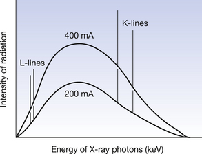
Figure 22.1 The effect of the mA on the X-ray spectrum. Note that the quantity of the radiation changes – as shown by the alteration of the area under each curve – but the quality of the radiation is unaltered – as shown by the maximum photon energy and the peak photon energy being at the same energy for each graph. Thus we can say that the mA selected for an exposure affects the quantity of the X-ray beam but does not affect the quality of the beam – an increase in the mA will produce an increase in the quantity of radiation from the target.
22.4 The effect of kVp on the X-ray beam
The kVp across the X-ray tube influences the force of attraction experienced by an electron released by the filament as it moves towards the anode. Thus, if the kVp is increased, then the kinetic energy of the electron at the point when it starts to interact with the target will be increased. As we saw in Section 21.4.2, the efficiency of X-ray production by Bremsstrahlung is proportional to E2 and so this improved efficiency means that:

As we already discussed in Section 21.4.2, increasing the kVp will also increase the energy of the maximum-energy photons in the beam – if the kVp is 50, then the maximum photon energy is 50 keV and if the kVp is 100, then the maximum photon energy is 100 keV. As the average photon energy is approximately 30–50% of the maximum photon energy, increasing the maximum photon energy will also increase the average photon energy.
As mentioned earlier, increasing the kVp will increase the kinetic energy of the electrons from the filament when they reach the target and so this may mean that characteristic radiation is seen on the higher kVp spectrum but not at the lower value – at 100 kVp the electrons reaching the target have energies up to 100 keV and so can displace K-shell electrons in tungsten, whereas at 50 kVp the 50-keV electrons have insufficient energy to displace a K-shell electron from the tungsten atom.
The spectrum produced at 100 kVp and at 50 kVp is shown in Figure 22.2 where the changes mentioned above can be identified.
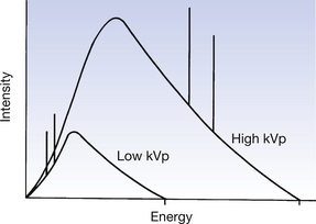
Figure 22.2 The effect of kVp on the X-ray spectrum. Note that the kVp affects both the quantity and the quality of the radiation beam. Also note that the lower of the two curves is unable to produce K-characteristic radiation, although both curves can produce L-lines.
Thus we can say that the kVp selected for an exposure affects both the quantity and the quality of the X-ray beam produced – an increase in kVp will produce an increase in the quantity and the quality of the radiation from the target.
22.5 The effect of the target material on the X-ray beam
As mentioned in Section 21.4.2, the atomic number of the target material has an effect on the X-ray beam from the tube. The higher the atomic number of the target material, the more positive the nucleus of the target atom and so the more it attracts the electrons from the filament which pass close to it. Thus the production of X-rays by the Bremsstrahlung process is more efficient and the intensity of the beam is increased. The maximum and minimum photon energies in the beam are not affected by the target material.
The target material also affects the characteristic radiation produced. The energies of the characteristic radiations from a tungsten and a molybdenum target are shown in Table 22.1. Thus, although the target material does not affect the quality of the Bremsstrahlung radiation, it does affect the energy of the characteristic radiation and this does have some effect on the overall quality of the X-ray beam – this effect may be enhanced by filtering the radiation with the same material as the target (this will be considered further in Ch. 23 when absorption mechanisms are discussed).
Table 22.1 Comparison of the characteristic radiation energies produced from a tungsten target and a molybdenum target
| ENERGY OF Kα CHARACTERISTIC RADIATION (KeV) | ENERGY OF Lα CHARACTERISTIC RADIATION (KeV) | |
|---|---|---|
| Tungsten | 59.32 | 8.39 |
| Molybdenum | 17.48 | 2.22 |
The radiation spectra from a tungsten and a molybdenum target are shown in Figure 22.3.
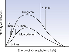
Figure 22.3 The effect of the target material on the X-ray spectrum. Note that the lower atomic number of molybdenum means that there is a reduction in the quantity of the radiation but the quality of the Bremsstrahlung radiation is not affected. The characteristic radiation is at a lower photon energy for the target with the lower atomic number.
22.6 The effect of rectification on the X-ray beam
The type of high-tension rectification of the X-ray generator affects the spectrum of radiation produced at the target of the X-ray tube. This is because of the changing energy of the electron beam striking the target each half-cycle. If we consider full-wave rectification with no capacitor smoothing, as produced by the two-pulse generator in many dental X-ray units, then the potential across the tube varies from zero to the kVp each half-cycle. Thus the energy of the electrons striking the target varies from zero to a keV with a numerical value equal to that of the kVp. The maximum photon energy at differing stages in the half-cycle will vary across the same range as the electron energies.
We can see how this affects the spectrum produced if we consider Figure 22.4A (see page 162) and consider the spectra produced at point B in the voltage waveform and at point C. The time-averaged spectrum for full-wave rectification is shown in Figure 22.4B. Now consider a constant potential, similar to the voltage waveform from the medium-frequency generator, being applied to the X-ray tube during the entire exposure. This would be the same as if voltage B is applied for the whole exposure, and so the spectrum for voltage B is labelled in Figure 22.4B as the spectrum from a constant-potential unit. Note that for constant potential, the area under the graph is increased and the value of the photon energy of the average photon is also increased.
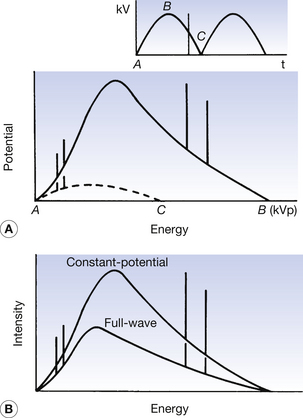
Figure 22.4 (A) The spectrum of radiation produced with a single-phase two-pulse rectified waveform as kVp values B and C are applied across the X-ray tube. (B) The effect of rectification on the X-ray spectrum where the constant potential generator produces radiation of higher quantity and higher quality.
Thus we can say that the rectification – or the type of X-ray generator – affects both the quantity and the quality of the X-ray beam produced – the nearer the voltage across the tube is to a constant potential, the higher the quantity and the quality of the radiation produced at the target.
22.7 The effect of filtration on the X-ray beam
In all the discussion so far we have considered the beam of X radiation produced at the target of the X-ray tube. Before this radiation can be utilized in radiography or radiotherapy, it must first leave the tube. In leaving the tube, the radiation beam must first pass through the glass of the tube insert, the oil in the housing and finally the window of the housing – plus any additional filtration – and it is filtered at each stage of this process. Thus, to consider the beam which will interact with the patient, we need to consider the effect of filtration on the spectrum of radiation produced at the tube target.
Spectrum A in Figure 22.5 shows the distribution of energies emitted from the tube target. This would be the spectrum of the radiation before it leaves the glass envelope of the tube. As we will discuss in Chapter 23, when a beam of radiation passes through any medium, the beam is attenuated by the processes of absorption and scattering. We will see that the lower the photon energy, the higher the chance of it being absorbed or scattered. So the passage of the X-ray beam through the glass envelope, the oil and the exit window of the shield results in selective attenuation of the lower-energy photons. Since this filtration is inherent to the tube construction, it is known as the inherent filtration of the X-ray tube. The spectrum emitted after the inherent filtration is shown as spectrum B in Figure 22.5.
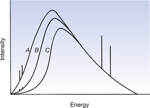
Figure 22.5 The effect of filtration on the X-ray spectrum. Line A represents the spectrum produced at the target of the X-ray tube; line B shows how this spectrum is modified because of the inherent filtration of the tube; and line C shows how the spectrum is modified because of the total filtration. Note that filtration causes a reduction in the quantity of the radiation but an increase in the quality.
Aluminium has a low atomic number (13) and so is able to absorb many of the low-energy X-ray photons in a beam of diagnostic energy, by photoelectric absorption (see Ch. 23). High-energy X-ray photons have a low probability of being absorbed or scattered by the aluminium and so are relatively unaffected by the filtration. This makes aluminium the ideal material for filtration of diagnostic energy X-ray beams.
The radiation beam from the therapy X-ray tube has a much higher average energy than the diagnostic beam. This beam is often hardened – low-energy radiations are removed – by the use of a composite filter. An example of this is when the beam is first filtered through copper (Z=29) and then is further filtered through an aluminium filter. The copper is responsible for the initial filtration but transmits some low-energy radiation due to the photoelectric absorption edge of copper. The copper also produces low-energy characteristic radiation as a result of photoelectric absorption in the copper atoms. The function of the aluminium filter is to absorb the radiation transmitted through the copper because of the K-shell photoelectric absorption edge of copper and also to remove the characteristic radiation produced by the photoelectric absorption in the copper. These concepts will be further discussed in the next chapter.
This beam still contains a significant number of low-energy photons. If these were allowed to interact with the tissues of a patient, they would be absorbed by superficial tissues and so would contribute to the patient dose but would make no contribution to the radiograph – or to the tumour treatment in the case of radiotherapy. The amount of low-energy photons in the spectrum can be significantly reduced by incorporating additional filtration into the beam, near the exit port of the tube, before it interacts with the patient’s tissues. Such a spectrum is shown as line C in Figure 22.5. The inherent filtration of diagnostic X-ray tubes is usually expressed in millimetres of aluminium equivalent, i.e. the inherent filtration is equivalent to the filtration of the beam achieved by the stated number of millimetres of aluminium. The inherent filtration of most diagnostic X-ray tubes is between 0.5 and 1.0 mm of aluminium equivalent. The total filtration in the beam is the sum of the inherent filtration and the additional filtration. This total filtration is between 1.5 and 2.5 mm depending upon the maximum kVp at which the tube is designed to operate.
As can be seen from Figure 22.5, filtration affects both the quantity and the quality of the X-ray beam – the greater the thickness of the filtration in the X-ray beam, the less the quantity but the greater the quality of the X-ray beam emerging from the X-ray tube.
22.8 Summary of the factors affecting the quantity, quality and intensity of the X-ray beam
The factors which affect the quantity and/or quality of the beam of X-radiation emerging from an X-ray tube are summarized in Table 22.2.
Table 22.2 Factors affecting the quantity and quality of the X-ray beam
| FACTOR | EFFECT ON THE QUANTITY OF THE X-RAY BEAM | EFFECT ON THE QUALITY OF THE X-RAY BEAM |
|---|---|---|
| Increase in the X-ray tube current (mA) | Produces an increase in the quantity of radiation directly proportional to the increase in mA | The quality of the radiation from the tube is unaffected by an increase in the tube current |
| Increase in the potential difference (kVp) across the X-ray tube | An increase in the kVp produces an increase in the quantity of radiation produced at the target proportional to the kVp2 | An increase in the kVp produces an increase in the average energy of the photons in the X-ray beam and so produces an increase in the quality of the beam |
| Target material | An increase in the atomic number of the target material will produce an increase in the quantity of radiation produced which is proportional to the increase in the atomic number | A change in the atomic number of the target material will produce no change in the quality of the beam produced by the Bremsstrahlung process. A higher atomic number will produce characteristic radiation of higher energy and, if this is in significant amounts, it will increase the quality of the overall spectrum |
| Rectification | The closer the voltage across the X-ray tube is to a constant potential, the greater the quantity of radiation produced | The closer the voltage across the X-ray tube is to a constant potential, the greater the quality of radiation produced |
| Filtration | Filtration reduces the quantity of radiation emerging from the X-ray tube. The reduction is related to the thickness and the atomic number of the filter | Filtration improves the quality of the radiation from an X-ray tube by selective removal of the low-energy photons |
In this chapter, you should have learnt the following:
• The meaning of the terms quantity and quality as applied to the X-ray beam (see Sect. 22.2).
• The effect of the current through the X-ray tube (mA) on the quantity of the X-ray beam produced (see Sect. 22.3).
• The effect of the potential difference across the X-ray tube (kVp) on the quantity and the quality of the X-ray beam produced (see Sect. 22.4).
• The effect of the atomic number of the target material on the quantity of the radiation produced and on the characteristic radiation from the target (see Sect. 22.5).
• The effect of the voltage waveform applied to the X-ray tube (rectification) on the quantity and the quality of the X-ray beam produced (see Sect. 22.6).
• The effect of filtration on the quantity and the quality of the radiation beam emerging from the X-ray tube (see Sect. 22.7).
Further reading
Ball J.L., Moore A.D., Turner S. Ball and Moore’s Essential Physics for Radiographers, fourth ed. London: Blackwell Scientific, 2008. (Chapter 16)
Carter P.R., Hyatt A.P., Pirrie J.R., Milne A. Chesneys’ Equipment for Student Radiographers. London: Blackwell Scientific, 1994. (Chapters 2 and 3)
Webb S., editor. The Physics of Medical Imaging. Bristol: Institute of Physics, 2000. (Chapter 2)