33 The Vertebral Column, Back, and Thorax of the Pig
THE VERTEBRAL COLUMN AND BACK
The vertebral formula is usually given as C7, T14–15, L6–7, S4, Cd20–23, but variation outside this range is common and almost always affects the thoracolumbar region where the total number of vertebrae varies between 19 and 23. An increase in number is more common, possibly the result of selective breeding for this character: the loins are the most valuable part of the carcass, apart from the hams (Figure 33–1).
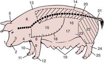
Figure 33–1 Parts of the pig. The position of the vertebral column is indicated. The hatched areas show ham and loin of the meat trade. 1, Snout; 2, mouth; 3, cheek; 4, jowls; 5, poll; 6, neck; 7, shoulder joint; 8, elbow joint; 9, carpus; 10, fetlock joint; 11, hoof; 12, accessory digit; 13, withers; 14, loin (lumbar area); 15, thorax; 16, flank; 17, abdomen; 18, ventral extent of bony thorax; 19, mammary glands; 20, position of coxal tuber; 21, tailhead; 22, thigh; 23, stifle joint; 24, hock joint; 25, metatarsus.
Among other features, the vertebrae of the cervical region are distinguished by a high spine on C2 and a very high one on C7. Since the neck is almost as deep as the cranial part of the thorax, the body of the first thoracic vertebrae is located near the middle of the trunk at this level. The vertebrae behind the first rise gradually until those of the caudal thoracic and lumbar regions run close to, and almost parallel with, the dorsal contour of the back. The four units of the sacrum lack spinous processes; thus, there is an abrupt drop in the height of the vertebral column at the lumbosacral junction. The iliac crest, which flanks the spinous process of the last lumbar vertebra, is the highest skeletal feature in this area (see Figure 32–1/32).
The lumbosacral space is available but rarely used for the epidural administration of anesthetic (Figure 8–56, C). It measures about 2 cm craniocaudally and 3 cm transversely and is situated between 2 and 5 cm caudal to the line connecting the coxal tubers, which are palpable in less fat animals. If this guide cannot be used, an indication of the location of the lumbosacral space is provided by the transverse plane of the flank fold. The space is 5 cm or more below the skin, and the arrival of the needle point at the interarcuate ligament is made known by the greater resistance encountered there. In young hogs, the spinal cord extends into the sacrum and is at risk in this procedure; in older animals the ascent of the cord carries it to safety within the lumbar part of the canal.
The most caudal vertebrae are incorporated in the curly tail, which carries the median caudal vessels near its ventral surface. Blood may be collected most easily at the tail head (Cd 4 or 5), but because the artery and accompanying veins run together, it cannot be predicted whether this blood will be of arterial, venous, or mixed origin. The tail is often removed when a piglet is a few days old to prevent the common vice of tail-biting, which sometimes results in ascending infection. Trichinosis (occurring in some countries) may also be transmitted in this way.
The contour of the back depends on breed and condition. In fat, old animals it may be flat, but in most modern hogs it is uniformly arched and, in those of top quality, also broad. A broad back and wide stance promise good muscling of the trunk and thick hams. The muscles of the back conform to the common pattern, and the longissimus (“loin eye”) and, most especially, psoas muscles (filet mignon) constitute particularly valuable parts of the carcass. Since subcutaneous fat has limited value, too thick a layer is undesirable; this indication of carcass quality may be measured by ultrasound. That deposited over the loins is especially well-formed and thick, and because it has to be trimmed, it represents a substantial loss to the producer. Some of it is rendered into lard, and some is cured to become the “pork” in the popular canned food “pork and beans.” Selective breeding has markedly reduced to 3 cm or less the thickness of back fat; consequently, caution is needed when intramuscular injections are performed.
THE THORAX
The body of a pig does not widen appreciably where the neck joins the trunk: the subcutaneous layer of fat allows the forelimb to blend in unobtrusively, and only a slight depression between the flabby jowls and the shoulder joint marks the junction. There is a similar depression between the elbow joint and the thoracic wall. The “points” of both joints are palpable. The olecranon of the elbow projects onto the ventral end of the fifth rib (Figure 33–2). The manubrium of the sternum is also easily found.
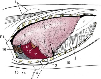
Figure 33–2 The thoracic viscera in situ, semischematic. 1, Scapula; 2, caudal border of triceps; 3, olecranon; 4, radius and ulna; 5, 6, cranial and caudal parts of cranial lobe of lung; 7, caudal lobe of lung; 8, basal border of lung; 9, 9′, muscular and tendinous parts of diaphragm; 10, line of pleural reflection; 11, heart; 12, 13, left and right auricles; 14, cranial mediastinum; 15, sternal lymph node; 16, thymus.
Most pigs have 14 or 15 pairs of ribs; asymmetry of number is common (see Figure 32–1). The first seven pairs are sternal. The rib cage is smaller than the external dimensions suggest; it is especially narrow and shallow between the forelimbs but deepens caudally with the upward sweep of the thoracic vertebrae. It is relatively long, depending to some extent on the number of vertebrae. The line of pleural reflection follows the dorsal half of the last rib before descending in a gentle curve to cross the seventh costochondral joint (Figure 33–2). The cranial mediastinum, like that of ruminants, attaches to the ventral parts of the left first and second ribs, but more dorsally it is separated from the thoracic wall by the cranial lobe of the left lung.
The left lung possesses a cranial lobe, divided by a cardiac notch, and a caudal lobe (Figure 33–2/5,6,7 and see Figure 4–23). The right lung possesses cranial, middle, caudal, and accessory lobes, separating the cardiac notch separates the first two (see Figure 4–23, A). The cranial lobe of this lung is ventilated by a separate tracheal bronchus (Figure 33–5/8 and see Figure 4–24). The lobulation of the lungs is relatively distinct.
The projection of the lungs onto the thoracic wall is small. The basal border of the left lung extends from the sixth costochondral junction to the upper end of the third last rib. This border of the right lung is less steep and reaches the penultimate rib. Auscultation and percussion of the lungs are usually reserved for young pigs of cooperative disposition.
The heart is small, providing as little as 0.3% of body weight (compared with 1.5% or more in athletic species such as the horse and dog), and this has been cited as a predisposing factor in “sudden death syndrome” commonly occurring in pigs. Heart size has not kept pace with the much-accelerated growth of modern, improved pigs, which reach a weight of 115 kg at 5 or 6 months; in striking contrast, 2 or 3 years was required to reach the much more modest weight of 40 kg in 1800. The heart occupies the ventral half of the thoracic cavity, extending between the second and fifth ribs (Figure 33–3/6 and Figure 33–4/1). It is thus covered by the forelimb in the standing animal but can be made accessible by drawing the limb forward. It exhibits no structural distinctions of note (see Figure 7–7).
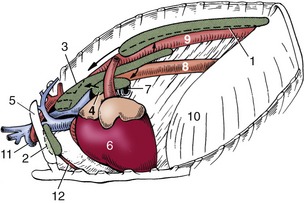
Figure 33–3 The lymph centers of the thorax, left lateral view. 1, Dorsal thoracic lymph center; 2, ventral thoracic lymph center; 3, mediastinal lymph center; 4, tracheobronchial lymph center; 5, first rib; 6, heart; 7, left bronchus; 8, esophagus; 9, aorta; 10, diaphragm; 11, axillary vein and artery; 12, internal thoracic artery.
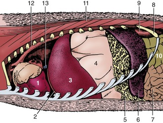
Figure 33–4 The heart in situ. 1, Heart; 2, diaphragm; 3, left lobe of liver; 4, stomach, greatly dilated; 5, greater omentum, gastrosplenic ligament; 6, spleen; 7, jejunum; 8, last rib; 9, left kidney; 10, ascending colon; 11, back muscles; 12, aorta; 13, caudal vena cava.
Paracentesis is best performed through the fifth left or the fourth right intercostal space; the needle is inserted about 5 cm dorsal to the olecranon (Figure 33–2).
THE LYMPHATIC STRUCTURES OF THE THORAX
The thoracic lymph nodes, arranged in four centers (Figure 33–3/6 and Figure 33–4/1), collect lymph from the thoracic walls and contents and from adjacent structures and channel it to the thoracic duct or, where some more cranial nodes are concerned, directly into veins at the thoracic inlet.
The dorsal thoracic center comprises a variable number of small aortic nodes that receive lymph from the dorsal part of the thoracic wall, the mediastinum, and mediastinal nodes. The ventral center consists of fewer but larger sternal nodes concerned with the ventral part of the thoracic walls and the first two or three pairs of mammary glands.
Inconstant numbers of cranial and caudal mediastinal nodes form a chain above the base of the heart. The cranial nodes drain structures of the neck in addition to mediastinal contents, including the tracheobronchial nodes. Their efferents are divided into some that open into veins directly and others that lead to the thoracic duct. The caudal nodes are not always to be found. When present, they drain neighboring structures and send their efferents to the tracheobronchial and aortic nodes.
The bronchial center (Figure 33–3/4) consists of a dozen or so tracheobronchial nodes arranged about the origin of the bronchi (Figure 33–5). They drain the lungs, heart, and pericardium and in turn drain to the cranial mediastinal nodes or directly into the thoracic duct.
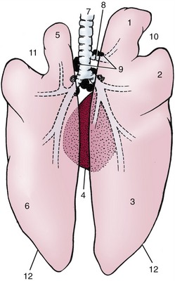
Figure 33–5 The lungs, dorsal view (see also Figure 4–23). 1, Right cranial lobe; 2, right middle lobe; 3, right caudal lobe; 4, accessory lobe of right lung; 5, divided left cranial lobe; 6, left caudal lobe; 7, trachea; 8, tracheal bronchus; 9, tracheobronchial lymph nodes; 10, right cardiac notch; 11, left cardiac notch; 12, basal border.
The thoracic duct runs from caudal to cranial between the aorta and esophagus, passing the trachea at its left side before joining the bloodstream.