6 The Permanent Maxillary Incisors
The maxillary incisors are four in number. The maxillary central incisors are centered in the maxilla, one on either side of the median line, with the mesial surface of each in contact with the mesial surface of the other. The maxillary and mandibular central incisors are the only neighboring teeth in the dental arches with mesial surfaces in contact. The right and left maxillary lateral or second incisors are distal to the central incisors.
The maxillary central incisor is larger than the lateral incisor. These teeth supplement each other in function, and they are similar anatomically. The incisors are shearing or cutting teeth. Their major function is to punch and cut food material during the process of mastication. These teeth have incisal ridges or edges rather than cusps such as are found on the canines and posterior teeth.
It might be good at this point to differentiate between the two terms incisal ridge and incisal edge. The incisal ridge is that portion of the crown which makes up the complete incisal portion. When an incisor is newly erupted, the incisal portion is rounded and merges with the mesioincisal and distoincisal angles and the labial and lingual surfaces. This ridge portion of the crown is called the incisal ridge. The term edge implies an angle formed by the merging of two flat surfaces. Therefore, an incisal edge does not exist on an incisor until occlusal wear has created a flattened surface linguoincisally, which surface forms an angle with the labial surface. The incisal edge is formed by the junction of the linguoincisal surface, sometimes called the incisal surface, and the labial surface (Figure 6-1).
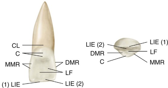
Figure 6-1 Maxillary right central incisor, lingual and incisal aspects. The labioincisal edge [LIE (1)] and linguoincisal edge [LIE (2)] border the incisal ridge. CL, Cervical line; C, cingulum; MMR, mesial marginal ridge; LF, lingual fossa; DMR, distal marginal ridge.
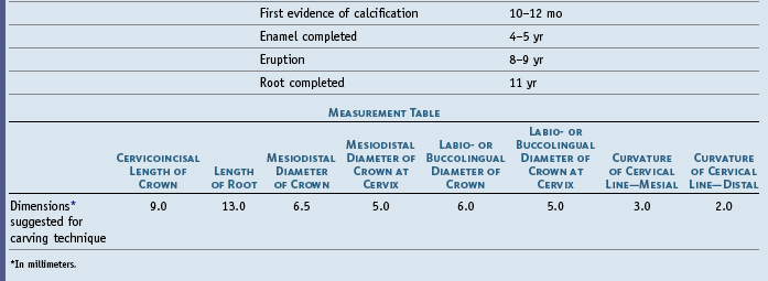
Preceding the description of each tooth in this and subsequent chapters, the chronology of calcification and eruption of each tooth will be given as taken from Table 2-2. Knowing the proportions of the individual tooth helps one learn the proportions of one tooth to another. Outline drawings of the five aspects of the teeth are explained more fully elsewhere.1
Maxillary Central Incisor
Figures 6-1 through 6-12 illustrate the maxillary central incisor in various aspects. The maxillary central incisor is the widest mesiodistally of any of the anterior teeth (Table 6-1). The labial face is less convex than that of the maxillary lateral incisor or canine, which gives the central incisor a squared or rectangular appearance (see Figures 6-7 and 6-8). From this aspect, the crown nearly always looks symmetrical and regularly formed, having a nearly straight incisal edge, a cervical line with even curvature toward the root, a mesial side with straight outline, the distal side being more curved. The mesial incisal angle is relatively sharp, the distal incisal angle rounded (see Figure 6-2).
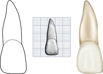
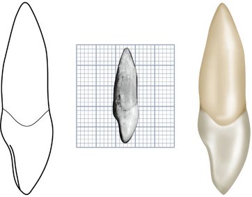
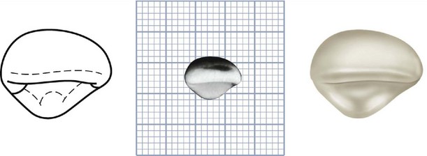
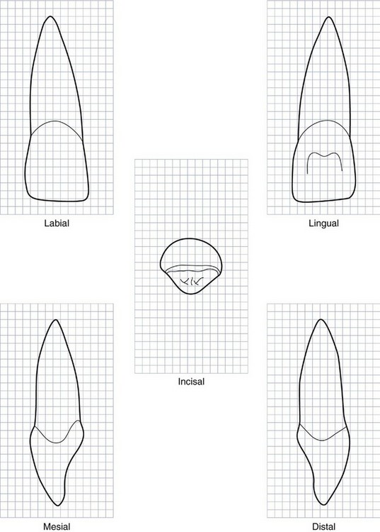
Figure 6-7 Maxillary right central incisor. Squared millimeter graph outlines of five aspects are shown. In the incisal view, the labial aspect is at the top of the drawing. (Grid = 1 sq mm.)
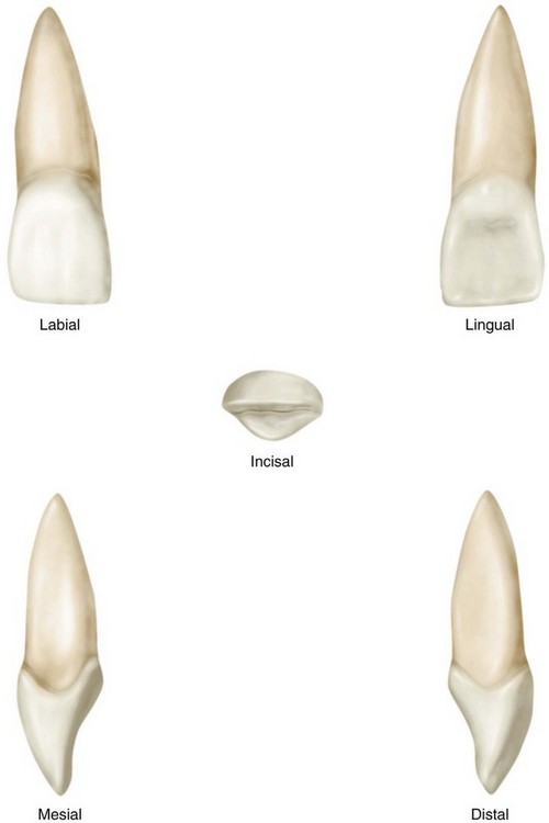
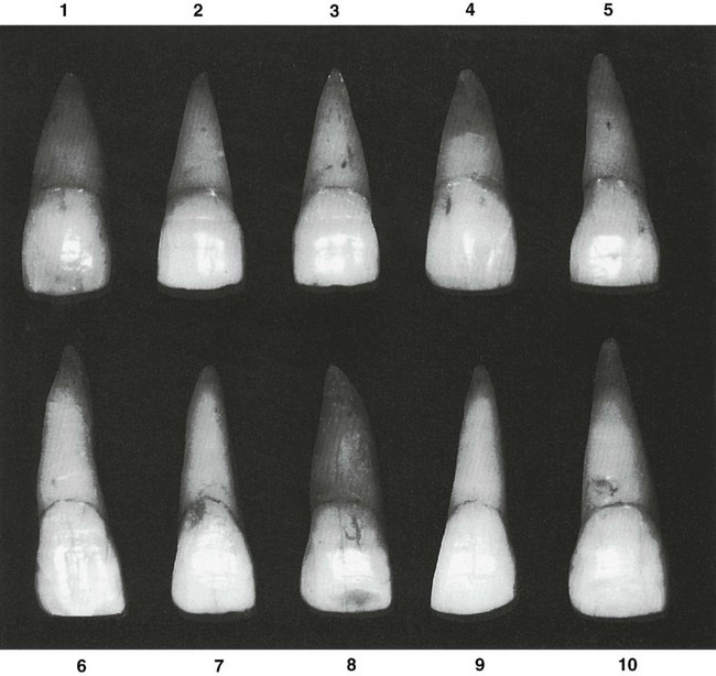
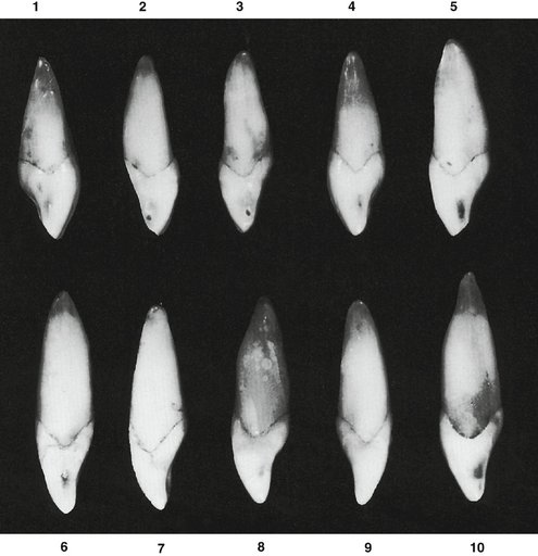
Figure 6-12 Maxillary central incisor. Ten specimens with uncommon variations are shown. 1, Extralingual inclination of incisal portion of crown. Note developmental (palatoradicular) groove traversing root and part of crown. 2, Root extremely long. 3, Specimen small in all dimensions. 4, Crown extremely long, root very short. 5, Specimen malformed; crown unusually long; cervix very wide. 6, Root short and tapering. 7, Same as specimen 6. 8, Crown nearly as wide at the cervix as at contact areas, crown long, root short. 9, Root with unusual curvature. 10, Crown and root narrow labiolingually; root comparable with that of specimen 2.
Although the labial surface of the crown is usually convex, especially toward the cervical third, some central incisors are flat at the middle and incisal portions. The enamel surface is relatively smooth. When the tooth is newly erupted or if little wear is evident, mamelons will be seen on the incisal ridge. The middle one is the smallest. The developmental lines on the labial surface that divide the surface into three parts are most noticeable at the middle portion if they can be distinguished at all (see Figure 2-12).
Lingually, the surface form of the maxillary central incisor is more irregular. The largest part of the middle and incisal portions of the lingual area is concave. Mesial and distal marginal ridges border the concavity, the lingual portion of the incisal ridge, and the convexity apically to the cingulum. The lingual topography gives a scooplike form to the crown (see Figure 6-3). An exaggeration of the marginal ridges, known as a shovel-shaped incisor, is a genetically determined variation seen in Mongoloid races, including North and South American Indians.2-4
The maxillary central incisor usually develops normally. One anomaly that sometimes occurs is a short root. Another variation is an unusually long crown (see Figure 6-12, 4 and 5). The maxillary central incisors are the most prominent teeth in the mouth. There are two basic forms: the first is relatively wide at the cervix, when viewed from the labial aspect, in comparison with the mesiodistal width at the contact areas (see Figure 6-9, 1 and 4); the second form is relatively narrow at the cervix, where the root joins the crown, in comparison with the mesiodistal width at the contact areas (see Figure 6-9, 5, 7, and 9).
In the description of the central incisor, an attempt will be made to strike an average between the extremes of the two forms, keeping in mind that crown sizes are gender dimorphic, with male larger than female. The extent of dimorphism varies among populations.5 However, gender-specific correlations between enamel thickness and crown width of dentin are low.6
DETAILED DESCRIPTION OF THE MAXILLARY CENTRAL INCISOR FROM ALL ASPECTS
Labial Aspect
The crown of the average central incisor will be 10 to 11 mm long from the highest point on the cervical line to the lowest point on the incisal edge (see Figures 6-2 and 6-9). The mesiodistal measurement will be 8 to 9 mm wide at the contact areas. The mesiodistal measurement, where the root joins the crown, will be 1.5 to 2 mm less. The crests of curvature mesially and distally on the crown represent the areas at which the central incisor contacts its neighbors. Any change in the position of this crest of contour affects the level of the contact area (see Figure 5-15, A).
The mesial outline of the crown is only slightly convex, with the crest of curvature (representing the contact area) approaching the mesioincisal angle (see Chapter 5).
The distal outline of the crown is more convex than the mesial outline, with the crest of curvature higher toward the cervical line. The distoincisal angle is not as sharp as the mesioincisal angle, the extent of curvature depending on the typal form of the tooth.
The incisal outline is usually regular and straight in a mesiodistal direction after the tooth has been in function long enough to obliterate the mamelons. The incisal outline tends to curve downward toward the center of the crown outline, so that the crown length is greater at the center than at the two mesial angles.
The cervical outline of the crown follows a semicircular direction with the curvature rootwise, from the point at which the root outline joins the crown mesially to the point at which the root outline joins the crown distally.
The root of the central incisor from the labial aspect is cone-shaped, in most instances with a relatively blunt apex, and the outline mesially and distally is regular. The root is usually 2 or 3 mm longer than the crown, although it varies considerably. (See illustrations of typical central incisors and those of variations from the labial aspects in Figures 6-9 and 6-12.)
A line drawn through the center of the root and crown of the maxillary central incisor tends to parallel the mesial outline of the crown and root.
Lingual Aspect
The lingual outline of the maxillary central incisor is the reverse of that found on the labial aspect (see Figure 6-3). The lingual aspect of the crown is different, however, when we compare the surface of the lingual aspect with that of the labial aspect. From the labial aspect, the surface of the crown is smooth generally. The lingual aspect has convexities and a concavity. The outline of the cervical line is similar, but immediately below the cervical line a smooth convexity is to be found; this is called the cingulum (see Figure 6-1).
Mesially and distally confluent with the cingulum are the marginal ridges. Between the marginal ridges, below the cingulum, a shallow concavity is present called the lingual fossa. Outlining the lingual fossa, the linguoincisal edge is raised somewhat, being on a level with the marginal ridges mesially and distally, completing the lingual portion of the incisal ridge of the central incisor.
From the foregoing description, we note that the lingual fossa is bordered mesially by the mesial marginal ridge, incisally by the lingual portion of the incisal ridge, distally by the distal marginal ridge, and cervically by the cingulum. Usually there are developmental grooves extending from the cingulum into the lingual fossa.
The crown and root taper lingually, so that the crown calibration at the two labial line angles is greater than the calibration at the two lingual line angles, and the lingual portion of the root is narrower than the labial portion. A cross section of the root at the cervix shows the root to be generally triangular with rounded angles. One side of the triangle is labial, with the mesial and distal sides pointing lingually. The mesial side of this triangle is slightly longer than the distal side (see Figure 13-8, C, 3, 4, 5, and 6).
Mesial Aspect
The mesial aspect of this tooth has the fundamental form of an incisor. The crown is wedge-shaped, or triangular, with the base of the triangle at the cervix and the apex at the incisal ridge (see Figure 4-16, A and Figures 6-4 and 6-10).
Usually a line drawn through the crown and the root from the mesial aspect through the center of the tooth will bisect the apex of the root and also the incisal ridge of the crown. The incisal ridge of the crown is therefore on a line with the center of the root. This alignment is characteristic of maxillary central and lateral incisors. A straight line drawn through the center of the crown and root from the mesial or distal aspects will rarely if ever pass lingual to the incisal edge. Maxillary incisors are occasionally seen with the incisal ridges lingual to the bisecting line (see Figure 6-12, 1).
Labially and lingually, immediately coronal to the cervical line are the crests of curvature of these surfaces. These crests of contour give the crown its greatest labiolingual measurement.
Normally, the curvature labially and lingually is approximately 0.5 mm in extent (see Figure 6-4) before continuing the outlines to the incisal ridge.
The labial outline of the crown from the crest of curvature to the incisal ridge is very slightly convex. The lingual outline is convex at the point where it joins the crest of curvature at the cingulum; it then becomes concave at the mesial marginal ridge and slightly convex again at the linguoincisal ridge and the incisal edge.
The cervical line outlining the cementoenamel junction (CEJ) mesially on the maxillary central incisor curves incisally to a noticeable degree. This cervical curvature is greater on the mesial surface of this tooth than on any surface of any other tooth in the mouth. The curvature varies in extent, depending on the length of the crown and the measurement of the crown labiolingually. On an average central incisor of 10.5 to 11 mm in crown length, the curvature is 3 to 4 mm (see Figure 5-26).
The root of this tooth from the mesial aspect is cone-shaped, and the apex of the root is usually bluntly rounded.
Distal Aspect
Little difference is evident between the distal and mesial outlines of this tooth (see Figure 6-5). When looking at the central incisor from the distal aspect, it may be noted that the crown gives the impression of being somewhat thicker toward the incisal third. Because of the slope of the labial surface distolingually, more of that surface is seen from the distal aspect; this creates the illusion of greater thickness. Actually, most teeth are turned a little on their root bases to adapt to the dental arch curvature. The maxillary central incisor is no exception.
The curvature of the cervical line outlining the CEJ is less in extent on the distal than on the mesial surfaces. Most teeth show this characteristic.
Incisal Aspect
The specimen of this tooth is posed in the illustrations so that the incisal edge is centered over the root (see Figures 6-6 and 6-11). A view of the crown from this aspect superimposes it over the root entirely so that the latter is not visible.
From this aspect, the labial face of the crown is relatively broad and flat in comparison with the lingual surface, especially toward the incisal third. Nevertheless, the cervical portion of the crown labially is convex, although the arc described is broad.
The incisal ridge may be seen clearly, and a differentiation between the incisal edge and the remainder of the incisal ridge, with its slope toward the lingual, is easily distinguished.
The outline of the lingual portion tapers lingually toward the cingulum. The cingulum of the crown makes up the cervical portion of the lingual surface.
The mesiolabial and distolabial line angles are prominent from the incisal aspect. The relative positions of these line angles should be compared with the mesiolingual and distolingual line angles, which are represented by the borders of the mesial and distal marginal ridges. The mesiodistal calibration of the crown at the labial line angles is greater than the same calibration at the lingual line angles.
The crown of this tooth shows more bulk from the incisal aspect than one would expect from viewing it from the mesial or distal aspect. Relatively broad surfaces are at the site of contact areas mesially and distally. Comparison should also be made between the dimensions of the crown labiolingually and mesiodistally. The labiolingual calibration of the crown is more than two thirds as great as the mesiodistal calibration. A cursory examination would not reveal this detail.
Bilaterally, the outline of the incisal aspect is rather uniform. The lingual portion shows some variation, however, in that a line drawn from the mesioincisal angle to the center of the cingulum lingually will be longer than one drawn from the same point on the cingulum to the distoincisal angle. The crown conforms to a triangular outline reflected by the outline of the root cross section at the cervix mentioned earlier.
Maxillary Lateral Incisor
Figures 6-13 through 6-21 illustrate the maxillary lateral incisor in various aspects. Because the maxillary lateral incisor supplements the central incisor in function, the crowns bear a close resemblance. The lateral incisor is smaller in all dimensions except root length (Table 6-2). Because it resembles the maxillary central incisor in form, direct comparisons are made with the central incisor in its description.
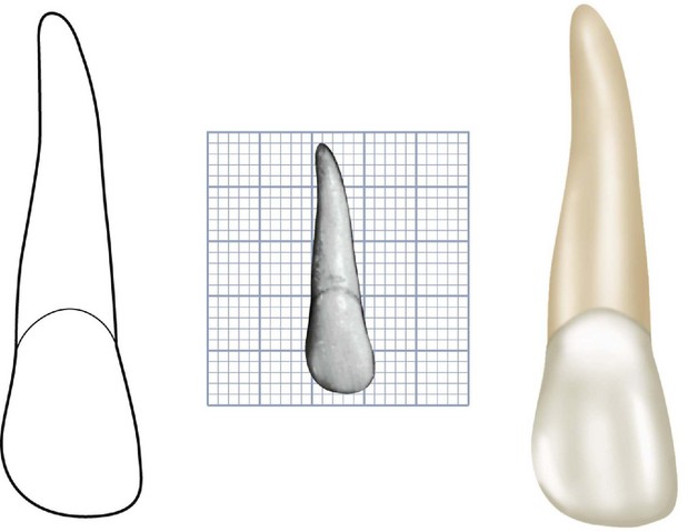
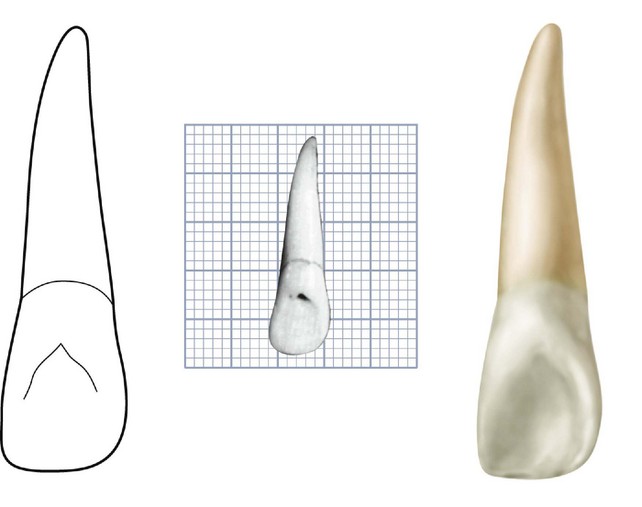
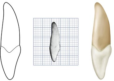
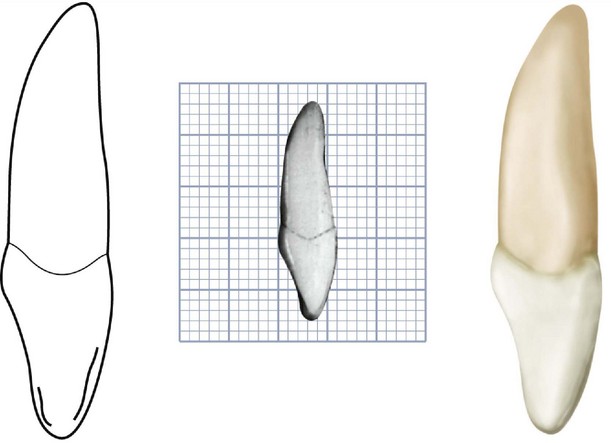
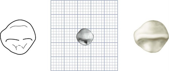
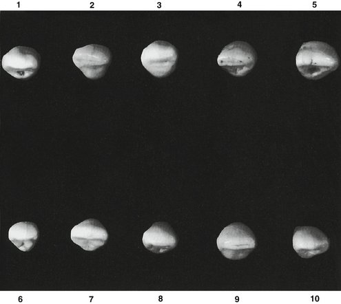
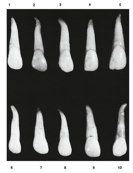
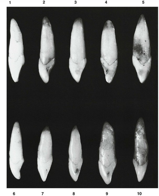
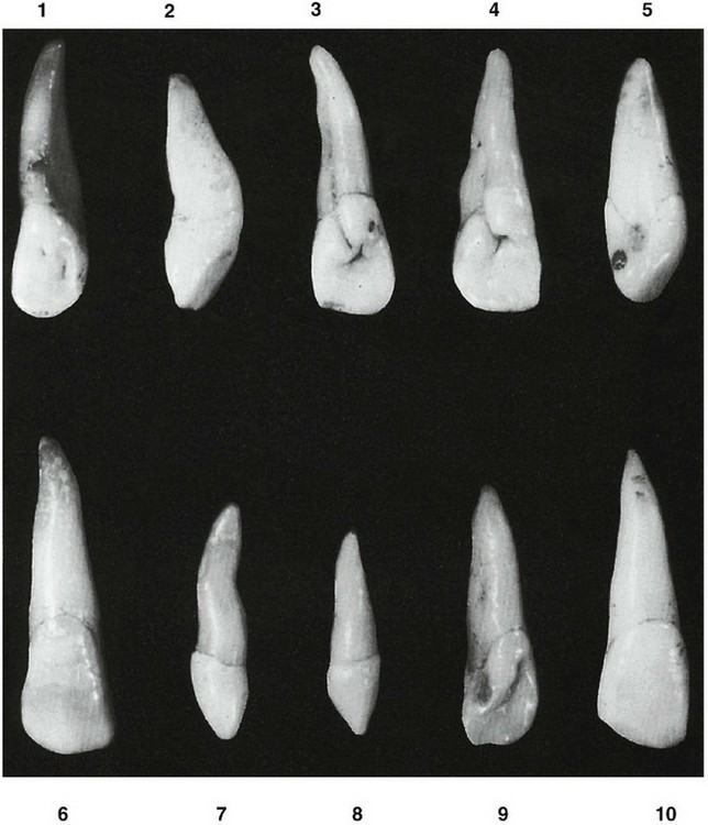
Figure 6-21 Maxillary lateral incisor. Ten specimens with uncommon variations are shown. 1, Odd twist to crown and root. 2, Malformed generally. 3, Deep developmental (palatoradicular) groove distally; note pit in lingual fossa. 4, Same as specimen 3 with pit and groove connected. 5, Deep concavity above contact area of the crown. 6, Abnormally large but well formed. 7, Single-cusp development and malformed root called peg lateral incisor. 8, Same as specimen 7, except root is straight. 9, Same as specimen 5, with deep lingual pit in addition. 10, Resemblance to a small maxillary central incisor more marked than the average.
This tooth differs from the central incisor in its development, which may vary considerably. Maxillary lateral incisors vary in form more than any other tooth in the mouth except the third molar. If the variation is too great, it is considered a developmental anomaly. A common situation is to find maxillary lateral incisors with a nondescript, pointed form; such teeth are called peg-shaped laterals (see Figure 6-21, 7 and 8). In some individuals, the lateral incisors are missing entirely7; in these cases, the maxillary central incisor may be in contact distally with the canine. The presence of a palatogingival groove in maxillary incisors may be a predisposing factor in localized periodontal disease8 (see Figure 6-21, 3). This groove is also referred to as the palatoradicular groove.9
One type of malformed maxillary lateral incisor has a large, pointed tubercle as part of the cingulum; some have deep developmental grooves that extend down on the root lingually with a deep fold in the cingulum; and some show twisted roots, distorted crowns, and so on (see Figure 6-21).
DETAILED DESCRIPTION OF THE MAXILLARY LATERAL INCISOR FROM ALL ASPECTS
Labial Aspect
Although the labial aspect of the maxillary lateral incisor may appear to favor that of the central incisor, usually it has more curvature, with a rounded incisal ridge and rounded incisal angles mesially and distally (see Figures 6-13 and 6-19). Although the crown is smaller in all dimensions, its proportions usually correspond to those of the central incisor.
The mesial outline of the crown from the labial aspect resembles that of the central incisor, with a more rounded mesioincisal angle. The crest of contour mesially is usually at the point of junction of the middle and incisal thirds; occasionally, in the so-called square forms, the mesioincisal angle is almost as sharp as that found on most maxillary central incisors (see Figure 6-19, 4 and 5). However, a more rounded mesioincisal angle is seen more often. The distal outline of the crown from the labial aspect differs somewhat from that of the central incisor.
The distal outline is always more rounded, and the crest of contour is more cervical, usually in the center of the middle third. Some forms describe a semicircular outline distally from the cervix to the center of the incisal ridge (see Figure 6-19, 3 and 7).
The labial surface of the crown is more convex than that of the central incisor except in some square and flat-faced forms.
This tooth is relatively narrow mesiodistally, usually about 2 mm narrower than the central incisor. The crown on the average measures from 2 to 3 mm shorter cervicoincisally than that of the central incisor, although the root is usually as long, if not somewhat longer, than that of the central incisor.
In general, its root length is greater in proportion to its crown length than that of the central incisor. The root is often about 1.5 times the length of the crown.
The root tapers evenly from the cervical line to a point approximately two thirds of its length apically. In most cases, it curves sharply from this location in a distal direction and ends in a pointed apex. Although the curvature distally is typical, some roots are straight (see Figure 6-19, 4, 7, and 9), and some may be found curving mesially. As mentioned previously, this tooth may show considerable variance in its crown form; the root form may be more characteristic.
Lingual Aspect
From the lingual aspect, mesial and distal marginal ridges are marked, and the cingulum is usually prominent, with a tendency toward deep developmental grooves within the lingual fossa, where it joins the cingulum (see Figure 6-14). The linguoincisal ridge is well developed, and the lingual fossa is more concave and circumscribed than that found on the central incisor. The tooth tapers toward the lingual, resembling a central incisor in this respect. It is not uncommon to find a deep developmental groove at the side of the cingulum, usually on the distal side, which may extend up on the root for part or all of its length. Faults in the enamel of the crown are often found in the deep portions of these developmental grooves (see Figure 6-21, 3 and 4).
Mesial Aspect
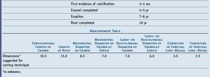
The mesial aspect of the maxillary lateral incisor is similar to that of a small central incisor except that the root appears longer (see Figures 6-15 and 6-20). The crown is shorter, the root is relatively longer, and the labiolingual measurement of the crown and root is a millimeter or so less than that of the maxillary central incisor of the same mouth.
The curvature of the cervical line is marked in the direction of the incisal ridge, although because of the small size of the crown the actual extent of curvature is less than that found on the central incisor. The heavy development of the incisal ridge accordingly makes the incisal portion appear somewhat thicker than that of the central incisor.
The root appears as a tapered cone from this aspect, with a bluntly rounded apical end. This varies in individuals, with the apical end sometimes being quite blunt, while at other times, it is pointed. In a good many cases, the labial outline of the root from this aspect is straight. As in the central incisor, a line drawn through the center of the root tends to bisect the incisal ridge of the crown.
Distal Aspect
Because of the placement of the crown on the root, the width of the crown distally appears thicker than it does on the mesial aspect from marginal ridge to labial face (see Figure 6-16). The curvature of the cervical line is usually a millimeter or so less in depth than on the mesial side. It is not uncommon to find a developmental groove distally on this crown extending on the root for part or all of its length.
Incisal Aspect
The incisal aspect of this tooth sometimes resembles that of the central incisor, or it may resemble that of a small canine (see Figures 6-17 and 6-18). If the tooth conforms in development to its central incisor neighbor in other respects, it will, from the incisal aspect, resemble a central incisor except in size (see Figure 6-18, 5 and 9). However, the cingulum may be large, as is the incisal ridge. In addition, the labiolingual dimension may be greater than usual in comparison with the mesiodistal dimension. If these variations are present, the tooth has a marked resemblance to a small canine (see Figure 6-18, 3 and 10).
All maxillary lateral incisors exhibit more convexity labially and lingually from the incisal aspect than do the maxillary central incisors.
1. Ash MM. Wheeler’s atlas of tooth form. Philadelphia: Saunders; 1984.
2. Carbonell VM. Variations in frequency of shovel-shaped incisors in different populations. In: Brothwell DR, editor. Dental anthropology. London: Pergamon Press, 1963.
3. Dahlberg AA, Mikkelson O. The shovel-shaped character in the teeth of the Pima Indians. Am J Phys Anthropol. 1947;5:234.
4. Hrdlicka A. Shovel-shaped teeth. Am J Phys Anthropol. 1920;3:429.
5. Garn SM, et al. Sexual dimorphism in the buccolingual tooth diameter. J Dent Res. 1966;45:1819.
6. Harris EF, Hicks JD. A radiographic assessment of enamel thickness in human maxillary lateral incisors. Arch Oral Biol. 1998;43:825.
7. Meskin LH, Gorlin RJ. Agenesis and peg-shaped permanent maxillary lateral incisors. J Dent Res. 1963;42:1576.
8. Lee KW, et al. Plato-gingival grooves in maxillary incisors. A possible predisposing factor to localized periodontal disease. Br Dent J. 1968;124:14.
9. Kogon SL. The prevalence, location, and conformation of palato-radicular grooves in maxillary incisors. J Periodontol. 1986;57:231.
 site for additional study resources.