Hypovolemic, hemorrhagic, maldistributive and obstructive shock

Synopsis

Etiology Shock due to a reduction in venous return (circuit failure) secondary to hypovolemia, hemorrhage, mal-distribution of blood or obstruction to venous return

Clinical findings Depression and weakness, subnormal temperature, elevated heart rate with weak thready pulse, cold skin and extremities, prolonged capillary refill time. Progressive development without aggressive fluid therapy and collapse and death from irreversible shock

Clinical pathology Increased blood or plasma l-lactate concentration, decreased venous oxygen tension, evidence of multiple organ dysfunction. Decreased central venous pressure, low mean arterial blood pressure terminally. Changes in heart rate, activity level and blood or plasma l-lactate concentration indicate the efficacy of treatment

Necropsy findings None specific for hypovolemic or maldistributive shock; the source of hemorrhage may be apparent in hemorrhagic shock

Diagnostic confirmation Clinical signs, blood or plasma l-lactate concentrations, venous oxygen tension

Treatment Aggressive fluid therapy based on intravenous isotonic crystalloid solutions and possibly colloid solutions. Blood transfusion or stroma-free hemoglobin administration for hemorrhagic shock. Initial treatment by rapid infusion with small-volume hypertonic saline solutions gives rapid but transient resuscitative affect. Antimicrobial agents and non steroidal anti-inflammatory drugs in maldistributive shock due to endotoxemia

ETIOLOGY

The circulatory system consists of a pump (the heart) and a circuit (the vasculature). Circulatory shock can result from abnormal functioning of the pump or circuit, or both. It is clinically very important to differentiate pump failure (cardiogenic shock due to acute or chronic heart failure) from circuit failure, because the diagnosis and treatment of cardiogenic shock is vastly different to that of circuit shock. Cardiogenic shock is covered in detail in Chapter 8, whereas circuit failure is addressed in the following section.

Circuit failure occurs whenever the cardiac output is reduced below a critical point because of inadequate venous return to the heart. There are four main ways that circuit failure occurs:

Hypovolemic shock occurs when there is a reduction in circulating blood volume due to plasma or free water loss

Hemorrhagic shock occurs when there is a reduction in circulating blood volume due to rapid blood loss

Maldistributive shock occurs when there is a reduction in circulating blood volume due to increased capillary permeability, pooling of blood in capacitance vessels (such as the veins in the splanchnic circulation), or pooling of plasma is a large third space such as the thoracic or abdominal cavities

Obstructive shock occurs when there is an acute reduction in venous return due to a mechanical obstruction, such as pericardial tamponade or pulmonary artery thrombosis. Obstructive shock is extremely rare in large animals.

Regardless of the initiating cause for circuit failure and inadequate venous return, tissue hypoperfusion results, leading to impaired oxygen uptake and anaerobic metabolism. The end result of inadequate tissue perfusion is the development of multiple organ failure, l-lactate acidemia, and strong ion (metabolic) acidosis, manifest as the hypodynamic stage of shock. Hypovolemia and poor tissue perfusion results in cold extremities, elevated heart rate, a weak thready pulse, decreased capillary refill times and altered mental status. Cardiac arrhythmias may occur because of myocardial ischemia and electrolyte and acid–base disturbance. There is anorexia and gastrointestinal stasis. Signs of renal failure include anuria or oliguria and azotemia.

Common causes of circuit failure in large animals are as follows.

Hypovolemic shock

Fluid loss and dehydration, such as in neonatal calf diarrhea and burn injury, especially when fluid loss is severe and rapid

Fluid loss into the gastrointestinal tract due to acute intestinal obstruction.

Hemorrhagic shock

Acute hemorrhage with loss of 35% or more of total blood volume, equivalent to an acute blood loss of 2.8% of body weight (assuming blood volume is 8% of body weight) will lead to clinical signs of severe hemorrhagic shock. In contrast, acute hemorrhage with loss of less than 10% of total blood volume (equivalent to an acute blood loss of less than 0.8% of body weight) produces minimal detectable clinical changes.

Traumatic injury or spontaneous rupture of large blood vessels are the common reasons for acute hemorrhage. Any sort of minor surgical wound, e.g. castration, dehorning, may lead to excess hemorrhage where there is a hemorrhagic tendency due to defects of clotting. Some of the more common causes of hemorrhagic shock areas follow.

Cattle and sheep

Spontaneous pulmonary hemorrhage associated with caudal vena caval syndrome

Abomasal ulcer, sometimes originating from a bovine viral leukosis lesion (cattle)

Enzootic hematuria with bleeding from a bladder lesion (cattle)

Pyelonephritis with bleeding from a renal lesion (cattle)

Intra-abdominal hemorrhage as a result of arterial aneurysm, possibly associated with copper deficiency (cattle)

Laceration of arteries in the wall of the vagina as a result of dystocia

Ruptured middle uterine artery during prolapse or torsion of uterus

Cardiac tamponade due to rupture of coronary artery or ventricular chamber, rupture of aorta (see Chapter 1)

Rupture of liver associated with dystocia in lambs, and in older lambs possibly associated with vitamin E deficiency.

Horses

Ethmoidal hematoma1

Exercise-induced pulmonary hemorrhage2

Rupture of the middle uterine, utero-ovarian (especially right side) or iliac artery associated with parturition, more commonly in aged mares3

Nasal bleeding from hemorrhage into the guttural pouch, from carotid or maxillary arteries with guttural pouch mycosis or associated with rupture of the longus capitis muscle following trauma4

Rupture of mesenteric arteries secondary to strongyle larval migration

Splenic hematoma5 or rupture following blunt trauma

Rupture of liver with hyperlipemia

Hemangioma, hemangiosarcoma and other neoplasia

Persistent bleeding from the vulva in association with ulcerated varicose veins on the dorsal wall of the vagina6

Congenital venous aneurysm (rare).7

Pigs

Esophagogastric ulceration

Proliferative hemorrhagic enteropathy

Rupture of liver in hepatosis dietetica

Congenital neonatal bleeding, e.g. umbilical hemorrhage.

Maldistributive shock

Endotoxemia in neonatal septicemia, salmonellosis, coliform mastitis in lactating dairy cattle, toxic metritis in cattle

Septic shock due to Gram-positive bacterial septicemia8-10

Too sudden reduction of pressure in a body cavity, e.g. by rapid withdrawal of ascitic fluid.

Obstructive shock

Pericardial tamponade.

PATHOGENESIS

Hypovolemic shock

When cardiac output falls as a result of decreased venous return, the carotid and aortic baroreceptors stimulate the sympathetic nerves and adrenal medulla to release catecholamines resulting in vasoconstriction in vessels with alpha-adrenergic receptors.8,11 Vasoconstriction leads to decreased renal perfusion, which activates the renin–angiotensin– aldosterone system, thereby inducing sodium and water retention. The decrease in renal perfusion can result in renal ischemia and nephrosis if the ischemia is sufficiently severe and prolonged (see Chapter 11). Hypovolemia also stimulates the release of antidiuretic hormone (vasopressin). There is contraction of the spleen and venous capacitance vessels, an increased peripheral vascular resistance and an increase in heart rate in an attempt to maintain cardiac output and blood perfusion through the coronary and cerebral blood vessels.

Water shifts from the interstitial space to the vascular space in response to the contraction of precapillary arterioles. In the initial stages of hypovolemic failure the primary signs are those of interstitial fluid depletion and dehydration, with dry mucous membranes, sunken eyes and decreased skin turgor. Peripheral vasoconstriction in the face of continued hypovolemia and falling cardiac output results in the opening of arteriovenous shunts and decreased perfusion of organ systems, with resultant damage from hypoxia and tissue acidosis and the development of clinical signs of peripheral vascular failure and shock. Arterial blood pressure falls terminally, and a decrease in mean arterial pressure indicates a complete lack of cardiovascular reserve. The rate at which hypovolemia develops profoundly affects the outcome because compensatory mechanisms are more readily overcome by acute than chronic changes.

Hemorrhagic shock

The major effects of hemorrhage are loss of blood volume (hypovolemic shock), loss of plasma protein (decreased plasma oncotic pressure), and loss of erythrocytes (decreased oxygen-carrying capacity).

With acute and severe hemorrhage, the rapid loss of blood volume results in hypovolemic shock and the loss of erythrocytes in anemic anoxia. The combination of these two factors is termed hemorrhagic shock and is often fatal. With less severe hemorrhage, the normal compensatory mechanisms, including release of blood stored in the spleen and liver and the withdrawal of fluid from the tissue spaces, may maintain a sufficient circulating blood volume, but the anemia is not relieved and the oncotic pressure of the blood is reduced by dilution of residual plasma protein. The resulting anemia and edema are repaired with time provided the blood loss is halted.

Maldistributive shock

In normal animals the healthy intestinal mucosa is an effective barrier to the absorption of endotoxin that is present in the gut and the small amounts of endotoxin that are absorbed into the portal blood are cleared by the liver and do not reach the systemic circulation. When the integrity of the intestine is compromised by factors such as ischemia, trauma or inflammation, sufficient endotoxin can be absorbed to overwhelm the clearance mechanisms of the liver, and endotoxin may also leak to the peritoneal cavity and thereby gain access to the systemic circulation. Endotoxin can also be absorbed from sites of local infection, as with diffuse peritonitis, coliform mastitis and toxic metritis, or released from Gram-negative bacteria in the blood stream. Intestinal mucosal integrity is lost in the terminal stages of circulatory shock due to tissue hypoxia, and endotoxin translocation from the intestinal tract is markedly increased in the terminal stages of shock, independent of the initiating cause.

Endotoxin and other bacterial toxins cause direct endothelial damage.11 Endotoxin also activates macrophages and neutrophils provoking the release of a multitude of inflammatory mediators, including TNF, interleukin-1, interleukin-6 and platelet activating factor, which lead to endothelial damage, leaky vessels, hypotension and vasculitis and eventually decreased intravascular volume.12 Inadequate perfusion of tissue with appropriately oxygenated blood impedes oxidative cellular metabolism and leads to the release of arachidonic acid, which is metabolized by the cyclooxygenase pathway to yield prostaglandins and thromboxane A2 or by the lipoxygenase pathway to yield leukotrienes.13,14 These eicosanoids are potent vasoactive compounds. They can act locally or be carried in the circulation to act at distant sites to further adversely affect vascular reactivity and vascular permeability.11 Endotoxin itself also provokes increased synthesis and release of eicosanoids13 and many of the early effects of endotoxin are mediated by these metabolites of arachidonic acid.15

A further consequence to tissue hypoxia is damage to endothelium with exposure of collagen; tissue thromboplastin can initiate the intrinsic and extrinsic coagulation cascades, leading to damage to other organ systems and further complications from the development of DIC,11 which may be central to the development of irreversible shock.

In the early hyperdynamic stage of endotoxemia and sepsis there is an increased oxygen demand by peripheral tissue and an increase in heart rate and cardiac output with pulmonary and systemic vasoconstriction.16 Pulmonary hypertension increases transvascular fluid filtration in the lung and pulmonary edema can develop when hypertension is accompanied by increased vascular permeability.17 There is hypoxemia and, despite the increase in cardiac output, blood flow may be inadequate to meet the needs of tissue in a hypermetabolic state.11 The late hypodynamic stage of endotoxemia and sepsis is characterized by decreased venous return, cardiac contractility, cardiac output and mean arterial pressure.

Obstructive shock

In severe pericardial tamponade, the rapid increase in pericardial fluid volume impedes diastolic filling of the heart and therefore results in decreased cardiac output. A similar response occurs in advanced traumatic reticulopericarditis in cattle that have ingested a wire; however, in the latter condition the obstruction is slow to develop.

CLINICAL FINDINGS

Depression, weakness and listlessness are accompanied by a fall in temperature to below normal. The skin is cold and skin turgor is decreased. The mucosae are pale gray to white and dry, and capillary refill time is extended beyond 3–4 s.

There is an increase in heart rate to 120–140 beats/min in horses and cattle, with abnormalities of the pulse including small and weak pressure amplitudes (a ‘thready’ pulse). Cardiac arrhythmias are present terminally. Venous blood pressure is greatly reduced in hypovolemic and hemorrhagic shock and the veins are difficult to raise. Arterial blood pressure, measured either directly by arterial puncture or by indirect oscillometric methods, is decreased terminally and fails to provide an early indicator of the severity of the circulatory failure.

Anorexia is usual but thirst may be evident and there is anuria or oliguria. Nervous signs include depression, listlessness and obtusion, and coma in the terminal stages.

During the early hyperdynamic stage of maldistributive shock the temperature is normal or elevated, mucous membranes are injected and brick-red in color, there is tachycardia but normal capillary refill time, and the extremities (particularly ears) are cool to the touch. Whereas these signs are not specific for shock, the recognition of this stage in animals that are at risk for maldistributive shock, such as the neonate or animals with early signs of acute intestinal accident, can allow the early institution of therapy, which will frequently result in a better outcome than therapy instituted when the later stages of shock are manifest.

Therapeutic reversal of maldistributive shock in its later stages is difficult. In contrast, circulatory failure that is a result of hypovolemic or hemorrhagic shock is relatively easily treated and can be successfully reversed even at stages of profound depression.

CLINICAL PATHOLOGY

The use of clinical pathology is directed at determining the cause and severity of shock and at monitoring the effectiveness of therapy. Volume expansion and restoration of tissue perfusion will usually correct acid–base and strong ion (metabolic) acidosis in the majority of animals with shock and abnormalities are addressed once fluid balance is established.18

Examination of the blood for hematocrit and plasma protein concentration are valuable in indicating the magnitude of the blood loss in hemorrhagic shock and provide a clinically useful index to the progress of the disease. However, there can be a delay in the fall of the hematocrit following hemorrhage for up to 4–6 hours because splenic contraction temporarily augments circulating red cell numbers. The hematocrit and plasma protein concentrations usually fall to their lowest levels 12–24 hours following hemorrhage, and determination at this time provides a clinically useful index of the amount of blood lost. Signs of a regenerative response (increased hematocrit, presence of reticulocytes, increased red blood cell volume) should be seen within 4 days of an acute hemorrhage in ruminants and pigs but cannot be used as a guide in the horse. In general, the hematocrit increases by 1% per day following acute hemorrhage in ruminants.

Abdominocentesis, thoracocentesis and ultrasound are used to identify sites of internal bleeding. Thrombocyte and clotting factor examinations are indicated in cases in which unexplained spontaneous hemorrhages occur.

Monitoring in shock

Clinical parameters of heart rate, pulse character, mucous membrane color, temperature of the extremities (particularly the ears) and activity level provide extremely useful guides to the efficacy of treatment when performed serially over time. The single most valuable index is the heart rate, although, in animals housed in a stable ambient temperature, peripheral skin temperature is also a useful clinical guide but not during rapid intravenous fluid administration because a thermal lag of at least 30 minutes before increased blood and heat flow to the periphery is manifest as an increase in skin surface temperature.19 Blood or plasma l-lactate concentration and venous oxygen tension provide the most useful measures of the adequacy of oxygen delivery and tissue perfusion, and therefore the efficacy of treatment. These two laboratory parameters are much more informative than measurement of central venous pressure or mean arterial blood pressure, and blood pressure measurement is discussed mainly for historical interest.

Blood or plasma L-lactate concentration, preferably measured in arterial blood or blood from a large vein such as the jugular vein, provides an indication of prognosis and an even more valuable serial measure of the efficacy of treatment. In general terms, plasma l-lactate concentrations are normally less than 1.5 mmol/L and fluctuate slightly depending on diet and time since feeding. Plasma l-lactate concentrations of more than 4 mmol/L indicate the presence of widespread anaerobic metabolism and the need for aggressive therapy, and plasma l-lactate concentrations above 10 mmol/L are associated with a high mortality in humans, pigs and horses.20 Blood l-lactate concentrations are increased in cows with abomasal volvulus (3.8 mmol/L;21 7.3 mmol/L;22 4.8 mmol/L23); however, blood lactate concentration did not provide an accurate prognostic indicator for survival. In general, it is the change in plasma l-lactate concentration after initiation of therapy that provides the most useful guide to treatment. In particular, failure to decrease the plasma l-lactate concentration despite aggressive and appropriate therapy is a poor prognostic sign.

Venous blood oxygen tension (Po2), preferably measured in a large vein such as the jugular vein, provides an indication of the adequacy of oxygen delivery and is a useful guide to the efficacy of treatment. In general terms, venous Po2 is normally 35–45 mmHg, arterial Po2 is normally 90 mmHg and the difference between the venous and arterial Po2 depends on the amount of oxygen extracted by tissues. Whenever tissues receive inadequate blood flow and therefore oxygen delivery, their oxygen extraction ratio increases, resulting in a greater difference between arterial Po2 and venous Po2 and a lower value for venous Po2. Venous Po2 below 30 mmHg indicates inadequate oxygen delivery and the need for aggressive therapy; hemoglobin in erythrocytes or stroma free solution in hemorrhagic shock, plasma volume expansion in hypovolemic and maldistributive shock. A venous Po2 below 25 mmHg indicates severe abnormalities in oxygen delivery, and venous Po2 below 20 mmHg indicates impending death. Aggressive resuscitation should always increase venous Po2 to more than 40 mmHg, and failure to substantially increase venous Po2 despite aggressive and appropriate therapy is a poor prognostic sign.

Central venous pressure (CVP) is another measure of hypovolemia but individual measurements can be misleading and serial measurements should be used. By definition, central venous pressure can only be measured by a catheter placed in a blood vessel within the thorax (typically the cranial vena cava), as this permits measurement of negative values for central venous pressure. ‘Central venous pressure’ is frequently measured in the jugular vein through a short intravenous catheter; this pressure is more correctly termed jugular venous pressure and, because it cannot be negative, is of much less clinical value than measuring CVP in shocked animals. The normal CVP of the standing horse referenced to the point of the shoulder (scapulohumeral joint) is 12 ± 6 cmH2O (1.2 ± 0.6 kPa) and is markedly influenced by factors such as head position and excitement.24 In contrast, the normal CVP of a standing calf is 0.6 ± 0.8 cmH2O (0.06 ± 0.08 kPa), with only a small decrease in CVP to –1.9 ± 1.0 cmH2O (0.19 ± 0.10 kPa) being present in hypovolemic calves that were severely dehydrated (14% body weight).25 A general rule of thumb in horses is to administer fluids as long as the CVP remains below 2 cmH2O (0.2 kPa), and to immediately discontinue fluid administration whenever CVP exceeds 15 cmH2O (1.5 kPa). The main clinical utility of CVP measurement is ensuring that volume overload is not occurring.

Mean arterial blood pressure is an insensitive but specific method for determining the severity of shock and the efficacy of therapy, in that mean arterial blood pressure only decreases in the terminal stages of shock, indicating a complete lack of cardiovascular reserve.

NECROPSY FINDINGS

In hemorrhagic shock there is extreme pallor of all tissues and a thin watery appearance of the blood may be accompanied by large extravasations of blood if the hemorrhage has been internal. Where the hemorrhage has been chronic, anemia and edema are characteristic findings. In obstructive shock there is a large increase in pericardial fluid (usually blood), or the presence of a large thrombus in the cranial or caudal vena cava or pulmonary circulation, or evidence of severe abdominal distension (such as in ruminal tympany). There are no specific findings in hypovolemic or maldistributive shock, although in maldistributive shock the capillaries and small vessels of the splanchnic area may be congested and there may be evidence of pulmonary edema. With death from septic shock the major findings relate to the changes associated with the infectious disease. Dehydration is evident in animals dying from hypovolemic shock.

DIFFERENTIAL DIAGNOSIS

Circulatory failure due to a circuit abnormality can be diagnosed when there is no detectable primary cardiac abnormality, and when a primary cause such as hemorrhage, dehydration, or endotoxemia is known to be present. Ideally, endotoxemic or septic shock should be diagnosed in its early hyperdynamic stage and aggressively treated at this stage. This requires a knowledge of the risks for shock with various conditions in each of the animal species. Hypovolemic, hemorrhagic, or maldistributive shock should be anticipated:

In septicemic disease, especially of the neonate

In acute localized infections

With intestinal disease, but especially with those in the horse that have acute intestinal accident as part of the differential diagnosis

When severe trauma occurs

Where there is severe fluid loss for any reason

Where decompression of an area is to be practiced (i.e. removal of fluid from a body cavity)

When there is to be a significant surgical procedure.

TREATMENT

Identification of cause

The identification and, if possible, the immediate elimination of the precipitating cause of the shock is important in cases where circulatory failure is initiated by conditions that are amenable to surgical correction. Prompt surgical intervention coupled with aggressive fluid therapy may save an animal, whereas delaying surgery until shock is advanced is almost always followed by fatality. This requires a full clinical examination and often ancillary laboratory examination to accurately identify the cause.

The identification of cause will also give some indication of the likelihood of success in treatment. In general there is greater success in the treatment and management of hypovolemic and hemorrhagic shock, especially if treatment is instituted early in the clinical course. Effective treatment and management of maldistributive shock is less successful unless the sepsis can be controlled and the source of the endotoxemia eliminated.

Hypovolemic and maldistributive shock

The rapid administration of intravenous fluids is the single most important therapy in animals with hypovolemic or maldistributive shock. The goal is to increase venous return and thereby restore circulatory function and tissue perfusion. Crystalloid solutions (fluids that contain electrolytes) and colloid solutions (fluids that increase the plasma oncotic pressure and expand plasma volume) can be used. The general principles and practice of fluid therapy are extensively discussed in the section on disturbances of free water, electrolytes and acid–base balance.

Isotonic crystalloid solutions

These are the least expensive and most commonly used treatment for hypovolemic and maldistributive shock in large animals. Balanced electrolyte solutions, such as lactated Ringer’s solution, are preferable to 0.9% NaCl solutions.26 Fluids for the restoration of the extracellular fluid volume must contain sodium but glucose solutions (fluids that provide free water when the glucose is metabolized) are not indicated in the treatment of shock. Large volumes of isotonic crystalloid fluids are required. There is no set dose and each case needs to be assessed individually; an initial administration of 100 mL/kg by rapid intravenous infusion is not unusual and 50 mL/kg is probably the minimum. Isotonic crystalloid solutions expand the interstitial fluid volume and promote urine flow; however, beneficial responses are absent shortly after the cessation of fluid administration unless the syndrome is resolved.17,27

More fluids are administered as required on the basis of clinical response and the monitoring measures discussed above; in general this involves continuous intravenous infusion during the clinical course. In calves, ruminants and horses the re-establishment of adequate tissue perfusion by intravenous fluid therapy can often be sustained by oral administration of large volumes of electrolyte solutions.28

The disadvantages of the use of isotonic crystalloid solutions are the large volume required for treatment, the requirement for repeated treatment, and a sustained increase in pulmonary artery pressure with the risk for production of pulmonary edema in animals with maldistributive shock due to endotoxemia.29 Moreover, the delivery of large volumes of isotonic fluid to large animals takes time and is difficult to accomplish in the field. This has led to the widespread use of small-volume hypertonic saline solutions for the initial resuscitation of shocked animals. The intravenous administration of small volumes of hypertonic salt solutions results in a transcompartmental and transcellular shift of fluid into the vascular compartment, with an increase in the circulating volume, cardiac contractility and stroke volume and an increase in blood pressure with a reduction in peripheral and pulmonary vascular resistance.17,26,27,29,30 However, there is little improvement in renal function, the improvement in hemodynamic function is very short-lived and their use must be followed by intravenous isotonic crystalloid fluids.

Hypertonic saline solution

This has been used successfully in fluid therapy of hypovolemic, maldistributive and hemorrhagic shock and is of value for the rapid resuscitative effect and the lower risk for induction of pulmonary edema in animals with endotoxemia27,29-31 Small volumes (4–5 mL/kg) of hypertonic saline (7.2%, 2400 mosmol/L) are infused intravenously over 4–5 min. Too rapid an infusion will result in vasodilation and death and too slow an infusion will diminish the resuscitative effect. There is a risk of phlebitis if there is perivascular deposition of hypertonic fluid.

Colloids

The intravenous administration of colloid solutions (dextran, gelatin polymers, hexastarch) induces a more sustained increase in plasma volume than crystalloid solutions and smaller volumes are required for therapy, but colloid solutions are expensive and are rarely used in cattle and occasionally used in horses, with the exception of blood transfusion. Colloid solutions also have a risk for the induction of pulmonary edema28 and may also increase risk for coagulopathy.11 For horses, equine plasma is available commercially but is expensive. The use of hypertonic saline in combination with colloids or infusions containing albumin gives a more sustained response and hypertonic saline–dextran solution (2400 mosmol/L sodium chloride with 6% Dextran-70) at a dose of 5 mL/kg is more effective than hypertonic saline alone.17,31

Hemorrhagic shock

The source of the hemorrhage should be determined and the cause corrected. The other immediate concern is to replenish the blood volume and a decision must be made if this will be with fluids, whole blood or stroma-free hemoglobin solutions. Blood transfusion replaces all elements of the blood and in cases of severe hemorrhage blood transfusion is the most satisfactory treatment. However, a decision for blood transfusion should not be made lightly as the procedure is time-consuming, costly and carries some risk.32 The collection of blood for transfusion and its administration is covered in detail in Chapter 9. The decision to use whole blood in addition to fluids for treatment is based on the need to replace erythrocytes. The hematocrit can be a guide, in combination with clinical assessment, if the hemorrhage started at least 4 hours previously. With acute hemorrhage (<4 h), transfusion is indicated solely on the basis of the severity of clinical signs.

In the period immediately following hemorrhage a hematocrit of 20% is indicative of a significant loss of erythrocytes and the hematocrit should be monitored over the next 24–48 hours. If there is a fall to less than 12%, a transfusion of blood is indicated, but a stable packed cell volume (PCV) between 12% and 20% is not usually an indication for transfusion.33

Blood should be administered intravenously with an in-dwelling catheter through an in-line filter. Administration of the blood at too rapid a rate may cause overloading of the circulation and acute heart failure, particularly in animals with both circuit and pump failure. A gallon (4.5 L) of blood usually requires an hour to administer to a cow and comparable rates in the smaller species are advisable; and an infusion rate of 10–20 mL/kg/h is recommended for the horse,32 with faster rates (40 mL/kg/h) for foals.33

Hypertonic saline solution is recommended in the initial treatment of hemorrhagic shock and has been shown to be effective in the treatment of experimental hemorrhagic shock in large animals.17,18,24 Hypertonic saline can be of particular value to the ambulatory clinician, as this therapy can be used in emergency situations for the initial resuscitation of cases of hemorrhagic shock pending transfusion.17,18 A further advantage to the ambulatory clinician is the ease of portability of this fluid. The use of hypertonic saline is contraindicated where the hemorrhage has not been controlled, as its use in these cases will result in more protracted bleeding.

Drugs to assist coagulation and arrest hemorrhage are used in some cases but there is limited information on their efficacy. Aminocaproic acid (10 g in 1 L of saline for an adult horse, administered intravenously) has been recommended34 for the management of hemoperitoneum in the horse. Formalin has traditionally been used to control hemorrhage and 10–30 mL of buffered neutral formalin in 500 mL of saline administered rapidly intravenously through an intravenous catheter has been recommended for the control of postparturient hemorrhage in mares.35 Ergonovine maleate, 1–3 mg intramuscularly at 3-hour intervals has also been used to control hemorrhage in the postparturient mare.36

Animals should be kept quiet and in a dark stall to minimize excitement and the risk of further hemorrhage. Analgesic drugs should be given with hemorrhagic disease where there is pain, such as rupture and hemorrhage of the broad ligament of the uterus.

Obstructive shock

The source for the obstruction should be identified and specific remedies applied. This is a rare cause of shock in large animals.

Ancillary treatment

A large number of drugs have been shown to influence various components of the inflammatory response in septic shock but none has been shown to alter the eventual outcome and the interference of one aspect of the inflammatory cascade triggered by endotoxin should not be expected to improve overall survival.11 The specific treatment of maldistributive shock has been discussed earlier.

Corticosteroids

There is considerable controversy over the use of corticosteroids in shock. Experimental studies have shown that they may have value in the prevention of maldistributive shock but for this to occur corticosteroids must be given prior to the bacterial or endotoxin challenge. There is little evidence that they are of value in the treatment of hypovolemic, hemorrhagic or maldistributive shock in animals once clinical signs have developed.11,26,37,38 Despite this, corticosteroids are frequently used in the treatment of shock in animals.26 The dose that is used is considerably higher than that used for other indications, for example a dose of 1–2 mg/kg BW of dexamethasone intravenously.

Cyclooxygenase inhibitors

The use of cyclooxygenase inhibitors such as flunixin meglumine (0.25 mg/kg BW) and ketoprofen (0.5–2.2 mg/kg BW) has attractions in that they inhibit the production of the vasoactive prostaglandins and thromboxane A2. This may not be entirely advantageous as the alternate path of metabolism of arachidonic acid is to leukotrienes, which are also potent mediators of inflammation. Treatment of horses with endotoxemia with cyclooxygenase inhibitors does result in a better maintenance of blood pressure and tissue perfusion but does not influence the eventual mortality.11 Tirilazad mesylate suppresses eicosanoid production and TNF activity and has been shown to be of benefit in the treatment of experimental endotoxemia in calves.39

Antibiotic therapy

With maldistributive shock the appropriate antibiotic therapy should be immediately instituted. Antibiotic therapy will not counteract the immediate effects of endotoxin and may theoretically increase the release of endotoxin in the short term but this should not be a contraindication to antibacterial therapy. Pending the result of bacterial culture and susceptibility testing a broad-spectrum bactericidal antibiotic, or a combination of antibiotics to achieve a broad spectrum, should be used. Gram-negative septicemia in calves or foals, or acute–diffuse peritonitis, must be treated with antibiotics as well as by aggressive fluid therapy if there is to be any chance of survival.

Vasoconstrictors and vasodilators

The administration of vasoconstrictors and vasodilators in cases of shock remains problematic unless the patient’s cardiovascular status is accurately known and can be continuously monitored. In general, their use is not currently recommended. The administration of a vasoconstrictor substance in a case of low-pressure distributive shock might seem rational because blood pressure would be elevated but it could reduce tissue perfusion still further. α-Adrenergic blockers improve tissue perfusion and cardiac function once the circulating blood volume has been restored but if hypotension is already present it will be further exacerbated.40 Dopaminergic agonists may be useful in the early stages of maldistributive shock as long as monitoring is adequate.26 This is seldom possible in large animal ambulatory practice and their use in large animals is confined to referral hospitals.

Immunotherapy

Immunotherapy with antibody directed against the core lipopolysaccharide antigens of Gram-negative bacteria may be of value in the therapy or prevention of shock produced by endotoxin in some diseases but not in others. Immunotherapy has shown some promise in the treatment of shock associated with experimental endotoxemia in horses but none for the control of maldistributive shock associated with Gram-negative sepsis in the neonate.41-43 Hyperimmune serum is available commercially and may be indicated in those cases where endotoxemia is a risk, in which case it is given before the onset of severe signs.44 Vaccination with these antigens has proved of value in the reduction of clinical disease produced by endotoxemia and in a reduction of the occurrence of endotoxin-induced shock associated with Gram-negative mastitis in cows, although it does not reduce the occurrence of infection of the udder.45,46

REVIEW LITERATURE

Wagner AE, Dunlop CI. Anesthetic and medical management of acute hemorrhage during surgery. J Am Vet Med Assoc. 1993;203:40-45.

Durham AE. Blood and plasma transfusions in horses. Equine Vet Educ. 1996;8:8-12.

Constable PD. Hypertonic saline. Vet Clin North Am Food Anim Pract. 1999;15:559-585.

Constable PD. Fluids and electrolytes. Vet Clin North Am Food Anim Pract. 2003;19:1-40.

Roy M. Sepsis in adults and foals. Vet Clin North Am Equine Pract. 2004;20:41-61.

Sykes BW, Furr MO. Equine endotoxemia — a state of the art review of therapy. Aust Vet J. 2005;83:45-50.

REFERENCES

1 Laing JA, Hutchins DR. Aust Vet J. 1992;69:57.

2 Weiss DJ, Smith CM. Equine Vet J. 1998;30:7.

3 McCarthy PF, et al. Vet Med. 1994;89:147.

4 Sweeney CR, et al. J Am Vet Med Assoc. 1993;202:1129.

5 Dyke TM, Friend SCE. Equine Vet J. 1988;20:138.

6 Foster RA, et al. Can Vet J. 1997;38:375.

7 Hilbert BJ, Rendano VT. J Am Vet Med Assoc. 1975;167:394.

8 Natanson C, et al. J Clin Invest. 1989;83:243.

9 Kenny K, et al. J Clin Microbiol. 1993;31:706.

10 Rapoport E, et al. Israel J Vet Med. 1994;49:127.

11 Morris DD. J Vet Intern Med. 1991;5:171.

12 Koterba AM. Equine Pract. 1992;142:28.

13 Bottoms GD, Adams HR. J Am Vet Med Assoc. 1992;200:1842.

14 Welch RD, et al. J Vet Intern Med. 1992;6:29.

15 Moore JN, Morris DD. J Am Vet Med Assoc. 1992;200:1903.

16 Green EM, Adams HR. J Am Vet Med Assoc. 1992;200:1834.

17 Constable PD, et al. Am J Vet Res. 1991;52:981.

18 Mandell DC, King LG. Vet Clin North Am. 1998;28:623.

19 Constable PD, et al. Am J Vet Res. 1998;59:874.

20 Lagutchick MS. J Vet Emerg Crit Care. 1998;6:83.

21 Constable PD, et al. In: Proceedings of the 20th World Buiatrics Congress, Sydney 1998; vol 1;121.

22 Hjortkjaer RK, Svendsen CK. Nord Vet Med. 1979;31(suppl 2):1.

23 Wittek T, et al. Am J Vet Res. 2004;65:597.

24 Schatzmann U, Battier B. Dtsch Tierärztl Wochenschr. 1987;4:137.

25 Walker PG, et al. Can J Vet Res. 1998;62:205.

26 Weeren FR, Muir WW. J Am Vet Med Assoc. 1992;200:1859.

27 Bertone JJ. Comp Cont Educ Pract Vet. 1991;13:665.

28 Mitchell AR. Ir Vet J. 1988:17.

29 Constable P, et al. Am J Vet Res. 1991;5:981.

30 Cambier C, et al. Vet Res Commun. 1997;21:303.

31 St Jean G, et al. Agri-Practice. 1993;14(7):6.

32 Morris DD. Equine Pract. 1989;115:34.

33 Durham AE. Equine Vet Educ. 1996;8:8.

34 Jeffery SC. Vet Med. 1996;91:850.

35 Hooper RN, et al. Vet Med. 1994;89:57.

36 Rossdale PD. Equine Vet Educ. 1994;6:135.

37 Cohn LA. J Vet Intern Med. 1991;5:95.

38 Ewart K, et al. Am J Vet Res. 1985;46:140.

39 Semrad SD, Dubielzig R. Can J Vet Res. 1994;58:67.

40 Hardie EM, Krues-Elliott K. J Vet Intern Med. 1990;4:306.

41 Spier SJ, et al. Circ Shock. 1989;28:235.

42 Morris DD, et al. Am J Vet Res. 1986;47:544.

43 Morris DD, et al. Am J Vet Res. 1986;47:2554.

44 Murray R. In Practice. 1998;20(2):88.

45 Hogan JS, et al. J Dairy Sci. 1992;75:78.

46 Cullor JS. J Am Vet Med Assoc. 1992;200:1894.

Allergy and anaphylaxis

When exposure of an animal to an antigen produces a state of increased reactivity of the animal’s tissues to that antigen, a state of specific immune responsiveness is achieved. In most animals these responses are defensive and beneficial but, on occasion, they can be detrimental to the host. In these cases a state of hypersensitivity is said to exist, which is clinically recognizable as allergy. When the reaction is sudden and clinically severe it is called anaphylaxis and if sufficiently severe it may result in anaphylactic shock.

There are a number of immune reactions that can be harmful to tissues but in large animals the immediate hypersensitivity reactions, especially those that result in severe anaphylaxis, pulmonary (potentially recurrent airway obstruction (RAO) horses) and dermatological diseases (such as Queensland itch) are most important. There are other immediate hypersensitivity reactions that should be noted. They include isoimmune erythrolysis of foals – a specific cytotoxic hypersensitivity – and the more generalized formation of circulating immune complexes, which cause vasculitis, thrombosis, hemorrhage and consequent tissue damage. Purpura hemorrhagica is probably the best example.

There are four major mechanisms for the induction of a hypersensitivity response. They are classified as types I– IV based on the immune mechanism that elicits the disease state.1 Types I–III are antibody-mediated responses to antigen and include such conditions as systemic anaphylactic shock (type I), autoimmune hemolytic anemia (type II) and the local Arthus reaction (type III). Type IV hypersensitivity is caused by the induction of sensitized T lymphocytes and thus has a cell-mediated mechanism.1

TYPE I

In immediate hypersensitivity reactions the antigen, or allergen, reacts with antibody, which may be either circulating or cell-bound, to set in train a series of complex biochemical and pharmacological reactions that culminate in the release of pharmacologically active mediators. There are a number of recognized mediators and the importance of any one varies with the host species and possibly the nature of the hypersensitivity reaction. In general they act to contract smooth muscle and increase capillary permeability. These agents may act immediately at the site of antigen–antibody reaction or they may be carried in the blood to produce effects in susceptible tissues at sites remote from the primary focus. The difference in manifestation of acute, immediate-type hypersensitivity reactions between species appears to depend largely on differences in the tissue site of antibody binding and the distribution of susceptible smooth muscle, as well as differences in the major pharmacological mediators of the reaction. The high incidence of atopic hypersensitivity with familial predisposition seen in humans and dogs does not occur in large animals.

The literature on immunoglobulin-E-mediated hypersensitivity in food-producing animals has been reviewed and the details are available.1

Immunological injury in the absence of significant release of pharmacological mediators also occurs but it is rarely approached from the clinical standpoint as a primary allergy and is generally considered in the disease complex in which it is occurring. The anemia and glomerulitis that accompany equine infectious anemia is an example. Serum sickness is rare in large animals.

TYPE II

Autoimmune reactions are uncommon in farm animals. They contribute to the formation of spermatic granulomas. Isoimmune hemolytic anemia and thrombocytopenic purpura could be considered as examples and are dealt with elsewhere under those headings.

TYPE III

Arthus-type reaction or the Arthus phenomenon is the development of an inflammatory lesion, with induration, erythema, edema, hemorrhage and necrosis, a few hours after intradermal injection of antigen into a previously sensitized animal producing precipitating antibody; it is classed as a type III hypersensitivity reaction in the Gell and Coombs classification of immune responses. The lesion results from the precipitation of antigen–antibody complexes, which causes complement activation and the release of complement fragments that are chemotactic for neutrophils; large numbers of neutrophils infiltrate the site and cause tissue destruction by release of lysosomal enzymes.

TYPE IV

Cell-mediated or delayed hypersensitivity is of importance in the tuberculin and other long-term skin sensitivity tests, but similar delayed reactions to topically applied antigens are not common in farm animals. Queensland and sweet itch are probably examples. Delayed hypersensitivity reactions may contribute to the pathology of many diseases such as mycoplasmal pneumonia in swine, but those are considered clinically under their initiating etiology.

TREATMENT

The treatment of allergic states is by the use of functional antagonists which have opposing effects to those of the allergic mediators, and the specific pharmacological antagonists, especially antihistamines and corticosteroids. The functional antagonists include the sympathomimetic drugs, those related to epinephrine and, to a less extent, the anticholinergic drugs. Of the sympathomimetic drugs there is a choice between those with an alpha-response (vasoconstriction and maintaining vascular permeability) and those with a beta-response (bronchodilatory and cardiac-stimulatory). Of the pharmacological antagonists, antihistamines have very limited usefulness, being effective only when the allergic mediator is histamine, the corticosteroids have very wide applicability, and the NSAIDs, including acetylsalicylic acid, phenylbutazone and meclofenamic acid, all inhibit prostaglandin synthesis and thus reduce inflammation.

ANAPHYLAXIS AND ANAPHYLACTIC SHOCK

Anaphylaxis is an acute disease caused by antigen–antibody reaction. If severe it may result in anaphylactic shock.

ETIOLOGY

Most commonly, severe anaphylactic reactions are seen in farm animals following the parenteral administration of a drug or biological product.1 Other routes of entry of the allergen, such as via the respiratory or gastrointestinal tract, may also result in anaphylactic reactions. The reaction may occur at the site of exposure or in other areas.

In general the reaction is due to sensitization to a protein substance entering the bloodstream and a second exposure to the same substance. In veterinary practice such incidents are not uncommon, although the sensitizing substance cannot always be isolated.

Although severe anaphylactic reactions occur usually after a second exposure to a sensitizing agent, reactions of similar severity can occur with no known prior exposure. In large-animal practice this is most likely to occur after the injection of sera and bacterins, particularly heterologous sera and bacterins in which heterologous serum has been used in the culture medium.

Hypersensitivity reactions are sometimes observed at a higher incidence than normal in certain families and herds of cattle.

Anaphylactic reactions can occur in the following circumstances:

Repeated intravenous injection of biological preparations such as glandular extracts

Repeated blood transfusions from the same donor

Repeated injections of vaccines, e.g. those against foot-and-mouth disease and rabies

Rarely, after a first injection of a conventional drug such as penicillin, usually procaine or benzathine penicillin, or a test agent such as bromsulfalein. The reaction is reminiscent of ‘serum sickness’ in humans because there has been no known previous exposure to the product, but occurs much earlier. It may in fact be immediate and is usually within a few hours after injection

Similar rare occurrences after the injection of lyophilized Brucella abortus strain 19 vaccine and Salmonella vaccine. These, like the preceding group, are really anaphylactoid reactions because there has been no apparent previous exposure to the sensitizing antigen

Assumed anaphylactic reaction to ingested protein occurs in animals at pasture or in the feedlot

Cows, especially Channel Island cattle, may develop anaphylaxis when milking is stopped because the cows are being dried off – severe urticaria and respiratory distress occur 18–24 hours later

A systemic reaction after Hypoderma spp. larvae are killed in their subcutaneous sites may be anaphylactic, but is more likely to be a toxic effect from breakdown products of the larvae

Anaphylactic like reactions can be produced experimentally in calves by injecting the endotoxin-like extract of ruminal contents. Acute toxemia develops about 30 minutes later, but an anaphylactic reaction occurs when the same extract is injected 15 days later. This response is more correctly termed endotoxemic shock.

PATHOGENESIS

Anaphylactic reactions occur as the result of antigen reacting with circulating or cell-bound antibody. In humans and dogs a specific class of reaginic antibody, IgE, has been identified and has particular affinity for fixed tissue mast cells.1 The tissue distribution of mast cells in part accounts for the involvement of certain target organs in anaphylactic reactions in these species. Homocytotropic antibody has been detected in farm animals but the classes of antibody involved in anaphylactic reactions have not been fully identified and are likely to be diverse. Anaphylactic antibodies may be transferred via colostrum.

Antigen–antibody reactions occurring in contact with, or in close proximity to fixed tissue mast cells, basophils and neutrophil leukocytes result in the activation of these cells to release pharmacologically active substances that mediate the subsequent anaphylactic reaction. These substances include biogenic amines such as histamine, serotonin and catecholamines; vasoactive polypeptides such as kinins, cationic proteins and anaphylatoxins; vasoactive lipids such as prostaglandins and slow reacting substance of anaphylaxis (SRS-A); and others. Knowledge of the type and relative importance of pharmacological mediators of anaphylaxis in farm animals rests with studies of severe anaphylactic reactions that have been induced experimentally, but it is likely that these mediators are also of significance in less severe reactions. From these studies it appears that histamine is of less importance as a mediator in farm animals than in other species and that prostaglandins and SRS-A are of greater importance. Bradykinin and 5-hydroxytryptamine (5-HT) are also known to act as mediators in cattle but the reactions in all species are complex and involve a sequence of mediator effects.

In the horse, there are four phases in the development of the anaphylactic response. The first is acute hypotension combined with pulmonary arterial hypertension 2–3 minutes after the injection of the triggering agent; it coincides with histamine release. In the second phase, blood plasma 5-HT levels rise, and central venous blood pressure rises sharply at about 3 minutes and onward. The third phase commences at about 8–12 minutes, and is largely reflex and manifested by a sharp rise in blood pressure, and alternating apnea and dyspnea. Finally, there is a second and more protracted systemic hypotension due to prostaglandin and SRS-A influence which persists until the return to normality.

In cattle, there is a similar diphasic systemic hypotension with marked pulmonary venous constriction and pulmonary artery hypertension. An increase in mesenteric venous pressure and mesenteric vascular resistance causes considerable pooling of blood on the venous side of the mesenteric vessels. In both cattle and horses these reactions are accompanied by severe hemoconcentration, leukopenia, thrombocytopenia and hyperkalemia.

Sheep and pigs also show a largely pulmonary reaction.

In horses and cattle the marked changes in vascular tone coupled with increased capillary permeability, increased secretion of mucous glands and bronchospasm are the primary reactions leading to the development of severe pulmonary congestion, edema and emphysema and edema of the gut wall. Death is due to anoxia.

Less severe reactions are also dependent upon the effect of mediators on capillary permeability, vascular tone and mucous gland secretion. The major manifestation depends on the distribution of antibody-sensitized cells and of susceptible smooth muscle in the various organs. In cattle, reactions are generally referable to the respiratory tract but the alimentary tract and skin are also target organs. Sheep and pigs show largely a pulmonary reaction and horses manifest changes in the lungs, skin and feet.

Sensitization of a patient requires about 10 days after first exposure to the antigen, and persists for a very long time: months or years.

CLINICAL FINDINGS

Cattle

In cattle, initially there is a sudden onset of severe dyspnea, muscle shivering and anxiety. In some cases there is profuse salivation, in others moderate bloat and yet others diarrhea. After an incompatible blood transfusion, the first sign is often hiccough. Additional signs are urticaria, angioneurotic edema and rhinitis. Muscle tremor may be severe and a rise in temperature to 40.5°C (105°F) may be observed. On auscultation of the chest there may be increased breath sounds, crackles if edema is present, and emphysema in the later stages if dyspnea has been severe. In most surviving cases the signs have usually subsided within 24 hours, although dyspnea may persist if emphysema has occurred.

In natural cases the time delay after injection of the reagin intravenously is about 15–20 min but in experimentally induced cases a severe reaction may be evident within 2 min and death within 7–10 min of the injection. Clinical signs include collapse, dyspnea, wild paddling, nystagmus, cyanosis, cough and the discharge of a creamy, frothy fluid from the nostrils. Recovery, if it occurs, is complete in about 2 hours.

Sheep and pigs

In sheep and pigs, acute dyspnea is common. Laminitis also occurs rarely in ruminants.

Horses

In the horse, naturally occurring anaphylactic shock is manifested by severe dyspnea, distress, recumbency and convulsions. Death may occur within less than 5 min but it usually requires about an hour. Laminitis and angioneurotic edema are also common signs in the horse. Experimentally induced anaphylaxis may be fatal but not in such a short time. Within 30 min of injecting the reagin the horse is showing anxiety, tachycardia, cyanosis and dyspnea. These signs are followed by congestion of conjunctival vessels, increased peristalsis, fluid diarrhea, generalized sweating and erection of the hair. If recovery occurs it is about 2 hours after the incident began. Death, if it occurs, takes place about 24 hours after the injection.

Pigs

In pigs, experimentally produced anaphylactic shock can be fatal within a few minutes, with systemic shock being severe within 2 min and death occurring in 5–10 min. The disease appears to occur in only one phase, in contrast to the four fairly distinct states in horses. Labored respiration, severe cyanosis, vomiting and edema of the larynx, stomach and gallbladder are the usual outcome.9

CLINICAL PATHOLOGY

Blood histamine levels may or may not be increased and few data are available on blood eosinophil counts. Tests for sensitivity to determine the specific sensitizing substance are rarely carried out for diagnostic purposes but their use as an investigation tool is warranted. Serological tests to determine the presence of antibodies to plant proteins in the diet have been used in this way.

Some significant changes occur during immediate anaphylaxis in cattle and horses but whether they have diagnostic importance is uncertain. There is a marked increase in packed cell volume, a high plasma potassium concentration and a neutropenia.

NECROPSY FINDINGS

In acute anaphylaxis in young cattle and sheep the necropsy findings are confined to the lungs and are in the form of severe pulmonary edema and vascular engorgement. In adult cattle there is edema and emphysema without engorgement. In protracted anaphylaxis produced experimentally in young calves, the most prominent lesions are hyperemia and edema of the abomasum and small intestines. In pigs and sheep pulmonary emphysema is evident and vascular engorgement of the lungs is pronounced in the latter. Pulmonary emphysema and widespread petechiation in the horse may be accompanied by massive edema and extravasations of blood in the wall of the large bowel. There may also be subcutaneous edema and lesions of laminitis.

DIFFERENTIAL DIAGNOSIS

A diagnosis of anaphylaxis can be made with confidence if a foreign protein substance has been injected within the preceding hour, but should be made with reservation if the substance appears to have been ingested.

Characteristic signs as described above should arouse suspicion and the response to treatment may be used as a test of the hypothesis.

Acute pneumonia may be confused with anaphylaxis, but there is usually more toxemia and the lung changes are more marked in the ventral aspects; in anaphylaxis there is general involvement of the lung.

TREATMENT

Treatment should be administered immediately; a few minutes’ delay may result in the death of the animal. Epinephrine is the most effective treatment for anaphylaxis and anaphylactic shock. Epinephrine administered intramuscularly (or one-fifth of the dose given intravenously) is often immediately effective, the signs abating while the injection is being made. Corticosteroids potentiate the effect of epinephrine and may be given immediately following the epinephrine. Antihistamines are in common use but provide variable results due to the presence of mediators other than histamine. Atropine is of little value.

The identification of mediators other than histamine in anaphylactic reactions in farm animals has led to studies of the effectiveness of drugs more active against these mediators than antihistamines. Acetylsalicylic acid, sodium meclofenamate and diethylcarbamazine have all shown ability to protect against experimentally induced anaphylaxis in cattle and horses and warrant trial in anaphylactic reactions in these species. One of the important clinical decisions, especially in horse practice, is to decide whether an animal is sufficiently hypersensitive to be at risk when being treated. An acute anaphylactic reaction, and even death, can occur soon after intravenous injection of penicillin into a horse. In suspect cases it is customary to conduct an intradermal or a conjunctival test for hypersensitivity with a response time of about 20 minutes, but these tests have their limitations. The types of sensitivity are not necessarily related and there is no sure relationship between anaphylactic sensitivity and either skin (or conjunctival) sensitivity or circulating antibody, and the test often gives false negatives. The reason why some animals develop systemic hypersensitivity and some develop cutaneous hypersensitivity does not appear to be related to the nature of the reagin but may be related to the size of the sensitizing dose.

OTHER HYPERSENSITIVITY REACTIONS

These reactions include anaphylaxis of a less severe degree than anaphylactic shock and cases of cell-mediated delayed hypersensitivity. The resulting clinical signs vary depending on the tissues involved, but are usually localized and mild.

ETIOLOGY

Exposure to any of the etiological agents described under anaphylaxis may result in this milder form of hypersensitivity. Exposure may occur by injection, by ingestion, by inhalation or by contact with the skin.

PATHOGENESIS

In anaphylactic reactions the clinical signs may depend on the portal of entry. Thus ingestion may lead to gastrointestinal signs of diarrhea, inhalation to conjunctivitis, rhinitis, and laryngeal and bronchial edema. Cutaneous lesions can result from introduction of the reagin via any portal. They are usually manifested by angioedema, urticaria or a maculopapular reaction. All the lesions result from the liberation of histamine, serotonin (5-HT) and plasma kinins as in anaphylactic shock.

CLINICAL FINDINGS

In ruminants inhalation of a sensitizing antigen may cause the development of allergic rhinitis. On ingestion of the sensitizing agent there may be a sharp attack of diarrhea and the appearance of urticaria or angioneurotic edema; in ruminants mild bloat may occur. Contact allergy is usually manifested by eczema. In farm animals the eczematous lesion is commonly restricted to the skin of the lower limbs, particularly behind the pastern, and at the bulbs of the heels, or to the midline of the back if the allergy is due to insect bites. In many cases of allergic disease the signs are very transient and often disappear spontaneously within a few hours. Cases vary in severity from mild signs in a single system to a systemic illness resembling anaphylactic shock. On the other hand, cases of anaphylaxis may be accompanied by local allergic lesions.

DIFFERENTIAL DIAGNOSIS

The transitory nature of allergic manifestations is often a good guide, as are the types of lesion and sign encountered. The response to antihistamine drugs is also a useful indicator. Skin test programs as applied to humans should be utilized when recurrent herd problems exist. The differential diagnosis of allergy is discussed under the specific diseases listed above.

TREATMENT

A combination of epinephrine, antihistamines and corticosteroids is usually highly effective. Skin lesions other than edema may require frequent local applications of lotions containing antihistamine substances. Continued exposure to the allergen may result in recurrence or persistence of the signs. Keeping the animals indoors for a week often avoids this, probably because the allergen occurs only transiently in the environment. Hyposensitization therapy, as it is practiced in human allergy sufferers, may have a place in small animal practice but is unlikely to be practicable with farm animals.

REVIEW LITERATURE

Black L. Hypersensitivity in cattle, Pt 1. Mechanism of causation; Pt 2. Clinical reactions; Pt 3. Mediators of anaphylaxis. Vet Bull. 1979;49:77. 303

Gershwin LJ. Immunoglobulin E-mediated hypersensitivity in food-producing animals. Vet Clin North Am Food Anim Pract. 2001;17:599-619.

REFERENCE

1 Gershwin LJ. Vet Clin North Am Food Anim Pract. 2001;17:599.

Edema

ETIOLOGY

Edema results from four causes: increased hydrostatic pressure in capillaries and veins due to chronic (congestive) heart failure or obstruction to venous return; decreased plasma oncotic pressure; increased capillary permeability in endotoxemia, part of the allergic response, vasculitis and damage to the vascular endothelium; or obstruction to lymphatic flow.

Increased hydrostatic pressure

Symmetric ventral edema in chronic (congestive) heart failure, symmetric pulmonary edema in acute heart failure

Generalized edema in enzootic calcinosis of cattle

Local symmetric ventral edema in udder edema in late pregnancy from compression of veins and lymphatics by the developing mammary gland (and possibly the enlarging fetus and uterus), causing mammary or ventral edema in cows (particularly heifers), mares and occasionally ewes.1 Sodium and potassium intakes and cation–anion differences in the diet contribute to the severity of udder edema.2 Edema resolves 5–10 days following parturition

Local edema by compressive lesions on veins (as in thymic lymphosarcoma with compression of the cranial vena cava)3 draining other anatomic locations

Local edema in portal hypertension due to hepatic fibrosis causing ascites (rare in large animals).

Synopsis

Etiology Increased hydrostatic pressure (chronic (congestive) heart failure or obstruction to venous return), decreased plasma oncotic pressure (hypoalbuminemia), increased capillary permeability (endotoxemia) or decreased lymphatic drainage (obstruction to lymph flow) result in accumulation of fluid in the interstitial space

Clinical findings Pitting, cool, usually dependent, subcutaneous swelling and fluid accumulation in peritoneal and pleural cavities. Distribution of edema varies with animal species

Clinical pathology Hypoalbuminemia except with obstruction to venous return or lymph flow. Examinations aimed at establishing the organ principally responsible for edema formation (commonly heart, kidney, gastrointestinal tract, and liver) and at the cause of the organ failure

Necropsy findings Fluid accumulation in tissues and body spaces. Specific changes with cause

Diagnostic confirmation Clinical findings coupled with clinical pathology

Treatment Diuretics (furosemide) and colloid replacement fluid therapy using blood, plasma, or plasma volume expanders. Correction of specific cause

Decreased plasma oncotic pressure

Decreased total protein concentration in plasma, and particularly decreased plasma albumin concentration, will result in symmetric ventral edema. Hypoalbuminemia is more important than hypoglobulinemia in inducing edema formation because albumin provides the largest contribution to plasma oncotic pressure. Hypoalbuminemia can result from increased loss (due to blood-sucking parasites or across the gastrointestinal tract, kidneys or into a large third space such as the pleural or peritoneal cavities), decreased production (as in chronic hepatic failure) or decreased intake:

Chronic blood loss, especially in heavy infestations with blood-sucking parasites such as Strongylus sp. in the horse, Fasciola sp. in ruminants, Haemonchus sp. in ruminants of all ages, especially goats, and Bunostomum sp. in calves

Protein-losing gastroenteropathies as in Johne’s disease and amyloidosis in adult cattle, right dorsal colitis in horses; heavy infestation with nematode parasites in ruminants, particularly Ostertagia sp. in young cattle and cyathostomiasis in horses

Glomerulonephropathies, such as amyloidosis in adult cattle, inherited glomerulonephritis in Finnish Landrace lambs

Chronic liver damage causing failure of plasma protein synthesis (rare and terminal in large animals)

Terminally in prolonged malnutrition with low dietary protein intakes, e.g. ruminants at range in drought time.

Increased capillary permeability

Increased capillary permeability due to endotoxemia

Allergic edema as in urticaria and angioneurotic edema caused by local liberation of vasodilators

Toxic damage to vascular endothelium or vasculitis – in anthrax, gas gangrene and malignant edema in ruminants, edema disease of pigs, mulberry heart disease in pigs, equine viral arteritis, equine infectious anemia, purpura hemorrhagica in horses, and heartwater (cowdriosis) in ruminants.

Obstruction to lymphatic flow

Part of the edema caused by tumors or inflammatory swellings is lymphatic obstruction. Extensive fluid loss also originates from granulomatous lesions on serous surfaces. Ascites or hydrothorax may result

Congenital in inherited lymphatic obstruction edema of Ayrshire and Hereford calves

Sporadic lymphangitis (bigleg) of horses

Edema of the lower limbs of horses immobilized because of injury or illness.

PATHOGENESIS

Edema is the excessive accumulation of fluid in the interstitial space of tissue caused by a disturbance in the mechanism of fluid interchange between capillaries, the interstitial space and the lymphatic vessels. At the arteriolar end of the capillaries the hydrostatic pressure of the blood is sufficient to overcome its oncotic pressure and fluid tends to pass into the interstitial space. At the venous end of the capillaries the position is reversed and fluid tends to return to the vascular system. The pressure differences are not great, but there is a large area for exchange, and a small increase in hydrostatic pressure or a small decrease in oncotic pressure leads to failure of the fluid to return to the capillaries.

Increased fluid passage into the interstitial space can also occur where there is increased vascular permeability due to vascular damage. Under these circumstances, fluid accumulates in the interstitial space when the fluid flux across the endothelium is greater than the ability of the lymphatic system to drain it. Alternatively, capillary hydrostatic pressure, oncotic pressure and vascular permeability might be normal, but fluid and vascular permeability can accumulate in the interstitial space when lymphatic drainage is occluded.

Edema of the lower limbs of immobilized horses (‘filling’) is usually ascribed to poor lymphatic or venous return due to inactivity of the ‘foot pump’. Lower limb edema in horses may also be related to changes in the hematocrit and plasma protein concentration in the distal limb vasculature as a result of inactivity.

CLINICAL FINDINGS

Accumulation of edematous transudate in subcutaneous tissues is referred to as anasarca, in the peritoneal cavity as ascites, in the pleural cavities as hydrothorax and in the pericardial sac as hydropericardium. Anasarca in large animals is usually confined to the ventral wall of the abdomen and thorax, the brisket and, if the animal is grazing, the intermandibular space because of the large hydrostatic pressure gradient between the submandibular space and heart. Intermandibular edema may be less evident in animals housed such that they do not have to lower their heads to feed. Edema of the limbs is uncommon in cattle, sheep and pigs but occurs in horses quite commonly when the venous return is obstructed or there is a lack of muscular movement. Hydrothorax is not common with generalized edema and is usually an indication of an obstructive intrathoracic lesion. Local edema of the head in the horse is a common lesion in African horse sickness and purpura hemorrhagica.

Edematous swellings are soft, painless and cool to the touch, and pit on pressure. In ascites there is distension of the abdomen and the fluid can be detected by a fluid thrill on tactile percussion, fluid sounds on succussion and by paracentesis. A level top line of fluid may be detectable by any of these means. In the pleural cavities and pericardial sac the clinical signs produced by the fluid accumulation include restriction of cardiac movements, embarrassment of respiration and collapse of the ventral parts of the lungs. The heart sounds and respiratory sounds are muffled, and the presence of fluid may be ascertained by percussion and thoracocentesis or pericardiocentesis.

More localized edemas cause more localized signs: pulmonary edema is accompanied by respiratory distress and in some cases by an outpouring of froth from the nose; cerebral edema is manifested by severe nervous signs of altered mentation. A not uncommon entity is a large edematous plaque around the umbilicus in yearling horses. The plaque develops rapidly, causes no apparent illness and subsides spontaneously after about 7 days. Thrombophlebitis is a common cause of localized edema, particularly of the head in horses and cattle with thrombophlebitis of both jugular veins. Head edema usually occurs in affected animals only when there is rapid and complete occlusion of both jugular veins by thrombophlebitis; a slower rate of jugular vein occlusion permits development of collateral veins for venous drainage of the head.

CLINICAL PATHOLOGY

Cytological examination of a sample of fluid reveals an absence of inflammatory cells where edema is the result of increased hydrostatic pressure, decreased plasma oncotic pressure (hypoalbuminemia), increased vascular permeability or obstruction to lymphatic flow. Thoracocentesis or abdominocentesis is useful to differentiate the causes of fluid accumulation, in conjunction with measurement of serum albumin concentration and central venous pressures.

Examinations should always be directed towards determining the mechanism for hypoalbuminemia; in particular, the renal and gastrointestinal systems and liver are examined for evidence of disease and altered function. In general, the serum albumin concentration is usually less than 15 g/L in animals with generalized edema due to decreased plasma oncotic pressure. Generalized edema should always be expected whenever serum albumin concentration is less than 10 g/L.

NECROPSY FINDINGS

The nature of the accumulation of fluid in most cases is obvious on gross postmortem examination but the determination of the cause of the disease that has resulted in hypoalbuminemia may require further histological and cultural examination. Necropsy findings for the specific diseases where edema is a feature are given in the individual disease sections.

DIFFERENTIAL DIAGNOSIS

Rupture of urethra or bladder for differentiation of ascites

Peritonitis or pleuritis for accumulation of fluid in abdominal or pleural cavities

Cellulitis for local edema

TREATMENT

The treatment of edema should be aimed at correcting the cause, whether it is increased hydrostatic pressure, decreased plasma oncotic pressure, increased endothelial permeability, or obstruction to lymphatic drainage. Chronic (congestive) heart failure may need to be treated with digoxin and thrombophlebitis of the jugular veins may need specific treatment (see Ch. 8). Hypoalbuminemia may require the administration of plasma or plasma substitutes, although this is only a short-term measure and is expensive. Parasitic gastroenteritis requires administration of the appropriate anthelmintic, obstructive edema requires removal of the physical cause, and increased permeability edema requires resolution of the cause of endothelial damage.

Ancillary nonspecific measures include restriction of the amount of salt in the diet and the use of diuretics. Diuretics may relieve the effects of pressure temporarily but the primary cause needs to be addressed for a satisfactory outcome. Aspiration of edema fluid is rarely successful and is not routinely recommended. Aspiration usually provides temporary relief because the fluid rapidly accumulates.

REFERENCES

1 Block E. J Dairy Sci. 1994;77:1437.

2 Al-Ani FK, Vestweber JGE. Vet Bull. 1986;56:763.

3 Alexander AN, et al. J Vet Intern Med. 1996;10:275.

Disturbances of free water, electrolytes and acid–base balance

There are many diseases of farm animals in which there are disturbances of body fluids (free water), electrolytes and acid– base balance. A disturbance of body water balance in which more fluid is lost from the body than is absorbed results in reduction in circulating blood volume and in dehydration of the tissues. In contrast, the rapid ingestion of large quantities of water can lead to overhydration (water intoxication).

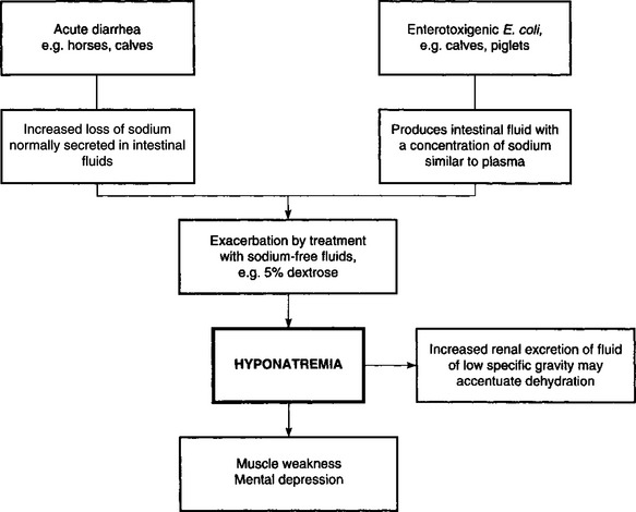
Electrolyte imbalances occur commonly as a result of loss of electrolytes, shifts of certain electrolytes or relative changes in concentrations due to loss of water. Common electrolyte imbalances include hyponatremia, hypokalemia, hypocalcemia and hypochloremia.

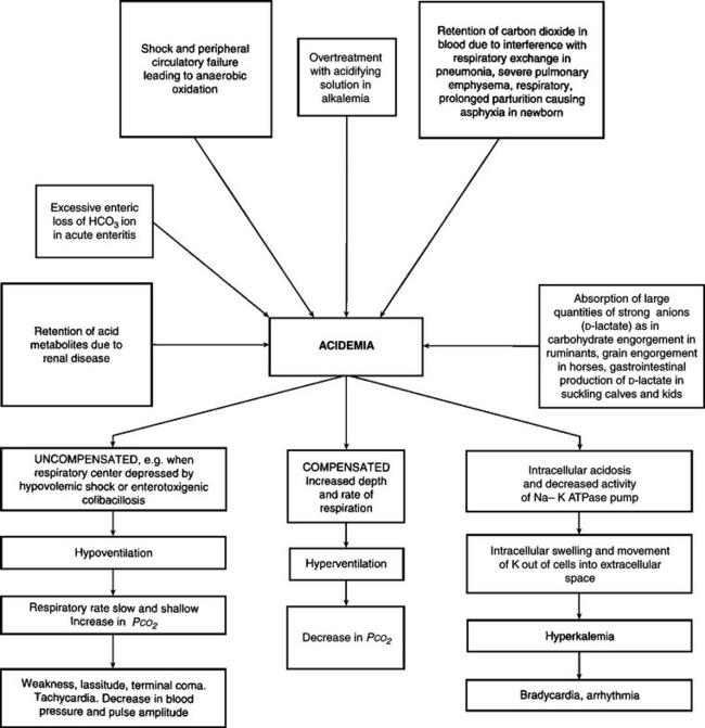
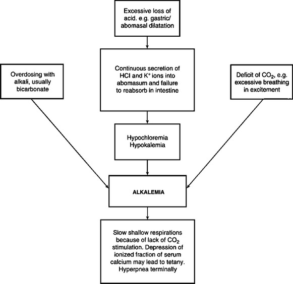
Acid–base imbalances, either acidemia or alkalemia, occur as a result of the addition of acid and depletion of alkali reserve, or the loss of acid with a relative increase in alkali reserve.

Under most conditions, the above disturbances of fluid and electrolyte balance will occur simultaneously, in varying degrees, depending on the initial cause. Each major abnormality will be described separately here with emphasis on etiology, pathogenesis, clinical pathology and treatment. However, it is important to remember that actual disease states in animals in which treatments with fluids and electrolytes are contemplated are rarely caused by single abnormalities. In most cases it is a combination of dehydration together with an electrolyte deficit, and often without a disturbance of the acid–base balance, that necessitates treatment.

DEHYDRATION

ETIOLOGY

There are two major causes of dehydration:

Inadequate water intake

Excessive fluid loss.

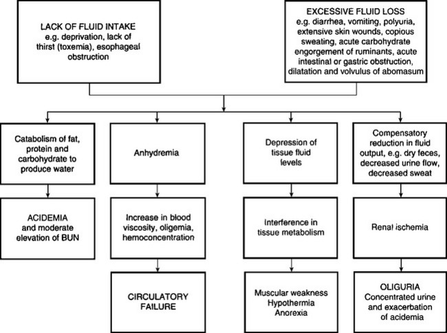
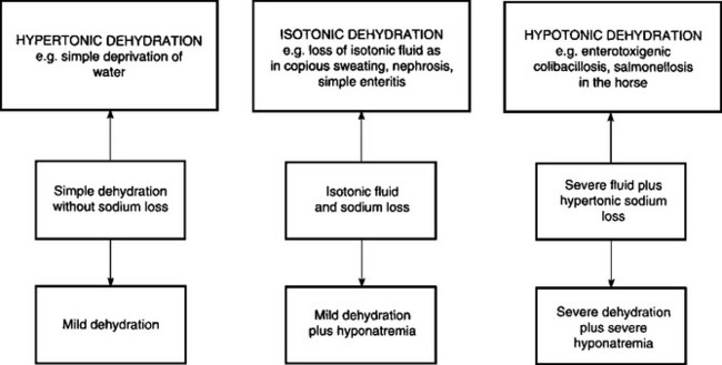
Deprivation of water, a lack of thirst due to toxemia, and the inability to drink water as in esophageal obstruction, are examples of dehydration due to inadequate water intake. The most common cause of dehydration is when excessive fluid is lost. Diarrhea is the most common reason for excessive fluid loss, although vomiting, polyuria and loss of fluid from extensive skin wounds or by copious sweating may be important in sporadic cases. Severe dehydration also occurs in acute carbohydrate engorgement in ruminants, acute intestinal obstruction and diffuse peritonitis in all species, and in dilatation and volvulus of the abomasum. In most forms of dehydration, deprivation of drinking water being an exception, the serious loss, and the one that needs correction, is not the fluid but the electrolytes (Fig. 2.1).

image

Fig. 2.1 Etiology and pathogenesis of dehydration.

The ability to survive for long periods without water in hot climates represents a form of animal adaptation that is of some importance. This adaptation has been examined in camels and in Merino sheep. In the latter, the ability to survive in dry, arid conditions depends on a number of factors, including insulation, the ability to carry water reserves in the rumen and extracellular fluid space, the ability to adjust electrolyte concentrations in several fluid locations, the ability of the kidney to conserve water and the ability to maintain the circulation with a lower plasma volume. Dehydrated mammals in hot environments can save water by reducing the rate of panting and sweating and regulating body temperature above hydrated levels. Sweating is a significant avenue of evaporative heat loss in goats when they are hydrated and exposed to high ambient temperatures above 40°C.

Observations of drinking behavior of cattle transported to the abattoir indicate that those animals that had been sold in livestock markets prior to arrival at the abattoir are more thirsty and more tired than cattle sent directly from farms.1 This indicates inadequate water intake and dehydration.

PATHOGENESIS

Two factors are involved in the pathogenesis of dehydration:

Depression of tissue water content with resulting interference in tissue metabolism

Reduction in the free water content of blood.

The initial response to negative water balance is the withdrawal of fluid from the tissues and the maintenance of normal blood volume. The fluid is drained primarily from the intracellular and interstitial fluid spaces. Essential organs including the central nervous system, heart and skeleton contribute little and the major loss occurs from connective tissue, muscle and skin. The loss of fluid from the interstitial and intracellular spaces results in loss of skin elasticity, dryness of the skin and mucosa, and a reduction and retraction of the eyeball (enophthalmia) due to reduction in the volume of the postorbital fat deposits. In the goat, total body water may be reduced as much as 44% before death occurs.

The secondary response to continued negative water balance is a reduction in the fluid content of the blood causing a reduction in circulating blood volume (volume depletion) and an increase in the concentration of the blood (hemoconcentration). Because of the hemoconcentration, there is an increase in the viscosity of the blood, which impedes blood flow and may exacerbate peripheral circulatory failure. The loss in circulating blood volume also contributes to the mental depression of dehydrated animals, which is also due to varying degrees of acidemia and toxemia depending on the cause of the dehydration. In deprivation of water and electrolytes or in deprivation of water alone or inability to consume water in an otherwise normal animal (e.g. esophageal obstruction), the dehydration is minimal because the kidney compensates effectively by decreasing urine output and increasing urine osmolality. In addition, water is preserved by reduced fecal output and increased absorption, which results in dehydration of the contents of the rumen and large intestine, which in turn results in dry, scant feces.

In calves with acute diarrhea there is increased fecal output of water compared to normal calves but the total water losses are not much greater than in normal calves. In the diarrheic calf the kidney compensates very effectively for fecal water loss, and the plasma volume can be maintained if there is an adequate oral fluid intake. Urine excretion decreases, the urine becomes progressively more concentrated and the renal insufficiency may accentuate pre-existing acidemia and electrolyte imbalance, hence the importance of restoring renal function. The newborn calf is able to concentrate urine at almost the same level as the adult. This illustrates the importance of oral fluid and electrolyte intake during diarrhea to compensate for continuous losses. However, it is possible for metabolic acidosis to occur in diarrheic calves and goat kids that are not dehydrated.2,3

Goats are more sensitive to water deprivation during pregnancy and lactation than during anestrus. Water deprivation for 30 hours causes a marked increase in the plasma osmolality and plasma sodium concentration in pregnant and lactating goats. Pregnant and lactating goats drink more than goats in anestrus.

The dehydration in horses used for endurance rides is hypotonic, in which both sodium and water are lost through sweating. This may account for the lack of thirst in some dehydrated horses with the exhaustion syndrome. Weight losses of 10–15 kg/h may occur in horses exercising in high environmental temperatures exceeding 32°C (89°F) and a horse weighing 450 kg can lose 45 L of fluid in a 3-hour ride.

Dehydration exerts important effects on tissue metabolism. There is an increase in breakdown of fat, then carbohydrate and finally protein, to produce water of metabolism. The increased endogenous metabolism under relatively anaerobic conditions results in the formation of acid metabolites and the development of metabolic acidosis. Urine formation decreases because of the restriction of renal blood flow and this, together with the increased endogenous metabolism, causes a moderate increase in blood levels of nonprotein nitrogen.4 The body temperature may increase slightly initially – dehydration hyperthermia – because of insufficient fluid to maintain the loss of heat by evaporation. The onset of sweating in steers after exposure to high environmental temperatures has been shown to be delayed by dehydration.

Dehydration may cause death, especially in acute intestinal obstruction, vomiting and diarrhea, but it is chiefly a contributory cause of death when combined with other systemic states, such as acidosis, electrolyte imbalances, toxemia and septicemia.

CLINICAL FINDINGS

The first and most important clinical finding in dehydration is dryness and wrinkling of the skin, giving the body and face a shrunken appearance. The eyes recede into the sockets and the skin subsides slowly after being picked up into a fold. The dehydration is usually much more marked if water and electrolyte losses have been occurring over a period of several days. Peracute and acute losses may not be obvious clinically because major loss will have occurred from the intravascular compartment and only minor shifts have occurred from the interstitial spaces. Sunken eyes and inelastic skin are not remarkable clinical findings of dehydration in the horse.

The best indicator of hydration status in calves has been demonstrated to be the degree of recession of the eye into the orbit. Hydration status is assessed by gently rolling the lower eyelid out to its normal position and estimating the distance of eye recession in millimeters. This distance is multiplied by 1.7 to provide an estimate of the degree of dehydration as a percentage of euhydrated body weight.5 The second best indicator of hydration status in calves is the elasticity of the skin of the neck and lateral thorax, which are assessed by pinching the skin between the fingers, rotating the skin fold 90° and noting the time required after release of the skin fold for the skin fold to disappear (normally <2 s). The elasticity of the skin fold on the upper or lower eyelid is a poor indicator of hydration status in calves and is not recommended. The best methods for assessing hydration status in adult cattle and other large animals has not been determined but it is likely that eye recession and skin tent duration in the neck region provide the most accurate and sensitive methods for estimating hydration status.

In diarrheic calves, the severity of dehydration, hypothermia and metabolic acidosis are associated with the degree of mental depression.6 The combined effects of acidemia and dehydration also contribute to hypothermia.

Loss of body weight occurs rapidly in dehydration and muscular weakness and inappetence or anorexia are common. In horses deprived of water for 72 hours there is a mean body weight loss of about 15%, and 95% of the animals have a urine specific gravity of 1.042, a urine osmolality of 1310 mosmol/kg and a urine osmolality/serum osmolality ratio of 4:14. Prerenal azotemia also develops.

The degree of thirst present will depend on the presence or absence of other diseases causing an inflammatory response or endotoxemia. In primary water deprivation, dehydrated animals are very thirsty when offered water. In dehydration secondary to enteritis associated with severe inflammation, acidemia and electrolyte imbalance, there may be no desire to drink. Horses that become dehydrated in endurance rides may refuse to drink and the administration of water by oral intubation and enemas may be necessary. In cattle on pasture and deprived of water for up to 9 days and then given access to water, there will be staggering, falling, convulsions and some death – signs similar to salt poisoning in pigs. Experimental restriction of the water intake in lactating dairy cattle for up to 4 days may reduce milk yield by 75% and decrease body weight by 14%. A 10% reduction in water intake causes a drop in milk production that may be difficult to detect. Behavioral changes are obvious: cows spend considerable time licking the water bowls. In cold climates, cattle are often forced to eat snow as a source of water. The snow must be soft enough so that it can be scooped up by the cattle and 3–5 days are necessary for the animals to adjust to the absence of water and become dependent on snow. During this time there is some loss of body weight. Lactating ewes relying on snow as a source of free water reduce their total water turnover by approximately 35%.

CLINICAL PATHOLOGY

Dehydration is characterized by an increase in the packed cell volume and total serum protein concentration, although the latter response may be modified by the presence of severe enteritis, peritonitis, or proteinuria.

REFERENCES

1 Jarvis AM, et al. Appl Anim Behav Sci. 1996;50:83.

2 Tremblay RRM, Butler DG. Aust Vet J. 1991;32:308.

3 Lorenz I. Vet J. 2004;168:323.

4 Groutides CP, Michell AR. Aust Vet J. 1990;146:205.

5 Constable PD, et al. J Am Vet Med Assoc. 1998;212:991.

6 Naylor JM. Can Vet J. 1989;30:577.

ACUTE OVERHYDRATION (WATER INTOXICATION)

Synopsis

Etiology Rapid ingestion of large quantities of water

Epidemiology Access to water by thirsty calves, or calves that have been marginally deprived of water for some time

Clinical findings Dark red urine, weakness and depression

Clinical pathology Hemoglobinuria, hemoglobinemia, hypo-osmolality, hyponatremia, hypochloremia

Necropsy findings Hemoglobinuria and renal cortical necrosis

Diagnostic confirmation Epidemiological, presence of hyponatremia and hypochloremia; rule out other causes of intravascular hemolysis

Treatment Time, possibly intravenous hypertonic saline but usually too late to be effective

The rapid ingestion of large amounts of water by young calves with normal serum sodium concentrations may result in intravascular hemolysis, hemoglobinemia and hemoglobinuria. In contrast, water ingestion in hypernatremic animals may result in cerebral edema but does not produce hemoglobinuria. The cerebral edema syndrome is described in sodium chloride poisoning. Water intoxication (acute overhydration) is described here.

ETIOLOGY

The ingestion of excessive quantities of water when animals are very thirsty may result in overhydration, which is also called water intoxication. The primary cause of acute overhydration is a rapid decrease in the osmolality of the small intestinal contents, which are normally isotonic to plasma. Such a rapid decrease in luminal osmolality occurs within 5 minutes of water ingestion2 because thirsty calves close their esophageal groove when drinking. This results in a large volume of water in the abomasum, which is subsequently emptied into the duodenum. Free water rapidly moves from the small intestinal lumen into the intravascular compartment because of the large surface area for absorption in the small intestine and development of an osmotic gradient between the small intestinal lumen and intestinal capillary bed. The end result is a rapid decrease in plasma osmolality and expansion and rupture of erythrocytes, leading to intravascular hemolysis, hemoglobinemia, hemoglobinuria, hyponatremia, hypochloremia and a decrease in plasma protein concentration from preingestion values.

EPIDEMIOLOGY

The syndrome has been reported from several countries but is uncommon. Calves 2–4 months of age are most commonly affected but the disease is also recorded in adult cattle,1 sheep3 and pygmy goats.4 Water intoxication occurs in calves in normal husbandry systems when animals that have had limited access to water are suddenly given free access. Commonly water intoxication occurs when calves previously fed a milk replacer diet but no other fluid, or weaned calves that have been on a starter diet but limited water, are turned out to pasture or to yards where water is freely available. Calves that are not fed supplementary salt or that have lost salt as a result of severe exercise or high environmental temperatures may be at higher risk5 but the syndrome also occurs where salt has not been restricted. The majority of calves show clinical signs within minutes to hours of access to water.

The condition has been reproduced in calves by gavage with water at 12% of body weight.6

CLINICAL FINDINGS

Hemoglobinuria as a result of intravascular hemolysis is prominent and there may be a moderate to severe hemolytic anemia. Dark red urine is passed shortly following access to water. Additional signs include tachycardia and hypothermia if the temperature of the water ingested is below body temperature. Affected animals are usually depressed and weak.

CLINICAL PATHOLOGY

Hemoglobinuria and hemoglobinemia are evident and there is hypo-osmolality, hyponatremia and hypochloremia.5 Serum total protein and albumin concentration may be decreased but are usually within the normal range because animals are usually mildly dehydrated and thirsty before ingesting large volumes of water.

Postmortem findings

There is marked pallor of the carcass and renal cortical necrosis due to hemoglobinemic nephrosis may be evident histologically.7

DIFFERENTIAL DIAGNOSIS

Other causes of intravascular hemolysis and hemoglobinuria.

TREATMENT

Treatment of affected animals is usually not attempted as the hypo-osmotic lysis has already occurred when clinical signs are manifest and serum osmolality is usually gradually increasing as the distal convoluted tubules eliminate excessive free water. Hypertonic saline (7.2% NaCl, 5 mL/kg BW over 5 min intravenously) is usually administered to correct the hyponatremia and hypochloremia but treatment is not necessary in mild cases. Case fatality is low and hemoglobinuria persists for only a few hours.

CONTROL

Water intoxication does not occur commonly and can be avoided by preventing thirsty animals from having unlimited access to water. Calves should have free access to water by the end of the first week of life.

REVIEW LITERATURE

Angelos SM, van Metre DC. Treatment of sodium balance disorders: water intoxication and salt toxicity. Vet Clin North Am Food Anim Pract. 1999;15:609-618.

REFERENCES

1 Bianca W. Br Vet J. 1970;126:121.

2 Shimizu Y, et al. Jpn J Vet Sci. 1979;41:583.

3 Abdelrakim AI, et al. Rev d’Elevage Med Vet Pay Trop. 1985;38:180.

4 Middleton JR, et al. J Vet Intern Med. 1997;11:382.

5 Gilchrist F. Can Vet J. 1996;37:490.

6 Slalina L, et al. Vet Med Prague. 1993;38:459.

7 Njoroge EM, et al. Onderstepoort J Vet Res. 1997;64:111.

ELECTROLYTE IMBALANCES

Most electrolyte imbalances are due to a net loss of electrolytes associated with diseases of the alimentary tract. Sweating, exudation from burns, excessive salivation and vomiting also result in electrolyte losses, but are of minor importance in farm animals, with the exception of sweating in the horse and dysphagia in ruminants. The electrolytes of major concern are sodium, chloride, potassium, calcium and phosphorus. Losses of bicarbonate are presented under acid– balance imbalance.

HYPONATREMIA

Sodium is the most abundant ion in the extracellular fluid and is chiefly responsible for the maintenance of osmotic pressure of the extracellular fluid. The most common cause of hyponatremia is increased loss of sodium through the intestinal tract in enteropathies (Fig. 2.2). This is particularly marked in the horse with acute diarrhea and to a moderate extent in calves with acute diarrhea. The sodium is lost at the expense of the extracellular fluid. In calves with acute diarrhea due to enterotoxigenic E. coli the sodium concentration of the intestinal fluid secreted in response to the enterotoxin is similar to that of plasma, and hyponatremia usually occurs (hypotonic dehydration). Animals affected with diarrhea of several days’ duration continue to lose large quantities of sodium and the hyponatremia may become severe. Hyponatremia can become severe when sodium-free water or 5% dextrose are used as the only fluid therapy in animals already hyponatremic. Hyponatremia can also occur in animals with proximal tubular dysfunction.

image

Fig. 2.2 Etiology and pathogenesis of hyponatremia.

Hyponatremia causes an increase in the renal excretion of water in an attempt to maintain normal osmotic pressure, which results in a decrease in the extracellular fluid space, leading to a decreased circulating blood volume, hypotension, peripheral circulatory failure and ultimately renal failure. Muscular weakness, hypothermia and marked dehydration are common findings.

Isotonic dehydration occurs when there is a parallel loss of sodium and water. Hypertonic dehydration, which is uncommon, occurs when there is a loss or deprivation of water with minor losses or deprivation of sodium. Hypertonic dehydration can occur in animals that are unable to consume water because of an esophageal obstruction. The dehydration in isotonic and hypertonic dehydration is mild compared to the marked clinical dehydration that can occur in hypotonic dehydration accompanied by marked loss of water and concentration of the extracellular space (Fig. 2.3).

image

Fig. 2.3 Types of dehydration.

There are no clinical signs that are characteristic of hyponatremia. There is usually dehydration, muscular weakness and mental depression, which occur with other disturbances of both water and electrolytes and with acid–base imbalance. Similarly, there are no clinical signs characteristic of hypochloremia. However, hyponatremia affects the osmotic pressure of the extracellular fluid, and hypochloremia promotes the reabsorption of bicarbonate and further development of alkalosis. Polyuria and polydipsia occur in cattle with dietary sodium chloride deficiency.

HYPOCHLOREMIA

Hypochloremia occurs as a result of an increase in the net loss of the electrolyte in the intestinal tract in acute intestinal obstruction, dilatation and impaction and volvulus of the abomasum and in enteritis (Fig. 2.4). Normally a large amount of chloride is secreted in the abomasum by the mucosal cells in exchange for bicarbonate, which moves into the plasma. The hydrogen, chloride and potassium ions secreted in gastric juice are normally absorbed by the small intestine. Failure of abomasal emptying and obstruction of the proximal part of the small intestine will result in the sequestration of large quantities of chloride, hydrogen and potassium ions which leads to a hypochloremic, hypokalemic metabolic alkalosis. A severe hypochloremia can be experimentally produced in calves by feeding them a low chloride diet and daily removal of abomasal contents. Clinical findings include anorexia, weight loss, lethargy, mild polydipsia and polyuria. A marked metabolic alkalosis occurs, with hypokalemia, hyponatremia, azotemia and death.

image

Fig. 2.4 Etiology and pathogenesis of hypochloremia.

HYPOKALEMIA

Hypokalemia may occur as a result of decreased dietary intake, increased renal excretion, abomasal stasis, intestinal obstruction and enteritis, and repeated administration of corticosteroids with mineralocorticoid activity (Fig. 2.5). The prolonged use of potassium-free solutions in fluid therapy for diarrheic animals may result in excessive renal excretion of potassium and hypokalemia. Alkalosis may result in an exchange of potassium ions for hydrogen ions in the renal tubular fluid, resulting in hypokalemia. Hypokalemia can cause muscle weakness, prolonged unexplained recumbency, inability to hold up the head, anorexia, muscular tremors and, if severe enough, coma. The treatment of ketosis in lactating dairy cows with multiple dosages of isoflupredone, a glucocorticoid with some mineralocorticoid activity, can cause hypokalemia and recumbency, with a high case fatality rate.1

image

Fig. 2.5 Etiology and pathogenesis of hypokalemia.

The most common occurrence of hypokalemia in ruminants is in diseases of the abomasum that cause stasis and the accumulation of fluid in the abomasum. Potassium becomes sequestered in the abomasum along with hydrogen and chloride, resulting in hypokalemia, hypochloremia and metabolic alkalosis.

Metabolic alkalosis and hypokalemia in cattle are often accompanied by muscular weakness and paradoxic aciduria. Hypokalemia causes muscle weakness by lowering the resting potential of membranes, resulting in decreased excitability of neuromuscular tissue. Thus, the differential diagnosis of the animal with muscle weakness should always include hypokalemia.

Hypokalemia and alkalosis also are often directly related because of the renal response to either. Hypokalemia from true body deficits of potassium will cause decreased intracellular concentration of this ion. The intracellular deficit of potassium and excess of hydrogen will cause hydrogen secretion into the urine when distal sodium reabsorption is required. This situation exists in metabolic alkalosis, where sodium bicarbonate reabsorption in the proximal nephron is decreased because of the excess of plasma bicarbonate. Distal nephron avidity for sodium is increased to protect extracellular fluid volume, and the increased distal sodium reabsorption is at the expense of hydrogen secretion, although it is contrary to the need of acid retention in the presence of alkalosis. In other words, the kidney prioritizes maintenance of plasma volume above that of acid–base balance, presumably because respiratory compensation can usually keep blood pH within the normal physiological range. Because electroneutrality of extracellular fluid must be maintained by reabsorbing an equivalent charge of cations and anions, the reabsorption of chloride and of bicarbonate in the kidneys are inversely proportional to each other. Thus, with excess trapping of chloride in the abomasum, the kidneys will compensate for the resulting hypochloremia by increasing bicarbonate reabsorption, which may proceed until metabolic alkalosis develops.

The treatment of hypochloremic, hypokalemic alkalosis requires correction of extracellular fluid volume and sodium and chloride deficits with 0.9% NaCl infusions and oral KCl. Providing adequate chloride ion allows sodium to be reabsorbed without bicarbonate. Increased proximal reabsorption of sodium will decrease distal acid secretion because less sodium is presented to the distal nephron. As less bicarbonate is reabsorbed and less acid secreted, the metabolic alkalosis is resolved. Specially formulated solutions containing potassium are necessary in cases of severe hypokalemia and small-intestinal obstruction.

Hypokalemia also occurs following treatment of the horse affected with metabolic acidosis and hyponatremia, and probably reflects whole-body potassium depletion. Horses used for endurance rides may be affected by hypokalemia, hypocalcemia and alkalosis due to loss of electrolytes during the competition. Synchronous diaphragmatic flutter also occurs, which may be the result of the electrolyte imbalance (particularly hypocalcemia) causing hyperirritability of the phrenic nerve.

Since potassium is the major intracellular cation, the measurement of plasma or serum potassium is not a reliable indication of whole-body potassium status. Extremely low levels or high levels are usually indicative of a potassium imbalance, often associated with other electrolyte and acid–base imbalances. In severe alkalosis, for example, potassium leaves the extracellular space and becomes concentrated in the cells. This may result in low serum potassium levels when, in fact, there might not be potassium depletion of the body. Conversely, in severe metabolic acidosis of calves with acute diarrhea, the potassium leaves the cells and moves into the extracellular fluid. This results in hyperkalemia in some cases where the body potassium is normal or even decreased. When changes occur in the concentration of intracellular and extracellular potassium, the ratio of intracellular to extracellular potassium may decrease by as much as 30–50%, which results in a decrease in the resting membrane potential. This is thought to be the explanation for the effects of hypokalemia and hyperkalemia on muscle function.

The potassium concentration of red blood cells may be a more accurate indicator of whole-body potassium deficit in diarrheic horses and provides a basis for a calculated oral dose of potassium chloride in horses with diarrhea, which is a safe therapeutic procedure.

Potassium should be administered intravenously or orally. The intravenous route is used only for the initial treatment of recumbent ruminants with severe hypokalemia and rumen atony, as it is much more dangerous and expensive than oral treatment. The most aggressive intravenous treatment protocol is an isotonic solution of KCl (1.15% KCl), which should be administered at less than 3.2 mL/kg per hour, equivalent to a maximal delivery rate of 0.5 mEq of K+/kg BW per hour. Higher rates of potassium administration run the risk of inducing hemodynamically important arrhythmias, including ventricular premature complexes that can lead to ventricular fibrillation and death. A less aggressive intravenous treatment is an isotonic equimolar mixture of NaCl (0.45% NaCl) and KCl (0.58%KCl), and the least aggressive intravenous treatment is the addition of 10 mmol of KCl/L of Ringer’s solution, which will increase the solution osmolarity to 329 mosmol/L. Clinical experience with oral administration of KCl has markedly decreased the number of adult ruminants treated with intravenous KCl.

Oral administration of potassium is the method of choice for treating hypokalemia. Inappetent adult cattle should be treated with 30–60 g of feed grade KCl twice with a 12-hour interval, with the KCl placed in gelatin boluses. Adult cattle with severe hypokalemia (<2.5 mEq/L) should initially be treated with 120 g of KCl, followed by two 60 g KCl treatment at 8-hour intervals, for a total 24-hour treatment of 240 g KCl. Higher doses have been administered to dairy cows but these are accompanied by diarrhea, and oral administration of 0.58 g KCl/kg BW was toxic in 6-month-old Holstein calves, manifest by excessive salivation, muscular tremors of the legs and excitability, and a peak plasma [K+] of 9.0 mEq/L. Extrapolating this toxic dose in normokalemic calves to hypokalemic 600 kg cows suggests that a daily dose of 240 g KCl approaches the upper limit of safety. The recommended doses are empirical but are effective in rapidly increasing serum [K+] and [Cl]. Inappetent horses often have whole-body potassium depletion and would benefit from supplementary dietary potassium (25–50 g/d KCl).

HYPERKALEMIA

Hyperkalemia is not as common in farm animals as hypokalemia, occurring most commonly in severe metabolic acidosis. The classic description for the development of hyperkalemia in metabolic acidosis involves a purported redistribution of potassium from the intracellular space to the extracellular space because a large proportion of the excess hydrogen ions are buffered intracellularly. Thus potassium is supposedly exchanged with hydrogen ions across the cell membrane in order to maintain electroneutrality. A more likely mechanism is that metabolic acidosis is accompanied by acidemia and a decreased intracellular pH; during intracellular acidosis the function of all enzyme systems is decreased. As a direct result of the intracellular acidosis, the Na–K-ATPase activity is decreased, with potassium leaving the cell down its concentration gradient.

Hyperkalemia is potentially more life-threatening than hypokalemia. Hyperkalemia (when over 7–8 mmol/L) has a profound effect on cardiac function. There is usually marked bradycardia and arrhythmia and sudden cardiac arrest may occur. The electrocardiogram (ECG) changes in experimentally induced hyperkalemia in the horse have been described. The changes include four successive stages as hyperkalemia increased. There was a widening and lowering of amplitude followed by inversion and disappearance of the P wave, an increase in the amplitude of the T wave, an increase in the QRS interval, with some irregularity in the ventricular rate, and periods of cardiac arrest that became terminal or were followed by ventricular fibrillation. The minimum plasma potassium concentration required to induce ECG changes was 6–7 mmol/L and severe cardiotoxic effects occurred at levels between 8–11 mmol/L. The effects of hyperkalemia on the ECG are exacerbated by the presence of hyponatremia.

Hyperkalemia has traditionally been treated by intravenous administration of sodium bicarbonate, glucose, insulin and sometimes calcium. Hypertonic saline is just as effective as is hypertonic sodium bicarbonate in decreasing hyperkalemia and hyperkalemia-associated bradyarrhythmias, as a result of sodium-induced intracellular movement of potassium, extracellular volume expansion and the strong ion effect of increasing the serum concentration of a strong cation. The long-held myth regarding the need to administer glucose and insulin to ‘drive’ potassium into the cells during hyperkalemia needs to be re-evaluated. Calcium counteracts the effect of hyperkalemia on the resting membrane potential by increasing the threshold potential to a higher value, thereby returning an appropriate difference between resting and threshold potentials. Calcium can be administered intravenously at 0.2–0.4 mL of a 23% calcium gluconate solution/kg BW. The focus of treatment in hyperkalemia should be correction of acidemia, plasma volume expansion and increasing the serum sodium concentration. Glucose and insulin are not routinely needed to correct hyperkalemia.

Hyperkalemic periodic paralysis occurs in heavily muscled Quarterhorse. Affected horses become weak, may stand base-wide and are reluctant to move. Sweating commonly occurs and generalized muscle fasciculations are apparent. Affected horses remain bright and alert but may yawn and do not eat or drink. Some horses become recumbent and may appear to be in a state of flaccidity. Attacks may occur in a rest period following exercise or at random. During the episode the serum potassium concentration is elevated by up to twofold and returns to normal values when the animal recovers. Treatment consists of sodium bicarbonate, hypertonic saline or 5% dextrose given intravenously, possibly with insulin.

HYPOCALCEMIA

Hypocalcemia or milk fever may occur in recently calved mature dairy cows that have been inappetent or anorexic for a few days. Hypocalcemia can be due to a reduction in dry matter intake because of illness or it may be the earliest stages of hypocalcemic parturient paresis. The clinical findings include anorexia, mild tachycardia with a reduction in the intensity of the heart sounds and occasionally an arrhythmia, a decrease in the frequency and amplitude of rumen contractions or complete ruminal stasis, and a decrease or complete absence of feces, which may last from 6–36 hours if untreated.

Hypocalcemia cases often mimic intestinal obstruction and create problems in the differential diagnosis. Affected cattle may not exhibit any evidence of muscular weakness and the detection of the hypocalcemic state can be elusive. The total serum calcium concentrations range from 1.5–2.0 mmol/L and the response to intravenous therapy is usually good, although recovery may require several hours before the appetite returns to normal and feces are passed.

Calcium should be administered by the intravenous, subcutaneous, or oral route. Calcium gluconate and calcium borogluconate are the preferred forms for intravenous and subcutaneous administration because CaCl2 causes extensive necrosis and sloughs of tissue when administered perivascularly. Compared to calcium gluconate, calcium borogluconate has improved solubility and shelf life. Plasma ionized calcium concentrations are increased to a greater extent following CaCl2 treatment when high equimolar solutions of CaCl2 and calcium gluconate are administered, leading to more cardiac arrhythmias during CaCl2 administration. A typical treatment to an adult lactating dairy cow with periparturient hypocalcemia is 500 mL of 23% calcium borogluconate by slow intravenous injection with cardiac auscultation, this provides 10.7 g of calcium. Although the calculated calcium deficit in a recumbent periparturient dairy cow is 4 g calcium, additional calcium should be provided to overcome the continued loss of calcium in milk. A field study comparing the effectiveness of different doses of calcium for treating periparturient milk fever determined that 9 g of calcium was superior to 6 g. A good rule of thumb for administering 23% calcium borogluconate solutions (2.14 g calcium/100 mL) to cows with periparturent hypocalcemia is therefore to administer 1 mL/kg BW. There do not appear to be any clinically important advantages to slow administration of the solution over 6 h, when compared to 15 min.2

The normal cardiac response to intravenous calcium administration is an increase in the strength of cardiac contraction and a slowing of the heart rate. Intravenous administration is continued until the first arrhythmia is detected (a bradyarrhythmia such as a prolonged pause); the rate of intravenous administration is then slowed until a second arrhythmia is detected, at which time intravenous administration is discontinued and the remainder of the solution is placed subcutaneously over the lateral thorax. This treatment method titrates the calcium dose required for each animal. Auscultation of the heart is an absolute requirement during treatment: visual monitoring of the jugular pulse at the base of the neck does not allow the early detection of bradyarrhythmias, making it more likely that the cow will receive a toxic and possibly lethal dose of calcium. The maximum safe rate of calcium administration in cattle is 0.07 mEq of Ca2+/kg BW/min, which is equivalent to 0.065 mL 23% calcium borogluconate/kg BW/min. For a 500 kg normocalcemic dairy cow, this corresponds to a maximum safe rate of administration of 33 mL/min. Typical rates of administration through a 14-gauge needle are 50 mL/min; this rate of administration is safe for cows with hypocalcemia, provided that cardiac auscultation is performed during administration.

Subcutaneous administration of calcium solutions has been practiced for many years. To facilitate absorption, it is preferable to administer no more than 125 mL at a site. A 14-gauge needle is placed subcutaneously over the lateral thorax, 125 mL is administered, the needle is redirected and another 125 mL is administered. The process is then repeated on the other side of the cow. Although the effectiveness of subcutaneous administration of calcium has been documented in healthy normal cows, there do not appear to be any reports documenting the rapidity by which subcutaneous calcium is absorbed by cows with periparturient hypocalcemia. Subcutaneous administration of calcium gluconate is not recommended in recumbent cows because poor peripheral blood flow is suspected to lead to slow absorption from the subcutaneous site. Calcium chloride is not recommended for subcutaneous administration because of extensive tissue damage; the addition of dextrose to the administered calcium is also not recommended because it increases the tonicity of the solution and propensity for bacterial infection and abscessation. Rectal calcium administration is not recommended because it causes severe mucosal injury and tenesmus but does not increase plasma concentrations of calcium.

Oral administration of calcium has also been practiced for many years, usually by ororuminal intubation of calcium borogluconate solutions designed for parenteral administration. Over the past decade there has been increased interest in improving the efficacy of oral calcium formulations. The results of a number of studies indicate that oral calcium salts are effective at increasing plasma calcium concentration; orally administered calcium is absorbed by a dose-dependent passive diffusion process across ruminal epithelium and a dose-independent calcium-binding protein mechanism in the small intestine that is modulated by vitamin D. Rapid correction of hypocalcemia by oral calcium administration is predominantly by passive ruminal diffusion, as small intestinal absorption is too slow to be of clinical value.

Two calcium formulations are currently recommended for oral administration to ruminants; CaCl2 and calcium propionate, but most commercially available products contain 50 g of CaCl2. Calcium chloride has the advantage of low cost and low volume (because of its high solubility), but CaCl2 can severely damage the pharynx and esophagus in ruminants with reduced swallowing ability, can lead to necrosis of the forestomach and abomasum when administered in high doses, and can lead to aspiration pneumonia when administered as a drench. Calcium propionate has the advantage that it is less irritating while providing a gluconeogenic substrate (propionate), but the disadvantages of higher volumes and cost. Oral calcium solutions should only be administered to cattle that have normal swallowing ability, precluding their administration to animals with advanced clinical signs of hypocalcemia. Higher plasma calcium concentrations are obtained more quickly when calcium solutions are drenched after administration of vasopressin to induce esophageal groove closure, or when the calcium solution is administered as a drench instead of ororuminal intubation. Calcium solutions are suspected to have a higher likelihood of aspiration pneumonia than calcium gels (with a consistency similar to toothpaste), although this supposition does not appear to have been verified. Commercially available formulations of calcium gels contain 50 g of CaCl2 and increase plasma calcium concentrations within 30–60 minutes and for at least 6 hours. Retreatment at 12-hour intervals (if needed) therefore appears indicated and provide 100 g of CaCl2 and 37 g of calcium over 24 hours, but more aggressive treatment protocols are not recommended.

HYPOPHOSPHATEMIA

Hypophosphatemia also occurs in cattle under conditions similar to those of hypocalcemia. A decrease in feed intake or alimentary tract stasis will result in a decrease in serum inorganic phosphate. Acute recumbency in lactating dairy cattle may be associated with marginal phosphorus deficiency,3 although a cause and effect relationship between hypophosphatemia and recumbency has not been established.4 However, many inappetent and weak cows have marginal hypophosphatemia and clinically appear to benefit from normalization of their plasma concentration of phosphate. As such, it is currently recommended that ruminants with marked hypophosphatemia and signs of illness should be treated with phosphorus-containing solutions.

Almost all commercially available intravenous solutions for treating hypophosphatemia use phosphite (PO22−) or hypophosphite (PO33−) salts as the source of phosphorus because these salts are very soluble, even in the presence of calcium and magnesium. However, the phosphorus in phosphite and hypophosphite is unavailable to mammals, meaning that the vast majority of ‘phosphate’-containing solutions have no efficacy in treating hypophosphatemia. Instead, the monobasic monophosphate form of sodium phosphate (NaH2PO4) should be administered. The pH of the solution should be mildly acidic (pH 5.8) to maintain phosphate solubility in cold weather but is not needed in warm ambient temperatures. A recommended treatment to an adult lactating dairy cow with severe hypophosphatemia is 300 mL of 10% NaH2PO4 (monohydrate) solution by slow intravenous injection; this provides 7 g of phosphate and increases plasma phosphate concentrations for at least 6 hours. Human enema formulations that contain a mixture of monobasic sodium phosphate monohydrate and dibasic sodium phosphate heptahydrate in a buffered solution have also been administered to cattle with hypophosphatemia but are not recommended. This human enema solution is extremely hypertonic and must therefore be diluted before administration. A major drawback with intravenous administration of phosphate solutions is that they should not be administered within 2 hours of intravenous calcium administration, because of concerns that calcium-phosphate precipitates may be formed in the plasma of cattle with treatment-induced hypercalcemia and hyperphosphatemia. This has traditionally been evaluated by calculating the calcium– phosphorus product, whereby metastatic calcification may occur if the product of serum calcium concentration and serum phosphate concentration (both in mg/dL) exceeds 70.

Hypophosphatemia is more safely treated by administration of oral monosodium phosphate, and this is the preferred method of administration in ruminants with rumen motility. Oral administration also results in a more prolonged increase in plasma phosphorus concentration. Recommended dose is 200 g of feed grade monosodium phosphate (contains 50 g of phosphate) administered in gelatin boluses, drench, or by ororuminal intubation. Phosphorus in other feed grade minerals (such as bone meal or dicalcium phosphate) is poorly available and is not recommended for the treatment of hypophosphatemia.

HYPOMAGNESEMIA

Magnesium is usually administered parenterally only when a ruminant exhibits clinical signs of hypomagnesemia. Treatment of hypomagnesemia is more dangerous (to the animal and clinician) and less satisfying than treatment of periparturient hypocalcemia; the response to treatment is much slower in hypomagnesemia presumably because magnesium concentrations must be normalized in cerebrospinal fluid, which turns over at approximately 1% per minute.

Treatment of hypomagnesemia has historically used 25% Epsom salts solution (magnesium sulfate heptahydrate; MgSO4.6H2O); this solution concentration was selected because it provided approximately 1 mmol of magnesium per liter. It should be noted that 25% Epsom salts solution is markedly hypertonic (2028 mosmol/L). A typical treatment for an adult cow has been slow intravenous administration (over at least 5 min) of 100 mL of the 25% Epsom salts solution, this provides 2.5 g of magnesium (25 mg of magnesium/mL of solution). More recently, hypomagnesemia has been treated using commercially available combined calcium and magnesium solutions; 500 mL of these solutions typically contain 1.6–2.7 g of magnesium in the form of a borogluconate, chloride or hypophosphite salt. Although the calculated extracellular deficit in a cow with hypomagnesemia is 2 g of magnesium, we should provide additional magnesium to correct presumed intracellular deficiencies and to overcome the anticipated urinary loss of magnesium. Combined calcium and magnesium solutions are preferred for intravenous administration to 25% Epsom salts solution because ruminants with hypomagnesemia frequently have hypocalcemia, and hypercalcemia provides some protection against the toxic effects of hypermagnesemia. Moreover, administration of solutions containing magnesium as the only cation increases the risk of developing cardiac and respiratory failure during treatment. The maximum safe rate of administration of magnesium in cattle is 0.08 mEq Mg2+/kg BW per minute, which is equivalent to 0.04 mL 25% Epsom salts/kg BW per minute. For a 500 kg beef cow with hypomagnesemia, this corresponds to a maximum safe rate of administration of 20 mL/min.

Magnesium-containing solutions (such as 25% Epsom salts solution) can also be administered subcutaneously, although this frequently leads to necrosis of the skin, particularly when 50% Epsom salts solution is administered. Only combined calcium and magnesium solutions should therefore be administered subcutaneously.

The oral bioavailability of magnesium is low and much lower than that of calcium. Accordingly, oral administration of magnesium is not recommended for the treatment of hypomagnesemia, but is essential for the prevention of hypomagnesemia. Magnesium absorption from the rumen is facilitated by volatile fatty acids but decreased by potassium and the ammonium ion.

Rectal administration may be the only practical and safe method for treating a convulsing hypomagnesemic beef cow. After evacuating the rectal contents, an enema containing 60 g of Epsom salts (magnesium sulfate heptahydrate) or magnesium chloride in 200 mL of water can be placed in the descending colon (and not the rectum) and the tail held down for 5 minutes; this increases plasma magnesium concentrations within 10 minutes. However, enema solutions can be prematurely evacuated, eliminating the chance for therapeutic success, and some degree of colonic mucosal injury is expected because of the high osmolarity of 30% solutions (approximately 2400 mosmol/L). The safety of this treatment protocol does not appear to have been evaluated, although a 50 mL enema of a 30% MgCl2.6H2O solution rapidly and effectively increased serum magnesium concentration in 7–10-week-old calves and relieved clinical signs of hypomagnesemia.

Oral administration of magnesium hydroxide and magnesium oxide excessively alkalinizes the rumen and can create a severe metabolic alkalosis (strong ion alkalosis), as absorption of magnesium leads to hypermagnesemia and increased plasma strong ion difference. Because oral administration of sodium bicarbonate causes expansion of the plasma volume and creates a metabolic alkalosis (strong ion alkalosis) without hypermagnesemia, it is likely that oral sodium bicarbonate is a more effective treatment for grain overload in ruminants.

REVIEW LITERATURE

Angelos SM, van Metre DC. Treatment of sodium balance disorders: water intoxication and salt toxicity. Vet Clin North Am Food Anim Pract. 1999;15:609-618.

Goff JP. Treatment of calcium, phosphorus, and magnesium balance disorders. Vet Clin North Am Food Anim Pract. 1999;15:619-640.

Sweeney RW. Treatment of potassium balance disorders. Vet Clin North Am Food Anim Pract. 1999;15:609-618.

Constable PD. Fluids and electrolytes. Vet Clin North Am Food Anim Pract. 2003;19:1-40.

REFERENCES

1 Seilman ES, et al. J Am Vet Med Assoc. 1997;210:240.

2 Braun U, et al. Vet Rec. 2004;154:336.

3 Gerloff BJ, Swenson EP. J Am Vet Med Assoc. 1996;208:716.

4 Metzner M, Klee W. Tier Umschau. 2005;60:13.

ACID–BASE IMBALANCE

The pH of mammalian blood is maintained within the normal range of 7.35–7.45 by its buffer systems, of which hemoglobin is the most important, because it has the greatest buffering capacity. However, because the blood hemoglobin concentration is regulated on the basis of oxygen delivery instead of acid–base balance, and because rapid changes in hemoglobin concentration occur only with marked changes in hydration status or splenic contraction associated with exercise, the bicarbonate system has traditionally been considered to be the most important buffer. Other buffers in blood are plasma proteins and phosphate. The addition of relatively large amounts of acid or alkali to the blood is necessary before its buffering capacity is exhausted and its pH changed. Changes from normal acid–base balance towards alkalemia or acidemia occur commonly in sick animals and make a significant contribution to the observed clinical signs.

The traditional approach for assessing acid–base balance focuses on how plasma carbon dioxide tension (Pco2), plasma bicarbonate concentration ([HCO3]), the negative logarithm of the apparent dissociation constant (pK1′) for plasma carbonic acid (H2CO3), and the plasma solubility of CO2 (S) interact to determine plasma pH. This relationship is most commonly expressed as the Henderson–Hasselbalch equation: pH = pK1′ + log([HCO3]/S × Pco2). The evaluation of acid–base balance using the Henderson–Hasselbalch equation has historically used pH as an overall measure of acid–base status, Pco2 as an independent measure of the respiratory component of acid–base balance, and extracellular base excess, actual HCO3concentration or standard HCO3 as a measure of the nonrespiratory (also called metabolic) component of acid–base balance.

When using the traditional Henderson– Hasselbalch approach, four primary acid–base disturbances can be distinguished: respiratory acidosis (increased Pco2), respiratory alkalosis (decreased Pco2), metabolic acidosis (decreased extracellular base excess or actual HCO3 concentration) and metabolic alkalosis (increased extracellular base excess or actual HCO3 concentration). The anion gap is easily calculated from the results of serum biochemical analysis and is used to determine whether unmeasured anions are present. The Henderson–Hasselbalch equation has a long history of use and remains widely and routinely used in the clinical management of acid–base disorders. These advantages should not be overlooked. The principal disadvantage of the Henderson–Hasselbalch equation is that it is more descriptive than mechanistic, decreasing the value of the approach in explaining the cause of acid–base changes during disease. This is because the Henderson–Hasselbalch equation fails to distinguish between the effects of independent and dependent variables on plasma pH.

Actual plasma HCO3 concentration in units of mmol/L is not measured but calculated using the Henderson– Hasselbalch equation and measured values for pH and Pco2, whereby:


[HCO3] = S × Pco2 × 10(pH−pK1′).

The values for pK1′ and S at 37°C are 6.12 and 0.0307/mmHg respectively for normal mammalian plasma. The equation at 37°C is therefore:


[HCO3] = 0.0307 × Pco2 × 10(pH−6.12).

Because actual HCO3 concentration is calculated from pH and Pco2, it can never provide an independent measure of the nonrespiratory component of an acid– base disturbance. A primary decrease in Pco2 (respiratory alkalosis) at normal pH always is accompanied by a decrease in plasma HCO3 concentration (which would be interpreted as a metabolic acidosis). Likewise, a primary increase in Pco2 (respiratory acidosis) at normal pH always produces an increase in plasma HCO3 concentration (which would be interpreted as a metabolic alkalosis). In both cases, the actual HCO3 concentration is dependent upon the pH and Pco2, thereby providing no additional information as to the cause of the acid–base imbalance than that obtained by knowledge of the pH and Pco2. It is therefore illogical to use actual HCO3 concentration to define the nonrespiratory (metabolic) component of an acid–base disturbance.

The current use of actual HCO3 concentration in the evaluation of acid– base status results from Van Slyke’s work in 1924, where pH and total CO2 (which is highly correlated with actual [HCO3]) could be measured more accurately than Pco2. This led to the graphical depiction of the curvilinear HCO3–pH relationship, the so-called Davenport diagram, to represent acid–base disturbances. With the later development of accurate and practical laboratory methods in the 1950s to measure Pco2, acid–base derangements were graphically depicted as approximately linear log(Pco2)–pH relationships. This development led directly to the base excess concept.

The normal range of plasma bicarbonate in large animals is 24–30 mmol/L (this should be compared to the normal range in humans, which is 22–24 mmol/L). In mild metabolic acidosis the bicarbonate concentration is in the range of 20–24 mmol/L, moderate metabolic acidosis is 14–18 mmol/L, and in severe cases the values are below 10 mmol/L and carry a grave prognosis. The levels of Pco2, Po2, plasma bicarbonate and blood pH can be used to determine the degree of compensation, if any, which has taken place. In metabolic acidosis there may be a compensatory decrease in Pco2 due to hyperventilation; in metabolic alkalosis there may be an increase in Pco2 due to hypoventilation. In respiratory acidosis due to severe pneumonia the arterial Po2 will be markedly decreased.

The base excess value directly expresses the amount (usually expressed in units of mEq/L) of strong base (or acid) added per liter of blood or plasma, when the normal mean base excess value is arbitrarily fixed at zero. As such, the base excess is defined as the amount of strong acid (such as HCl) needed to titrate the pH of 100% oxygenated human blood to 7.40 at 37°C and at a Pco2 of 40 mmHg. By definition, the normal base excess value for humans is 0 mEq/L (range is –2 to +2 mEq/L), and a base excess of more than +2 mEq/L indicates metabolic alkalosis, whereas a value of less than –2 mEq/L (negative base excess value or base deficit) reflects metabolic acidosis. The normal range of base excess in large animals is 0–6 mmol/L.

Mathematical formulas and nomograms are available to calculate base excess from measured pH, Pco2 and blood hemoglobin concentration. Base excess is usually expressed as BEECF (also called standard base excess or in vivo base excess). Extracellular base excess is the preferred measurement as this formulation provides the best clinical estimate of the required mmol/L of HCO3 required to correct metabolic acidosis, as it assumes a fixed hemoglobin concentration of 5 g/dL. Clearly, the BEECF value will be incorrect when applied to animals with anemia or polycythemia; however, the error introduced by this approximation is small and usually clinically insignificant.

Most blood gas analyzers calculate base excess in units of mEq/L using Siggaard-Andersen’s empirical equation derived from his nomogram with hemoglobin concentration [Hb] and actual HCO3 concentrations in mmol/L:


BEblood = (1 − 0.023 × [Hb]) × ([HCO3] − 24.4 + (7.7 + 2.3 ×[Hb]) × (pH − 7.40)),

which is equivalent to the following expression when [Hb] = 3.1 mmol/L = 5 g/dL:


BEECF = 0.93 × ([actual HCO3] − 24.4 + 14.83 × (pH − 7.40)).

The calculated BEECF value assumes normal serum protein concentration (7.2 g/dL) and therefore provides an inaccurate estimate of the magnitude of a metabolic acidosis or alkalosis in domestic animals with hypoproteinemia or hyperproteinemia. The ability of extracellular base excess (BEECF) and actual HCO3 concentration to accurately characterize the metabolic component of acid–base status has been controversial for many years, although BEECF has advantages compared to actual HCO3concentration. The major advantages of the base excess approach are that BEECF is theoretically related to strong ion difference and is independent of respiratory activity. On this basis, when using the traditional Henderson–Hasselbalch approach to acid–base balance, the recommended approach is to use pH as an overall index of acid–base status, Pco2 as an index of the respiratory component and standard (in vivo) base excess as an index of the nonrespiratory (metabolic) component.

The strong ion approach to acid–base balance provides a revolutionary method to assess acid–base balance that is becoming more widely adopted. This strong ion approach differs in three important areas from the traditional bicarbonatecentric application of the Henderson–Hasselbalch equation: 1) acid–base balance is examined using a systems approach; 2) a clear conceptual distinction is made between dependent variables (such as pH and [HCO3]) and the independent variables; and 3) the effects of protein concentration on acid– base balance are considered.

The strong ion approach reduces the chemical reactions in plasma to that of simple ions in solution. This assumption can be made because the quantitatively important plasma cations (Na+, K+, Ca2+, Mg2+) and anions (Cl, HCO3, protein, lactate, sulfate, ketoacids) bind each other in a salt-like manner. Plasma ions (such as Cu2+, Fe2+, Fe3+, Zn2+, Co2+ and Mn2+) that enter into oxidation–reduction reactions, complex ion interactions and precipitation reactions are not categorized as simple ions but are assumed to be quantitatively unimportant in determining plasma pH, primarily because their plasma concentrations are low.

Simple ions in plasma can be differentiated into two main types, nonbuffer ions (strong ions or strong electrolytes) and buffer ions. Strong ions are fully dissociated at physiological pH and therefore exert no buffering effect. Strong ions do, however, exert an electrical effect because the sum of completely dissociated cations does not equal the sum of completely dissociated anions. Stewart termed this difference the strong ion difference (SID). Because strong ions do not participate in chemical reactions in plasma at physiological pH, they act as a collective positive unit of charge.

In contrast to strong ions, buffer ions are derived from plasma weak acids and bases that are not fully dissociated at physiological pH. The conventional dissociation reaction for a weak acid (HA), conjugate base (A) pair is:


HA image H+ + A

and, at equilibrium, an apparent weak acid dissociation constant (Ka) can be calculated adopting the accepted convention regarding hydrated solutes as Ka = [H+][A]/[HA]. For a weak acid to act as an effective buffer, its pKa (defined as the negative logarithm of the weak acid dissociation constant Ka) lies within the range of pH ± 1.5.

Conceptually, the buffer ions can be subdivided into volatile buffer ions (HCO3) and nonvolatile buffer ions (non-HCO3). Bicarbonate is considered separately because this buffer system is an open system in arterial plasma; rapid changes in carbon dioxide tension and hence arterial plasma HCO3concentration can be readily induced through alterations in respiratory activity. In contrast, the non-HCO3 buffer system is a closed system containing a fixed quantity of buffer. Another important physiological distinction between these two buffer systems is that an open buffer system such as HCO3 can be effective beyond the limits of pH = pKa ± 1.5. Finally, it should be appreciated that HCO3 is a homogeneous buffer ion while the nonvolatile buffer ion (A) represents a diverse and heterogeneous group of plasma buffers (albumin, globulin and phosphate) that is being modeled as a single buffer. Another assumption in Stewart’s strong ion model is that HA and A do not take part in plasma reactions that result in the net destruction or creation of HA or A. This is because when HA dissociates, it ceases to be HA (therefore decreasing plasma [HA]) and becomes A (therefore increasing plasma [A]). The sum of [HA] and [A] (called ATOT) therefore remains constant through conservation of mass, whereby: [ATOT] = [HA] + [A].

In summary, the strong ion approach assumes that plasma ions act as either strong ions, volatile buffer ions (HCO3) or nonvolatile buffer ions (A). Plasma therefore contains three types of charged entity: SID, HCO3 and A. The requirement for electroneutrality dictates that at all times the SID equals the sum of bicarbonate buffer ion activity (HCO3) and nonvolatile buffer ion activity (A), such that: SID – HCO3 – A = 0. This equation obviously assumes that all ionized entities in plasma can be classified as either a strong ion (SID), a volatile buffer ion (HCO3) or a nonvolatile buffer ion (A).

An equation relating plasma pH to three independent variables(Pco2, SID, ATOT) and three constants (Ka, K1, S) has been developed based on these assumptions. The most important factors that determine plasma pH are Pco2, SID and the concentrations of individual nonvolatile plasma buffers (albumin, globulins, phosphate). A change in any one of these variables will produce a direct and predictable change in plasma pH. Using the strong ion approach, six primary acid–base disturbances can be distinguished, instead of the four primary acid–base disturbances (respiratory acidosis, respiratory alkalosis, metabolic acidosis, metabolic alkalosis) differentiated when using the traditional Henderson– Hasselbalch approach. The strong ion approach indicates that acidemia results from an increase in Pco2 and nonvolatile buffer concentration, or from a decrease in SID. Alkalemia results from a decrease in Pco2 and nonvolatile buffer concentration, or from an increase in SID. The unmeasured strong anion concentration is quantified by calculating the strong ion gap (SIG).

ACIDEMIA

ETIOLOGY

The traditional Henderson–Hasselbalch approach to acid–base balance indicates that general causes of nonrespiratory (metabolic) acidosis can be divided into three categories on the basis of pathogenesis (Fig. 2.6):

Excessive loss of base (bicarbonate)

Accumulation of endogenous or exogenous acid

Combination of both of the above processes.

image

Fig. 2.6 Etiology and pathogenesis of acidema.

For comparison, the strong ion approach indicates that general causes of nonrespiratory (metabolic) acidosis can be divided into two categories: strong ion acidosis due to a decrease in strong cation concentration (hyponatremia) or increase in strong anion concentration (hyperchloremia, hyper l-lactatemia, hyper d-lactatemia, ketoacidosis), and nonvolatile buffer ion acidosis due to an increase in albumin, globulin and phosphate concentration.

Some common specific causes include acute diarrhea in newborn animals, acute enteritis in adult cattle and horses and carbohydrate engorgement in ruminants and horses. Metabolic acidosis without dehydration, which is probably due to hyper d-lactatemia, has been described in neonatal goat kids1 and neonatal calves.2 Respiratory acidosis also occurs where there is retention of carbon dioxide in the blood as a result of interference with normal respiratory exchange. Thus pneumonia, severe pulmonary emphysema, depression of the respiratory center and left-sided heart failure may all be accompanied by respiratory acidosis. Metabolic acidosis occurs in the newborn at the time of parturition if this is prolonged and difficult. It is also common in shock with peripheral circulatory failure and anaerobic oxidation. A decrease in renal excretion of acid in renal insufficiency or renal failure also contributes to a metabolic acidosis. The administration of excessive quantities of acidifying solutions for the treatment of metabolic alkalosis also may cause acidosis. Acute intestinal obstruction in the horse is commonly accompanied by metabolic acidosis, whereas in other species alkalosis occurs, at least initially.

PATHOGENESIS

The traditional Henderson–Hasselbalch approach indicates that metabolic acidosis is characterized by a low arterial blood pH and a reduced plasma bicarbonate concentration, following the loss of bicarbonate or the addition of hydrogen ions. Extra- and intracellular buffering and respiratory compensation minimize the change in pH until the kidney can excrete sufficient hydrogen ions to correct the acid–base imbalance.3 In general, the body will tolerate a pH range of 7.0–7.6, although survival has been reported at pH values beyond these limits for short periods, particularly in neonatal animals with diarrhea.

Acidemia generally depresses cardiac contractility and cardiac output in the denervated heart. In the intact animal, however, activation of the sympathetic nervous system in response to acidemia causes increased cardiac contractility, increased heart rate and increased cardiac output. In acidemia, the myocardial response to catecholamines is not depressed until the blood pH is decreased to below 7.0–7.1.4 The increased carbon dioxide tension of the blood and depletion of bicarbonate causes an increase in the depth and then the rate of respiration by stimulation of the respiratory center (Kussmaul breathing). However, when hypovolemic shock is severe enough, there is often depressed respiratory function, resulting in the additional accumulation of hydrogen ions, and so the acidemia is accentuated.

Acidemia causes varying degrees of depression of the central nervous system and muscular weakness.1,5 Central nervous abnormalities may develop in neonatal foals that develop severe respiratory compromise, resulting in hypoxemia and hypercapnia, because of the reduced ability of the cerebrospinal fluid to buffer acid–base changes.6 Carbon dioxide concentration within the central nervous system (CNS) may have an effect on respiratory rate, neurotransmitter activity, CNS activity, cerebral blood flow and cerebral extracellular fluid volume. If the blood–CSF and brain–CSF interfaces in the neonate are immature and unable to adequately compensate for vascular changes in CO2, the hypercapnia may contribute to the CNS abnormalities that are often seen in sick newborn foals. The increased cerebral blood flow may be associated with cerebral edema, resulting in the depression of cerebral activity observed in these sick foals.

The increased urinary excretion of acids in acidosis also causes polyuria, which may be sufficiently severe to cause dehydration or accentuate concomitant dehydration.

CLINICAL FINDINGS

The major clinical manifestation of metabolic acidosis is mental depression and varying degrees of muscular weakness. Newborn calves and goat kids with metabolic acidosis are depressed, weak and reluctant to suck.5 In severe acidemia, affected animals may be in lateral recumbency and appear to be in a state of coma. The depth and rate of respirations may be increased because of the increased Pco2. Respiratory compensation is normally evident when the bicarbonate level is diminished to 50% of normal. Calves affected with severe acidemia and dehydration due to acute diarrhea may be unable to compensate because of depressed respiratory function. Their respiratory rate will be much slower and the depth of respiration much more shallow than normal. There is usually tachycardia, which becomes worse as the acidosis becomes more severe, and the amplitude of the pulse and blood pressure both decrease. A concomitant hyperkalemia will cause bradycardia, heart block, sudden collapse and rapid death. This is particularly evident when animals with acidosis and hyperkalemia are transported and handled for treatment. The increased muscular activity appears to accentuate the abnormalities and sudden death is not uncommon. Weakness, lassitude and terminal coma are frequent observations.

A syndrome of metabolic acidosis with minimal signs of dehydration or diarrhea has been described in calves from 1–4 weeks of age.2,7 Affected calves are depressed, weak and ataxic, and the suck and menace reflexes may be weak or absent. Some calves appear comatose. On succussion of the abdomen fluid-splashing may be audible, which suggests that the syndrome may be related to diarrhea, which most of the calves may have had but from which they appeared to recover. The same abnormality has also occurred in goat kids with no apparent history of previous diarrhea.1 The abnormal laboratory findings include a reduced venous blood pH, Pco2 and bicarbonate ion concentration, marked hyper d-lactatemia, elevated blood urea nitrogen, increased anion gap and a neutrophilic leukocytosis with a left shift. Many of the clinical signs appear to be primarily the consequence of hyper d-lactatemia.2 The intravenous administration of 2.5–4.5 L of isotonic (1.3%) sodium bicarbonate solution, the amount depending on the severity of the condition,7 is necessary.

ALKALEMIA

ETIOLOGY AND PATHOGENESIS

Alkalemia is caused by an increased absorption of alkali, excessive loss of acid or a deficit of carbon dioxide (Fig. 2.7). Abomasal atony due to dilatation, impaction or torsion of the abomasum is one of the commonest causes of alkalemia in cattle. There is continuous secretion of hydrochloric acid and potassium into the abomasum, with failure of evacuation of the abomasal contents into the duodenum for absorption. Sequestration of hydrochloric acid and potassium occurs in the abomasum, along with reflux into the rumen, all of which results in a hypochloremic, hypokalemic alkalosis. In metabolic alkalosis, potassium will shift from the extracellular to the intracellular space, resulting in a hypokalemia when in fact there may not be depletion of total body potassium. In cattle with metabolic alkalosis there is a paradoxical aciduria, which is not well understood but may be due to severe electrolyte depletion placing limits on the kidney to regulate acid–base balance. Paradoxic aciduria must be differentiated from postparturient aciduria, which has been reported to occur in dairy cows.

image

Fig. 2.7 Etiology and pathogenesis of alkalemia.

Metabolic alkalosis has been recorded in cows with severe coliform mastitis but the pathogenesis is unknown.8

CLINICAL FINDINGS

The clinical findings of alkalosis are not characteristic enough to be recognized reliably. Alkalosis results in slow, shallow respirations in an attempt to preserve carbon dioxide. Muscular tremors and tetany with tonic and clonic convulsions may occur because of depression of the ionized fraction of serum calcium. Hyperpnea and dyspnea may also occur in the terminal stages.

REVIEW LITERATURE

Constable PD. Clinical assessment of acid-base status: strong ion difference theory. Vet Clin North Am Food Anim Pract. 1999;15:447-471.

Constable PD. Clinical assessment of acid-base status: comparison of the Henderson-Hasselbalch and strong ion approaches. Vet Clin Path. 2000;29:115-128.

Constable PD. Fluids and electrolytes. Vet Clin North Am Food Anim Pract. 2003;19:1-40.

REFERENCES

1 Tremblay RRM, Butler DG. Aust Vet J. 1991;32:308.

2 Lorenz I. Vet J. 2004;168:323.

3 Lunn DP, McGuirk SM. Vet Clin North Am Food Anim Pract. 1990;6:1.

4 Brobst D. J Am Vet Med Assoc. 1983;183:773.

5 Naylor JM. Can Vet J. 1989;30:577.

6 Geiser DR, et al. Am J Vet Res. 1996;57:1483.

7 Kasari TR, Naylor JM. Can J Vet Res. 1986;50:502.

8 Ohtsuka H, et al. J Vet Med Sci. 1997;59:471.