MULTIDISCIPLINARY CARE
History and physical examination
Sputum specimen for Gram stain and culture
Cessation of cigarette smoking
Bronchodilator therapy (see Table 28-4)
Long-acting theophylline preparations and blood levels
Corticosteroids (oral for exacerbations, inhaled corticosteroids)
Chest physiotherapy and postural drainage
Breathing exercises and retraining
Hydration of 3 L/day (if not contraindicated)
Pneumovax immunisation (if indicated)
Long-term O2 therapy (if indicated)
Progressive plan of exercise, especially walking and upper body strengthening
Pulmonary rehabilitation program
Nutritional supplementation if low BMI
ABGs, arterial blood gases; BMI, body mass index; ECG, electrocardiogram; PEFR, peak expiratory flow rate.
TABLE 28-11 Correlation of FEV1 with probable clinical manifestations
Approximate FEV1 (mL) | Probable clinical manifestation |
---|---|
1500 | Shortness of breath just beginning to be noticed |
1000 | Shortness of breath with activity |
500 | Shortness of breath at rest |
A history and physical examination are extremely important in a diagnostic analysis of the patient.1 Pulmonary function studies are useful in diagnosing and assessing the severity of COPD. Usually spirometry before and after bronchodilation is ordered. The most significant findings are related to increased resistance to expiratory airflow. Typical findings include the following:
• reduced FEV1, FEF25–75%, maximum voluntary ventilation (MVV), vital capacity (VC), FEV1/FVC ratio, diffusing capacity for carbon monoxide
When the FEV1/FVC ratio is less than 70%, it suggests the presence of obstructive lung disease. The value of FEV1 in millilitres can provide a rough guideline to determine the severity of the patient’s lung disease and the degree of disease progression (see Tables 28-10 and 28-11).
The body mass index (BMI) and degree of dyspnoea are useful in predicting outcomes, such as survival. Current practice guidelines recommend that the BMI and dyspnoea be evaluated in all patients. BMI is obtained by dividing weight (in kg) by height (in m2). A BMI of less than 21 kg/m2 is associated with an increase in mortality. Functional dyspnoea can be assessed by the Medical Research Council Dyspnoea Scale from 0 (no trouble with breathlessness except with strenuous exercise) to level 4 (too breathless to leave the house or breathless when dressing or undressing).1
ABGs are usually monitored. In the later stages of COPD, typical findings are low PaO2, elevated PaCO2, decreased pH and increased bicarbonate levels. In the early stages there may be a normal or only slightly decreased PaO2 and a normal PaCO2. An exercise test (6-minute walk) to determine oxygen saturation in the blood with pulse oximetry may be performed to evaluate how much oxygen desaturation occurs with exercise. An ECG may be normal or show signs indicative of right ventricular failure (e.g. low voltage, right-axis deviation, P pulmonale). An echocardiogram or gated pool nuclear blood studies (see Ch 31) can be used to evaluate right-sided ventricular function as well as left ventricular function. Sputum for culture and sensitivity may be obtained if the patient is hospitalised for an acute exacerbation and has not responded to empiric therapy with antibiotics.
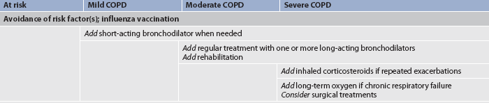
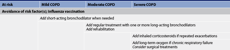
The GOLD is the main evidence base and, together with systematic reviews and meta-analyses from the Cochrane Database, is used by the Thoracic Society of Australia and New Zealand and the Australian Lung Foundation to inform the development of guidelines for the management of COPD. The GOLD report lists a summary of recommended treatment at each stage (see Table 28-12).3 The Australian and New Zealand guidelines place the emphasis on the role of non-pharmacological interventions and self-management and are titled COPD-X after the key components: Confirm diagnosis, Optimise function, Prevent deterioration, Develop a self-management plan and manage eXacerbations.1 The primary goals of the guidelines are to:
• effect changes in clinical practice based on sound evidence
• shift the emphasis from a predominant reliance on pharmacological treatment of COPD to a range of interventions that include patient education, self-management of exacerbations and pulmonary rehabilitation.
TABLE 28-12 Therapy at each stage of COPD
Source: Adapted from global Initiative for Chronic Obstructive lung Disease. Global strategy for diagnosis, management, and prevention of COPD (p 54). Available at www.goldcopd.org/guidelineitem.asp?l1=2&l2=1&intId=989, accessed 22 January 2011.
Despite the prevalence and serious consequences of COPD, much can be done to improve quality of life and exercise capacity and reduce morbidity and mortality for people with COPD. The majority of people with COPD are treated as outpatients and are hospitalised only for acute exacerbations and complications such as respiratory failure, pneumonia and chronic heart failure.
Environmental or occupational irritants should be evaluated for their possible negative effect and ways to control or avoid them should be determined. For example, aerosol hairsprays and smoke-filled rooms should be avoided. The patient with COPD should have an influenza vaccination annually in the autumn and a pneumococcal vaccination is recommended for patients with a reduced respiratory reserve.1
Exacerbations of COPD should be treated as soon as possible, especially if the patient is in the severe stages of COPD. Often the best indication of the presence of a respiratory infection is the increasing quantity, viscosity or purulence of sputum. Some patients are given a 7–10-day supply of antibiotics and are instructed to begin taking them at the first signs of a change in sputum. The most common antibiotics given for outpatients are macrolides (e.g. azithromycin), doxycycline and cephalosporins (e.g. cefpodoxime). If the patient has failed prior antibiotic therapy or is hospitalised, common antibiotics are amoxicillin/clavulanate (Augmentin) or respiratory fluoroquinolone.3,42
Source: Global Initiative for Chronic Obstructive Lung Disease. Global strategy for the diagnosis, management and prevention of chronic obstructive pulmonary disease. Available at www.goldcopd.com, accessed 22 January 2011.
EVIDENCE-BASED PRACTICE
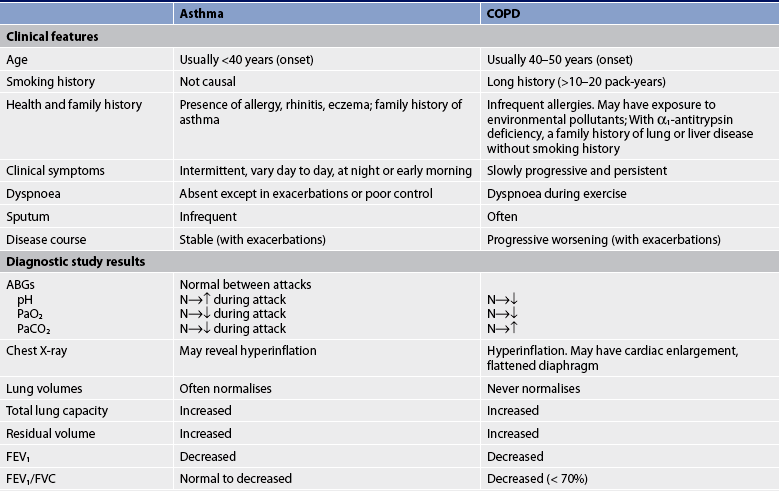
Cessation of cigarette smoking in the early stages is the most significant factor in slowing the progression of the disease. After discontinuation of smoking, the accelerated decline in pulmonary function slows and pulmonary function usually improves. Normally individuals after the age of 35 years lose approximately 20–25 mL (as measured by FEV1) of lung function per year as measured by spirometry. Persons with COPD who continue to smoke lose approximately 50 mL per year. With the cessation of smoking the loss can fall to almost non-smoking levels at 35 mL per year. Thus the sooner the smoker stops, the less pulmonary function is lost and the sooner the symptoms decrease, particularly cough and sputum production. (Smoking cessation techniques are discussed in Ch 10.)
Medications for COPD can reduce or abolish symptoms, increase the capacity to exercise, improve overall health and reduce the number and severity of exacerbations. Presently no drug modifies the decline of lung function with COPD. Bronchodilator drug therapy decreases airway resistance and dynamic hyperinflation of the lungs, which results in reducing the degree of breathlessness.1 Although patients with COPD do not respond to bronchodilator therapy as dramatically as those with asthma, a reduction in dyspnoea and an increase in FEV1 are usually achieved. The inhaled route of medication is preferred and given on an as-required or regular basis. Medications are given in a stepwise fashion, stepping up but usually not stepping down as in asthma, because in COPD there are probably continual symptoms (see Table 28-12).
Reducing risk factors for COPD
Source: Global Initiative for Chronic Obstructive Lung Disease. Global strategy for the diagnosis, management, and prevention of chronic obstructive pulmonary disease. Available at www.goldcopd.com, accessed 22 January 2011.
EVIDENCE-BASED PRACTICE
Bronchodilator medications commonly used are β2-adrenergic agonists, anticholinergic agents and methylxanthines (see Table 28-4). The choice of bronchodilator depends on the availability and the patient’s response. However, when the patient has mild COPD or intermittent symptoms, a short-acting bronchodilator is used as needed. Short-acting bronchodilators increase exercise tolerance. Salbutamol or ipratropium may be used as a single agent, but combining bronchodilators improves their effect and decreases the risk of adverse effects, compared to the use of a single agent. These two agents can be nebulised together or delivered by one MDI.
As symptoms persist or moderate stages of COPD develop, a long-acting bronchodilator is used in addition to a short-acting bronchodilator (see Table 28-4). Salmeterol is a widely used long-acting β2-adrenergic agonist and, unlike in drug therapy for asthma, it can be used in COPD as monotherapy. Formoterol is another long-acting β2-agonist. Tiotropium, a long-acting anticholinergic, can be used for daily therapy of bronchospasm and dyspnoea in COPD. Tiotropium also improves bronchodilation, results in less dyspnoea, improves quality of life and decreases the number of COPD exacerbations when compared to ipratropium.47
The use of long-acting theophylline in the treatment of COPD is controversial as it interacts with many drugs. Although it has some action as a mild bronchodilator in the patient with partial reversibility of airflow obstruction, its main value may be to improve contractility of the diaphragm and decrease diaphragmatic fatigue.
Inhaled corticosteroid therapy may be beneficial for moderate-to-severe COPD (stage III or IV). However, it does not appear to help patients with mild COPD. Inhaled corticosteroid therapy combined with long-acting β2-adrenergic agonists (e.g. fluticasone/salmeterol) is more effective than the single drug therapy. Some patients are on triple therapy with salmeterol/fluticasone (Advair) and tiotropium (Spiriva).48 Oral corticosteroids should not be used for long-term therapy in COPD but are effective in the short term for exacerbations.
Oxygen (O2) therapy is frequently used in the treatment of COPD and other problems associated with hypoxaemia. Long-term O2 therapy improves survival, exercise capacity, cognitive performance and sleep in hypoxaemic patients.3 O2 is a colourless, odourless, tasteless gas that constitutes 20.95% of the atmosphere. Administering supplemental O2 raises the partial pressure of O2 (PO2) in inspired air. Used clinically it is considered a drug but, for reimbursement purposes, it is considered durable medical equipment.
Goals for O2 therapy are to reduce the work of breathing, maintain the PaO2 and/or reduce the workload on the heart, keeping the SaO2 >90% during rest, sleep and exertion or PaO2 >8 kPA (60 mmHg). O2 is usually administered to treat hypoxaemia caused by: (1) respiratory disorders such as COPD, cor pulmonale, pneumonia, atelectasis, lung cancer and pulmonary emboli; (2) cardiovascular disorders such as myocardial infarction, arrhythmias, angina pectoris and cardiogenic shock; and (3) central nervous system disorders such as overdose of opioids, head injury and disordered sleep (sleep apnoea). In COPD, level A evidence supports the use of long-term O2 therapy for patients who have SaO2 levels of 88% or less on air.1 There is limited evidence for the effectiveness of short-term use and since it is expensive, it must be prescribed with caution. In Australia and New Zealand, O2 therapy for COPD receives government funding as long as a respiratory physician is the prescriber.49
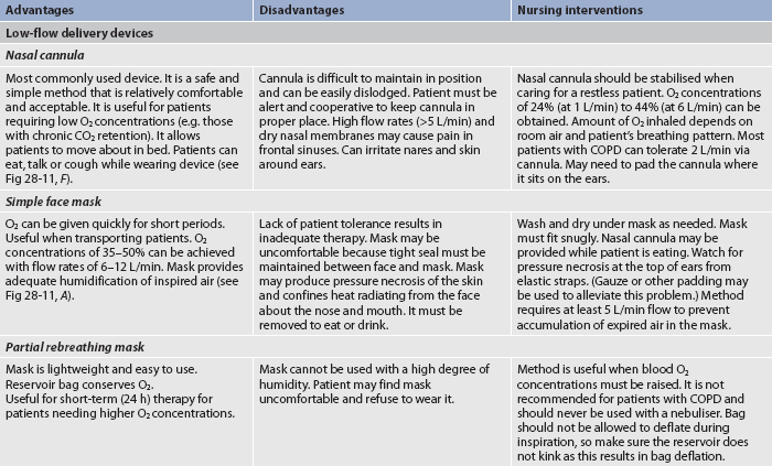
O2 administration aims to supply the patient with adequate O2 to maximise the O2-carrying ability of the blood. There are various methods of O2 administration (see Table 28-13 and Figs 28-11 to 28-14). The method selected depends on factors such as the fraction of inspired O2 (FiO2) and the mobility of the patient, the humidification required, patient cooperation, comfort, cost and available financial resources.
TABLE 28-13 Methods of oxygen administration
ABGs, arterial blood gases; COPD, chronic obstructive pulmonary disease.
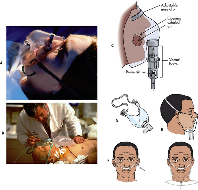
Figure 28-11 Methods of oxygen administration. A, Simple face mask. B, Plastic face mask with reservoir bag. C, Venturi mask. D, Tracheostomy mask. E, Face tent. F, Standard nasal cannulae.
O2 delivery systems are classified as low- or high-flow systems. Most methods of O2 administration use low-flow devices that deliver O2 in concentrations that vary with the person’s respiratory pattern. In contrast, the Venturi mask is a high-flow device that delivers fixed concentrations of O2 independent of the patient’s respiratory pattern. With the Venturi mask, O2 is delivered to a small jet (Venturi device) in the centre of a wide-based cone (see Fig 28-11, C). Air is entrained (pulled through) openings in the cone as O2 flows through the small jet. The mask has large vents through which exhaled air can escape. The degree of restriction or the narrowness of the jet determines the amount of entrainment and dilution of pure O2 with room air and thus the concentration of O2. Nasal cannulae can be used short term. Mechanical ventilators are another example of a high-flow O2 delivery system. Because room air is mixed with O2 in low-flow systems, the percentage of O2 delivered to the patient is not as precise as with high-flow systems.
O2 obtained from cylinders or wall systems is dry. Dry O2 has an irritating effect on mucous membranes and dries secretions. Therefore it is important that O2 be humidified when administered, either by humidification or nebulisation when flow rates are higher than 4 L/min. A common device used for humidification when the patient has a catheter, cannula or low-flow mask is a bubble-through humidifier. This is a small plastic jar filled with sterile distilled water that is attached to the O2 source by means of a flow meter. O2 passes into the jar, bubbles through the water and then goes through tubing to the patient’s catheter, cannula or mask. The purpose of the bubble-through humidifier is to restore the humidity conditions of room air. However, the need for bubble-through humidifiers at flow rates between 1 and 4 L/min is controversial when humidity in the environment is adequate. The amount of flow at this level depends on patient comfort.
Another means of administering humidified O2 is via a nebuliser. It delivers particulate water mist (aerosols) with nearly 100% humidity. The humidity can be raised by heating the water, which increases the ability of the gas to hold moisture. Heated (37°C) and humidified (100%) gas is required when the upper airway is bypassed in acute care. However, patients with established tracheotomies do not always require 100% humidity. When nebulisers are used, large-size tubing should be employed to connect the device to a face mask or T-bar. If small-size tubing is used, condensation can occlude the flow of O2.
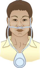
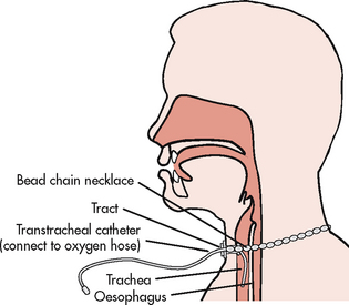
Vapotherm can deliver high flows (15–20 L/min) of warm humidified air (either sterile air or O2) to the patient through a nasal or transtracheal cannula using technology to warm and saturate the gas stream. Preliminary findings in COPD patients in pulmonary rehabilitation suggest increases in exercise tolerance with Vapotherm high-flow therapy.35
O2 supports combustion and increases the rate of burning. This is why it is important that smoking be prohibited in the area in which O2 is being used. A ‘no smoking’ sign should be prominently displayed on the patient’s door. The patient should also be cautioned against smoking cigarettes when O2 prongs or cannulae are in place.
The two chemoreceptors in the respiratory centre that control the drive to breathe are CO2 and O2. Normally, CO2 accumulation is the major stimulant of the respiratory centre. Over time COPD patients develop a tolerance for high CO2 levels (the respiratory centre loses its sensitivity to the elevated CO2 levels). Theoretically, for these individuals the ‘O2 drive’ to breathe is hypoxaemia. Thus there has been concern regarding the dangers of administering O2 to COPD patients and wiping out their drive to breathe. This has been a pervasive myth but is not regarded as a serious threat. In fact, not providing adequate O2 to these patients is much more detrimental. Although O2 administration should be titrated to the lowest effective dose, many patients who have end-stage chronic obstructive disease require high-flow rates and higher concentrations for survival. They may, in fact, exhibit higher than normal levels of CO2 in their blood, but this is of little concern. What is important is careful, ongoing assessment when providing O2 to these patients.
It is critical to start O2 at low-flow rates until ABGs can be obtained. ABGs are used as a guide to determine what FiO2 level is sufficient and can be tolerated. The patient’s mental status and vital signs should be assessed before starting O2 therapy and frequently thereafter.
Pulmonary oxygen toxicity may result from prolonged exposure to high levels of O2 (PaO2). The development of O2 toxicity is determined by patient tolerance, exposure time and effective dose. High concentrations of O2 damage alveolar–capillary membranes, inactivate pulmonary surfactant, cause interstitial and alveolar oedema and decrease compliance. These individuals develop acute respiratory distress syndrome (ARDS; see Ch 67).3
Early manifestations of O2 toxicity are reduced vital capacity, cough, substernal chest pain, nausea and vomiting, paraesthesia, nasal stuffiness, sore throat and malaise. The later stages of O2 toxicity affect the alveolar–capillary gas exchange unit, causing oedema and production of copious sputum. The end stage of O2 toxicity is progressive fibrosis of the lungs. Prevention of O2 toxicity is important for the patient who is receiving O2. The amount of O2 administered should be just enough to maintain the PaO2 within a normal or acceptable range for the patient. ABGs should be monitored frequently to evaluate the effectiveness of therapy and to guide the tapering of supplemental O2. A safe limit of O2 concentrations has not yet been established. All levels above 50% and used for longer than 24 hours should be considered potentially toxic. Levels of 40% and below may be regarded as relatively non-toxic and may not result in the development of significant O2 toxicity if the exposure period is short.
Normally nitrogen, which constitutes 79% of the air that is breathed, is not absorbed into the bloodstream. This prevents alveolar collapse. When high concentrations of O2 are given, nitrogen is washed out of the alveoli and replaced with O2. If airway obstruction occurs, the O2 is absorbed into the bloodstream and the alveoli collapse. This process is called absorption atelectasis.
Infection can be a major hazard of O2 administration. Heated nebulisers present the highest risk. The constant use of humidity supports bacterial growth, with the most common infecting organism being Pseudomonas aeruginosa. Disposable equipment that operates as a closed system should be used. Each hospital has a policy stating the required frequency of equipment changes based on the type of equipment used at that particular institution.
Improved prognosis and enhanced quality of life have been noted in patients with COPD who receive long-term O2 therapy to treat hypoxaemia.1,49 The improved prognosis results from preventing progression of the disease and subsequent cor pulmonale. The benefits of long-term continuous O2 therapy include improved neuropsychological function, increased exercise tolerance, decreased haematocrit and reduced pulmonary hypertension. It also improves sleep, may reduce nocturnal arrhythmias and may extend life.35
Some patients believe they will become ‘addicted’ to O2 and are very reluctant to use it. They need to be educated that it is not ‘addictive’ and that it needs to be used for the prescribed times during the day because of the positive effects on the heart, lungs and brain. The need for long-term O2 therapy should be evaluated when the patient’s condition has stabilised. The goal of O2 therapy is to maintain SaO2 >90% during rest, sleep and exertion.
Short-term home O2 therapy (1–30 days) may be indicated for the patient in whom hypoxaemia persists after discharge from the hospital. For example, the patient with underlying COPD who develops a serious respiratory tract infection may continue to have clearing of the infection after completion of antibiotic therapy and discharge from the hospital. This patient may demonstrate continued hypoxaemia for 4–6 weeks after discharge. It is important to measure the patient’s oxygenation status by pulse oximetry 2–3 months after an acute episode to determine if the O2 is still warranted.
Desaturation only during exercise or sleep suggests consideration of O2 therapy specifically under those conditions. Patients may receive O2 only during exercise or sleep, or at both times. The need for O2 during these periods should be evaluated with oximetry. (Pulse oximetry is discussed in Ch 25.) Sleep disordered breathing may be seen in some of these patients and they will require a full sleep study.
Periodic re-evaluations are necessary for the patient who is using chronic supplemental O2. The recommendation is generally that the patient should be re-evaluated 1–2 months after commencing O2 therapy and then annually or more frequently depending on the clinical condition.49
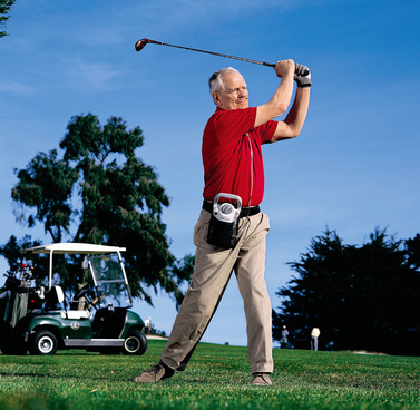
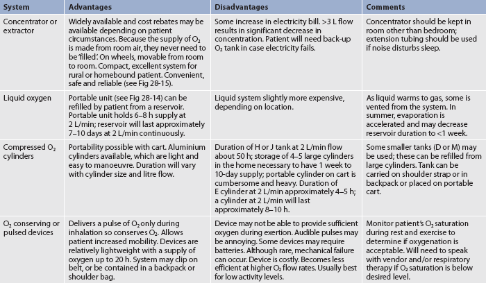
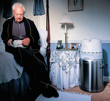
Nasal cannulae, either regular or the O2-conserving type (see Table 28-13 and Figs 28-11 and 28-14), are usually used to deliver O2 from a central source in the home. The source may be a liquid O2 storage system, compressed O2 in tanks or an O2 concentrator or extractor, depending on the patient’s home environment and activity level (see Table 28-14). The concentrator is the least expensive option. The patient can use extension tubing (up to 15 m) without adversely affecting the O2 flow delivery to increase mobility in the home, provided that the flow meter is the back pressure-compensated type. Small portable systems, such as liquid O2 or, more commonly, an O2 concentrator, may be provided for the patient who remains active outside the home (see Fig 28-15).
Reservoir cannulae operate on the principle of storing O2 in a small reservoir during exhalation. The O2 is then delivered to the patient during the subsequent inhalation, similar to a bolus effect. The reservoir cannulae can reduce flow requirements by approximately 50%. Another type that fits onto the frame of spectacles is also available and is less visible on the face.
It is important that patients who require home O2 systems are taught how to use the system, how to care for it and how to recognise when the supply is running low and needs to be reordered. A patient and family teaching guide for the use of O2 at home is presented in Box 28-8. Care must also be taken to ensure that the patient or carer notifies their electricity provider if the system is dependent on electricity to operate.
PATIENT & FAMILY TEACHING GUIDE
• Post ‘no smoking’ warning signs in home where they can be seen
• Do not use electric razors, portable radios, open flames, wool blankets or mineral oils in the area where oxygen is in use
A good resource is: The Australian Lung Foundation. Home oxygen treatment. Available at www.lungfoundation.com.au/lung-information/patient-educational-material/getting-started-on-home-oxygen, accessed 22 January 2011.
The patient who uses home O2 should be encouraged to remain active and travel normally. If travel is by car, arrangements can be made for O2 to be available at the destination point. O2 supply companies can often assist in these arrangements. If a patient wishes to travel by bus, train or aeroplane, the patient should inform the appropriate people when reservations are made that O2 will be needed for travel. If there is a potential for the patient to become hypoxic, oxygen needs for flying can be determined via a hypoxia inhalation test or through a mathematical formula. Portable oxygen concentrators are a ready source of renewable O2 and can be available by recharging at home or in a DC (e.g. car) power supply. These systems are widely approved by airlines for in-flight use. The patient should contact the specific airline to determine the particular accommodations and policies for in-flight O2.
Three different surgical procedures have been used in severe COPD, although none provides any surgical advantage.1
Lung volume reduction surgery (LVRS) reduces lung volume and improves lung and chest wall mechanics.50 The goal is to reduce the size of the hyperinflated emphysematous lungs by removing much of the diseased tissue. This can decrease airway obstruction and increase room for the remaining healthy lung tissue to function more effectively. There is evidence of improvements in exercise capacity among those having LVRS compared with medical therapy, but overall the procedure is associated with increased mortality and negligible gain.51
Bullectomy is used for carefully selected patients with emphysematous COPD who have large bullae (>5 cm).1 The bullae are usually resected via thoracoscope. The procedure has resulted in improved lung function and reduction in dyspnoea,3 and it has the greatest success in patients who have very large cysts that are preventing expansion of adjacent apparently normal lung.52
Lung transplantation is appropriate for some carefully selected patients with advanced COPD.53 Although single-lung transplantation is the most commonly used technique, bilateral transplantation can be performed and evidence suggests that this may be the future preferred technique due to the improved longer term outcomes. However, rejection and the effects of immunosuppressive therapy remain obstacles for these patients. (Lung transplantation is discussed in Ch 27.)
The patient with COPD develops an increased respiratory rate with a prolonged expiration to compensate for obstruction to airflow resulting in dyspnoea. In addition, the accessory muscles of breathing in the neck and upper part of the chest are used excessively to promote chest wall movement. These muscles are not designed for long-term use and as a result the patient experiences increased fatigue. Breathing exercises can assist the patient during rest and activity (e.g. lifting, walking, stair climbing). The main types of breathing exercises are: (1) pursed-lip breathing; and (2) diaphragmatic breathing.
The purpose of using pursed-lip breathing (PLB) is to prolong exhalation and thereby prevent bronchiolar collapse and air trapping. PLB is simple and easy to teach and learn and gives the patient more control over breathing, especially during exercise and periods of dyspnoea (see Box 28-9). Patients should be taught to use ‘just enough’ positive pressure with the pursed lips, because excessive resistance may increase the work of breathing. The issue of whether and how extensively pursed-lip breathing affects dyspnoea is still questioned, but current evidence seems to support its use to improve the breathing of patients with COPD.54–56
BOX 28-9 Guidelines for pursed-lip breathing
PATIENT & CAREGIVER TEACHING GUIDE
Teach the patient how to do pursed-lip breathing using the following guidelines.
1. Use PLB before, during and after any activity causing you to be short of breath.
2. Inhale slowly and deeply through the nose.
3. Exhale slowly through pursed lips, almost as if whistling.
4. Relax your facial muscles without puffing your cheeks—like whistling—while you are exhaling.
5. Make breathing out (exhalation) three times as long as breathing in (inhalation).
6. The following activities can help you get the ‘feel’ of PLB:
7. Practice 8–10 repetitions of PLB three or four times a day.
Source: Adapted from Guidelines for the diagnosis and management of asthma. Expert Panel Report 3. National Asthma Education and Prevention Program, National Heart, Lung, and Blood Institute; 2007.
Diaphragmatic (abdominal) breathing focuses on using the diaphragm instead of the accessory muscles of the chest to: (1) achieve maximum inhalation; and (2) slow the respiratory rate. However, there is some controversy regarding its effectiveness for COPD patients.57 For patients with severe COPD, diaphragmatic breathing may result in hyperinflation because of increased fatigue and dyspnoea and abdominal paradoxical breathing (the inward movement of the abdomen and the outward movement of the upper chest during inspiration) rather than with normal chest wall motion (the outward movement of the abdomen and upper chest simultaneously during inspiration).
Pursed-lip breathing slows the respiratory rate and is much easier to learn than diaphragmatic breathing. In the setting of extreme acute dyspnoea when the patient is hospitalised for infection or heart failure, it is important to focus on helping the patient slow the respiratory rate by using the principles of pursed-lip breathing.
Many patients with COPD and conditions such as cystic fibrosis and bronchiectasis retain secretions and require help to adequately clear their airways. Airway clearance techniques (ACTs) loosen mucus and secretions so that they can be cleared by coughing. A variety of techniques can be used to achieve airway clearance. No one technique is really better than another and it is largely a matter of patient preference.58,59 Respiratory therapists (RTs), physiotherapists and nurses are involved in performing these techniques. ACTs are often used with other treatments. Typically, the patient will receive bronchodilator therapy via an inhaled device (e.g. nebulisation) before ACT, then an ACT, followed by effective coughing (e.g. huff coughing).
Many patients with COPD have developed ineffective coughing patterns that do not adequately clear their airways of sputum. In addition, they fear they may develop spastic coughing, resulting in increased dyspnoea. Guidelines for effective coughing are presented in Box 28-10. Huff coughing is an effective technique that the patient can be taught easily. The main goals of effective coughing are to conserve energy, reduce fatigue and facilitate removal of secretions. This technique clears secretions with less change in pleural pressure and less likelihood of bronchial collapse. Before coughing the nurse should ensure that the patient is breathing deeply from the diaphragm. The nurse should place the patient’s hands on the lower, lateral chest wall and then ask the patient to breathe deeply through the nose; the nurse should feel the patient’s hands move outwards, which represents a breath from the diaphragm.
BOX 28-10 Guidelines for effective coughing
PATIENT & FAMILY TEACHING GUIDE
1. Patient assumes a sitting position with head slightly flexed, shoulders relaxed, knees flexed and forearms supported by pillow and, if possible, with feet on the floor.
2. Patient then drops head and bends forwards while using slow, pursed-lip breathing to exhale.
3. Sitting up again, patient uses diaphragmatic breathing to inhale slowly and deeply.
4. Patient repeats steps 2 and 3 three to four times to facilitate mobilisation of secretions.
5. Before initiating a cough, patient should take a deep abdominal breath, bend slightly forwards and then huff cough (cough three to four times on exhalation). Patient may need to support or splint thorax or abdomen to achieve a maximum cough.
Chest physiotherapy is indicated for the patient with excessive bronchial secretions who has difficulty clearing them (e.g. because of cystic fibrosis, bronchiectasis). It consists of postural drainage, percussion and vibration (see Box 28-11). Postural drainage uses the principle of gravity to assist in bronchial drainage. Percussion and vibration are manual or mechanical techniques used to augment postural drainage and are used after the patient has assumed a postural drainage position to assist in loosening the mobilised secretions (see Fig 28-16). Postural drainage, percussion and vibration may assist in bringing secretions into larger, more central airways. Effective coughing is then necessary to help raise these secretions.
BOX 28-11 Steps in chest physiotherapy
1. Perform procedure 1 h before meals or 1–3 h after meals.
2. Administer bronchodilator (if nebulised or metered dose inhaler is ordered) approximately 15 min before procedure.
3. Collect equipment such as tissues, emesis basin, paper bag and pillows.
4. Help patient to assume correct position for postural drainage based on findings from X-ray, auscultation, palpation and percussion of chest. If patient’s tolerance is limited, start with the lower lung fields. Position should be maintained for 5–15 min to mobilise secretions via gravity.
5. Observe patient during treatment to assess tolerance. Particularly observe breathing and colour changes, especially duskiness in face.
6. Have patient take several deep abdominal breaths.
7. Percuss appropriate area for 1–2 min keeping the patient’s face in full view.
8. Vibrate the same area while the patient exhales four to five deep breaths.*
9. Assist patient to cough while assuming same position. Splinting with towel or hands may be necessary to aid in effective coughing. Patient may have to assume sitting position to generate enough airflow to expel secretions. (Coughing productively may be a long waiting process that may occur 30 min after procedure.) Suction may be necessary if coughing is not effective.
10. Repeat percussion, vibration and coughing until patient no longer expectorates mucus.
11. Repeat same procedure in all necessary positions.
12. After procedure, help patient to assume a comfortable position, assist with oral hygiene and discard used tissues.
13. Monitor for hypoxaemia if patient has any respiratory difficulty during the procedure.
14. Evaluate and chart effectiveness of treatment by amount of sputum produced and the results of auscultation. Also chart patient tolerance.
*If using an electronic vibrator, use for periods of 5–20 min in each position according to the patient’s tolerance.
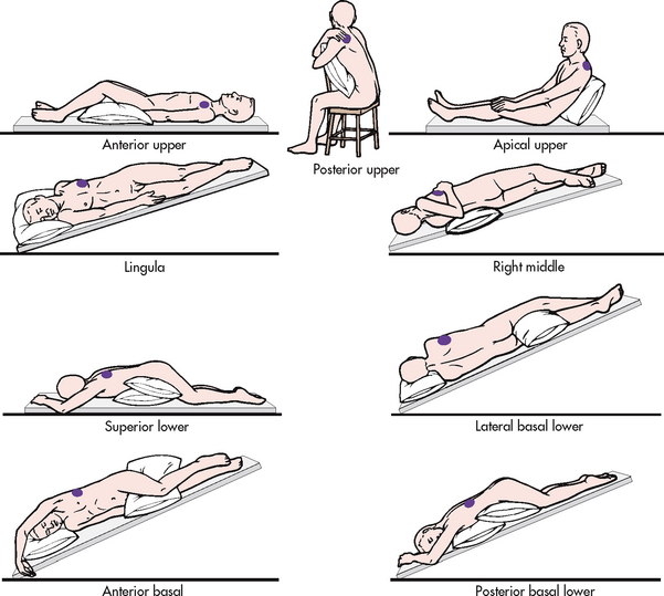
Figure 28-16 Representative positions for postural drainage. Shaded areas in each drawing indicate the segment of the lung in which drainage is promoted.
Chest physiotherapy should be performed by an individual who has been properly trained. Contraindications for chest physiotherapy include situations in which there is head, neck, chest or back instability and/or injuries; anatomical deformities; severe spasticity; mental limitations; or the patient cannot tolerate the position for other reasons. Complications associated with improperly performed chest physiotherapy include fractured ribs, bruising, hypoxaemia and discomfort to the patient. Chest physiotherapy may not be beneficial and may be stressful for some patients.
Postural drainage involves the use of positioning techniques that drain secretions from specific segments of the lungs and bronchi into the trachea. The lungs are divided into five lobes, with three on the right side and two on the left side. There are 18 segments in the lungs, which can be drained by 18 positions. Figure 28-16 shows the modified postural drainage positions most often used in clinical practice.
The purpose of various positions in postural drainage is to drain each segment towards the larger airways. The postural drainage positions used depend on the areas of involved lung, determined by patient assessment (including the patient’s preference), chest X-rays and chest auscultation. For example, some patients with left lower lobe involvement will require postural drainage of only the affected region, whereas patients with cystic fibrosis may require postural drainage of all segments.
Aerosolised bronchodilators and hydration therapy are frequently administered before postural drainage. The chosen postural drainage position is maintained for 5–15 minutes. The degree of slope can be obtained with pillows, blocks, books or a tilt board. The frequency and choice of postural drainage positions depend on the location of retained secretions and patient tolerance to dependent positions. A common order is two to four times a day. In acute situations, postural drainage may be performed as frequently as every 1–2 hours. The procedure should be planned to occur and be completed at least 1 hour before meals or 3 hours after meals.
There are beds available that can rotate and percuss in various postural drainage positions, and these are quite effective. Some positions for postural drainage (e.g. Trendelenburg) should not be performed on the patient with chest trauma, haemoptysis, heart disease or head injury and in other situations where the patient’s condition is not stable.
Percussion is performed in the appropriate postural drainage position with the hands in a cup-like position (see Fig 28-17). The hands are cupped and the fingers and thumbs are closed. The cupped hand should create an air pocket between the patient’s chest and the hand. Both hands are cupped and used in an alternating rhythmic fashion. Percussion is accomplished with flexion and extension of the wrists. If it is performed correctly, a hollow sound should be heard. The air-cushion impact facilitates the movement of thick mucus. A thin towel should be placed over the area to be percussed or the patient may choose to wear a T-shirt or hospital gown.
Vibration is accomplished by tensing the hand and arm muscles repeatedly and pressing mildly with the flat of the hand on the affected area while the patient slowly exhales a deep breath. The vibrations facilitate movement of secretions to larger airways. Mild vibration is tolerated better than percussion and can be used in situations where percussion may be contraindicated. Commercial vibrators are available for hospital and home use.
Various airway clearance devices are available to mobilise secretions. They are easier to tolerate than chest physiotherapy and take less than half the time of conventional chest physiotherapy sessions.40,41 These devices include the Flutter mucus clearance device, the TheraPEP therapy system, the ThAIRaphy vest and Acapella.
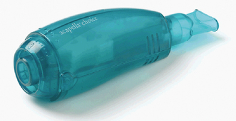
The Flutter mucus clearance device is a hand-held device that provides positive expiratory pressure (PEP) treatment for patients with mucus-producing conditions (see Fig 28-18). The Flutter has a mouthpiece, a high-density stainless steel ball and a cone that holds the ball. When the patient exhales through the Flutter, the steel ball moves, which causes vibrations in the lungs and loosens mucus. It helps move mucus up through the airways to the mouth where the mucus can be expectorated. Although the Flutter valve is mostly used in patients with cystic fibrosis, it has been used effectively in patients with COPD and bronchiectasis who have excessive secretions.
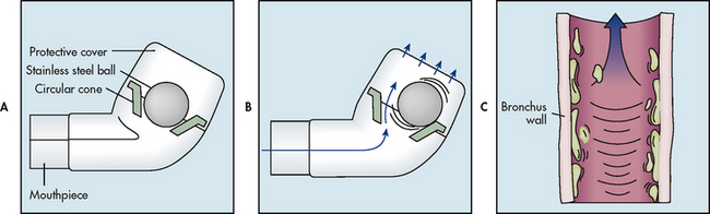
Figure 28-18 The Flutter mucus clearance device is a small hand-held device that provides positive expiratory pressure therapy. It is used to facilitate the removal of mucus from the lungs. A, It consists of a hard plastic mouthpiece, a plastic perforated cover and a high-density stainless steel ball resting in a circular cone. B, The Flutter effect occurs during expiration. Before exhalation, the steel ball blocks the conical canal of the Flutter. During exhalation, the position of the steel ball is the result of an equilibrium between the pressure of the exhaled air, the force of gravity on the ball and the angle of the cone where the contact with the ball occurs. As the steel ball rolls and moves up and down, it creates an opening and closing cycle, which repeats itself many times throughout each exhalation. The net result is that vibrations occur in the airways, resulting in the ‘fluttering’ sensation. C, These vibrations loosen mucus from the airway walls and facilitate their movement up the airways.
The TheraPEP therapy system also provides sustained PEP and simultaneously delivers aerosols so the patient can inhale and exhale through it. The device comprises a mouthpiece attached to tubing connected to a small cylindrical resistor and a pressure indicator. The pressure indicator provides visual reinforcement about the pressure the patient needs to hold in an exhalation to receive the PEP. The device is available in Australia and New Zealand and a description and photo are available on the TheraPEP therapy system website (see Resources on pp 727–728).
The ThAIRaphy vest delivers high-frequency chest compression via an inflatable vest with hoses connected to a high-frequency pulse generator. The pulse generator delivers air to the vest, which vibrates the chest. The high-frequency airwaves clear all lobes of the lungs. The vest has been found to be more effective than conventional chest physiotherapy in clearing mucus and it can be used without the aid of another person. The unit is light, quiet and portable.
Acapella is a small hand-held device that combines the benefits of PEP therapy and airway vibrations of the Flutter valve to mobilise pulmonary secretions (see Fig 28-19). It works by using oscillating vibrations that travel to the lung, shaking free mucus plugs that the patient can then cough up. It can be used in virtually any setting as the patient is free to sit, stand or recline. It improves clearance of secretions, is easier to tolerate and quicker than conventional chest physiotherapy and facilitates opening of the airways.
Approximately one-third of COPD patients are underweight, experiencing loss of muscle mass and cachexia, especially in the severe stages of the disease.60 Weight loss is a predictor of poor prognosis and increased frequency of COPD exacerbations.61 Weight gain after nutritional support can decrease the mortality risk. The cause of weight loss is not entirely known. Eating becomes an effort because of dyspnoea, which occurs as a result of the energy expended to chew, the reduction of airflow while swallowing and O2 desaturation. Therefore weight loss and muscle wasting are likely. Weight loss is also thought to occur because systemic inflammation causes the metabolism to increase. This hypermetabolism could explain why some individuals with COPD lose weight despite having an adequate nutritional intake.
Patients with COPD should try to keep their BMI between 21 and 25 kg/m2. Being either overweight or underweight can be a problem with COPD. The dietician can serve as a good resource to determine what supplements are the best for patients.
To decrease dyspnoea and conserve energy, patients should rest for at least 30 minutes before eating, use the bronchodilator before meals and select foods that can be prepared in advance. They should eat five to six small, frequent meals to avoid feelings of bloating and early satiety. A full stomach puts pressure on the diaphragm and decreases lung movement. Liquid, blended or commercial diets may be helpful. Foods that require a great deal of chewing should be avoided or served in another manner (e.g. grated, pureed). Cold foods may give less of a sense of fullness than hot foods. Exercises and treatments should be avoided for at least 1 hour before and after eating. The exertion involved in the preparation and eating of food is often fatiguing. The use of frozen foods and a microwave oven may help conserve patient energy in food preparation.
Many patients with COPD feel bloated and experience early satiety when eating. This can be attributed to swallowing air while eating, the side effects of medications (especially corticosteroids and theophylline) and the abnormal position of the diaphragm relative to the stomach in association with hyperinflation. Intestinal gas-forming foods, such as cabbage, brussel sprouts and beans, should be avoided.
Underweight patients with emphysematous COPD have a greater than normal nutritional requirement for protein and kilojoules. A high-energy, high-protein diet is recommended and can be divided into five or six small meals a day. High-protein, high-energy nutritional supplements may be offered between meals. Ice-cream added to these supplements can help increase kilojoules. Drinking low-fat milk rather than whole milk may cause less mucus production. (Nutritional supplements are discussed in Ch 39.) Non-protein energy should be divided evenly between fat and carbohydrate while not overfeeding the patient.62 In most cases just getting the patient to eat adequate amounts of any foods can be difficult. If the patient has O2 prescribed, use of supplemental O2 by nasal prongs (cannulae) while eating may also be beneficial because eating expends energy. Fluid intake should be at least 3 L per day unless contraindicated for other medical conditions, such as heart failure. Fluids should be taken between meals (rather than with them) to prevent excess stomach distension and to decrease pressure on the diaphragm. Sodium restriction may be indicated if there is accompanying heart failure.
In contrast, some patients with COPD are obese, which also causes dyspnoea. These patients may have increased appetite if on oral corticosteroids and may have little mobility. Advice about controlling food portions and increasing their exercise may be needed.
Subjective and objective data that should be obtained from a person with COPD are presented in Table 28-15.
The nursing diagnoses for the patient with COPD may include, but are not limited to, those presented in NCP 28-2.
The overall goals are that the patient with COPD will have: (1) return of baseline respiratory function; (2) ability to perform ADLs; (3) relief from dyspnoea; (4) no complications related to COPD; (5) knowledge and ability to implement a long-term treatment regimen; and (6) overall improved quality of life.
The incidence of COPD would decrease if more people did not begin smoking or stopped smoking. (Techniques to help patients stop smoking are discussed in Ch 10.) Avoiding or controlling exposure to occupational and environmental pollutants and irritants is another preventive measure to maintain healthy lungs. (These factors are discussed in the section on nursing management of lung cancer in Ch 27.)
Early detection of small-airway disease is important. The person who has smoked for only a few years may have early evidence of obstructive airway disease on spirometry testing. However, using spirometry to screen the general population to detect COPD is not recommended as having a favourable benefit/harm ratio. The changes often cannot be detected from pulmonary function studies until extensive damage is present. It is extremely important for the person to stop smoking and to avoid inhaling irritants while the disease is still reversible. Failure to follow this advice will inevitably lead to irreversible COPD. Unfortunately confronting a person who smokes with abnormal spirometry findings does not always motivate them to stop smoking.63 Nurses who smoke should re-evaluate their own smoking behaviour and its relationship to their health. Nurses, healthcare providers and physiotherapists who smoke and smell of cigarette smoke should be aware that the odour of their clothes can be offensive to patients.
Early diagnosis and treatment of respiratory tract infections are other ways to decrease the incidence of COPD. Avoiding exposure to large crowds in the peak periods for influenza may be necessary, especially for older adults and those with a history of respiratory problems. Influenza and pneumococcal pneumonia vaccines are recommended for patients with COPD.
Families with a history of COPD, as well as AAT deficiency, should be aware of the genetic nature of the disease and family members should have spirometry screening regularly during their adult life even if they do not have symptoms.63 Genetic counselling is appropriate for the patient with AAT deficiency who is planning to have children.
Patients with COPD require acute intervention for complications such as pneumonia, cor pulmonale and acute respiratory failure. (The nursing care for these conditions is discussed in Chs 27 and 67.) Once the crisis in these situations has been resolved, the nurse can assess the degree and severity of the underlying respiratory problem. The information obtained will help plan the nursing care.
By far the most important aspect in the long-term care of patients with COPD is teaching (see Table 28-16). With the rising cost of healthcare, caring for patients in communities and patients’ homes is becoming commonplace.
The widely accepted definition of pulmonary rehabilitation is an evidence-based intervention that includes many disciplines working together to individualise treatment of the patient with chronic respiratory disease who has symptoms and decreased quality of life. Pulmonary rehabilitation should be considered for all patients with symptomatic COPD.1 Pulmonary rehabilitation is one of the most effective interventions in COPD and can reduce symptoms and disability to improve functioning by: (1) improving cardiovascular fitness, muscle function and exercise endurance; (2) enhancing self-confidence and coping strategies and improving medication adherence and use of treatment devices; and (3) controlling mood by controlling anxiety and panic, decreasing depression and reducing social barriers.64 Significant research has validated the benefits of rehabilitation in patients with mild to severe COPD, which include increased exercise tolerance and reduced dyspnoea and fatigue.61 The overall goal of pulmonary rehabilitation is to increase the quality of life.
Pulmonary rehabilitation can be done in an inpatient or outpatient setting, or in the home setting. A mandatory component of any pulmonary rehabilitation program is exercise that focuses on the muscles used in ambulation.65 Ideally, pulmonary rehabilitation should include exercise training, nutrition counselling and education. Other important topics include health promotion, psychological counselling and vocational rehabilitation. Smoking cessation is critical to success: some rehabilitation programs will not accept patients who are current smokers and are not committed to quitting. Physiotherapists or nurses who have experience in pulmonary care are often responsible for the management of pulmonary rehabilitation programs. A large part of the nursing role is to teach patients self-management of their disease. The minimum length of an effective program is 6 weeks, but the longer the program, the more effective the results.
Nutritional counselling is integral to a pulmonary rehabilitation program and this has been discussed previously in the chapter. Education is also an important component and it should include information on self-management and prevention and treatment of exacerbations (see Table 28-16).
Energy conservation is another important component in COPD rehabilitation. Patients are typically upper thoracic and neck breathers who use accessory muscles rather than the diaphragm and thus have difficulty performing upper-extremity activities, particularly those activities that require arm elevation above the head. Exercise training of the upper extremities may improve function and reduce dyspnoea. Frequently patients have already adapted alternative energy-saving practices for ADLs. Alternative methods of hair care, shaving, showering and reaching may need to be explored. An occupational therapist may help with ideas in these areas. Assuming a tripod posture (elbows supported on a table, chest in fixed position) in front of a mirror placed on a table when using an electric razor or hair dryer conserves much more energy than standing in front of a wall mirror to shave or blow-dry hair. If the patient uses home O2 therapy, it is essential that the patient use the O2 during activities of hygiene because these are energy consuming. The patient should be encouraged to make a schedule and plan daily and weekly activities so as to leave plenty of time for rest periods. The patient should also try to sit as much as possible when performing activities. Another energy-saving tip is to exhale when pushing, pulling or exerting effort during an activity and inhale during rest.
Walking is by far the best physical exercise for COPD patients. Coordinated walking with slow, pursed-lip breathing without breath holding is a difficult task that requires conscious effort and frequent reinforcement. During coordinated walking and breathing, the patient is taught to breathe in through the nose while taking one step, then to breathe out through pursed lips while taking two to four steps (the number depends on the patient’s tolerance). Walking should occur at a slow pace with rest periods when necessary so the patient can sit or lean against an object such as a tree or post. The patient may need to walk using O2. Once the patient is able to perform coordinated walking with pursed-lip breathing successfully, diaphragmatic breathing may also be incorporated if the patient has practised and mastered this technique at rest. The nurse should walk with the patient, giving verbal reminders when necessary regarding breathing (inhalation and exhalation) and steps. Walking with the patient helps decrease anxiety and helps maintain a slow pace. It also enables the nurse to observe the patient’s actions and physiological responses to the activity. Many patients with moderate or severe COPD are anxious and fearful of walking or performing exercise. These patients and their families require much support while they build the confidence they need to walk or to perform daily exercises.
In some situations pulmonary rehabilitation programs are not an option, and patients are advised to exercise on their own. The patient should be encouraged to walk 15–20 minutes a day by building up gradually. Patients with severe disabilities can begin at a slow pace by walking for 2–5 minutes three times a day and slowly build up to 20 minutes a day, if possible. Adequate rest periods should be allowed. Some patients benefit from using their β2-adrenergic agonist MDI approximately 10 minutes before exercise. Parameters that may be monitored in the patient with mild COPD are resting pulse and pulse rate after walking. Pulse rate after walking should not exceed 75–80% of the maximum heart rate (maximum heart rate is age in years subtracted from 220). In the patient with other than mild COPD and without significant heart disease, it is usually dyspnoea and the limitation in breathing rather than increased heart rate that limits the exercise. Thus it is better to use the patient’s perceived sense of dyspnoea as an indication of exercise tolerance. The Borg category-ratio scale (see Fig 25-9) can be used to help the patient determine the intensity of dyspnoea.
The patient should be told that shortness of breath will probably increase during exercise (as it does for a healthy individual) but that the activity is not being overdone if this increased shortness of breath returns to baseline within 5 minutes after ceasing the exercise. The patient should also be told to wait 5 minutes after completing the exercise before using the β2-adrenergic agonist MDI to allow a chance to recover. During this time, slow, pursed-lip breathing should be used. If it takes longer than 5 minutes to return to baseline, the patient most likely has overdone it and should proceed at a slower pace during the next exercise period. The patient may benefit from keeping a diary or log of the exercise program. The diary can help provide a realistic evaluation of the patient’s progress. In addition, the diary can help motivate the patient and add to the patient’s sense of accomplishment. Stationary cycling can also be used either alone or with walking. Cycles and treadmills are particularly valuable when weather prevents walking outside.
Fatigue, sleep disturbances and dyspnoea are common complaints of patients with COPD. Of these symptoms, dyspnoea appears to be the only symptom that affects the patient’s ability to carry out daily activities. Therefore it is suggested that the nurse and other health team members should focus their interventions on improving dyspnoea, which would then improve the patient’s functional performance.66 In addition to a traditional pulmonary rehabilitation program, preliminary studies show an internet-based self-management program decreases the dyspnoea associated with ADLs.67
Modifying but not abstaining from sexual activity can also contribute to a healthy psychological wellbeing. Most patients with COPD are older. Nurses need to assess and reflect on their own attitudes and feelings about sexuality, sexual functioning and ageing before exploring sexual issues with older patients. They need to first assess the patient related to sexuality and concerns of functioning. It is important to ask open-ended questions to determine whether the patient wants to discuss any of these concerns. For example: ‘How has your breathing problem affected how you see yourself as a woman or man?’ Or ‘How does your shortness of breath affect your desire for intimacy with your partner?’ Moving through these types of questions will give the patient an opening to discuss any concerns. Many of the sexual performance issues experienced by patients with COPD are changes related to ageing and if the nurse is aware of these changes, patients can be taught the ‘normalcy’ of these changes.
Dyspnoea is the predominant symptom in COPD, but it should not be a major problem with success in sexual functioning, except for those patients in stage III or IV of the disease. Erectile dysfunction may occur, and is related to the severity of the underlying disease. Using an inhaled bronchodilator before sexual activity can help ventilation. Patients may also find these suggestions helpful: (1) plan sexual activity during the part of the day when breathing is best; (2) use slow pursed-lip breathing; (3) refrain from sexual activity after eating or other strenuous activity; (4) do not assume a dominant position; and (5) do not prolong foreplay. These aspects of sexual activity require open communication between partners regarding their needs and expectations.
Adequate sleep is extremely important, but getting adequate amounts of sleep can be difficult for patients with COPD. Medications may cause restlessness and insomnia. Many patients have a postnasal drip or nasal congestion that may cause coughing and wheezing at night. Nasal saline sprays before sleep and in the morning may help. The healthcare provider may also prescribe a nasal decongestant or nasal steroid inhaler that may be used at bedtime. Theophylline preparations frequently aid in promoting sleep by decreasing bronchospasm and airway obstruction. If the patient is a restless sleeper, snores, stops breathing while asleep and has a tendency to fall asleep during the day, the patient may need to be tested for sleep apnoea (see Ch 26).
Healthy coping is often the most difficult task for patients with COPD to accomplish. People with COPD frequently have to deal with many lifestyle changes that may involve decreased ability to care for themselves, decreased energy for social activities and loss of a job.
When a patient with COPD is first diagnosed or when a patient has complications that require hospitalisation, the nurse should expect a variety of emotional responses. Emotions frequently encountered include guilt, depression, anxiety, social isolation, denial and dependence. Guilt may result from the knowledge that the disease was caused largely by cigarette smoking. There is a lack of research concerning the effects of stopping smoking on quality of life in the late stages of the disease.68 Therefore the nurse must personally decide the advice to give patients in the terminal stages of the disease. Depression affects approximately 40% of patients as the severity and chronicity of the disease are realised. The nurse should convey a sense of understanding and caring to patients.69 Patients may benefit from stress management techniques (e.g. massage, progressive muscle relaxation). Acupressure has demonstrated some promise in decreasing dyspnoea.70 Support groups can also be helpful.
Patients frequently ask whether moving to a warmer or drier climate will help, but generally this is not greatly beneficial. Moving to places with an elevation of 1200 m or more above sea level should be discouraged because of the lower partial pressure of O2 found in the air at such elevations. A disadvantage with moving is that the person leaves an occupation, friends and familiar environment, which could be psychologically stressful. Any advantage gained from a different climate may be outweighed by the psychological effects of the move.
Patients need to know that symptoms can be controlled for the most part, but COPD cannot be cured. End-of-life issues and advance directives are important topics for discussion in the terminal stages of the disease. However, this may be difficult for the patient and family to consider because of the uncertainty of the disease.
Cystic fibrosis (CF) is an autosomal recessive, multisystem disease characterised by altered function of the exocrine glands involving primarily the lungs, pancreas and sweat glands. (Autosomal recessive disorders are discussed in Ch 13.) Abnormally thick, abundant secretions from mucus glands can lead to a chronic, diffuse, obstructive pulmonary disorder in almost all patients. Exocrine pancreatic insufficiency is associated with most cases of CF. Sweat glands excrete increased amounts of sodium and chloride.
CF affects approximately 1 in 3000 people in Australia71 and New Zealand,72 with 1 in 25 people being symptomless carriers of the CF gene. Both sexes are equally affected, but the disease is rarely seen in the Indigenous populations of Australia and New Zealand. The first signs and symptoms typically occur in children and in Australia and New Zealand all babies are screened at birth. The severity and progression of the disease vary from person to person. In the last decade, it has been shown that with early diagnosis and improvements in therapy, the prognosis has been significantly improved. Approximately 34% of patients reach adulthood and nearly 10% live past age 30. Approximately 12% of adults with CF live past age 40 and on into older years.
The CF gene is located on chromosome 7 and produces a protein called CF transmembrane regulator (CFTR). The CFTR protein localises to the lining of the exocrine portion of particular organs such as airways, the pancreatic duct, the sweat gland duct and the reproductive tract. CFTR regulates sodium and chloride channels. Mutations in the CFTR gene alter this protein in such a way that the channels are blocked. As a result, cells that line the passageways of the lungs, pancreas and other organs produce abnormally thick, sticky mucus. This mucus obstructs the airways and glands, and the glands distal to the duct eventually undergo fibrosis.73 The high concentrations of sodium and chloride in the sweat of patients with CF result from decreased chloride reabsorption in the sweat duct.
HEALTH DISPARITIES
• Most common autosomal recessive disease in Caucasians
• Wide range of clinical expression of disease
• Requires long-term medical management
• Advances in medical care have improved life expectancy
• Current recommendations are that CF screening be offered to individuals with a family history of CF and reproductive partners of individuals who have CF
In the respiratory system, both the upper and the lower respiratory tracts can be affected. Upper respiratory tract manifestations may be present and include chronic sinusitis and nasal polyposis. The hallmark of respiratory involvement in CF is its effect on the airways. Obstruction of the exocrine glands by mucus is the main reason for morbidity and mortality in patients with CF.74 The disease progresses from being a disease of the small airways (chronic bronchiolitis) to involving the larger airways and finally causes destruction of lung tissue. Thick secretions obstruct bronchioles and lead to air trapping and hyperinflation of the lungs. The stasis of mucus provides an excellent growth medium for bacteria.
CF is characterised by chronic airway infection, which is difficult to eradicate. Organisms commonly cultured from the sputum of patients with CF are Staphylococcus aureus, Haemophilus influenzae, Burkholderia cepacia and Pseudomonas aeruginosa, with the latter being by far the most common. Pulmonary inflammation may precede the chronic infection and can cause respiratory decline. Inflammatory mediators, such as interleukins, tumour necrosis factor and leukotrienes, are increased and contribute to the progression of lung disease.75
Lung disorders that initially occur are chronic bronchiolitis and bronchitis, but after months or years changes in the bronchial walls lead to bronchiectasis (see Fig 28-20). Over a long period of time pulmonary vascular remodelling occurs because of local hypoxia and arteriolar vasoconstriction, with pulmonary hypertension and cor pulmonale resulting in the later phases of the disease. Blebs and large cysts in the lungs are also severe manifestations of lung destruction, and pneumothorax may develop. Other pulmonary complications include haemoptysis occurring because of erosion of enlarged pulmonary arteries. Haemoptysis may range from scant streaking to major bleeding; it can sometimes be fatal.
Figure 28-20 Pathological changes in bronchiectasis. A, Longitudinal section of bronchial wall where chronic infection has caused damage. B, Collection of purulent material in dilated bronchioles, leading to persistent infection. C, Bronchiectasis in a patient with cystic fibrosis who underwent lung transplantation. Cut surfaces of the lung show markedly distended peripheral bronchi filled with mucopurulent secretions.
The sweat glands of CF patients secrete normal volumes of sweat but are unable to absorb sodium chloride from sweat as it moves through the sweat duct. Therefore they excrete four times the normal amount of sodium and chloride in sweat. This abnormality does not seem to affect the patient’s general health but it is useful as a diagnostic indicator.
Pancreatic insufficiency is caused primarily by mucus plugging of the pancreatic duct and its branches, which results in fibrosis of the acinar glands of the pancreas. The exocrine function of the pancreas is altered and may be lost completely. Pancreatic enzymes such as trypsinogen, lipase and amylase do not reach the intestine to digest ingested nutrients. There is malabsorption of fat, protein and fat-soluble vitamins (vitamins A, D, E and K). Fat malabsorption results in steatorrhoea and protein malabsorption results in failure to grow and gain weight. In advanced pancreatic insufficiency, endocrine function may also be affected.76
CF-related diabetes mellitus (CFRD) results from fibrotic scarring of the pancreas and is found in 35% of adults aged 20–29 years and 43% of those aged over 30.77 CFRD is a unique type of diabetes with characteristics of both type 1 and type 2 diabetes. In people with CF, the pancreas produces small amounts of insulin, but not enough to fully respond to carbohydrate intake. Insulin is used to manage CFRD.
Other common disorders that develop in CF include osteopenia and osteoporosis. The aetiology relates to malnutrition, insufficient testosterone levels, chronically elevated inflammatory cytokines and the direct effect of the CFTR mutation on the development of bone.78 Individuals with CF often have other gastrointestinal problems including abdominal pain, which may be caused by conditions such as GORD. The liver and gallbladder may become damaged by mucus deposits; biliary cirrhosis may not be recognised until late in the disease. Hepatobiliary disease is common in older patients. Chronic cholestasis, inflammation, fibrosis and portal hypertension can occur.79
Distal intestinal obstructive syndrome (DIOS) is a syndrome that results from intermittent obstruction in the ileal–caecal area in patients with pancreatic insufficiency. DIOS develops because of chronic malabsorption related to exocrine dysfunction, non-adherence to enzyme supplementation, dehydration, swallowing of mucus and use of opioids. The degree to which the bowel is obstructed may vary with each episode and a partial obstruction may progress to a complete obstruction. Constipation develops in the sigmoid colon and progresses proximally, whereas DIOS develops in the ileal–caecal area and progresses distally. Careful monitoring of bowel habits and patterns is essential.
The clinical manifestations of CF vary depending on the severity of the disease. The manifestations of the disorder are caused by the production of abnormally thick, sticky mucus in the body’s organs. Carriers are not affected by the mutation. Usually within a family the clinical manifestations are consistent among family members. However, there may be a great variation between different families.33
The median age of diagnosis of CF is 6 months, with the most common symptoms being respiratory or gastrointestinal.77 An initial finding of meconium ileus in the newborn infant is present in 10–15% of persons with CF. Early manifestations in childhood are failure to grow, digital clubbing, persistent cough with mucus production, tachypnoea and large, frequent bowel movements. A large, protuberant abdomen may develop with an emaciated appearance of the extremities. A diagnosis may also be prompted by wheezing, coughing or frequent pneumonia.
The first symptom of CF in the adult is frequent cough. With time the cough becomes persistent and produces viscous, purulent, often greenish-coloured sputum, as the prevalence of Pseudomonas occurs in up to 80% of adolescents with CF.75 Other respiratory problems that may be indicative of CF are recurring lung infections such as bronchiolitis, bronchitis and pneumonia. As the disease progresses, periods of clinical stability are interrupted by exacerbations characterised by increased cough, weight loss, increased sputum and decreased pulmonary function. Over time the exacerbations become more frequent, bronchiectasis develops and the recovery of lost lung function is less complete, ultimately leading to respiratory failure.
Patients who develop DIOS present with abdominal pain in the right lower quadrant, loss of appetite, emesis and often a palpable mass. Insufficient pancreatic enzyme release causes the typical pattern of protein and fat malabsorption with frequent, bulky, foul-smelling stools.
The function of the reproductive system is altered. This finding is important because more persons with CF are living to adulthood. The male adult is usually sterile (although not impotent) as a result of glandular obstruction of the vas deferens in utero and some men have a congenital absence of the vas deferens.74 The female adult usually has delayed menarche. During exacerbations, menstrual irregularities and secondary amenorrhoea are fairly common. Women with CF may be unable to become pregnant because of generally poor health or the increased viscosity of the cervical mucus. Women with CF do become pregnant, but the fertility rate is lower than in healthy women.80 The baby is heterozygous (and hence a carrier) for CF if the father is not a carrier. If the father is a carrier, there is a 50% chance that the baby will have CF. (See Figs 13-2 and 13-3 for an explanation of the genetic transmission of CF.)
Pneumothorax is common, because of the formation of bullae and blebs, with approximately 20% of adults with CF experiencing it in their lifetime.79 The presence of small amounts of blood in the sputum is also common in CF patients with lung infection. Massive haemoptysis is life-threatening. With advanced lung disease, digital clubbing becomes evident in almost all patients with CF. Respiratory failure and cor pulmonale are late complications of CF.
The diagnostic criteria for CF involve a combination of clinical presentation, laboratory testing and genetic testing to confirm the diagnosis. The sweat chloride test is seen as the gold standard and is performed using the pilocarpine iontophoresis method, which is abnormal in more than 90% of adults diagnosed with CF.81 Pilocarpine is placed on the skin and carried by a small electric current to stimulate sweat production. This part of the process takes about 5 minutes and the patient will feel a slight tingling or warmth. The sweat is collected on filter paper or gauze and then analysed for sweat chloride concentrations. The test takes approximately 1 hour. Values greater than 60 mmol/L for sweat chloride are consistent with the diagnosis of CF, especially in a person who has other clinical features of the disease. The degree of sweat chloride elevation does not necessarily correlate with the severity of the disease. The sweat chloride test will be positive from birth and remain so throughout a patient’s life. Other diagnostic studies used to support the diagnosis include chest X-ray, pulmonary function tests and faecal analysis for fat.
In a genetic test, a blood sample or cells taken from the inside of the cheek are sent to a laboratory that specialises in genetic testing. Most laboratories test only for the most common mutations of the CF gene. Because there are more than 1400 mutations that cause CF, screening for all mutations is not possible. A genetic test is often used if the results from a sweat chloride test are unclear.
Fetal diagnosis can be undertaken using samples obtained by amniocentesis or chorionic villus sampling.
For improved patient outcomes a multidisciplinary team should be involved in the patient’s care. Team members should include a nurse, doctor, respiratory therapist, physiotherapist, dietician and social worker who ideally have specialised training in CF. The major objectives of therapy in CF are to: (1) promote clearance of secretions; (2) control infection in the lungs; and (3) provide adequate nutrition.
Management of pulmonary problems aims at relieving airway obstruction and controlling infection. Drainage of thick bronchial mucus is assisted by aerosol and nebulisation treatments of medications used to liquefy mucus and to facilitate coughing. The abnormal viscosity of CF secretions is primarily caused by concentrated DNA from degenerated neutrophils involved in chronic infection. Agents that degrade the high concentrations of DNA in CF sputum (e.g. DNase) decrease sputum viscosity and increase airflow. Inhaled hypertonic saline (7%) is effective in clearing mucus and also decreases the frequency of exacerbations. Hypertonic saline is safe, but some patients require concomitant bronchodilators to avoid bronchoconstriction.82 Bronchodilators (e.g. β2-adrenergic agonists, theophylline) may be used to control bronchoconstriction but the long-term benefit is not proven.83
Airway clearance techniques are critical in reducing mucus. These techniques include CPT, PEP devices and breathing and high-frequency chest wall oscillation systems (e.g. Flutter device, ThAIRaphy vest, Acapella). No clear evidence exists that any one airway clearance technique is superior to the others.59,84 Individuals with CF may have a preference for a certain technique that works well for them in a daily routine.81 (These airway clearance techniques are discussed on p 710.)
Lung damage due to repeated infections is the leading cause of death among patients with CF. Standard treatment includes antibiotics for exacerbations and chronic suppressive therapy. The use of antibiotics should be carefully guided by sputum culture results. Early intervention with antibiotics is useful and long courses of antibiotics are the usual treatment. Prolonged high-dose therapy may be necessary because many drugs are abnormally metabolised and rapidly excreted in patients with CF. Most patients will have Pseudomonas, which is difficult to treat. An oral agent commonly used for mild-to-moderate exacerbations is trimethoprim-sulfamethoxazole. Oral quinolones, especially ciprofloxacin, are rarely used because of the rapid emergence of resistant organisms.
Although oral antimicrobial therapy is often helpful, some patients require a 2–4-week course of IV antimicrobial therapy. If home support and resources are adequate, the CF patient and family may choose to continue parenteral therapy at home. The usual treatment is two antibiotics with different mechanisms of action (e.g. cephalosporin and an aminoglycoside).78,85
Adults who are chronically infected with P. aeruginosa may need to receive chronic suppressive antibiotics. The concern of using long-term antibiotics is antimicrobial resistance. One treatment found to significantly improve lung function, decrease the number of days in hospital and decrease the density of P. aeruginosa is aerosolised tobramycin (TOBI). TOBI is administered twice daily for 28 days on and 28 days off the medication until improvement is noticed. In addition, azithromycin has been shown to be effective against P. aeruginosa.86 There is no evidence to support the chronic use of oral antibiotics in adults with CF.
Aerosolised bronchodilators and anti-inflammatory agents (e.g. inhaled corticosteroids) are used in selected patients, particularly before CPT. The patient with cor pulmonale or hypoxaemia may require home O2 therapy. (O2 therapy is discussed on p 704.) Patients with a large pneumothorax will require chest tube drainage, perhaps repeatedly. Sclerosing of the pleural space or partial pleural stripping and pleural abrasion performed surgically may be indicated for recurrent episodes of pneumothorax. However, sclerosing makes subsequent surgical procedures for transplant more difficult.79 CF has become a leading indication for lung transplantation. (Lung transplantations are discussed in Ch 26.) Lung transplantations for patients with CF have resulted in significant improvement of pulmonary function and prolonging of life.
The management of pancreatic insufficiency includes pancreatic enzyme replacement of lipase, protease and amylase administered before each meal and snack. Adequate intake of fat, kilojoules, protein and vitamins is important. Fat-soluble vitamins (vitamins A, D, E and K) must be supplemented. Use of energy supplements improves nutritional status. Added dietary salt is indicated whenever sweating is excessive, such as during hot weather, when fever is present or from intense physical activity.
For patients who develop DIOS with complete bowel obstruction, gastric decompression and surgery may be needed. Partial and uncomplicated episodes of DIOS are treated with ingestion of a balanced polyethylene glycol (PEG) electrolyte solution used to thin bowel contents. In addition, water-soluble contrast enemas may be used.78 Careful monitoring of bowel habits and patterns is essential for CF patients.
Aerobic exercise seems to be effective in clearing the airways. Important needs to consider when planning an aerobic exercise program for the patient with CF are: (1) frequent rest periods interspersed throughout the exercise regimen; (2) meeting increased nutritional demands of exercise; (3) observing for manifestations of hyperthermia; and (4) drinking large amounts of fluid and replacing salt losses.
Subjective and objective data that should be obtained from the patient with cystic fibrosis are presented in Table 28-17.
Nursing diagnoses for the patient with CF may include, but are not limited to, the following:
• ineffective airway clearance related to abundant, thick bronchial mucus, weakness and fatigue
• ineffective breathing pattern related to bronchoconstriction, anxiety and airway obstruction
• impaired gas exchange related to recurring lung infections
• imbalanced nutrition: less than body requirements related to dietary intolerances, intestinal gas and altered pancreatic enzyme production
• ineffective coping related to multiple life stressors such as decreased life expectancy, cost of treatment and limitation on career choices.
The overall goals are that the patient with CF will have: (1) adequate airway clearance; (2) reduced risk factors associated with respiratory infections; (3) adequate nutritional support to maintain appropriate BMI; (4) ability to perform ADLs; (5) no complications related to CF; and (6) active participation in planning and implementing a therapeutic regimen.
The nurse and other healthcare professionals can assist young adults to gain independence by helping them to assume responsibility for their care and for their vocational or school goals. An important issue that should be discussed is sexuality. Delayed or irregular menstruation is not uncommon. There may be delayed development of secondary sex characteristics such as breasts in girls. The patient may use the illness to avoid certain events or relationships. The healthy person may hesitate to make friends with someone who is sick. Other crises and life transitions that must be dealt with in the young adult include building confidence and self-respect on the basis of achievements, persevering with employment goals, developing motivation to achieve, learning to cope with the treatment program and adjusting to the need for dependence if health fails. Disclosing the CF diagnosis to friends, potential partners or employers may pose challenges emotionally and financially.
The issue of marrying and having children is difficult. Genetic counselling may be an appropriate suggestion for the couple considering having children. Most men with CF are sterile, and women with the disease may have difficulty becoming pregnant. In addition, any children produced will either be a carrier of CF or have the disease. Another concern is the shortened life span of the parent with CF, and the parent’s ability to care for the child must be taken into consideration.
Acute intervention for the patient with CF includes relief of bronchoconstriction, airway obstruction and airflow limitation. Interventions include aggressive CPT, antibiotics, O2 therapy and corticosteroids in severe disease. Good nutrition is important to support the immune system. Advances in long-term vascular access (e.g. implanted ports) have made IV access and administration of medication much easier. This has also eased the transition for IV treatment at home.
Home management of CF includes an aggressive plan of postural drainage with percussion and vibration, use of mucus-clearing devices and techniques (discussed earlier in chapter), aerosol-nebulisation therapy and breathing retraining. The patient is taught controlled coughing techniques, deep-breathing exercises and progressive exercise conditioning such as a cycling program.
The family and the person with CF have a great financial and emotional burden. The cost of medication, special equipment and healthcare is often a financial hardship. Because most CF patients live to childbearing age, family planning and genetic counselling are important. The burden of living with a chronic disease at a young age can be emotionally overwhelming. Community resources are often available to help the family. In addition, associations such as Cystic Fibrosis Australia and the Cystic Fibrosis Association of New Zealand can be of assistance (see Resources on pp 727–728). As the person continues towards and into adulthood, nurses and other skilled health professionals should be available to help the patient and family cope with complications resulting from the disease.
Bronchiectasis is characterised by permanent, abnormal dilation of one or more large bronchi in either a localised or diffuse pattern. The pathophysiological change that results in dilation is destruction of the elastic and muscular structures supporting the bronchial wall. The disease process results in a reduced ability to clear mucus from the lungs and decreased expiratory airflow. Thus bronchiectasis is classified as an obstructive lung disease.
A variety of pathophysiological processes can result in bronchiectasis. There can be primary disorders of structures in the bronchi (cartilage defects), diseases of mucus clearance (CF), infectious aetiologies (severe childhood bronchial infections) and inflammatory diseases (ulcerative colitis).87 Bronchiectasis can present with generalised effects on the lungs as seen with CF or the pattern may be more localised in a segment of the lung. Most frequently, infection is the primary reason for the continuing cycle of inflammation, airway damage and remodelling. Bronchiectasis can follow a severe pneumonia with a wide variety of infectious agents initiating bronchiectasis, including adenovirus, influenza virus, S. aureus, Klebsiella and anaerobes. Infections cause the bronchial walls to weaken and pockets of infection begin to form (see Fig 28-20). When the walls of the bronchial system are injured, the mucociliary mechanism is damaged, allowing bacteria and mucus to accumulate within the pockets. The infection becomes worse and results in bronchiectasis.
The hallmark of bronchiectasis is persistent or recurrent cough with production of large amounts of purulent sputum. However, some patients with severe disease and upper lobe involvement may have no sputum production and little cough. The other manifestations of bronchiectasis are dyspnoea, wheezing, pleuritic chest pain and haemoptysis. On auscultation of the lungs, crackles are the most common finding, but wheezing is also found in about one-third of patients. Haemoptysis may occur during the frequent infections and it can be massive, thus necessitating immediate medical care.
An individual with a chronic productive cough with copious purulent sputum (which may be blood-streaked) should be suspected of having bronchiectasis. Chest X-rays are usually done and may show some non-specific abnormalities. High-resolution CT (HRCT) scan of the chest is the gold standard for diagnosing bronchiectasis. Bronchoscopy as a diagnostic tool is now largely obsolete with the advent of the non-invasive HRCT. Sputum may provide additional information regarding the severity of impairment and the presence of active infection. Patients are frequently colonised with H. influenzae or P. aeruginosa. Pulmonary function studies usually show an obstructive pattern including a decrease in FEV1 and FEV1/FVC.87
Bronchiectasis is difficult to treat. Therapy is aimed at treating acute flare-ups and preventing decline in lung function. Antibiotics are the mainstay of treatment and are often given empirically, but attempts are made to culture the sputum. Long-term suppressive therapy with antibiotics is reserved for those patients who have symptoms that recur a few days after stopping antibiotics. There has been some success with rotating antibiotics prophylactically to reduce exacerbation frequency to prevent antibiotic resistance. Antibiotics may be given orally, intravenously or inhaled. Inhaled tobramycin (TOBI) is quite effective in patients with P. aeruginosa. Concurrent bronchodilator therapy is given to prevent bronchospasm. β2-agonists have been show to stimulate mucociliary clearance. Other forms of drug therapy may include mucolytic agents and anti-inflammatory agents such as ICS. Maintaining good hydration is important to liquefy secretions. Chest physiotherapy and other airway clearance techniques are important to facilitate expectoration of sputum. (These techniques are discussed on p 710.) The individual should reduce exposure to excessive air pollutants and irritants, avoid cigarette smoking and obtain pneumococcal and influenza vaccinations.
Surgical resection of parts of the lungs, although not used as often as previously, may be done if more conservative treatment is not effective. Surgical resection of an affected lobe or segment may be indicated for the patient with repeated bouts of pneumonia, haemoptysis and disabling complications. Surgery is not advisable when there is diffuse or widespread involvement. For selected patients who are disabled in spite of maximal therapy, lung transplantation is an option. Massive haemoptysis may require surgical resection or embolisation of the bronchial artery.88,89
The early detection and treatment of lower respiratory tract infections will help prevent complications such as bronchiectasis. Any obstructing lesion or foreign body should be removed promptly. Other measures to decrease the occurrence or progression of bronchiectasis include avoiding cigarette smoking and decreasing exposure to pollution and irritants.
An important nursing goal is to promote drainage and removal of bronchial mucus. Various airway clearance techniques can be effectively used to facilitate secretion removal. The patient should be taught effective deep-breathing exercises and effective ways to cough (see Box 28-10). Chest physiotherapy with postural drainage should be done on affected parts of the lung (Fig 28-16). Administration of the prescribed medications is important. The patient needs to understand the importance of taking the prescribed regimen of drugs to obtain maximum effectiveness and should be aware of possible side effects or adverse effects that must be reported to the doctor.
Rest is important to prevent overexertion. Bed rest may be indicated during the acute phase of the illness, especially with haemoptysis. If haemoptysis occurs, patients should know when they should contact their healthcare provider. Some patients may periodically expectorate a ‘spot’ of blood that is usual for them: the healthcare provider will give explicit instructions regarding when emergency contact is needed. In the acute care setting, if the patient has haemoptysis, contact the patient’s healthcare provider immediately, elevate the head of the bed and place the patient in a side-lying position with the suspected bleeding side down.88
Good nutrition is important and may be difficult to maintain because the patient is often anorexic. Oral hygiene to cleanse the mouth and remove dried sputum crusts may improve the patient’s appetite. Offering foods that are appealing may also increase the desire to eat. Adequate hydration to help liquefy secretions and thus make it easier to remove them is extremely important. Unless there are contraindications, such as renal disease, the patient should be instructed to drink at least 3 L of fluid daily. To accomplish this, the patient should be advised to increase fluid consumption from the baseline by increasing intake by one glass per day until the goal is reached. Generally the patient should be counselled to use low-sodium fluids to avoid systemic fluid retention.
Direct hydration of the respiratory system may prove beneficial in the expectoration of secretions. Usually a bland aerosol with normal saline solution delivered by a jet-type nebuliser is used. Alternatively, hypertonic saline may be used for a more aggressive effect. At home a steamy shower can prove effective; expensive equipment that requires frequent cleaning is usually unnecessary.
The patient and family should be taught to recognise significant clinical manifestations to be reported to the healthcare provider. These manifestations include increased sputum production, grossly bloody sputum, increasing dyspnoea, fever, chills and chest pain.
CASE STUDY
Mrs Simmons, a 30-year-old Indigenous Australian mother of two preschool children, comes to the emergency department (ED) with severe wheezing, dyspnoea and anxiety. She was in the ED only 6 hours ago with an acute asthma attack.
• Treated in the ED previously with nebulised salbutamol and responded quickly
• Can speak only one- to three-word sentences
• Is allergic to cigarette smoke
• Began to experience increased shortness of breath and tightness in her chest when she returned home
• Used salbutamol MDI (without a spacer) repeatedly at home with no relief
• Coughing at night wakes her up 3–4 times a week.
• Has no healthcare provider she sees regularly; on no medications
1. Why did this patient return to the ED? Explain the pathophysiology of this exacerbation of asthma.
2. What are the nursing care priorities for this patient?
3. What are the complications the nurse must be ready for based on the assessment of this patient?
4. What should be included in the discharge plan of care?
5. Based on the assessment data presented, write one or more nursing diagnoses. What are the collaborative problems?
1. What effect does a planned exercise program have on respiratory function in the patient with COPD?
2. Can the use of relaxation techniques reduce dyspnoea in the patient with asthma or COPD? If so, how?
3. What types of breathing retraining techniques result in the greatest improvement in oxygenation?
4. What are the most common patient care problems with an adult who has CF?
5. What are the most effective measures to improve upper arm strength and endurance and reduce dyspnoea in the patient with COPD?
1. Asthma is best characterised as:
2. In evaluating the asthmatic patient’s knowledge of self-care, the nurse recognises that additional instruction is needed when the patient says:
3. A plan of care for the patient with chronic obstructive pulmonary disease (COPD) would include:
4. The effects of cigarette smoking on the respiratory system include:
5. One of the most important things a nurse can teach a patient with emphysema is to:
6. The major advantage of a Venturi mask is that it can:
7. Diagnostic studies that the nurse would expect to be abnormal in a person with cystic fibrosis (CF) are:
8. A primary goal for the patient with bronchiectasis is that the patient will:
1 McKenzie DK, Abramson M, Crockett NG, et al. The COPD-X plan: Australian and New Zealand guidelines for the management of chronic obstructive pulmonary disease. Available at www.copdx.org.au. accessed 5 January 2011.
2 Carter R, Tiep B, Tiep R. The emerging chronic obstructive pulmonary disease epidemic: clinical impact, economic burden, and opportunities for disease management. Dis Manag Health Out. 2008;16(5):275–284.
3 Global Initiative for Chronic Obstructive Lung Disease (GOLD). Global strategy for the diagnosis, management and prevention of COPD. Available at www.goldcopd.org, 2008. accessed 18 January 2011.
4 National Asthma Council Australia (NACA). Asthma management handbook. Melbourne: NACA; 2006. Available at www.nationalasthma.org.au/cms/index.php, 2006. accessed 18 January 2011.
5 Holt S, Beasley R. The burden of asthma in New Zealand. Wellington: the Asthma and Respiratory Foundation of New Zealand (Inc) and the Medical Research Institute of New Zealand; 2002. Available at www.asthmanz.co.nz/files/PDF-files/burdenfull.pdf accessed 18 January 2011.
6 Australian Institute of Health & Welfare (AIHW). Australia’s health. Canberra: AIHW, 2010. Australia’s Health Series No. 12. Cat. no. AUS 122.
7 Australian Bureau of Statistics (ABS). Causes of death, Australia, 2008: Asthma. Available at www.abs.gov.au/ausstats/abs@.nsf/Products/DBCC0E6329D9E5AFCA2576F6001220C7?opendocument. accessed 18 January 2011.
8 National Heart, Lung, and Blood Institute, National Asthma Education and Prevention Program. Guidelines for the diagnosis and management of asthma. Expert Panel Report 3. NIH Pub No. 08-4051. Bethesda, MD: National Institutes of Health; 2007. Available at www.nhlbi.nih.gov/guidelines/asthma/index.htm accessed 18 January 2011.
9 Storms WW. Asthma associated with exercise. Immunol Allergy Clin North Am. 2005;25(1):3–43.
10 Global Initiative for Asthma (GINA). Global strategy for asthma management and prevention. Available at www.ginasthma.org, 2008. accessed 18 January 2011.
11 Asthma and Respiratory Foundation of New Zealand. Asthma in the workplace. Available at www.asthmafoundation.org.nz/resources.php. accessed 18 January 2011.
12 Shaaban R, Zureik M, Soussan D, et al. Rhinitis and onset of asthma: a longitudinal population based study. Lancet. 2008;372(9643):1012.
13 Pfaar O, Klimek L. Aspirin desensitization in aspirin intolerance: update on current standards and recent improvements. Curr Opin Allergy Clin Immunol. 2006;6(3):161–166.
14 Ruigomez A, Rodriguez LA, Wallander MA, et al. Gastroesophageal reflux disease and asthma: a longitudinal study in UK general practice. Chest. 2005;128(1):85–93.
15 Owen CE. Immunoglobulin E: role in asthma and allergic disease. Lessons from the clinic. Pharmacol Ther. 2007;113(1):121–133.
16 Holgate ST, Polosa R. The mechanisms, diagnosis, and management of severe asthma in adults. Lancet. 2006;368(9537):780–793.
17 Scottish Intercollegiate Guidelines Network. British guidelines on the management of asthma. Available at www.sign.ac.uk/pdf/sign101.pdf, 2008. accessed 19 January 2011.
18 New Zealand Guidelines Group. The diagnosis and treatment of adult asthma: best practice evidence based guideline summary. Available at www.nzgg.org.nz/guidelines/0003/Summary.pdf. accessed 19 January 2011.
19 Castro M, Rubin AS, et al. Effectiveness and safety of bronchial thermoplasty in the treatment of severe asthma: a multicenter, randomised, double-blind, sham-controlled clinical trial. Am J Respir Crit Care Med. 2010;181(2):116–124.
20 National Institute for Health and Clinical Excellence. Asthma (in adults)—corticosteroids. Available at guidance.nice.org.uk/TA138. accessed 18 January 2011.
21 Wright J, Brocklebank D, Ram F. Inhaler devices for the management of asthma and COPD. Effective Healthcare 2003; 8(1). Available at www.york.ac.uk/inst/crd/EHC/ehc81.pdf. accessed 20 January 2011.
22 Day P, Weir R. Effectiveness of therapeutic agents in the treatment of asthma. Christchurch: NZHTA; nd. Available at www.otago.ac.nz/christchurch/otago013991.pdf. accessed 20 January 2011.
23 Reid J. Update on the use of LABAs for the treatment of asthma. BPAC NZ 2006; 2:16–19. Available at www.bpac.org.nz/magazine/2006/december/labas.asp. accessed 20 January 2011.
24 Kallstrom TJ. Evidence-based asthma management. Respir Care. 2004;49(7):783–792.
25 Van den Bemt L, van Knapen L, de Vreis MP, et al. Clinical effectiveness of a mite allergen-impermeable bed-covering system in asthmatic mite-sensitive patients. J Allergy Clin Immunol. 2004;114(4):858–862.
26 Gøtzsche PC, Johansen HK, Schmidt LM, et al. House dust mite control measures for asthma. Cochrane Database Syst Rev. (4):2004. CD001187.
27 Cortes T, Lee A, Boal J, et al. Using focus groups to identify asthma care and education issues for elderly urban-dwelling minority individuals. Appl Nurs Res. 2004;17(3):207–212.
28 Buist AS, McBurnie MA, Vollmer WM, et al. International variation in the prevalence of COPD (The BOLD Study): a population-based prevalence study. Lancet. 2007;370:741–750.
29 Maori Health. About Maori health. Available at www.maorihealth.govt.nz/moh.nsf/indexma/respiratory-disease. accessed 18 January 2011.
30 Town I, Taylor R, Garrett J, Patterson J. The burden of COPD in New Zealand. Wellington: Asthma and Respiratory Foundation of New Zealand; 2003. Available at www.asthmafoundation.org.nz/files/PDF-files/COPD/thoracic_rpt_nov2003.pdf accessed 20 January 2011.
31 Barnes PJ. Chronic obstructive pulmonary disease. N Engl J Med. 2000;343(4):269–280.
32 Woodward A, Laugesen M. Deaths in New Zealand attributable to second hand cigarette smoke. A report to the New Zealand Ministry of Health. Available at www.ndp.govt.nz/moh.nsf/pagescm/1012/$File/deathssecondhandsmoke.pdf, 2000. accessed 20 January 2011.
33 Workman LM, Winkelman C. Genetic influences in common respiratory disorders. Crit Care Nurs Clin North Am. 2008;20:171.
34 Banasik J. Diagnosing alpha1-antitrypsin deficiency. Nurse Pract. 2001;26(1):58–62. 64, 67.
35 Desai TJ, Karlinsky JB. COPD: clinical manifestations, diagnosis, and treatment. In Crapo JD, Glassroth J, Karlinsky JB, et al, eds.: Baum’s textbook of pulmonary disease, 7th edn., Philadelphia: Lippincott Williams & Wilkins, 2004.
36 Celli BR. Pathophysiology of chronic obstructive pulmonary disease. In Hodgkin JE, Celli BR, Connors GL, eds.: Pulmonary rehabilitation: guidelines to success, 4th edn., St Louis: Mosby, 2009.
37 Brashers VL. Alterations of pulmonary function. In Huether SE, McCance KL, eds.: Understanding pathophysiology, 4th edn., St Louis: Mosby, 2008.
38 Decramer M, Rennard S, Troosters T, et al. COPD as a lung disease with systemic consequences—clinical impact, mechanisms, and potential for early intervention. COPD. 2008;5(4):235.
39 Eisner MD, Blanc PD, Yelin EH, et al. COPD as a systemic disease: impact on physical functional limitations. Am J Med. 2008;121(9):789.
40 Theander K, Unosson M. Fatigue in patients with chronic obstructive pulmonary disease. J Adv Nurs. 2004;45(2):172–177.
41 Rubin L. Pulmonary heart disease. In Crapo JD, Glassroth J, Karlinsky JB, et al, eds.: Baum’s textbook of pulmonary disease, 7th edn., Philadelphia: Lippincott Williams & Wilkins, 2004.
42 Sethi S, Murphy TF. Infection in the pathogenesis and course of chronic obstructive pulmonary disease. N Engl J Med. 2008;359(22):2355.
43 Hill NS. Chronic respiratory failure and noninvasive ventilation. In Crapo JD, Glassroth J, Karlinsky JB, et al, eds.: Baum’s textbook of pulmonary disease, 7th edn., Philadelphia: Lippincott Williams & Wilkins, 2004.
44 Salpeter SR, Ormiston TM, Salpeter EE. Cardioselective β-blockers in patients with reactive airway disease: a meta-analysis. Ann Intern Med. 2002;137(9):715–725.
45 Prakash UBS, King TE. Gastrointestinal diseases. In Crapo JD, Glassroth J, Karlinsky JB, et al, eds.: Baum’s textbook of pulmonary disease, 7th edn., Philadelphia: Lippincott Williams & Wilkins, 2004.
46 Kunik ME, Veazey C, Cully JA, et al. COPD education and cognitive behavioural therapy group treatment for clinically significant symptoms of depression and anxiety in COPD patients: a randomized controlled trial. Psychol Med. 2008;38(3):385.
47 Laustsen G, Wimmett L. 2004 drug approval highlights: FDA update. Nurse Pract. 2005;30(2):14–29.
48 Singh D, Brooks J, Hagan G, et al. Superiority of ‘triple’ therapy with salmeterol/fluticasone propionate and tiopropium bromide versus individual components in moderate to severe COPD. Thorax. 2008;63(7):592.
49 McDonald CF, Crockett AJ, Young IH. Adult domiciliary oxygen therapy. Position statement of the Thoracic Society of Australia and New Zealand. Med J Aust. 2005;182(12):621–626.
50 Brasher VL, Huether SE. Structure and function of the pulmonary system. In Heuther SE, McCance KL, eds.: Understanding pathophysiology, 3rd edn., St Louis: Mosby, 2004.
51 Fishman A, Martinez F, Naunheim K, et al. A randomized trial comparing lung-volume-reduction surgery with medical therapy for severe emphysema. N Engl J Med. 2003;348(21):2059–2073.
52 Mehran RJ, Deslauriers J. Indications for surgery and patient work-up for bullectomy. Chest Surg Clin N Am. 1995;5(4):717–734.
53 Todd JL, Palmer SM. Lung transplantation in advanced COPD: is it worth it? Semin Respir Crit Care Med. 2010;31(3):365–372.
54 Miller JD, Malthaner RA, Goldsmith CH, et al. A randomized clinical trial of lung volume reduction surgery versus best medical care for patients with advanced emphysema: a two-year study from Canada. Ann Thorac Surg. 2006;81(1):314–320.
55 Bianchi R, Gigliotti F, Romagnoli I, et al. Chest wall kinematics and breathlessness during pursed-lip breathing in patients with COPD. Chest. 2004;125(2):459–465.
56 Dechman G, Wilson CR. Evidence underlying breathing retraining in people with stable chronic obstructive pulmonary disease. Phys Ther. 2004;84(12):1189–1197.
57 Gosselink R. Controlled breathing and dyspnea in patients with chronic obstructive pulmonary disease (COPD). J Rehabil Res Dev. 2003;40(5 suppl 2):25–33.
58 Morrison L, Agnew J. Oscillating devices for airway clearance in people with cystic fibrosis. Cochrane Database Syst Rev. (1):2009. CD006842.
59 Elkins MR, Jones A, van der Schans C. Positive expiratory pressure physiotherapy for airway clearance in people with cystic fibrosis. Cochrane Database Syst Rev. (2):2006. CD003147.
60 Schols A. Nutritional assessment and support. In Hodgkin JE, Celli BE, Connors GL, eds.: Pulmonary rehabilitation: guidelines to success, 4th edn., St Louis: Mosby, 2009.
61 Gronkiewicz C, Borkgren-Okonek M. Acute exacerbation of COPD: nursing application of evidence-based guidelines. Crit Care Nurs Q. 2004;27(4):336–352.
62 Grodner M, Long S, DeYoung S. Foundations and clinical applications of nutrition: a nursing approach, 3rd edn. St Louis: Mosby, 2004.
63 US Preventive Services Task Force. Screening for chronic obstructive pulmonary disease using spirometry: US Preventive Services Task Force recommendation statement. Ann Intern Med. 2008;148(7):529.
64 American Thoracic Society/European Respiratory Society (ATS/ERS). ATS/ERS statement on pulmonary rehabilitation. Am J Respir Crit Care Med. 2006;173:1390.
65 Ries AL, Bauldoff GS, Carlin BW, et al. Pulmonary rehabilitation executive summary: joint American College of Chest Physicians/American Association of Cardiovascular and Pulmonary Rehabilitation evidence-based clinical practice guidelines. Chest. 2007;131(5 suppl):1S.
66 Reishtein JL. Relationship between symptoms and functional performance in COPD. Res Nurs Health. 2005;28(1):39–47.
67 Nguyen HQ, Carrieri-Kohlman V, Rankin SH, et al. Is internet-based support for dyspnea self-management in patients with chronic obstructive pulmonary disease possible? Results of a pilot study. Heart Lung. 2005;34(1):51–57.
68 Blackler L, Mooney C, Jones CH. Palliative care in the management of chronic obstructive pulmonary disease. Br J Nurs. 2004;13(9):518–521.
69 Yohannes AM. Depression and COPD in older people: a review and discussion. Br J Community Nurs. 2005;10(1):42–46.
70 Wu H, Wu S, Lin J, et al. Effectiveness of acupressure in improving dyspnoea in chronic obstructive pulmonary disease. J Adv Nurs. 2004;45(3):252–259.
71 Cystic Fibrosis Australia. Understanding cystic fibrosis. Available at www.cysticfibrosis.org.au/pdf/Understanding_CF.pdf. accessed 20 January 2011.
72 Cystic Fibrosis Association of New Zealand. Available at www.cfnz.org.nz/about-cystic-fibrosis accessed 20 January 2011.
73 Yankaskas JR. Cystic fibrosis. In Crapo JD, Glassroth J, Karlinsky JB, et al, eds.: Baum’s textbook of pulmonary disease, 7th edn., Philadelphia: Lippincott Williams & Wilkins, 2004.
74 Rowe SM, Miller S, Sorscher EJ. Cystic fibrosis. N Engl J Med. 2005;352(19):1992–2001.
75 Heuther SE, McCance KL, eds. Understanding pathophysiology, 3rd edn., St Louis: Mosby, 2004.
76 Modolell I, Guarner L, Malagelada JR. Digestive system involvement in cystic fibrosis. Pancreatology. 2002;2(1):12–16.
77 Cystic Fibrosis Foundation. Patient registry: 2007 annual data report. Bethesda, MD: Cystic Fibrosis Foundation; 2008. Available at www.cff.org/UploadedFiles/research/ClinicalResearch/2008-Patient-Registry-Report.pdf accessed 20 January 2011.
78 Boyle MP. Adult cystic fibrosis. JAMA. 2007;298:1787.
79 Yankaskas JR, Egan TM, Mauro MA. Major complications. In: Yankaskas JR, Knowles MR, eds. Cystic fibrosis in adults. Philadelphia: Lippincott-Raven, 1999.
80 Orenstein D. Cystic fibrosis: a guide for patient and family, 3rd edn. Philadelphia: Lippincott, 2004.
81 Yankaskas JR, Marshall BC, Sufian B, et al. Cystic fibrosis adult care: consensus conference report. Chest. 2004;125(1 suppl):1–39S.
82 Elkins MR, Robinson M, Rose BR, Harbour C, Moriarty CP, et al. A controlled trial of long-term inhaled hypertonic saline in patients with cystic fibrosis. N Eng J Med. 2006;354(3):229–240.
83 Halfhide C, Evans HJ, Couriel J. Inhaled bronchodilators for cystic fibrosis. Cochrane Database Syst Rev. (4):2005. CD003428.
84 Wilkins RL, Stoller JK, Kacmarek RM, eds. Egan’s fundamentals of respiratory care, 9th edn., St Louis: Mosby, 2009.
85 Boucher RC. Cystic fibrosis. In Fauci AS, Braunwald E, Kaspar DL, et al, eds.: Harrison’s principles of internal medicine, 17th edn., New York: McGraw-Hill, 2008.
86 Baumann U, King M, et al. Long term azithromycin therapy in cystic fibrosis patients: a study on drug levels and sputum properties. Can Respir J. 2004;11(2):151–155.
87 O’Regan AW, Berman JS. Bronchiectasis. In Crapo JD, Glassroth J, Karlinsky JB, et al, eds.: Baum’s textbook of pulmonary disease, 7th edn., Philadelphia: Lippincott Williams & Wilkins, 2004.
88 Tino G, Weinberger SE. Bronchiectasis and lung abscess. In Fauci AS, Braunwald E, Kaspar DL, et al, eds.: Harrison’s principles of internal medicine, 17th edn., New York: McGraw-Hill, 2008.
89 Lazarus A, Myers J. Bronchiectasis in adults: a review. Postgrad Med. 2008;120(3):113.
American Lung Association. www.lungusa.org
American Thoracic Society. www.thoracic.org
Asthma Foundation of New Zealand. www.asthmafoundation.org.nz
Asthma and Respiratory Foundation of New Zealand. www.asthmanz.co.nz
Australian and New Zealand COPD reference site. www.copdx.org.au
Australian Centre for Asthma Monitoring (ACAM). www.asthmamonitoring.org
Australian Institute of Health & Welfare. www.aihw.gov.au
Australian Lung Foundation. www.lungfoundation.com.au
Cystic Fibrosis Association of New Zealand. www.cfnz.org.nz
Cystic Fibrosis Australia. www.cysticfibrosis.org.au
Global Initiative for Asthma. www.ginasthma.com
Global Initiative for Chronic Obstructive Lung Disease (GOLD). www.goldcopd.com
National Asthma Council Australia. www.nationalasthma.org.au
National Cancer Control Initiative. www.canceraustralia.gov.au/publications-resources/cancer-control-plans-and-reports/national-cancer-control-initiative-reports
National Heart, Lung, and Blood Institute, USA. www.nhlbi.nih.gov
New Zealand Guidelines Group. www.nzgg.org.nz
Quit Now National Tobacco Campaign. www.quitnow.info.au
TheraPEP therapy system. www.smiths-medical.com/catalog/bronchial-hygiene/therapep/therapep-system.html
Thoracic Society of Australia and New Zealand. www.thoracic.org.au