The Child with a Musculoskeletal Condition
1 Define each key term listed.
2 Demonstrate an understanding of age-specific changes that occur in the musculoskeletal system during growth and development.
3 Discuss the musculoskeletal differences between the child and adult and how it influences orthopedic treatment and nursing care.
4 Describe the management of soft tissue injuries.
5 Discuss the types of fractures commonly seen in children and their effect on growth and development.
6 Differentiate between Buck’s extension and Russell traction.
7 Compile a nursing care plan for the child who is immobilized by traction.
8 Describe a neurovascular check.
9 Discuss the nursing care of a child in a cast.
10 List two symptoms of Duchenne’s muscular dystrophy.
11 Describe the symptoms, treatment, and nursing care for the child with Legg-Calvé-Perthes disease.
12 Describe two topics of discussion applicable at discharge for the child with juvenile rheumatoid arthritis.
13 Describe three nursing care measures required to maintain skin integrity for an adolescent child in a cast for scoliosis.
14 Identify symptoms of abuse and neglect in children.
15 Describe three types of child abuse.
16 State two cultural or medical practices that may be misinterpreted as child abuse.
 , p. 563)
 , p. 564)
 , p. 563)
 , p. 563)
 , p. 563)
 http://evolve.elsevier.com/Leifer
The musculoskeletal system supports the body and provides for movement. The muscular and skeletal systems work together to enable a person to sit, stand, walk, and remain upright. In addition, muscles move air into and out of the lungs, blood through vessels, and food through the digestive tract. They also produce heat, which aids in numerous body chemical reactions. Bones act as levers and provide support. Red blood cells are produced in the bone marrow, and minerals such as calcium and phosphorus are also stored there.
The musculoskeletal system arises from the mesoderm in the embryo. A great portion of skeletal growth occurs between the fourth and eighth weeks of fetal life. As the limbs elongate before birth, muscle masses form in the extremities. The Ballard scoring system (see Figure 13-2) is one measure of assessing neuromuscular maturity at birth. Testing various reflexes is another.
Locomotion develops gradually and in an orderly manner in the growing child. A marked deceleration of growth is always a signal for investigation.
Musculoskeletal System: Differences Between the Child and the Adult
The pediatric skeletal system differs from the adult skeletal system in that bone is not completely ossified, epiphyses are present, and the periosteum is thicker and produces callus more rapidly than in the adult. The lower mineral content of the child’s bone and its greater porosity increase the bone’s strength. However, rotational or angular forces can stress ligaments that insert at the epiphyseal area of the bone, and injury to the epiphysis can affect bone growth. Because of the presence of the epiphysis and hyperemia caused by the trauma, bone overgrowth is common in healing fractures of children under 10 years of age. Figure 24-1 describes some differences between the child’s and adult’s skeletal and muscular systems.
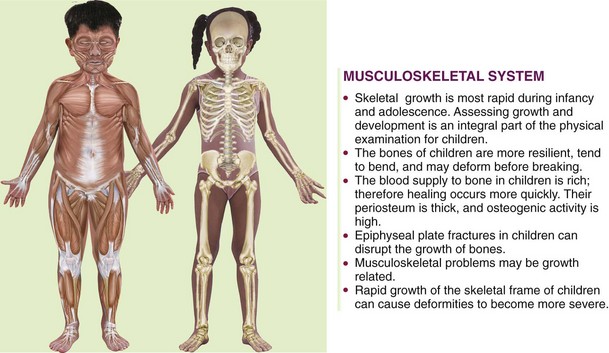
FIGURE 24-1 Some musculoskeletal system differences between the child and the adult. The muscular system consists of the large skeletal muscles that enable movement as well as the cardiac muscle of the heart and the smooth muscle of the internal organs. The skeletal system consists of bones and cartilage. This system helps to support and protect the body.
Observation and Assessment of the Musculoskeletal System in the Growing Child
To assess the musculoskeletal system of the growing child and to identify deviations, the nurse must have a basic understanding of the effect of growth, neurological development, and motor milestones at various ages. The newborn hip has limited internal rotation range of motion (ROM). The legs are maintained in a flexed position, and the lower leg has an internal rotation (internal tibial torsion) caused by the effects of uterine positioning; this can last 4 to 6 months. The general curvature of the newborn spine is a C shape from the thoracic to the pelvic level; it changes with the mastery of motor skills to a double S curve in childhood (see Figure 17-1). The newborn’s feet normally turn inward (varus) or outward (valgus), but the turning-in self-corrects when the sole of the foot is stroked. The toddler’s feet appear flat because of the presence of a fat pad at the arch. Any delay in neurological development can cause a delay in the mastery of motor skills, which can result in altered skeletal growth.
Assessment of the musculoskeletal system includes observation, palpation, ROM, and gait assessment in children who can walk. Children who do not walk independently by 18 months of age have a serious delay and should be referred to a health care provider for follow-up.
Observation of Gait
A gait is a characteristic manner of walking. The toddler who begins to walk has a wide, unstable gait. The arms do not swing with the walking motion. By 18 months of age the wide base narrows and the walk is more stable. By 4 years of age the child can hop on one foot, and arm swings occur with walking. By 6 years of age the gait resembles the adult walk with equal stride lengths and associated arm swing. The trunk is centered over the legs, and movement is symmetrical. When a child favors one side, pain may be present. Toe walking after 3 years of age can indicate a muscle problem.
In most cases, excessive in-toeing, or pointing of the toe inward, will resolve by 4 years of age. These children trip and fall easily. Teaching proper sitting and body mechanics is the treatment of choice. Participation in ballet classes and in-line skating will enhance hip flexibility. If the problem does not resolve, a brace may be prescribed. Failure to treat can result in hip, knee, or back problems in adulthood.
Young children appear bowlegged (genu varum) or knock-kneed (genu valgum), with the knees turned inward until 5 years of age. Bowing is seldom pathological. The ligaments that support the arch are not mature before 6 years of age, and therefore the child may appear to have flat feet. If the condition interferes with walking, an orthotic appliance can be prescribed for the child to wear inside the shoes. When the flat foot is painful, a referral for follow-up examination should be initiated. The role of the nurse is to reassure parents that unless there is associated pain or a problem with motor or nerve functions, many minor abnormal-appearing alignments will spontaneously resolve with activity.
Observation of Muscle Tone
The nurse should assess symmetry of movement and the strength and contour of the body and extremities. The strength of the extremities can be tested by having the child push away the examiner’s hand with his or her foot or hand.
Neurological Examination
A neurological assessment is a vital part of a comprehensive musculoskeletal examination. An assessment of reflexes, a sensory assessment, and the presence or absence of spasms should be noted.
Diagnostic Tests and Treatments
Radiographs (x-ray films) are taken to confirm a suspected pathological condition, and the affected area is compared with the unaffected area.
Bone Scans: Bone scans are helpful in identifying pathological conditions that may not clearly be seen on a routine x-ray study, such as septic arthritis or tumors.
Computed Tomography: Computed tomography (CT) provides a cross-sectional picture of the bone and its relationship to other structures within the area of examination.
Laboratory Tests and Treatments
A complete blood count (CBC) and erythrocyte sedimentation rate (ESR) may rule out septic arthritis or osteomyelitis. Human leukocyte antigen (HLA) B-27 may help diagnose rheumatological disorders.
A thorough history is necessary to determine the basis for musculoskeletal problems, which are often insidious. The nurse determines the history of the injury; the location of pain; when symptoms started; any weakness, numbness, or loss of function in an extremity; and whether the problem is affecting the daily activities of the child. An arthroscopy is commonly performed on adolescents with sports injuries. The physician is able to look inside the joint (usually the knee or shoulder) to determine the extent of injury. The area is inspected, foreign particles are removed, or repairs are made to the torn menisci. A bone biopsy may reveal a malignancy. Muscle biopsy may detect muscular dystrophy.
Traction, casting, and splints are used in accordance with the patient’s needs. Three types of skin traction are often used for the lower extremities of children: Bryant’s traction, Buck’s extension, and Russell traction. Children with musculoskeletal disorders may require lengthy hospitalization. Immobility causes a deceleration in body metabolism. Nursing interventions focus on maintaining body functions. ROM exercises and the use of a trapeze prevent muscle atrophy. Foods high in roughage stimulate the digestive tract and prevent constipation. Respiratory exercises prevent pneumonia. These and other measures can prevent complications that can lengthen hospitalization for the child. (Clubfoot and congenital hip dysplasia are discussed in Chapter 14. Rickets is discussed in Chapter 28.)
Pediatric Trauma
Soft tissue injuries usually accompany traumatic fractures in the child at play or the adolescent involved in sports activities and include the following:
• Contusion: A tearing of subcutaneous tissue results in hemorrhage, edema, and pain. The escape of blood into the soft tissue is referred to as a hematoma, or a “black and blue mark.”
• Sprain: When the ligament is torn or stretched away from the bone at the point of trauma, there may be resulting damage to blood vessels, muscles, and nerves. Swelling, disability, and pain are major signs of a sprain.
• Strain: A microscopic tear to the muscle or tendon occurs over time and results in edema and pain.
Treatment of Soft Tissue Injuries
Soft tissue injuries should be treated immediately to limit damage from edema and bleeding. A cold pack and elastic wrap will reduce edema and bleeding and relieve pain, and should be applied at alternating 30-minute intervals. (After a 30-minute period, ischemia can occur and impede the tissue perfusion.) Elevating the extremity above heart level reduces edema. When an elastic bandage is used for compression, a priority nursing responsibility is to perform frequent neurovascular checks to ensure adequate tissue perfusion.
 Memory Jogger
Prevention of Pediatric Trauma
Accidents are common in childhood, but much can be done to prevent morbidity and mortality. Parents are responsible for maintaining a safe environment for their children. Nurses are responsible for educating parents and schoolteachers about how to prevent accidental injury and maintain a safe environment.
The proper use of pedestrian safety practices, car seat restraints, bicycle helmets and other athletic protective gear, pool fences, window bars, deadbolt locks, and locks on cabinets can prevent many injuries to children. Pediatric trauma can cause permanent disability or premature death. Nursing assessment and interventions can assist the injured child toward recovery. The nurse also has a community responsibility to support legislation that would maintain safe environments for children.
Traumatic Fractures
Pathophysiology: A fracture is a break in a bone and is mainly caused by accidents. It is characterized by pain, tenderness on movement, and swelling. Discoloration, limited movement, and numbness may also occur. In a simple fracture the bone is broken, but the skin over the area is not. In a compound fracture a wound in the skin accompanies the broken bone, and there is an added danger of infection. A greenstick fracture is an incomplete fracture in which one side of the bone is broken and the other is bent. This type of fracture is common in children because their bones are soft, flexible, and more likely to splinter. In a complete fracture the bone is entirely broken across its width. Figure 24-2 illustrates various types of fractures. When an x-ray film reveals multiple fractures in various stages of healing, child abuse should be suspected.

FIGURE 24-2 A, Types of fractures. B, Reduction of a fractured bone. A gradual pull is exerted on the distal (lower) fragment of the bone until it is in alignment with the proximal fragment. C, Various methods of internal fixations, using plates, pins, nails, and screws to hold fragments of bone in place.
A fracture heals more rapidly in a child than it does in an adult. The child’s periosteum is stronger and thicker, and there is less stiffness on mobilization. Injury to the cartilaginous epiphysis, which is found at the ends of the long bones, is serious if it happens during childhood because it may interfere with longitudinal growth. Care of a child in a cast is discussed in Chapter 14. Casts may be made of plaster or fiberglass.
Fractures of the Femur in Early Childhood: The femur (the thigh bone) is the largest and strongest bone of the body. It is one of the most prevalent serious breaks that occur during early childhood. A spiral fracture of the femur is caused by a forceful twisting motion. When the history of an injury does not correlate with x-ray findings, child abuse should be suspected because spiral fractures can be the result of manual twisting of the extremity. The child complains of pain and tenderness when the leg is moved and cannot bear weight on it. Clothes are gently removed, starting at the uninjured side and proceeding to the injured side. It may be necessary to cut the clothes; x-ray films confirm the diagnosis. Skin traction is used to reduce the fracture and keep the bones in proper alignment.
Treatment of Fractures with Traction
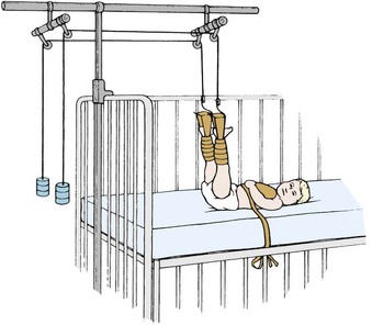
Traction in the Younger Child: Bryant’s traction is used for treating fractures of the femur in children under 2 years of age or under 20 to 30 lb. Weights and pulleys extend the limb as in the Buck’s extension; however, the legs are suspended vertically (Figure 24-3). The weight of the child supplies the countertraction.
Traction in the Older Child: Traction is used when the cast cannot maintain alignment of the two bone fragments. Skeletal muscles act as a splint for the fracture. Traction aligns the injured bone by the use of weights and countertraction. Immobilization is maintained until the bones fuse.
Buck skin traction (Buck’s extension) is a type of skin traction used in fractures of the femur and in hip and knee contractures. It pulls the hip and leg into extension. Countertraction is supplied by the child’s body; therefore it is essential that the child not slip down in bed and that the bed not be placed in high-Fowler’s position. Buck’s extension is sometimes used preoperatively, either unilaterally or bilaterally, to reduce pain and muscle spasm associated with a slipped capital femoral epiphysis. Russell traction is similar to Buck’s extension. In Russell traction, however, a sling is positioned under the knee, which suspends the distal thigh above the bed (Figure 24-4). Skin traction is applied to the lower extremity. Pull is in two directions, vertically from the knee sling and longitudinally from the foot plate (Figure 24-5). This prevents posterior subluxation of the tibia on the femur, which can occur in children in traction. Split Russell traction uses two sets of weights, one suspending the thigh and the other exerting a pull on the leg, with weights at the head and foot of the bed. Balanced suspension using the Thomas splint and Pearson attachments is used to treat diseases of the hip as well as fractures in older children and adolescents. It may be used both before and after surgery.
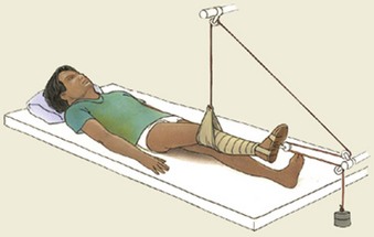
FIGURE 24-4 Russell skin traction. A skin traction system applied to the lower extremity using a foot plate and a knee sling. When there are separate weights attached to both the foot plate and the knee sling, it is known as a split Russell traction.
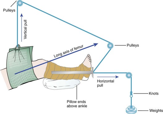
FIGURE 24-5 Forces involved in traction. The placement of pulleys and the angle of the joints determine the line of pull. In this case the combined vertical and horizontal pull results in a pull on the long axis of the femur to reduce fracture displacement.
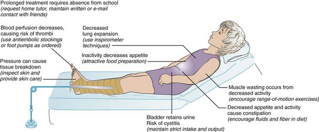
In skeletal traction a Steinmann pin or Kirschner wire is inserted into the bone, and traction is applied to the pin. Daily care of the pin site is essential. “Ninety-ninety” (or ninety degree–ninety degree) traction with a boot cast or sling on the lower leg may be used (Figure 24-6). Crutchfield, or Barton, tongs may be used in the skull to provide cervical traction (Figure 24-7). Skeletal traction carries the added risk of infection from skin bacteria that may cause osteomyelitis. The child in traction experiences certain effects as a result of immobilization (Figure 24-8). Visitors are important to the child in traction, and a school tutor should be contacted so that the child will be able to return to class after healing occurs.
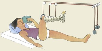
FIGURE 24-6 90 degree–90 degree skeletal traction. A wire pin is inserted into the distal segment of the femur. The lower leg may be placed in a boot cast or is supported by a sling.
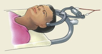
FIGURE 24-7 Cervical traction. A special bed may be used to turn a patient who is in cervical traction.
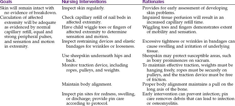
Nursing Responsibilities for Traction: The nurse observes the traction ropes to be sure they are intact and in the wheel grooves of the pulleys and that the child’s body is in good position. In Bryant’s traction, the legs should be at right angles to the body, with the buttocks raised sufficiently to clear the bed. In all types of traction, elastic bandages should be neither too loose nor too tight. A jacket restraint may be used to keep the child from turning from side to side. The weights are not removed once applied. Continuous traction is necessary. The weights must hang free, and the pull of the weights must not be obstructed by room furnishings, such as a chair. The weights are not lifted or supported when the bed is moved.
The nurse performs frequent neurovascular checks to the toes to see that they are warm and that their color is good (Figure 24-9). Observations such as cyanosis, numbness, or irritation from attachments; tight bandages; severe pain; or the absence of pulse rates in the extremities are reported immediately to the nurse in charge. A specific and serious complication of any traction is Volkmann’s ischemia (ischein, “to hold back,” and haima, “blood”), which occurs when the circulation is obstructed. When the legs are elevated overhead, as in Bryant’s traction, there is gravitational vascular drainage. Arterial occlusion can cause anoxia of the muscles and reflex vasospasm, which when unnoticed could result in contractures and paralysis.
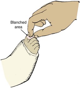
FIGURE 24-9 Checking circulation to the toes or fingers. To check circulation (capillary refill), squeeze or press a toe or finger to blanch the skin. If the circulation is adequate, the color should return in less than 3 seconds when the pressure is released. (1) If the toes do not blanch, congestion may be present and should be reported to the physician. (2) If the blanching persists after pressure is released, the circulation is impaired. The physician must be notified. (3) Report to the physician if extreme pain results from touching or moving the toes.
The child is bathed, and back and buttock care is given. A sheepskin padding may also be used. The sheets are pulled taut and are kept free of crumbs. The jacket restraint is changed when it is soiled. The child is encouraged to drink plenty of fluids and to eat foods that are high in roughage to prevent constipation caused by a lack of exercise. Because the child in traction is unable to sit upright when eating, special precautions to prevent choking and aspiration during mealtime are a priority nursing responsibility. Stool softeners may be necessary. A fracture pan is used for bowel movements, and a careful record is kept of eliminations. Deep-breathing exercises are encouraged to prevent the collection of fluid in the lungs caused by the child’s immobility. These exercises may be done by blowing bubbles or blowing a pinwheel.
Diversional therapy is important because hospitalization may be lengthy. Toys may be securely suspended over the child’s head so they are within easy reach. The child’s crib is taken to the playroom when possible so the child can experience the excitement of the activities there.
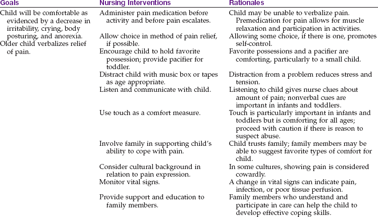
DVDs, CDs, MP3 players, video games, stories, and other forms of entertainment are important aspects of a total nursing care plan. Pain control is essential. Parents are encouraged to visit the child as often as possible. With proper treatment the prognosis for the child with this condition is good.
 Nursing Tip
Checklist for the patient in traction:
• Head of bed no higher than 20 degrees (for countertraction)
• Heels of feet elevated from bed
• ROM of unaffected parts checked at regular intervals
• Antiembolism stockings or foot pumps in place as ordered
• Neurovascular checks performed regularly
• Skin integrity monitored regularly
• Pain and its relief by medication recorded
Neurovascular checks: A priority nursing responsibility in the care of a child with a fracture who is in traction or has a cast or Ace bandage in place is to perform neurovascular assessments or neurovascular checks at regular intervals (Skill 24-1).
Skill 24-1 Neurovascular Checks


1. Pain: Assess and record the location and quality of pain. Initiate pain control strategies and medication as soon as possible. Pain at the trauma site that does not respond to medication may indicate a serious complication called compartment syndrome. Compartment syndrome is a term used to describe ischemia to an extremity caused by pressure on the tissues as a result of excessive edema. A surgical procedure called a fasciotomy (a cutting into the tissue) may be needed to reduce the pressure and increase tissue perfusion.
2. Pulse: Compare the quality of the pulse on the affected extremity to that on the unaffected extremity. A strong pulse indicates good blood flow necessary for healing.
3. Sensation: Reduced sensation to touch (numbness or tingling) at a site distal to the fracture may indicate poor tissue perfusion and should be reported.
4. Color: Pallor at the site distal to the fracture can indicate arterial insufficiency, whereas cyanosis of the site distal to the fracture can indicate venous stasis. Adequate blood supply and vascular drainage are essential for optimal healing.
5. Capillary refill: A compressed nail bed should return to its original color in less than 3 seconds. The findings should be compared with the unaffected extremity and the results recorded frequently (see Figure 24-9).
6. Movement: Test the toes and fingers distal to the fracture site for movement. Because nerve injury can occur as a complication of skeletal fractures, the movement associated with specific nerve supply should also be tested (Figure 24-10).
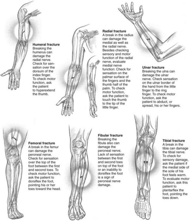
 Safety Alert!
The “neurovascular check” for tissue perfusion is performed on the toes or fingers distal to an injury or cast and includes the following:
Nursing Care Plan 24-1 describes interventions for the child in traction.
 Nursing Care Plan
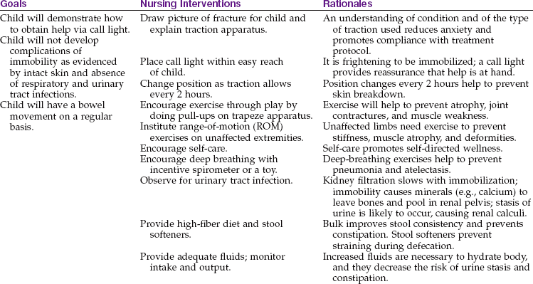
Treatment of Fractures with Casts
Nursing Care of a Child in a Cast: A cast is a device used to immobilize a fracture site, usually including the joints above and below the fracture. Casts are constructed of plaster of Paris or a synthetic material such as fiberglass. Fiberglass casts are lighter than plaster casts and come in various bright colors but cannot be written on as easily as plaster casts can. If the proper lining is used under the fiberglass cast, the cast is water resistant and can be washed and dried, whereas the plaster cast will deteriorate if it becomes wet. When applied, the fiberglass cast dries within half an hour compared to the plaster cast that takes 10 to 72 hours to dry and must be handled carefully with open palms and extended fingers to prevent indents that can create pressure areas on the skin during this drying time.
Nursing responsibilities include elevating the affected extremity on a pillow and performing frequent neurovascular checks on the distal digits (see Skill 24-1). Before discharge, the nurse teaches the child and parents how to care for the cast and how to support the cast to prevent the extremity from assuming a dependent position that could compromise circulation. The child is taught safe transfer from bed to wheelchair and safe crutch-walking techniques (e.g., being sure to keep the body weight on the hands and not the axillae). Parents are also taught how to check if the cast is too loose or too tight and when to return to the clinic or health care provider.
Safety Alert!
Pain unrelieved by medication or swelling, cyanosis or pallor of digits, decreased pulse and skin temperature should be immediately reported because they may be signs of compartment syndrome.
The child should be prepared for the experience of cast removal because the cast cutter can appear and sound threatening. After cast removal, the skin can be expected to be dry and caked. Lotion and soothing baths are advised. Care of a child in a spica cast is discussed in Chapter 14.
Disorders and Dysfunction of the Musculoskeletal System
Pathophysiology: Osteomyelitis is an infection of the bone that generally occurs in children younger than 1 year of age and in those between 5 and 14 years of age. Long bones contain few phagocytic cells (white blood cells [WBCs]) to fight bacteria that may come to the bone from another part of the body. The inflammation produces an exudate that collects under the marrow and cortex of the bone.
Staphylococcus aureus is the organism responsible in 75% to 80% of cases in children more than 5 years of age, and Haemophilus influenzae is the most common cause in young children. This incidence may be reduced by the widening practice of routine infant immunization against this organism. Other causative organisms include group A streptococci and pneumococci. Salmonella and Pseudomonas are organisms often found in adolescents who are intravenous (IV) drug users. Osteomyelitis may be preceded by a local injury to the bone, such as an open fracture, burn, or contamination during surgery. It may also follow a furuncle, impetigo, and abscessed teeth. In neonates a heel puncture or scalp vein monitor can be the predisposing site of infection. Infective emboli may travel to the small arteries of the bone, setting up local destruction and abscess. For this reason, a careful search for infection in other bones and soft tissues is necessary.
The vessels in the affected area are compressed, and thrombosis occurs, producing ischemia and pain. The collection of pus under the periosteum of the bone can elevate the periosteum, which can result in necrosis of that part of the bone. If the pus reaches the epiphysis of the bone in infants, infection can travel to the joint space, causing septic arthritis of that joint.
Local inflammation and increased pressure from the distended periosteum can cause pain. Older children can localize the pain and may limp. Younger children and infants will show decreased voluntary movement of that extremity. Associated muscle spasms can cause limited active ROM. The child may refuse to stand or walk. Signs of local inflammation may be present. A detailed history may reveal possible sources of primary infection. Blood cultures to identify the organism may be valuable if the child has not been given antibiotics for the primary infection. A urine test for the presence of bacterial antigens and a tissue biopsy may be helpful to establish the diagnosis.
Diagnosis: There is an elevation in WBC count and sedimentation rate. X-ray examination may initially fail to reveal the infection. A microbiology culture of aspirated drainage or a bone scan may be diagnostic.
Treatment and Nursing Care: Prompt and vigorous treatment is essential to ensure a favorable prognosis. Intravenous antibiotics are prescribed for a 4- to 6-week period. The high doses required indicate a nursing responsibility to monitor the infant or child for toxic responses and to ensure long-term compliance. The joint may be drained of pus arthroscopically or surgically to reduce pressure and prevent bone necrosis. A complication should be suspected if fever lasts beyond 5 days. The use of appropriate pain-relieving medications and gentle handling to minimize pain are essential. The child should be positioned comfortably with the limb supported by pillows or blanket rolls.
Bed rest is followed by wheelchair access, but weight bearing should be avoided. Diversional therapy, physical therapy, and tutorial assistance for school-age children should be provided so they can return to their class and classmates after discharge. Close interaction with home care providers is indicated.
Duchenne’s or Becker’s (Pseudohypertrophic) Muscular Dystrophy
Pathophysiology: The muscular dystrophies are a group of disorders in which progressive muscle degeneration occurs. The childhood form (Duchenne’s muscular dystrophy) is the most common type. It has an incidence of about 1 in 3600 live born male infants of all races and ethnic groups. It is a sex-linked inherited disorder that occurs only in boys. Dystrophin, a protein in skeletal muscle, is absent.
Manifestations: The onset is generally between 2 and 6 years of age; however, a history of delayed motor development during infancy may be evidenced. The calf muscles in particular become hypertrophied. The term pseudohypertrophic (pseudo, “false,” and hypertrophy, “enlargement”) refers to this characteristic. Other signs include progressive weakness as evidenced by frequent falling, clumsiness, contractures of the ankles and hips, and Gowers’ maneuver (a characteristic way of rising from the floor).
Laboratory findings show marked increases in serum creatine phosphokinase. Muscle biopsy reveals a degeneration of muscle fibers and their replacement by fat and connective tissue. A myelogram (a graphic record of muscle contraction as a result of electrical stimulation) shows decreases in the amplitude and duration of motor unit potentials. A serum blood polymerase chain reaction (PCR) for the gene mutation is diagnostic for this condition (Sarnat, 2007). The disease becomes progressively worse, and wheelchair confinement may be necessary. Death usually results from cardiac failure or respiratory tract infection. Mental retardation is not uncommon.
Treatment and Nursing Care: The use of prednisone has shown some promise (Moxley et al., 2005). Studies are in place concerning the use of glutamine and creatine for muscle weakness, the enzyme transferase to block muscle wasting, and urotrophin to replace the missing dystrophin. Treatment at this time is mainly supportive to prevent contractures and maintain the quality of life. Cardiac and respiratory complications are common. A multidisciplinary team should provide psychological support, nutritional support, physiotherapy, social and financial assistance, and when necessary, respite and hospice care.
Compared with other children with disabilities, some children with muscular dystrophy may appear passive and withdrawn. Early on, depression may be seen because the child cannot compete with peers. Social and emotional pressures on the child and family are great.
Slipped Femoral Capital Epiphysis
Pathophysiology: A slipped femoral capital epiphysis, also known as coxa vara, is the spontaneous displacement of the epiphysis of the femur. It most often occurs during rapid growth of the preadolescent and is not related to trauma. The epiphysis of the femur widens, and then the femoral head and epiphysis remain in the acetabulum. The head of the femur then rotates and displaces. Symptoms include thigh pain and a limp or the inability to bear weight on the involved leg. An x-ray study confirms diagnosis.
Treatment: The child is placed in traction to minimize further slippage, and then surgery is scheduled to insert a screw to stabilize the bone. Serious complications may include disturbance of circulation to the epiphysis, resulting in necrosis of the head of the femur.
Nursing Care: The general principles of caring for a child in traction and preoperative and postoperative care are used (see Chapters 14 and 22). The nurse assists in carrying out orders regarding range-of-motion activities, guides gradual resumption of weight bearing with the help of crutches, and encourages referrals to maintain school studies.
Legg-Calvé-Perthes Disease (Coxa Plana)
Pathophysiology: Legg-Calvé-Perthes disease is one of a group of disorders called the osteochondroses (osteo, “bone,” chondros, “cartilage,” and osis, “disease”) in which the blood supply to the epiphysis, or end of the bone, is disrupted. The tissue death that results from the inadequate blood supply is termed avascular necrosis (a, “without,” vasculum, “vessels,” and nekros, “death”). Legg-Calvé-Perthes disease affects the development of the head of the femur. Its cause and incidence are unknown. The disease is seen most commonly in boys between 5 and 12 years of age. It is more common in Caucasians. This disease is unilateral in about 85% of cases. Healing occurs spontaneously over 2 to 4 years; however, marked distortion of the head of the femur may lead to an imperfect joint or degenerative arthritis of the hip in later life. Symptoms include a painless limp and limitation of motion. X-ray films and bone scans confirm the diagnosis.
Treatment: Legg-Calvé-Perthes disease is a self-limiting disorder that heals spontaneously but slowly over the course of 1 to 2 years. The treatment involves keeping the femoral head deep in the hip socket while it heals and preventing weight bearing. This is accomplished through the use of ambulation-abduction casts or braces that prevent subluxation (sub, “beneath,” and luxatio, “dislocation”) and enable the acetabulum to mold the healing head in such a way that it does not become deformed. This treatment may be preceded by bed rest and traction. The prognosis in Legg-Calvé-Perthes disease is fair. Some affected people may require hip joint replacement procedures as adults.
Nursing Care: Nursing considerations depend on the age of the patient and the type of treatment. The general principles of traction, cast, and brace care are used when immobilization of the child is necessary. Teaching and counseling are directed toward a holistic understanding of and interest in the individual child and family. Total immobility or partial mobility is particularly trying for children. The natural inclination to compete physically is thwarted. In some cases surgical immobilization is required. Preoperative and postoperative care and care of a patient in a cast are discussed in Chapters 14 and 22.
Osteosarcoma
Pathophysiology: Osteosarcoma (osteo, “bone,” sarx, “flesh,” and oma, “tumor”) is a primary malignant tumor of the long bones. The two most common types of bone tumors in children are osteosarcoma and Ewing’s tumor. The mean age of onset of osteosarcoma is between 10 and 15 years of age. Children who have had radiation therapy for other types of cancer and children with retinoblastoma have a higher incidence of this disease. Metastasis occurs quickly because of the high vascularity of bone tissue. The lungs are the primary site of metastasis; the brain and other bone tissue are also sites of metastasis.
Manifestations: The patient experiences pain and swelling at the site. In adolescents this is often attributed to injury or “growing pains.” The pain may be lessened by flexing the extremity. Later a pathological fracture may occur. Diagnosis is confirmed by biopsy. A complete physical examination, including CT and a bone scan, is done. Radiological studies help to confirm the diagnosis.
Treatment and Nursing Care: Treatment of the patient with osteosarcoma consists of radical resection or amputation surgery. Internal prostheses are available for most sites. Long-term survival is possible with early diagnosis and treatment.
The nursing care is similar to that for other types of cancer. Problems of body image are particularly important to the self-conscious adolescent. If amputation is necessary, the family and patient will need much support. The nurse anticipates anger, fear, and grief. Immediately after surgery the stump dressing is observed frequently for signs of bleeding. Vital signs are monitored. The child is positioned as ordered by the surgeon.
Phantom limb pain is likely to be experienced. This is the continued sensation of pain in the limb even though the limb is no longer there. It occurs because nerve tracts continue to report pain. This pain is very real, and an analgesic may be necessary. Rehabilitation measures follow surgical recovery.
Ewing’s Sarcoma
Pathophysiology: Ewing’s sarcoma was first described in 1921 by Dr. James Ewing. It is a malignant growth that occurs in the marrow of the long bones. It occurs mainly in older school-age children and early adolescents. When metastasis is present on diagnosis, the prognosis is poor. Without metastasis there is a 60% survival rate. The primary sites for metastasis are the lungs and long bones.
Treatment and Nursing Care: Amputation is not generally recommended for Ewing’s sarcoma because the tumor is sensitive to radiation therapy and chemotherapy. This is a relief to the child and family. The child is warned against vigorous weight bearing on the involved bone during therapy to help prevent pathological fractures. Patients must be prepared for the effects of radiation therapy and chemotherapy. The nurse supports the family members in their efforts to gain equilibrium after such a crisis.
Juvenile Idiopathic Arthritis (Juvenile Rheumatoid Arthritis)
Pathophysiology: Juvenile rheumatoid arthritis (JRA) is now referred to as juvenile idiopathic arthritis (JIA) and is the most common arthritic condition of childhood. It is a systemic autoimmune disease that involves the joints, connective tissues, and viscera, and it differs somewhat from adult rheumatoid arthritis. It is not a rare disease. Approximately 113 of 100,000 children per year in the United States are diagnosed with JIA (Miller & Cassidy, 2007). The exact cause is unknown, but infection and an autoimmune response have been implicated.
Manifestations and Types: JIA has several distinct classifications. The systemic form is manifested by intermittent spiking fever above 39.5° C (103° F) persisting for over 10 days, a nonpruritic macular rash, abdominal pain, an elevated ESR, C-reactive protein in laboratory tests, presence of antinuclear antibodies, and possibly an enlarged liver and spleen. It occurs most often in children ages 1 to 3 years and 8 to 10 years. Joint symptoms may be absent at onset, but usually arthritis develops in most patients.
Other classifications involve criteria of increased number of joints involved, age of onset, and the presence or absence of the rheumatoid factor. Psoriatic arthritis is a classification that includes psoriasis in its manifestation. Enthesitis-related arthritis involves the tendon insertion sites and is associated with uveitis (inflammation of the eye) and irritable bowel disease.
Treatment: The goals of therapy are to accomplish the following:
• Reduce joint pain and swelling
• Promote mobility and preserve joint function
• Promote growth and development
• Promote independent functioning
• Help the child and family to adjust to living with a chronic disease
Medications such as nonsteroidal antiinflammatory drugs (NSAIDs) and methotrexate may also be given. The child should be monitored closely for side effects with frequent CBC tests. Steroids may be given for incapacitating arthritis, uveitis or life-threatening complications. Etanercept is a tumor necrosis factor–inhibitor used in children unresponsive to methotrexate. It is administered subcutaneously, weekly, and may have serious side effects. Slow-acting antirheumatic drugs (SAARDs) such as sulfasalazine, hydroxychloroquine, gold and d-penicillamine can be used with NSAIDs but are not as popular today as in the past.
Nursing Care: The nurse functions as a member of a multidisciplinary health care team that includes the pediatrician, rheumatologist, social worker, physical therapist, occupational therapist, psychologist, ophthalmologist, and school and community nurses. Physical and occupational therapy preserve joint function and mobility. Occupational therapy helps with activities of daily living. Resting in a bed with a supportive, flat mattress is helpful. Resting splints during sleep can prevent flexion contractures from developing. Moist heat and exercise are advised, and whirlpool baths and hot packs relieve pain and stiffness. Therapeutic play facilitates compliance with exercise regimens. Pool exercise, kick ball, and biking can help maintain joint mobility. Sleep, rest, and general health measures are important. The school nurse can be contacted to help promote normal growth and development in school-related activities. Home schooling, if needed, is available in most school districts. Unnecessary restrictions should be avoided because they can lead to rebellion and noncompliance. JIA inhibits social interactions of the child and can interfere with development of a positive self concept. The Arthritis Foundation provides services to parents and nurses involved in patient care.
This long-term disease is characterized by periods of remission and exacerbations. Nurses can serve as advocates for the child; that is, they can help alleviate stress by recognizing the impact of the disease and by openly communicating with the child, the family, and other members of the health care team. Nurses support the child and family members and instill hope.
Torticollis (Wry Neck)
Pathophysiology: Torticollis (tortus, “twisted,” and collium, “neck”) is a condition in which neck motion is limited because of shortening of the sternocleidomastoid muscle. It can be either congenital or acquired and can also be either acute or chronic. The most common type is a congenital anomaly in which the sternocleidomastoid muscle is injured during birth. It is associated with breech and forceps delivery and may be seen in conjunction with other birth defects, such as congenital hip.
Manifestations: In congenital torticollis the symptoms are present at birth. The infant holds the head to the side of the muscle involved. The chin is tilted in the opposite direction. There is a hard, palpable mass of dense fibrotic tissue (fibroma). This is not fixed to the skin and resolves by 2 to 6 months of age. Passive stretching and ROM exercises and physical therapy may be indicated. Feeding and playing with the infant can encourage turning to the desired side for correction. Surgical correction is indicated if the condition persists beyond age 2 years.
Acquired torticollis is seen in older children. It may be associated with injury, inflammation, neurological disorders, and other causes. Nursing intervention is primarily that of detection. Infants who have limited head movement require further investigation.
Scoliosis
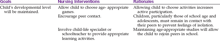
Pathophysiology: The most prevalent of the three skeletal abnormalities shown in Figure 24-11 is scoliosis. Scoliosis refers to an S-shaped curvature of the spine. During adolescence, scoliosis is more common in girls. Many curvatures are not progressive and may necessitate only periodic evaluation. Untreated progressive scoliosis may lead to back pain, fatigue, disability, and heart and lung complications. Skeletal deterioration does not stop with maturity and may be aggravated by pregnancy.
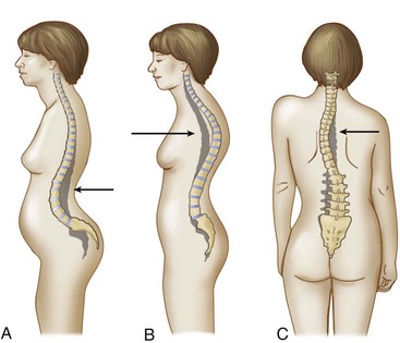
FIGURE 24-11 Abnormal spinal curvatures. A, Lordosis, known as “sway back,” is commonly seen during pregnancy. B, Kyphosis, known as “hunchback,” is an increased roundness in the thoracic curve commonly found in the elderly. C, Scoliosis, an abnormal side-to-side curvature of the spine, is commonly found in adolescents.
Causes: There are two types of scoliosis: functional and structural. Functional scoliosis is usually caused by poor posture, not by spinal disease. The curve is flexible and easily correctable. Structural or fixed scoliosis is caused by changes in the shape of the vertebrae or thorax. It is usually accompanied by rotation of the spine. The hips and shoulders may appear uneven. The patient cannot correct the condition by standing in a straighter posture.
There are many causes of structural scoliosis. Some are congenital and develop in utero. These are noticeable at birth or during periods of rapid growth. Neuromuscular scoliosis is the result of muscle weakness or imbalance. It is seen in children with cerebral palsy, muscular dystrophy, and other conditions. The cause of idiopathic scoliosis is unknown; there may be a hereditary link.
Signs and Symptoms: Symptoms develop slowly and are not painful so that detection is usually via an incidental screening at school by the school nurse. The shoulders are different heights with one higher than the other, and a one-sided rib hump and a prominent scapula are seen. This asymmetry is seen from the back when the child leans forward. Diagnosis is made by moiré photography that highlights the asymmetry, and a spinal x-ray study.
Treatment: Treatment is aimed at correcting the curvature and preventing more severe scoliosis. Curves up to 20 degrees do not necessitate treatment but are carefully followed with exercise to increase muscle tone and posture. Curves between 20 and 40 degrees require the use of a Milwaukee brace (Figure 24-12). This apparatus exerts pressure on the chin, pelvis, and convex (arched) side of the spine. It is worn approximately 23 hours a day and is worn over a T-shirt to protect the skin. An underarm modification of the brace (the Boston brace) is proving effective for patients with low curvatures. It is less cumbersome and is more acceptable to the self-conscious young person. Transcutaneous electrical muscle stimulation (TENS) and exercise have not proven to be effective in the treatment of scoliosis (Kliegman et al., 2007).

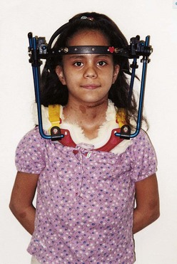
Severe scoliosis necessitates surgery to fuse the bones. A Harrington rod, Dwyer instrument, or Luque wires may be inserted for immobilization during the time required for the fusion to become solid. Halo traction may be used when there is associated weakness or paralysis of the neck and trunk muscles (Figure 24-13); it is also used in treating cervical fractures and fusions.
Community nursing: The management of scoliosis begins with screening. This is done before middle school. It should be a part of every yearly physical examination given to prepubescent youngsters. Camp nurses also need to be aware of symptoms. Early recognition is of utmost importance in detecting mild cases amenable to nonsurgical treatment.
The adolescent is prepared by explaining the purpose of the procedure and by being reassured that it merely entails observing the back while standing and bending forward. Adolescents must know that it is simple, quick, and painless and that privacy will be afforded. They are instructed to wear clothing that is easy to remove, such as a pullover top. Boys disrobe to the waist, girls generally to the bra. No slip or undershirt should be worn.
The procedure consists of the nurse examining the spine from the front, side, and back while the adolescent stands erect and then observing the back as the adolescent bends forward. One looks initially for general body alignment and asymmetry (lack of proportion). In scoliosis, one shoulder may be higher than the other, a scapula may be prominent, the arm-to-body spaces may be unequal, or a hip may protrude; one arm may appear longer than the other when the person bends forward. Referrals are made as indicated to those who must obtain further treatment.
The nurse’s knowledge of preadolescent and adolescent developmental tasks is imperative, because therapy often conflicts with these tasks.
Routine preoperative nursing care of the adolescent is necessary for spinal fusion. This includes instructing the adolescent and family about the purpose and extent of the cast or brace that may be prescribed. It is important that the nurse evaluate and document the adolescent’s neuromuscular status at this time so that it may be used as a basis for comparison after the procedure is completed.
Much postoperative nursing care is directly related to combating the physical results of immobilization (see Figure 24-8). The body systems become sluggish because of inactivity. This is evidenced in the gastrointestinal tract by anorexia, irregularity, and constipation. Allowing the adolescent to select foods with the aid of the dietitian is helpful in improving appetite. Increasing fluid intake reduces constipation. The adolescent and parents are introduced to the multidisciplinary health care team. Postoperative exercise, physical therapy, and promotion of adolescent school and developmental tasks are essential aspects of nursing care.
Sports Injuries
A high percentage of adolescent males and females participate in athletic activities. The American Academy of Pediatrics (AAP) recommends that a complete physical examination be given at least every other year during adolescence and that sports-specific examinations be given for those involved in strenuous activity on entry into middle school or senior high school. Such examinations should be updated by an annual questionnaire. The family history and an orthopedic screening are important in identifying risk factors.
Prevention: Several factors help to prevent sports injuries. Some of these are adequate warm-up and cool-down periods; year-round conditioning; careful selection of activity according to physical maturity, size, and skill necessary; proper supervision by adults; safe, well-fitting equipment; and avoidance of participation when in pain or injured. Proper diet and fluids are also necessary. A few of the more common injuries are listed in Table 24-1. The nurse has a major role in educating and directing parents to sources of accurate information to ensure that the physical, emotional, and maturational levels of the adolescent are appropriate for the activity (see Health Promotion box). Parents are encouraged to inquire about the capabilities of personnel and the availability of emergency services before the beginning of the competition.
Table 24-1
| TYPE | DESCRIPTION |
| Concussion | Any blow to the head followed by alterations in mental functioning should be treated as a possible concussion; observe carefully for sequelae |
| “Stingers” or “burners” | A common neck injury when a player hits another in the head in such sports as football or soccer; caused by brachial plexus trauma; feels like an electrical jolt; usually mild and disappears suddenly; restrict sports activity until symptoms disappear; reassess protective gear |
| Injured knee | Usually a result of stress on the knee ligaments; potentially serious; should be evaluated by an experienced trainer or physician; may necessitate arthroscopic surgery |
| Sprain or strained ankle | May injure growth plate; x-ray films important in adolescent |
| Muscle cramps | Caused by injury, alterations in blood flow, or electrolyte deficiencies; important to warm up before activity; ensure fluid intake is adequate |
| Shin splints | Pain and discomfort in lower leg caused by repeated running on a hard surface such as concrete; avoid such activity; use well-fitting shoes; decrease inflammation by rest |
 Health Promotion
Selected Recreational Activities and Their Risks
| Activity | Risk |
| Gymnastics | Common problems associated with children engaging in gymnastics are delayed menstruation and eating disorders. Trauma and overuse injuries of the large joints and spine are common. Prevention includes muscle strengthening exercises, flexibility exercises, and wrist braces. Nutritional planning and education are essential. |
| Ballet | Common problems associated with ballet include delayed menarche and eating disorders. There is a high risk for stress fractures, strains, shin splints, and spinal deviations. Prevention includes skilled coaching and nutrition planning and education. |
| Wrestling | Placement in a wrestling match is based on weight. A bingeing and purging behavior is commonly found in athletes participating in wrestling. Concussions, neck strains, and spinal injuries are common. Skin dermatitis and infection occur from contact with floor mats. Skilled coaching and nutrition education and planning are important. |
| Football | Improvements in techniques, protective equipment, and game exercises have decreased serious injuries in children participating on football teams. |
| Hockey | Injuries can occur from both collisions with other athletes and collisions with pucks or sticks. Proper protective equipment and sporting rules decrease the seriousness of injuries. |
| Basketball, volleyball | These are jumping sports that involve injury risk to ankles, knees, and fingers. Ankle sprain, tendonitis, stress fractures, and blisters are common complications of these sports. |
| Running | Involves repeated, poorly absorbed foot impact. Muscle fatigue, environmental temperature, and running surface contribute to injuries. Engaging in proper exercises before running, using good impact-absorbing shoes, cross-training, and adequate rest are essential. Shin splints involve pain over the anterior tibia and result from tearing the collagenous fibers that connect muscle to bone. Shoe orthotics, running on a soft surface, cross-training, and rest are the priorities of management. |
| Skiing, snowboarding | Injuries are usually related to falls. Better equipment, controlled slope conditions, and separation of skiers by skill level contribute to a decreased rate of injury. |
| Cheerleading | Includes acrobatics, pyramid climbing, and tossing. Falls related to injuries such as sprains, fractures, dislocations, and head injuries. Safety instruction is important, and a certified coach should be present. |
Family Violence
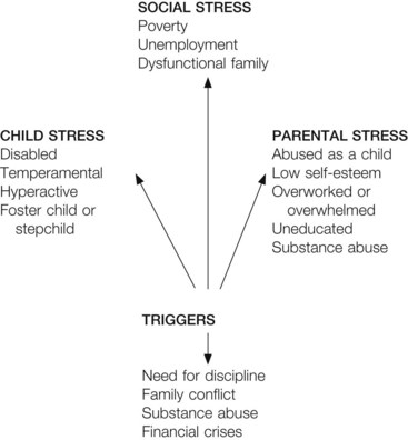
Violence has become a problem that affects children of all social classes across the nation. Family violence includes spousal abuse and child abuse, neglect, and maltreatment. Community violence is seen in neighborhoods where even non–gang-related adolescents arm themselves with guns and knives for protection. Preschoolers who are repeatedly allowed to watch violent programs on television or play aggressive computer games may be learning antisocial coping skills they will use as they grow and mature. The time spent watching television and the content of the programs should be controlled by parents so that television exposure results in the acquisition of knowledge, skills, and information that will motivate learning. In homes where spousal and/or child abuse occurs, children learn the behaviors they will practice when they become adults, and the abuse cycle continues. Parents who are abusive are not usually psychotic or criminal. They may have a knowledge deficit about child care needs and child growth and development. Abusive parents are often without a support system and are perhaps alone, angry, or in crisis, or have unrealistic expectations.
Child Abuse
The term battered child syndrome was coined by Kempe (1962) in his landmark paper in the Journal of the American Medical Association. It refers to “a clinical condition in young children who have received serious physical abuse, generally from a parent or foster parent.” The impact of Kempe’s research was considerable and focused the attention of physicians on unexplained fractures and signs of physical abuse. Today, most authorities consider Kempe’s definition narrow and have broadened it to include neglect and maltreatment.
According to the National Center on Child Abuse and Neglect (Nooner et al., 2010), the incidence of the following aspects of child abuse has been increasing:
• Emotional abuse: Intentional verbal acts that result in a destruction of self-esteem in the child; can include rejection or threatening the child
• Emotional neglect: An intentional omission of verbal or behavioral actions that are necessary for development of a healthy self-esteem; can include social or emotional isolation of a child
• Sexual abuse: Involves an act that is performed on a child for the sexual gratification of the adult
• Physical neglect: The failure to provide for the basic physical needs of the child, including food, clothing, shelter, and basic cleanliness
• Physical abuse: The deliberate infliction of injury on a child; suspected when an injury is not consistent with the history or developmental level of the child
The temperament of the child and the parent can be a causal factor in child abuse. Children who are different from others in any way are at particular risk. This includes preterm infants, sick or disabled children, and merely unattractive children. Unwanted or illegitimate infants and stepchildren are especially vulnerable. It has been noted that people are often reluctant to report occurrences in middle- and upper-income families or when the incident involves friends or relatives.
Federal Laws and Agencies
By 1963 the United States Children’s Bureau drafted a model mandatory state reporting law that has been adopted in some form in all states (see Legal and Ethical Considerations box). This law aids in establishing statistics and is based on the need to provide therapeutic help to both child and family. Immunity from liability is provided for persons reporting suspected cases. Most states have penalties for failure to report suspected child abuse. Referrals usually are made to the local child protective services, and a case worker is assigned.
 Legal and Ethical Considerations
Reporting Suspected Abuse or Neglect
A citizen can report suspected child abuse or neglect by contacting the child protective services in the yellow pages of the telephone directory under “Social Service Organizations.” This can be done anonymously. After obtaining the facts, the agency informs the parents that a report is being filed and checks the condition of the child. A visit must be initiated within 72 hours. In most cases, this is accomplished within 48 hours or earlier if the situation is life threatening. All persons who report suspected abuse or neglect are given immunity from criminal prosecution and civil liability if the report is made in good faith. Many professionals, such as physicians, nurses, and social workers, must report child abuse.
Nursing Care and Interventions
Nursing intervention for high-risk children is of utmost importance (Box 24-1). One approach currently taken is to identify high-risk infants and parents during the prenatal and perinatal periods. Predictive questionnaires are being used as screening tools in some clinics. Many hospitals also provide closer follow-up of mothers and newborns. Maternal-infant bonding and its significance to later parent-child relationships have been explored.
Nurses in obstetrical clinics have the opportunity to observe parents and their abilities to cope. The history of the parent(s), desirability of the pregnancy, number of children already in the family, financial and personal stability of the family, types of support systems, and other factors may have a bearing on how the parents accept the new offspring. Pertinent observations include a description of parent-newborn interaction. Both verbal and nonverbal communications are important, as is the level of body and eye contact. Lack of interest, indifference, or negative comments about the sex, looks, or temperament of the infant could be significant.
In other areas, a cooperative team approach is necessary. This may include services such as family planning, protective services, day care centers, homemakers, parenting classes, self-help groups, family counseling, child advocates, and a continued effort to reduce the incidence of preterm birth. Other related areas include financial assistance, employment services, transportation, emotional support and encouragement, and long-term follow-up care.
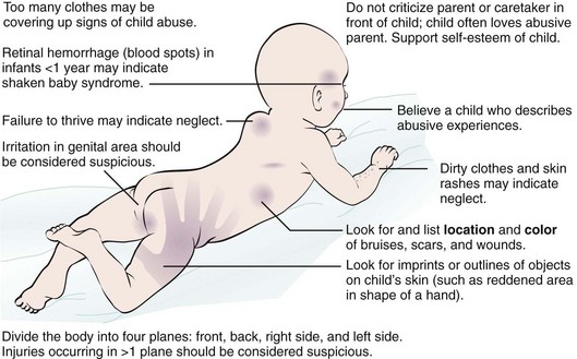
Individual nurses can help to detect child abuse by maintaining a vigilant approach in their work settings. Record keeping should be factual and objective. The pediatric nurse should make a point of reviewing old records of their patients, which may reveal repeated hospitalizations, x-ray films of multiple fractures, persistent feeding problems, a history of failure to thrive, and a history of chronic absenteeism in school. Neglect or delay in seeking medical attention for a child or failure to obtain immunization and well-child care can be significant findings. Children who seem overly upset about being discharged must be brought to the attention of the health care provider. Runaway teenagers are often victims of abuse.
The abused child is approached quietly, and preparation for any treatment is carefully explained in advance. The number of caretakers should be kept to a minimum. The child may be able to express some hostility and fear through play or drawing. It is not unusual for these children to be unresponsive or openly hostile or to show affection indiscriminately. Direct questioning is kept to a minimum. Praise is used when appropriate. Activities that promote physical and sensory development are encouraged. The nurse avoids speaking to the child about the parents in a negative manner. Other professionals are consulted about setting limits for poor behavior.
The nurse must acknowledge that there are always two victims in cases of child abuse: the child and the abuser. Because of personal problems, the abuser often leads an isolated life. Some have been battered or neglected children themselves. Many have unrealistic expectations about the child’s intelligence and capabilities. There may be a role reversal in which the child becomes the comforter. Although removing the child from the home is one answer, many authorities believe this can be more detrimental in the long run.
Being open to parents during this type of crisis is difficult but essential if the nurse wishes to be part of the solution rather than part of the problem. When placement in a foster home is necessary, parents experience grief, loss, and remorse. The child also mourns the loss of the family, even though there has been abuse. The nurse should be aware of the child’s needs and facilitate the expression of feelings of loss. The nurse who recognizes the potential for violence within us all is better able to respond to this complex problem.
Cultural and Medical Issues
Multiple factors should be considered when evaluating the child. A culturally sensitive history is essential. The nurse should be aware that what appears to be a cigarette burn could be a single lesion of impetigo. Mongolian spots can be mistaken for bruises. A severe diaper rash caused by a fungal infection can look like a scald burn. In some cases, loving parents can injure infants when shaking them to wake or feed them. They are not aware of the danger of “shaken baby syndrome.”
Some cultural practices can be interpreted as physical abuse if the nurse is not culturally aware of folk healing and ethnic practices. For example, “coining” of the body by the Vietnamese to allay disease can cause welts on the body (Chapter 34). Burning small areas of the skin to treat enuresis is practiced by some Asian cultures. Forced kneeling is a common Caribbean discipline technique. Yemenite Jews treat infections by placing garlic preparations on the wrists, which can result in blisters. The Telugu people of Southern India touch the penis of a child to show respect.
The nurse should document all signs of abuse and interaction as well as verbal comments between the child and parents (Figure 24-14). Child protective services should oversee any investigation that is warranted. Providing support to parents and child, including an opportunity to talk in privacy, and planning for follow-up care are basic nursing responsibilities. Parent education concerning growth and development is valuable.
Get Ready for the NCLEX® Examination!
Key Points
• The age, neurological development, and motor milestones achieved will influence the nursing assessment of the musculoskeletal system in a growing child.
• The normal gait of a toddler is wide and unstable. By 6 years of age, the gait resembles an adult walk.
• Immobility causes a deceleration of body metabolism.
• Injury to the epiphyseal plate at the ends of long bones is serious during childhood because it may interfere with longitudinal growth.
• In a compound fracture a wound in the skin accompanies the broken bone, and there is added danger of infection.
• Any delay in neurological development can cause a delay in mastery of motor skills, which can result in altered skeletal growth.
• Children who do not walk by 18 months of age should be referred for follow-up care.
• Rest, ice, compression, and elevation are the principles of managing soft tissue injuries.
• Pain over a muscle area that does not respond to medication may indicate a complication known as compartment syndrome.
• A neurovascular check includes color, warmth, capillary refill time, movement, pulse, sensation, and pain.
• Frequent neurovascular checks should be performed on the distal digits of a patient with a cast to determine adequate tissue perfusion.
• Tutorial assistance should be provided to school-age children who are hospitalized or immobilized for long periods of time.
• A complication of any traction is an arterial occlusion termed Volkmann’s ischemia.
• Legg-Calvé-Perthes disease affects the blood supply to the head of the femur.
• Juvenile idiopathic arthritis is the most common arthritic condition of childhood.
• Juvenile idiopathic arthritis can inhibit social interaction and the development of a positive self-image.
• Treatment of scoliosis includes bracing, exercise, and surgery (spinal fusion).
• Adolescents who participate in sports are subject to injuries such as concussions and ligament injuries. Activities must be selected carefully according to physical maturity, size, and skill required.
• A spiral fracture of the femur or humerus may be a sign of child abuse.
• Child abuse may be physical, emotional, or sexual, or may involve neglect.
Additional Learning Resources
 Go to your Study Guide for additional learning activities to help you master this chapter content.
 Go to your Evolve website (http://evolve.elsevier.com/Leifer) for the following FREE learning resources:
• Answer Guidelines for Critical Thinking Questions
• Answers and Rationales for Review Questions for the NCLEX® Examination
• Glossary with English and Spanish pronunciations
• Interactive Review Questions for the NCLEX® Examination
• Patient Teaching Plans in English and Spanish
 Online Resources
• Arthritis Foundation: www.arthritis.org
• Scoliosis: www.kidshealth.org/kid/health_problems/bone/scolio.html
Review Questions for the NCLEX® Examination
1. A disorder in which the blood supply to the epiphyses of the bone is disrupted is called:
2. A teenager who had a cast applied after a tibia fracture complains that his pain medication is not working and his pain is still a 9 or 10. The nurse notices some edema of the toes and a capillary refill of 6 seconds. The priority action of the nurse would be:
1. call the health care provider immediately.
2. check if there is an order for a stronger pain medication.
3. try nonpharmacological techniques of pain relief.
4. explain to the teen that a new fracture is expected to be painful the first day.
3. Buck’s extension is an example of:
4. An abnormal S-shaped curvature of the spine seen in school-age children is: