CHAPTER 22 Reflexes
What is reflex testing?
Reflex testing, commonly referred to as a tendon jerk, involves evaluating the response of the phasic component of the stretch reflex pathway (S2.13). The test is referred to as a tendon reflex because a sharp stretch applied to the tendon by the therapist produces a corresponding stretch in most or all of the stretch receptors (muscle spindles) within the muscle itself. The result is temporal summation of the excitatory action potentials (S2.6) at the alpha motor neuron, which leads to a muscle contraction.
Why do I need to assess reflexes?
Assessing reflexes can give the therapist important information related to:
Nerve integrity
By testing the reflex arc, the therapist is assessing the integrity of the neural pathway from the spinal cord to the muscle and vice versa. This is crucial information in conditions affecting the lower motor neuron pathway or peripheral nervous system, such as spinal cord injury, motor neuron disease or Guillain–Barré syndrome. An absent response indicates a complete lesion somewhere along the neural pathway or in the muscle itself.
Reflexic properties of the muscle
Having established that the neural pathway is intact, the muscle response can also provide useful information to the therapist.
Hyper-reflexia
A brisk response to testing is termed an exaggerated reflex or hyper-reflexia and is one of the positive signs of spasticity (S3.21). Hyper-reflexia is considered to be a consequence of reduced descending inhibition from the cerebral cortex and in particular is associated with damage to the cortico-reticulospinal pathway (Rathore et al. 2002). The presentation of hyper-reflexia therefore implies a lesion of the central nervous system (CNS) and is cause for concern in disorders where CNS damage would not be initially suspected.
Hypo-reflexia
A diminished response to testing is termed hypo-reflexia and may be observed in pathologies affecting either the central or peripheral nervous system. More specifically it can relate to a lesion of one or more components of the reflex arc, the modulating descending systems or the muscle itself (Kandel et al. 2000).
 Caution
How do I assess reflexes?
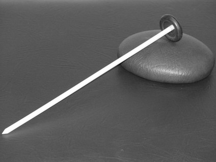
Tendon jerk
Patient
The patient should be comfortable and relaxed so that the testing can be completed with the muscle in a resting state. The position will therefore alter depending upon the muscle being tested.
Therapist
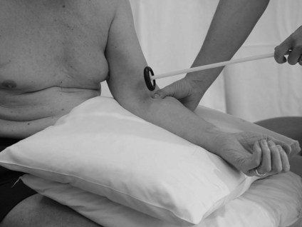
Biceps (C5, C6) (Fig. 22.2)
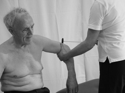
Triceps (C6, C7) (Fig. 22.3)
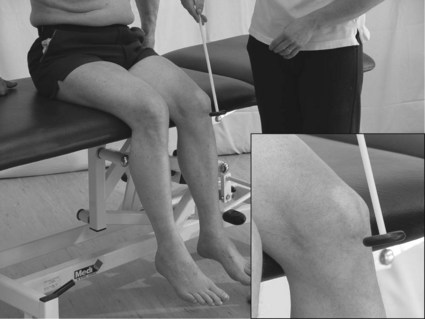
Quadriceps (L2, L3, L5) (Fig. 22.4)
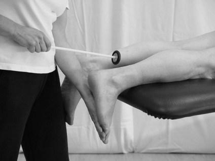
Plantar flexors (S1, S2) (Fig. 22.5)
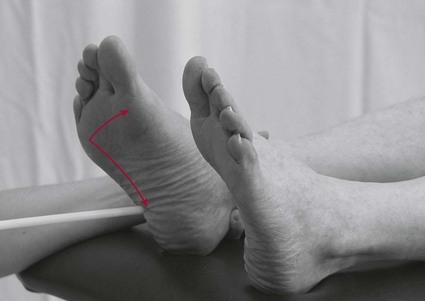
Plantar response (Babinski sign) (Fig. 22.6)
Patient
The patient should be in supine or long sitting and be well supported so that the testing can be completed with the muscles of the lower limb in a resting state.
Recording
Recording by the therapist usually consists of a simple list comprising the muscle tested and the response gained. For example:
Analysis
It is important to remember that the findings from reflex testing need to be considered alongside other assessment findings. For example, in pathologies affecting the peripheral nervous system analysis should be in conjunction with results from the assessments of myotomes (S3.31) and dermatomes (S3.24). In pathologies affecting the central nervous system analysis may be in conjunction with findings related to the assessment of muscle tone (S3.21).
References and Further Reading
Kandel ER, Schwartz JH, Jessell TM. Principles of neural science, ed 4. New York: McGraw-Hill; 2000.
Petty NJ. Neuromusculoskeletal examination and assessment: a handbook for therapists. Edinburgh: Churchill Livingstone; 2006.
Rathore SS, Hinn AR, Cooper LS, et al. Characterization of incident stroke signs and symptoms: findings from the Atherosclerosis Risk in Communities Study. Stroke. 2002;33:2718-2721.