Liver Abscesses and Granulomas: Bacteria can reach the liver via a number of different routes and form abscesses (Figs. 8-41 to 8-43). Routes include the following:
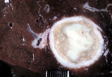
Fig. 8-41 Chronic hepatic abscesses, Corynebacterium pseudotuberculosis, liver, sheep.
Note the thick fibrous capsule and the characteristic pale caseous exudate produced by Corynebacterium pseudotuberculosis in sheep. (Courtesy College of Veterinary Medicine, North Carolina State University.)
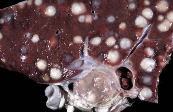
Fig. 8-42 Hepatic abscesses, Rhodococcus equi, liver, goat.
Disseminated hepatic abscesses in a goat caused by Rhodococcus equi. This lesion is more commonly found in foals. (Courtesy Dr. P. Stromberg, College of Veterinary Medicine, The Ohio State University.)
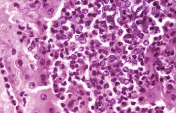
Fig. 8-43 Hepatic abscess, liver, cow.
An abscess in the liver is similar to those in other tissues and consists of an infiltrate of neutrophils, degenerating neutrophils, and necrotic tissue debris. H&E stain. (Courtesy Dr. M.D. McGavin, College of Veterinary Medicine, University of Tennessee.)
• The umbilical veins from umbilical infections in newborn animals
• The hepatic artery, as part of a generalized bacteremia
• Ascending infection of the biliary system
• Direct extension of an inflammatory process from tissues immediately adjacent to the liver, such as the reticulum
Both Gram-positive and Gram-negative organisms can cause hepatic abscesses. In adult small animals, hepatic abscesses are often caused by any of a variety of enteric species, as well as Francisella spp., Nocardia asteroides, and Actinomyces spp. Bacterial infections of the liver and subsequent formation of hepatic abscesses or foci of necrosis are especially common in neonatal foals and ruminants, in addition to feedlot cattle. In feedlot cattle, hepatic abscesses usually occur as a sequel to toxic rumenitis because damage to the ruminal mucosa allows ruminal microflora, particularly Fusobacterium necrophorum, to enter the portal circulation. After initially localizing within the liver, bacteria proliferate and produce focal areas of hepatocellular necrosis and hepatitis that can in time develop into hepatic abscesses (Fig. 8-44). Liver abscesses of cattle frequently are incidental lesions, but they can cause weight loss and decreased milk production. Less commonly, a hepatic abscess encroaches on the lumen of either a hepatic vein or the caudal vena cava. This can cause phlebitis that results in mural thrombosis, and because of the obstruction of the outflow to the venous drainage of the liver, passive congestion of the liver and portal hypertension can occur (Fig. 8-45). Detachment of portions of these mural thrombi can produce septic thromboemboli that lodge in the lungs. Rupture of hepatic abscesses directly into the hepatic vein or into the caudal vena cava occurs sporadically in cattle and may result in fatal septic embolization of the lungs. Sometimes death can be sudden from the blockage of large areas of pulmonary capillaries by the exudate. Hepatic abscesses derived from bacteria arriving via the portal vein may not be evenly distributed throughout the liver, possibly because of selective distribution of portal blood into different liver lobes, termed portal streaming. Occasionally, fungi, such as Mucor sp., that proliferate in areas of ruminal ulceration invade the portal circulation and are carried to the liver and there cause extensive areas of necrosis and inflammation (Fig. 8-46).
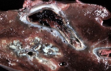
Fig. 8-44 Hepatic abscess, Fusobacterium necrophorum, liver, cow.
Foci of necrosis and abscess formation. Abscesses, such as this one, can erode the wall of a hepatic vein or the caudal vena cava, rupture, and release their contents into the bloodstream. (Courtesy Dr. P. Stromberg, College of Veterinary Medicine, The Ohio State University.)
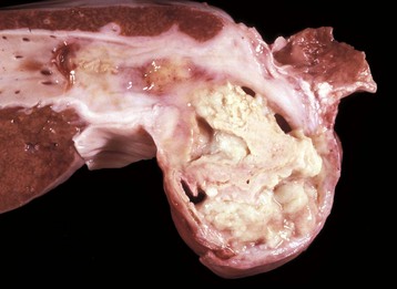
Fig. 8-45 Hepatic abscess, caudal vena cava, cow.
A hepatic abscess has eroded the wall of the vena cava, ruptured, and released its contents into the caudal vena cava. (Courtesy College of Veterinary Medicine, North Carolina State University.)
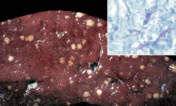
Fig. 8-46 Multiple necrotic foci, disseminated fungal infection (Mucor spp.), liver, cow.
Mucor spp. enter the portal blood after ulcerative rumenitis and cause focal necrosis and inflammation in the liver. Inset, The hyphae of the causative organism (pink) are usually evident within the granuloma. Periodic acid–Schiff (PAS) reaction. (Figure courtesy College of Veterinary Medicine, University of Illinois. Inset courtesy Dr. M.D. McGavin, College of Veterinary Medicine, University of Tennessee.)
Tuberculosis (Mycobacterium bovis) has been eradicated from almost all of the United States (US), but its occurrence in other countries varies with the effectiveness of control efforts. The primary site of the disease is pulmonary with subsequent dissemination to other organs, including the liver. Other domestic animal species can be infected with Mycobacterium bovis, and it is also a zoonotic microbe. Mycobacterium avium-intracellulare complex can occur in domestic animals, especially dogs, in the southern areas of the US. Granulomas are randomly distributed (i.e., hematogenous spread) in the liver. They have a central core of cell debris, caseation, and granulomatous inflammation surrounded by a fibrous capsule (Fig. 8-47).
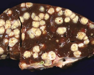
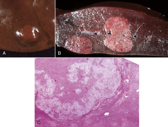
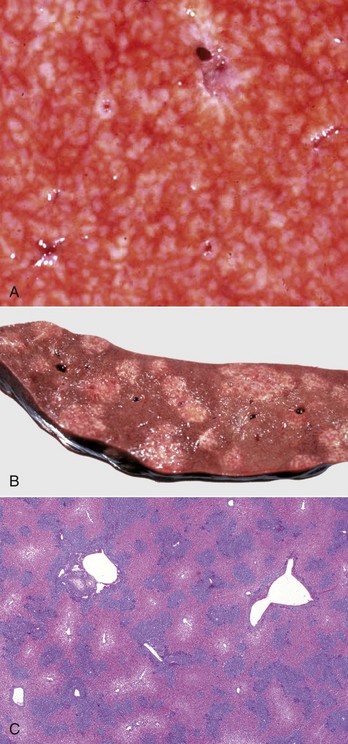
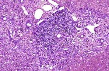
Fig. 8-47 Multiple caseous granulomas, tuberculosis, Mycobacterium bovis, liver, cow.
Hepatic tuberculosis is characterized by random multifocal pale white-to-yellow caseous granulomas on the capsular and cut surfaces. (Courtesy Dr. M. Domingo, Autonomous University of Barcelona; and Noah’s Arkive, College of Veterinary Medicine, The University of Georgia.)
Tyzzer’s Disease: This disease is caused by Clostridium piliforme (formerly Bacillus piliformis), a Gram-negative obligate intracellular parasite. It is well recognized in laboratory animals but occurs only sporadically in domestic animals. Infection is most common in foals but has been described in calves, cats, and dogs and many other species. Typically, only very young or immunocompromised animals are affected. The bacteria are found in the intestinal tract of rodents. Infection is most likely through the oral route. The mechanisms of attachment and entry into host cells are unknown. After colonization of the gastrointestinal tract, organisms penetrate into the portal venous drainage and enter the liver. The disease is characterized by enlarged, edematous, and hemorrhagic abdominal lymph nodes, hepatic enlargement, and the presence of randomly distributed, pale foci of hepatocellular necrosis surrounded by a variably intense inflammatory infiltrate of neutrophils and mononuclear cells (Fig. 8-48, A). Diagnosis requires the demonstration of the characteristic, elongated large bacilli within viable hepatocytes at the margins of necrotic foci (Fig. 8-48, B). Silver stains, such as Warthin-Starry or Gomori’s silver stain, are frequently used for this purpose (Fig. 8-48, C).
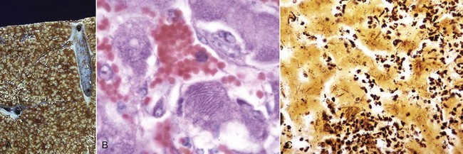
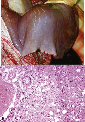
Fig. 8-48 Tyzzer’s disease (Clostridium piliforme).
A, Liver, horse. Disseminated gray-white 1- to 2-mm foci of necrosis surrounded by suppurative inflammation. B, Foal. Clostridium piliforme can be identified by the haphazard distribution of filamentous bacteria in the cytoplasm of hepatocytes. Giemsa stain. C, Foal. Clostridium piliforme can be readily seen with special stains such as Giemsa and Warthin-Starry. Warthin-Starry stain. (A courtesy Dr. R.C. Giles, University of Kentucky; and Noah’s Arkive, College of Veterinary Medicine, The University of Georgia. B and C courtesy Dr. M.D. McGavin, College of Veterinary Medicine, University of Tennessee.)
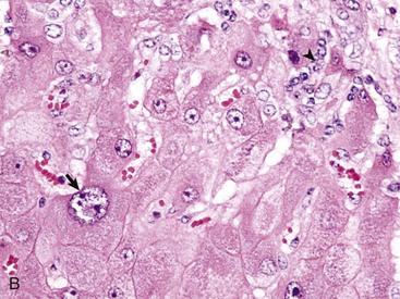
Leptospirosis: Leptospirosis is caused by infection with the Gram-negative, thin, spiral, and motile bacterium of the genus Leptospira. There are two species, of which Leptospira interrogans is capable of causing disease in animals. The taxonomy of these organisms is complicated because there are more than 23 antigenically distinct pathogenic serogroups and 200 serovars. Each serovar can differ with respect to the species affected, organs affected, and severity of disease. Leptospires enter the body through the mucous membranes or through the skin if its barrier functions have been disrupted. Contaminated water, bedding, and soil are common sources of infection because the organism is shed in urine. Fetuses can develop transplacental infection and are often aborted. Infection can involve red blood cells, kidney, liver, and a number of other tissues, depending on the infecting serovar. The liver is often involved in acute, severe leptospirosis of all domestic species because a number of serovars cause intravascular hemolytic anemia leading to ischemic injury to centrilobular areas. Furthermore, organisms can be seen in large numbers in the liver after silver staining methods, although the direct effects of leptospira toxins on hepatocytes are less well established.
Gross lesions include icterus when animals are infected with serovars that produce hemolysis. Hepatic hemorrhage and ascites can occur, depending on the course of infection and the serovar involved. In some cases, acute infection can cause focal necrosis in addition to or instead of centrilobular necrosis. A common but nonspecific change in the liver of infected dogs is dissociation of hepatocytes. Affected cells become rounded and have eosinophilic granular cytoplasm and dark, shrunken hyperbasophilic nuclei. Bile casts in canaliculi are often apparent. Kupffer cells may contain abundant hemosiderin. Infection of dogs with Leptospira grippotyphosa has been reported to produce chronic (chronic-active) hepatitis, but it is unlikely that leptospira are involved in the pathogenesis of the majority of spontaneous cases of chronic hepatitis.
Other Bacterial Infections: These diseases are grouped together because they all arise from a bacteremia that occurs during a systemic infection. A comprehensive list of systemic infections that may produce hepatocellular necrosis and hepatitis is beyond the scope of this chapter, but examples include Yersinia pseudotuberculosis, Salmonella spp. (lesions present within the liver are discrete accumulations of mixed mononuclear inflammatory cells, which often are referred to as paratyphoid nodules) and Brucella spp. infection in many species (Fig. 8-49, A and B). Haemophilus agni and Pasteurella haemolytica can present as infections in sheep. Other infections include Arcanobacter pyogenes (Actinomyces pyogenes) of the bovine fetus and neonate, Campylobacter fetus ssp. fetus in fetal and neonatal lambs (Fig. 8-50), Actinobacillus equuli infection of neonatal foals, and Nocardia asteroides infection of dogs. Yersinia tularensis (Francisella tularensis), the cause of tularemia, can occur in cats and dogs. Bacterial infections such as these may produce lesions within the liver that range from small foci of hepatic necrosis to multiple, large abscesses. Determination of the specific causative agent often depends on bacterial isolation and characterization.
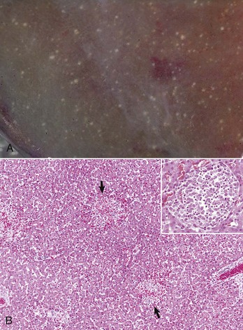
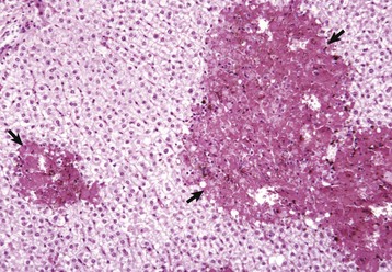
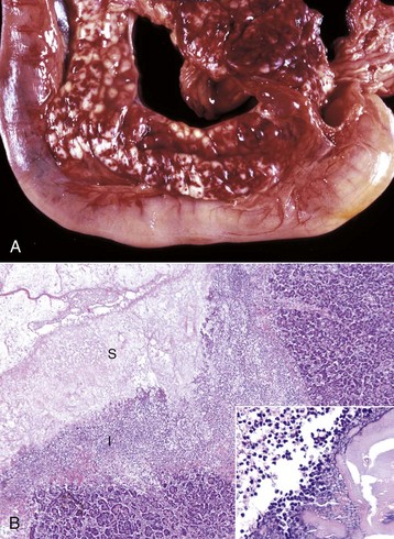
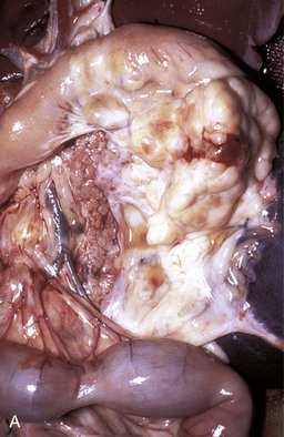
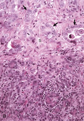
Fig. 8-49 Hepatic salmonellosis, liver.
A, Diaphragmatic surface, cow. Random 1- to 2-mm foci of focal necrosis in a cow with Salmonella septicemia. Multiple pale subcapsular foci of necrosis are evident. B, Pig, Later in the disease process, the necrotic foci are infiltrated by macrophages and form discrete granulomas termed paratyphoid nodules (arrows). H&E stain. Inset, Higher magnification of a paratyphoid nodule. H&E stain. (A courtesy Dr. M.D. McGavin, College of Veterinary Medicine, University of Tennessee. B courtesy Dr. M.D. McGavin, College of Veterinary Medicine, University of Tennessee. Inset courtesy Dr. J. Simon, College of Veterinary Medicine, University of Illinois.)
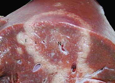
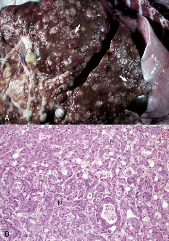
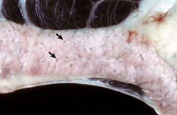
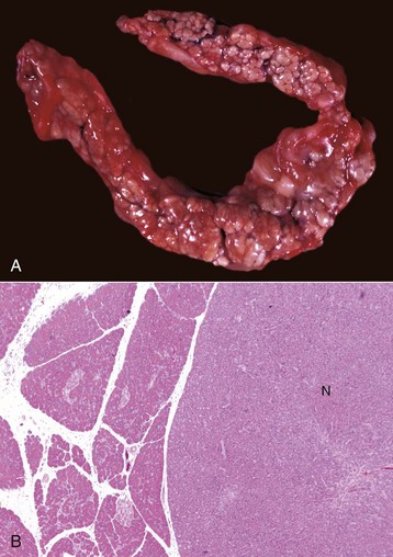
Fig. 8-50 Hepatic campylobacteriosis, multifocal necrotizing hepatitis, liver, capsular and cut surfaces, lamb fetus.
The lesion consists of a necrotic center (coagulation necrosis) (N), which in older lesions is distinctly tan and depressed. This center is surrounded by a white to gray rim of inflammatory cells (I). The cut surface (lower) illustrates the same changes and the extent of the necrosis into the hepatic parenchyma. (Courtesy Drs. C. Lichtensteiger and R. Doty, College of Veterinary Medicine, University of Illinois.)
Bacillary hemoglobinuria and infectious necrotic hepatitis, both due to species of Clostridia, are described in detail in the section on Disorders of Ruminants.
The liver can be involved in systemic infections with Toxoplasma gondii, Neospora sp., and other less common protozoa (Web Fig. 8-8). Liver lesions are usually characterized by multifocal necrosis and inflammation. Inflammatory cells include neutrophils, macrophages, and smaller numbers of other inflammatory cells. Free tachyzoites or cysts containing bradyzoites can be found within necrotic areas or adjacent to them. Although there are subtle physical differences between the organisms, immunologic testing, such as immunohistochemical staining, is a more reliable means to separate the two organisms.
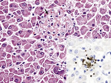
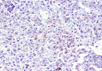
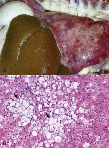
Web Fig. 8-8 Hepatitis with necrosis, toxoplasmosis, liver, cat.
Note the focus of acute hepatic necrosis (arrow). Several cysts filled with bradyzoites are present in a cell, likely a Kupffer cell. H&E stain. Inset, The brown oval structures are bradyzoites that have been labeled with DAB chromogen in an immunohistochemical reaction to confirm toxoplasmosis as the agent. IHC stain. (Courtesy Dr. K. Vashisht, College of Veterinary Medicine, University of Illinois.)
Systemic involvement with dimorphic fungi often includes the liver. There are several genera of fungi that may involve the liver including, Blastomyces, Coccidioides, Aspergillus, and Histoplasma. Histoplasmosis is a fungal disease that is endemic in the US and Canada and can occur occasionally in other areas. It is caused by Histoplasma capsulatum, a soil-dwelling organism. Dogs are affected most often. The route of infection is primarily through inhalation, although ingestion is also a possible route. In some circumstances pulmonary infections become disseminated and affect a variety of visceral organs, including the liver. Lesions in the liver consist of a multifocal distribution of granulomas with intralesional yeast forms of the organism. Numerous yeast forms can be found in the cytoplasm of macrophages and can be readily stained with the periodic acid–Schiff (PAS) reaction (Fig. 8-51, A and B).
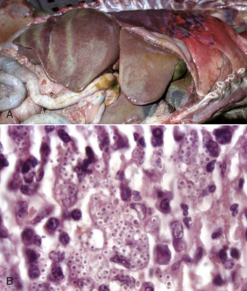
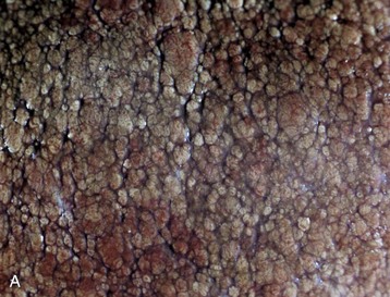
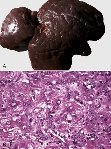
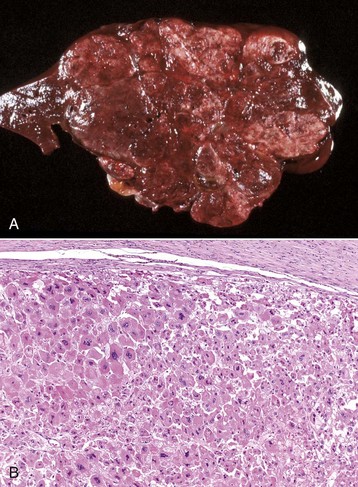
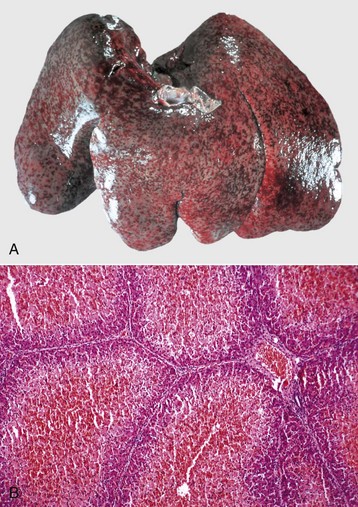
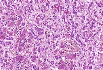
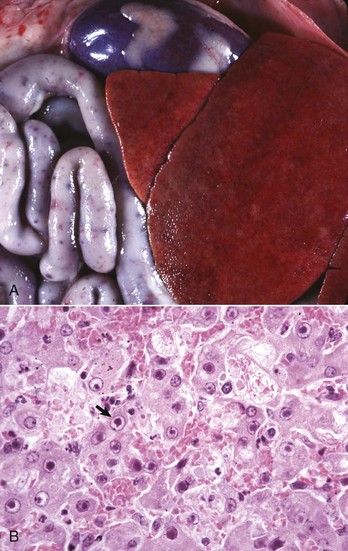
Fig. 8-51 Hepatic histoplasmosis, liver, dog.
A, In disseminated cases, Histoplasma capsulatum can involve the liver. Affected livers tend to be enlarged and pale mahogany from the diffuse hypertrophy and proliferation of Kupffer cells and macrophages. B, Note the yeast form of Histoplasma in the cytoplasm of Kupffer cells and macrophages. H&E stain. (A courtesy College of Veterinary Medicine, University of Illinois. B courtesy Dr. J. Simon, College of Veterinary Medicine, University of Illinois.)
Nematodes: Migration of larvae through the liver is a common component of a nematode’s life cycle in domestic animals. As larvae travel through the liver, they produce local tracts of hepatocellular necrosis that are accompanied by inflammation. These tracts are eventually replaced with connective tissue that matures into fibrous scars and which are especially prominent on the capsular surface (Fig. 8-52). These capsular scars appear as pale areas, and the term milk-spotted liver has been used to describe livers in pigs scarred by migrating larvae of Ascaris suum. Larvae occasionally become entrapped within the liver or its capsule and are walled off within abscesses or granulomas. Examples of chronic hepatitis or hepatic scarring as a consequence of larval migration include migration of ascarids in several species of domestic animals, such as Stephanurus dentatus in pigs, and Strongylus sp. in the horse. Infection of the liver with adult nematodes is considerably less common than larval migration. Calodium hepatica occasionally may be found in the hepatic parenchyma of dogs and cats where the ova provoke granulomatous inflammation.
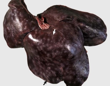
Fig. 8-52 Capsular and portal fibrosis (milk-spotted liver), Ascaris suum larval migration, liver, diaphragmatic surface, pig.
Fibrous tissue (scars) has been deposited in the migration tracks of the ascarid larvae and in adjacent portal areas (arrows). (Courtesy Dr. M.D. McGavin, College of Veterinary Medicine, University of Tennessee.)
Dogs with heartworm infection (Dirofilaria immitis) occasionally develop vena caval syndrome, also known as the postcaval syndrome, which is characterized by DIC, intravascular hemolysis, and acute hepatic failure. The syndrome typically occurs in dogs with large numbers of adult worms in the vena cava and their more usual location within the right side of the heart and pulmonary artery (Fig. 8-53). The liver is engorged with blood as a consequence of severe passive congestion from the partial blockage of the caudal vena cava. It is proposed that mechanical factors produced by the presence of large numbers of worms in the right atrium or caudal vena cava are the cause of intravascular hemolysis, which characterizes vena caval syndrome, although other theories suggest that there may be a hypersensitivity reaction to antigens released by the worms.
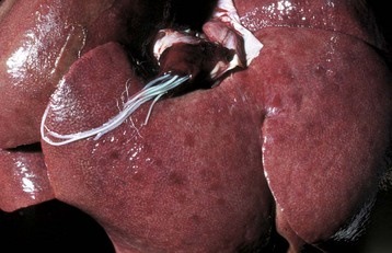
Fig. 8-53 Dirofilariasis, vena caval syndrome, caudal vena cava at the level of the liver, dog.
Large collections of adult Dirofilaria immitis are present in the caudal vena cava. The condition is rapidly fatal unless the nematodes are removed. (Courtesy Dr. C.S. Patton, College of Veterinary Medicine, University of Tennessee.)
Cestodes: A number of cestodes occur within the hepatobiliary system of domestic animals. Those cestode parasites of greatest clinical significance develop encysted forms within the liver of the intermediate hosts. The most important are larval cestodes of the genus Taenia; adults inhabit the gastrointestinal tract of carnivores and usually are innocuous to their definitive host. The ova ingested by an intermediate host develop into embryos, which penetrate the wall of the gut and then are distributed via the blood to virtually any site in the body. Parasitic cysts develop within the tissue of the intermediate host, and the life cycle of the parasite is completed when the cysts are ingested by the definitive host. Although the liver is but one organ in the intermediate host that may be affected, hepatic involvement is common because portal blood, in which embryos migrate, drains into the liver before flowing to the systemic circulation.
The adult cestode Taenia hydatigena shows up in the small intestine of dogs, whereas its intermediate stage, Cysticercus tenuicollis, appears in the peritoneal cavity of a variety of species, including horses, ruminants, and pigs (Fig. 8-54). Immature cysticerci migrate in the liver and can induce extensive damage if infection is heavy; lesions present are comparable to those induced by migration of immature Fasciola hepatica.
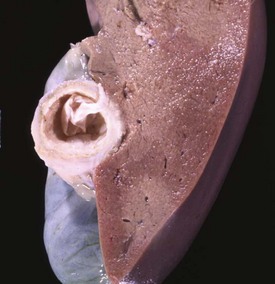
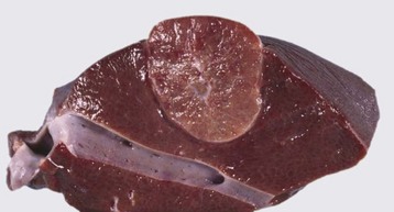
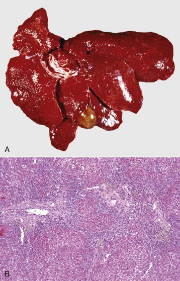
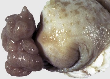
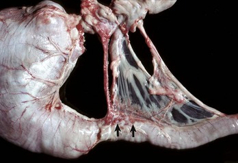
Fig. 8-54 Cysticercosis, liver, cut surface, sheep.
The thick fibrous capsule usually indicates the death of the larva. (Courtesy Dr. K. Read, College of Veterinary Medicine, Texas A&M University; and Noah’s Arkive, College of Veterinary Medicine, The University of Georgia.)
Hydatid liver disease is common in some countries. Echinococcus granulosus is a cestode that parasitizes canids as the definitive host, and hydatid cysts can develop in many different intermediate host animal species, including humans. The dog-sheep cycle is most important in many geographic areas. Pastured cattle are also commonly affected in other geographic locations. Adult worms in the intestines of dogs pass proglottids into the dog’s stool and thereby contaminate pastures. Ova are then ingested by sheep, cattle, or other species. Embryos may develop into hydatid cysts in virtually any organ in the intermediate host, but the liver and lungs are commonly affected. These cysts are usually less than 10 cm in diameter but can attain quite a spectacular size, particularly in humans. Hydatid cysts, even when present in large numbers, rarely cause overt clinical signs of disease in domestic animals.
Cestode adults occurring within the hepatobiliary system include Stilesia hepatica, Stilesia globipunctata, and Thysanosoma actinoides, all of which can inhabit the bile duct of ruminants. Infections with these parasites may result in chronic inflammation of the biliary tract, but they usually do not produce clinical signs of hepatic dysfunction.
Trematodes: The majority of parasitic hepatic injury caused by trematodes is produced by members of three major families. These include the families Fasciolidae, Dicrocoelidae, and Opisthorchidae.
The principal liver fluke disease of sheep and cattle and occasionally other species is caused by Fasciola hepatica. Hepatic fascioliasis occurs throughout the world in areas where climatic conditions, typically in low swampy areas, are suitable for the survival of aquatic snails, which serve as intermediate hosts for the parasites. Adult Fasciola hepatica are leaf-shaped parasites that inhabit the biliary system; their eggs pass via the bile to the intestinal tract and eventually are passed in the feces. Larvae (miracidium) then must develop in the snail intermediate host (genus Lymnaea). Cercariae that leave the snail encyst on herbage where they develop into infectious metacercariae. Metacercariae are ingested by the ruminant host and penetrate the wall of the duodenum to enter the peritoneal cavity and subsequently enter the liver. They migrate within the liver before taking up residence within the bile ducts. Migration of immature flukes through the liver produces hemorrhagic tracts of necrotic liver parenchyma. These tracts are grossly visible and in acute infection are dark red, but with time become paler than the surrounding parenchyma. Repair is often by fibrosis. A variety of untoward sequelae can follow these migrations, including acute peritonitis; hepatic abscesses; death of the host as a consequence of acute, widespread hepatic necrosis produced by a massive infiltration of immature flukes; and the proliferation of spores of Clostridium haemolyticum or Clostridium novyi in necrotic tissue, which causes the subsequent development of bacillary hemoglobinuria or infectious necrotic hepatitis, respectively.
Mature flukes reside in the larger extrahepatic and intrahepatic bile ducts and cause cholangitis. Chronic cholangitis and bile duct obstruction lead to ectasia and stenosis of the ducts and periductular fibrosis that thickens the walls so that the ducts become increasingly prominent. Mineralization may occur producing the classic “pipestem” appearance of diseased bile ducts. The contents of the bile ducts are often dark brown and viscous caused by a combination of abnormal bile, cellular debris, and the iron-porphyrin pigment excreted by the flukes. Obstruction of the ducts leads to cholestasis. Animals with chronic liver fluke disease are often in poor body condition.
Fasciola gigantica and Fascioloides magna are important causes of liver fluke disease of ruminants in some parts of the world. Fasciola gigantica is most common in areas of Africa and surrounding countries, and Fascioloides magna is found in North America. The adults of Fasciola gigantica and Fasciola hepatica reside in the bile ducts (Fig. 8-55). In contrast, adult Fascioloides magna, whose normal hosts are elk and white-tailed deer, reside in the hepatic parenchyma in aberrant hosts, such as cattle and sheep. In cattle, the immature Fascioloides magna flukes cause extensive tissue damage as they migrate through the liver (Fig. 8-56), but the adults are enclosed by fibrous connective tissue in cysts containing a black fluid. In sheep and goats, the flukes continuously migrate through the liver, causing extensive damage and eventual death.
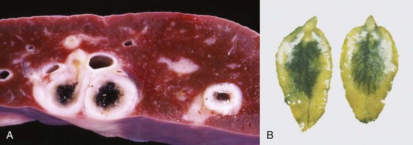
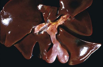
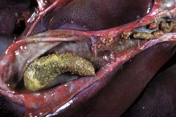
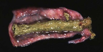
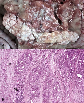
Fig. 8-55 Fasciola hepatica infection.
A, Chronic intrahepatic cholangitis (Fasciola hepatica), liver, cow. When Fasciola hepatica metacercariae are ingested, they migrate to the liver and then take up residence within the bile ducts. Mature flukes reside in the larger extrahepatic and intrahepatic bile ducts and cause chronic cholangitis and bile duct obstruction that lead to ectasia and stenosis of the ducts and periductular fibrosis that thickens the walls so that the ducts become increasingly prominent, as shown here. B, Adult Fasciola hepatica are leaf-shaped flukes that inhabit the biliary system; their eggs pass via the bile into the intestinal tract and eventually are passed in the feces. (A courtesy Dr. K. Read, College of Veterinary Medicine, Texas A&M University; and Noah’s Arkive, College of Veterinary Medicine, The University of Georgia. B courtesy Dr. T. Boosinger, College of Veterinary Medicine, Auburn University; and Noah’s Arkive, College of Veterinary Medicine, The University of Georgia.)
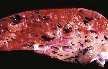
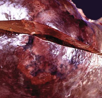
Fig. 8-56 Fluke migration tracts, fascioloidiasis, liver, cow.
Migration of Fascioloides magna through the bovine liver produces extensive parenchymal damage. A black excretory pigment deposited by the fluke discolors the migration tracks black. (Courtesy Dr. J. Wright, College of Veterinary Medicine, North Carolina State University; and Noah’s Arkive, College of Veterinary Medicine, The University of Georgia.)
Other trematodes that may inhabit the bile ducts include Dicrocoelium dendriticum in horses, ruminants, pigs, dogs, and cats; Eurytrema pancreaticum and Eurytrema coelomaticum in ruminants; Opisthorchis tenuicollis in pigs, dogs, and cats and Opisthorchis felineus in dogs and cats; Pseudamphistomum truncatum, Metorchis conjunctus, Metorchis albidus, Parametorchis complexus, Concinnum procyonis, and Platynosomum fastosum in dogs and cats. All are capable of inducing changes similar to but usually considerably milder than those caused by Fasciola hepatica. In addition, they occasionally cause obstruction of the biliary ducts.
Cats, and less often dogs, can develop pronounced chronic cholangitis from infections with flukes, most often Opisthorchiidae. Microscopically, larger intrahepatic bile ducts are dramatically thickened by concentric fibrosis and the duct lumen is usually dilated, often with papillary projections of biliary epithelium into the lumen (Fig. 8-57). A mild-to-moderate inflammatory infiltrate of neutrophils and macrophages is often found in and around the ducts, and the portal tracts are infiltrated by neutrophils, lymphocytes, and plasma cells. Eosinophils are generally uncommon. It is often difficult to detect adult flukes or ova in affected animals.
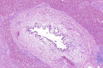
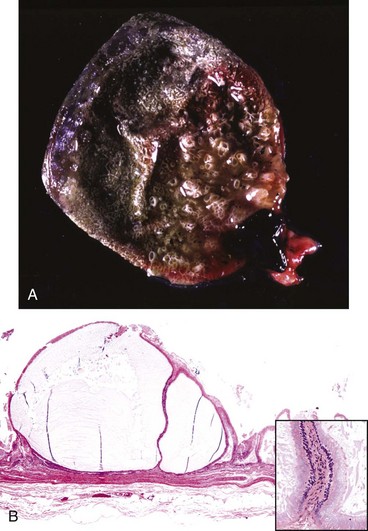
Fig. 8-57 Chronic intrahepatic cholangitis, liver, cat.
Fluke infections of the biliary tree of cats produce a characteristically pronounced periductular fibrosis, dilated bile duct, and papillary projections of biliary epithelium, although flukes may be difficult to find. H&E stain. (Courtesy Dr. J.M. Cullen, College of Veterinary Medicine, North Carolina State University.)
Dogs can be infected with the schistosome Heterobilharzia americana, which is normally a parasite in raccoons. Ova shed into water by infected raccoons release miracidia, which penetrate host snails. Dogs become infected when their skin is penetrated by cercariae, which are released from the intermediate snail hosts. Granulomatous lesions of the liver, pancreas, intestines, and mesentery result when ova released by adult schistosomes lodge in affected tissue and incite an inflammatory reaction.
The liver is subjected to toxic injury more often than any other organ. This is not surprising because the portal vein blood that drains from the absorptive surface of the intestinal tract flows directly to the liver. Thus the liver is exposed to virtually all ingested substances, including plant, fungal, and bacterial products, and metals, minerals, drugs and other chemicals that are absorbed into the portal blood. Hepatotoxic injury can range along a spectrum from pure hepatocellular injury to pure biliary injury and a mixed pattern of injury that involves both components of the liver.
Hepatotoxic drugs can be divided into two basic categories. Predictable hepatotoxins are those that affect the large majority of animals that are exposed, and the effect is evident within a similar dose range. The majority of recognized hepatotoxins in veterinary medicine fall into this category; acetaminophen and pyrrolizidine alkaloids are examples of predictable hepatotoxins. Toxic injury, even with predictable toxins, is not always uniform, however. A variety of factors influence the severity of injury induced by a toxin; these factors include age, sex, diet, endocrine function, genetic constitution, and diurnal factors. It is therefore not surprising that responses of individual animals exposed to the same toxin can vary considerably. Idiosyncratic drug reactions are characterized as responses seen in only a small minority of exposed individuals. There are a number of possible mechanisms for idiosyncratic drug reactions, including atypical metabolism as a result of inheritance of rare genes encoding enzymes involved in drug metabolism or deletions of genes encoding certain enzymes or immunologic responses to drugs or modified hepatocyte proteins. Interactions with other drugs or effects of diet and health status can also play a role in idiosyncratic toxicity. Diazepam toxicity in cats is an example of an idiosyncratic toxicity.
The response of the liver to acute hepatotoxic injury depends on the mechanism and site of toxic insult. By far, the most common pattern of acute liver toxicity is centrilobular necrosis. The mechanisms for this pattern of injury involve metabolism by the cytochrome p450 system and are discussed later. Certain chemicals that are uncommonly encountered produce periportal necrosis. These chemicals are able to produce a toxic effect without requiring metabolism by the cytochrome p450 system and include white phosphorus (once used as a rodenticide) and allyl alcohol.
It should be kept in mind that a single episode of nonlethal hepatotoxic injury in an otherwise healthy animal is difficult to detect histologically within a few days after the episode. Within 48 to 72 hours, macrophages clear cell debris, and hepatocytes begin to undergo mitosis to replace the lost cells. Within a week or less, the liver regains a normal histologic appearance, unless there is massive necrosis, which can lead to collapse of the hepatic connective tissue scaffolding and subsequent fibrosis surrounding the central vein. Chronic toxic liver injury, manifest either as repeated bouts of toxin exposure or more consistent daily exposure (e.g., through dietary contamination) can lead to activation of hepatic stellate cells within the space of Disse or related myofibroblasts in the portal areas and the connective tissue of the central vein area, which may then initiate synthesis of ECM leading to hepatic fibrosis. In addition, chronic liver injury can lead to disruption of the normal framework that supports the hepatic architecture and leads to hepatic fibrosis. Sufficient injury also can produce nodules of regenerative hepatocytes that are surrounded by bands of fibrosis that connect central vein areas to each other, connect portal tracts to each other, or bridge portal tracts to centrilobular areas. This pattern is recognized as cirrhosis.
Hepatocytes are not the only cell type in the liver that can be affected by toxic drugs. The biliary epithelium is susceptible to injury from trimethoprim-sulfa and the mycotoxin, sporidesmin, Kupffer cells to endotoxin, sinusoidal endothelial cells to arsenicals and some pyrrolizidine alkaloids, and the hepatic stellate cells to vitamin A excess. Bile duct necrosis or proliferation can disrupt bile flow. Activated Kupffer cells can release cytokines that affect the type and degree of inflammation within the liver. Hepatic stellate cells play a central role in hepatic fibrosis, as is discussed later. Damage to endothelial cells can affect blood flow through the liver.
Hepatotoxic liver injury can be classified into the following six categories based on the cellular target involved:
1. The most frequent mechanism of hepatocellular injury involves production of injurious metabolites by the cytochrome p450 system. This family of enzymes is located in the smooth endoplasmic reticulum of hepatocytes primarily, although they are also found in many other cells of the body. A major role of cytochrome p450 enzymes is to metabolize lipid-soluble chemicals into water-soluble compounds for excretion from the body in bile or urine. In the first step of this three-step process, termed biotransformation, chemicals are bioactivated to a high-energy reactive intermediate molecule, termed phase I, in preparation for the second step, phase II, which involves formation of covalent bonds with polar molecules, such as glucuronic acid. This conjugation forms a water-soluble metabolite that can be excreted. Phase III involves the transport of these molecules across the cell membrane into the lumen of the canaliculus by molecular pumps. In some circumstances, such as an overdose, the high-energy reactive metabolites can form covalent bonds with other cellular constituents, such as proteins, and nucleic acids termed adducts. In acute toxicity, adducts with essential cellular enzymes may lead to cell injury or death. Toxic hepatocellular injury of this category occurs most often in the centrilobular area of the liver because this is the region of the liver with the highest concentration of cytochrome p450 enzymes. For example, acetaminophen is metabolized by cytochrome p450 enzymes to N-acetyl-p-benzo-quinone imine (NAPQI), a free radical that is responsible for the toxicity of the parent compound. Lesions induced by acetaminophen are most severe in the centrilobular (periacinar) areas, where the active form of the chemical is present in greatest concentration. Many plant toxicities are examples of injury produced by this mechanism.
2. Adduct formation between drugs and cellular enzymes, other proteins, or nucleic acids can alter the cellular constituents sufficiently that they become neoantigens, as may be the case with halothane toxicity. These neoantigens, like other foreign antigens, can be processed in the cytoplasm, transported to the cell surface, presented as antigens, and recognized by the immune system. Consequently, the immune system may develop an inflammatory response toward hepatocytes or biliary epithelium that contain the adducts. Both cellular and humoral immunity can be involved. Injury can occur through direct cellular cytotoxicity and antibody-dependent cellular cytotoxicity. Although this mechanism is not well characterized in clinical veterinary medicine, it is likely to occur on occasion.
3. Certain toxins, including retained or excess hydrophobic bile acids, can trigger apoptosis (individual cell necrosis), by direct stimulation of proapoptotic pathways in the hepatocytes. Alternatively, apoptosis can be stimulated by immune-mediated events, such as those discussed previously, which lead to the release of TNF-α or activate Fas pathways.
4. Injury that damages cell membranes and disables enzymes responsible for calcium homeostasis, as seen in carbon tetrachloride toxicity, can lead to an influx of calcium. One consequence of the increased intracellular calcium is activation of proteases that damage actin filaments. Blebbing and lysis of the cell membranes can result.
5. Chemicals that bind to and disrupt the molecular pumps that secrete bile constituents into the canaliculi, such as estrogen and erythromycin, can produce cholestasis. More extensive hepatocellular injury that affects canalicular pumps and hepatocytes may produce cholestasis by disrupting the actin filaments situated around the bile canaliculi and preventing the normal pulsatile contractions that move bile through the canalicular system to the bile ducts.
6. Hepatocyte injury or death can follow mitochondrial damage, as seen with some toxic antiviral nucleosides or intravenous tetracycline administration. Chemical or reactive oxygen species–induced injury to mitochondrial membranes, enzymes, or DNA can inhibit or disrupt mitochondrial function. Disruption of the electron transport chain can release reactive oxygen species, such as superoxide, which can produce widespread cellular damage. Damaged mitochondria do not produce sufficient adenosine triphosphate (ATP) to power the essential functions of the hepatocytes. Also, β-oxidation of lipids is reduced once the mitochondria are damaged, which leads to intrahepatic lipid accumulation (microvesicular steatosis) and diminished energy production. Damaged mitochondria may release cytochrome-c, triggering apoptosis, or if disruption of mitochondrial function is sufficient, hepatocyte necrosis ensues.
Hepatotoxic Bacteria Blue-Green Algae: Blue-green algae are classified in the phylum Monera, division Cyanophyta; are considered to be more closely related to bacteria; and are no longer considered members of the plant family. Several genera of blue-green algae, including Anabaena, Aphanizomenon, and Microcystis, can cause lethal poisoning of livestock and less commonly small animals such as dogs and cats. Algal blooms usually occur in late summer or early fall because of the warm temperatures, long hours of sunlight, and abundance of essential nutrients. Dead and dying algae, which contain preformed toxins, such as microcystin LR, a cyclic heptapeptide, accumulate on the surface of bodies of water and are ingested by livestock. Secondary bacterial growth in dying algae may contribute to toxin formation. Signs develop rapidly and include diarrhea, prostration, and death. Gross lesions include hemorrhagic gastroenteritis and a red, swollen, hemorrhagic liver. Histologically, centrilobular, or even massive, hepatic necrosis and hemorrhage is evident. Animals that survive the acute manifestations may develop clinical signs of chronic liver disease. Other preformed toxins that affect different organ systems, including the nervous system, have also been identified in blue-green algae.
Hepatotoxic Plants: Toxic plants of great variety cause hepatic injury in domestic animals. A comprehensive discussion of each is beyond the scope of this chapter.
Pyrrolizidine Alkaloid-Containing Plants: Pyrrolizidine alkaloids are found in many plant families, including Compositae, Leguminosae, and Boraginaceae, that occur throughout much of the world. The most important genera are Senecio, Cynoglossum, Amsinckia, Crotalaria, Echium, Trichodesma, and Heliotropium. Approximately 100 different alkaloids are recognized; toxic effects depend on which alkaloids are present within ingested plants. Ingested alkaloids are converted to pyrrolic esters by hepatic cytochrome p450 enzymes. These esters are alkylating agents, which react with cytosolic and nuclear proteins and nucleic acids. Pigs are particularly susceptible to pyrrolizidine alkaloid intoxication, sheep considerably less so, and cattle and horses are intermediate in susceptibility. Most cases of intoxication arise from chronic intoxication, and the gross lesion is typically hepatic fibrosis (Fig. 8-58, A). The characteristic histologic lesions of pyrrolizidine alkaloid intoxication are the megalocytes, which are hepatocytes with enlarged nuclei and increased cytoplasmic volume. Megalocytes may be many times the size of normal hepatocytes (Fig. 8-58, B). Megalocytes are the result of the antimitotic effects of pyrrolizidine alkaloids, which prevent cell division but not DNA synthesis because the hepatocytes attempt to divide to replace those that have undergone necrosis. This change, although indicative of pyrrolizidine alkaloid intoxication, is not pathognomonic because it occurs with other toxins such as aflatoxins and nitrosamines. Typically, chronic pyrrolizidine intoxication is accompanied by hepatic fibrosis, biliary proliferation, and in some circumstances, nodular regeneration of parenchyma. Nodular regeneration does not always occur because hepatocyte proliferation can be inhibited by pyrrolizidines; however, exposure is not likely to be constant, and there may be periods during which hepatocyte replication can occur, such as the end of the dry season when more desirable plant species reappear. Species differences may also have an effect on the hepatic response to pyrrolizidines because cattle have regenerative nodules more often than horses. Chronic hepatic damage can lead to hepatic failure and its associated constellation of signs (described in detail earlier).
Fig. 8-58 Chronic pyrrolizidine hepatotoxicity, cow.
A, Chronic pyrrolizidine intoxication produces a fibrotic and sometimes distorted liver with an irregular capsular surface. B, Greatly enlarged hepatocytes (megalocytes) (arrow) and hyperplasia of biliary epithelium (arrowhead) in the persisting parenchyma are typical of pyrrolizidine toxicity. H&E stain. (A courtesy Dr. P. Carbonell, School of Veterinary Science, Melbourne. B courtesy Dr. M.D. McGavin, College of Veterinary Medicine, University of Tennessee.)
Cycads: Cycads are primitive palmlike plants that inhabit tropical and subtropical regions. They contain cycasin and macrozamin, nontoxic glycosides, which after ingestion are deconjugated by intestinal bacteria to release a toxic metabolite, methylazoxymethanol. After absorption into the portal vein, hepatic metabolism of this compound yields alkylating agents, leading to acute or chronic liver injury. Acute injury is manifested as acute centrilobular necrosis. Chronic hepatic lesions in cattle include hepatocellular megalocytosis caused by the mitoinhibitory effects of alkylating agents and nuclear hyperchromasia and varying degrees of hepatic fibrosis. Chronic cycad poisoning in cattle causes a nervous disease with progressive proprioceptive deficits in the hind legs because of “dying back” of axons in the dorsal funiculus and the spinocerebellar and corticospinal tracts. Acute intoxication is more common in sheep than in other species and produces acute gastrointestinal dysfunction and centrilobular hepatic necrosis. Dogs can also be intoxicated by cycads.
Cholestatic or Crystal-Associated Plant Intoxications: The ornamental shrub Lantana camara produces toxic pentacyclic triterpenes, lantadene A and B, that primarily produce a syndrome of chronic cholestasis in grazing animals. Bile accumulation is evident within the canaliculi, hepatocytes, and Kupffer cells.
Primarily in South Africa, Tribulus terrestris (puncture vine) ingestion by sheep can produce a fatal disorder (geeldikkop) characterized by icterus, biliary injury, and secondary photosensitization. Histologically, there is abundant crystalline material within and obstructing the bile ducts. Crystals may also be found in Kupffer cells. In various parts of the world, a similar pattern of crystal deposition can be seen in sheep and goats that graze a number of grass species, including Panicum sp., that contain steroidal saponins.
Secondary photosensitization and icterus can occur in all of these conditions as the result of impaired bile secretion.
Mycotoxins: Mycotoxins are secondary metabolites of fungi, that is, their production is not necessary for the survival of the fungus. The amount of toxin synthesized by a given strain of fungus reflects the genetic constitution of the particular strain, presence of appropriate substrate, temperature, humidity, and available nutrients. There are several hepatotoxic mycotoxins of veterinary significance.
Aflatoxin: The fungus Aspergillus flavus is the most important source of aflatoxins. Aflatoxin B1 is the most common form and is also the most potent toxin and carcinogen. Aflatoxins are usually elaborated during storage of fungus-contaminated feed, particularly in humid conditions, and may be present in many crops, including corn, peanuts, and cottonseed. These can be incorporated into commercial food leading to significant outbreaks of acute toxicity in dogs. Aflatoxins are converted to toxic intermediates by hepatic cytochrome p450 enzymes. Carcinogenic, toxic, and teratogenic effects of aflatoxins reflect binding of the toxic intermediates to cellular DNA, RNA, or proteins. Pigs, dogs, horses, cattle, and ducks, especially younger animals, are sensitive to the toxic effects of aflatoxins, whereas sheep are more resistant. Acute aflatoxin intoxication is rare in horses and cattle because an inordinately large amount of contaminated feed would have to be ingested to achieve a sufficient dose. Acute aflatoxicosis in dogs is characterized by hemorrhagic central to massive necrosis. Steatosis and biliary proliferation also may occur. Chronic intoxication is more common than acute intoxication and results in ill-thrift, increased susceptibility to infection, and occasionally signs of hepatic failure. Affected livers are firm and pale and microscopically are characterized by steatosis and necrosis of hepatocytes, biliary hyperplasia, centrilobular to bridging fibrosis, and cellular atypia of hepatocytes, characterized by variable cell size and variable nuclear size (Fig. 8-59, A and B).
Fig. 8-59 Chronic hepatic aflatoxicosis.
A, Postnecrotic scarring, pig. Chronic aflatoxicosis produces a shrunken and fibrotic liver from collapse of areas of massive necrosis and condensation of the fibrous stroma. B, Histologic appearance. Chronic aflatoxicosis is characterized by variable amounts of steatosis (fatty change), biliary hyperplasia, and cellular atypia in hepatocytes. H&E stain. (A courtesy Dr. M.D. McGavin, College of Veterinary Medicine, University of Tennessee. B courtesy Dr. J. Simon, College of Veterinary Medicine, University of Illinois.)
Phomopsins: Phomopsins are toxic metabolites of the fungus Phomopsis leptostromiformis. The fungus grows on lupines (Lupinus sp.), and cattle, sheep, and occasionally horses that graze contaminated lupine stubble develop hepatic injury. Hepatic dysfunction is usually chronic, and the liver is atrophic and fibrotic. The microscopic appearance of affected livers is characterized by diffuse scattered hepatocyte necrosis with a background of mitotic figures, often appearing to be arrested in metaphase. Later in the course of the disease, diffuse fibrosis and biliary hyperplasia predominate. Signs of hepatic failure, including photosensitization, may occur in affected animals. This mycotoxicosis should not be confused with the condition known as lupinosis, which is caused by naturally occurring alkaloids in lupines that are capable of inducing skeletal deformities but not obvious hepatic injury.
Sporidesmin: The mycotoxin sporidesmin is produced by Pithomyces chartarum, a fungus that grows particularly well in dead rye grass (Lolium perenne), a common pasture plant in New Zealand and Australia. The majority of the toxin is concentrated into the fungal spores, and when a sufficient amount of the spores is ingested by sheep and to a lesser extent, cattle, the toxin is secreted into the biliary tree in an unconjugated form that produces necrosis of the epithelium of large intrahepatic and extrahepatic biliary ducts with minimal inflammation. Cholestasis with a concurrent failure to excrete phylloerythrin frequently leads to photosensitization with skin lesions predominantly on the head, thus the common name facial eczema. Acute cases are characterized by a bile-stained liver with prominent small-caliber bile ducts. These are dilated by bile in the lumens and surrounded by periductal edema. In chronic cases of facial eczema, the bile ducts become thickened by fibrosis secondary to biliary epithelial necrosis and subsequent inflammation (chronic cholangitis). Perhaps because of streaming of blood in the portal vein, the left lobe of the liver (although this lobe occupies the ventral portion of the ruminant liver), which may have an increased proportion of blood draining from the small intestine, is usually most severely affected and in severe cases undergoes atrophy and fibrosis.
Mushrooms: Poisonous mushrooms, such as Amanita sp. and others, can cause acute fatal liver necrosis. Intoxication by Amanita phalloides, known as the Death Cap, is caused by a group of toxins termed toxic cyclopeptides. This species is particularly toxic; a single gram of this mushroom is sufficient to kill a human, and even smaller amounts are likely to prove fatal to dogs. Amatoxin, an octapeptide, is in particular responsible for hepatocellular injury. The mechanism of injury is attributed to inhibition of RNA polymerase II function disrupting DNA and RNA transcription. Gross lesions usually consist of hepatic hemorrhage and a shrunken liver because of the loss of hepatocytes. Hepatocellular steatosis, hemorrhage, and centrilobular to massive necrosis are the typical lesions. Death from liver failure may occur 3 to 4 days after the onset of clinical signs. Phalloidin, a toxic heptapeptide found in Amanita sp., causes disruption of intracellular actin filaments, leading to cell injury or death. It is a less significant toxin in natural exposure because of the limited absorption from the digestive tract. Other mushroom species contain different toxic agents.
Xylitol: The artificial sweetener xylitol is used in various food items and snacks prepared for diabetics or dieters. Although innocuous to humans, xylitol can be acutely toxic to dogs. After ingesting as little as 0.5 gm/kg, affected dogs can develop hyperinsulinemia, hypoglycemia, icterus, and acute, severe centrilobular to massive hepatic necrosis.
Phosphorus: Phosphorus occurs in two forms: red phosphorus and white phosphorus. Red phosphorus is unimportant as a toxin, but white phosphorus was previously used as a rodenticide. The mechanism of phosphorus toxicity is unclear, although it apparently is directly toxic. Poisoning is first indicated by signs of gastroenteritis and subsequently by microscopic lesions of steatosis of hepatocytes and periportal necrosis. The pattern of periportal necrosis is unusual because most toxic liver injury occurs in the centrilobular region of the liver. This is explained by the fact that white phosphorus does not require metabolic transformation to a reactive intermediate by cytochrome p450 enzymes, which are most concentrated in the centrilobular region of the liver lobule.
Carbon Tetrachloride: Carbon tetrachloride is the classic example of a hepatotoxin that must be bioactivated by the mixed-function oxidase system to produce a toxic intermediate form. Although once used widely, it is only occasionally used as an anthelmintic. Carbon tetrachloride produces centrilobular hepatic necrosis and steatosis of surviving hepatocytes (see Chapter 1).
Metals: Several metals can cause toxic hepatic injury. Excessive iron supplementation in dogs and cats may result in excessive storage of iron and subsequently hepatic disease caused by iron overload, termed hemochromatosis. Two specific syndromes of iron poisoning are iron-dextran intoxication of piglets and ferrous fumarate intoxication of newborn foals. Severe cases of these two toxicities are characterized by massive hepatic necrosis. Intoxication of foals with ferrous fumarate occurred after its use as a component of a specific dietary supplement, and was characterized by massive necrosis and also a remarkable amount of hyperplasia of bile ducts and cholangioles, possibly with oval cell proliferation, despite the short clinical course of the disease. Iron-dextran is frequently administered intramuscularly to suckling pigs to prevent anemia, but administration of iron-dextran has occasionally resulted in significant mortality, and affected pigs die soon after injection.
Copper toxicity is discussed in separate sections on Disorders of Ruminants and Canine Chronic Hepatitis.
Hepatotoxic Therapeutic Drugs: There are a variety of drugs that have a proven therapeutic application but can cause significant acute or chronic hepatic injury in some animals. A partial list of hepatotoxic therapeutic drugs in dogs and cats is presented in Web Table 8-2. Clearly, these drugs would not be used if the proportion of injured animals was high, but it is important to keep in mind that many drugs have the potential to cause hepatic injury in some patients. The mechanisms by which these drugs cause injury vary by species and by individual. Some therapeutic drugs are predictable toxins, and all members of a particular species are susceptible to liver injury if a sufficient dose is given. But because the therapeutic effect occurs at a lower dose than the toxic dose, liver injury occurs only when overdoses are ingested. Hepatic metabolism (bioactivation) of these compounds is likely to be involved because the site of liver injury is typically centrilobular. Cats are more susceptible than dogs to intoxication by many chemicals because they are relatively deficient in hepatic glucuronyltransferase activity. This phase II enzyme forms conjugates between bioactivated (phase I) xenobiotics and glutathione. When phase II metabolism is overwhelmed, injurious bioactivated products cause liver injury. Cats are more sensitive to acetaminophen intoxication than dogs because of this relative enzyme deficiency. Other therapeutic drugs are idiosyncratic toxins, and they affect only a small minority of patients. The mechanism of injury is not known but may be a consequence of inherited differences in hepatic enzyme content and activity, atypical immune reactions to drug metabolites, or novel antigens created when drug metabolites bind to cellular proteins. For example, the antiinflammatory drug carprofen can occasionally cause acute hepatic necrosis in a variety of dogs, but certain breeds of dogs, such as Labrador retrievers, may be affected more often than others. The tranquilizer diazepam can cause acute fatal hepatic injury in some cats, but the majority of treated cats are unaffected, and dogs do not seem to be adversely affected.
WEB TABLE 8-2
Selected Therapeutic Agents with Potential to Cause Hepatic Injury in Small Animals
Drug | Species Primarily Affected |
Acetaminophen | Dog, cat |
Amiodarone | Dog |
Aspirin | Dog, cat |
Carprofen | Dog |
Diazepam | Cat |
Diethylcarbamazine-oxibendazole | Dog |
Glipizide | Dog |
Glucocorticoids | Dog |
Griseofulvin | Cat |
Halothane | Dog |
Ketoconazole/Itraconazole | Dog |
Lomustine (CCNU) | Dog |
Manganese chloride | Dog |
Mebendazole | Dog |
Megestrol acetate | Cat |
Methimazole | Dog |
Methoxyflurane | Dog |
Mibolerone | Dog |
Methotrexate | Dog |
Oil of Pennyroyal | Cat |
Phenobarbital | Dog |
Phenylbutazone | Dog |
Phenytoin | Dog |
Primidone | Dog |
Stanozolol | Cat |
Tetracycline | Dog, cat |
Thiacetarsamide | Dog, cat |
Trimethoprim-sulfa | Dog |
Chronic liver toxicity has been described in dogs receiving any of the anticonvulsants, primidone, phenytoin, and phenobarbital for prolonged periods. The mechanism of hepatotoxicity is unknown. Only a small proportion of dogs receiving these drugs are affected, and these dogs frequently have signs of hepatic failure. The liver is small and has widespread hepatic fibrosis and nodular regeneration (end-stage liver).
A variety of extrahepatic disorders, usually affecting the gastrointestinal tract, can result in hepatocellular injury and hepatic dysfunction. Acute hemorrhagic pancreatitis of dogs, for example, sometimes is accompanied by icterus and increased activities of hepatic enzymes in serum. Release of various toxins and inflammatory mediators from the injured pancreas into the portal vein showers the liver with a variety of injurious substances. Similarly, movement of hepatotoxic substances, such as endotoxins, into the portal vein can occur as a consequence of diseases that disrupt the mucosal barrier of the intestine. Some cases of chronic inflammation of the colon can result in chronic hepatic inflammation as well. Accumulation of inflammatory cells within the portal triads may accompany blood-borne infection or abdominal sepsis (nonspecific reactive hepatitis).
The liver is particularly susceptible to the effects of hypoxia; thus any disease that causes anemia can produce centrilobular or paracentral degeneration and necrosis. Also, the hepatocytes in hemolytic anemias must remove and conjugate the increased amounts of circulating bilirubin and hemoglobin, and Kupffer cells must remove either erythrocytes during extravascular hemolysis or erythrocytic fragments during intravascular hemolysis.
Hepatocellular Nodular Hyperplasia
Hepatocellular nodular hyperplasia is common only in the dog. The incidence increases with age, starting around 6 years of age, without predilection for either sex or breed. Nodular hyperplasia is not the result or the cause of significant hepatic dysfunction, but nodular hyperplasia should be distinguished from regenerative nodules and hepatic neoplasms, with which they are often confused. Multiple hyperplastic nodules are frequently present. Nodules that can be seen on the capsular surface are typically raised and hemispherical, yellow to tan (although they can be dark red when congested), 0.5 to 3 cm in diameter, and are more friable than normal liver. On incision, the hyperplastic nodules are well demarcated from normal parenchyma and usually compress adjacent parenchyma (Fig. 8-60, A and B). Hyperplastic nodules contain all the elements of normal liver, but the lobular pattern is distorted. The lobules in areas of nodular hyperplasia contain an increased proportion of hepatocytes and decreased numbers of portal tracts and central veins compared with a normal liver. Hepatocytes are variably sized and frequently contain cytoplasmic lipid or glycogen-containing vacuoles (Fig. 8-60, C).
Fig. 8-60 Hepatic nodular hyperplasia, liver, dog.
A, A nodule protrudes above the surface of the adjacent, normal parenchyma. B, Nodular hyperplasia, cut surface of liver. Two hyperplastic nodules are shown. C, A hyperplastic nodule compresses adjacent hepatocytes, and the hepatocytes of the nodule can be prominently vacuolated as in this case. H&E stain. (A courtesy Dr. M.D. McGavin, College of Veterinary Medicine, University of Tennessee. B courtesy Dr. R. Fairley, Lincoln University. C courtesy Dr. J.M. Cullen, College of Veterinary Medicine, North Carolina State University.)
Regenerative nodules are another type of nodular hepatocellular lesion. Regenerative nodules are unlikely to be related to nodular hyperplasia because regenerative nodules arise from the proliferation of hepatocytes in response to loss of hepatocytes, and the incidence is not related to age. Often the insult is unknown, but the response of some dogs to anticonvulsant drugs, such as phenobarbital or phenytoin, is a well-recognized cause. Regenerative nodules are readily distinguished from nodular hyperplasia because the process occurs in the presence of significant fibrosis and disruption of normal hepatic parenchymal architecture. Because these lesions result from the outgrowth of surviving hepatocytes, there is usually only a single portal tract apparent in sections of the regenerative nodules.
Hyperplasia of biliary ductules commonly occurs as a nonspecific response to a variety of hepatic injuries and has been described in the Response of the Liver to Injury section. Reactive proliferation of the bile ductules of the liver must be distinguished from neoplastic proliferations.
Primary neoplasms of the hepatobiliary system can arise from epithelial elements, including hepatocytes, biliary epithelium of bile ducts or the gallbladder, and mesenchymal elements such as connective tissue and blood vessels. The liver is a common site of metastasis for many malignant tumors; in fact, the majority of neoplasms within the liver are metastases from other organs.
Hepatocellular Adenoma: Hepatocellular adenomas are benign neoplasms of hepatocytes. Hepatocellular adenomas have been described most commonly in young ruminants, although hepatic adenomas are likely under-diagnosed in older dogs where they may be diagnosed as well-differentiated hepatocellular carcinomas. The neoplasms usually are single, unencapsulated, variably sized, and red or brown masses that compress adjacent parenchyma. They are typically spherical but may be pedunculated (Fig. 8-61). They are composed of well-differentiated hepatocytes, which form uniform plates that may be two to three cells thick. Hepatic plates in adenomas tend to abut normal adjacent hepatocytes at right angles. Portal tracts and central veins are scarce within the neoplasm, if they can be found at all. Diagnostic criteria to distinguish hepatocellular adenomas from hepatocellular nodular hyperplasia can be somewhat subjective because both arise in livers with no background abnormality, unlike regenerative nodules that arise in damaged livers. Histologically, adenomas are characterized by only one or very few portal tracts, whereas hyperplastic nodules retain normal lobular architecture elements, although the portal tracts are more separated than normal. In other cases, it may be difficult to distinguish hepatocellular adenomas from well-differentiated hepatocellular carcinomas.
Hepatocellular Carcinoma: Hepatocellular carcinomas are malignant neoplasms of hepatocytes. They are uncommon in all domestic species but may occur more frequently in ruminants, particularly sheep. These neoplasms are often solitary, frequently involve an entire lobe, and are well demarcated. They typically consist of friable, gray-white or yellow-brown tissue, which is subdivided into lobules by multiple fibrous bands (Fig. 8-62, A). Malignant hepatocytes characteristically form irregular plates (trabeculae) three or more cells thick, and vascular spaces are present between the trabeculae (Fig. 8-62, B). Crude acini forming a pseudoglandular pattern of neoplastic cells are sometimes present. Within an individual tumor, trabecular, pseudoglandular, and solid patterns may be found. Cells present in the neoplasm range from well-differentiated hepatocytes to atypical or bizarre forms. In the absence of metastasis, which is obviously indicative of malignancy, distinction of well-differentiated carcinoma from adenoma can be difficult, although invasion by malignant hepatocytes at the margin of the adjacent compressed normal hepatocytes and hepatocellular atypia are useful indicators of malignancy. Metastasis to a variety of sites may occur, particularly to lymph nodes within the cranial abdomen, lungs, and seeding into the tissue of the peritoneal cavity. Some hepatocellular carcinomas extensively spread within the liver (intrahepatic metastasis).
Fig. 8-62 Hepatocellular carcinoma, liver, dog.
A, A multilobular carcinoma has replaced much of the normal liver. B, Hepatocellular carcinomas contain pleomorphic hepatocytes that can form trabeculae, a glandular-like pattern or solid sheets of cells, as in this case. H&E stain. (A courtesy College of Veterinary Medicine, North Carolina State University. B courtesy Dr. J.M. Cullen, College of Veterinary Medicine, North Carolina State University.)
Cholangiocellular (Bile Duct) Adenoma: Adenomas of the biliary ducts are uncommon in most species but may be the most common primary hepatic neoplasm in cats. They are usually discrete, firm, and gray or white masses consisting of well-differentiated biliary epithelium. Cholangiomas are glandlike structures formed by tubules lined with cuboidal epithelium and moderate amounts of stroma. The tubules may have narrow lumens or may be distended by fluid-forming cystic structures of variable sizes. Cystic variants, termed biliary cystadenomas, typically have a nonencapsulated, multilocular cystic structure. Hepatocytes are usually compressed at the margins or may on occasion be entrapped by expanding cysts. The stroma of the cyst wall consists of fibrovascular tissue with moderate amounts of collagen. Cysts are lined with benign biliary epithelium (simple cuboidal to flattened). The lining epithelium tends to be more flattened in the biliary cystadenomas, presumably because of compression. Biliary epithelial cells may form papillary projections extending into the cystic spaces.
In cats, large cystic cavities that are lined with flattened biliary epithelium are regarded as adenomas or cystadenomas by some investigators, but it may be more appropriate to consider them to be congenital malformations. Congenital biliary cysts are multiloculated and can involve extensive areas of the liver. Typically, they have flattened epithelium and varying amounts of fibrous tissue, and islands of hepatocytes are often scattered between the cysts.
Cholangiocellular (Bile Duct) Carcinoma: Cholangiocellular carcinomas are malignant neoplasms of biliary epithelium, which usually arise from the intrahepatic ducts, but extrahepatic bile ducts can be affected. These neoplasms occur in all species. A large single mass or multiple nodules may be present within the liver; these typically are firm, raised, often with a central depression (umbilicated), pale gray to tan, and unencapsulated (Fig. 8-63, A). The tumors are composed of cells that retain a resemblance to biliary epithelium. Characteristically, well-differentiated carcinomas are organized into a tubular or acinar arrangement. In less differentiated neoplasms, some acinar arrangements can be detected among solid masses of neoplastic cells. Poorly differentiated carcinomas are composed of packets, islands, or cords, and areas of squamous differentiation can occur. The epithelial components of the neoplasms are usually separated by fibrous connective tissue (Fig. 8-63, B). The amount of connective tissue varies among tumors, but an abundant deposition of collagen, termed a scirrhous response, is relatively common and is responsible for the firm texture of these neoplasms. The margins of cholangiocarcinomas are characterized by multiple sites of local invasion by tumor cells of surrounding hepatic parenchyma. Multiple sites of hepatic necrosis are also common in the adjacent parenchyma.
Fig. 8-63 Cholangiocellular carcinoma, liver.
A, Dog. Multiple nodules of tumor, some of which are umbilicated (arrows). B, Cat. Cords and acini of neoplastic bile duct epithelial cells (N) invade the adjacent normal hepatic parenchyma (H). H&E stain. (A courtesy Dr. M.D. McGavin, College of Veterinary Medicine, University of Tennessee. B courtesy Dr. J. Simon, College of Veterinary Medicine, University of Illinois.)
Metastasis to extrahepatic sites is common, particularly to the adjacent lymph nodes of the cranial abdomen, lungs, or by seeding into the abdominal cavity. Metastasis into the peritoneal cavity can produce variably sized nodules within the mesentery and on the serosal surface of the abdominal viscera.
A unique cutaneous paraneoplastic syndrome can be observed in cats with pancreatic adenocarcinoma or cholangiocarcinoma. This manifests grossly as symmetric alopecia of the ventral trunk and limbs with a glistening appearance. Histologically, affected areas have marked follicular and adnexal atrophy and loss of the stratum corneum of the epidermis.
Carcinoids are uncommon tumors that are believed to arise from neuroendocrine cells that lie within the biliary epithelium. They can form within the intrahepatic or extrahepatic biliary system. Often, they form a single mass, but multiple nodules can occur, probably secondary to intrahepatic metastasis. Cells tend to be small, elongated, or spindle-shaped and form ribbons or rosettes (Fig. 8-64). Immunohistochemical detection of neuroendocrine markers, such as chromogranin A, can be used to confirm the diagnosis.
Fig. 8-64 Neuroendocrine tumors, carcinoid, liver, dog.
Carcinoids are malignant neoplasms of neuroendocrine cells, including those of the liver or bile ducts. Histologically, the tumor is composed of small elongated or spindle-shaped basophilic cells that form ribbons or rosettes and contain numerous vascular spaces. H&E stain. See Chapter 12 and Figs. 12-53, 12-54, and 12-55) for more information on carcinoids. (Courtesy Dr. J.M. Cullen, College of Veterinary Medicine, North Carolina State University.)
Primary neoplasms can arise from any of the cellular constituents of the liver, including mesenchymal neoplasms derived from the liver’s connective tissue (fibrosarcoma, leiomyosarcoma, and osteosarcoma) and endothelium (hemangioma and hemangiosarcoma). Primary hepatic hemangiosarcoma is well recognized in dogs, although it is a relatively uncommon site of origin for this neoplasm as compared with the skin and spleen and heart. Primary mesenchymal neoplasms of the liver must be distinguished from metastases; the presence of disseminated masses throughout the liver is more typical of metastatic sarcomas than of primary hepatic sarcomas.
The liver and the lung are the two most common sites for metastatic spread of malignant neoplasms. Metastatic neoplasms must be distinguished from primary hyperplasia or neoplasia of the hepatobiliary tissue. It is important therefore when evaluating a neoplasm within the liver to determine if a neoplasm is present at some extrahepatic site that might be the primary neoplasm. The animal’s medical history should also be reviewed to determine if masses have been removed previously. Malignant lymphoma is the most common metastatic neoplasm found in the liver of most, if not all, species.
Some metastatic neoplasms have a typical appearance within the liver; for example, melanomas frequently are black because of the presence of melanin, and hemangiosarcomas are usually dark red to brown because of blood. Hematopoietic neoplasms, such as lymphoma and the myeloproliferative disorders, can diffusely expand the liver and can be diffusely infiltrative (Fig. 8-65, A), producing hepatomegaly and an enhanced lobular pattern on the cut surface, or may have a nodular appearance (Fig. 8-65, B). This characteristic appearance of diffuse involvement is attributable to centrilobular hepatocellular degeneration because of anemia in both lymphoma and myeloproliferative disorders and because of the specific location of accumulations of neoplastic cells; locations include portal and periportal for lymphomas (Fig. 8-65, C) and sinusoidal for myeloproliferative disorders. Metastatic carcinomas often have an umbilicated appearance, similar to that seen with cholangiocellular carcinomas, but umbilication is rarely a feature of sarcomas.
Fig. 8-65 Hepatic lymphoma, liver.
A, Cut surface, high magnification, dog. The entire liver is enlarged (not shown here), and there are multiple pale foci caused by infiltrating neoplastic lymphocytes. The regular distribution of neoplastic foci apparent on the cut surface is due to the preferential infiltration of the portal tracts by neoplastic cells. B, Cow. As shown here, hepatic lymphoma can have a nodular rather than a diffuse pattern, as seen in Fig. 8-65, A. C, Dog. Neoplastic lymphocytes (blue areas) are typically distributed within and around the portal tracts and central veins. H&E stain. See Chapter 6 and Fig. 6-9 for more information on lymphomas. (A and C courtesy Dr. J.M. Cullen, College of Veterinary Medicine, North Carolina State University. B courtesy College of Veterinary Medicine, University of Illinois.)
Equine serum hepatitis was first described by Theiler in South Africa at the beginning of the twentieth century but is now recognized in many countries. It occurs frequently but not invariably in horses that have received an injection of a biologic that contains equine serum; for example, equine antisera such as tetanus antitoxin or pregnant mare serum gonadotropin. It appears that an infectious agent is responsible, although none has been identified. The incubation period is prolonged, but the clinical course of the disease is very rapid and is invariably fatal. Affected horses typically have hepatic failure, which manifests as hepatic encephalopathy and icterus. Intravascular hemolysis occurs in the terminal stages of the disease. The livers of affected animals may be normal size or even enlarged but are typically small, flabby, and discolored greenish brown to dark brown (Fig. 8-66, A). The liver of affected animals has an enhanced lobular pattern because of diffuse centrilobular degeneration and necrosis of hepatocytes and subsequent congestion of these necrotic areas (Fig. 8-66, B). Frequently, only narrow rims of periportal hepatocytes survive and these cells may be prominently vacuolated with lipid. The centrilobular areas usually contain only remnants of necrotic hepatocytes, apoptotic bodies, pigmented Kupffer cells, and dilated sinusoids. Sometimes the portal areas contain proliferating tubules or columns of small basophilic cells, which are probably a regenerative response of bipotential hepatic precursor cells, termed a ductual reaction.
Fig. 8-66 Equine serum hepatitis, liver, horse.
A, Livers from horses affected with equine serum hepatitis can be small, flabby, and pale or discolored by bile pigment. B, In horses with equine serum hepatitis, most hepatocytes are necrotic, although there may be a few remaining hepatocytes with lipid-containing vacuoles in the periportal regions (P). Inflammation consists of mononuclear cells predominantly. H&E stain. (A courtesy Dr. K. Bailey, College of Veterinary Medicine, University of Illinois. B courtesy Dr. J.M. Cullen, College of Veterinary Medicine, North Carolina State University.)
In ruminants, particularly sheep, copper can accumulate within the liver over a period of time due to dietary excess or insufficient molybdenum to antagonize bioavailability of copper. When such sheep ingest hepatotoxins, such as pyrrolizidine-containing plants or mycotoxins, the resultant hepatocellular injury can, when sufficiently serious, trigger a sudden release of copper, which is followed by acute, severe intravascular hemolysis and additional hepatocellular necrosis, mostly because of acute anemia. Necrosis of the liver is extensive and affects centrilobular and midzonal regions most consistently because of hypoxia, but massive necrosis can occur in severe cases. Affected animals are icteric with swollen pale to orange livers, and hemoglobinuria is prominent. Despite the acute and fulminant nature of the terminal event, this process is referred to as chronic copper poisoning to distinguish it from disease caused by simple copper intoxication that causes gastroenteritis.
Rift Valley fever is an acute, arthropod (mosquito)-transmitted zoonotic viral disease that principally affects ruminants, causing extensive mortality among calves and lambs, and abortion in ewes and cows, although adults can also be affected. The causative virus is a member of the family Bunyaviridae in the genus Phlebovirus. The disease is enzootic in southern and eastern Africa, but significant outbreaks can spread throughout much of Africa and extend into the Middle East. It is especially prevalent after periods of unusually high rainfall. Disease manifestations are typically more severe in the epidemic outbreaks than in enzootic cases. In severe cases, affected animals are febrile, may abort, and have respiratory and gastrointestinal signs, including prominent diarrhea. Mortality is highest in sheep, with lambs most severely affected, but even in cattle up to a third of calves may die.
Hepatic involvement is consistently present in fulminant cases, typically in neonates, and is characterized by hepatomegaly with a yellow-orange discoloration. Areas of congestion may be present. In older animals, pale, 1 to 2 mm randomly scattered foci of hepatocellular necrosis impart a mottled appearance and sometimes an enhanced lobular pattern. Microscopic lesions are characterized by the presence of both randomly distributed foci of hepatocellular necrosis and apoptosis. Secondarily, more widespread zonal necrosis, which ranges from centrilobular to include the midzonal region can develop (Fig. 8-67). These lesions, particularly random hepatic necrosis, are more severe and widespread in young animals and aborted fetuses. Fibrin deposition within sinusoids is common, but cholestasis is not a consistent feature. Eosinophilic intranuclear inclusion bodies may be present in degenerate hepatocytes in areas of necrosis.
Fig. 8-67 Focal hepatic necrosis, Rift Valley fever, liver, sheep.
This disease produces randomly distributed focal areas of necrosis (arrows) in the liver of lambs and fetuses, often with a central area of older necrosis surrounded by a rim of hepatocytes, which have been killed at a later stage in the infection. H&E stain. (Courtesy Armed Forces Institute of Pathology.)
Diffuse petechiae and ecchymoses are also characteristic of the disease, as are edema and hemorrhages of the intestinal tract and the wall of the gallbladder. DIC probably contributes to the hemorrhagic diathesis and perhaps to the development of zonal hepatic necrosis.
Wesselsbron disease of sheep is caused by Wesselsbron virus, a flavivirus, and like Rift Valley fever, is a zoonotic arthropod (mosquito)-transmitted viral disease that occurs in Africa. The virus can cause disease in newborn lambs and abortion in ewes, but adults rarely have apparent clinical signs. Affected lambs have multifocal areas of generalized petechiae, intestinal hemorrhage, and an enlarged pale-to-orange liver. Icterus may develop. Canalicular cholestasis is often apparent and is occasionally prominent. Scattered individual hepatocyte and sinusoidal lining cell necrosis accompanied with pigmented macrophages and mononuclear inflammatory cells within the parenchyma are typical. Eosinophilic inclusions can be found in hepatocytes. In Wesselsbron disease, foci of hepatic necrosis are typically less extensive than in Rift Valley fever, although cholestasis is usually more prominent.
Bacillary hemoglobinuria is an acute and highly fatal disease of cattle and sheep that occurs in various areas of the world and can be endemic in those regions in which liver fluke, particularly Fasciola hepatica, infection also occurs. Spores of Clostridium haemolyticum, the causative agent of bacillary hemoglobinuria, are ingested and come to reside within Kupffer cells, but they proliferate only in areas of low oxygen tension. Migration of immature liver flukes, or less commonly other parasites, or an event, such as liver biopsy, produces a nidus of necrotic hepatic parenchyma in which bacterial spores can germinate. Bacteria proliferate and release exotoxins, including the beta toxin, phospholipase C, that induces the hepatocellular necrosis, intravascular hemolysis, anemia, and hemoglobinuria that characterize the disease. Grossly, these foci (or often a large single lesion), which have been misnamed infarcts, are sharply delineated from the adjacent parenchyma, and usually are pale and surrounded by an intensely hyperemic zone (Fig. 8-68). The causative organisms, Gram-positive spore-containing rods, may be visible in histologic sections. Migration tracts of the immature flukes that typically precipitate the disease may be present. Serous cavities (pleura, peritoneum, and pericardium) can be flecked with fibrin.
Fig. 8-68 Focal hepatic necrosis, Clostridium haemolyticum (bacillary hemoglobinuria), liver, cut surface, cow.
These large areas of necrosis are sharply delineated from the adjacent parenchyma, usually pale, and surrounded by an intensely hyperemic zone of acute inflammation. (Courtesy Dr. J. King, College of Veterinary Medicine, Cornell University.)
Infectious necrotic hepatitis, also known as black disease, is most common in sheep and cattle but also occurs in pigs and horses. This disease is somewhat analogous to bacillary hemoglobinuria in that dormant spores of Clostridium, in this circumstance, Clostridium novyi (type B), germinate in areas of lowered oxygen tension and release exotoxins that produce discrete foci of coagulation necrosis and hemorrhage within the liver, hemolysis, and eventually death of the host. In endemic areas, germination of spores is usually initiated by hepatic necrosis caused by the migration of immature liver flukes; however, a variety of other initiating factors that produce low oxygen tension within the liver parenchyma have been described. Parasitic migration tracts are usually present within the affected liver. Other lesions that may be present include diffuse venous congestion and accumulation of fluid within the pericardial sac and pleural and peritoneal cavities. Affected animals typically have one or more areas of hepatocellular necrosis, which usually manifests as discrete, pale areas of variable size. A zone of intense hyperemia often surrounds these foci. Histologically, well-demarcated areas of necrosis with peripheral neutrophils and subjacent, abundant Gram-positive rods within the necrotic areas are found. The carcass of affected animals typically putrefies rapidly because of high fever before death.
White liver disease derives its name from the pale, fatty livers in sheep that develop from a nutritional deficiency caused by insufficient cobalt intake. Animals grazing on soil that is depleted in cobalt either by natural deficiency or previous use of the area for plants, such as potatoes, that deplete soil of cobalt are affected. Cobalt is a necessary cofactor in the synthesis of vitamin B12 and other enzymes. Deficiency of vitamin B12 can lead to anemia, and the liver lesions may be attributed to the effects of anemia.
Hepatosis dietetica (nutritional hepatic necrosis) is a syndrome of acute hepatic necrosis that occurs in young, rapidly growing pigs. This is but one manifestation of a variety of disorders that are likely to be at least in part caused by deficiency of vitamin E and/or selenium. The pathogenesis of hepatosis dietetica is incompletely defined. Although it is apparent that affected animals respond to the provision of vitamin E or selenium, it has been difficult, on an experimental basis, to produce the syndrome consistently by feeding diets deficient in vitamin E and selenium. Because vitamin E and selenium-containing enzymes are antagonists of free radical formation and are therefore important for the maintenance of stability and integrity of cellular membranes, it is believed that oxidative injury leads to hepatocyte necrosis.
Regions of massive necrosis in the affected liver are initially distended, deep red, and friable. Hepatosis dietetica is characterized by hemorrhagic centrilobular to massive hepatic necrosis (Fig. 8-69, A and B). The appearance of the liver reflects the extent of hepatic necrosis, the severity of the hemorrhage, and the duration of the deficiency. Later, in animals that survive the acute disease, parenchymal collapse and dense tracts of connective tissue (postnecrotic scarring) are usually evident.
Fig. 8-69 Massive necrosis, hepatosis dietetica, liver, pig.
A, Areas of hemorrhagic centrilobular necrosis and massive necrosis appear as dark regions of different size scattered throughout the liver. B, Acute centrilobular necrosis is the principal lesion of this disorder. (A courtesy Dr. R. Michel, College of Veterinary Medicine, University of Tennessee. B courtesy College of Veterinary Medicine, North Carolina State University; Dr. A.R. Doster, University of Nebraska; and Noah’s Arkive, College of Veterinary Medicine, The University of Georgia.)
Canine Chronic Hepatitis (Chronic-Active Hepatitis)
Chronic hepatitis in dogs is poorly understood. The terminology of this entity, in keeping with the inflammatory process, has been a persistent topic of dispute. Chronic-active hepatitis is a descriptive term that has been used to identify a particular pattern of inflammation in the human liver. Originally, the purpose of this classification was to identify hepatic lesions, regardless of cause, that were predictive of a progressive course of inflammation and fibrosis. This term was adopted by veterinary pathologists and used to indicate hepatic disorders of the dog that have microscopic changes similar to those seen in human livers. Based on usage, the term chronic-active hepatitis has incorrectly evolved from a morphologic description into a disease entity. The appropriateness and usefulness of this designation is conjectural in humans and dogs. Recent publications in the medical literature have argued that this terminology should be abandoned because it is no longer regarded as useful in predicting the course of liver disease, and it ignores the cause of the liver inflammation. Accordingly, the term chronic hepatitis is preferred to describe this entity in dogs.
Chronic hepatitis, with modifiers indicating the type and degree of inflammation and fibrosis, is used to fully characterize the activity and stage of the lesion. If the cause of the inflammation is known, it should be included in the diagnosis. The cause of most of the spontaneous cases of canine chronic hepatitis is uncertain. Some cases have been hypothesized to be caused by leptospira infection or experimental canine adenovirus I infection. Immune-mediated, toxin, or drug-related mechanisms and breed-associated metabolic abnormalities have also been described.
Excessive copper retention is the best characterized and the likely, most common recognizable cause of chronic hepatitis in dogs. Progressive chronic hepatitis has been described in cases of excessive copper retention leading to toxicosis in variety of breeds. Bedlington terriers are the only breed with a recognized mutation (COMMD1), although several breeds appear to have a familiar involvement with copper retention, including Skye terriers, West Highland white terriers, Labrador retrievers, and Dalmatians. Dobermans and American and English cocker spaniels are also recognized for their potential to accumulate excess copper. The cause of the abnormal concentrations of hepatic copper is not well understood. Although copper is excreted in bile, extrahepatic cholestasis does not seem to significantly increase hepatic copper levels. Copper can be detected within hepatocytes and in macrophage or Kupffer cell aggregates using special stains such as rhodanine. Copper accumulates first in the centrilobular hepatocytes in dogs with excessive copper storage.
The liver in cases of chronic hepatitis is usually small, often with an accentuated lobular pattern; severely affected livers are characterized by architectural distortion, which ranges from a coarsely nodular texture to an end-stage liver (Fig. 8-70, A). Chronic hepatitis, depending on the duration of inflammation and injury, is characterized by portal and periportal mononuclear cell inflammation, intrahepatic cholestasis, and fibrosis of portal areas that may extend into adjacent periportal areas of the lobule, leading to the prominent lobular pattern (Fig. 8-70, B).
Fig. 8-70 Chronic hepatitis.
A, Liver, diaphragmatic surface, dog. The liver is characterized by scattered regenerative nodules of different sizes and extensive fibrosis that gives the liver an irregular surface. B, End-stage liver with chronic hepatitis (also see Fig. 8-22). The liver lobular architecture is replaced by irregular nodules of regenerative parenchyma separated by tracts of connective tissue with an inflammatory infiltrate and pigment accumulation. H&E stain. (A courtesy College of Veterinary Medicine, University of Illinois. B courtesy Dr. J.M. Cullen, College of Veterinary Medicine, North Carolina State University.)
Lobular dissecting hepatitis is a form of cirrhosis usually seen in young dogs. The condition is frequently fatal and has no known cause. Affected livers tend to be smooth and small, rather than the multinodular livers seen in typical cirrhosis. Histologically, the livers are characterized by fine septa with increased fibrosis that dissect the hepatic plates, distort the lobular architecture, and isolate small aggregates or individual hepatocytes (Fig. 8-71). Inflammation in the tissue is usually mild to moderate, and the inflamed area contains mononuclear cell infiltrates.
Fig. 8-71 Lobular dissecting hepatitis, liver, dog.
Lobular dissecting hepatitis is a form of end-stage liver characterized microscopically by fine septa of extracellular matrix (chiefly collagen) that divide hepatocyte plates into small clusters of individual hepatocytes. Because of the disruption of blood flow through the liver and the failure of hepatocytes to come in contact with blood, there is profound hepatic dysfunction. H&E stain. (Courtesy Dr. J.M. Cullen, College of Veterinary Medicine, North Carolina State University.)
Copper accumulates continuously in the livers of Bedlington terriers shown to have a mutation (deletion of exon 2) or other mutations in the COMMD1 gene, which encodes a chaperone protein involved in copper excretion by hepatocytes. Copper accumulates in the centrilobular regions of the liver and leads to ongoing necrosis of hepatocytes, chronic inflammation, replacement fibrosis, and eventually to an end-stage liver and signs of hepatic failure (Fig. 8-72). Excessive concentrations of hepatic copper may be present in other breeds of dog including the Dalmatian, West Highland white terrier, Skye terrier, Doberman Pinscher, Anatolian shepherd dog, and Labrador retriever, although the significance or role of copper in the hepatic disease of these breeds of dog is uncertain. These diseases are discussed above in the section on Canine Chronic Hepatitis.
Fig. 8-72 Hepatic copper, liver, dog.
The red-brown copper-containing granules are indicative of excess copper in the lysosomes of hepatocytes. Copper is not readily visible with H&E staining but can be confirmed by special stains. Rhodanine stain. (Courtesy Dr. J.M. Cullen, College of Veterinary Medicine, North Carolina State University.)
Glucocorticoid-induced hepatocellular degeneration is a specific disorder characterized by excessive hepatic accumulation of glycogen (Fig. 8-73, A). Excessive amounts of endogenous or exogenous glucocorticoids cause extensive swelling of hepatocytes from the accumulation of glycogen. Glucocorticoids induce glycogen synthetase and so enhance hepatic storage of glycogen. Glycogen accumulation leads to pronounced swelling of hepatocytes (up to 10 times normal volume), particularly those in the midzonal areas (Fig. 8-73, B). In severe cases of glucocorticoid-induced hepatocellular degeneration (often referred to as steroid-induced hepatopathy), the liver is enlarged and pale but otherwise unremarkable. The disorder occurs in dogs and frequently is iatrogenic but can also be a consequence of hyperadrenocorticism. The diagnosis can be confirmed on the basis of the characteristic microscopic appearance of the liver and identification of the source of the excess glucocorticoids.
Fig. 8-73 Glucocorticoid-induced hepatopathy, liver, dog.
A, In dogs with glucocorticoid excess (Cushing’s disease) from endogenous or exogenous sources, an extensive accumulation of glycogen in hepatocytes results in an enlarged, pale-brown to beige liver. B, Note the swollen hepatocytes (arrows) with extensive cytoplasmic vacuolation from glycogen accumulation. H&E stain. (A courtesy Dr. K. Bailey, College of Veterinary Medicine, University of Illinois. B courtesy Dr. J.M. Cullen, College of Veterinary Medicine, North Carolina State University.)
Infectious canine hepatitis is, as the name implies, a viral infection of the liver of dogs and other canids, including foxes and coyotes. The disease is caused by canine adenovirus 1. The majority of infections are asymptomatic, and infections that result in disease may not be fatal. Young dogs, in the first 2 years of life, are more likely to die of the infection than older dogs. The virus has a predilection for hepatocytes, vascular endothelium, and mesothelium; fulminant disease is characterized by hepatic necrosis and widespread serosal hemorrhage that can affect a variety of organs.
Exposure of susceptible dogs is most often via the oral route by contact with urine from infected dogs. Viremia lasts for 4 to 8 days, but virus is shed in the urine of infected dogs for prolonged periods. Virus multiplication initially occurs in the tonsils and produces tonsillitis, which can be severe, with spread to local lymph nodes and then to the systemic circulation. Viremia is associated with leukopenia and fever. Spread of virus to the liver, endothelial cells, and mesothelial cells follows. Infection of Kupffer cells may precede hepatocytic injury. Adenoviruses are cytolytic and cause necrosis of infected cells.
Lesions of infectious canine hepatitis include widespread petechiae and ecchymoses, accumulation of clear fluid in the peritoneal and other serous cavities, the presence of fibrin strands on the surface of the liver, and enlargement and reddening of the tonsils and lymph nodes (Fig. 8-74, A). The liver is moderately enlarged and friable and may contain small foci of hepatocellular necrosis centered on centrilobular areas. An enhanced lobular pattern is sometimes evident because of the centrilobular hepatic necrosis. Characteristically, the wall of the gallbladder is thickened by edema. Foci of hemorrhage in the lung, brain, kidneys, and the metaphysis of the long bones may also be evident.
Fig. 8-74 Infectious canine hepatitis, hepatic necrosis, liver, dog.
A, The liver from a dog infected with infectious canine hepatitis (ICH) can be slightly enlarged and friable with a blotchy yellow discoloration. Sometimes, fibrin is evident on the capsular surface. Note the petechiae on the serosal surface of the intestines caused by vascular damage from canine adenovirus type I infection. B, Infection of hepatocytes and endothelial cells with canine adenovirus type I produces characteristic deeply eosinophilic to amphophilic intranuclear inclusions surrounded by a clear zone that separates them from the marginated chromatin (arrow). H&E stain. (A courtesy Dr. W. Crowell, College of Veterinary Medicine, The University of Georgia; and Noah’s Arkive, College of Veterinary Medicine, The University of Georgia. B courtesy Dr. M.D. McGavin, College of Veterinary Medicine, University of Tennessee.)
The severity of microscopic lesions present in individual dogs may reflect the duration of the disease. Susceptible puppies rapidly succumb to infection and have only scattered foci of hepatocellular necrosis, whereas fulminant disease in more mature dogs often produces both randomly scattered foci of hepatocellular necrosis and widespread centrilobular necrosis. The predilection for centrilobular necrosis may be related to the virus’s penchant for infection and necrosis of endothelial cells that may lead to vascular stasis and local hypoxia rather than to any increased propensity for the virus to damage centrilobular hepatocytes, although this issue is not resolved. Large deeply eosinophilic to amphophilic intranuclear inclusions are found in hepatocytes, vascular endothelium, and Kupffer cells (Fig. 8-74, B). Inflammation tends to be mild and neutrophils are the most abundant cell type. Virus-induced endothelial damage may lead to DIC and hemorrhagic diathesis, which contribute to the hemorrhage observed in affected dogs. Some dogs recovering from infectious canine hepatitis develop an immune-complex uveitis (type III hypersensitivity), which produces degeneration and necrosis of the corneal endothelium and resultant corneal edema clinically known as “blue eye.”
Lymphocytic cholangitis, a relatively common disorder of cats, is slowly progressive and chronic. Affected cats are usually older than 4 years of age and may have icterus as a consequence of intrahepatic cholestasis. Small lymphocytes infiltrate the portal tracts and usually center on bile ducts directly. The intensity of the infiltration can make it difficult to identify the original bile duct within an affected portal tract. Often, by the time a biopsy is obtained, the liver is characterized by extensive aggregations of inflammatory cells, typically lymphocytes and plasma cells in portal tracts, and surrounding numerous small bile ducts (Fig. 8-75). Inflammation usually is accompanied by bile duct proliferation, hepatic or biliary fibrosis, and intrahepatic cholestasis. The cause or causes of this syndrome are unknown. The disease might have an immunologic basis. The main differential diagnosis is lymphoma. Chronic lymphocytic cholangitis should be distinguished from suppurative cholangitis, which is caused most often by ascending bacterial infection of the biliary tree.
Fig. 8-75 Feline lymphocytic cholangitis, liver, cat.
Large numbers of lymphocytes surrounding bile ducts and biliary hyperplasia in portal areas are the hallmarks of this disease. The inflammation most often affects the periphery of the bile ducts and could be termed a pericholangitis, but the syndrome is referred to as cholangitis. H&E stain. (Courtesy Dr. J.M. Cullen, College of Veterinary Medicine, North Carolina State University.)
Developmental anomalies of the gallbladder may be most common in the cat; bilobed and occasionally trilobed gallbladders can occur (Fig. 8-76). These anomalies usually are of no clinical significance but rarely can be associated with cholecystitis or cholelithiasis.
Choleliths, or gallstones as they are commonly called, occur infrequently in all the domestic species, but they are especially well described in ruminants. Choleliths are concretions of normally soluble components of bile (Fig. 8-77). They form when these components become supersaturated and precipitate. Choleliths in the gallbladder usually do not become clinically significant unless they migrate and obstruct the extrahepatic bile ducts.
Cholecystitis is inflammation of the gallbladder and can be acute or chronic. Acute inflammation of the gallbladder may be produced by viral infections, such as Rift Valley fever in ruminants and infectious canine hepatitis, and produces characteristic edema and hemorrhage in the gallbladder. Fibrinous cholecystitis may occur in calves with acute salmonellosis, particularly that caused by Salmonella enteritidis serotype Dublin (Fig. 8-78). Other bacteria, either derived from the blood or ascended from the intestine, can cause acute or chronic cholecystitis. Chronic cholecystitis typically accompanies prolonged bacterial infection of the biliary tree, or ongoing irritation from choleliths or parasites of the gallbladder. Rupture of the gallbladder is rare but can occur as a result of acute or chronic infection. The resultant release of bile, with or without accompanying bacteria, can cause life-threatening peritonitis because of the irritating effect of bile on the serosal surfaces of the abdomen.
Occasionally, infarction without significant inflammation of the gallbladder can be seen in dogs. Thrombi can be found in the arteries of the muscular wall of the affected gallbladders. Rupture of the gallbladder can occur secondarily to the infarction of the wall. The cause is not known.
Gallbladder mucocele refers to a syndrome in dogs characterized by a distended gallbladder filled with mucus that is often associated with signs of biliary obstruction and that can occasionally lead to gallbladder rupture. Small breed dogs appear to be most often affected. The mucosa of the gallbladder is usually hyperplastic. Occasionally, the common bile duct is similarly affected and cholestasis can develop. The pathogenesis of gallbladder mucocele is uncertain, although dogs with hyperadrenocorticism are reported to have a higher incidence of the disorder, as are Shetland sheepdogs, than other dogs.
Cystic Mucinous Hyperplasia of the Gallbladder
Cystic mucinous hyperplasia of the gallbladder mucosa has only been reported in dogs and sheep. There are no apparent abnormalities evident from the exterior of the gallbladder, and the features of cystic hyperplasia can only be appreciated by opening the gallbladder and draining residual bile that may obscure the mucosa. When the bile is rinsed away, affected mucosa is gray-white and has a diffusely thickened, spongelike consistency. Sessile or polypoid masses or large cysts are occasionally found, and they are evident as papillary projections into the lumen of the gallbladder (Fig. 8-79). Numerous 1- to 3-mm cysts within the hyperplastic mucosa impart the characteristic appearance. Histologically, the hallmark of cystic hyperplasia of the gallbladder is the abundance of variably sized cystic spaces that distort and thicken the entire mucosa of the gallbladder. Most of the cysts contain a copious amount of mucus. The majority of the lining epithelial cells are typical of the normal gallbladder epithelium (i.e., tall columnar with abundant apical cytoplasmic mucus). The entire mucosa may be affected. These lesions are usually of no significance to the host. In all likelihood, cystic hyperplasia of the gallbladder frequently goes undetected visually. The cause is not known.
Fig. 8-79 Cystic mucinous hyperplasia, gallbladder, dog.
A, The gallbladder mucosa is thickened and contains multiple mucous cysts. B, The mucosa contains a mucous cyst. H&E stain. Inset, The mucosa is hyperplastic with prominent goblet cells that produce the mucus that fills the cysts. H&E stain. (A courtesy Dr. W. Crowell, College of Veterinary Medicine, The University of Georgia; and Noah’s Arkive, College of Veterinary Medicine, The University of Georgia. B and inset courtesy Dr. M.D. McGavin, College of Veterinary Medicine, University of Tennessee.)
Adenomas of the gallbladder are rare neoplasms but are most common in young cattle and have also been described in dogs, cats, and sheep. They are multinodular or papillary masses that protrude from the mucosal surface and consist of a loose connective tissue stalk that is lined with well-differentiated biliary epithelium (Fig. 8-80).
Malignant neoplasms of the gallbladder epithelium are rare in domestic animals but have been described in dogs, cats, and cattle. They typically are composed of mucin-secreting epithelial cells and often have a papillary arrangement. Carcinoma of the gallbladder may invade the liver by direct extension and may metastasize to the hepatic lymph nodes and to more distant sites.
A distinct syndrome characterized by a dramatically diminished exocrine pancreas has been recognized in several breeds of dog. It is particularly common in the German shepherd dog and rough-coated collies, in which it appears to be inherited as an autosomal recessive trait. In German shepherd dogs, decreased expression of the gene gp25L is observed within the affected pancreas. This lesion is most likely one of atrophy rather than hypoplasia, given recent evidence that suggests that an autoimmune pancreatitis precedes the loss of normal pancreatic parenchyma. The pancreas in affected dogs is small (Fig. 8-81), but islands of normal exocrine pancreatic tissue usually remain. Histologically, there is marked depletion of exocrine pancreatic acinar cells with relative uninvolvement of endocrine islet cells. Young animals are affected, usually between 6 to 12 months of age. Affected dogs have signs typical of maldigestion secondary to exocrine pancreatic insufficiency and rapidly lose weight despite a voracious appetite.
Fig. 8-81 Pancreatic atrophy/hypoplasia, pancreas, dog.
Virtually no pancreatic tissue is present in this case. Pancreatic remnants are indicated by arrows. (Courtesy Dr. M.D. McGavin, College of Veterinary Medicine, University of Tennessee.)
There may be several conditions in which the pancreas is found to be smaller than expected, and the pathogenesis of the lesions may vary, depending on the species. A syndrome termed hypoplasia of the exocrine pancreas occurs sporadically in calves and is characterized by signs of exocrine pancreatic insufficiency. The distinction between atrophy and hypoplasia can be difficult to determine because both processes lead to an abnormally small organ with diminished function. Cells of the hypoplastic exocrine pancreas do not usually contain lipofuscin, which can be seen in atrophic cells.
A recent entity of juvenile pancreatic atrophy has been reported in Greyhounds, in which atrophy of both the exocrine and endocrine pancreas is observed.
The arrangement of the major pancreatic duct or ducts varies between and within species, so a variety of normal arrangements occur. Sheep, for example, have only one pancreatic duct that drains into the common bile duct, whereas cattle and horses typically have two ducts, and several distinct arrangements of the pancreatic ducts have been described in dogs. The pancreatic duct of cats enters the duodenum immediately adjacent to or confluent with the common bile duct. Specific anomalies include congenital stenosis of the pancreatic ducts and cystic dilation of the ducts. Congenital cysts within the pancreas occasionally occur in lambs. Congenital ductal cysts within the pancreas can also be observed in animals with polycystic kidney and/or liver disease.
Nodules of ectopic pancreatic tissue sometimes are present in the duodenum or other sections of the small bowel, stomach, spleen, gallbladder, and mesentery of the dog and cat. This type of anomaly, normal tissue in an abnormal location, is termed a choristoma.
Pacinian corpuscles are normally present within the interlobular connective tissue of the pancreas and mesentery of the cat, and appear as discrete 1- to 3-mm nodules (Fig. 8-82 and Web Fig. 7-23). The corpuscles should not be mistaken for abnormal structures.
Autolysis of the pancreas is very rapid after death, particularly if the pancreas is traumatized. Postmortem release and activation of pancreatic proteolytic enzymes within the pancreas can hasten tissue breakdown. Thus autolysis may be advanced in the pancreas before it is evident in other organs. As autolysis progresses, the color of the gland may change from its normal pink to dark red or green. The metabolic activity of intestinal bacteria, which can easily gain access to the pancreas, can contribute to the discoloration of the pancreas through hemolysis and tissue decomposition.
The formation of concretions or “stones” within the pancreatic duct system is termed pancreolithiasis, and occurs uncommonly in cattle. It is usually an incidental finding at slaughter, and apparently is slightly more common in cattle older than 4 years of age than in younger animals.
Fat cell infiltration of the interstitial connective tissue of the pancreas occurs occasionally, especially in obese cats. The pancreas itself is usually unaffected, so exocrine pancreatic function is normal, but the dispersion of the parenchyma creates the impression that the pancreas has been replaced by adipose tissue.
Degeneration of the acinar cells of the exocrine pancreas is a nonspecific process that can occur as a consequence of a variety of local and systemic diseases. For example, starvation results in loss of zymogen granules within the cytoplasm of acinar cells of the exocrine pancreas because the rate of synthesis of the granules is diminished, and available protein is used to maintain serum protein concentrations when dietary protein is limited. Obstruction of the pancreatic ducts, whatever the cause, can also cause degeneration and atrophy of the exocrine pancreas. Obstruction of the pancreatic duct(s) can be caused by neoplasms or chronic inflammation and associated fibrosis that compress the duct, or by foreign bodies, such as parasites or pancreoliths, that occlude the ductal lumen. Exocrine pancreatic atrophy also may occur secondary to widespread interstitial fibrosis of the pancreas, as occurs, for example, in dogs with chronic pancreatitis.
Pancreatic pseudocysts are fluid-filled nonepithelialized fibrous sacs containing cellular debris and pancreatic enzymes that form within the pancreas or adjacent to the organ following pancreatic inflammation. They are described in dogs and cats. They should be distinguished from abscesses, cystic neoplasms, and congenital cysts seen with polycystic disease.
Vacuolation of exocrine pancreatic acinar cells can be observed in several types of lysosomal storage diseases. This lesion is most often seen in addition to vacuolation of neurons, macrophages, hepatocytes, and/or other cells.
Acute pancreatitis is a condition characterized primarily by necrosis and varying degrees of inflammation of the pancreas. In fact, the predominance of necrosis over inflammation supports using the term acute pancreatic necrosis over acute pancreatitis for dogs and cats in most instances. Obese, sedentary bitches are especially predisposed. Acute pancreatitis occurs less often in cats than dogs but more often in cats than most other species. Acute pancreatitis has been described in a variety of species, although the cause usually is different in each species. In dogs, an increased incidence of acute pancreatitis has been observed in cocker spaniels.
The three major proposed mechanisms of pancreatitis are as follows:
• Direct injury to acinar cells
• Disturbances of enzyme trafficking within the cytoplasm of acinar cells
Obstruction of ductal flow by calculi or parasites can lead to interstitial edema that compresses small-caliber vessels and compromises local blood flow, leading to ischemic damage to acinar cells. Direct damage to acinar cells can be caused by a few specific agents in animals, including compounds found in Cassia occidentalis and T-2 toxin, a trichothecene mycotoxin that affects pigs, and zinc toxicosis of dogs, veal calves, and sheep. Certain therapeutic drugs, such as sulfonamides and potassium bromide–phenobarbital combinations, can damage the pancreas in dogs, and other species are probably similarly affected. Ischemia to the pancreas from a variety of causes may also produce direct injury to the acinar cells. A third mechanism involves aberrant transport of proenzymes within the acinar cells, leading to inappropriate activation of the enzymes within the cells. The association between corticosteroid administration in dogs and an increased risk of acute pancreatitis could possibly be explained by this mechanism. However, many cases of acute pancreatitis commonly occur after dogs have consumed a meal high in fat or some other dietary indiscretion, and the specific mechanism triggering the disease remains unclear. Pancreatitis is occasionally initiated by trauma, usually in dogs and cats as a consequence of some accidental crushing or impact trauma to the abdomen or surgical trauma. Acute pancreatitis in dogs occurs as a consequence of release of activated pancreatic enzymes into the pancreatic parenchyma and adjacent tissue producing autodigestion. Trypsin is believed to be a key player in pancreatitis. Once activated, trypsin in turn can activate proelastase and prophospholipase into elastase and phospholipase A. These enzymes digest pancreatic tissue and adjacent fat and damage blood vessels. Trypsin also activates prekallikrein, leading to involvement of the kinin system, complement, and clotting cascades in affected tissue. These in turn amplify the process, promote thrombosis and hemorrhage, and attract inflammatory cells.
The gross lesions of acute pancreatitis are referable to proteolytic degradation of pancreatic parenchyma, vascular damage and hemorrhage, and necrosis of peripancreatic fat by lipolytic enzymes of the pancreas. Mild cases of pancreatitis are characterized by edema of the interstitial tissue of the pancreas. Acute hemorrhagic pancreatitis is more severe, and characteristically, the pancreas is edematous and contains areas that are gray-white, the result of coagulation necrosis, and other areas that are dark red or blue-black, which are hemorrhagic (Fig. 8-83, A). Areas of fat necrosis are manifest as chalky-white foci as a result of saponification of necrotic adipose tissue in the mesentery adjacent to the pancreas. Portions of normal pancreatic parenchyma may be interspersed between affected portions. The peritoneal cavity frequently contains blood-stained fluid, which may contain droplets of fat in the early stage. Peritonitis is manifest by fibrinous adhesions between the affected portions of the pancreas and adjacent tissues.
Fig. 8-83 Acute pancreatic necrosis, acute pancreatitis, pancreas, dog.
A, Note the expansion of the pancreas by areas of hemorrhage and edema. B, Acute pancreatitis (histologic appearance of the pancreas depicted in A). Note the accumulation of fibrinous exudate and edema within the interlobular septa (S) and inflammatory cell infiltrate (I). H&E stain. Inset, Higher magnification of acute pancreatitis. Note the abundant neutrophils and the area of “saponification” of fat (lower right). H&E stain. (A courtesy Dr. R. Fairley. B courtesy Dr. J.M. Cullen, College of Veterinary Medicine, North Carolina State University. Inset courtesy Dr. M.D. McGavin, College of Veterinary Medicine, University of Tennessee.)
The microscopic appearance of acute hemorrhagic pancreatitis reflects the gross lesions just described. Characteristic lesions include focally extensive areas of hemorrhage, influx of leukocytes, and coagulation necrosis of the pancreatic parenchyma; accumulation of fibrinous exudate in the interlobular septa; and necrosis and inflammation of fat in the mesentery adjacent to the affected portions of pancreas (Fig. 8-83, B).
Species differences in acute pancreatitis are recognized. For example, in cats, there appear to be two distinct syndromes of acute pancreatitis, one characterized by an acute pancreatic necrosis and a distinct suppurative pancreatitis that is most likely the consequence of ascending bacterial infection.
Acute pancreatitis is usually characterized by vomiting, diarrhea, anorexia, and abdominal tenderness. Acute, severe pancreatitis also produces systemic effects secondary to the release of inflammatory mediators and activated enzymes from the damaged pancreas; these effects include widespread vascular injury and subsequent hemorrhage, shock, and DIC. The liver also is affected in many cases of pancreatitis, as indicated by increased concentrations of serum hepatic enzymes (such as alanine aminotransferase), and sometimes, focal hepatic necrosis.
Acute pancreatitis sufficient to cause clinical disease apparently is considerably less common in species other than the dog and cat. Acute pancreatic necrosis and pancreatitis have been described in the horse, but the pathogenesis of pancreatitis in this species differs from that in the dog and cat. Necrosis and inflammation are the result of migration of strongyle larvae through the pancreas, which results in the release of pancreatic enzymes and enzymatic digestion of the pancreas and surrounding tissue.
Chronic pancreatitis is typically accompanied by fibrosis and parenchymal atrophy. It can occur in all species as a consequence of obstruction of the pancreatic ducts and presumably all of the other mechanisms associated with acute pancreatitis. In the dog, pancreatic fibrosis and chronic pancreatitis are the result of progressive destruction of the pancreas by repeated mild episodes of acute pancreatic necrosis and pancreatitis. An increased incidence of chronic pancreatitis has been reported in cocker spaniels, cavalier King Charles spaniels, collies, and boxer dogs. The pancreas has modest regenerative capacity and responds to injury with replacement fibrosis and atrophy of persisting parenchyma. Thus ongoing destruction of pancreatic tissue causes progressive loss of glandular tissue without replacement (Fig. 8-84). Grossly, the pancreas in affected animals is a distorted, shrunken, nodular mass with fibrous adhesions to adjacent tissue. If a significant portion of the pancreas is affected, dogs may develop signs of exocrine pancreatic insufficiency, with or without signs of endocrine pancreatic insufficiency (diabetes mellitus). However, destruction of pancreatic tissue frequently is not of sufficient magnitude to cause exocrine pancreatic insufficiency, and pancreatic fibrosis is sometimes found as incidental lesions at necropsy of dogs with apparently normal digestive function. In cats, chronic pancreatitis most always manifests as extensive fibrosis, with little inflammation. Fibrosis of the exocrine pancreas also occurs after the necrosis of exocrine pancreatic cells from zinc toxicosis in sheep. Ectasia of the pancreatic ducts with cyst formation also is relatively common in cats with interstitial pancreatic fibrosis.
Fig. 8-84 Chronic pancreatitis, pancreas, dog.
A, Lobules are more prominent as the result of fibrosis, and the pancreas is paler than normal. The white, raised, granular areas in the pancreas and mesentery are foci of fat necrosis that result from enzymatic digestion of lipids that then become mineralized. B, Remaining exocrine pancreatic cells are separated into small lobules by abundant fibrous connective tissue (F), which contains chronic inflammatory cells (arrow). H&E stain. (A courtesy College of Veterinary Medicine, North Carolina State University. B courtesy Dr. J.M. Cullen, College of Veterinary Medicine, North Carolina State University.)
Chronic pancreatitis and replacement fibrosis occurs sporadically in the horse, usually as a consequence of either parasitic migration or from ascending bacterial infection of the pancreatic ducts. In addition, pancreatitis may occur in horses with chronic eosinophilic gastroenteritis. However, chronic pancreatitis usually is not clinically apparent in the horse, as signs of exocrine pancreatic insufficiency rarely, if ever, occur in this species. Chronic inflammation of the pancreas, characterized by lymphoplasmacytic infiltrates, is most common and important in the dog, but does occur in the cat, horse, and cattle, in which it is rarely of clinical significance.
A variety of parasites may inhabit the pancreatic ducts of domestic animals. Parasitic infections of the pancreatic ducts are important if they occlude the ducts, either by direct physical obstruction or by inducing inflammation within and around ducts. Examples include flukes of the families Opisthorchiidae (Opisthorchis tenuicollis, Opisthorchis viverrini, Clonorchis sinensis, Metorchis albidus, Metorchis conjunctus) and Dicrocoeliidae (Eurytrema pancreaticum, Concinnum procyonis, Dicrocoelium dendriticum), which may inhabit the pancreatic ducts of a variety of animal species and occasionally cause fibrosis and/or pancreatitis. Nematodes, particularly ascarids, and cestodes are common gastrointestinal parasites of the domestic species; occasionally, they may lodge within the pancreatic ducts.
Pancreatic Nodular Hyperplasia
Nodular hyperplasia of the exocrine pancreas occurs in dogs, cats, and cattle. It is especially common in older dogs and cats. The lesion is of no clinical significance, but it must be distinguished from neoplasms of the endocrine and exocrine pancreas.
These hyperplastic nodules typically are multiple, raised, smooth, and a uniform gray or white on cut surface (Fig. 8-85, A). The nodules may be firmer than the adjacent normal pancreas. Microscopically, these nodules consist of unencapsulated aggregates of acinar cells that may lack zymogen granules or contain an abundance of them (Fig. 8-85, B). Some nodules contain a mixture of the two types of acinar cells. The distinction between hyperplasia and adenoma of the exocrine pancreas is poorly defined in domestic animals.
Fig. 8-85 Pancreatic nodular exocrine hyperplasia, pancreas, dog.
A, Hyperplastic nodules are pale beige to white and project above the surface. B, Microscopically hyperplastic nodules (N) are composed of numerous small acini, most of which, in this case, lack typical zymogen granules. H&E stain. (A courtesy Dr. M.D. McGavin, College of Veterinary Medicine, University of Tennessee. B courtesy Dr. J.M. Cullen, College of Veterinary Medicine, North Carolina State University.)
Adenomas of the exocrine pancreas are extremely rare but have been described in the cat. Those of acinar cell origin share all the features of hyperplastic nodules but are single and larger than normal pancreatic lobules, whereas hyperplastic nodules are not larger than normal lobules; this distinction clearly is somewhat arbitrary.
Carcinoma of the ductular epithelium or acinar cells of the exocrine pancreas is uncommon in all species. It is most often reported in the dog and cat. The neoplasms may consist of single or multiple nodules of variable size within the pancreas, each of which consists of gray or yellow tissue. Lesions may consist of a single nodule or affect the organ diffusely. Tumors are typically grayish-white to pale yellow with a firm-to-hard consistency (Fig. 8-86, A). Tumors are often gritty when cut. Areas of hemorrhage, mineralization, or necrosis may be present within the neoplasm. The neoplasm is usually firmer than the adjacent pancreas because of proliferation of fibrous connective tissue. Adhesion of the affected pancreas to adjacent tissue may occur. This neoplasm often invades adjacent tissue and seeds the peritoneal cavity. Peritoneal implants form nodules over the mesentery, omentum, and serosa of the abdominal viscera. Metastasis to the regional lymph nodes (pancreatoduodenal, which is inconstantly present and the right hepatic lymph node) is also common, and some carcinomas metastasize widely.
Fig. 8-86 Pancreatic carcinoma.
A, Stomach and pancreas (center), ventral-dorsal view, dog. Pancreatic carcinoma has invaded the mesentery, wall of the stomach, and gastrosplenic ligament. Note the lobulated appearance of the mass, which is formed by neoplastic exocrine pancreatic epithelial cells and scirrhous connective tissue. Proximal duodenum (bottom), liver (top), and spleen (right [left anatomically]). B, Pancreas, cat. Pancreatic carcinoma tends to form crude acini or tubules (arrows) that aggressively invade adjacent normal tissue. Prominent fibrosis, termed scirrhous response (S), is commonly caused by this type of tumor. H&E stain. (A courtesy College of Veterinary Medicine, University of Illinois. B courtesy Dr. M.D. McGavin, College of Veterinary Medicine, University of Tennessee.)
Microscopic features of carcinomas of the exocrine pancreas range from well-differentiated adenocarcinomas with tubular patterns to undifferentiated carcinomas with solid patterns. The amount of fibrous stroma varies considerably and usually is greatest in poorly differentiated neoplasms (Fig. 8-86, B). Zymogen granules similar to those present in normal acinar cells of the pancreas are often absent within the cytoplasm of the neoplastic cells. Mitotic figures are common.
A unique cutaneous paraneoplastic syndrome can be observed in cats with pancreatic adenocarcinoma or cholangiocarcinoma. This manifests grossly as symmetric alopecia of the ventral trunk and limbs with a glistening appearance. Histologically, affected areas have marked follicular and adnexal atrophy and loss of the stratum corneum of the epidermis.
Bunch, SE. Hepatotoxicity associated with pharmacologic agents in dogs and cats. Vet Clin North Am Small Anim Pract. 1993;23:659–670.
Center, S. Diseases of the gallbladder and biliary tree. Vet Clin North Am Small Anim Pract. 2009;39:543–598.
Charles, J. The pancreas. In Maxie G, ed.: Pathology of domestic animals, ed 5, Philadelphia: Saunders, 2007.
Cullen, JM, Popp, JA. Tumors of the liver and gall bladder. In Meuten DJ, ed.: Tumors in domestic animals, ed 4, Ames: Iowa State Press, 2002.
De Cock, H, Forman, MA, Farver, TB, et al. Prevalence and histopathologic characteristics of pancreatitis in cats. Vet Pathol. 2007;44:39–49.
Newman, S, Steiner, JM, Woosley, K, et al. Histologic assessment and grading of the exocrine pancreas in the dog. J Vet Diag Invest. 2006;118:115–118.
Stalker, MJ, Hayes, MA. The liver and biliary system. In Maxie G, ed.: Pathology of domestic animals, ed 5, Philadelphia: Saunders, 2007.
Poldervaart, JH, Favier, RP, Penning, LC, et al. Primary hepatitis in dogs: a retrospective review (2002-2006). J Vet Int Med. 2009;23:72–80.
Rothuizen, J, Bunch, SE, Charles, J. WSAVA standards for clinical and histological diagnosis of canine and feline liver disease. New York: Elsevier; 2006.
van den Ingh, TS, Rothuizen, J, Meyer, HP. Circulatory disorders of the liver in dogs and cats. Vet Q. 1995;17:70–76.