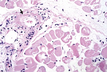
Various endocrinologic abnormalities can result in myopathic conditions (Table 15-9). The most common are hypercortisolism and hypothyroidism in dogs. In horses, pituitary hyperfunction resulting in Cushing’s disease also causes muscle disease. In most cases of endocrine myopathy, the end result is myofiber atrophy, particularly of type 2 fibers. A unique syndrome of muscle hypertrophy and pseudomyotonia occurs in dogs associated with hypercortisolism. Endocrine myopathies can also be complicated by the fact that endocrinopathy can also cause pathologic changes in peripheral nerves, leading to a mixture of myopathic (type 2 fiber atrophy) and neuropathic changes (denervation atrophy and alteration in fiber-type pattern) within muscle. Denervation followed by reinnervation leading to fiber-type grouping can be seen in dogs with chronic hypercortisolism (see Fig. 15-20, B) and hypothyroidism.
TABLE 15-9
Myopathies Caused by Endocrine and Electrolyte Abnormalities
Disorder | Species Affected |
Hypothyroidism | Dogs |
Hypercortisolism | Dogs |
Hypokalemia | Cattle, cats |
Hypophosphatemia | Cattle |
Hypernatremia | Cats |
Hypocalcemia | Cattle |
Hypothalamic/pituitary dysfunction | Horses |
Normal electrolyte status is vital to normal skeletal muscle function. Hypocalcemia, hypokalemia, hypernatremia, and hypophosphatemia can cause profound skeletal muscle weakness, sometimes associated with myofiber necrosis, in various species.
Dysfunction of the lower motor neurons, peripheral nerves, or neuromuscular junction can have profound effects on muscle function.
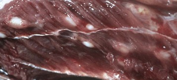
Neuropathic Disorders: There are many peripheral nerve disorders and a few motor neuron disorders that can lead to denervation atrophy of muscle in animals. These can be inherited or acquired. Long nerves, such as the sciatic and left recurrent laryngeal nerves, appear to be particularly sensitive to development of acquired neuropathy. Many of the peripheral nerve disorders of animals are discussed in Chapter 14. Characteristic features of denervation atrophy are described in the section on Responses of Muscle to Injury.
Neuromuscular Junction Disorders: The neuromuscular junction is a modification of the postsynaptic myofiber membrane. At the neuromuscular junction, the membrane is folded to increase surface area and is studded with specialized ion channels known as acetylcholine receptors. After arrival of an action potential at the distal end of a motor nerve, the terminal axons release acetylcholine, which diffuses across the synaptic space to bind to the acetylcholine receptors. Binding opens these channels, leading to sodium influx, which initiates the skeletal muscle action potential that culminates in muscle contraction. Acetylcholine is rapidly degraded by acetylcholinesterase released from the postsynaptic membrane, which prevents continued stimulation and thus contraction of the muscle fiber.
Disorders that impair the ability of nerve impulses to travel across the neuromuscular junction have profound effects on skeletal muscle function. Technically, however, the myofibers are still innervated, so denervation atrophy does not occur and no light microscopic abnormalities in the muscle or nerve are present. Various neurotoxins (i.e., in snake and spider venom and in curare-containing plants) and drugs can affect the neuromuscular junction, but the most common neuromuscular junction disorders affecting animals are myasthenia gravis, botulism, and tick paralysis.
Myasthenia Gravis: Myasthenia gravis can be either acquired or congenital. Acquired myasthenia gravis is an immune-mediated disorder caused by circulating autoantibodies against skeletal muscle acetylcholine receptors (Fig. 15-29). Binding of these antibodies to the acetylcholine receptor on the postsynaptic membrane leads to a severe decrease in the number of functional receptors. The mechanisms by which antibodies damage these receptors are (1) direct damage to the neuromuscular junction, which may be visible with electron microscopy as simplification of the folding of the membrane, and (2) formation of cross-linked antibodies leading to receptor internalization. Sufficient functional acetylcholine receptors are present to initially allow normal neuromuscular transmission, but if there is sustained muscular activity the decrease in the number of available receptors leads to progressive weakness and collapse. Therefore acquired myasthenia gravis results in episodic collapse, and repetitive nerve stimulation causes a characteristic rapid decrease in amplitude of the muscle compound motor action potential. Diagnosis of myasthenia gravis can also be made after intravenous injection of cholinesterase inhibitors such as edrophonium chloride (Tensilon, ICN Pharmaceuticals, Costa Mesa, CA) in collapsed animals. The reduction in cholinesterase activity leads to more active acetylcholine being available within the synapse and rapid, although transient, restoration of skeletal muscle contraction. Detection of autoantibodies to acetylcholine receptors in the blood confirms the diagnosis of acquired myasthenia gravis.
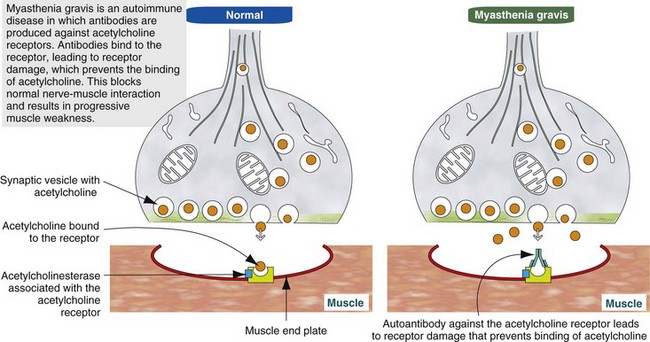
Fig. 15-29 Schematic diagram of the pathogenesis of acquired (autoimmune) myasthenia gravis. (Modified from Kierszenbaum AL: Histology and cell biology: an introduction to pathology, ed 2, St Louis, 2006, Mosby.)
The origin of the autoantibodies causing myasthenia gravis is not always known, but there is a strong link between thymic abnormalities and development of myasthenia gravis in both humans and animals. Specialized cells within the thymic medulla, known as myoid cells, express skeletal muscle proteins, including those of the acetylcholine receptor. It is thought that these cells participate in development of self-tolerance. Abnormalities of the thymus, most commonly thymoma in animals and thymic follicular hyperplasia in humans, can lead to loss of self-tolerance to acetylcholine receptors. In such cases, removal of the abnormal thymus can result in restoration of normal neuromuscular junction activity. When thymic abnormalities are not present, treatment with long-acting anticholinesterase agents and in some cases immunosuppressive agents, such as corticosteroids, is necessary.
Congenital myasthenia gravis is an inherited disorder that is much less common than acquired myasthenia gravis. To date it has been described only in humans, dogs, and cats. Animals with congenital myasthenia gravis are born with defective neuromuscular junctions that often have a decreased membrane surface area, best visualized with electron microscopy, and as a consequence an inherently reduced acetylcholine receptor density. Such animals may be normal at birth because there are sufficient functional acetylcholine receptors to support muscle contraction in a neonate. But, with rapid postnatal growth, clinical signs of profound, sustained, and progressive weakness occur as a consequence of insufficient functional receptors to support the function of growing muscles.
Botulism: Botulism is a neuromuscular disorder caused by the exotoxin of the bacterium Clostridium botulinum. Botulinum toxin is considered one of the deadliest of the known toxins. Botulism is characterized by profound generalized flaccid paralysis. Seven serologically distinct but structurally similar forms of botulinum toxin are designated A, B, C, D, E, F, and G. Sensitivity to these toxin types varies among different species. Dogs are most sensitive to type C toxin, ruminants to types C and D, and horses to types B and C.
Botulinum toxin consists of a light chain and a heavy chain linked by a disulfide bond. Binding of botulinum toxin to receptors on the presynaptic terminals of peripheral nerves is followed by endocytosis of the toxin. Within the endocytotic vesicle of the terminal nerve, the disulfide bond is cleaved, and the released light chain is translocated into the axonal cytoplasm (see Fig. 4-27). Botulinum toxin light chains are metalloproteinases. Numerous proteins are involved in the release of acetylcholine from presynaptic vesicles, and botulinum toxin blocks release of acetylcholine by irreversible enzymatic cleavage of one or more of these proteins. Different forms of botulinum toxin affect different proteins, but the end result is the same. Active neuromuscular junctions are the most sensitive, which has led to the use of low concentrations of locally injected botulinum toxin as a treatment for localized muscular disorders resulting in spasm.
Clostridium botulinum spores are commonly present in the gastrointestinal tract of animals and in the soil. Under favorable anaerobic and alkalinic conditions, these spores become active, with resultant toxin production. Botulism can occur because of ingestion of preformed toxin, such as in feed contaminated by dead rodents or soil-borne organisms, or from toxin produced by Clostridium botulinum organisms within the gastrointestinal tract or superficial wounds (Box 15-9). Dogs and cats are the species most likely to ingest dead rodents containing botulinum toxin and are quite resistant to developing botulism. In veterinary medicine, horses are the most sensitive to botulinum toxin. Death of horses, most often the result of respiratory muscle paralysis, can result from exposure to only very small amounts of botulinum toxin. The damage to presynaptic axon terminals is irreversible, and recovery from botulism occurs only after terminal axon sprouting and reestablishment of new functioning synapses.
Tick Paralysis: Dermacentor and Ixodes ticks can elaborate a toxin that also blocks release of acetylcholine from axon terminals. Tick paralysis is seen most often in dogs and children. Recovery after tick removal can be rapid (within 24 to 48 hours), indicating that the mechanism of toxin action in tick paralysis does not result in irreversible presynaptic damage and thus is different from that of botulinum toxin.
Neoplasms involving skeletal muscle are most often those that arise within the muscle or its supporting structures or that invade muscle from adjacent tissue. Neoplasms metastatic to muscle are rare.
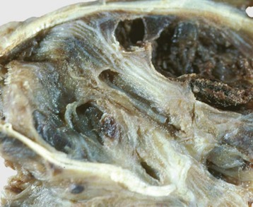
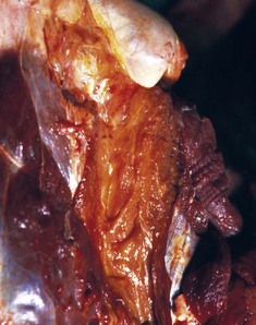
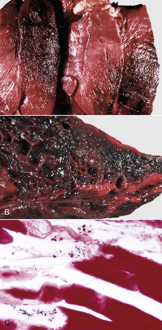
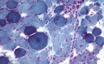
Primary Muscle Tumors: Tumors with striated muscle differentiation are thought to arise from intramuscular pluripotential stem cells rather than from satellite cells. These tumors are uncommon and are either benign (rhabdomyoma) or malignant (rhabdomyosarcoma [Fig. 15-30]). Primary intramuscular tumors can also arise from fibrous tissue, vasculature, or neural elements. The most common tumor to arise from muscle-supporting structures is hemangiosarcoma.
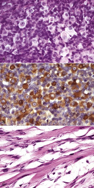
Fig. 15-30 Rhabdomyosarcoma.
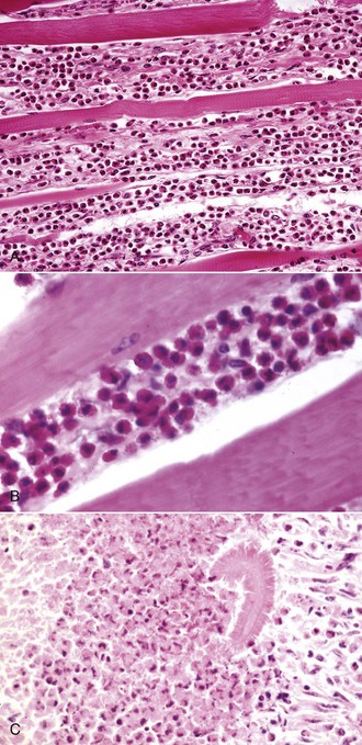
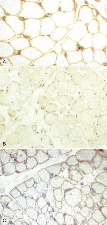
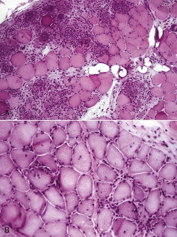
A, Skeletal muscle, cat. An admixture of small round basophilic cells with a lesser number of larger round cells with prominent eosinophilic cytoplasm is characteristic of embryonal rhabdomyosarcoma. Nuclei are central and euchromatic, most often with a single large nucleolus. H&E stain. B, Immunostaining reaction of the same rhabdomyosarcoma as depicted in A, showing intense cytoplasmic expression of desmin in many tumor cells, indicative of muscle origin (skeletal, cardiac, or smooth). These cells also express myoglobin and sarcomeric actin (not shown), which differentiates skeletal muscle tumors from smooth muscle tumors. Immunoperoxidase reaction for desmin. C, Botryoid rhabdomyosarcoma, urinary bladder, large breed dog. Cross-striations, characteristic of a well-differentiated rhabdomyosarcoma, are present in the elongated multinucleate tumor cells. H&E stain. (A and B courtesy College of Veterinary Medicine, Cornell University. C courtesy Dr. B.A. Valentine, College of Veterinary Medicine, Oregon State University.)
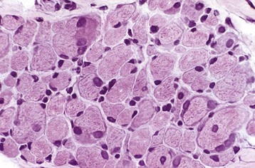
Rhabdomyoma and Rhabdomyosarcoma: Tumors of striated muscle that occur at sites other than within muscle are rhabdomyomas of the heart or lung and botryoid rhabdomyosarcomas of the urinary bladder; these are not discussed in this section. Rhabdomyoma and rhabdomyosarcoma arising within skeletal muscle are most common in the dog, followed by the horse and cat. Morphologic variants include round cell, spindle cell, and mixed round and spindle cell, reflecting the developmental stages of skeletal muscle. Historically, diagnosis of tumors of skeletal muscle has relied on identification of cross striations indicative of sarcomeric differentiation. Cross striations are most often seen in elongated multinucleate cells known as strap cells (Fig. 15-30, C) and in ovoid cells known as racquet cells. They are most easily recognized after staining with phosphotungstic acid hematoxylin (PTAH) stain, but the search for cross striations can be extremely frustrating and often unrewarding. These days the diagnosis of tumors of skeletal muscle origin relies primarily on results of immunohistochemical examination using antibodies for muscle-specific proteins. Muscle actin and desmin are expressed by smooth and skeletal muscle tumors, but myoglobin, sarcomeric actin, myogenin, and MyoD1 are specific for skeletal muscle. Evidence of muscle differentiation, such as primitive myofilaments and Z-band structures, can also be detected by electron microscopy.
Rhabdomyoma is most often a round cell tumor and occurs most commonly in the larynx of adult dogs. The youngest reported age is 2 years. Tumors are generally smooth and nodular, pink, and unencapsulated. Histologic features are closely packed plump round cells that have central euchromatic nuclei, generally with a single prominent nucleus, and abundant vacuolated to granular eosinophilic cytoplasm. A small number of multinucleate and elongate strap cells can also be seen. Mitoses are rare, and evidence of invasion is uncommon.
Similar to the situation in humans, rhabdomyosarcomas in animals most often occur at a young age and are most common in the neck or oral cavity, especially in the tongue. These tumors are pink and fleshy, and they often have prominent local invasion. The most common and most distinctive form of rhabdomyosarcoma in animals is embryonal rhabdomyosarcoma, composed of primitive round cells with prominent euchromatic nuclei, a single prominent nucleolus, and either indistinct or prominent eosinophilic cytoplasm (“rhabdomyoblasts”; see Fig. 15-30, A and B). Rhabdomyosarcoma can also contain elongate multinucleate strap cells (see Fig. 15-30, C) and ovoid racquet cells. Cellular and nuclear pleomorphism is common, as is mitotic activity. These tumors are locally invasive and frequently metastasize, although too few cases have been studied to document any pattern of metastasis.
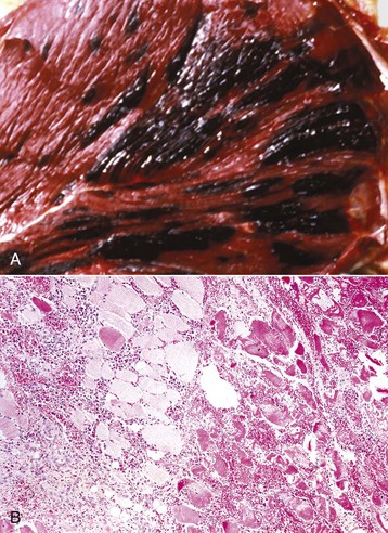
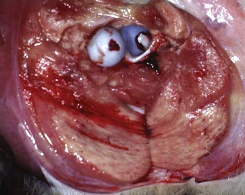
Hemangiosarcoma: Malignant vascular neoplasms (hemangiosarcoma) arising within muscle are most common in the horse and dog (Fig. 15-31). Clinical signs include swelling within a muscle, often with associated lameness. Cytologic preparations frequently reveal only peripheral blood, which is suggestive of a hematoma. Pathologic diagnosis can be difficult if multiple sites within the lesion are not sampled, as the amount of hemorrhage often far exceeds the area composed of proliferating neoplastic endothelial cells. Intramuscular hemangiosarcoma has a high incidence of metastasis, often to the lungs.
Other Tumors Involving Skeletal Muscle: A variant of lipoma, known as infiltrative lipoma, is often located in skeletal muscle. Characteristic gross pathologic and histopathologic findings are mature adipocytes invading skeletal muscle. This tumor is most common in the dog but has also been reported in young horses. Wide excision is the treatment of choice because this tumor recurs as a result of local invasion, but it does not metastasize.
Infiltration of skeletal muscle by neoplastic lymphocytes is not uncommon. Neoplastic lymphocytic infiltrates surround myofibers and can cause myofiber atrophy. These cells do not invade myofibers, however, and myonecrosis is rare. This helps to distinguish intramuscular lymphoma from lymphocytic myositis. Careful examination of infiltrating neoplastic cells typically reveals a relatively monomorphic population of lymphocytes, which may be atypical in appearance. Immunohistochemistry to confirm a single infiltrating cell type is also useful.
Vaccine-associated sarcoma in the muscle of the cat can arise within an intramuscular vaccination site or extend into underlying skeletal muscle from a subcutaneous injection site. Occasionally, mast cell tumors and carcinomas exhibit prominent skeletal muscle invasion. Melanoma arising in the skin of older gray horses often metastasizes to muscle fascia and may exhibit some extension into the muscle itself. Intramuscular metastasis of tumors is rare (see the section on Defense Mechanisms). Intramuscular metastasis of carcinoma, particularly prostatic, can occur in dogs. When carcinomas with areas of sclerosis involve muscle, either by extension or by metastasis, the muscle basement membrane of adjacent myofibers is typically destroyed, often resulting in bizarre multinucleate cells representing attempts at muscle regeneration (see Fig. 15-17). These bizarre cells should not be misidentified as tumor cells.
Adequate muscle function is essential for the survival of any species. Many domestic animals have been selectively bred for improved musculature for meat production, performance, or appearance. Therefore muscle disease in animals can have a significant economic impact. In some cases, it is selection pressure imposed by humans that has resulted in development and perpetuation of various myopathic conditions in animals. It is likely that continued selection for what appears to be a phenotypically desirable trait will lead to the recognition of new genetic mutations and myopathic conditions in the future.
It is interesting to compare the effects of muscular disorders that affect humans and animals. The four-footed stance of animals allows for greater stability, which can allow an animal to remain ambulatory for some time, when a similarly affected person would be confined to a wheelchair. However, disorders that result in recumbency, even if it is transitory, can be devastating in livestock. It is much more difficult to nurse a large animal through a period of recumbency than it would be for a hospitalized human or small animal.
The most common and important muscle disorders of animals are discussed by species because this is the way diseases are considered clinically. The same disease may occur in different species. Details of less common muscle disorders are presented in Web Appendix 15-1.
There is perhaps no other domestic animal species for which optimal muscle development and function is so critical as the horse. Selective breeding for better muscling has occurred in virtually all horse and pony breeds. The ability of such selection pressure to perpetuate equine muscle mutations is exemplified by the relatively recent occurrence of hyperkalemic periodic paralysis (HYPP), in which a muscle mutation results in visually appealing increased muscle bulk and definition. Unfortunately, as you will see in the discussion of HYPP later, such mutations do not often result in improved muscle function.
Infection by various bacterial organisms and clostridial toxins can cause myopathy in the horse. Protozoa (Sarcocystis spp.) are common incidental findings in equine muscle, but Sarcocystis-induced muscle damage resulting in clinical signs of muscle disease is rare.
Clostridial Myositis (Malignant Edema; Gas Gangrene): Clostridial myositis in the horse is an often fatal disorder caused by infection by various toxin-producing clostridial species, which are large Gram-positive anaerobic bacilli. Clostridium septicum is the most common cause of clostridial myositis in horses, but Clostridium perfringens types A to E, Clostridium chauvoei, Clostridium novyi, and Clostridium fallax can also cause infection. Infection can involve more than one clostridial species. Clostridium spp. are ubiquitous organisms that form spores within the soil and within the gastrointestinal tract. Unlike cattle, in which nonpenetrating trauma can cause muscle bruising and anaerobic conditions that activate clostridial spores already in the muscle, clostridial myositis in horses is virtually always secondary to a penetrating wound. Most often, this is an injection site of a nonantibiotic substance, but infection of sites of puncture wounds and of perivascular leakage of irritants in intravenously administered compounds are also possible. It is also possible that clostridial bacteria entering the blood from an injured gastrointestinal tract can colonize damaged muscle. This is one possible explanation for the frequent occurrence of signs of colic before development of clostridial myositis at the site of intramuscular injection of medications such as flunixin meglumine that cause localized muscle damage. Under anaerobic conditions, clostridia proliferate and produce toxins that damage blood vessels, resulting in hemorrhage and edema, and cause necrosis of adjacent muscle fibers (see Table 15-3).
Clinical signs are acute onset of heat, swelling, and pain within a muscle group and adjacent fascia, with concurrent fever, depression, dehydration, and anorexia. If sufficient muscle necrosis is present, serum CK and AST concentrations may be mildly to moderately increased. Death from toxemia and/or septicemia often occurs within 48 hours. Affected muscle and adjacent fascia are swollen and often hemorrhagic, with edema, suppurative inflammation, and necrosis; gas may also be present (Fig. 15-32). Vasculitis is not seen. Gram-positive bacilli characteristic of Clostridium spp. are generally demonstrable within affected tissue.
Fig. 15-32 Clostridial myositis, malignant edema, horse.
A, Clostridium septicum is the most common cause of clostridial myositis in horses. Affected muscle (shown here) and adjacent fascia (not shown here) are swollen and often hemorrhagic. B, Interstitial edema, hemorrhage, and inflammatory cells surround numerous swollen and fragmented necrotic myofibers. Formalin fixation, H&E stain. (A courtesy College of Veterinary Medicine, Cornell University. B courtesy Dr. B.A. Valentine, College of Veterinary Medicine, Oregon State University.)
The diagnosis can be made with reasonable certainty based on typical historic, gross pathologic, cytologic, and histopathologic findings. Clostridium spp. can also be identified by culture under anaerobic conditions or by a fluorescent antibody test. Treatment must be initiated rapidly and includes surgical incisions into affected muscle to allow drainage and oxygenation, antibiotic therapy, and supportive care.
Botulism: Technically, this disease is a neuromuscular junction disorder and is included in this section for convenience. Botulism is caused by Clostridium botulinum toxin and is often not associated with Clostridium botulinum infection. The portals of entry of botulinum toxin in horses are summarized in Box 15-9. Clostridium botulinum bacteria are found as spores within the gastrointestinal tract of many mammals, and spores are common in the soil. Preformed toxin within contaminated feed or soil is the most common cause of botulism in adult horses. However, in foals, usually between 1 week and 6 months of age, ingestion of Clostridium botulinum spores can lead to proliferation of toxin-producing Clostridium botulinum within the intestinal tract, resulting in toxicoinfectious botulism (shaker foals). Wound infection is an uncommon cause of botulism in horses.
The pathogenesis of botulism has been previously discussed in the section on Neuropathic and Neuromuscular Junction Disorders). Irreversible binding of toxin to presynaptic nerve terminals and blockage of acetylcholine release lead to the profound generalized flaccid paralysis that is the hallmark of botulism. Clinical signs are acute and progress rapidly, generally resulting in recumbency. Dysphagia and tongue weakness are common findings that help to distinguish botulism from other neuromuscular diseases causing recumbency. Serum concentrations of CK and AST are within normal limits (indicating the absence of damage to myofibers) or are possibly slightly increased as a result of ischemic myopathy secondary to recumbency (see later discussion).
No specific gross or histopathologic lesions are present in horses dying with botulism, although aspiration pneumonia caused by dysphagia can occur. Muscle fibers are intact unless recumbency has compromised their blood supply, causing ischemia and localized myofiber necrosis.
Evaluation of stomach contents or contaminated feed may reveal the presence of toxin. However, horses are exquisitely sensitive to botulinum toxin, and since only a small concentration of the toxin may be present in an affected horse, available tests may not detect such a low concentration of toxin. In most equine cases, the diagnosis is made based on the clinical history after elimination of other possible causes of profound muscular weakness. Affected animals should be treated with polyvalent botulinum antitoxin to prevent further binding of toxin. Recovery occurs after terminal axon sprouting and reestablishment of functional neuromuscular junctions. Vaccination with botulinum toxoid is an effective preventive measure.
Corynebacterium pseudotuberculosis (Pigeon Fever): Intramuscular abscesses caused by Corynebacterium pseudotuberculosis occur almost exclusively in horses in arid regions of the western United States and Brazil. Corynebacterium pseudotuberculosis is a Gram-positive pleomorphic facultative anaerobic bacillus present within the soil. It can enter muscle via penetrating wounds, including injection sites. The biotype most common in horses is different from that which affects sheep and goats because it is unable to reduce nitrates to nitrites. The high lipid content of the bacterial cell wall contributes to the survival of C. pseudotuberculosis within macrophages. Bacterial exotoxins, such as phospholipase D, contribute to vascular damage and inhibition of neutrophil function. Equine infections occur most frequently during the fall and early winter, and a higher incidence of the disease is often seen after rainy winters. Infections are most common in the pectoral musculature, but other locations are possible. Affected muscles are swollen and edematous and contain variably sized zones of localized suppurative inflammation. Fever is common. The causative agent is readily isolated from affected tissue and can be seen in aspirates from intramuscular abscesses. Treatment is generally curative and includes antibiotic therapy and establishment of drainage of abscesses. Rarely, infection with Corynebacterium pseudotuberculosis in horses leads to immune-mediated vasculitis (purpura hemorrhagica; see next section).
Streptococcal-Associated Myopathies: Two distinct degenerative myopathies are associated with infection or exposure of the horse to Streptococcus equi ssp. equi. One, known as purpura hemorrhagica, has been recognized for many years. The other, known as streptococcal-associated rhabdomyolysis and muscle atrophy, has only recently been recognized.
Purpura Hemorrhagica: In this disease, muscle damage is not caused by the direct infection of the muscles but rather by an immune response to the bacterial pathogen. Streptococcus equi is the most common cause of purpura hemorrhagica in horses, but Corynebacterium pseudotuberculosis and possibly other bacteria can also cause purpura hemorrhagica. In cases caused by Streptococcus equi, circulating immune complexes composed of immunoglobulin A (IgA) antibodies and streptococcal M antigen deposit in the walls of small vessels. This leads to vasculitis and vascular wall necrosis (Fig. 15-33), with resultant hemorrhage and infarction of myofibers. It is also possible that antibodies to streptococcal M protein cross-react with skeletal and cardiac muscle myosins to cause direct injury.
Fig. 15-33 Intramuscular vasculitis, purpura hemorrhagica, skeletal muscle, transverse section, horse.
In the wall of the blood vessel (arrow) is a band of circumferential fibrinoid necrosis containing nuclear debris. Many of the adjacent myofibers are necrotic (center to lower right areas). Some of these myofibers are fragmented, and a small number contain fine basophilic deposits of mineral. Formalin fixation, H&E stain. (Courtesy Dr. B.A. Valentine, College of Veterinary Medicine, Oregon State University.)
Signs of myopathy often accompany systemic signs of poststreptococcal purpura in horses (i.e., depression, fever, dependent edema, petechiae or ecchymoses, leukocytosis, increased serum fibrinogen, and anemia), but myopathy can also be the primary presenting disease process. Affected horses are weak, may have a short-strided gait, and can become recumbent. Myoglobinuria and very high increases in serum concentrations of CK and AST are common.
Multiple muscles are involved (as opposed to the locally extensive lesion of clostridial myositis), and affected muscles contain multifocal to locally extensive hemorrhage and edema that dissects between necrotic muscle fibers and muscle fasciculi. Gross pathologic findings are similar to those seen in clostridial myositis (see Fig. 15-32, A), but lesions do not contain gas bubbles. Vascular injury (leukocytoclastic vasculitis and fibrinoid necrosis of blood vessels; see Fig. 15-33) is seen on microscopic examination and is the diagnostic feature.
Diagnosis is based on a history of exposure of the horse to Streptococcus equi and the typical clinical, clinicopathologic, and histopathologic findings. Because this is an immune-complex disorder, histopathology, cytology, and bacterial cultures of affected muscle do not reveal Streptococcus equi. This bacterium or other causative bacteria may be cultured from other affected tissues, especially lymph nodes or guttural pouch. A high serum titer to Streptococcus equi M protein is strongly supportive of a diagnosis of streptococcal-associated purpura hemorrhagica. Treatment includes corticosteroid therapy and supportive care, but horses frequently succumb to other sequelae of systemic vasculitis, such as gastrointestinal infarcts.
Streptococcal-Associated Rhabdomyolysis and Muscle Atrophy: A syndrome of severe acute rhabdomyolysis resulting in profound rapidly progressive generalized loss of muscle mass has also been seen in horses with clinical infection by Streptococcus equi or in horses that have been exposed to this bacterium but that did not develop obvious clinical signs of infection. This syndrome occurs most frequently in young to young adult quarter horses, but young horses of other breeds can also be affected. Clinically recognizable muscle atrophy is often most evident in paraspinal and gluteal muscles. Some cases have microscopic evidence of concurrent EPSSM (see the section on Inherited or Congenital Myopathies), which may be a predisposing factor. In others, nonsuppurative perivascular and interstitial inflammation has been detected, and the proposed mechanism is immune-mediated damage caused by cross-reaction of streptococcal antibodies with muscle proteins. Affected horses do not show typical signs of purpura hemorrhagica but often have very high serum concentrations of CK (often greater than 100,000 units per liter) and AST (often greater than 10,000 units per liter). Affected horses may respond to corticosteroid therapy. Most will recover, but recurrence after subsequent exposure to Streptococcus equi is possible.
Protozoal Myopathy: Protozoa (Sarcocystis spp.) are common incidental findings in equine skeletal and cardiac muscles. As the protozoa are in cysts within the myofiber itself and thus are protected from the body’s surveillance, there is no inflammatory response. Massive infection by Sarcocystis fayeri is suspected of causing a degenerative myopathy in horses, but this is rare. Rarely, localized thickening of the tongue has been found in horses with granulomatous myositis, the result of sarcocystis organisms within tongue musculature. The cause of the intense inflammation apparently incited by protozoa in these rare cases is unknown.
Ear Tick–Associated Muscle Spasms: Episodic muscle spasms of various muscle groups can occur in horses with ear ticks (Otobius megnini). The mechanism is not known. Dimpling of affected muscles after percussion can be seen, but myotonic discharges are not found with electromyography. Treatment for ear ticks results in rapid recovery.
Nutritional deficiency, most often of selenium, and various toxins are relatively common causes of degenerative myopathy in the horse.
Nutritional Myopathy: Foals (most commonly up to 2 weeks of age) and young adult horses are most susceptible to nutritional myopathy because of a deficiency of the antioxidants selenium or (less commonly) vitamin E. In severely selenium-deficient areas, such as the Pacific Northwest, selenium deficiency myopathy can occur in horses of any age. Normally the selenium present in the soil is taken up by growing plants. In many areas, the soil is selenium deficient, and selenium supplements to the animal’s ration must be provided. Vitamin E deficiency occurs in horses that eat marginal- to poor-quality grass hay and have little or no access to pasture and no supplemental vitamin E. Oxidative injury to actively contracting muscle fibers occurs as a result of a lack of antioxidant activity.
Affected foals are most likely to be those born to selenium-deficient mares. Foals have generalized weakness, which may be present at birth or become apparent soon after birth. Affected foals may become recumbent but are generally bright and alert. They often continue to suckle if bottle fed, but weakness of the tongue and pharyngeal muscles can lead to weak suckling.
Affected adult horses are most often stabled horses fed only selenium-deficient hay, with clinical disease being seen most commonly in the late winter or early spring. In the Pacific Northwest, selenium deficiency myopathy can occur in adult horses fed only pasture or hay, and it can occur at any time of year. Affected adult horses often show preferential involvement of the temporal and masseter muscles (the condition is sometimes inappropriately termed maxillary myositis or masseter myositis) with swelling and stiffness of these muscles and impaired mastication. Involvement of pharyngeal muscle results in dysphagia and involvement of the tongue results in impaired prehension of food, which can be mistaken for botulism. In more chronic cases, bilaterally symmetric atrophy of the masseter muscles may be evident, which can be mistaken for atrophy secondary to protozoal myeloencephalitis. Careful examination of these horses often reveals generalized weakness, evident as a stiff, short-strided gait. Severely affected horses can have an acute onset of recumbency that mimics neurologic disease.
Serum concentrations of CK and AST are generally mildly to moderately increased, although extremely high concentrations can be seen in severely affected foals and horses. Concentric needle EMG of affected muscles results in abnormal spontaneous activity (positive sharp waves, fibrillations, and myotonic bursts).
Muscles of affected horses appear pale (hence the common name white muscle disease), often in a patchy distribution (see Fig. 15-40). The most severely affected muscles are those that have the highest workload (e.g., cervical muscles in foals used during suckling and “bumping” the udder, proximal limb muscles, tongue, and masticatory muscles). The gross appearance depends on the extent of the necrosis and the stage. In early stages, yellow and white streaks are present, and later pale, chalk white streaks often appear. Horses with impaired swallowing can have cranioventral aspiration pneumonia. Severely selenium-deficient foals and horses also have pale areas of necrosis within the myocardium, especially the left ventricular wall and septum, which are areas that have a high workload. The stage of the necrosis depends on the age of the lesions. In foals with severe, acute myopathy leading to death or euthanasia, lesions are at the stage of massive muscle necrosis and mineralization with minimal macrophage infiltration (monophasic). In animals that have lived longer (i.e., subacute cases), the lesions are polyphasic, and active necrosis, macrophage infiltration, and regeneration are present. Although type 1 fibers may be more likely to develop necrosis because of nutritional myopathy, in severely affected muscles almost all fiber types are affected. In cases with myocardial involvement, myocardiocyte necrosis and mineralization are present. If the animal survives, the necrotic myocardiocytes are replaced by fibrovascular connective tissue that matures to form a scar.
A provisional diagnosis of nutritional myopathy is based on typical history, increases in serum concentrations of CK and AST, and characteristic gross and histopathologic findings. The diagnosis is confirmed by detecting deficient concentrations of selenium or vitamin E in blood of live animals or in liver samples obtained at necropsy. If horses live long enough, myofiber regeneration can restore the muscles to normal. This disorder in foals can be prevented by supplementing the ration of mares with selenium during gestation. Foals born in selenium-deficient areas can also be given injectable vitamin E and selenium soon after birth. Young adult horses should be given sufficient dietary vitamin E and selenium. Treatment with selenium and vitamin E after the onset of clinical signs is far less effective than prevention.
Ionophore Toxicity: The pathogenesis of ionophore toxicity is discussed in the section on Toxic Myopathies. Horses are exquisitely sensitive to ionophores and succumb to very small doses. Ionophores may be present as contaminants within horse feed, or the horse may be accidentally fed ionophore-containing feeds intended for other domestic animals.
Most of the available literature relates to monensin toxicity, but the effects of other ionophores should be similar. In acute monensin toxicity, death occurs because of shock and cardiovascular collapse, and no specific lesions are seen on postmortem examination within the first 48 hours, although these may be stained diffusely pink by myoglobin. If the horse survives 3 to 4 days, affected skeletal and cardiac muscles often contain pale streaks (Fig. 15-34, A) and, microscopically, cardiac muscle necrosis and segmental necrosis of skeletal muscle is present (Fig. 15-34, B; see Fig. 15-13, B), with concurrent increases in serum concentrations of CK and AST, which may be severe. Given the profound sensitivity of horses to ionophores, ionophore toxicity in horses is typically the result of a single dose and thus the lesion is a monophasic multifocal process. This helps to differentiate ionophore toxicity from nutritional myopathy, which is often polyphasic. Both type 1 and type 2 fibers are affected. If the horse survives, necrosis is followed by myofiber regeneration, which can restore the muscles to normal, but necrotic myocardiocytes are replaced by fibrosis because of the lack of significant regeneration by myocardiocytes. Horses dying at 14 days after ionophore exposure often have normal skeletal muscles and extensive myocardial fibrosis. Acute cardiac failure and death because of myocardial scarring can occur months to years after apparent clinical recovery from ionophore exposure.
Fig. 15-34 Ionophore toxicity, monensin, skeletal muscle.
A, Necrosis. The pale white to gray foci are areas of necrotic myofibers. Myocardium will often contain similar lesions. B, Segmental myofiber necrosis (2 days old), longitudinal section, horse. The segment of myofiber (arrow) visible here is necrotic, fragmented, and infiltrated by macrophages and neutrophils. Note the intact basal lamina and endomysium on both sides of the myofiber, which will contain the regenerating myofiber and thus facilitate resolution. Ionophore toxicity results in calcium overload and death of skeletal (also cardiac) myocytes. Formalin fixation, H&E stain. (A courtesy Dr. J. Wright, College of Veterinary Medicine, North Carolina State University; and Noah’s Arkive, College of Veterinary Medicine, The University of Georgia. B courtesy Dr. M.D. McGavin, College of Veterinary Medicine, University of Tennessee.)
Diagnosis is based on a history that includes both ingestion of ionophores and the presence of the characteristic gross or histopathologic findings. Analysis of feed or stomach contents for ionophores is definitive. Treatment for ionophore-intoxicated horses is supportive, as there is no specific therapy.
Plant Toxicities: A number of toxic plants are known to cause muscle necrosis in horses (see also Toxic Myopathies). These include Cassia occidentalis (coffee senna) and Thermopsis spp. Most plant-associated toxicities in horses are associated with plants growing in pastures or in baled hay. Necrosis is most often polyphasic indicating a prolonged period of ingestion. Cardiac myonecrosis may or may not also be present. In the United Kingdom and less commonly in the midwestern United States, a syndrome of pasture-associated myonecrosis has been documented in horses. The cause of this disorder, also known as atypical myoglobinurium, is still unknown. Clostridial toxins (specifically Clostridium sordellii and Clostridium bifermentans) and environmental mycotoxins are considered possible causes.
Hyperkalemic Periodic Paralysis: Hyperkalemic periodic paralysis (HYPP) is a myotonic disorder that affects horses whose ancestry traces back to a quarter-horse stallion named Impressive. Affected horses generally have remarkably well-defined muscle groups, which has led to their popularity for showing in halter. The disease is inherited as an autosomal dominant disease; therefore affected horses can be either heterozygotes or homozygotes. Homozygous foals often have a distinctive laryngeal muscle dysfunction that results in laryngospasm and labored breathing. Most homozygous horses do not survive; if they do, they are invalids.
The underlying defect in HYPP is a point mutation in the gene encoding the α subunit of the skeletal muscle sodium channel. This defect causes abnormal (delayed) inactivation of sodium channel activity, resulting in membrane instability and continuous muscle fiber electrical activity, which is reflected in EMG findings (see later discussion). The pathogenesis of clinical signs of HYPP is complex and not entirely understood, either in horses or in humans with a similar disorder. Affected heterozygotes have a mosaic of abnormal and normal sodium channels, and resting muscle membrane potentials are typically lower than normal. This leads to an increased likelihood of electrical generation of a prolonged muscle action potential, resulting in transient myotonia. When abnormal sodium channels are activated, the response to the resulting abnormally increased intracellular sodium is release of potassium into the extracellular space and bloodstream, resulting in hyperkalemia. Hyperkalemia is not, however, a consistent finding. Feeding of high potassium feeds, such as alfalfa products or feeds with added molasses, can precipitate clinical signs of HYPP, possibly by activating abnormal sodium channels. Another potential consequence of prolonged activation of abnormal sodium channels is inactivation of normal sodium channels, resulting in flaccid paralysis and collapse. This result would explain the typical signs seen during episodes, which include transient muscle spasm (myotonia), with protrusion of the third eyelid, followed by generalized flaccid paralysis. Decreased muscle temperature, as can occur as a result of a chilling rain, can precipitate episodic collapse in HYPP horses, possibly by decreasing the activity of the muscle sodium-potassium exchanger (the Na-K ATPase), an important means by which affected muscle compensates for abnormal sodium channel activity. Postanesthetic recumbency and anesthesia-associated hyperthermia have also been seen in HYPP horses. Affected horses can appear normal for many years, can have multiple episodes of collapse, or can die acutely. Serum concentrations of CK and AST are generally normal. Abnormal ionic fluxes occur at all times in affected horses, and concentric needle EMG between paralytic episodes reveals characteristic persistent myotonic bursts.
There are no gross pathologic findings in HYPP horses other than gross prominent muscling. Skeletal muscle dysfunction in HYPP horses is due to abnormal ionic fluxes that can lead to spasms and weakness, therefore affected skeletal muscle is generally histologically normal. In some cases, scattered intracytoplasmic vacuoles (vacuolar myopathy) can be present in type 2 fibers. The characteristic pathologic finding of HYPP is only evident at the ultrastructural level, where dilated terminal cisternae of the sarcoplasmic reticulum are found.
Diagnosis can be made with reasonable certainty based on characteristic clinical signs (muscle spasms often leading to flaccid paralysis) and clinicopathologic findings (hyperkalemia) in a horse of Impressive line breeding. Myotonic bursts with concentric needle EMG are also diagnostic. The simplest and most reliable test, however, is a DNA-based test performed on peripheral white blood cells or, as described more recently, on cells obtained from the base of pulled mane or tail hairs. Treatment consists of feeding a low-potassium diet, which means avoiding alfalfa products and molasses. A low-potassium diet can be successful in controlling signs in many cases. More severe cases can be treated with the diuretic acetazolamide, which causes increased urinary excretion of potassium. Acute episodes can be treated with intravenous dextrose or insulin or oral sugar solutions such as sugar syrup. Administration of glucose to stimulate insulin secretion, or of insulin itself, aids in alleviating signs by helping drive the intracellular movement of potassium along with glucose.
Equine Polysaccharide Storage Myopathy (EPSSM): EPSSM is a myopathy most commonly recognized in quarter horse, warm blood, Arabian, Morgan, pony of the Americas, and draft-related breeds. It also occurs in many other horse and pony breeds, including miniature horses. Surveys of equine muscle samples have revealed an astonishingly high incidence of approximately 66% in all draft-related horses and approximately 30% in all light horses. Not all affected horses exhibit obvious clinical signs of muscle dysfunction. This disorder is inherited as an autosomal dominant trait.
In contrast to other glycogenoses affecting skeletal muscle, to date no abnormality in the glycolytic or glycogenolytic pathways in skeletal muscle has been identified, making this equine disorder unique, but an underlying carbohydrate metabolic disorder is still suspected. Affected horses appear to have a more rapid intramuscular uptake of blood glucose than controls, although the exact mechanism for this phenomenon is still unknown. A point mutation in the skeletal muscle glycogen synthase 1 (GYS1) gene has recently been associated with some, but not all, cases of EPSSM. A DNA test for this mutation is available. Abnormal accumulation of intracytoplasmic glycogen (confirmed by being PAS-positive, amylase-sensitive) within type 2 fibers is the histologic finding. In severe cases, aggregates of abnormal glycogen are eventually ubiquitinated, resulting in amylase-resistant inclusions composed of glycogen and filamentous protein. Certain breeds, such as quarter horse and draft-related breeds, seem to be most prone to the development of amylase-resistant inclusions, whereas glycogen aggregates are more common in other breeds. The explanation for this difference is as yet unknown, although breeds prone to developing amylase-resistant inclusions are also those breeds most likely to have the GYS1 mutation.
Clinical signs are variable, but all are thought to be caused by insufficient energy production by affected muscle fibers. Abnormal myofiber function caused by architectural alteration secondary to intramyofiber deposition of complex polysaccharide is also a possible mechanism, but the excellent response to therapy, even in horses with severe intramyofiber inclusions of accumulated polysaccharide, suggests that this is less significant than is altered energy metabolism. Recurrent exertional rhabdomyolysis (see later discussion) is a commonly recognized sign, but unexplained pelvic limb lameness is even more common than clinical rhabdomyolysis. Affected horses can also have a stiff gait, symmetric muscle atrophy, back soreness, muscle-cramping resulting in abnormal hindlimb flexion characteristic of shivers, and bilateral pelvic limb or generalized weakness. In draft horses, sudden onset of spontaneous recumbency or postanesthetic recumbency because of myopathy can occur. Serum concentrations of CK and AST are markedly increased after episodes of exertional rhabdomyolysis but may be only mildly to moderately increased in affected horses after exercise or onset of recumbency. Normal serum concentrations of CK and AST in affected horses are thought to indicate that the muscle dysfunction is not accompanied by overt myonecrosis. Concentric needle EMG may reveal abnormal spontaneous activity (scattered positive sharp waves and fibrillations).
In severe cases, in which horses have died or been euthanatized because of rhabdomyolysis or recumbency, muscles may be pale pink or diffusely red-tinged (myoglobin staining), which can be mistaken for autolysis. Multifocal pale zones may be present (see Fig. 15-36, A). In draft horses and sporadically in horses of other breeds, chronic myopathy can result in overall reduction in muscle mass. Muscles in severely affected draft horses can also be of normal size but may contain pale streaks where myofibers have been replaced by fat. The most severely affected muscles are those of the proximal hind limb (especially gluteal, semimembranosus, and semitendinosus muscles) and epaxial muscles of the back (e.g., longissimus), although any of the large “power” muscle groups, including pectoral and shoulder girdle muscles, can be affected. Swollen, dark kidneys (pigmentary nephrosis) caused by myoglobinuria can be seen in horses dying with severe rhabdomyolysis. The extent of overt myofiber necrosis is extremely variable; massive necrosis or regeneration can be seen after severe rhabdomyolysis, whereas only minimal scattered necrotic fibers may be seen in recumbent horses. Lesions are monophasic if there has been only a single bout of exertional rhabdomyolysis, or polyphasic if there have been repeated bouts of less severe exercise-induced injury. Abnormal polysaccharide is always present, but fiber necrosis is uncommon in muscle biopsy samples taken from affected horses while they are clinically normal.
The characteristic histologic finding is aggregates of intracytoplasmic material that stain positively with the PAS reaction for glycogen (Fig. 15-35, A). In severe cases, multiple pale intracytoplasmic inclusions are also present in H&E stained sections (Fig. 15-35, B). These inclusions are PAS-positive (Fig. 15-35, C) and resist digestion by amylase and are thus not glycogen. Terms used to describe this amylase-resistant material include amylopectin, polyglucosan, and complex polysaccharide. In chronic cases, myofibers also have chronic myopathic change (atrophy, hypertrophy, or internal nuclei), and fat replacement of myofibers after myofiber loss can occur in severely affected cases.
Fig. 15-35 Equine polysaccharide storage myopathy, semimembranosus muscle, transverse sections, horse.
A, Note the increased amount of and irregularly distributed dark-pink staining glycogen. Abnormal aggregates are present both beneath the sarcolemma and within the cytoplasm. Formalin fixation, PAS reaction. B, Severe form. Numerous myofibers contain multiple pale (very light pink) subsarcolemmal and intracytoplasmic inclusions of stored polysaccharide. Formalin fixation, H&E stain. C, These inclusions shown in B stain intensely with PAS but are not digested by amylase (not shown) and are characteristic of what is called complex polysaccharide, amylopectin, or polyglucosan. Formalin fixation, PAS reaction. (Courtesy Dr. B.J. Cooper, College of Veterinary Medicine, Oregon State University.)
At this time, the detection of the GYS1 mutation provides a definitive diagnosis of EPSSM, but this test is not very sensitive. The most sensitive test for diagnosis of EPSSM depends on finding characteristic histopathologic changes in muscle samples of horses with appropriate clinical signs. Gluteal, semimembranosus, or semitendinosus muscle samples are preferred, although changes in longissimus muscle are also found, especially in horses with back pain. A presumptive diagnosis of EPSSM can be made based on characteristic clinical findings in a predisposed breed. Treatment has relied on altering the diet to minimize starch and sugar intake (less than 15% of total daily calories) and maximize fat intake (at least 20% to 25% of total daily calories from fat). Grains and sweet feeds are replaced by high-fiber, low-starch, low-sugar feeds, with added fat in the form of vegetable oil, powdered fat, or high-fat rice bran supplements. Providing the horse with regular exercise and as much time as possible in a pasture or paddock are also important. Treatment is very successful in most cases.
Glycogen Brancher Enzyme Deficiency: Glycogen brancher enzyme (GBE) deficiency, or glycogenosis type IV, is a disorder caused by a congenital lack of a glycogenic enzyme, GBE, and is an emerging disease in quarter horses and American paint horses. It is inherited as an autosomal recessive trait. Affected foals may be aborted, stillborn, or weak at birth or can have contracted tendons, rhabdomyolysis, or cardiac failure at an early age. The consequence of GBE deficiency is the accumulation of long unbranched chains of glucose within cells that leads to abnormal glycogen formation and intramyofiber deposits. These molecules would normally be converted into glycogen in the presence of GBE in the final step in the formation of glycogen. There are no specific gross pathologic findings. Pulmonary edema may be found in foals that die from cardiac failure. Characteristic histologic findings are round hyaline inclusions resembling amylopectin (polyglucosan bodies) within skeletal and cardiac myocytes, especially Purkinje fibers, and to a lesser degree within hepatocytes. Unlike glycogen, inclusions are PAS-positive and resistant to amylase digestion. As with other carbohydrate metabolic defects, a lack of energy production by affected fibers is thought to underlie cellular dysfunction. Disruption of cytoarchitecture caused by amylopectin deposition may also contribute. Analysis of peripheral blood or skeletal muscle for GBE activity identifies affected animals with severely reduced GBE activity and carriers in which GBE activity is moderately reduced. A DNA test to detect carriers and affected horses using pulled mane or tail hairs is now available. There is no treatment for this disorder.
Myotonia and Mitochondrial Myopathy: A myotonic disorder occurs occasionally in horses, and a mitochondrial myopathy has been described in an Arabian horse. These disorders are discussed in more detail in Web Appendix 15-1.
Exertional Rhabdomyolysis: Equine exertional rhabdomyolysis (tying up, azoturia, Monday morning disease, setfast, blackwater) is characterized clinically by sudden onset of stiff gait, reluctance to move, swelling of affected muscle groups (especially gluteal), sweating, and other signs of pain and discomfort. Serum concentrations of CK and AST are often markedly increased. Signs may appear during or immediately after exercise, but only rarely is exertional rhabdomyolysis associated with exhaustive exercise. In severely affected horses, even minimal exercise, such as walking out of a stall, can cause clinical signs. High grain feeding and lack of regular exercise have been recognized to be factors leading to exercise-induced muscle injury for many years. Previous theories regarding the pathogenesis of equine exertional rhabdomyolysis include development of muscle lactic acidosis, vitamin E and/or selenium deficiency, hypothyroidism, and systemic electrolyte abnormalities. It is only recently that studies have concluded that lactic acidosis is not a finding in horses with exertional rhabdomyolysis, that hypothyroid horses show no signs of degenerative myopathy, and that electrolyte abnormalities as a primary cause of equine exertional rhabdomyolysis are rare. It is still thought that vitamin E or selenium deficiency can exacerbate signs of exertional rhabdomyolysis in predisposed horses, but neither vitamin E nor selenium deficiency is considered a primary cause. Recent studies have found that affected horses typically have an underlying myopathy, most often EPSSM. There is some evidence to suggest that recurrent exertional rhabdomyolysis in thoroughbreds is the result of abnormal calcium homeostasis within skeletal muscle, although some affected thoroughbreds have been found to have EPSSM. As muscle necrosis per se is not painful and does not cause muscle swelling, it is suspected that other factors play a role in this disorder in the horse. These factors include oxidative injury to muscle membranes occurring secondary to segmental necrosis and the subsequent production of oxygen-derived free radical compounds and vascular compromise resulting in ischemia (i.e., compartment syndrome when muscle damage occurs in a muscle with a tight and relatively nonexpandable fascia such as the gluteal and longissimus muscles). Oxidative injury may explain the perceived benefit of supplemental vitamin E and selenium to affected horses.
Gross findings are similar to those described for EPSSM (i.e., initially areas of muscle that are pale pink or diffusely red-tinged; Fig. 15-36, A). Histologic findings are localized or widespread muscle fiber necrosis (Fig. 15-36, B), followed by the usual sequence of events: macrophage infiltration and regeneration. Affected fibers are primarily type 2 fibers. Lesions can be either monophasic or polyphasic.
Fig. 15-36 Acute rhabdomyolysis, skeletal muscle, horse.
A, Affected muscles may be pale pink or diffusely red-tinged, which can be mistaken for autolysis. Multifocal pale zones may also be present. B, Segmental myofiber necrosis, semitendinosus muscle, transverse section. Most of the myocytes are necrotic and at the stage of coagulation necrosis. In a few myofibers, necrosis is at a later stage and the necrotic sarcoplasm has lysed, leaving empty sarcolemmal tubes (arrows). A couple of necrotic myofibers are at an even later stage and contain a small number of macrophages. Formalin fixation, H&E stain. (A courtesy Dr. W. Crowell, College of Veterinary Medicine, University of Georgia; and Noah’s Arkive, College of Veterinary Medicine, The University of Georgia. B courtesy Dr. B.J. Cooper, College of Veterinary Medicine, Oregon State University.)
Diagnosis is based on typical clinical signs and clinicopathologic evidence of muscle injury (increased activity of CK or AST). Treatment for an acute episode includes nonsteroidal antiinflammatory agents, acepromazine, and rest. Careful evaluation of the patient for evidence of renal damage ([pigmentary] myoglobinuric nephrosis) because of myoglobin released from damaged muscle is indicated. Long-term treatment and prevention include correction of any concurrent electrolyte, mineral, or vitamin deficiencies and most importantly, a change in diet to one that is high in fat and fiber and low in starch and sugar, as described for horses with EPSSM (see the section on Inherited or Congenital Myopathies). Thoroughbreds with recurrent exertional rhabdomyolysis caused by suspected underlying skeletal muscle calcium-handling abnormalities also respond well to this type of diet.
Malignant Hyperthermia: In horses, malignant hyperthermia (MH) can occur during general anesthesia. Hyperthermia can also occur during recovery from anesthesia, which is sometimes called hypermetabolism to distinguish it from true MH. A genetic defect in the skeletal muscle ryanodine receptor, similar to that in MH in humans, dogs, and pigs, has been identified in some horses with MH triggered by anesthetic agents. A genetic test for the MH mutation is available. Some horses affected with a hyperthermia-like syndrome during anesthesia or during recovery from anesthesia have HYPP or EPSSM, but in some cases the exact cause of the hyperthermia is not clear. It is likely that, as is similar to hyperthermia in humans, a variety of underlying myopathies, especially those that result in uncoupling of mitochondria within skeletal myocytes (see the section on Malignant Hyperthermia), can predispose animals to anesthesia-associated hyperthermia. Studies of muscle from horses with exertional rhabdomyolysis have detected loosely coupled mitochondria, which could predispose them to MH–like episodes. The extent of overt muscle fiber necrosis caused by hyperthermia varies but is often severe.
Ischemic Myopathy: In addition to vascular damage resulting from clostridial toxins or immune-mediated vasculitis, ischemic myopathy of pectoral and limb muscle can be seen in recumbent horses as the result of pressure interfering with vascular perfusion. Once the horse is moved or is standing, reperfusion injury can occur. Development of compartment syndrome can contribute to ischemic injury (see the section on Disturbance of Circulation). Ischemic myopathy of the abdominal muscles can be seen after prolonged pressure from being supported in a sling. In these cases, affected muscles generally show degenerative or regenerative changes that are all at about the same stage (monophasic necrosis). Concurrent necrosis and regeneration (polyphasic necrosis) can also be seen in horses that are in a sling or recumbent for an extended period of time such as for several days. Recovery depends on the extent of the ischemic area and the ability of the muscle to regenerate (i.e., depending on whether the basal lamina is intact and whether satellite cells have become necrotic from ischemia).
Transient pelvic limb muscle ischemia as the result of aortoiliac mural thrombosis occurs in horses. The cause of the thrombosis is unknown, although it has been attributed to migration of strongyle larvae through the aortic wall, damaging the intima. Typically the thrombus is not occlusive, and clinical signs of pelvic limb dysfunction occur only during or after strenuous exercise, such as racing. A short-stride gait and a decreased surface temperature of the distal portion of the affected limb during episodes are characteristic. Because the ischemia is transient, pathologic studies are few. But overt myofiber necrosis is thought to be minimal, and recovery is typically rapid. Surgery to remove the thrombus can be curative.
Postanesthetic Myopathy: Degenerative myopathy can occur in horses undergoing prolonged recumbency during general anesthesia. In some cases, muscle damage may be the result of ischemia from systemic hypotension leading to muscle hypoxia or from pressure caused by the weight of large muscle masses during recumbency, especially when adequate padding has not been provided. Underlying myopathy of various types also predisposes to postanesthetic myopathy. In ischemic damage, the location of the lesions depends on the position of the horse during anesthesia. In dorsal recumbency, the gluteal and the longissimus muscles are ischemic; in lateral recumbency, the triceps brachii, pectoralis, deltoideus, and brachiocephalicus muscles of the leg under the body become ischemic. The basic mechanism is that the pressure in the muscle exceeds the perfusion pressure in the capillaries. The use of adequate padding under the recumbent horse and the maintenance of normal blood pressure during anesthesia have greatly reduced the incidence of postanesthetic myopathy from muscle ischemia in horses. These days, underlying myopathy, particularly EPSSM, appears to be the most common cause of postanesthetic myopathy in horses.
Endocrine Myopathies: Although hypothyroidism is often suggested to be a cause of muscle dysfunction in the horse, studies of experimentally thyroidectomized horses have failed to support hypothyroidism as a cause of equine myopathy. Pituitary hyperfunction caused by adenoma or hyperplasia in older horses, causing equine Cushing’s disease, is the most common equine endocrine disorder causing muscle atrophy (preferentially of type 2 fibers) and weakness. The characteristic pot-bellied appearance of affected horses is thought to be secondary to abdominal muscle weakness.
Localized or generalized muscle dysfunction can be caused by disorders affecting motor neurons or peripheral nerves. Several syndromes of peripheral nerve dysfunction are recognized in the horse.
Peripheral Neuropathy: Injury to the motor nerves in a peripheral nerve results in localized muscle atrophy and dysfunction of those myofibers innervated by those nerves. Damage to the suprascapular nerve results in unilateral scapular muscle (supraspinatus and infraspinatus) atrophy, and the clinical condition is known as sweeney. In working draft horses, this nerve can be compressed by a poorly fitted harness collar. In nonharness horses, trauma is the most common cause. Traumatic injury to the radial nerve or axillary plexus is also relatively common in horses.
Stringhalt is a sporadic pelvic limb neuropathy characterized by an exaggerated flexion of one or both hindlimbs. It can be caused by trauma to the hind leg, ingestion of plant toxins, or can be of unknown cause. Outbreaks of stringhalt in pastured horses in Australia and New Zealand are the result of ingestion of Hypochoeris radicata and related species, also known as flatweed, false dandelion, and hairy cat’s ear. Lesions of denervation atrophy are found in the distal lateral digital extensor muscle, and surgical removal of this muscle is one method of correction. Hypochoeris radicata grows prolifically in the Pacific Northwest, and a similar syndrome of plant-induced stringhalt is said to occur there, but evidence to support this hypothesis has been hard to find. Feeding trials at Oregon State University have failed to reproduce the syndrome.
Fibrotic myopathy is a condition most often attributed to hamstring (semitendinosus, semimembranosus, and biceps femoris) muscle trauma, but pelvic limb neuropathy as a result of trauma or unknown causes can also cause fibrotic myopathy. Fibrotic myopathy causes a restriction of the forward swing of the affected pelvic limb. Gross examination often reveals pale, firm muscle caused by collagen deposition. When fibrotic myopathy is actually the result of neuropathy, affected muscle shows characteristic microscopic lesions of chronic denervation atrophy.
Laryngeal hemiplegia is a well-documented condition in horses in which degeneration of nerve fibers within the left recurrent laryngeal nerve results in unilateral laryngeal muscle denervation atrophy (see Fig. 15-18) and laryngeal dysfunction. Affected horses often make a characteristic respiratory noise during exercise, hence the name roaring. There are many possible causes of injury to the left recurrent laryngeal nerve, including extension of infections from the guttural pouches or tumors in that area, lead toxicity, and direct trauma. Most cases, however, are considered idiopathic. Although the exact cause of idiopathic laryngeal hemiplegia in horses is not known, the fact that it occurs only in tall, long-necked horses, and virtually never in ponies, suggests that whatever the mechanism of injury, very long nerves (particularly the very long left recurrent laryngeal nerve of tall long-necked horses) are predisposed.
Lead intoxication can also cause generalized peripheral neuropathy resulting in muscle atrophy and weakness mimicking equine motor neuron disease (see later discussion). Polyneuritis equi (neuritis of the cauda equina) and peripheral nerve lymphoma also cause denervation atrophy in horses. Polyneuritis equi most often involves the caudal nerve roots and facial nerves, and lymphoma has been found affecting multiple nerve roots or selectively involving the facial nerve.
Motor Neuronopathy: Damage to motor neurons in the nuclei of the brainstem or in the ventral horns of the spinal cord will result in Wallerian degeneration of peripheral nerves. In the horse, protozoal myeloencephalitis caused by Sarcocystis neurona is a common cause of unilateral denervation atrophy, usually of facial or gluteal musculature because of preferential damage to cranial nerve nuclei in the brainstem or motor neurons of the lumbosacral intumescence. Affected horses often also have Wallerian degeneration in spinal cord white matter and exhibit ataxia and proprioceptive deficits.
Equine motor neuron disease occurs as the result of severe and prolonged vitamin E deficiency, which leads to motor neuron degeneration. Clinical signs are sudden onset of rapid muscle wasting, weakness, trembling, and increased time spent in recumbency. Type 1 motor neurons and muscles are preferentially affected, supporting the proposed pathogenesis of oxidative injury to motor neurons secondary to vitamin E deficiency. The severe denervation atrophy occurring in postural muscles (medial head of the triceps, vastus intermedius, and sacrocaudalis dorsalis medialis) in horses with motor neuron disease often results in a remarkable pale yellow-tan color (Fig. 15-37, see Fig. 15-9, C) and gelatinous texture of the affected muscle. Severely affected horses may become persistently recumbent, leading to death or euthanasia. In some cases, high-dose vitamin E supplementation (10,000 IU or more per day) can halt the progression of the disorder, and affected horses on vitamin E therapy can even develop some compensatory muscle hypertrophy and regain muscle mass. There is little or no evidence of reinnervation in this disorder, and affected horses are considered disabled for life.
Fig. 15-37 Denervation atrophy, equine motor neuron disease, medial triceps muscle, horse.
The medial triceps muscle (center, top to bottom), a type 1 predominant postural muscle deep in the foreleg, is diffusely pale tan and gelatinous in appearance because of severe denervation atrophy. The adjacent muscles (left and right) have a normal appearance. (Courtesy College of Veterinary Medicine, Cornell University. For histopathologic findings, see Fig. 15-19.)
Although cattle have not been selected for muscle performance, many breeds have been selected for meat quality. This process has led to selection for at least one genetic disorder. Disorders affecting muscle can have a profound economic effect on the cattle industry.
Clostridial Myositis (Blackleg): Clostridial myositis (blackleg), due to Clostridium chauvoei, is an extremely economically important disease that is most common in beef cattle. It can also occur in dairy cattle, especially those housed in free-stall barns where jostling and muscle bruising are possible. Clostridium chauvoei is a spore-forming, Gram-positive anaerobic bacillus. Its spores are ubiquitous in the soil, and after ingestion they are capable of crossing the intestinal mucosa, entering the bloodstream, and being carried to skeletal muscles. The spores lie dormant until localized trauma to the muscle, which in cattle is most often caused by bruising during handling in a chute or from trauma in a crowded feedlot, results in muscle damage and localized hypoxia and anoxia. The resultant anaerobic conditions allow the spores to activate and the bacteria to proliferate and produce toxins (see Table 15-3) that cause capillary damage with resultant hemorrhage, edema, and necrosis of adjacent myofibers.
The most common presentation is acute death. Signs before death are referable to toxemia; to the heat, swelling, crepitus, and dysfunction of the affected muscle group; and to fever. Serum concentrations of CK and AST are typically increased. Locally extensive hemorrhage and edema, often with crepitus caused by gas bubbles, are seen in affected muscles and in overlying fascia and subcutaneous tissue. Necrotic muscle fibers appear dark red to red-black. Lesions are either wet and exudative (early lesions) or dry (later lesions) (Fig. 15-38, A). A characteristic odor of rancid butter from butyric acid is typical. Cardiac muscle can also be involved. In other parts of the body, hemorrhages and edema can occur from the toxemia. Affected carcasses autolyze rapidly, likely because of the effects of clostridial toxins on tissue and of high body temperature before death. Histologically, locally extensive areas of muscle fibers undergoing coagulation necrosis and fragmentation, and interstitial edema and hemorrhage are seen. Overt vasculitis is not seen. Gas bubbles are typical. Gram-positive bacilli whose appearance is compatible with that of Clostridium chauvoei may be demonstrable within affected muscle (Fig. 15-38, C).
Fig. 15-38 Blackleg, hemorrhagic-necrotizing myositis (Clostridium chauvoei), thigh muscle, cow.
A, The dark red areas are caused by hemorrhagic necrosis of the affected muscle. These lesions are characteristic of blackleg. B, Clostridium chauvoei can also produce substantial quantities of gas within infected tissues as shown here by the numerous (“pseudocystic”) spaces within hemorrhagic and necrotic muscle. C, Gram-positive bacilli are present in the serous exudate. Formalin fixation, Gram stain. (A courtesy College of Veterinary Medicine, Cornell University. B and C courtesy Dr. M.D. McGavin, College of Veterinary Medicine, University of Tennessee.)
Isolation of Clostridium chauvoei on anaerobic media or visualization by fluorescent antibody techniques are useful for the diagnosis of blackleg but are confirmatory only if typical gross and histopathologic lesions are present because dormant spores of Clostridium chauvoei can be found in normal muscle. The vaccination history and evaluation of husbandry practices are also important; unvaccinated or inadequately vaccinated (i.e., vaccinations not up-to-date) animals in situations in which muscle trauma is possible are most at risk. There is generally no effective treatment for cattle with blackleg, and death occurs rapidly. Prevention is the best approach. Vaccination against clostridial toxins and maintenance of a safe environment are critical.
Botulism: Botulism caused by ingestion of Clostridium botulinum toxin from contaminated feed or soil occurs in cattle, and clinical signs of flaccid paralysis and pathogenesis are similar to those in the adult horse. Cattle are most susceptible to type C and D botulinum toxins, and herd outbreaks are possible. Cattle, however, are much more resistant to botulism than are horses. Botulinum toxin, most often from animal cadavers such as mice or rats, within silage, haylage, or hay is the most common cause of outbreaks of botulism in cattle. Abnormal eating habits (pica) can result in ingestion of Clostridium botulinum toxin from the soil or carrion. Botulism in cattle is usually fatal.
Pyogenic Bacteria: Cattle are prone to develop abscesses and cellulitis (fasciitis) from infections with pyogenic bacteria, most commonly Arcanobacterium pyogenes. Abscesses in muscle occur most commonly in the hindleg. Swelling and lameness of the affected limb caused by widespread necrotizing cellulitis and myositis are seen.
Arcanobacterium pyogenes is a ubiquitous bacterium that can infect muscle by two routes: by direct contamination of wounds and injection sites and hematogenously. The bacterium can be found within the reproductive tract of cows and within the rumen wall, and it has been speculated that Arcanobacterium pyogenes from a transient bacteremia after parturition or from disruption of the rumen wall can result in colonization of damaged muscle. Lesions vary, depending on the virulence of the bacteria and the age of the lesions. They vary in extent from encapsulated intramuscular abscesses adjacent to the site of injection to a diffuse purulent cellulitis extending down the tissue and fascial planes. The cellulitis may be so severe as to involve much of the musculature of the affected limb. When abscesses are present, the gross appearance is of an encapsulated mass filled with thick, yellow-green, foul-smelling pus. In cases of cellulitis, pus dissects along fascial planes outside the muscle and between perimysial sheaths within muscles. Inflammation extends into the adjacent myofibers, resulting in myonecrosis and subsequent replacement by fibrous tissue. The greenish color of the exudate is distinctive, and small Gram-positive pleomorphic bacteria are often seen within tissue sections or cytologic preparations. Arcanobacterium pyogenes is readily isolated on aerobic culture.
Actinobacillus lignieresii (Wooden Tongue): Infection of oral tissue, particularly of the tongue musculature (see Figs. 7-26 and 7-27), by Actinobacillus lignieresii results in a severe chronic granulomatous to pyogranulomatous and fibrosing myositis. Infection occurs through oral wounds or by penetrating plant fragments. Affected cattle have difficulty prehending and swallowing and often have excessive salivation. Histologic features include marked fibrosis caused by tissue destruction and chronicity and foci of inflammation containing eosinophilic material (“radiating clubs”) and characteristic Gram-negative bacilli. Aggressive antibiotic therapy can be curative.
Actinomyces bovis (Lumpy Jaw): Actinomyces bovis frequently involves bones of the jaw, causing chronic granulomatous to pyogranulomatous and fibrosing osteomyelitis (see Fig. 16-58). Occasionally Actinomyces bovis involves the musculature of the tongue, causing gross and histologic lesions similar to those caused by Actinobacillus lignieresii. Gram stain reveals Gram-positive bacilli, which distinguishes this lesion from the Gram-negative Actinobacillus lignieresii infection.
Protozoal Myopathies: Sarcocystis spp. forming intracytoplasmic cysts (see Fig. 15-27) is a common incidental finding that may even be grossly visible as nodules within skeletal and cardiac myofibers of cattle (see Fig. 15-41). Massive infection may result in fever, anorexia, and progressive wasting, but this is uncommon. More often, Sarcocystis infection is diagnosed as an incidental finding at necropsy or during meat inspection at slaughter. If the cyst wall breaks down, a focus of myofiber necrosis and later granulomatous inflammation result.
Eosinophilic myositis is a disease of cattle thought to be a relatively uncommon manifestation of Sarcocystis infection that may involve hypersensitivity. There is overt green discoloration (see Fig. 15-10) of affected muscles caused by the massive infiltration of eosinophils (Fig. 15-39, A and B). This is accompanied by myofiber necrosis and, in chronic cases, fibrosis. Fragments of degenerating intralesional protozoa can sometimes be found (Fig. 15-39, C).
Fig. 15-39 Bovine eosinophilic myositis, skeletal muscle, longitudinal section, cow.
A, Dense interstitial infiltrate of eosinophils has separated the muscle fibers, some of which are atrophic. Formalin fixation, H&E stain. B, Higher magnification demonstrating the large population of eosinophils in the inflammatory exudate. Formalin fixation, H&E stain. C, Degenerate Sarcocystis organism surrounded by degenerate eosinophils. Formalin fixation, H&E stain. (A courtesy Dr. M.D. McGavin, College of Veterinary Medicine, University of Tennessee; and Noah’s Arkive, College of Veterinary Medicine, The University of Georgia. B courtesy Dr. M.D. McGavin, College of Veterinary Medicine, University of Tennessee. C courtesy Dr. R. Bildfell, College of Veterinary Medicine, Oregon State University.)
Neospora caninum can also infect cattle. Adults have no clinical disease, but infection of the fetus can cause multifocal nonsuppurative inflammation of skeletal muscle and heart and brain.
Nutritional Myopathy: Similar to horses, calves and young cattle are susceptible to nutritional myopathy caused by a selenium or (less commonly) vitamin E deficiency. But the profound involvement of temporal and masseter muscles (“maxillary myositis”) that can occur in horses is not seen in cattle. In the latter species, the postural muscles and muscles of locomotion are most commonly affected. Muscles of affected calves appear pale pink to white, often in a patchy distribution and in cervical muscles used during suckling and “bumping” the udder. The gross appearance depends on the extent of the necrosis and the stage of the lesion. In early stages, yellow and white streaks are present, and later pale, chalk white streaks from calcification often appear, thus the common name white muscle disease (Fig. 15-40). Confirmation of the diagnosis is based on blood or liver analysis for selenium and vitamin E.
Fig. 15-40 Nutritional myopathy (white muscle disease), skeletal muscles of the caudal thigh, sagittal section, calf.
In this early stage, affected muscles have yellow and white streaks, often in a patchy distribution. These streaks are areas of necrotic myofibers. Later as the necrotic myofibers calcify, white streaks (chalky texture, mineralization) are visible grossly. (Courtesy Dr. G.K. Saunders, Virginia-Maryland Regional College of Veterinary Medicine; and Noah’s Arkive, College of Veterinary Medicine, The University of Georgia.)
Plant Toxicities: Cassia occidentalis (coffee senna, coffee weed) is the most common cause of degenerative myopathy in cattle as the result of plant toxicity. This plant grows throughout the southeastern United States. Pale areas within skeletal muscle, with lesser involvement of cardiac muscle, are caused by myofiber necrosis, generally with minimal to no mineralization. Other plant toxicities are discussed in the toxic myopathies section.
Ionophore Toxicity: The pathogenesis of ionophore toxicity is discussed in the toxic myopathy section. Ionophore toxicity in cattle is seen only with overdoses because of improper feed mixing. Anorexia, diarrhea, and weakness are the primary clinical signs. Serum concentrations of CK and AST are often extremely high (e.g., CK greater than 50,000 U/L and AST greater than 5000 U/L). Pale areas within skeletal and cardiac muscle are due to myofiber necrosis. In animals that survive, regeneration will restore the skeletal muscle completely, but cardiac lesions heal by fibrosis.
Steatosis: Steatosis in cattle, sometimes called lipomatosis, is most often recognized as an incidental finding at necropsy or at slaughter. This disorder is thought to be the result of defective in utero muscle development, in which large areas of myofibers are replaced by adipocytes. An inherited basis has not been established. Lesions can be symmetric or asymmetric, with the most severely affected muscles being those of the back and loin (longissimus muscles; see Fig. 15-9, D). The most severely affected muscles are composed entirely of fat, whereas less severely affected muscles appear streaked because of partial replacement by fat. Histologically, the space normally occupied by myofibers is filled with mature adipocytes. In utero denervation or failure of innervation results in a similar muscle lesion (see Fig. 15-25), and careful evaluation of the peripheral nerves and spinal cord is indicated.
Diagnosis is readily made on gross examination and can be confirmed by histologic examination, specifically in frozen sections stained with oil-red-O or Sudan black for fat. Because this condition is usually not diagnosed during life and the loss of myofibers is irreversible, treatment is neither necessary nor possible.
Other Bovine Congenital or Inherited Myopathies and Neuronopathies: Congenital muscular hyperplasia (“double muscling”) resulting from defects in the myostatin gene occurs in a variety of cattle breeds. An unusual multisystemic disease with characteristic necrotizing vasculopathy occurs in young Gelbvieh cattle. Glycogenosis type II (acid maltase deficiency) has been recognized in shorthorn and Brahman cattle, and glycogenosis type V (myophosphorylase deficiency) occurs in Charolais cattle. An inherited motor neuron degenerative disease occurs in Brown Swiss cattle. These disorders are discussed in more detail in Web Appendix 15-1.
Hypokalemic Myopathy: Decreased potassium interferes with normal muscle cell function and can lead to muscle weakness and myofiber necrosis. Type 2 fibers are preferentially affected. The pathogenesis of hypokalemic myopathy is not clear, but myofiber necrosis may be the end result of either decreased myofiber energy production or of focal ischemia secondary to vasoconstriction. Hypokalemia can also interfere with normal cardiac conduction, and atrial fibrillation is common. Hypokalemia in cattle can be the result of anorexia. A history of ketosis occurring within a month of parturition is common. Glucocorticoids with high mineralocorticoid activity, such as isoflupredone acetate used to treat ketosis, are a recognized cause of hypokalemic myopathy in cattle. Activation of glucose transport into cells by intravenously administered glucose or insulin also causes intracellular movement of potassium and can result in hypokalemia. No specific findings are present at postmortem examination, although ischemic necrosis secondary to recumbency can be seen in muscles of the hindlimbs (see later discussion). Lesions of multifocal polyphasic myofiber necrosis and vacuolated myofibers (vacuolar degeneration) are present in all muscles, including those not involved in weight-bearing, and are indicative of myodegeneration as a direct effect of hypokalemia.
Affected cows are profoundly weak and become recumbent and unable to support the weight of their heads. Serum concentration of potassium is below normal (<2.3 mEq/L), and CK and AST levels are moderately high (CK up to approximately 25,000 U/L, AST up to approximately 2000 U/L). The diagnosis is based on typical historic and clinical findings and a low serum potassium concentration. Intravenous and oral supplementation with potassium salts and supportive therapy may result in recovery in some cases, but this disorder is often fatal.
Other Electrolyte Abnormalities: Both hypocalcemia and hypophosphatemia can result in profound muscle weakness and recumbency in cattle. In hypocalcemia, weakness is primarily the result of disruption of neuromuscular transmission. Significant changes are not seen in affected muscles, although ischemic necrosis can occur secondary to recumbency (see later discussion). Diagnosis relies on clinical findings and identification of abnormally low serum calcium or phosphorus concentrations. Treatment includes correction of the electrolyte defect by intravenous administration of the appropriate electrolyte-containing fluids, supportive care, and correction of any dietary abnormalities that may predispose to electrolyte problems.
Ischemic muscle necrosis caused by recumbency is common in cattle. The muscular lesion is similar to that seen in other species, but in cattle, prolonged sternal recumbency is more common than lateral recumbency, and pectoral muscles and muscles of limbs tucked under the body or splayed out limbs are most prone to injury (see Fig. 15-26).
Selection pressures and economic consequences of muscle disorders similar to those in beef cattle exist in small ruminants raised for meat. In goats, selection for an interesting mutant has resulted in perpetuation of myotonia.
Clostridial Myositis (Blackleg): Clostridial myositis (blackleg) occurs occasionally in sheep and goats and is similar to the disease in cattle.
Protozoal Myopathy: Intracytoplasmic cysts of Sarcocystis spp. are commonly found within skeletal and cardiac muscle fibers of sheep and goats as an incidental finding, similar to findings in muscle and heart of cattle. Eosinophilic myositis as a result of sarcocystosis is rare in sheep and is not recognized in goats. In camelids, massive infection with Sarcocystis can occur (Fig. 15-41), especially in animals imported from South America where sarcocystosis is common. In rare cases, Sarcocystis infection in camelids is associated with widespread eosinophilic myositis.
Degenerative myopathy caused by nutritional deficiency or toxin ingestion is relatively common in many small ruminant species. The nonselective eating habits of goats make them particularly likely to ingest poisonous plants.
Nutritional Myopathy: Young goats and sheep are susceptible to degenerative myopathy associated with selenium or, less commonly, vitamin E deficiency. A similar disorder occurs rarely in young camelids. The disease in these species is similar to the disease in young cattle.
Toxic Myopathies: Sheep and goats are susceptible to plant and ionophore toxicities, similar to those in cattle. In goats, ingestion of honey mesquite (Prosopis glandulosa) causes degeneration of the motor nucleus of the trigeminal nerve, resulting in denervation atrophy of the muscles of mastication, and consequent inability to adequately chew feed, leading to progressive emaciation.
Myotonia in Goats: Myotonia in the goat is inherited as an autosomal dominant trait, and the variable clinical severity is attributable to increased severity in homozygotes compared with heterozygotes. The genetic defect affects the skeletal muscle chloride channel, resulting in decreased chloride conductance and associated ionic instability of the sarcolemma. Starting at about 2 weeks of age, affected goats develop severe muscle spasms in response to sudden voluntary effort, for example, when startled by the blowing of a locomotive’s horn, Episodes of myotonia can last from 5 to 20 seconds and are characterized by generalized stiffness and adoption of a “sawhorse” stance. Goats often fall over. Sustained dimpling of muscle occurs after percussion. Serum concentrations of CK and AST are normal. Concentric needle EMG reveals the characteristic waxing and waning (“dive bomber”) spontaneous activity of myotonia. There are no gross pathologic findings. Histologically, muscle fibers in affected goats may show moderate hypertrophy. But characteristic abnormalities are revealed only with ultrastructural examination in which dilated and proliferated T tubules and terminal cisternae of sarcoplasmic reticulum are seen. Diagnosis is based on characteristic clinical signs and EMG findings. There is no treatment for this disorder, and it is rarely fatal. Affected animals are actually prized by collectors of so-called fainting goats. If nothing else, housing for these animals is simplified, as fencing need not be nearly as high as that required for normal goats.
Other Inherited Myopathies: An inherited myopathy (ovine muscular dystrophy) in Merino sheep and an inherited glycogen storage myopathy have been identified in sheep in Australia. These disorders are discussed in more detail in Web Appendix 15-1.
The tunica muscularis of the esophagus of camelids contains a large amount of skeletal muscle, and adult llamas and alpacas are prone to develop abnormal motility and dilation of the esophagus (megaesophagus). Affected animals often lose body condition and exhibit abnormal rumination of feed boluses. Histopathologic findings of angular atrophy of type 1 and type 2 fibers in the esophagus of affected older llamas suggest that this disorder is an acquired denervating disease, but further studies are necessary. Megaesophagus in alpacas occurs in young animals and to date no diagnostic muscular lesions have been detected.
The economic impact of muscle disease in pigs is profound. The high percentage of pigs with the genetic defect that predisposes to malignant hyperthermia is another example of selection pressure leading to skeletal muscle genetic mutations.
Clostridial Myositis (Malignant Edema): Pigs occasionally develop clostridial myositis (usually Clostridium septicum), particularly at sites of intramuscular injection. The resulting disease is similar to that seen in cattle, sheep, and goats, although heart involvement appears to be rare.
Pyogenic Bacteria: Abscesses within muscles and their fascia as a result of infection by pyogenic bacteria, such as Arcanobacterium pyogenes, are common in pigs and are similar to those in cattle.
Trichinosis: Infection of pigs by the nematode parasite Trichinella spiralis is of major economic importance to the porcine industry and poses a serious health hazard to humans. Pigs infected with Trichinella spiralis show no clinical signs.
The adult nematode resides in the mucosa of the small intestine. Larvae penetrate the intestinal mucosa and enter the bloodstream, through which they gain access to the muscle. Larvae invade and encyst within myocytes. Encysted larvae are typically not visible on gross examination, although dead larvae can calcify and be visible as 0.5- to 1-mm white nodules (Fig. 15-42, A). Active muscles, such as the tongue, masseter, diaphragm, and intercostal, laryngeal, and extraocular muscles, are preferentially affected. Focal inflammation consisting of eosinophils, neutrophils, and lymphocytes occurs associated with invasion of the muscle by Trichinella larvae. After cyst formation, the larvae are protected from the host’s immune response and inflammation is minimal to absent (Fig. 15-42, B).
Fig. 15-42 Trichinosis, encysted larvae, diaphragm, bear.
A, Encysted larvae of Trichinella spiralis appear as pale elongated gray-white foci in the muscle. B, Encysted larvae (center) of Trichinella spiralis incite minimal inflammation until they die. Formalin fixation, H&E stain. (Courtesy Dr. M.D. McGavin, College of Veterinary Medicine, University of Tennessee.)
Diagnosis is based on identification of the characteristic nematode larvae encysted within muscle fibers. In those cases in which the larvae have died and calcified, a presumptive diagnosis of trichinosis can still be made.
Nutritional Myopathy: Young pigs are susceptible to degenerative myopathy caused by selenium or vitamin E deficiency, and the pathologic changes are similar to those seen in calves. A distinctive clinical disorder seen in very young Vietnamese pot-bellied pigs, in which affected piglets have a short, stilted gait and tend to stand on their toes, is thought to be related to selenium or vitamin E deficiency. Histologically, there is multifocal polyphasic myofiber necrosis. Affected piglets appear to recover spontaneously.
Toxic Myopathies: Pigs are susceptible to poisoning by Cassia occidentalis and develop segmental necrosis of myofibers, especially in the diaphragm. Monensin toxicity results in segmental necrosis of skeletal muscle and necrosis of cardiac muscle, particularly of the atria. The pathogenesis of ionophore toxicity is discussed in the earlier section on Toxic Myopathies. Gossypol present in cottonseed products is toxic to pigs when these products are fed at 10% or more of the ration and causes skeletal and cardiac muscle necrosis, as well as lesions in the liver and lung.
Myofibrillar Hypoplasia (Splay Leg): Myofibrillar hypoplasia (splay leg) is a congenital disorder that affects young piglets and results in splaying of the limbs laterally (abduction). Affected animals propel themselves by pushing against the ground with the pelvic limbs. This posture results in progressive flattening of the sternum. Although delayed myofibril development has been suggested, the histopathologic findings are inconclusive because similarly poorly developed myofibers can be seen in normal littermates. Affected piglets can recover with treatment, which includes the use of a harness that partially supports their bodies, holds their legs under their bodies, and encourages locomotion. Providing affected pigs with a nonslip floor is also important.
Steatosis: Pigs can have large areas of muscle replaced by mature adipose tissue, similar to that described in cattle.
Malignant Hyperthermia (Porcine Stress Syndrome; Pale Soft Exudative Pork): MH (porcine stress syndrome, pale soft exudative pork) affects several strains of pigs, most commonly those with unpigmented hair coats. A similar syndrome occurs in Vietnamese pot-bellied pigs. Incidence varies but can be very high within certain herds. The disease in pigs is an accurate animal model of the disease in humans and is an important cause of economic losses in the pig industry. Susceptibility to MH is inherited as an autosomal recessive trait. The genetic defect results in abnormal activity of the skeletal muscle ryanodine receptor. The ryanodine receptor is a calcium release channel located in the sarcoplasmic reticulum terminal cisternal membrane that links the T tubule to the sarcoplasmic reticulum during excitation-contraction coupling. Uncontrolled intracytoplasmic calcium release because of abnormal ryanodine receptor activity leads to excessive contraction with resultant heat production. Clinical disease occurs only in pigs homozygous for the defect, although human heterozygotes can also be susceptible to hyperthermic episodes after halothane anesthesia. It is suspected that this defect originated more than 50 years ago in a foundation animal and resulted in offspring with increased muscling and reduced body fat. Affected pigs are clinically normal until an episode of hyperthermia is triggered by a precipitating factor such as halothane anesthesia or stress. Episodes consist of severe muscle rigidity and dramatically increased body temperature. Severe cases progress rapidly to death. Serum concentrations of CK and AST are markedly increased during episodes.
In animals dying during a hyperthermic episode, affected muscles are pale, moist, and swollen and appear “cooked” (Fig. 15-43), thus the common name “pale, soft, exudative pork.” Muscles of the shoulder, back, and thigh are preferentially affected. Affected fibers are either hypercontracted or if the animal has survived for some hours, are undergoing coagulation necrosis. Histopathologic findings in susceptible pigs sampled during clinically normal periods include chronic myopathic change (fiber-size variation, internal nuclei) and rare necrotic fibers.
Fig. 15-43 Malignant hyperthermia (porcine stress syndrome, pale soft exudative pork), lumbar epispinal muscles, transverse section, pig.
The affected muscles are pale pink, moist, and swollen and have a “cooked” pork appearance (“parboiled”). (Courtesy Dr. J. Wright, College of Veterinary Medicine, North Carolina State University; and Noah’s Arkive, College of Veterinary Medicine, The University of Georgia.)
This disorder is most commonly diagnosed in pigs dying acutely and is made based on the clinical history of a precipitating stress and on the characteristic gross and histopathologic findings. Given that the precise defect is known, genetic testing allows for identification of carrier and affected animals. Avoidance of precipitating stress factors in susceptible pigs and removal of carrier and affected animals from the breeding stock reduces the incidence of this disorder.
Selection pressures for a certain type of muscular development are far less frequent in dogs than in livestock. A few disorders, such as myotonia, have been suggested to occur more often in dogs originally bred for meat, but this is pure speculation. The canine genome may have genes prone to new mutations, similar to humans, leading to genetic disorders, such as X-linked muscular dystrophy. In general, the impact of muscular disorders in dogs is much less than in livestock. Dogs with muscular weakness can still make good house pets.
Protozoal Myopathy: The parasitic diseases affecting skeletal muscle in the dog are primarily caused by protozoal organisms, of which Neospora caninum is the most important. It is now suspected that early reports of myositis and radiculoneuritis attributed to Toxoplasma gondii in young dogs were actually the result of Neospora caninum infections. Neospora caninum is often transmitted in utero, and evidence suggests that affected bitches are chronic carriers of the organism. Both the peripheral nervous system and the skeletal muscle are invaded by organisms. Ventral spinal roots are preferentially involved, and damage results in denervation atrophy of muscles. Signs of progressive neuromuscular weakness, most profound in the pelvic limbs, begin in affected pups several weeks of age. Marked muscle atrophy of the pelvic limb muscles occurs rapidly, and fixation of pelvic limb joints occurs as a result of denervation of muscle in an actively growing limb. Serum concentrations of CK and AST may be slightly increased. Concentric needle EMG reveals dense, sustained spontaneous activity (fibrillations and positive sharp waves) consistent with denervation.
Pelvic limb muscles are severely atrophied, firm, and pale. Fixation of the pelvic limb joints persists after anesthesia or death. Scattered foci of mixed inflammation with associated segmental myofiber necrosis are often seen within skeletal muscle, and characteristic intracytoplasmic protozoal cysts may be present.
Neospora caninum infection should be suspected based on characteristic progressive neuromuscular dysfunction in a young growing pup. Infection of older dogs is also possible but is uncommon. The finding of a mixed inflammatory-neuropathic lesion within affected skeletal muscle should prompt a search for protozoa, although these are often present in small numbers and may not be seen. Serologic tests can detect antibodies to Neospora caninum, and antibodies are available for immunohistochemical studies of paraffin-embedded, formalin-fixed tissue. Antiprotozoal treatment may kill the organisms, but denervation atrophy and pelvic limb fixation will persist.
Hepatozoon americanum and Trypanosoma cruzi are other protozoal organisms that can affect canine skeletal muscle. These parasitic diseases are discussed in more detail in Web Appendix 15-1.
X-Linked Muscular Dystrophy (Duchenne’s Type): X-linked muscular dystrophy (Duchenne’s type) has been confirmed or suspected in several breeds of dogs, including Irish terrier, golden retriever, Labrador retriever, miniature schnauzer, Rottweiler, Dalmatian, Shetland sheepdog, Samoyed, Pembroke Welsh corgi, Japanese spitz, and Alaskan malamute. This canine disorder is homologous to Duchenne’s muscular dystrophy of humans and involves defects in the dystrophin gene, which codes for a membrane-associated cytoskeletal protein present in skeletal and cardiac muscle. The absence of dystrophin renders skeletal muscle fibers susceptible to repeated bouts of necrosis and regeneration. Necrosis of cardiac myocytes also occurs and is followed by replacement with connective tissue, resulting in a progressive cardiomyopathy. This disorder is inherited as an X-linked recessive trait, affecting approximately 50% of males born to a female carrier. Experimentally, affected females have been produced from breeding of an affected male to a carrier female. It is suspected that new mutations in the canine dystrophin gene may be relatively common, as is the case in humans. Therefore this disorder could occur in any breed, including crossbreeds. There is variable severity of clinical disease even within littermates, and small breed dogs are often less severely affected than are large breed dogs.
Severely affected pups develop a rapidly progressive weakness and die within the first few days of life. In less severely affected dogs, clinical signs are a stiff, short-strided gait and exercise intolerance beginning at 8 to 12 weeks of age, followed by progressive weakness and muscle atrophy. Development of a degree of joint contracture and splaying of the distal limbs is typical (Fig. 15-44). Weakness of the tongue, jaw, and pharyngeal muscles results in difficulty with prehension and swallowing of food, and affected dogs often drool excessively. Involvement of skeletal muscle within the esophagus can result in megaesophagus, which can cause regurgitation, and aspiration pneumonia. Markedly increased concentrations of serum CK, AST, and ALT are characteristic, even before the onset of obvious clinical disease. Concentric needle EMG reveals remarkable spontaneous activity in the form of pseudomyotonic bursts. Muscles do not dimple with percussion.
Fig. 15-44 Canine muscular dystrophy, X-linked muscular dystrophy, adult golden retriever.
Note the diffuse muscle wasting and splaying (outward rotation) of the forelimbs. (Courtesy Dr. B.A. Valentine, College of Veterinary Medicine, Oregon State University.)
In pups dying within the first few days of life, the thin superficial muscles of the shoulder, neck, and pelvic limbs (trapezius, brachiocephalicus, deltoid, and sartorius), and the diaphragm have pale yellow-to-white streaks throughout (see Fig. 15-9, A). Death in these cases is thought to be caused by respiratory failure related to severe diaphragmatic myonecrosis. In animals with clinical disease beginning at 8 to 12 weeks, pale streaks within muscle are much less evident, although affected muscles often appear diffusely pale and may be fibrotic. All skeletal muscles, with the exception of the extraocular muscles, appear to be affected to varying degrees. Overt myofiber necrosis is most severe in earlier stages of the disorder and typically affects small clusters of contiguous myofibers. Scattered large, darkly stained myofibers (“large dark fibers”) in the early stages of hypercontraction and segmental necrosis are common (see Fig. 15-13, A) Regeneration of affected segments occurs rapidly, and characteristically both myofiber necrosis and fiber regeneration are present within the same section (i.e., the lesion is a multifocal polyphasic necrosis) (Fig. 15-45). Scattered mineralized fibers can also be found. With time, ongoing necrosis and regeneration are less common, and endomysial fibrosis occurs. Chronically affected muscles can have remarkable fibrosis, infiltration by adipocytes, and other chronic myopathic changes. Fiber-type conversion can also be seen as a chronic myopathic change.
Fig. 15-45 Canine muscular dystrophy, X-linked muscular dystrophy, biceps femoris muscle, transverse section, dog.
The numerous large dark fibers (left) are undergoing acute necrosis, and the cluster of small-diameter fibers with large prominent nuclei (top right) are regenerating. The presence of both necrotic and regenerating fibers is indicative of polyphasic necrosis. Frozen section, modified Gomori’s trichrome stain. (Courtesy Dr. B.A. Valentine, College of Veterinary Medicine, Oregon State University.)
In all dogs 6 months of age or older, multifocal pale yellow-to-white zones will be present within the heart, predominantly involving the subepicardial region of the left ventricular wall, the papillary muscles, and the ventricular septum. Histologically, necrosis, mineralization, and progressive dissecting myocardial fibrosis are found. Death in older animals is the result of either aspiration pneumonia secondary to dysphagia or to progressive cardiac failure, although affected dogs may survive for many years.
The diagnosis should be suspected based on characteristic clinical findings in a young male dog but must be confirmed by muscle biopsy and analysis of muscle for dystrophin. The absence of dystrophin in muscle fibers of affected dogs can be confirmed using immunohistochemical staining on frozen sections (Fig. 15-46) or by Western blot analysis. There is no treatment for this disorder.
Fig. 15-46 Dystrophin localization in transverse sections of canine muscle, immunostain for dystrophin, skeletal muscle.
A, Normal dog. Note that the dystrophin is localized at the sarcolemma. Frozen section, immunoperoxidase reaction for dystrophin. B, X-linked muscular dystrophy, dog. Dystrophin is completely absent. Frozen section, immunoperoxidase reaction for dystrophin. C, X-linked muscular dystrophy carrier, young carrier female dog. Note the mosaic pattern in which some fibers contain normal dystrophin and others completely lack dystrophin. Frozen section, immunoperoxidase reaction for dystrophin. (Courtesy Dr. B.J. Cooper, College of Veterinary Medicine, Oregon State University.)
Carrier females show no clinical signs, but scattered necrotic and regenerating fibers and moderate increases in serum CK and AST are common in young carriers. At birth, dystrophin in carriers is expressed as a mosaic pattern in individual cardiac and skeletal myofibers (Fig. 15-46, C). Because they are multinucleate, skeletal myofibers are able to eventually upregulate and translocate dystrophin to restore this protein to those segments where it is missing throughout the entire myofiber. Fiber necrosis is therefore rare in older carriers. Cardiac muscle, however, remains mosaic for life. Foci of necrosis and development of fibrosis occurs in the cardiac muscle of carrier females, but to date, none have developed overt cardiac failure. Any female dog producing affected pups is a carrier, and approximately half of all of her female offspring will also be carriers. Carrier females can also be identified either by dystrophin or DNA analysis and should be spayed.
Other Canine Muscular Dystrophies: Dystrophin has been found to be associated with a series of dystrophin-associated proteins, forming a membrane complex. The genes for many of these proteins are autosomally inherited; therefore not all canine muscular dystrophies are X-linked disorders. Autosomal recessive inheritance of dystrophin-associated gene defects leading to muscular dystrophy is common in humans, and defects in dystrophin complex proteins leading to non-Duchenne’s type muscular dystrophy have also been identified in various breeds of dogs. These are discussed in more detail in Web Appendix 15-1.
Labrador Retriever Centronuclear Myopathy: Labrador retriever centronuclear myopathy is inherited as an autosomal recessive trait. Affected dogs occur within the working or sporting breed lines rather than the show dog lines. Studies suggest similarity to inherited centronuclear myopathy of humans and a genetic test to detect carrier and affected dogs has been developed. Affected Labrador retrievers develop signs of neuromuscular weakness within the first 6 months of life. Exercise intolerance leads to collapse during prolonged exercise, and episodes of collapse can also be elicited by exposure to cold. Loss of triceps and patellar reflexes is characteristic. Affected dogs usually do not develop normal muscle mass. Concentric needle EMG reveals intense and abnormal spontaneous activity with normal motor nerve conduction velocities. Serum concentrations of CK and AST are often normal, although they can be mildly to moderately increased. Megaesophagus can be present.
The only specific abnormalities seen at necropsy are generalized poor muscling and possibly megaesophagus. On histologic examination, affected dogs have remarkable myopathic changes characterized by clusters of atrophic myofibers, myofiber hypertrophy, and internal nuclei (Fig. 15-47). Abnormal mitochondrial distribution, often with peripheral mitochondrial aggregates (identified as ragged red fibers in frozen sections stained with modified Gomori’s trichrome stain), can also be seen (see Fig. 15-24, B). Segmental necrosis and regeneration are rare, therefore this disorder does not qualify as a muscular dystrophy. Although the initial reports described this disorder as a type 2 deficiency myopathy, further studies have shown that fiber-type proportions vary remarkably between muscles and between dogs, although an increase in type 1 fibers (type 1 fiber predominance) is often seen. Alteration of the normal mosaic pattern of myofiber types is also seen. There is fiber-type grouping, usually considered a neuropathic change, despite the absence of peripheral nerve lesions. These changes are thought to reflect fiber-type conversion unassociated with denervation.
Fig. 15-47 Labrador retriever centronuclear myopathy, skeletal muscle, transverse section, Labrador retriever dog.
There is excessive fiber-size variation, and some fibers contain one or rarely two internal nuclei. Nuclei are abnormally large. Frozen section, H&E stain. (Courtesy Dr. B.A. Valentine, College of Veterinary Medicine, Oregon State University.)
Based on the clinical findings, the diagnosis may be suspected but should be confirmed by genetic testing. There is no treatment for the disorder, although the disease is nonprogressive after 6 months to 1 year of age, and affected animals can still be kept as pets. Dogs producing affected pups should not be rebred.
Congenital Myotonia: Myotonia is seen most commonly in the Chow Chow dog, miniature schnauzer, and Staffordshire terrier. Autosomal recessive inheritance has been confirmed in the miniature schnauzer, and available evidence supports similar inheritance in the Chow Chow. The underlying cellular defect in miniature schnauzers is decreased chloride conduction, and a similar defect is suspected in Chow Chow dogs. Affected pups can begin to show signs of a stiff gait as early as 6 weeks of age. The signs progress for several months and then stabilize with variable severity. Affected dogs move with splayed, stiff thoracic limbs and often a “bunny hop” gait in the pelvic limbs. Signs are most severe on initiation of movement and improve with continued exercise. But affected dogs are never clinically normal. During severe episodes, dogs can fall over, and laryngospasm can result in transient dyspnea and even cyanosis. The musculature becomes remarkably hypertrophied, and sustained muscle dimpling occurs after percussion. Characteristic waxing and waning (“dive bomber”) myotonic bursts are found with concentric needle EMG. Serum concentrations of CK and AST are normal or mildly increased.
Overall muscle hypertrophy, with prominently defined muscle groups, is the only finding on postmortem examination. In early stages of the disease, muscle appears relatively normal on histologic examination. With time, myofiber hypertrophy and myofiber atrophy of both type 1 and type 2 fibers and rare scattered segmental necrosis or regeneration are seen. Fibrosis is mild to inapparent.
Diagnosis is based on clinical signs and can be confirmed by concentric needle EMG or by examination of a muscle biopsy. Molecular testing is available to detect carrier and affected miniature schnauzers. Therapeutic agents that act to stabilize excitable cell membranes, such as quinidine, procainamide, and phenytoin, can relieve some of the signs of myotonia.
Swimmer Pups: Swimmer pups are clinically similar to piglets with splay leg. Affected pups cannot adduct the limbs beneath their bodies and develop a characteristic “swimming” gait and, because of the weight of the body, progressive dorsoventral flattening of the sternum and thoracic wall. Although this syndrome can occur in pups with neuromuscular disease of any sort that leads to weakness, it is more commonly associated with overfeeding leading to excess body weight. Affected overfed pups often recover completely after reduction in total daily milk intake, provision of a nonslippery floor surface, and development of harnesses and physical therapy to encourage them to bring their legs underneath their bodies and walk. In pups that die or are euthanized, sternal flattening and abnormal lateral deviation of the limbs are consistent necropsy findings. Histopathologic abnormalities in muscle vary, depending on the cause (e.g., myofiber necrosis and regeneration in pups with muscular dystrophy, denervation atrophy in denervating disease) and are absent in pups in which this disorder simply reflects overfeeding.
Hypothyroidism: Because of its role in muscle metabolism, decreased thyroid hormone often results in skeletal myofiber weakness and atrophy. Hypothyroidism can also cause a peripheral neuropathy, and damage to motor nerves can cause denervation atrophy and contribute to the neuromuscular weakness. Signs of neuromuscular dysfunction caused by hypothyroidism are extremely varied and include generalized weakness, muscle atrophy, laryngeal paralysis, and megaesophagus. EMG studies are often normal; abnormal spontaneous activity and decreased motor nerve conduction velocities can be found if there is concurrent peripheral neuropathy. Serum concentrations of CK and AST are generally normal. Other systemic manifestations of hypothyroidism may or may not be present.
At necropsy, overall muscle atrophy can be seen. Thyroid glands are often bilaterally atrophied, and megaesophagus can be present. Symmetric alopecia (endocrine dermatopathy) can also be seen. Type 2 myofibers are preferentially atrophied. Axonal degeneration can occur in peripheral nerves and because of denervation, can lead to angular atrophy of both type 1 and type 2 fibers and to fiber-type grouping as a result of reinnervation.
Diagnosis is suspected on the basis of clinical findings and selective type 2 atrophy or evidence of denervation or reinnervation in affected muscles, but should be confirmed by evaluation of thyroid function. In many cases, replacement thyroid hormone improves the signs of neuromuscular weakness.
Hypercortisolism: Hypercortisolism can occur because of either increased adrenocortical cortisol production or administration of exogenous corticosteroids. Clinical findings of neuromuscular weakness can be very similar to those in hypothyroidism. A unique manifestation of hypercortisolism in some dogs is development of a remarkably stiff, stilted pelvic limb gait, with increased bulk and tone of proximal thigh muscles (Cushingoid pseudomyotonia). The cause of Cushingoid pseudomyotonia is not known, although induction of sarcolemmal ionic instability is postulated. Concentric needle EMG of these muscles reveals myotonic bursts that do not wax and wane (pseudomyotonic activity). Muscles do not dimple after percussion. Other systemic signs of hypercortisolism, such as symmetric muscle atrophy and alopecia, can also be present. Serum concentrations of CK and AST are normal. Adrenal glands have bilateral cortical atrophy caused by exogenous corticosteroid administration or bilateral hypertrophy caused by stimulation secondary to pituitary neoplasia. Adrenal cortical neoplasia causes enlargement of the affected gland and atrophy of the contralateral gland. Findings in affected muscle and peripheral nerves are similar to those seen in hypothyroid myopathy (i.e., selective type 2 fiber atrophy) and evidence of axonal degeneration in peripheral nerves, type 1 and type 2 fiber atrophy indicative of denervation atrophy, and fiber-type grouping reflecting reinnervation are possible (see Fig. 15-20, B).
Diagnosis is suspected on the basis of clinical and histopathologic findings but should be confirmed by evaluation of adrenocortical function and total serum cortisol. Cessation of exogenous corticosteroids, removal of adrenal neoplasms, or chemical destruction of hyperplastic adrenal cortical tissue results in improvement in muscle mass and strength, although signs of pseudomyotonia may persist.
Polymyositis: Polymyositis is the result of immune-mediated inflammation that attacks components of the skeletal myofibers and results in myofiber necrosis (see Fig. 15-28). The immunologic injury can be directed against skeletal muscle only or can be part of a more generalized immune-mediated disease such as systemic lupus erythematosus. Polymyositis can also occur in dogs with thymoma. This inflammatory myopathy can have an acute and rapidly progressive course or an insidious onset of muscle atrophy and generalized weakness. Muscles throughout the body are affected, but atrophy of temporal and masseter muscles may be most obvious, mimicking the appearance of dogs with masticatory myositis (see later discussion). Esophageal muscle involvement can lead to esophageal fibrosis and esophageal dysfunction, including megaesophagus. Respiratory muscle involvement can occur and if severe, will cause respiratory distress. Pain on palpation of muscles is rare. Serum concentrations of CK, AST, and ALT can be increased, but in chronic cases these concentrations can also be within normal limits. Concentric needle EMG often reveals scattered foci of abnormal spontaneous activity, and motor nerve conduction velocities are normal.
At necropsy, overall muscle atrophy may be the only finding. Aspiration pneumonia can occur secondary to megaesophagus. Histologic findings within affected muscles are extremely variable. In acute, fulminating cases, the muscle sections are filled with inflammatory cells, predominantly lymphocytes (Fig. 15-48, A), although interstitial eosinophils and neutrophils can also be present. The degree of myofiber necrosis is variable. Necrotic fibers in early stages are surrounded by lymphocytes that can be seen to invade intact myofibers (see Fig. 15-28, B). Similar to human polymyositis, CD8+ cytotoxic/suppressor T lymphocytes are the primary infiltrating cells. Necrosis is followed by regeneration, but basal lamina damage is common and results in some degree of healing by fibrosis. In more chronic and insidious cases, the only lesion consists of scattered lymphocytes in the interstitial tissue adjacent to myofibers, with a variable degree of fibrosis and chronic myopathic change (Fig. 15-48, B). Sampling multiple muscles for histopathologic examination is recommended.
Fig. 15-48 Canine polymyositis, skeletal muscle, transverse section, dog.
A, Acute polymyositis. Dense interstitial and intramyofiber mononuclear inflammatory cell infiltrates are associated with myofiber necrosis. Frozen section, H&E stain. B, Chronic polymyositis. At this stage, there are only scattered interstitial mononuclear inflammatory cell infiltrates, scattered degenerate fibers, and chronic myopathic change (excessive fiber-size variation, internal nuclei, endomysial fibrosis). Frozen section, H&E stain. (A courtesy Dr. L. Fuhrer, Clinic Vétérinaire de St. Avertin, France. B courtesy Dr. B.A. Valentine, College of Veterinary Medicine, Oregon State University.)
Polymyositis should be suspected based on the clinical findings, but identification of characteristic changes within muscle sections is often necessary to confirm the diagnosis. A positive circulating antinuclear antibody titer (ANA) is useful but is not always present. Treatment with immunosuppressive drugs, such as corticosteroids, can be curative, but affected animals may require lifelong therapy.
Masticatory Myositis (Eosinophilic Myositis; Atrophic Myositis): The type 2 myofibers in the masticatory muscles of the dog contain a unique myosin isoform (type 2M myosin). On occasions, antibodies to this myosin form, and the result is an inflammatory myopathy confined to the temporalis and masseter muscles. Severe, acute cases display bilaterally symmetric swelling of, and pain in, those muscles, and an inability to fully open the jaw. Affected dogs can have difficulty prehending food. More chronic or insidious cases have bilaterally symmetric atrophy of the temporal and masseter muscles (Fig. 15-49) and decreased jaw mobility. Pain may or may not be evident at this stage. Concentric needle EMG often reveals foci of spontaneous activity in active cases but can be normal in more chronic cases. Serum concentrations of CK and AST are normal or only mildly increased.
Fig. 15-49 Chronic masticatory myositis, skeletal muscle, dog.
Note the severe atrophy of the temporalis and masseter muscles. (Courtesy Dr. W. Hornbuckle, College of Veterinary Medicine, Cornell University.)
Severely atrophied muscles often contain pale streaks. The degree and nature of the inflammation are variable. In acute cases, infiltrates of lymphocytes and plasma cells, similar to those in polymyositis, are present. But, in contrast to canine polymyositis, the infiltrating cells in masticatory myositis are primarily B lymphocytes. There can also be numerous eosinophils, and these can be the predominant cell type. Neutrophils are much less common. Inflammation is associated with myofiber necrosis. Regeneration can restore myofibers, but because the basal lamina is often damaged, healing by fibrosis is common. The presence of fibrosis is an important prognostic indicator because fibrosis is an irreversible change.
The diagnosis is suggested on the basis of characteristic clinical findings. Masticatory myositis must be differentiated from polymyositis, which can also have severe involvement of the temporal and masseter muscles. Serologic testing to detect anti–type 2M myosin antibodies specific to masticatory muscle myositis is available, and serum from affected dogs will bind to type 2M fibers (Fig. 15-50). EMG and histopathologic evaluation of multiple muscles can also help to differentiate these two disorders. Treatment with immunosuppressive doses of corticosteroids generally alleviates pain and results in increased mobility of the jaw and an increase in muscle mass. Some degree of atrophy and loss of complete jaw mobility can persist. A single course of corticosteroids can be curative; however, some cases require extended therapy.
Fig. 15-50 Canine masticatory myositis, skeletal muscle, temporalis muscle, transverse sections, normal dog.
A, A single type 1 fiber (light staining, M) surrounded by type 2 fibers (dark staining). Frozen section, ATPase pH 9.8. B, After incubation with serum from a dog with masticatory myositis, type 2 fibers stain positively because of binding of anti–type 2M myosin antibodies from the affected dog. Notice that the type 1 fiber (M) is unstained. Frozen section, staphylococcal protein A-peroxidase. (Courtesy Dr. G.D. Shelton, University of California, San Diego.)
Extraocular Muscle Myositis: An immune-mediated attack directed specifically at extraocular muscles is the suspected cause of this disorder. Acute onset of bilateral exophthalmos is seen. Affected dogs are usually less than 2 years of age, and golden retriever dogs appear to be predisposed. Serum concentrations of CK and AST are generally normal.
The extraocular muscles, with the exception of the retractor bulbi muscle, are swollen and pale yellow. A predominantly lymphocytic inflammation resulting in myofiber necrosis and regeneration is seen. Because it is difficult to obtain a biopsy sample of the extraocular muscles, diagnosis is generally based on typical clinical findings. Corticosteroid therapy is effective, but episodes can recur.
Myasthenia Gravis: The pathogenesis of myasthenia gravis is discussed in the section on neuropathic and neuromuscular junction disorders (see Fig. 15-29). In most cases, myasthenia gravis is an acquired disease, with circulating antibodies directed at the acetylcholine receptors of the neuromuscular junction. An inherited predisposition to development of acquired myasthenia gravis has been reported in Newfoundland dogs. In some cases, onset of myasthenia gravis occurs because of thymoma or less commonly, thymic hyperplasia. Myasthenia gravis associated with hypothyroidism also occurs in dogs but is rare. Congenital myasthenia gravis is due to abnormal development of the neuromuscular junction and is inherited as an autosomal recessive trait in Jack Russell terriers, smooth fox terriers, and Springer spaniels. Congenital myasthenia gravis also occurs in smooth-haired miniature dachshunds. Typical signs of acquired disease are episodic collapse in an adult dog, with normal gait and strength after rest. Clinical signs can, however, be variable. The canine esophagus contains a large percentage of skeletal muscle throughout the length of the tunica muscularis; therefore megaesophagus is common in dogs with myasthenia gravis and may be the only presenting sign. This differs from humans, in which only the proximal one-third of the esophageal muscularis is completely skeletal muscle and the lower third is completely smooth muscle. In some cases, mild weakness persists between episodes. Clinical signs of congenital myasthenia gravis appear at an early age (6 to 8 weeks of age) and in most affected breeds are progressive and typically quite severe. Affected dachshunds, however, appear to recover by 6 months of age. Repetitive motor nerve stimulation reveals an initial sharp decremental response, followed by relatively uniform amplitude potentials. Serum concentrations of CK and AST are normal.
No findings are evident at postmortem examination unless megaesophagus, thymic abnormalities, or thyroid abnormalities are present, and no abnormalities in muscle are seen on light microscopic examination. Ultrastructural abnormalities of the neuromuscular junctions (simplification of the postsynaptic membrane) may be present.
Diagnosis is suspected on the basis of typical clinical findings and results of repetitive nerve stimulation. In patients with acquired myasthenia gravis, dramatic transient improvement in muscle strength after administration of intravenous acetylcholinesterase inhibitors such as edrophonium (Tensilon) is seen, and the diagnosis is confirmed by identification of circulating antibodies to skeletal muscle acetylcholine receptors. In cases of acquired myasthenia gravis, the presence of a thymic abnormality should be determined, because removal of a thymoma or of a hyperplastic thymus results in resolution of clinical signs. In other cases, long-acting acetylcholinesterase inhibitor therapy, sometimes combined with corticosteroid therapy, is often beneficial. There is no effective treatment for congenital myasthenia gravis.
Tick Paralysis: In a dog with flaccid tetraparesis, a diagnosis of tick paralysis should be considered along with polyradiculoneuritis (coonhound paralysis; see Chapter 14) and botulism. Clinical signs of tick paralysis appear 5 to 7 days after infestation with causative Dermacentor or Ixodes ticks. Initial clinical signs are pelvic limb weakness, with progression to recumbency within 48 to 72 hours. Cranial nerve function is normal. Clinical signs of tick paralysis are very similar to those of coonhound paralysis (see Chapter 14). Treatment for tick infestation can result in recovery within a few days, although death from respiratory muscle paralysis is still possible.
Botulism: Botulism occurs in dogs, resulting in rapid onset of flaccid tetraparesis, but is rare. Reported cases of canine botulism are most often the result of types C and D of Clostridium botulinum neurotoxins. Diagnosis is often presumptive, based on a failure to identify other causes of diffuse neuromuscular weakness and with luck, a history of consumption of a rotted carcass. Recovery has been reported in dogs with botulism, although many cases are fatal.
Exertional Rhabdomyolysis: Massive acute rhabdomyolysis associated with exertion occurs in racing greyhounds and sled dogs. Muscles of the back (longissimus) and thigh (gluteal) are most often affected and may be severely swollen. Predisposing factors are not clear, but in sled dogs a change to a very high-fat diet has resulted in a decrease in exercise-induced muscle injury.
Malignant Hyperthermia (MH): MH occurs sporadically in dogs, and breeding studies indicate an autosomal dominant inheritance. The cause has been determined to be a genetic defect in the muscle ryanodine receptor, which is also the cause of MH in pigs and humans. MH-like episodes can also occur in any dog after ingestion of hops used for brewing beer.
Other Breed-Specific Myopathies: A number of breed-specific myopathies have been reported in the dog, and include dermatomyositis in collies and Shetland sheepdogs, mitochondrial myopathy in Old English sheepdogs and other breeds, central core myopathy in Great Danes, exercise-induced collapse in Labrador retrievers, and myopathy of Bouvier des Flandres dogs, English Springer spaniels, and Rottweilers. Myoclonus and intramuscular Lafora-like bodies occur in wirehaired miniature dachshunds. These are discussed in more detail in Web Appendix 15-1.
Dogs can develop a progressive atrophy of temporal and masseter muscles that is not associated with pain or difficulty opening the jaw or prehending food. Examination of affected muscle from these dogs reveals mild generalized atrophy of myofibers, but there is no evidence of inflammation, degeneration, fibrosis, or denervation. The cause is not known, and there is no treatment.
There are numerous causes of inherited and acquired peripheral nerve disorders causing axonal damage and resultant denervation in dogs (see Chapter 14). Motor neuron disease is most often inherited, as in the Brittany spaniel and Rottweiler. Such disorders cause symmetric atrophy of affected muscles. Neoplasms arising in peripheral nerves (nerve sheath neoplasms) cause compression of the nerve resulting in Wallerian degeneration, leading to progressive gait abnormalities and ultimately denervation atrophy of muscles of the affected limb.
Relatively few muscular disorders have thus far been identified in cats. This may in part be the result of low performance expectations of the average house cat. It is entirely possible that there are many cats lying around with muscular disorders that have as yet gone unrecognized.
X-Linked Muscular Dystrophy (Duchenne’s Type): Dystrophic cats lack the muscle cytoskeletal protein dystrophin, which is also the cause of Duchenne’s dystrophy in boys and X-linked muscle dystrophy in the dog. Affected cats develop a progressive, persistent, stiff gait associated with marked muscular hypertrophy. The cause of the remarkable muscular hypertrophy seen in affected cats, as opposed to the muscle atrophy seen in the dog and in humans, and the pseudohypertrophy as a result of fat infiltration into affected muscle that can occur in humans, is not known. Age of onset is from a few months to 21 months of age. Affected cats have difficulty grooming, jumping, and lying down. Concentric needle EMG reveals dense and sustained abnormal spontaneous activity, similar to findings in the dystrophic dog. Serum concentrations of creatine kinase (CK), AST, and ALT are elevated, typically to very high levels. Affected cats can die under anesthesia or after restraint or sedation because of a MH-like syndrome.
At necropsy, all muscles are severely hypertrophied and may contain pale areas. Focal pale or chalky areas within the myocardium are typically found. Histologically, muscles show a range of changes. Concurrent segmental myonecrosis and myofiber regeneration (polyphasic necrosis) are characteristic. Chronic myopathic changes, found in older animals, include severe myofiber hypertrophy, myofiber atrophy, internal nuclei, and mild-to-moderate endomysial fibrosis. Myocardial lesions consist of multifocal necrosis and mineralization of cardiac myofibers and fibrosis, primarily in the left ventricular free wall, papillary muscles, and septum. Affected cats may have a relatively normal lifespan, although unexpected death during anesthesia or forced restraint is common. The exact cause of this is unclear.
The diagnosis is suspected on the basis of characteristic clinical, clinicopathologic, and histopathologic findings in a young male cat. Confirmation relies on assay of muscle samples for dystrophin or on immunohistochemical staining for dystrophin in frozen sections.
Other Feline Inherited or Congenital Myopathies: A form of autosomal recessively inherited muscular dystrophy due to deficiency of the dystrophin complex protein α-dystroglycan occurs in Sphinx and Devon Rex cats. Glycogenosis type IV (GBE defect) affecting skeletal muscle is seen as an inherited disorder in Norwegian Forest cats. A histologically similar condition occurs occasionally in other breeds. Feline nemaline myopathy is a rare congenital myopathy in the cat. These disorders are discussed in more detail in Web Appendix 15-1.
Similar to cattle, cats with severe electrolyte abnormalities can show signs of neuromuscular weakness that can be caused by degenerative myopathy. Although degenerative myopathy has been reported secondary to increased blood sodium concentrations (hypernatremia), hypokalemic myopathy occurs far more frequently.
The cause of the weakness and myofiber necrosis associated with electrolyte abnormalities is complex and involves abnormal skeletal muscle energy metabolism and possible ischemia because of vasoconstriction. Hypokalemia (serum potassium concentration less than 3.5 mEq/L) can occur because of decreased dietary intake or increased urinary excretion of potassium. In cats, hypokalemia is often a consequence of chronic renal disease. It can also occur secondary to gastrointestinal disease or inappropriate fluid therapy. Hyperthyroidism has been associated with development of hypokalemic myopathy in cats. Hypernatremic myopathy is less common but has been reported in a 7-month-old cat with hydrocephalus and transient hypopituitarism.
Affected cats show severe generalized weakness, with notable ventroflexion of the neck. Concentric needle EMG often demonstrates foci of abnormal spontaneous activity. Serum concentrations of CK, AST, and ALT are often increased, sometimes severely. Clinically diagnosed cases of hypokalemia and hypernatremia can be confirmed by determining if the serum potassium is low or the serum sodium is high, respectively.
No specific gross pathologic findings are present except in cats with hypokalemia as a result of chronic renal disease, in which the kidneys are small and fibrotic. In hypokalemic myopathy, myofiber necrosis and regeneration of variable severity are present concurrently (polyphasic necrosis). Chronic renal disease is commonly due to chronic interstitial nephritis. No abnormalities were detected in a muscle biopsy from a cat with hypernatremic myopathy, although the mildly increased serum concentration of CK and the abnormal EMG suggest mild and perhaps transient myofiber necrosis and regeneration.
Diagnosis is based on characteristic clinical findings of weakness and concurrent hypokalemia or hypernatremia. Treatment of affected cats has been very successful. Immediate fluid therapy is used to correct the electrolyte abnormality, followed by diet change to maintain normal electrolyte concentrations. If there is an underlying hyperthyroidism, this should also be treated.
An immune-mediated myositis has been described in cats infected with feline immunodeficiency virus. Serum concentration of CK is moderately increased, but clinical signs of muscle dysfunction are not apparent. Infiltration of muscle by CD8+ lymphocytes, similar to human immunodeficiency virus (HIV)-associated polymyositis is characteristic.
Disorders affecting peripheral motor nerves are much less common in cats as compared with dogs. A chronic relapsing polyneuritis primarily affecting ventral spinal roots has been seen in young adult cats, which can cause denervation atrophy in affected muscles. Diabetes mellitus can also result in peripheral neuropathy in cats.
Porcine encephalomyelitis is caused by a coronavirus of the Enterovirus genus. Besides the destruction of the neurons, which results in paralysis, the virus can also cause multifocal necrosis of myofibers, accompanied by a focal interstitial and perivascular infiltrate of lymphocytes, macrophages, and a few neutrophils.
The major lesions of foot and mouth disease virus in ruminants and pigs are vesicles in the skin and mucous membranes. In addition, the heart and skeletal muscles can have yellow streaks and pale foci, which microscopically are areas of segmental myofiber necrosis accompanied by an intense lymphocytic and neutrophilic infiltration.
Akabane virus (Bunyaviridae family) can produce a nonsuppurative myositis in the bovine fetus.
Bluetongue, caused by a virus of the family Reoviridae, is a noncontagious, insect-borne viral disease of sheep that causes vasculitis in a wide array of tissue, particularly the oral mucosa. Gross lesions in muscles are foci of necrosis (infarctions) and hemorrhage. Depending on the age of the lesions, necrosis, calcification, or regeneration may be present. Because of the size of the infarcts, regeneration is usually not possible, and healing is by fibrosis.
The larval forms of Ancylostoma caninum migrate somatically, primarily in humans. After entering the muscles of paratenic hosts, development is arrested. The larvae cause inflammation and myonecrosis. As they continue to migrate, they leave a trail of inflammation and segmental myofiber necrosis.
Toxocara canis larvae migrate through numerous tissues of the dog (visceral larval migrans). Some larvae are arrested, and granulomas form around them. These have been found in a wide array of tissue, including kidney, liver, lung, myocardium, and skeletal muscle. The lesion in muscle is a focal granulomatous myositis, with the larvae and granulomas lying between myofibers.
Dirofilaria immitis, a nematode normally found in the hearts of dogs and cats, can occasionally involve the external and internal iliac arteries and their branches. Thromboemboli from debris and parasites can cause multiple infarcts in the muscles of the hindlimbs (see the section on Disturbance of Circulation).
Cysticercus is a larva with a solid caudal portion and a bladderlike proximal portion. It is the intermediate stage in the life cycle of several tapeworms. Taenia solium and Taenia saginata, both tapeworms of humans, have a cysticercus stage in the pig (Cysticercus cellulosae) and cattle (Cysticercus bovis). These cysticerci preferentially lodge in the most active muscles, especially the heart, masseter, diaphragm, and tongue, where they appear as small white or gray cysts. Histologically, there is displacement of myofibers by the cyst, but little myositis; there may be a few lymphocytes, macrophages, and eosinophils around the cyst, which lies in the interstitial tissue, not within the myofiber. With time, the immunologic system of the host kills the cysticercus. Cysticercus cellulosae in pigs can become calcified. Cysticercus ovis in the heart and shoulder muscles of sheep and goats is the intermediate stage of Taenia ovis, a tapeworm of dogs.
Hepatozoon americanum is a protozoal organism, previously classified as Hepatozoon canis, that infects multiple tissues, including the skeletal muscle of dogs. It is most common in South Africa and the Middle East but also occurs in areas of the United States (primarily Oklahoma and the Gulf Coast area). Young dogs, up to 6 months of age, are most susceptible to infection. The organism is transmitted by ingestion of an infected tick, such as Rhipicephalus sanguineus. Sporozoites invade through the intestinal wall and travel to multiple tissues, particularly liver and skeletal muscle, where they undergo schizogony. Suppurative to granulomatous inflammation occurs after rupture of schizonts within tissue. Encysted stages, however, do not elicit an inflammatory response. Clinical signs include fever, anorexia, weight loss, body pain, and gait abnormalities. Respiratory signs can also occur. Serum CK activity is often mildly increased. Radiographs often reveal a characteristic periosteal proliferation of long bones similar to that of hypertrophic osteopathy. Diagnosis is made by identification of the organism either within peripheral neutrophils or within affected tissue.
In dogs, infection by Trypanosoma cruzi (Chagas’ disease) causes myocarditis with lesser involvement of skeletal muscle. Inflammation consists of lymphocytes admixed with macrophages. Protozoal organisms are typically readily identified in affected tissues.
Congenital Muscular Hyperplasia (Double Muscling) In Cattle
Congenital muscular hyperplasia (double muscling) is seen in several beef breeds, including Charolais, Angus, Belgian blue, Belgian white, South Devon, Santa Gertrudis, and Piedmontese cattle. This disorder is inherited as an autosomal recessive trait with incomplete penetrance. The genetic defect is inactivation of the myostatin gene, which regulates the number of myofibers. Affected calves have large, bulky muscles, especially of the shoulder and rump, because of an increased number of otherwise normal fibers. This increased muscle bulk predisposes to dystocia. Body fat deposits and intramuscular fat are reduced to about 60% of normal, which is considered desirable in a meat-producing animal. The diagnosis of this disorder is readily made based on typical clinical findings. There is no treatment.
Bovine Diaphragmatic Dystrophy: A muscular dystrophy affecting diaphragm and respiratory muscles has been recognized in Meuse-Rhine-Yssel cattle in Europe and Holstein cattle in Japan. This disorder appears to be inherited as an autosomal recessive trait. The most common clinical sign is recurrent bloat. Clinical signs appear in adults 2 years of age or older and include loss of condition, decreased rumen activity, and recurrent bloat. Serum activity of muscle enzymes is normal. The diaphragm is found to be thickened and pale. Examination of affected muscle indicates a progressive myopathy with severe cytoarchitectural alterations and other chronic myopathic changes, including fibrosis. Scattered necrotic fibers can be found, but this myopathy does not have the characteristic ongoing progressive myofiber necrosis and regeneration of muscular dystrophy. Central corelike lesions are prominent and have been found to contain actin and ubiquitin with immunohistochemical studies. This disorder would be best defined as a progressive inherited myopathy, possibly a myofibrillar myopathy. There is no treatment, and animals producing affected offspring should not be rebred.
Ovine Muscular Dystrophy: A progressive disorder known as ovine muscular dystrophy is recognized in Merino sheep in Australia. The underlying defect is not known. The disease is inherited as an autosomal recessive trait. Clinical signs of neuromuscular weakness occur as early as 1 month of age and are characterized by a stiff gait and exercise intolerance. Serum concentrations of CK and AST are increased. Because the disease affects only type 1 myofibers, gross lesions are most easily seen in muscles that consist primarily or only of type 1 myofibers (e.g., vastus intermedius). The appearance depends on the age of the animal. Initially the muscle is pale and lacks tone but is close to normal size. In the next few years, the muscle becomes firm, more atrophic, and pale gray to almost white as the space formerly occupied by the myofibers is filled with adipocytes and fibrosis. There is atrophy and hypertrophy of the myofibers, along with myopathic features such as internal nuclei and subsarcolemmal masses. Lesions do not have the characteristic ongoing progressive myofiber necrosis and regeneration of muscular dystrophy, and this disorder may be best defined as a progressive inherited myopathy. Diagnosis is based on characteristic clinical and histopathologic findings. There is no treatment for this progressive disorder, and animals producing affected lambs should not be rebred.
Other Canine Muscular Dystrophies: Defects in sarcoglycan, a protein that is part of the sarcolemmal dystrophin glycoprotein complex, have been found in both male and female dogs of various breeds. Affected dogs exhibit signs of neuromuscular disease by 1 year of age. Serum activities of CK, AST, and ALT are increased. EMG detects abnormal spontaneous activity, including myotonic bursts, and histopathologic findings of multifocal polyphasic necrosis are consistent with muscular dystrophy.
Myopathy of Gelbvieh Cattle: A necrotizing myopathy of juvenile Gelbvieh cattle has been recognized. An inherited basis is suspected. Clinical signs include neuromuscular weakness. The characteristic histopathologic change in affected muscles is necrotizing vasculitis that results in myofiber necrosis. The pathogenesis of this disorder is not known; both vitamin E deficiency and immune-mediated disease have been suggested. Pathologic changes are also found in the kidney, dorsal spinal tracts of the spinal cord, and in peripheral nerves. Cardiac lesions can occur but are uncommon. Treatment with vitamin E may be of some benefit.
Brown Swiss Cattle Neuronopathy: An inherited neuronal degenerative disease designated as a form of spinal muscular atrophy occurs in brown Swiss cattle. Clinical signs of a progressive lower motor neuron weakness appear by 2 to 6 weeks of age. Neuronal degeneration within the ventral gray matter of the spinal cord leads to axonal degeneration of peripheral nerves and denervation atrophy of muscle. The disorder is inherited as an autosomal recessive trait, and pedigree analysis has identified a common ancestor thought to be the founder animal. Animals producing affected calves should not be rebred.
A condition involving skin and muscle has been described in collies and Shetland sheepdogs, and has been compared with dermatomyositis of humans. In humans, characteristic skin lesions and immune-mediated damage to muscle capillaries occur. In dogs, the dermatopathologic changes are distinctive, but muscle involvement is much less common, and the muscle lesions seen are not always convincingly vascular in nature. In cases studied by one of the authors, occasional muscle inflammation appeared to reflect extension of inflammation from overlying ulcerated skin.
A progressive degenerative myopathy affecting males and females is recognized in Bouvier des Flandres dogs. Onset of clinical signs of neuromuscular weakness varies from about 2 months to 2 years of age. Esophageal and pharyngeal muscles are often most severely affected. Generalized muscle atrophy, weakness, and abnormal gait are typical. Serum activities of CK and AST are often moderately increased. EMG reveals abnormal spontaneous activity (myotonic bursts). Generalized muscle atrophy and megaesophagus are common necropsy findings. Histopathologic changes are generally severe chronic myopathic change with notable cytoarchitectural changes. Multifocal fiber necrosis and regeneration occurs but is not common. Cardiac necrosis and fibrosis can also be seen.
In distal myopathy of Rottweiler dogs, both males and females are affected. Clinical signs of progressive muscle weakness and development of a plantigrade and palmigrade stance are apparent by about 2 months of age. This disorder is characterized histologically by severe fiber atrophy and fat infiltration, primarily of distal limb musculature. Myonecrosis and fibrosis are mild. Serum activities of CK and AST can be normal or slightly increased. EMG reveals rare spontaneous activity (fibrillations and positive sharp waves). Decreased serum and muscle carnitine concentrations suggest that this may be a lipid metabolic disorder.
A myopathy with involvement of esophageal muscle occurs in English Springer spaniel dogs. Affected dogs also have dyserythropoiesis and cardiomegaly. Histologic findings include chronic myopathic change with central linear or granular inclusions within myofibers.
A progressive myopathy characterized by central “core-like” structures occurs in young Great Dane dogs. Clinical signs are progressive weakness and muscle atrophy. Serum concentration of CK is either normal or only mildly increased.
A syndrome of sustained muscle contraction (myoclonus), seizures, and early dementia is recognized in related wirehaired miniature dachshunds. Inclusions of PAS-positive, amylase-resistant polyglucosan bodies similar to Lafora bodies described in humans occur in skeletal muscle and central nervous tissue.
Feline nemaline myopathy is a congenital disorder described in domestic short-haired cats. Affected cats develop a characteristic progressive gait abnormality and muscle atrophy at an early age. The characteristic pathologic finding of expanded Z-line material (nemaline rods) within skeletal muscle fibers is only apparent in frozen sections or ultrastructurally. The mode of inheritance is not known.
An autosomal recessively inherited muscular dystrophy caused by absence of the dystrophin-related protein α-dystroglycan occurs in Devon Rex and Sphinx cats. Clinical signs of neuromuscular weakness are apparent beginning at 1 month to 6 months of age. The disease is either slowly progressive or remains static. Megaesophagus is also possible. Muscular dystrophy associated with deficiency of β-sarcoglycan also occurs in cats.
Congenital or early-onset myotonia is seen in thoroughbreds, standardbreds, and quarter horses. Various similar disorders, designated as myotonic dystrophy–like or muscular dystrophy–like, are likely to be the same or similar disorders. As with all congenital myotonias, an underlying abnormal ion channel leading to continuous abnormal muscle activity is suspected. But to date the defect and potential for inheritance have not been defined. Affected horses have remarkable exercise intolerance, with stiffness of posture and gait apparent at birth or soon thereafter, and often, remarkably well-defined to hypertrophied muscle groups. Clinical signs of stiffness are most apparent when the animals first begin to move, with some decrease in stiffness with exercise. Serum concentrations of CK and AST are generally normal to only slightly increased. Muscles often show prolonged dimpling after percussion. Concentric needle EMG demonstrates characteristic waxing and waning (“dive bomber”) myotonic bursts.
No specific gross lesions are present, other than prominent muscling. On histologic examination, affected muscle fibers vary tremendously in size and shape, with numerous internal nuclei, altered cytoplasmic areas beneath the sarcolemma (sarcoplasmic masses), and other cytoarchitectural alterations such as ring fibers. Scattered fiber necrosis and regeneration may be seen but is not a prominent feature. In chronic cases, affected muscles can develop a variable degree of replacement of myofibers by fat, indicating a previous loss of myofibers.
The diagnosis of myotonia is based on characteristic clinical signs in a young horse and can be confirmed by EMG or muscle biopsy. No specific treatment is known at this time.
The pathogenesis of feline congenital myotonia is not known at this time, although a skeletal muscle ion channel defect is suspected. Cats with congenital myotonia have signs similar to those of cats with X-linked muscular dystrophy, but the muscular hypertrophy is less remarkable. A stiff gait is the most obvious sign. Serum concentrations of CK and AST are normal or only slightly increased. Concentric needle EMG reveals waxing and waning (“dive bomber”) potentials characteristic of myotonia. Other than mild muscular hypertrophy, there are no findings at necropsy. Significant myofiber hypertrophy and increased variation in myofiber diameter are the only histopathologic findings. Dilation of sarcotubular elements is the characteristic ultrastructural finding. The diagnosis of congenital feline myotonia is based on characteristic clinical findings. At this time, no type of treatment has been attempted.
Acid Maltase Deficiency (Glycogenosis Type II; Pompe’s Disease)
Acid maltase deficiency (glycogenosis type II; Pompe’s disease) is a defect that has been described in shorthorn and Brahman cattle and is inherited as an autosomal recessive trait. The enzyme defect results in blockage of the glycolytic metabolic pathway and in cellular dysfunction, which is most evident in skeletal muscle, Purkinje cells of the heart, and neurons. Myofiber necrosis is thought to be a result of a cellular “energy crisis” (i.e., energy deprivation).
Affected shorthorn cattle often show clinical signs of weakness by 3 to 7 months of age and die as a result of respiratory and cardiac failure. Affected shorthorn cattle may also develop relatively normally until 1 to years of age, at which time weakness and neurologic deficits are evident. Affected Brahman cattle grow poorly and have muscular weakness and neurologic disease. Electrocardiographic studies reveal abnormalities of cardiac conduction. Serum concentrations of CK and AST can be increased, with notable increases evident in severely weak animals before death.
There may be no obvious changes within the skeletal and cardiac muscle at necropsy, although pale streaks may be evident in those animals undergoing myofiber necrosis before death. No gross pathologic lesions are evident in the nervous system. On histopathologic examination, affected myofibers, cardiac myocytes, and neurons are filled with vacuoles containing glycogen (vacuolar myopathy and neuronopathy), which can be demonstrated by PAS reaction. Glycogen accumulation in skeletal myofibers is segmental, whereas in neurons it is diffuse. Both degeneration and regeneration of skeletal muscle fibers and chronic myopathic change (fiber atrophy, hypertrophy, and internal nuclei) are present.
Diagnosis of a glycogenosis can be made based on characteristic clinical and histopathologic findings. Assay of affected tissue for glycolytic enzyme activities is necessary to determine the specific enzyme defect. There is no effective treatment for this disorder, and cattle known to produce affected calves should not be rebred.
Myophosphorylase deficiency is an autosomal recessive disorder with glycogen storage similar to acid maltase deficiency but with only skeletal muscle involvement. This disorder has been identified in Charolais cattle. Clinical signs of exercise intolerance and inability to keep up with herd mates are recognized at a relatively early age. If forced to exercise, affected cattle become recumbent for up to 10 minutes. Serum concentrations of CK and AST are often mildly to markedly increased. No specific findings are evident at necropsy. Histopathologic findings in skeletal muscle are similar to those of acid maltase deficiency. Diagnosis can be based on characteristic clinical and histopathologic findings. Affected animals and carriers can be detected after analysis of peripheral blood leukocyte DNA by polymerase chain reaction assay. There is no treatment, and carrier animals should not be used for breeding.
A glycogen storage myopathy caused by myophosphorylase deficiency has been identified in sheep in Australia and is similar to the disease in cattle.
Phosphofructokinase deficiency (glycogenosis type VII) is an autosomal recessive disorder in dogs caused by a point mutation in the muscle isoenzyme of phosphofructokinase, an important enzyme in the glycolytic pathway. This disorder has been recognized in English Springer spaniels and American cocker spaniels. Muscles from older affected dogs can have myopathic changes and inclusions of a PAS-positive, amylase-resistant substance classified as complex polysaccharide. Clinical signs of neuromuscular dysfunction do not occur, however, because skeletal muscle upregulates expression of the liver isoenzyme of phosphofructokinase. Absence of erythrocyte phosphofructokinase results in hemolysis during periods of increased respiratory activity (panting) and resultant mild respiratory alkalosis.
Glycogenosis type IV occurs in Norwegian Forest cats because of decreased activity of GBE, resulting in defective carbohydrate metabolism and a generalized glycogen storage disease. The disorder is inherited as an autosomal recessive trait. Affected cats may be stillborn or die within a few hours of birth. Those that survive lack energy and develop muscle tremors and a bunny-hopping pelvic limb gait at about 5 months of age. The disease is progressive, resulting in severe generalized muscle atrophy and tetraplegia. Concentric needle EMG reveals abnormal spontaneous activity with normal motor nerve conduction velocities. Serum concentrations of CK and AST are mildly to moderately increased. Muscle atrophy and fibrosis are evident in affected pelvic limb muscles of cats surviving 1 year or longer. Storage of PAS-positive, amylase-resistant material forming “lakes” within skeletal muscle fibers is the characteristic histopathologic finding. Myofiber atrophy is also prominent, and myofiber necrosis and regeneration can be seen. Cardiac myocytes have similar inclusions and undergo necrosis and replacement by fibrosis. Abnormal glycogen storage is also seen within smooth muscle and neurons in the central nervous system. Diagnosis can be suspected on the basis of characteristic clinical and histopathologic findings. Confirmation is based on assay of GBE concentration in blood leukocytes. There is no treatment for this disorder.
Similar intramyofiber inclusions of PAS-positive, amylase-resistant inclusions resulting in clinical signs of neuromuscular weakness are found rarely in older cats of mixed breeding, suggesting that there is more than one cause for this finding in cats.
A single case of mitochondrial myopathy in a 3-year-old Arabian filly has been reported. Deficiency of mitochondrial respiratory chain complex I was detected. Clinical signs were stiff gait and profound exercise intolerance. Lactic acidosis developed with minimal exercise. Skeletal muscle samples exhibited increased muscle mitochondrial content with bizarre cristae formation on ultrastructural examination.
A mitochondrial myopathy has been recognized in Old English sheepdogs. Clinical signs are exercise intolerance leading to episodic weakness and exercise-induced lactic acidosis. A suspected mitochondrial myopathy occurs in Welsh terrier dogs. Involvement of skeletal muscle occurs in Alaskan husky and Australian cattle dogs as part of a mitochondrial encephalomyopathy syndrome.
Beech, J. Equine muscle disorders 2. Eq Vet Educ. 2000;20(4):208–213.
Blot, S. Disorders of the skeletal muscles. In Ettinger SJ, Feldman EC, eds.: Textbook of veterinary internal medicine, ed 6, Philadelphia: Saunders, 2004.
de Lahunta, A, Glass, E, Lower motor neuron-general somatic efferent system. Veterinary neuroanatomy and clinical neurology ed 3. Philadelphia: Saunders; 2009.
Evans, J, Levesque, D, Shelton, GD. Canine inflammatory myopathies: a clinicopathologic review of 200 cases. J Vet Intern Med. 2004;18(5):679–691.
Frost, RA, Lang, CH. Skeletal muscle cytokines: regulation by pathogen-associated molecules and catabolic hormones. Curr Opin Clin Nutr Metab Care. 2005;8(3):255–263.
Glass, DJ. Skeletal muscle hypertrophy and atrophy signaling pathways. Int J Biochem Cell Biol. 2005;37(10):1974–1984.
Shelton, GD. Myasthenia gravis and disorders of neuromuscular transmission. Vet Clin North Am Small Anim Pract. 2002;32(1):189–206.
Shelton, GD, Cardinet, GH, III. Pathophysiologic basis of canine muscle disorders. J Vet Intern Med. 1987;1(1):36–44.
Taylor, SM. Selected disorders of muscle and the neuromuscular junction. Vet Clin North Am Small Anim Pract. 2000;30(1):59–75.
Van Vleet, J, Valentine, BA. Muscle and tendon. In Jubb KVF, Kennedy PC, Palmer N, eds.: Pathology of domestic animals, ed 5, New York: Academic Press, 2007.