Edema Disease (Escherichia coli): The pathogenesis of edema disease begins as an alimentary enterotoxemia and ends as a fibrinoid arteriopathy/arteriolopathy of the vascular system, especially of the brain, leading to ischemia and malacia. The enterotoxemia phase is discussed in the section on the Alimentary System; the nervous system phase is discussed in the section on the Nervous System. The mechanism of injury is injury and death (coagulative necrosis) of endothelial and smooth muscle cells of arteries and arterioles caused by Shiga toxin 2e (also known as verotoxin 2e) produced by hemolytic strains of E. coli. After colonization of the intestinal mucosae, the toxin is absorbed from the alimentary system and circulates in the blood vascular system. Cells susceptible to the affects of this toxin include endothelial and smooth muscle cells of arteries and arterioles that express receptors for the toxin such as globotetraosylceramide, galactosylgloboside, and globotriaosylceramide. Toxin acts to disrupt protein synthesis leading to vascular permeability changes and cell death and thus edema of affected organs, most notably the eyelids, ventral neck (jowls), the gastric and colonic mesenteries, and the nervous system (see Figs. 7-126 and 7-127). Additionally, endothelial injury caused by this toxin may lead to hemorrhage, intravascular coagulation, microthrombosis, and infarction.
Embolic Vasculopathy/Vasculitis (Actinobacillus equuli, Escherichia coli, Staphylococcus spp., Streptococcus spp., Fusobacterium necrophorum): This section covers a variety of diseases in which the key component of the underlying pathogenesis is embolization through the blood vascular system, leading to vasculitis and potentially thrombosis and ischemia. Such embolic diseases most commonly begin in the skin/subcutis or mucosae but end in a wide variety of highly vascularized organ systems. Examples of embolic diseases include white spotted kidney disease (E. coli), embolic nephritis (foal shigellosis [Actinobacillus equuli]), milk spots in the liver (E. coli), bacterial endocarditis (E. coli), and bacterial hepatitis (Fusobacterium necrophorum). Embolization also occurs in diseases caused by angioinvasive fungi, and they are covered in the section on fungal diseases. The mechanism of injury in embolic vasculopathy/vasculitis is cell death, probably acute coagulative necrosis, caused by bacterial toxins and inflammation and its mediators and degradative enzymes. Gross lesions include gray-white foci of necrosis and inflammation distributed at random (vascular embolization pattern) in tissue such as occur in renal actinobacillosis of foals (see Fig. 11-42).
Bacteria are able to enter and spread in the vascular system by two mechanisms: (1) direct entry into a blood vessel or (2) establishment of a local infection followed by invasion of the vascular system. The former category usually results from bacteria gaining direct access to blood vessels secondary to penetrating trauma, bite wounds, or lacerations, whereas the latter category usually is caused by similar traumatic injury that leads to local inflammation and often abscess formation. In the direct entry mechanism, access to the vascular system, embolization, and entrapment in capillary beds are likely physical interactions based on the anatomy of vascular distribution patterns, physiology of vascular flow and pressures, and pathobiology of endothelial cell surface molecules. As an example in the cerebral cortices, lesions caused by bacterial emboli tend to be observed at the interface between gray and white matter. Anatomically, at this location, capillaries penetrate through the gray matter from the overlying meninges and as they run into the white matter they make abrupt turns (90-degree), so capillaries can run parallel to fiber tracts in the white matter. This flow change causes vascular turbulence and endothelial cell surface perturbations, and under the proper conditions, activation of Virchow’s triad can result in the formation of vascular endothelial surfaces that may be sticky or have attached fibrin that can entrap bacteria. Many of the bacterial virulence determinates discussed throughout this chapter, as well as ligand-receptor interactions, probably are involved to some extent in the origination and entrapment and growth of bacterial emboli in the direct entry mechanism.
In the establishment of a local infection mechanism, contamination of the umbilicus at birth and the skin/subcutis through management practices, such as tail docking, castration, and ear notching, are the common means of establishing local infections. Injury of mucosae, such as occurs in the abomasum from lactic acidosis in grain overload, also provides opportunities for bacteria to enter the portal blood vascular system and then embolize to and colonize the liver. Finally, bacteria that induce biofilms or cause irresolvable inflammatory processes, such as in a dermatitis, otitis, cellulitis, periodontal disease, arthritis, or abscesses, can serve as a site for intermittent bacteremia and embolization. Many of the bacterial virulence determinates discussed throughout this chapter, as well as ligand-receptor interactions, probably are involved to some extent in the origination and entrapment and growth of bacterial emboli in the local infection mechanism.
Septicemic Anthrax (Bacillus anthracis): The sections in this chapter on alimentary and inhalation anthrax should be reviewed for background information pertinent to understanding septicemic anthrax (see Fig. 7-135). Once vegetative forms of the bacteria enter the circulatory system from the respiratory or alimentary systems, septicemia ensues and vascular collapse occurs, resulting from massive release of toxins into the blood plasma. Septicemic anthrax is characterized by animals found dead unexpectedly, often in a classic sawhorse stance and with hemorrhage (unclotted) from body orifices (Fig. 4-23). If inadvertently opened, the spleen will be enlarged and unclotted blood will exit from cut surfaces; lymph nodes will be enlarged, edematous, and hemorrhagic; and body tissues and serosal surfaces will be edematous and hemorrhagic (see Fig. 4-23). Necropsies should not be performed on animals that are suspected of dying from anthrax because the vegetative form proliferates in large numbers in blood; when they are released from lacerated blood vessels onto the ground, they form endospores, which contaminate the area long-term.
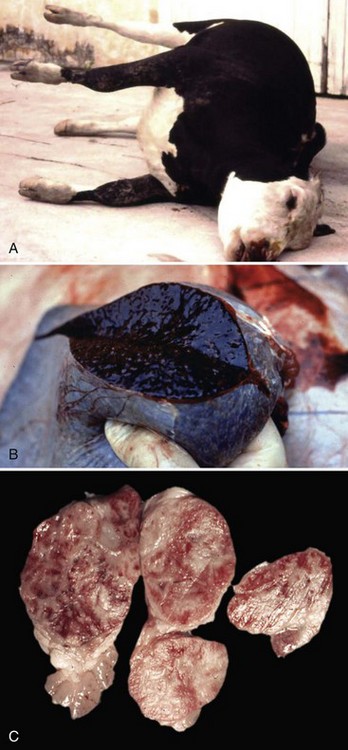
Fig. 4-23 Anthrax, ox.
A, Because of the high fever, cadavers of cattle dying of anthrax decompose rapidly with the usual result of excessive gas formation in the GI tract, abdominal distention and resultant “saw horse” position of the legs. B, The spleen is enlarged and bloody (splenomegaly, bloody spleen). A postmortem examination should not be performed on an animal suspected of dying from anthrax. Air-dried impression smears of blood from external orifices or from an ear vein can be stained and the bacterium identified (see Fig. 7-135). C, Lymph nodes are also enlarged and bloody as a result of anthrax toxins that destroy vascular endothelial cells (see Fig. 13-53). Anthrax toxin can also cause severe injury to the intestines (see Fig. 7-135) and lungs. (A courtesy Dr. D. Driemeier, Federal University of Rio Grande do Sul, Brazil. B and C courtesy Dr. J. King, College of Veterinary Medicine, Cornell University.)
In the circulatory system, vegetative forms proliferate in large numbers and are arranged in long chains in the capillary beds of many organ systems, including the spleen (see Fig. 13-53). Large quantities of edema toxin and lethal toxin are released into the blood, causing dysfunction and cell death of endothelial cells and their barrier systems, thus the toxins increase the permeability of the capillary wall, leading to edema, vasodilation, and hemorrhage in infected organ systems. Anthrax toxins also disrupt the clotting cascade likely through massive activation of DIC and consumption of clotting factors, resulting in unclotted blood at body orifices and within tissues and organs.
Vascular Leptospirosis (Leptospira spp.): The mechanism of injury in vascular leptospirosis is cell death caused by (1) physical properties (penetrating movements) of bacteria that disrupt functions of endothelial cells and (2) bacterial toxins that act directly on membranes of endothelial cells of small blood vessels, including capillaries of the systemic vasculature in all organ systems, leading to coagulative necrosis of affected cells. Gross lesions include acute vasculitis (endothelial cell necrosis) with systemic petechial and ecchymotic hemorrhages, edema, and DIC affecting all organ systems and serosal surfaces (see Fig. 2-18).
Animals encounter Leptospira spp. through direct contact of their oral or conjunctival mucous membranes or skin with leptospira-infected urine or with water from reservoirs or ponds into which infected urine drains. Ingestion may also serve as a portal of entry if water contaminated with leptospira is consumed and the bacteria encounter mucosae of the intestine. During mastication and swallowing, it is likely that the mucosae of the oral pharynx trap the bacteria in its mucus layer. After swallowing and through intestinal peristalsis, the bacteria are moved into contact with villi and crypts, where they are probably trapped in the mucus layer and encounter enterocytes. In the conjunctiva, it is also likely that the mucosae trap the bacteria in a mucus layer. For infection to occur, it has been suggested that the skin and mucosae must have small cuts or abrasions that allow the bacteria to penetrate into the vascularized submucosal or subcutaneous connective tissues and gain access to capillaries and/or postcapillary venules. However, the bacteria are motile and likely able to penetrate mucus layers and invade mucosae by moving directly through mucosal epithelial cells or between the cells through intracellular junctional complexes. In all three of these portals of entry, the goal is for the bacteria to reach well-vascularized ECM tissues. As a group, these spirochetes are highly motile and invasive and using its invasive motility (virulence determinate), it is able to penetrate the vascular wall and endothelial cells of capillaries and postcapillary venules to gain access to the circulatory system. Leptospira spp. may also invade lymphatic vessels and through this system and the thoracic duct eventually gain access to the circulatory system. Leptospira spp. are able to grow and replicate in the circulatory system and then spread systemically to all organ systems. To infect other organs such as the kidney and liver, it appears that the bacteria must first attach to endothelial cell membranes via adhesins before invading these cells and their underlying vascularized tissues and then interact with renal tubular epithelial cells and hepatocytes.
Leptospira spp. initially spread through the vascular system to all tissues of the body and do not appear to specifically target the kidney or liver via a tropism (attraction to a specific cell type or tissue) mechanism. However, once epithelial cells are infected, the reason for dominance of lesions in these organs is unclear and may be related to some essential trophism (nourishment of tissues) provided by these cells to the bacteria for colonization and proliferation.
In the context of interacting with capillary endothelial cells and probably renal epithelium, surface-associated proteins (outer membrane leptospiral protein) appear to be involved in ligand-receptor interactions that facilitate adhesion to host cell receptors like cell-adhesion molecules and ECM proteins (Len protein family). Adhesion to cells also appears to cause an increased expression of adhesion receptors such as E-selectin on endothelial cells resulting in additional adhesion of bacteria, platelets, and neutrophils (acute inflammatory response). This response may be attributable to bacteria wall LPS, peptidoglycans, and outer membrane proteins, thus promoting inflammation in capillaries leading to vasculitis and hemorrhage. Bacterial LPS likely activates cells by binding to TLRs on host cell membranes. Leptospira spp. also produce pore-forming hemolysins, proteases, sphingomyelinases, and collagenases that may assist in this process, but their role in causing endothelial cell injury remains to be determined.
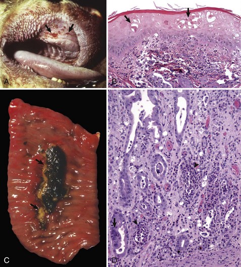
Glanders (Burkholderia mallei; Farcy, Malleus, Droes): The mechanism of injury in glanders is cell death caused by pyogranulomatous inflammation and its mediators and degradative enzymes. It is a disease of lymphatic vessels (and surrounding integument) and of the respiratory system. Gross lesions include ulcers, pustules, and nodules that can affect any part of the body but most frequently involve the skin and lymphatic vessels of the legs and flanks (Fig. 4-24). Nodules typically occur along the course of lymphatic vessels resulting in a raised beaded appearance of the skin. These nodules often rupture because of trauma to the skin or from pressure necrosis caused by the expanding volume of exudate within the nodules. This process results in craterlike ulcers of the skin that discharge a thick yellowish-white viscid and sticky purulent material containing abundant bacteria (see Fig. 4-24), and the outcome has been called farcy pipes. In the respiratory system, pyogranulomas can occur in mucosae of the nasal cavity and in all lobes of the lungs (random pattern).
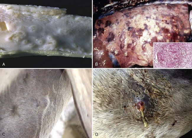
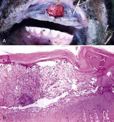
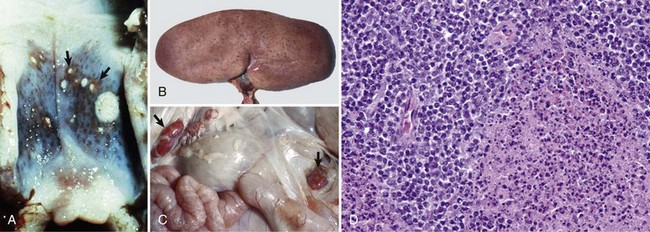
Fig. 4-24 Glanders disease.
A, Mucosa, nasal turbinates, multiple nasal ulcers, and granulomas. Burkholderia mallei colonizes the nasal turbinates resulting in pyogranulomatous inflammation, necrosis, and ulceration of the mucosa. B, When the bacterium colonizes the mucosae of the conductive system of the lung, it spreads into pulmonary parenchyma resulting in the formation of pyogranulomas (inset) throughout the lung. Inset, H&E stain. C, When the bacterium spreads to the skin, it colonizes subcutaneous lymphatic vessels resulting in the formation of pyogranulomatous nodules that typically occur along the course of lymphatic vessels (pyogranulomatous lymphangitis) resulting in a raised beaded appearance of the skin. D, These nodules frequently rupture because of trauma to the skin or from pressure necrosis due to the expanding volume of exudate within the nodules. This process results in crater-like ulcers of the skin that discharge a thick yellowish-white viscid and sticky purulent material containing abundant bacteria. (A courtesy Dr. D.D. Harrington, School of Veterinary Medicine, Purdue University; and Noah’s Arkive, College of Veterinary Medicine, The University of Georgia. B courtesy United States Animal Health Association, St. Joseph, MO. B inset courtesy Dr. Tyler, College of Veterinary Medicine, University of Georgia and Noah’s Arkive, College of Veterinary Medicine, University of Georgia. C courtesy Dr. D. Driemeier, Federal University of Rio Grande do Sul, Brazil. D courtesy Dr. R. Mota, Universidad Federal Rural de Pernambuco, Recife, Brazil and Dr. M. Brito, Universidad Federal Rural do Rio de Janeiro, Brazil.)
Horses, mules and donkeys encounter Burkholderia mallei, a facultative intracellular bacterium that resides in the soil, via fomites that (1) penetrate the skin by trauma or foreign objects or (2) are inhaled and/or ingested. In either case, these fomites are contaminated with bacteria-laden exudate derived from other infected animals with cutaneous lesions or nasal discharge. It appears that cutaneous glanders can occur through two mechanisms: (1) direct penetration of the skin or (2) through systemic spread to the skin via a systemic bacteremia or leukocytic trafficking from an initial infection of the respiratory or intestinal mucosae. For direct penetration, the skin apparently must have small cuts or abrasions that allow the bacterium to penetrate the subcutaneous connective tissues, gain access to lymphatic vessels, and cause infection and inflammation. Target cells for infection in the cutaneous lymphatic vessels and skin are unknown. Additionally, the role of ligand-receptor interactions in this process has not been determined. It appears that lesions characteristic of glanders probably result from inflammatory responses directed against a variety of molecules within the bacterium, resulting in pyogranulomatous inflammation.
For systemic spread to the skin, the bacterium appears to encounter mucosae of the nasal cavity and conductive component of the respiratory system. It is likely trapped in the mucus layer and then colonizes the mucosae. The development of a biofilm (virulence determinate) may be important in the colonization process. Once mucosae are colonized, the bacterium could then be phagocytosed by mucosal macrophages and/or dendritic cells and carried through mucosae to MALTs such as BALT. It is also possible, but unknown, that the bacterium has a mechanism to penetrate the mucus layer, enter the apical surfaces of mucosal epithelial cells via endocytosis or transcytosis, and exit the lateral-basal surfaces to gain access to mucosal-associated lymphoid tissues. In these lymphoid tissues, the target cells for infection and the role of ligand-receptor interactions have not been determined but appear to result in the nasal and pulmonary pyogranulomas characteristic of the disease. Cells of the monocyte-macrophage system and possibly mucosal dendritic cells probably play a role in the infection, replication, and spread of the bacterium. Once in MALTs, the bacterium could spread to regional lymph nodes in lymphatic vessels (1) as a cell-free bacteremia or (2) via leukocytic trafficking in macrophages, and after infection of and replication in lymphoid tissues, the bacterium could then spread systemically via lymphatic vessels and the thoracic duct to the circulatory system. Via the circulatory system, the bacterium ultimately arrives (cell free or in macrophages) at capillary beds of the skin, passes through the endothelial cells (leukocytic trafficking, endocytosis, or transcytosis), enters the subcutaneous tissues, encounters cutaneous lymphatic vessels, and elicits a pyogranulomatous inflammatory response in these tissues. Pyogranulomas have also been described in the spleen and liver.
Lesions in the respiratory system probably arise from an extension of the processes involved in colonizing mucosae as previously described, with secondary spread into adjacent interstitial tissues and alveoli. Additionally, it is possible that pyogranulomas in the lung could arise from spread of the bacterium through the circulatory system as a cell-free bacteremia or in macrophages as described. The existence of this mechanism of spread has not been determined.
Through ingestion and gastrointestinal peristalsis, Burkholderia mallei can also potentially reach the alimentary system. Herein, they could encounter the mucus layer of intestinal mucosae or M cells overlying Peyer’s patches. How the bacterium could penetrate the mucus layer is unknown, but it could involve mucosae-associated macrophages as discussed previously in respiratory system mucosae. Bacteria could also infect M cells via endocytosis, undergo endocytosis or transcytosis, and exit the basal surfaces via exocytosis to gain access to macrophages within Peyer’s patches. For either of these two pathways, macrophages within Peyer’s patches could then be infected with bacteria and be used to spread bacteria systemically. The likely goal of these types of mucosal encounters is to provide the bacterium with the opportunity to infect the local lymphoid tissues and then gain access to local, regional, and systemic lymph nodules and the circulatory system directly or via macrophages.
How and if the bacterium evades killing by phagosome-lysosome fusion, replicates, and spreads to cutaneous lymphatic vessels via the circulatory system are unknown. Type III and IV secretion systems (virulence determinates) may be possible mechanisms for the invasion, escape from lysosomes or phagolysosomes, and survival in target cells. The bacterium is surrounded by a type I O-antigenic polysaccharide (capsular) antigen, a virulence determinate, that may also block phagocytosis or phagosome-lysosome fusion. LPS, which likely contains a lipid A component, may play a role in tissue injury. Additionally, an interaction of the bacterium with cell membranes is a prerequisite for infection to occur. A type IV pilin-like protein may be involved in the adherence of the bacterium to target cells.
Pulpy Kidney (Overeating) Disease (Clostridium perfringens): The pathogenesis of pulpy kidney disease begins as an enterotoxemia caused by Clostridium perfringens, and this disease in the section on the alimentary system should be reviewed. Because ε-toxin is a permease that alters cell permeability, the vascular beds in affected intestinal tissues readily absorb toxins into the circulatory system. It appears that the sequence of events leading to pulpy kidney disease occur in the first phase or early in the second phase of alimentary enterotoxemia before toxin-induced massive necrosis of the intestine occurs. The mechanism of injury in pulpy kidney disease is cell death caused by ε-toxin that acts directly on renal endothelial and tubular epithelial cell membranes leading to vascular permeability changes and acute coagulative necrosis of tubular cells. Microthrombosis and ischemia from capillary endothelial injury are plausible but unproved mechanisms of tubular cell death. Gross lesions include soft pliable kidneys with hemorrhages; however, the lesions are often attributed to postmortem change. Experimental data suggest that vascular endothelial cells, such as those in the renal cortex supplying epithelial cells of renal tubules, express receptors (ligand-receptor interactions) for ε-toxin. Because ε-toxin is an angiotoxic permease, it increases the permeability of targeted endothelial cells, allowing plasma containing ε-toxin to leak into the ECM surrounding renal tubules. Renal tubular epithelial cells also express receptors for ε-toxin, and toxin binding may lead to membrane-mediated cytotoxicity and cell death.
Necrohemorrhagic Urocystitis (Escherichia coli, Corynebacterium renale, Pseudomonas spp., Proteus vulgaris, or Klebsiella pneumoniae): Necrohemorrhagic urocystitis is a term used to group bacteria whose virulence determinates can cause acute inflammation and hemorrhage of the mucosae of the urinary bladder, especially affecting transitional epithelial cells and the lamina propria and its capillary beds. Because more is known about virulence determinates for uropathogenic E. coli, it is discussed in greater detail; however, the other bacteria listed in this group likely use similar or related mechanisms to cause disease. The mechanism of injury is probably cell death (coagulative necrosis) caused by bacterial toxins that act directly on mucosal epithelial cells and capillaries in the lamina propria of the bladder and acute and chronic inflammation and their effector molecules and degradative enzymes. Gross lesions of the bladder include mucosal edema and mucosae that are rough and granular, red to dark red, and covered with white-gray flecks of fibrin mixed with cellular debris (see Fig. 11-64). Blood vessels in the wall and serosa of the bladder are prominent; this change is due to active hyperemia of the fluidic vascular phase of acute inflammation.
Animals encounter these bacteria through contact with them in fomites or fluid droplets of urinary or fecal origin. They commonly become commensal organisms that reside in the mucous membranes of the vagina and prepuce. Physical changes in pressure across the tubular components of the urinary and reproductive systems caused by parturition and breeding appear to force these commensal bacteria via reflux mechanisms into the urethra and urinary bladder. The length of the urethra, in part, appears to determine why females have cystitis more commonly than males. Environmental stressors, such as peak lactation, traumatic mucosal injury, and a high-protein diet that increases the pH of the urine, make mucosae more susceptible to colonization and alter the commensal relationship, allowing the bacteria to replicate in sufficient numbers to colonize the mucosae of the urinary and reproductive systems and spread bacteria to other animals. Once in the lumen of the urinary bladder, bacteria gain access to mucosal surfaces via random movement of the urine. The urinary mucosae lacks goblet cells, thus there is no mucus layer to penetrate. These bacteria encounter the apical surface of transitional epithelial cells, and using ligand-receptor interactions characteristic of other bacterial diseases, begin the process of adherence, binding, and colonization of the mucosae. Uropathogenic E. coli expresses adhesins, such as type 1 fimbriae, P fimbriae, and S fimbriae, that are involved in this process. These fimbriae (also known as pili) bind to hexagonal arrays of mannosyl-glycoprotein receptors known as uroplakins. They are expressed on the apical (and luminal) surfaces of specialized transitional epithelial cells called umbrella cells. The tips of the type 1 fimbriae express a ligand called FimH adhesin that binds to these uroplakin receptors.
Once bound, the bacteria begin a complicated process of entering and colonizing the cells and mucosae, respectively, through a series of conformational changes in the apical surface of the umbrella cells, leading to cytoskeletal rearrangements and cell entry by a zipper mechanism. Bacterial flagella may also have a role in this mechanism. This process results in colonization of the mucosae and the development of a biofilm-like arrangement affecting the mucosae (also known as biomasses of bacteria or intracellular bacterial communities). After formation of intracellular bacterial communities, bacteria kill infected umbrella cells via hemolysins that produce pores in membranes, thus releasing bacteria into the lumen of the bladder where they colonize new umbrella cells and repeat the infective process or are released into the environment during urination. The edema, hemorrhage, and necrosis characteristic of necrohemorrhagic urocystitis appear to be caused by acute inflammation and a variety of virulence determinates in highly pathogenic strains of uropathogenic E. coli and likely the other bacteria listed previously. Acute inflammation is probably induced via TLRs that recruit neutrophils from the vascular system into the lamina propria and mucosae and in response to cell necrosis, loss of the mucosal barrier, and interaction of the vascularized lamina propria with bacterial toxins. In umbrella cells infected by bacteria, bacterial toxins, such as LT and ST toxins, Shiga-like toxin, cytotoxins, and endotoxin, likely diffuse through the mucosae and cause membrane injury, leading to cell death (necrosis) and loss of the mucosal barrier system. These toxins may also stimulate apoptotic cell death, leading to release of bacteria into the urine. Once dead, these cells slough into the urine and endotoxins and other toxic molecules can be absorbed into the highly vascularized lamina propria, resulting in injury to the capillaries and acute vasculitis with active hyperemia.
Other virulence determinates that contribute to the pathogenesis of necrohemorrhagic urocystitis include bacterial surface molecules such as capsular K antigens and LPS that block phagocytosis and killing of the bacteria by neutrophils and macrophages. Uropathogenic E. coli usually produces siderophores that play a role in iron acquisition for the bacteria during and after colonization. The lytic actions of hemolysins also increase the availability of iron and other nutrients for bacterial growth in colonized mucosae. Hemolysins also can kill lymphocytes and block phagocytosis and chemotaxis by phagocytic cells. Some strains of uropathogenic E. coli have a virulence determinate for the production of urease, which hydrolyzes ammonia in urine into urea, resulting in an alkaline urine that causes additional injury to mucosae. Finally, these bacteria can readily exchange genetic information with less virulent bacterial strains by transduction and conjugation using drug resistance, toxin, and other virulence plasmids. These factors are a few of the reasons why it is often difficult to treat and resolve certain types of acute and chronic bladder infections.
Contagious Bovine Pyelonephritis (Corynebacterium renale or Escherichia coli): Contagious bovine pyelonephritis is most commonly caused by Corynebacterium renale, but E. coli may also cause this disease. These bacteria are likely commensal organisms that reside in the mucous membranes of the vagina and prepuce. Mechanisms that contribute to the occurrence of cystitis that preceded pyelonephritis are discussed in the previous section. Information about the mechanisms used by Corynebacterium renale or E. coli to cause contagious bovine pyelonephritis in cattle is limited. Thus portions of this section are speculative and based on (1) what is known mechanistically about other diseases of the respiratory system caused by Corynebacterium spp. or E. coli and (2) a reasonable probability that inflammation, responses to injury, and lesions that have been described in contagious bovine pyelonephritis are the result of underlying and known pathobiologic mechanisms. The mechanism of injury in contagious bovine pyelonephritis is probably cell death (acute coagulative necrosis) caused by (1) bacterial toxins that act directly on transitional mucosal and tubular epithelial cells of the urinary system possibly by inducing tubular cell and collecting duct cell apoptosis and (2) acute and chronic inflammation and their effector molecules and degradative enzymes. Gross lesions include white-tan streaks often mixed with narrow red streaks (hemorrhage) that radiated from the pelvis, through the medulla, often extending to the cortical medullary junction or deeper into the cortex (see Fig. 11-53). In many ways, these lesions resemble inverted renal cortical infarcts with their bases against the pelvis and their apices extending into the medulla.
Cattle (and probably sheep and goats) encounter these bacteria through contact with them in fomites or fluid droplets of urinary or fecal origin. Environmental stressors such as parturition, peak lactation, traumatic mucosal injury, and a high-protein diet that increases the pH of the urine, making the mucosae more suitable to colonization, could alter the commensal relationship, allowing the bacteria to replicate in sufficient numbers to colonize the mucosae of the urinary and reproductive systems and spread the bacterium to other animals. It has been proposed that contagious bovine pyelonephritis occurs secondary to a chronic and often insidious cystitis that ascends in the lower urinary tract and reaches the renal pelvis via the ureters to then spread though the mucosal barrier formed by transitional epithelium of the renal pelvis into the interstitium of the renal medulla. The mechanism of ascension may be caused by the reflux of infected urine from the ureters and urinary bladder into the renal pelvis. In the pelvis, it may occur via spread of bacteria through the mucosal barrier formed by the transitional epithelium of the pelvis and into vascularized ECM connective tissues supporting the tubules. How each of the events occurs has not been specifically determined; however, ligand-receptor interactions characteristic of other bacterial diseases and their interaction with mucosae likely occur in contagious bovine pyelonephritis.
Although Corynebacterium renale is a nonmotile bacterium, more pathogenic strains of E. coli are motile and this virulence determinate may help in ascension of the bacterium up the ureter to the kidney. It has been shown that pili are required for Corynebacterium renale to adhere to transitional epithelium of the urinary system and to attach to and colonize mucosae of the reproductive system. Additionally, pili may serve to disrupt phagocytosis of the bacteria by neutrophils and macrophages. Binding is strongest to the mucosal epithelial cells of the vulva and vagina. The receptors used to bind to mucosae have not been determined; however, once bound, colonization of the mucosae begins. Once bacteria replicate to sufficient numbers, they then spread by ascending in the lower urinary tract to encounter and colonize the mucosae of the urethra and bladder and then by vesicoureteral reflux to ascend to the ureters and renal pelvis. The development of a chronic insidious urocystitis is often an intervening stage in the disease that serves to produce large numbers of bacteria. After colonization, it is not known how these bacteria cross mucosae to gain access to the medullary interstitium at the pelvis. Degradative enzymes and inflammatory mediators combined with bacterial virulence determinates, such as Renalin, which is an extracellular cytolytic protein produced by Corynebacterium renale, may facilitate spread across mucosal barriers and inflammation and cell death within the medulla. It has been suggested that lesions in the medulla (resembling inverted renal cortical infarcts) may actually begin as a vasculitis from inflammation resulting in thrombosis, ischemia, and necrosis. It has been shown that toxins from E. coli may stimulate apoptotic cell death in renal tubular cells in pyelonephritis.
Renal Leptospirosis (Leptospira spp.): The pathogenesis of renal leptospirosis begins as vascular leptospirosis (see the section on the Cardiovascular System and Lymphatic Vessels) caused by Leptospira spp. The mechanism of injury in renal leptospirosis is cell death caused by (1) physical properties (penetrating movements) of bacteria that disrupt functions of endothelial cells, (2) bacterial toxins that act directly on membranes of tubular epithelial cells, and (3) acute and chronic inflammation and their effector molecules and degradative enzymes. Gross lesions include discrete and coalescing, often linear to radiating white to gray foci of tubular cortical necrosis and acute inflammation intermixed with hemorrhage (see Fig. 11-73). In chronic renal leptospirosis, lesions include discrete and coalescing, often linear to radiating white to gray foci of chronic inflammation and fibrosis (see Fig. 11-20). In the kidney, primary target cells for infection appear to be epithelial cells of the proximal convoluted tubules (cortex) (Fig. 4-25, A) and then later, epithelial cells of the loops of Henley (medulla) (Fig. 4-25, B). Once Leptospira spp. gain access to the circulatory system, they disseminate in glomerular capillaries and then intertubular capillaries of proximal convoluted tubules. Bacteria could access proximal tubular cells via their apical or basolateral surfaces by two routes, vascular by glomerular capillaries and migration into the lumen of the urinary space (apical) or vascular via intertubular capillaries and migration into the interstitium (basolateral). Because glomerular changes are usually unremarkable and bacteria and inflammation are observed in the interstitium, it appears that epithelial cells of the proximal convoluted tubules are infected via the basal-lateral surfaces of the cells via migration through intertubular capillaries.
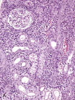
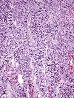
Fig. 4-25 Renal leptospirosis.
A, Kidney, outer cortex. Note the infiltration of mononuclear cells, chiefly macrophages, lymphocytes and plasma cells in the interstitium between the proximal convoluted tubules, the result of the leptospires infecting the proximal tubule cells after exiting the intertubular capillaries. B, Kidney (same as A), inner cortex. Numerous neutrophils distend the interstitium between the loops of Henle. This acute inflammatory response further down the nephron from the area in A, supports the concept that the cells of the loop of Henle are infected later than those in the proximal convoluted tubule. H&E stain. (A and B courtesy Dr. J. F. Zachary, College of Veterinary Medicine, University of Illinois.)
The bacteria likely attach to endothelial cell membranes of intertubular capillaries via adhesins then penetrate the vessel wall by moving directly through the cells or through their junctional complexes to gain access to the interstitium. In reality, the distance in the interstitium between capillaries and proximal tubular epithelial cells is probably no more than 100 µm and it is likely that the flagella of these motile bacteria propel them to the epithelial cells. It is unclear why the bacteria target proximal tubular epithelial cells for infection. Although undetermined, such specificity could be attributed to ligand-receptor interactions or to a chemical gradient, such as an iron concentration, that could be required for bacterial growth and replication. The bacteria are present in the cytoplasm of these cells; endocytosis and phagosome-lysosome fusion are not involved in cell entry. It appears that the bacteria are able to directly enter these cells via their motility.
The cause of death of proximal tubular cells is probably multifactorial, involving vasculitis and ischemia, trauma from physical injury caused by bacterial motility, inflammatory mediators and degradative enzymes, and bacterial toxins. The inflammatory cells in this lesion progress from neutrophils (suppurative) to lymphocytes, macrophages, and plasma cells (chronic). Epithelial cells lining the loop of Henle could also be infected by an intertubular capillary-interstitial route. This mechanism has not been confirmed. Additionally and based on inflammatory cell responses, it is unclear why cells of the proximal tubules appear to be infected at an earlier point in the disease than those of the loop of Henle. When proximal tubular cells die, they release bacteria into the urinary lumen where they are carried in urine and spread into the environment via urination. During this luminal transit, the bacteria also encounter the apical surfaces of epithelial cells lining the loop of Henle. It is plausible that Leptospira spp. infect epithelial cells of the loop of Henle via their apical surfaces projecting into the urinary lumen, using mechanisms similar to those previously described. Infection appears to result in the same cascade of cell alterations and inflammatory responses as those described for proximal tubular cells. Biofilms (virulence determinate) formed by Leptospira spp. may also play a role in tubular injury. These outcomes in both proximal tubular and loop of Henle cells serve as the basis for characterizing this disease as tubulointerstitial nephritis.
Strangles (Streptococcus equi ssp. equi): The mechanism of injury in strangles is death (coagulative necrosis) of cells of lymphatic vessels, lymph nodes, and the monocyte-macrophage system attributable to acute suppurative inflammation and its mediators and degradative enzymes. Gross lesions include the formation of acute abscesses within regional lymph nodes resulting in enlarged firm lymph nodes that on a cut surface have discrete and coalescing areas of yellow-white suppurative exudate infiltrating and compressing contiguous parenchyma (see Fig. 13-74). Affected retropharyngeal and mandibular lymph nodes may also have draining fistulous tracts between affected nodes and the surface of the skin, the guttural pouches, and the nasal cavities and sinuses, resulting in release of bacteria into the environment. This outcome occurs because degradative enzymes released from dead neutrophils in abscesses digest the capsule of the lymph node and the structures of all contiguous tissues until a fistulous tract is formed.
Foals encounter Streptococcus equi ssp. equi through inhalation or ingestion of fomites or body fluids contaminated with the bacterium. It is deposited on mucosae of the nasal (centrifugal turbulence) and oral pharynx and trapped in the mucus layer. The bacterium is nonmotile and it has not been clearly shown how it penetrates the mucus layer and gains access to mucosal epithelial cells. Mucosal epithelial cells of the tonsils and tonsillar crypts appear to be important target cells for adherence and binding, and this specificity may be determined by unique ligand-receptor interactions. Once in contact with cell membranes, several bacterial cell wall surface proteins, such as M-like proteins (SeM, SzPSe), may act as adhesins and attach to receptors expressed on the membranes of these cells. The characteristics of these receptors are unknown. It has not been determined if the bacterium needs to first colonize the mucosal surface before spreading into subjacent local lymphoid tissues. Additionally, it has not been determined how the bacterium crosses the mucosal barrier of the tonsil. Mucosal macrophages could phagocytose the bacteria in the mucus layer and carry it via leukocyte trafficking through the mucosal barrier into the local lymphoid tissues, or once bound to host cell receptors, it could be transported through the cell via transcytosis and released via exocytosis from the basal membranes of the cells into local lymphoid tissues of the tonsil. Dendritic cells could also be involved in the spread of bacteria across the mucosal barrier and into local and regional lymphoid tissues and lymph nodes. In local lymphoid tissues of the tonsil, the bacterium is able to replicate extracellularly and then spread to regional nodes such as the mandibular or retropharyngeal. Although unknown, the bacterium could spread by means of lymphatic vessels to these regional nodes in macrophages via leukocyte trafficking or through cell-free migration. The bacterium multiplies extracellularly in the lymph tissues and nodes, and the exudate contains large numbers of viable bacteria.
Mechanistically, several bacterial virulence determinates contribute to the character of this exudate (suppurative) and the large number of viable bacteria. Bacterial virulence determinates that act as chemoattractants for neutrophils and that disrupt phagocytosis and killing by neutrophils appear to explain the abundance of exudate and viable bacteria, respectively. Early in the sequence of events when bacteria encounter mucosal and tissue macrophages in local and regional lymphoid tissues and nodes, a bacterial cell wall protein, SeeH, interacts with these cells, resulting in the release of proinflammatory cytokines, increased vascular permeability, and edema. The occurrence of acute suppurative inflammation is facilitated by several virulence determinates. When peptidoglycan of the bacterial cell wall interacts with C3 of complement in the edema fluid via the alternative complement pathway, it produces complement-derived chemotactic factors that attract large numbers of neutrophils from capillary beds into local vascularized connective tissues. Furthermore, bacterial streptokinase interacts with plasminogen in the edema fluid to form active plasmin, which hydrolyzes fibrin. This process appears to increase the spread and dispersion of bacteria in tissue. Normally, fibrin confines bacteria by isolating them within its polymerized fibrillar meshwork so they can be phagocytosed and killed by neutrophils and macrophages, but when fibrin is hydrolyzed, large numbers of bacteria can accumulate in the exudate of an abscess and be readily available for release into the environment (see later). The outcome of these processes also contributes to the initiation of the cellular (leukocytic) phase of acute inflammation.
The surface of Streptococcus equi ssp. equi is coated with numerous protein virulence determinates, such as hyaluronic acid, SeM, and Se18.9 proteins that disrupt phagocytosis and killing. The bacterium also secretes leukocidal toxin and streptolysin S, a cell membrane pore-forming toxin, which kills leukocytes and disrupts phagocytosis. These processes lead to the accumulation of large numbers of viable bacteria in the exudate of lymphoid tissues and nodes and in abscess formation. Additionally, hyaluronic acid in the capsule appears to block interactions between bacteria and neutrophils by increasing the negative charge and hydrophobicity of the bacterial surface and by producing a localized oxygen-reduced environment that protects the activity of oxygen-labile proteases and toxins such as streptolysin S. It is likely but undetermined that bastard strangles occurs because of spread of the bacterium to systemic lymph nodes and organ systems by leukocyte trafficking or cell-free migration via efferent lymphatic vessels and/or capillaries or postcapillary venules in lymph nodes to gain access to the systemic circulatory system.
Caseous Lymphadenitis (Corynebacterium pseudotuberculosis): The mechanism of injury in caseous lymphadenitis is cell death attributable to inflammation and its mediators and degradative enzymes affecting cells of the monocyte-macrophage system and cell populations in lymph nodes and other organ systems. Gross lesions include chronic active pyogranulomatous lymphadenitis (see Figs. 11-75 and 11-76) with enlarged firm lymph nodes that on a cut surface have discrete and coalescing areas of yellow-white caseous exudate infiltrating and compressing contiguous parenchyma and abundant connective tissue. In other organs, such as the lung, abscesses encapsulated by dense bands of fibrous connective tissue and containing yellow-white caseous exudate are common findings.
Sheep and goats encounter Corynebacterium pseudotuberculosis through penetrating wounds and potentially ingestion. The bacterium is a common contaminate of the environment usually from animals that have the cutaneous form of caseous lymphadenitis leading to fistulous tracts from draining cutaneous lymph nodes. Penetrating wounds most commonly involve the skin and mucous membranes of the oral cavity. Management practices, such as shearing, may cause skin abrasions, whereas objects like wire, sticks, and protruding barn or fence nails may puncture the skin. Similar types of injury may occur in the oral cavity by similar objects and mechanisms. Once the bacterium reaches the submucosa or subcutis, it is phagocytosed by neutrophils and macrophages and spread via leukocyte trafficking to regional lymph nodes via lymphatic vessels. The bacterium replicates in the lymph nodes and the inflammatory response results in multiple pyogranulomas (abscesses) that grow in size and with time, notably enlarge and affect the entire lymph node. Macrophages infected with bacteria leave the node via leukocyte trafficking and spread via lymphatic vessels and probably the thoracic duct to the systemic circulation system or spread by entering the capillary or venous circulation within the node to gain access to the systemic circulatory system. Via leukocyte trafficking, macrophages then spread the bacterium to other visceral lymph nodes, especially the mediastinal and bronchial, and to tissues in a wide variety of organ systems, especially the lung. Because the bacterium is able to replicate in large numbers in macrophages and neutrophils, the death of these cells attributable to mycolic acid or cell aging results in the release of bacteria into vascularized ECM tissues. This process activates integrins and adhesins in vascular endothelium and causes massive recruitment of additional neutrophils and macrophages into the tissues as part of a chronic active inflammatory response, thus repeating the inflammatory process of forming pyogranulomas (abscesses). The mechanisms used by Corynebacterium pseudotuberculosis to gain access to lymph nodes via ingestion (if it occurs) are unknown. Two potential pathways could be used and both focus on macrophages and leukocyte trafficking. First, bacteria could interact with the mucus layer and mucosae of the oral pharynx, be phagocytized by macrophages and carried to the tonsils, then to regional lymph nodes, and then systemically. Second, bacteria could be swallowed and via alimentary peristalsis encounter M cells of small intestinal crypts, spread via M cells to macrophages in contiguous Peyer’s patches, then to regional lymph nodes, and then systemically. It is likely that the mechanisms and responses to injury described for the penetrating wounds portal of entry would also apply to these two scenarios.
Corynebacterium pseudotuberculosis has two known bacterial virulence determinates, phospholipase D and mycolic acid, that allow it to colonize tissues and produce pyogranulomas. Phospholipase D increases vascular permeability, which is thought to assist macrophages in migrating in and out of tissues infected with the bacterium, thus favoring the systemic spread of the bacterium. As a potent exotoxin, it also injures cell membranes leading to macrophage and neutrophil dysfunction, disruption, and death and interferes with neutrophil chemotaxis. Corynebacterium pseudotuberculosis does not have a protective capsule but has a waxy mycolic acid coat on the cell wall surface. Mycolic acid induces acute inflammation, has a role in the formation of granulomas, is toxic for macrophages, and prevents killing of the bacterium with phagosome-lysosome fusion likely by protecting against hydrolytic enzymes present within lysosomes.
Brucellosis (Brucella spp.): The mechanism of injury in brucellosis is cell death caused by inflammation and its mediators and degradative enzymes. Brucella spp. do not have virulence determinates for exotoxins or endotoxins that cause direct injury to cells. Gross lesions include chronic active pyogranulomatous lymphadenitis with enlarged firm lymph nodes that on a cut surface have discrete and coalescing areas of yellow-white exudate infiltrating and compressing contiguous parenchyma.
Animals encounter Brucella spp. through inhalation or ingestion of bacteria in fomites contaminated with infected exudates from other body systems such as the female reproductive tract. The bacterium encounters mucosae and their mucus layers through centrifugal turbulence and entrapment in the mucus layer of the nasal pharynx and through mastication, gravity, and entrapment in the mucus layer of the oral pharynx. Bacteria are phagocytosed by macrophages or dendritic cells migrating through, on, or in the mucosae and spread to local lymphoid tissues via leukocyte trafficking by lymphatic vessels, to regional lymph nodes via lymphatic vessels, and then systemically to superficial and visceral lymph nodes and other organs such as spleen, liver, bone marrow, mammary glands, and reproductive organs. Brucella spp. can also cross the mucosal barrier via endocytosis, exit the basal surface of mucosal epithelial cells through exocytosis, and spread cell free in lymphatic vessels to local and regional lymph nodules and nodes where they are phagocytosed by macrophages and then spread systemically as discussed above. Through ingestion and peristalsis, Brucella spp. can also reach the alimentary system where they encounter M cells. Bacteria infect M cells via endocytosis, undergo transcytosis, and exit the basal surfaces via exocytosis to gain access to macrophages within Peyer’s patches. Macrophages within Peyer’s patches are then infected with the bacteria and used to spread bacteria systemically. The goals of these three types of mucosal encounters are to provide Brucella spp. with ample opportunity to infect macrophages and subsequently gain access to local, regional, and systemic lymph nodules and nodes.
In lymph nodes, bacteria-infected macrophages are killed by the bacterium or die through aging and the bacteria are released into vascularized ECM. These bacteria elicit an acute inflammatory response that is quickly replaced by a pyogranulomatous inflammation because of LPS in the bacterial cell wall. The material is not readily degradable, and macrophages (as monocytes) are recruited from the systemic circulation to phagocytose and degrade such material and kill the bacteria. Brucella spp. are able to evade the killing mechanisms when phagocytosed by neutrophils and macrophages. Additionally, they are able to grow and replicate in macrophages and dendritic cells. Once Brucella spp. encounter cell membranes of macrophages, they use ligand-receptor interactions to attach to and enter cells; however, the details remain unclear. Outer membrane protein of the bacterial cell wall and class A scavenger receptors on host target cells are likely involved but not necessarily with each other. TLRs are also likely involved in attachment and entry into macrophages. Entry occurs via endocytosis through a phagosome, but phagosome-lysosome fusion does not occur because bacteria are able to block fusion through rapid acidification of the phagosome. LPS (a PAMP]), a type IV secretion system, and a long list of other putative virulence determinates such as cyclic β-1,2-glucan and heat shock proteins may also be involved in blocking phagosome-lysosome fusion and promoting bacterial growth and replication. The virulence of Brucella spp. strains appears related to the LPS composition of its capsule, with the encapsulated smooth phenotypes generally being more virulent. Additionally, the presence of a smooth capsule enhances bacterial growth and replication in phagosomes.
When Brucella spp. spread via leukocyte trafficking systemically in macrophages, they are able to gain access to tissues in the male and female reproductive systems and the mammary gland (Fig. 4-26; see also Fig. 19-18). In summary, infected macrophages interact with and infect placental trophoblasts in placentomas and epithelial cells of other reproductive tissues. Once such cells are infected, the bacteria likely infect fetal macrophage-like cells that serve to spread the bacteria through the fetus and to other lymphoid tissues in reproductive organs (see Fig. 4-26, C and D). Brucella spp. also survive in macrophages of these tissues by inhibiting the phagosome-lysosome fusion. Bacterial growth and replication with concurrent death of bacteria-infected macrophages results in pyogranulomatous inflammation of these tissues and organ systems.
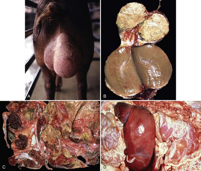
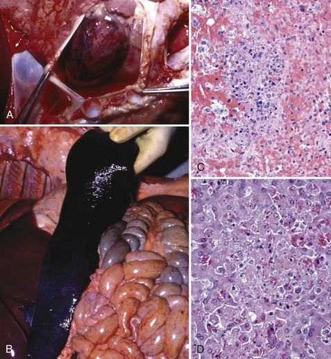
Fig. 4-26 Brucellosis.
Brucellosis is a disease in which the bacterium initially targets lymphocytes and macrophages in mucosae-associated lymphoid tissues, regional lymph nodes, systemic lymph nodes, and the spleen to replicate and grow in number. It uses macrophages to spread to and through these tissues and then systemically to infect cells and tissues in the placenta, sex organs of males and females, and fetuses. Therefore brucellosis is initially and long term characterized by a chronic active pyogranulomatous lymphadenitis with sequelae that affect the reproductive systems. A, Boar, swollen testis. The testis in enlarged due to chronic active pyogranulomatous inflammation. Brucella spp. spread in macrophages via leukocyte trafficking from lymphoid tissues systemically to the testis. B, The epididymis can be filled with pyogranulomatous exudate, which obstructs the flow of spermatozoa and causes infertility. Infected animals can also serve as carriers and spread the bacterium via sexual contact (also see Fig. 19-18). C, Fetal cotyledons. Note the roughened granular yellow-brown surface of cotyledons (arrows) infected with the bacterium. This lesion is caused by pyogranulomatous inflammation leading to severe necrosis of the affected cotyledons. Normal cotyledons are dark red and have a smooth shiny surface. D, Fetal brucellosis, hepatomegaly and fibrinous polyserositis. The bacterium is thought to be spread from infected cotyledons to fetal organs via leukocytic trafficking in fetal macrophage-like cells. The bacterium causes extensive injury of the vascular system and organs through inflammatory responses induced in the fetus. (A courtesy Dr. C. Wallace, College of Veterinary Medicine, The University of Georgia; and Noah’s Arkive, College of Veterinary Medicine, The University of Georgia. B courtesy Dr. K. McEntee, Reproductive Pathology Collection, University of Illinois; and Dr. J. King, College of Veterinary Medicine, Cornell University. C and D courtesy Dr. K. McEntee, Reproductive Pathology Collection, University of Illinois.)
Rhodococcal Mesenteric Lymphadenitis (Rhodococcus equi): The pathogenesis of rhodococcal mesenteric lymphadenitis begins as an infection of the respiratory system (see section on the Respiratory System, Mediastinum, and Pleurae), followed by infection of the alimentary system (see section on the Alimentary System and the Peritoneum, Omentum, Mesentery, and Peritoneal Cavity). The mechanism of injury in rhodococcal mesenteric lymphadenitis is death of cells of the monocyte-macrophage system and of all cell populations in the lymph node secondary to inflammation and its mediators and degradative enzymes. Gross lesions include chronic active pyogranulomatous lymphadenitis (see Fig. 7-137) with enlarged firm lymph nodes that on a cut surface have discrete and coalescing areas of yellow-white exudate infiltrating and compressing contiguous parenchyma. Rhodococcus equi enters the alimentary system through M cells and is released into Peyer’s patches where it is phagocytosed by tissue macrophages. Bacteria-infected tissue macrophages spread via leukocyte trafficking in lymphatic vessels within the intestinal mesentery to mesenteric lymph nodes, leading to pyogranulomatous lymphadenitis, and then systemically via the thoracic duct and the blood vascular system to additional lymph nodes and lymphoid tissues such as the spleen. The pathogenesis of pyogranulomatous lymphadenitis appears to progress much like that which occurs in the lung.
Edema Disease (Escherichia coli): The pathogenesis of edema disease begins as an enterotoxemia of the alimentary system (see section on the Alimentary System and the Peritoneum, Omentum, Mesentery, and Peritoneal Cavity). This disease is caused by a specific strain of hemolytic E. coli having virulence determinates for a bacterial toxin called Shiga toxin 2e (also known as verotoxin 2e). It was initially called edema disease principle. The mechanism of injury is death of endothelial and smooth muscle cells of arterioles (fibrinoid arteriopathy/arteriolopathy), thus the toxin biologically behaves as an angiotoxin. In the brain, vascular lesions are followed by secondary ischemia and necrosis of neural cells, particularly neurons in brainstem nuclei. Gross lesions include symmetric areas of yellow-gray malacia in the brainstem overlying affected nuclei (see Web Fig. 10-24). Pigs encounter E. coli through ingestion and colonization of the intestinal mucosae. The enterotoxemia phase is discussed in the section on the Alimentary System and the Peritoneum, Omentum, Mesentery, and Peritoneal Cavity. Shiga toxin 2e is absorbed in the alimentary system and circulates systemically in the blood vascular system. Endothelial and smooth muscle cells of arteritis and arterioles express receptors for this toxin. This toxin causes vascular permeability changes and edema followed by endothelial injury and death leading to hemorrhage, intravascular coagulation, microthrombosis, and infarction (grossly malacia). Shiga toxin 2e acts to disrupt protein synthesis in affected cells leading to cell death.
Focal Symmetric Encephalomalacia (Clostridium perfringens): The pathogenesis of focal symmetric encephalomalacia begins as an enterotoxemia caused by Clostridium perfringens. Because ε-toxin is a permease that alters cell permeability, the vascular beds in affected intestinal tissues readily absorb toxins into the circulatory system. It appears that the sequence of events leading to focal symmetric encephalomalacia occurs in the first phase or early in the second phase of enterotoxemia before toxin-induced massive necrosis of the intestine occurs. The mechanism of injury is cell death caused by bacterial toxins that act directly on endothelial cell membranes causing permeability changes leading to acute coagulative necrosis of affected cells, including endothelial cells and neurons. Gross lesions include bilaterally symmetric malacia (acute coagulative necrosis of neuron cell bodies) and liquefactive necrosis of the basal ganglia, internal capsule, thalamus, and substantia nigra with edema and hemorrhage. Cerebral edema causes indistinct sulci and flattened gyri and in severe cases, coning of the cerebellar vermis through the foramen magnum. Because ε-toxin is a permease that alters cell permeability of the microvascular system, in the nervous system, it behaves as an angiotoxin. The vascular beds in affected intestinal tissues with enterotoxemia readily absorb toxins into the circulatory system. They are then carried to the brain via the blood vascular system where they act to increase the permeability of capillary beds leading to the release of blood plasma containing toxins into the neuropil, resulting in severe generalized vasogenic cerebral edema. Circulating ε-toxin accumulates preferentially in the brain via ligand-receptor interactions. It is likely that receptors expressed on different populations of endothelial cells in the body and within the brain determine in part the specificity for certain neurons and nuclear groups within the nervous system. The cell membrane of cerebral endothelial cells is the probable site of toxin binding, and it appears that toxin-induced injury of endothelial cells leads to the expression of more receptors for circulating ε-toxin. Injury to the endothelium disrupts the integrity of the blood-brain barrier, leading to increased vascular permeability, vasogenic edema, and the diffusion of toxin into the neuropil where it encounters neuron cell bodies. Acute coagulative necrosis of neuron cell bodies has been attributed to toxin-induced microthrombosis of capillaries, resulting in neuronal ischemia and by direct cytotoxic action on neurons and other neural cells. The selective nature of neuronal death caused by ε-toxin may be explained by ligand-receptor interactions, selective metabolic vulnerability of specific populations of neurons, or the concentration of ε-toxin.
Botulism and Tetanus (Clostridium botulinum, Clostridium tetani): The mechanism of injury in botulism is disruption of neurotransmitter vesicle exocytosis at myoneural (flaccid paralysis) junctions (i.e., synapses) by botulinum (Clostridium botulinum) neurotoxin. The mechanism of injury in tetanus is disruption of neurotransmitter vesicle exocytosis at neural-neural (spastic paralysis) junctions (i.e., synapses) by tetanus (Clostridium tetani) neurotoxin (tetanospasmin). These neurotoxins are produced in anaerobic microenvironments (a lowered oxidation-reduction [redox] potential) such as in necrotic tissue occurring in traumatic wounds (e.g., nail penetrating sole of the hoof, gastric ulcers in foals, muscle necrosis). Gross or microscopic lesions are not observed in the nervous system with these diseases.
Animals encounter these bacteria through contact with bacterial endospores present in soil and on environmental objects. Spores are carried into wounds and germinate to vegetative forms, and when this latter form dies, neurotoxins are released into dead tissue. Examples of such wounds include penetration of the skin or sole of the hoof or gastric ulcers. In addition, botulinum neurotoxin can also be released from lysed vegetative forms in the anaerobic environment of decaying vegetable matter (e.g., spoiled silage, hay, grain) and decomposing carcasses and absorbed into the circulatory system from the alimentary system with their ingestion. From a wound (or the alimentary system), neurotoxins access myoneural (botulinum neurotoxin) and neural-neural (tetanus neurotoxin) junctions by two routes, either hematogenously (botulinum neurotoxin) or via retrograde axonal transport (tetanus neurotoxin).
Botulinum neurotoxin enters the blood from (1) wounds as it diffuses via a concentration gradient to the periphery of the wound to areas with adequate circulation in which it is absorbed into the blood via capillaries and (2) absorption through intestinal villi and transfer to capillary beds within the lamina propria of the villi. Botulinum neurotoxin gains access to myoneural junctions via capillary beds that supply muscular tissues. On its release from capillaries, the neurotoxin diffuses in interstitial fluids until contacting the cell membrane of peripheral nerves (e.g., lower motor neuron), where it enters the cytoplasm of the neuron through the formation of endocytotic vesicles.
In contrast, tetanus neurotoxin (tetanospasmin) enters the nervous system and gains access to neural-neural junctions by initially entering the cytoplasm of distal processes of neurons through the formation of endocytotic vesicles in viable nerve endings located in tissue surrounding the site of the wound. The endocytotic vesicles are transported into the central nervous system (CNS) by retrograde axonal transport, and tetanus neurotoxin is released into the interstitial fluid of the neural-neural junctions by exocytosis. Free tetanus neurotoxin binds to the cell membrane of inhibitory interneurons of the spinal cord, is internalized via endocytosis, and acts to disrupt the release of inhibitory neurotransmitters via the same mechanism as used by botulinum toxin: disruption of the synaptic fusion complex. Presynaptic neurons (upper motor neurons) excite postsynaptic neurons (lower motor neurons) on a nearly continuous basis. Inhibitory interneurons acting on lower motor neurons serve to counterbalance and smooth the excitatory effects of acetylcholine released from presynaptic neurons (upper motor neurons) to excite the same lower motor neurons. Thus skeletal muscle groups (opposing flexor and extensor muscles) are given time to relax; as a result, skeletal muscle contractions initiated by lower motor neuron are well regulated and coordinated. Failure to have adequate inhibitory interneuron regulation of lower motor neurons leads to the spastic paralysis observed in tetanus.
Although botulinum and tetanus toxins gain access to targets in the nervous system by different mechanisms, from this point forward in the pathogenesis, both botulinum and tetanus neurotoxins share a common mechanism of injury, the disruption of neurotransmitter vesicle exocytosis by disrupting the synaptic fusion complex. The mechanisms are demonstrated in Figs. 4-27 and 4-28 for botulinum toxin and tetanus toxin, respectively. Thus diseases (clinical signs) that occur are the direct result of disruption of the function of myoneural (flaccid paralysis) and neural-neural (spastic paralysis) junctions. Botulinum and tetanus toxins have heavy and light chains and behave as typical A-B toxins (i.e., diphtheria, cholera, pertussis, and shigellae toxins) composed of two units: a binding B-domain (heavy chain) that mediates transport via endocytosis and exocytosis and an enzymatically active A-domain (light chain) that serves to cleave proteins within the target cell. The heavy chain binds to the neuronal membrane of myoneural junctions (botulinum toxin) and nerve endings (tetanus toxin), and the entire toxin molecule enters the neuron via receptor-mediated endocytosis. The A-domain is cleaved from the B-domain within the target cell endocytotic vesicle and then released into the cytoplasm where it is active. The A-domain (light chain), a zinc-containing endopeptidase, leaves the endocytotic vesicle and enters the cytoplasm of the neuron and acts to cleave proteins that form the synaptic fusion complex. This complex, formed by fusion of synaptic vesicle proteins with presynaptic plasma membrane proteins, serves to bring neurotransmitter vesicles into contact with the neuronal cell membrane at the myoneural (botulinum toxin) and neural-neural (tetanus toxin) junctions and facilitates membrane fusion and release of excitatory (acetylcholine) and inhibitory neurotransmitters (glycine and γ-aminobutyric acid [GABA]), respectively.
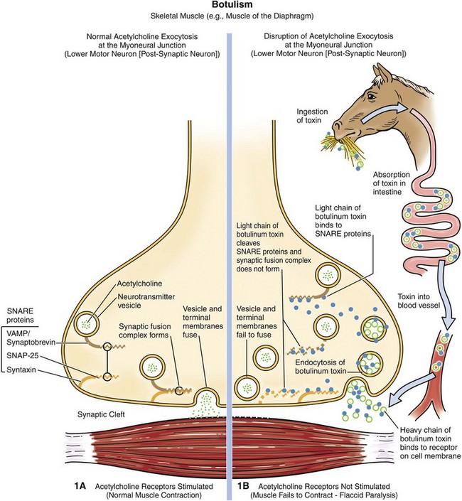
Fig. 4-27 Mechanism of myoneural junction dysfunction in botulism.
Note that botulinum toxin reaches the myoneural junction via the circulatory system. (Courtesy Dr. J. F. Zachary, College of Veterinary Medicine, University of Illinois.)
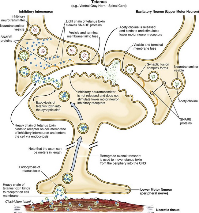
Fig. 4-28 Mechanism of neural-neural junction dysfunction in tetanus.
Note that tetanus toxin reaches the neural-neural junction via retrograde axonal transport. The selectivity of tetanus toxin for inhibitory interneurons is likely mediated by the expression of different glycosylphosphatidylinositol-anchored protein(s) on different types of neurons. The B-domain of tetanus toxin appears to bind only to the type of glycosylphosphatidylinositol-anchored protein expressed on inhibitory interneurons. (Courtesy Dr. J. F. Zachary, College of Veterinary Medicine, University of Illinois.)
Different types of glycosylphosphatidylinositol-anchored protein(s) may be expressed on different types of neurons, which may explain why B-domain of tetanus toxin only binds to inhibitory interneurons and not other types motor neurons. Disruption of the synaptic fusion complex prevents the neurotransmitter vesicles from fusing with the membrane, which in turn prevents release of neurotransmitters into the synaptic cleft. Proteins that form the synaptic fusion complex (SNARE proteins) include neurotransmitter vesicle proteins (such as vesicle-associated membrane proteins [VAMPs]/synaptobrevin) and presynaptic plasma membrane proteins (syntaxin, synaptosomal-associated protein [SNAP-25]). Different types of Clostridium botulinum produce different types of toxins (toxin type A to G), and these types target and cleave specific types of SNARE proteins, synaptobrevin (cleaved by toxin types B, D, F, and G), syntaxin (cleaved by toxin type C), and synaptosomal-associated protein (cleaved by toxin types A, C, and E). Botulinum toxin seemingly does not cross the blood-brain barrier; therefore neural-neural junction functions in the CNS remain intact. Notwithstanding the profound neurologic signs of spastic paralysis and flaccid paralysis that occur with tetanus (Clostridium tetani) and botulism (Clostridium botulinum), respectively, macroscopic or microscopic lesions are not observed in the nervous system.
Listeriosis (Listeria monocytogenes): The mechanism of injury in listeriosis is cell death that is uniquely localized to the brainstem caused by acute inflammation and its mediators and degradative enzymes. Gross lesions are often not observed, but when present consist of nodules and linear bands of gray-yellow exudate (perivascular microabscesses formed by neutrophils) mixed with active hyperemia and/or hemorrhage commonly arranged in a perivascular pattern (see Fig. 14-88).
Cattle, sheep, and goats encounter Listeria monocytogenes in soil, animal feed, water, and feces; however, the greatest risk of contracting the disease occurs when ruminants are fed improperly stored silage in which the pH is not acidic enough to prevent overgrowth of the bacterium. Consumption of Listeria monocytogenes–contaminated silage is not sufficient to cause CNS disease, unless it occurs with a penetrating injury of the oral cavity caused by a stick or other sharp object (nail) that carries the bacterium in the silage into the submucosal connective tissue of the oral cavity or tongue. At this point, the bacterium colonizes oral tissues, enters nerve endings in the oral cavity, and ascends into the CNS via retrograde axonal transport in cranial nerves. The oral cavity is primarily innervated by the trigeminal and other cranial nerves that terminate in the brainstem. Thus Listeria monocytogenes ultimately localizes to the brainstem (i.e., pons, medulla oblongata, and proximal cervical spinal cord). The mechanism of entry into nerve endings is unknown; however, it has been shown experimentally in cell culture systems that Listeria. monocytogenes gains entry into typically nonphagocytic cells through endocytosis and endocytic vesicles. Bacterial internalization, the entry process, is mediated by internalins (type A and B) that utilize host cell receptor E-cadherin, a transmembrane glycoprotein. Because Listeria monocytogenes resides intracellularly within cell bodies of neurons when it arrives in the brainstem, it initially does not disrupt the blood-brain barrier and thus does not activate defense mechanisms provided by the innate (inflammation) and adaptive immune responses. The cytoplasm of infected neuron cell bodies appears to be permissive and allows free proliferation of the bacterium. This permissive environment also appears to be promoted by a bacterial virulence determinate called listeriolysin O that inhibits immune responses and allows infected cells to hide from defense mechanisms.
Once free in the cytoplasm, the bacterium replicates to sufficient numbers and then begins the process of infecting other cells. In the cytoplasm, the bacterium has a doubling time of approximately 1 hour. When the number of bacterium in the cytoplasm reaches a level sufficient to facilitate infection of adjacent cells, bacteria move themselves, facilitated by a virulence determinate in the cytoplasm, to the inner side of the cell membrane via polymerization and depolymerization of host cell actin filaments. Once near the cell membrane, aggregates of bacteria use a bacterial surface protein called surface protein actA to propel themselves via actin polymerization (listerial actin-based motility) and a pseudopod into cell membranes of adjacent cells, forming invaginations of the membrane that ultimately result in double-membrane endocytotic phagocytic vesicles (Fig. 4-29). This process is random, so it does not appear to target specific cells in the nervous system only neighboring cells. These double-membrane endocytotic phagocytic vesicles are lysed by listeriolysin O, phospholipase C, and lecithinase, and the bacteria are released into the cytoplasm of newly infected cells. Experimentally, Listeria monocytogenes has been shown to infect neutrophils, macrophages, fibroblasts, endothelial cells, and various types of nerve cells, including neurons and microglial cells. It appears that infection of and injury to endothelial cells of capillaries initiates the inflammatory process. Once initiated, the blood-brain barrier is disrupted and activation of the entire inflammatory cascade ensues. Neutrophils are the primary effector cells utilized by animal defense mechanisms to kill the bacterium. Experimentally, Listeria monocytogenes infected endothelial cells express exuberant endothelial adhesion molecules (P- and E-selectin, intercellular adhesion molecule-1 [ICAM-1], and vascular cell-adhesion molecule-1 [VCAM-1]) resulting in activation of the neutrophil adhesion cascade and neutrophil binding, both components of the acute inflammation. The bacterium can also spread from macrophages to endothelial cells.
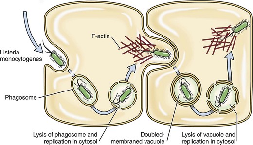
Fig. 4-29 Mechanism of infection in listeriosis.
Listeria monocytogenes propels itself via actin polymerization (Listeria actin-based motility) within a membrane pseudopod into cell membranes of adjacent neural cells forming invaginations of the membrane that ultimately result in double-membrane endocytotic phagocytic vesicles.
Thrombotic Meningoencephalitis (Histophilus somni): The pathogenesis of thrombotic meningoencephalitis (TME) begins as pulmonary histophilosis (see the section on Respiratory System, Mediastinum, and Pleurae). The mechanism of injury in the nervous system is infarction secondary to occlusive ischemia by bacterial-induced arteritis (vasculitis) and subsequent thrombosis caused by acute inflammation and its mediators and degradative enzymes. Gross lesions are red hemorrhagic infarcts of varied sizes distributed at random throughout nervous tissue, especially in the cerebral cortices (see Fig. 14-89).
Cattle encounter Histophilus somni (formerly Haemophilus somnus) via inhalation of fomites or water droplets contaminated with the bacterium. It likely exists in nasal or oral biofilms as a commensal bacterium of mucosae. Environmental stressors, such as overcrowding, combined with other factors, such as poor ventilation and humidity or abrupt changes in ambient air temperature, could alter the commensal relationship, allowing the bacteria to replicate in sufficient numbers to colonize mucosae and spread the bacterium to other animals. After colonization of the respiratory mucosae, the bacterium spreads to the lung (see the section on Pulmonary Histophilosis, Respiratory System, Mediastinum, and Pleurae) and then gains access to the vascular system in areas of inflammation, embolizes to the CNS (septicemia), and colonizes and infects small arterioles likely via ligand-receptor interactions. The first encounter with endothelial cells of arterioles occurs at anatomic sites in the brain in which there are abrupt changes in the laminar flow of blood resulting in turbulence, such as occurs at the interface between gray and white matter in the cerebral cortex. Turbulence favors perturbation of the endothelium and contact of the bacterium with platelets and the endothelium. Experimentally, Histophilus somni and its membrane LOS (a truncated form of LPS) have been shown to activate bovine platelets and increase the expression of adhesion molecules, such as ICAM-1 and E-selectin and tissue factor (factor III) on endothelial cells. Tissue factor is a protein necessary for activation of blood coagulation cascades. Thus, because strains of Histophilus somni have virulence determinates that enhance the adherence of the bacterium to endothelial cells, such areas are prone to endothelial injury, exposure of collagen, platelet aggregation and activation, activation of clotting cascades, arterial thrombosis and obstruction, and infarction (the lesion of TME).
Meningitis (Escherichia coli and Other Bacterial Species): The pathogenesis of meningitis shares many of the mechanisms discussed for porcine polyserositis (see the section on the Respiratory System, Mediastinum, and Pleurae) and embolic vasculopathy/vasculitis (see the section on the Cardiovascular System and Lymphatic Vessels).
Blackleg (Clostridium chauvoei): The mechanism of injury in blackleg is necrosis (acute gangrenous myositis) of muscle, connective, and nervous tissues caused by α- and β-toxins released from vegetative forms of Clostridium chauvoei. Gross lesions occur in large striated muscle groups and include dark red to black muscle that appears dry and contains gas bubbles (see Fig. 15-38). Affected muscle may have a rancid (spoiled butter) smell. Cattle, sheep, and goats encounter spores through ingestion of plant matter and topsoil contaminated with spores often after disturbances (excavations) of the soil and pasture bed. Spores are carried by swallowing and peristalsis through the oral pharynx, esophagus, abomasum, and rumen to their final destination, the small intestine. It appears that spores can remain dormant in the small intestine or germinate into vegetative forms and become normal inhabitants of the alimentary system. How spores interact with mucosae and gain access to epithelial cells and mucosal macrophages is unknown. Other similar pathogens use M cells to enter Peyer’s patches, so it is plausible that spores may gain access to and infect macrophages in Peyer’s patches via this mechanism. Although suggested but unproved, spores are likely the form of the bacterium that spreads systemically to muscle. However, it is possible that vegetative forms of the bacterium spread to muscle and produce spores in dendritic cells and macrophages of muscle after their phagocytosis.
Spread to muscle could also occur via leukocyte trafficking followed by interaction with and infection of endothelial cells and then dendritic cells and macrophages of muscle. If it occurs, tropism for dendritic cells and macrophages of muscle is probably mediated by ligand-receptor interactions. Blackleg often follows some form of traumatic injury to muscle. It is thought that injury creates a microenvironment suitable with lowering of the oxidation-reduction (redox) potential (anaerobic environment) required for germination of spores. Such a hypothesis also assumes that the injury has damaged the dendritic cells and macrophages of muscle, allowing them access to this microenvironment. Spores germinate into vegetative forms of the bacterium and produce large quantities of a variety of α- and β-toxins such as oxygen-stable hemolysin, deoxyribonuclease (DNase), hyaluronidase, oxygen-labile hemolysin, and neuraminidase. These toxins diffuse out from the site of bacterial replication and coagulate muscle tissue and its vascular supply, resulting in acute gangrenous myositis.
Malignant Edema (Clostridium septicum): The pathogenesis of malignant edema is similar to that of blackleg (see previous section) regarding bacterial replication, the production of toxins, tissue injury, and gross lesions affecting striated muscle and blood vessels. However, the mechanism of spread to muscle is different. Cattle, sheep, and goats encounter spores through wounds caused by penetrating objects, such as a wire, which carries spores into the wound. Wounds caused by castration, tail docking, unsanitary vaccination, and other management practices can also be infected with spores. The injury must be sufficient to create an anaerobic microenvironment in the wound with lowering of the oxidation-reduction (redox) potential suitable for germination of spores. Once spores germinate, vegetative forms release toxins that injure and coagulate muscles and vascular tissues resulting in necrosis and edema much like in blackleg.
Big Head and Black Disease (Clostridium novyi): The pathogeneses of big head and black disease are very similar to malignant edema and blackleg, respectively. In big head of sheep, penetrating wounds of the skin of the head caused by horns during fighting establish the initial anaerobic microenvironment for spores to germinate. The resulting pathogenesis is similar to that which occurs in malignant edema. Black disease of cattle and sheep occurs because of fluke migration (Fasciola hepatica) through the liver (see Figs. 8-55 and 8-56) that causes hepatocellular necrosis and establishes an anaerobic microenvironment suitable for germination of spores. Kupffer cells likely contain dormant spores. As in blackleg, spores are likely ingested, enter through the mucosae of the small intestine, become phagocytosed by cells of the monocyte-macrophage system, and spread via leukocyte trafficking to Kupffer cells of the liver.
Lumpy Jaw (Actinomyces bovis): The mechanism of injury in lumpy jaw is cell death attributable to pyogranulomatous inflammation and its mediators. Gross lesions include enlarged and misshapen bones of the mandible and/or maxilla attributable to abscesses, fibrosis, and fistulous tracts in the bone with secondary remodeling of the bone (i.e., pyogranulomatous alveolar osteomyelitis). The cut surface has numerous randomly distributed discrete and coalescing yellow-white granulomas surrounded by remodeled bone intermixed with bands of fibrous connective tissue (see Fig. 16-58). Actinomyces bovis is a commensal bacterium of mucosae of the oral cavity of cattle and sheep, likely existing in a biofilm. The bacterium can infect bone by several routes: (1) genetic or developmental defects of the tooth root and/or socket that provide access to bone, (2) injury to a tooth and its socket opening a pathway into the bone, and (3) penetrating wounds that give the bacterium access to the bone and its periosteum. During chewing, the bacterium is carried by direct extension through the mucosae into submucosal connective tissues via penetrating wounds such as those caused by sharp foreign bodies like sticks or wires. The object may penetrate the periosteum and the bone, giving the bacterium direct access to these tissues. The bacterium colonizes submucosal connective tissue, and LPS of the cell wall, in part, likely plays a role in the type of inflammatory response that occurs. Little is known about virulence determinates, ligand-receptor interactions, target cells, toxins, capsule antiphagocytic molecules, or other factors that may contribute to the pathogenicity of this bacterium. Actinomyces bovis can spread via lymphatic vessels to regional lymph nodes and cause a similar inflammatory response in these tissues.
Greasy Pig Disease (Staphylococcus hyicus): The mechanism of injury in greasy pig disease is cell death and exfoliation of cells of the skin secondary to inflammation and its mediators and degradative enzymes. Gross lesions include areas of patchy red skin (active hyperemia of acute inflammation) followed by thickening of the reddened skin and the formation of reddish brown macules, vesicles, and pustules first around the eyes, nose, lips, and ears and then the flanks and abdomen (see Fig. 17-47). Affected skin, mostly through inflammation, exudes large quantities of a greasy exudate consisting of serum and sebum mixed with inflammatory cells, degradative enzymes, and cell debris. This exudate is the basis for naming the disease.
Pigs encounter Staphylococcus hyicus through fomites and body fluids contaminated with the bacterium. This bacterium is likely a commensal organism that resides in the skin and hair follicles of healthy pigs. Environmental stressors, such as skin trauma caused by overcrowding combined with other factors such as poor ventilation and humidity or abrupt changes in ambient air temperature, could alter the commensal relationship, allowing bacteria to replicate in sufficient numbers to colonize the skin, spread the bacterium to other animals, and cause disease. Infected droplets are deposited on the surface of the skin, but the bacterium under most conditions is not able to infect and colonize intact skin. It appears that skin trauma is usually a prerequisite for colonization because abrasions on the feet and legs or lacerations on the body precede the onset of the disease.
The role of virulence determinates, ligand-receptor interactions, and cells of the monocyte-macrophage system, such as Langerhans’ cells, are poorly understood in the pathogenesis of the disease. An exfoliative toxin that induces separation of epithelial cells of the stratum corneum and spinosum aids in the invasion of the bacterium into the skin. It serves to expose vascularized ECM tissues in traumatized skin. Fibronectin-binding proteins expressed on the surface of the bacteria appear to act as adhesins, allowing the bacteria to bind to the fibronectin present in collagen, fibrin, and heparin sulfate proteoglycans of traumatized skin. Fibronectin is a glycoprotein of vascularized ECM tissues and is produced by cells such as fibroblasts. Once the skin is colonized, the infection appears to spread to the hair follicles leading to suppurative inflammation and sebaceous gland hyperplasia and hypersecretion (i.e., greasy pig). It also appears that acute inflammation and its effector cells, such as neutrophils, play a central role in the onset and progression of the skin lesions. Capsule polysaccharides and protein A in the bacterial wall appear to block phagocytosis of the bacterium by neutrophils and increase the ability of bacteria to survive and replicate in vascularized ECM tissues of the skin.
Canine Pyoderma (Staphylococcus intermedius): The pathogenesis of canine pyoderma appears to be similar to that of greasy pig disease (see previous section). Skin trauma arising from pruritus and scratching or from existing skin disease leads to the exposure of vascularized ECM tissues and its colonization by bacteria. Although incompletely characterized, it is likely that a variety of virulence determinates are involved in canine pyoderma, including surface proteins (colonization of host tissues); invasins such as leukocidin, kinases, hyaluronidase (promote bacterial spread in tissues); surface factors such as capsule polysaccharides and protein A (inhibit phagocytosis); and exotoxins and exfoliative toxins such as hemolysins, leukotoxin, and leukocidin (cause cell death).
Diamond Skin Disease (Erysipelothrix rhusiopathiae): The mechanism of injury in diamond skin disease is cell death and infarction of skin secondary to cutaneous vasculitis. Gross lesions include active hyperemia and red-purple skin affecting the ears, ventral abdomen, and legs followed by thrombosis, ischemia, and infarction resulting in rhomboidal (diamond) red-purple areas of skin (cutaneous infarcts) (see Figs. 17-51 and 10-81).
Pigs encounter Erysipelothrix rhusiopathiae through ingestion of fomites and body fluids contaminated with the bacterium. This bacterium is likely a commensal organism that resides in a biofilm of the mucosae of the pharynx and tonsillar epithelia of healthy pigs. Environmental stressors, such as overcrowding combined with other factors such as poor ventilation and humidity or abrupt changes in ambient air temperature, can alter the commensal relationship, allowing the bacteria to replicate in sufficient numbers to colonize mucosae and spread the bacterium to other animals. Infected droplets are deposited on pharyngeal mucosae, where bacteria encounter the mucus layer and mucosal epithelial cells. It is unclear how this nonmotile bacterium is able to penetrate the mucus layer and gain direct access to the luminal membrane of epithelial cells. Additionally, it is unclear if and how the bacterium colonizes the mucus layer and mucosae. It appears that neuraminidase may be a virulence determinate for Erysipelothrix rhusiopathiae potentially involved in initial interactions with and invasion of the mucus layer of pharyngeal mucosae. It acts to remove sialic acid from glycoproteins, glycolipids, and oligosaccharides expressed on host cells, potentially exposing new receptors for the bacterium. It is also probably important later in the disease process in the attachment, colonization, and invasion of endothelial cells by the bacterium leading to vasculitis, thrombosis, infarction, and DIC.
Other virulence determinates involved in mucosal colonization and systemic spread of the bacterium include capsular polysaccharides (antiphagocytic properties), surface proteins (adhesins, antiphagocytic properties, biofilm formation), invasins such as hyaluronidase (invade ECM tissues), and enzymes such as superoxide dismutase and catalase (block the effects of the respiratory burst of phagocytosis and oxygen free radicals). Transcytosis could move bacteria through mucosal cells to the basal surface of mucosal epithelial cells to encounter local macrophages and lymphoid cells in the tonsils. Alternatively, mucosal macrophages could phagocytose bacteria in the mucus layer, migrate through the mucosal barrier, and spread them via leukocyte trafficking to the same cells.
Macrophages within the tonsil are likely used by the bacterium for replication and growth and then to spread them via leukocyte trafficking in lymphatic vessels to regional lymph nodes to infect additional macrophages. Ligand-receptor interactions are likely involved and bacterial surface proteins appear to serve as adhesins to macrophage and endothelial cell membrane receptors. Specific bacterial adhesins and host cell receptors for them on macrophages and endothelial cells have not been identified. Once bound to cell membrane, the bacterium is phagocytosed and retained in a phagosome within the cell cytoplasm. Erysipelothrix rhusiopathiae grows and replicates intracellularly in phagosomes and phagolysosomes. Capsular polysaccharides are able to inhibit phagocytosis of the bacterium by neutrophils and to a limited extent by macrophages. However, macrophages are used by the bacterium to isolate it from host innate and adaptive immune responses. Although it appears that phagosome-lysosome fusion occurs, capsular polysaccharides appear to block the oxidative burst and prevent killing of the bacterium by molecules present in the lysosome. Although undetermined, bacteria-infected macrophages in regional lymph nodes could spread the bacterium systemically via leukocyte trafficking using lymphatic vessels and the thoracic duct or postcapillary venules and the venous system to the systemic circulatory system and then to capillary beds within the skin. Cutaneous infarcts (also vascular-related lesions in other organs such as the kidney) suggest these bacteria may have tropisms for vascular endothelial cells. It is unclear why this occurs but is likely linked to the expression of bacterial virulence determinates and ligand-receptor interactions with host endothelial cells. In addition to cell death attributable to direct infection of endothelial cells by the bacterium, bacterial neuraminidase may also activate the alternative complement pathway and induce thrombocytopenia, producing complement-derived chemotactic factors that could contribute to injury of capillary beds in local vascularized connective tissues. These mechanisms may contribute, in part, to the development of vegetative valvular endocarditis and arthritis that occurs in the chronic septicemic form of this disease.
Brucellosis (Brucella spp.): The pathogenesis of Brucellosis begins as an infection of the regional and systemic lymph nodes (see the section on Bone Marrow, Blood Cells, and Lymphatic System) facilitated by entry through mucosae of the respiratory and alimentary systems. The mechanism of injury is cell death caused by pyogranulomatous inflammation and its mediators and degradative enzymes. Gross lesions include aborted fetuses; necrosis, inflammation, and fibrinoid exudation of uterine caruncles and fetal cotyledons (see Fig. 4-26); and a yellow-white uterine exudate. Bacteria spread in macrophages via leukocyte trafficking from regional lymph nodes to the caruncular side of placentomas when they likely leave the vascular system to migrate into and through these tissues. Although it is unclear what additional cells in the placentoma are infected during transplacental spread to the fetus, trophoblasts are infected with bacteria. Other cell types may be involved. Bacteria then could spread in fetal macrophage-like cells within the fetal circulatory system to the fetus via the umbilical cord or within the allantoic and amniotic membranes and infect the fetus via contact with fetal mucosae of the respiratory or alimentary systems, but if this occurs or what cells facilitate this spread are unclear.
Bovine Mastitis (Staphylococcus aureus, Streptococcus agalactiae, Streptococcus dysgalactiae, and Escherichia coli): The mechanism of injury in bovine mastitis is death of all cell populations in the mammary gland from (1) bacterial toxins, (2) inflammation and its mediators and degradative enzymes, and (3) induced reparative responses such as fibrosis. Gross lesions in acute mastitis include firm, swollen, edematous, and occasionally hemorrhagic glands and ectatic ducts and sinuses containing yellow-white exudate (see Figs. 18-53 to 18-55, 18-57, and 18-58). In chronic mastitis, tissues are firm and consist of large zones of fibrous connective tissue that have replaced and displaced remaining normal glands (see Fig. 18-59). Inflammatory exudate is difficult to observe unless abscesses have formed. Ducts and sinuses may be ectatic.
Animals encounter these bacteria through physical contact in fomites or fluid droplets from mammary gland, uterine, or fecal origin on milking equipment and human hands. They commonly become commensal organisms that reside in biofilms of the mucous membranes of the teat canal and mammary duct and sinuses. Trauma to mucosae in the gland induced by pressure changes acting on the duct system caused by milking likely makes the mucosae more suitable for colonization and alters the commensal relationship, allowing the bacteria to replicate in sufficient numbers to spread the bacterium within the gland and to other animals mechanically during milking. Mastitis is an ascending infection, and milk in canals and sinuses is a suitable culture media for initial growth of bacteria. This environment is not suitable in the long-term for survival of the bacteria, thus they attempt to colonize mucosae to sustain the infection. Ligand-receptor interactions are likely involved in the adherence of these bacteria to receptors on mucosal epithelial cells; however, specific bacterial adhesins and host cell receptors have not been clearly identified. Once mucosae are colonized, bacteria employ mechanisms to sustain the infection. For example, Staphylococcus aureus produces toxins, such as superantigens, leukocidins, hemolysins, coagulase, and likely α-, β-, and δ-toxins (virulence determinates) that result in cell membrane injury and cell death and the activation of mucosal macrophages. The severity of this lesion and its progression to gangrenous mastitis in the peracute and acute forms are dependent on the type and quantity of toxins secreted by the bacterium as determined by its virulence determinates. Additionally, activated mucosal macrophages secrete proinflammatory cytokines resulting in the recruitment of neutrophils from the systemic circulation, through the mucosae, and ultimately into the milk, thus increasing the somatic cell count. With the focus of inflammation on the mucosae, epithelial cells and subjacent basement membrane are injured, killed, and sloughed, providing bacteria with access to vascularized ECM tissues of the gland. Using bacterial surface proteins, they are able to adhere to and colonize ECM tissues likely using receptors expressed on molecules such as fibronectin, vitronectin, laminin, and collagen in the matrix. This process allows bacteria to evade many of the harmful actions of the innate and adaptive immune responses. Additionally, capsular polysaccharides block phagocytosis by neutrophils and macrophages. As a result, acute inflammation progresses with time to chronic inflammation with fibrosis (see Chapter 3), which is a common manifestation of mastitis caused by Staphylococcus aureus.
Chronic mastitis is often linked with the formation of mucosal biofilms. In mastitis caused by Streptococcus agalactiae and Streptococcus dysgalactiae, the bacteria use most of the mechanisms employed by Staphylococcus aureus with one important exception. They lack virulence determinates that injure the mucosae and allow the bacterium to invade vascularized ECM tissues and colonize this area. Thus the bacterium is limited to colonizing the mucosae and causing inflammation at the mucosal barrier. The outcome of this process is loss of mucosal epithelial cells lining glands, collapse of the glands, and replacement of the glands with fibrous connective tissue. In mastitis caused by E. coli and other coliforms, the bacterium uses most of the mechanisms as discussed previously. However, in the initial phases of colonizing the mucosae, endotoxins (LPS) and other toxic molecules released from Gram-negative bacteria cause tissue injury and cell death, affecting the mucosae, submucosae, and capillary beds. The concurrent acute inflammatory response with neutrophils and their degradative enzymes exacerbate the severity of the injury. This outcome leads to tissue necrosis, edema, and hemorrhage. Endotoxin is also absorbed by the capillaries and can cause endotoxic shock of the circulatory system and death of affected animals (see Chapters 2 and 3).
Brucellosis (Brucella spp.): The pathogenesis of Brucellosis begins as an infection of regional and systemic lymph nodes (see the sections on Bone Marrow, Blood Cells, and Lymphatic Systems and Female Reproductive System) facilitated by entry through the mucosae of the respiratory and alimentary systems. The mechanism of injury is cell death caused by pyogranulomatous inflammation and its mediators and degradative enzymes. Brucella spp. spread in macrophages via leukocyte trafficking from regional lymph nodes to the testes, epididymides, and other male reproductive tissues. Gross lesions include enlarged and deformed testes and epididymides attributable to a yellow-white pyogranulomatous exudate against the bacteria in the tissues (see Fig. 4-26).
Viruses are about a hundred times smaller than bacteria, and viruses, like bacteria, are genetically programmed to replicate endlessly, if all growth factors, metabolic needs, and microenvironments for replication are satisfactorily met. However, viruses are unable to produce energy and contain a limited number of enzymes; therefore they are completely dependent on animal cells for such resources and are obligate intracellular parasites. They have evolved to specifically utilize target cells in animals that are susceptible to and suitable for completion of their viral replication cycle. The term target cell assigns specificity to which cells are infected by viruses, and this process is based on the typical ligand (viral envelope or capsid proteins)-receptor (host cell membrane proteins) interactions common to all cells (Fig. 4-30). The phrase virus replication cycle is used here to merge under a single key concept the chronologic sequence of the events that occurs when a virus encounters and enters cells, hijacks the functions of cellular organelles and metabolic processes, produces new virus, and ultimately injures or kills cells and causes disease.
Fig. 4-30 Ligand-receptor interactions.
Ligand (viral envelope or capsid proteins)-receptor (host cell membrane proteins) interactions common to all cells are used by viruses to attach to and infect specific target cells.
Target cells that allow replication of the virus are called permissive cells, whereas those that do not are called nonpermissive cells. Generally, virus-infected permissive cells are usually killed by the virus (cell death), whereas virus-infected nonpermissive cells are not killed. For example, the pathogenesis of the lentivirus disease, maedi-visna, is determined, in part, by nonpermissive cells (immature progenitor monoblasts and promonocytes in bone marrow) and permissive cells (mature monocytes and macrophages in the blood vascular system and tissues). Infection of nonpermissive progenitor cells in bone marrow is used to provide a reservoir of immunologically protected virus-infected cells that become permissive when they mature into monocytes and macrophages in the vascular system and when they migrate into specific tissues and organs. These permissive macrophages are ultimately killed by virus replication, and the cell is lysed to release new virus from the cell.
Viral pathogenicity is a phrase used to convey the severity of disease, clinical signs, and lesions caused by viruses. It is determined, in large part, by the expression of viral genes that are used to produce structural or functional proteins needed to sustain or enhance the virus replication cycle. Similar to bacteria, these viral genes and proteins biologically behave as viral virulence determinates; however, the occurrence of viral virulence determinates is nowhere near the complexity and diversity expressed by bacteria. As would be anticipated, viral virulence determinates focus on attachment, replication, and shedding of viruses and on the processes of modulating and/or evading host defense mechanisms. Thus the type, quantity, and arrangement of nucleic acid in viruses provide the basis for genomic diversity and the transfer of virulence determinates among viruses. The encounter between viruses with virulence determinates and host cells with innate and adaptive defense mechanisms determines the occurrence and severity of disease.
The severity of disease, and in large part the life or death of the host cell, is determined by how a virus (1) utilizes and truncates the processes of host cell organelles and transcriptional and translational processes in virus replication and (2) escapes from virus-infected host cells. The organ system specific clinical symptomatology of diseases caused by viruses is mechanistically linked to the following three key stages in the replication cycle of a virus (Fig. 4-31):
Fig. 4-31 Virus replication cycle.
Stages in the infection, replication, and egress of a virus in a target cell. Several thousand virus particles may be formed within each infected cell. (From Rosenthal KS, Tan JS: Rapid review microbiology and immunology, ed 2, St. Louis, 2007, Mosby.)
• Determines what organs are infected, thus determining the clinical signs of disease.
• Viruses bind to receptors normally expressed on host cell membranes and these receptors are used to infect target cells.
Once viruses encounter host cells, they attach to them via cell membrane receptors. Attachment is the process through which binding occurs between ligands (viral attachment proteins) on the surface of the virus and receptors on the host cell membrane. Receptors are often expressed in unique patterns on target cells, and these patterns appear to frequently determine the route used by viruses to infect target cells. For example, parvoviruses and herpesviruses use specific receptors with specific distribution patterns to attach to and enter target cells. Parvovirus (canine parvovirus enteritis) infects intestinal crypt epithelial cells through receptors expressed on the basolateral surface of the cells, thus using a circuitous route via leukocyte trafficking to Peyer’s patches and M cells to gain access to this surface. Although this route is not likely the most direct route to intestinal epithelial cells, it may be advantageous for survival of the virus to avoid contact with gastric acids, bile, and other potentially toxic molecules in the alimentary lumen. Additionally, junctional complexes of intestinal epithelial cells would probably prevent the virus from reaching, and thus encountering, basolateral cell surfaces. Bovine herpesvirus 1 (infectious bovine rhinotracheitis) infects epithelial cells of the respiratory system through receptors expressed on the apical and lateral surface of the cells. These receptors are distributed above junctional complexes formed with adjacent epithelial cells; therefore virus in the lumen of the respiratory tract can encounter appropriate receptors on mucosae. Receptors on host target cells used by viruses include those for complement, growth factors, neurotransmitters, integrins, adhesion molecules, complement regulatory proteins, phospholipids, and carbohydrates. In general, specific viruses use one of these receptor types to infect a specific type of cell; however, some viruses use several receptors (coreceptors), which allows for invasion of a variety of cell types (i.e., pantropic viruses such as canine distemper virus). Experimental studies suggest that there are approximately 104 to 106 receptors for a virus expressed on a single target cell. Attachment is accomplished through the interaction of the host cell membrane receptors with the outermost layer of the virus.
Specific groups of viruses (nonenveloped viruses) attach to host cells using a protein coat (viral coat, capsid, or capsomeres) (Fig. 4-32, A); others attach via a viral envelope (enveloped viruses) (Fig. 4-32, B). Protein molecules, derived from viral genes and expressed in the protein capsid or envelope, are called attachment proteins. Attachment proteins are viral virulence determinates that provided a basis for genomic diversity among viruses. Once attachment occurs, viruses can enter target cells by one of two main mechanisms: receptor-mediated endocytosis or fusion. Once inside target cells, viruses initiate a variety of virus-specified processes to complete their replication cycles, such as replication of its genome, core proteins, and capsid and envelope proteins; assembly of new viruses; and release of new viruses from the cell (Fig. 4-33). When a DNA virus enters a cell and its components are released into the cytoplasm, the DNA genome is transferred into the nucleus where it utilizes host cell nuclear organelles to transcribe viral messenger RNA (mRNA) and later replicate new viral DNA. Viral mRNA leaves the nucleus and in the cytoplasm is translated into structural and nonstructural proteins of the virus by host cell organelles. After all viral proteins are translated in the host cell cytoplasm, new viral DNA is replicated (transcribed) and transferred into the cytoplasm where it is assembled with structural and nonstructural proteins to form new virus.
Fig. 4-32 Morphology of viruses.
A, Nonenveloped viruses. They attach to host cells using a protein coat (viral coat, capsid, capsomeres) and usually kill infected cells to release newly formed virus. B, Enveloped viruses. They attach to host cells using a viral envelope and usually do not kill infected cells to release newly formed virus. (From Goering R, Dockrell H, Roitt I, et al: Mims’ medical microbiology, ed 4, St. Louis, 2008, Mosby.)
Fig. 4-33 Replication of DNA and RNA viruses. (From Goering R, Dockrell H, Roitt I, et al: Mims’ medical microbiology, ed 4, St. Louis, 2008, Mosby.)
When a RNA virus enters a cell and its components are released into the cytoplasm, the RNA genome, depending on the virus, can (1) replicate new viral RNA from the cytoplasmic viral RNA via its own viral RNA-dependent RNA polymerase or (2) make viral DNA from viral RNA via RNA-dependent DNA polymerase (viral reverse transcriptase) and then use host cell nuclear and cytoplasmic organelles to transcribe and translate new proteins and viral RNA. Thus the genome of RNA viruses must express genes that code for enzymes such as RNA-dependent RNA polymerase and RNA-dependent DNA polymerase. Detailed coverage of these processes is outside of the scope of this chapter and can be reviewed in virology textbooks; however, these replicative processes often lead to injury and cell death. Generally, nonenveloped viruses (viral protein coats or capsids) are released from host target cells only when the cell death occurs.
Enveloped viruses (viral envelope glycoproteins) must acquire an envelope by budding through cellular membranes such as the plasma membrane, membranes of the Golgi complex or rough endoplasmic reticulum, or nuclear membrane. Viral envelope glycoproteins localize to zones in the membrane where the virus buds from the membrane. Most viruses that bud from the cell membrane do not cause cell lysis except for those that bud from the Golgi complex or rough endoplasmic reticulum (flavivirus, coronavirus, Arterivirus, and bunyavirus) or the nuclear membrane (herpesvirus).
Virus capsid proteins and envelope glycoproteins are used immunologically as a means to clinically prevent (e.g., vaccination) or control (e.g., pharmaceutical products) diseases caused by viruses by developing strategies to block one of more of the steps in the viral attachment or replication cycle. Antibiotics have no effect on viruses; however, fortunately, viral infections (viral antigens) usually activate host innate and adaptive defense mechanisms and cause an immune response (cell-mediated), which can completely eliminate a virus or prevent an infection by a virus (vaccination). However, these defensive responses can also injure and kill host cells leading to disease. The list of structural and biochemical effects that viruses have on the host cell is extensive. These effects are often called cytopathic effects, and as a general rule, many viral infections result in death of the host cell. Depending on the virus and its replication cycle, injury to and death of the host cell can occur at any point during the attachment, fusion, penetration, synthesis, assembly, or release phases. Generally, viruses cause injury and death most commonly by two mechanisms: (1) as a result of taking over cell transcriptional and translational processes and (2) when they exit from infected cells. Additionally, causes of cell death include alterations in cell membrane function, including ion transport and secondary messenger systems; alterations in metabolic processes, including activation cascades leading to altered cellular activities; alterations of host cell antigenic or immune properties, shape, and growth characteristics; inhibition of the synthesis of host cell macromolecules, including DNA, RNA, and protein; and direct (protein messenger molecules) and indirect (inflammatory mediators) activation of cell lysis and apoptosis cascades.
Virulence determinates also have been identified for viruses. The purpose of these determinates is to improve a virus’s ability to complete its replication cycle in the host cell, thus spreading and propagating the virus to naïve animals. Virulence determinates control the processes involved in (1) replication, including attachment to, replication in, and release of virus from host cells and (2) escaping, modulating, or suppressing the host innate and adaptive immune responses. For example, feline immunodeficiency virus hides within the immune system and replicates and spreads within macrophages and T lymphocytes. Other viruses have evolved mechanisms to evade cytotoxic T lymphocyte and natural killer (NK) cell killing of virus-infected cells, disrupt complement activation, synthesize cytokine homologs that interfere with normal immunologic functions, and synthesize molecules that inhibit interferon responses or block the induction of apoptosis in virus-infected cells. Other viral virulence determinates include viral proteins, as well as by-products of virus replication, such as caspases and caspase-like molecules, that accumulate in the cell and have toxin-like activities on host cells (Fig. 4-34). As an example of a viral toxin, rotavirus-infected enterocytes secrete a viral-directed toxin called NSP4 into the intestinal lumen. Adjacent enterocytes not infected with virus absorb this toxin and it acts on a cytoplasmic messenger system to cause a secretory diarrhea. This diarrhea occurs before there is death of virus-infected enterocytes.
Fig. 4-34 Actions of viral proteins.
Proteins synthesized by viruses can affect normal cell function through mechanisms illustrated here. (From Kumar V, Abbas A, Fausto N, et al: Robbins & Cotran pathologic basis of disease, ed 8, Philadelphia, 2009, Saunders.)
The number of virulence determinates for viruses when compared to bacteria is extremely small and is directly related to the number of genes in the respective microbes. The number of genes in viruses range between 101 to 102, whereas the range in bacteria is 103 to 104 genes. Correspondingly, the number of virulence determinates is low in viruses and much higher in bacteria. The introduction of a new viral virulence determinate to a viral family results from genomic variation through genetic drift, reassortment, recombination, or defective interfering viruses. Breaks in protection normally provided by commercial vaccines or the reemergence of a vaccinated/protected disease in certain regions of the country are often the result of genomic variation in the street virus and the introduction of a new viral strain such as has occurred with canine distemper and parvovirus infections.
Viruses are often classified as DNA or RNA viruses based on the nucleic acid used to form their genes. In general, the competitive advantages for infecting host cells favor RNA viruses because they have an extremely high mutation rate, which increases their chances of expressing virulence determinates that improve their ability to complete their replication cycle. However, it is likely that this advantage is counterbalanced by their slower replication speed allowing host defense mechanisms to intervene in the replication process and kill the virus or virus-infected cell. Genomic variation is a broad term used to categorize a group of biologic processes that allow viruses to acquire new virulence determinates (genetic diversity) that favor their survival through infective and replicative mechanisms in host cells. The most common form of genomic variation, called genetic drift, is caused by a spontaneous point mutation of individual nucleic acid bases in viral DNA or RNA. These point mutations are usually silent and do not change the protein encoded by the affected gene; however, some mutations can result in a new protein (e.g., capsid or envelop proteins), thus providing an opportunity for the virus to improve its chances of infectivity, replication, and spread during its replication cycle. During virus replication, mutation of RNA viruses occurs at a much higher frequency than in DNA viruses and thus RNA viruses have the ability to more rapidly adapt to new situations and host environments via point mutations when compared to DNA viruses. Antigenic shift also leads to genomic variation and results in major changes in the genome of a virus. It occurs because of reassortment or recombination of viral genes. Reassortment occurs only in RNA viruses because they have discrete genomic segments, much like chromosomes, that behave independently of one another. These genomic segments can undergo reassortment during virus replication, resulting in new viruses with genomes different from the original infecting virus. Segmented genomes confer evolutionary advantages to RNA viruses. Reassortment is a powerful initiator of divergence, especially among viruses that can exchange genetic material between animals and humans like influenza viruses (Fig. 4-35). Recombination occurs in DNA viruses and results in rearrangements within the viral genome and deletion or duplication of viral genes, as well as the acquisition of unrelated genetic material. Genetic recombination comes about when a strand of DNA is broken and then rejoined to the end of a different DNA molecule. A final mechanism for genomic change occurs in RNA and DNA viruses and involves defective interfering viruses that cannot replicate by themselves and therefore compete with nondefective viral genomes for a limited supply of replication enzymes. They can interfere with the replication of complete viruses in host cells and significantly decrease the numbers of newly replicated virus, thus favoring success of new mutants that may arise in the virus replication process.
Fig. 4-35 Antigenic shifts in influenza virus.
One theory proposes that antigenic shifts occur when a human influenza virus (blue) and an avian influenza virus (red) coinfect a species that is permissive for both. The 8 ssRNA strands are co-expressed in the same infected cell, resulting in mixing of the strands so that a hybrid virus can be produced. The hybrid virus indicated here contains all the genetic information of the original virus that infected humans, but contains a new hemagglutinin (HA)-containing strand from the avian virus. This virus expresses a new HA antigen and will be less susceptible to residual immunity that normally provides partial protection against yearly influenza infections. (From McCance KL: Pathophysiology: The biologic basis for disease in adults and children, ed 6, St. Louis, 2010, Mosby.)
Defense mechanisms include many of the systems, biologic processes, and molecules described in Chapters 3 and 5. Host genes probably determine susceptibility to some viral infections via expression of or lack of expression of viral membrane receptors or through effects on the immune system. Stress (overcrowding), nutritional status, and environmental factors, such as temperature, humidity, and ventilation, also affect the susceptibility of animals to viral infections. Innate and adaptive mechanisms are actively involved in protection against viruses. However, it is important to remember that actions of the innate and adaptive immune systems, especially of T lymphocytes, against viral infections have both beneficial and harmful outcomes (Fig. 4-36). Beneficial outcomes include the return to normal structure and function of infected host cells and tissues and a host animal that is free of and fully protected (vaccinated) against the virus. Harmful outcomes include the failure to return to normal structure and function of infected host cells because the cells and tissues, their stem cells, supporting stroma, basement membrane, and vascularized ECM tissues have been degraded by enzymes from neutrophils of acute inflammation or macrophages of chronic inflammation and replaced by fibrous connective tissue. The innate immune system and TLRs, in response to viral antigens, induce inflammatory responses, cause secretion of cytokines and interferon, and activate the adaptive immune system. Cell-mediated immunity is the most important adaptive defense mechanism against viral infections. The monocyte-macrophage system, through phagocytosis, is active in containing the spread of viruses, whereas phagocytosis by neutrophils does not play an important role. Antibody deficiencies usually do not affect the outcome of viral infections, whereas antibodies are important in preventing reinfection (autoimmunization or vaccination). Although viruses are obligate intracellular parasites that have evolved sophisticated mechanisms to hijack host cell transcriptional and translational processes, this approach to replication results in alteration of host cell membranes that are recognized as foreign by lymphocytes of the adaptive immune system.
Fig. 4-36 Possible roles for T lymphocytes in immunity to intracellular microorganisms.
A, The T lymphocyte activates intracellular killing mechanisms by secretion of cytokines such as IFNγ, e.g., in a macrophage. B, The T lymphocyte directly kills cell and parasite. C, The T lymphocyte destroys vital tissue in the process of killing the parasite. D, By lysing cells the T lymphocyte allows still-living parasites to disseminate. E, Parasites released in this way may be phagocytosed by a more effective host cell. (From Goering R, Dockrell H, Roitt I, et al: Mims’ medical microbiology, ed 4, St. Louis, 2008, Mosby.)
Virus replication and spread are abruptly stopped when virus-infected host cells are killed by cytotoxic NK cells and cytotoxic T lymphocytes. Interferons, a group of molecules that act on virus-infected cells to inhibit virus replication, function by inducing the synthesis of host cell proteins that inhibit translational activities of the virus (Fig. 4-37). The synthesis of interferon is induced by virus infection of host cells and by the action of proinflammatory molecules on host cells. Viral infection of host cells can also activate complement cascades independent of an antibody response. Complement components also act as opsonins (e.g., phagocytosis of viruses) and can cause death of viruses or virus-infected cells. Many of the viruses discussed in this chapter are able to infect cells of the lymphoid and monocyte-macrophage systems and dendritic cells. Under normal conditions, such cells are migratory immunosurveillance cells, behaving as sentinel cells for the adaptive immune system and monitoring for the presence of foreign antigens expressed by microbes or microbe-infected cells throughout the body. As part of their normal immunosurveillance functions, these cells migrate via lymphatic and blood vascular systems throughout all tissues and organs of the body, including the brain. It is through these normal migratory pathways that viruses within these infected cells are able to spread to other tissues and organs. This process is termed cell-associated viremia or leukocyte trafficking. Viruses can also spread to other cells as a cell-free viremia in the blood vascular or lymphatic systems.
Although viral diseases often affect several different organ systems, diseases in this section are placed into a specific organ system based on which organ system demonstrates the primary gross lesion (or lesions) that is most commonly used to initially recognize and identify the viral disease. The heading for each viral disease includes information on whether the virus is enveloped or nonenveloped (type of injury) and the type of nucleic acid (virulence determinates, genomic diversity) it contains. This information is useful in understanding the mechanisms of injury in specific viral diseases. Viral diseases are identified by a primary mechanism of injury in Table 4-4.
Parvovirus Enteritis (Parvovirus, Nonenveloped DNA Virus): Parvovirus enteritis is a general name used to group two closely related strains of parvovirus that cause canine parvovirus enteritis and feline panleukopenia (feline parvovirus enteritis). The mechanism of injury is death of crypt epithelial cells and lymphocytes, including those in the bone marrow. Specificity for these mitotically active cells occurs because parvoviruses require a host cell–derived duplex transcription template, which is only available when cells divide during the S-phase of the cell cycle. Parvoviruses are unable to turn on DNA synthesis in host cells, so they must wait for host cells to enter the S-phase of the cell cycle before infecting these cells. Gross lesions include segmental areas of the mucosae that are rough and granular (enterocyte necrosis, villus atrophy) with areas of hemorrhage, acute inflammation, and fibrin exudation (see Fig. 7-160).
Dogs and cats encounter parvoviruses in fomites from body fluids contaminated with virus-infected fecal matter through direct contact with virus-infected animals. The virus is inhaled or ingested; deposited on mucosae of the oral, nasal, and pharyngeal cavities; and trapped in the mucus layer. It has not been determined if and how virus penetrates the mucus layer to gain access to mucosal epithelial cells, mucosal macrophages, lymphocytes, and/or dendritic cells. Virus probably infects macrophages or dendritic cells migrating in the mucus layer and on the surface of mucosae. Virus replicates in these cells and is then spread via leukocyte trafficking to the lamina propria of the tonsils. Here, additional macrophages and lymphocytes are infected and spread the virus via leukocyte trafficking in lymphatic and blood vascular systems to regional lymph nodes and systemically to the spleen, thymus, lymph nodes, bone marrow, and mucosa-associated lymphoid nodules such as Peyer’s patches of the small intestine. Virus may also be spread as a cell-free viremia in lymph via lymphatic vessels to regional lymph nodes.
In these diseases, the majority of virus-infected intestinal epithelial cells are found in crypts neighboring Peyer’s patches in the small intestine. Experimental studies demonstrate that virus arrives at Peyer’s patches before it reaches contiguous crypt enterocytes. Although not yet shown with canine or feline parvovirus, other similar viruses spread from Peyer’s patches to M cells in Peyer’s patches. Morphologically, M-cell processes extend into the mucosa and are contiguous with crypt enterocytes forming intestinal crypts. Additionally, virus entry into intestinal epithelial cells has a polarized pattern in which entry is restricted to the basolateral areas of crypt enterocytes, the areas nearest Peyer’s patches and M cells. Collectively, these findings suggest that virus initially spreads to the intestine via the blood vascular system and not in ingesta via peristalsis. It is unclear whether a virus arrives as a cell-free viremia or within cells of the monocyte-macrophage and/or lymphoid systems; however, (1) virus infects such cells in the oral, nasal, pharyngeal mucosa, and tonsil and regional lymph nodes and (2) leukocyte trafficking is commonly used by other viruses to spread virus systemically to lymphoid and other organ systems, suggesting that parvovirus is spread to the intestine via leukocyte trafficking.
Infection is initiated through capsid-mediated attachment to one or more glycosylated receptors on target cell membranes and is followed by entry via receptor-mediated endocytosis. Parvoviruses appear to use coreceptors for the attachment and entry processes. Attachment receptors may assist in aggregating virus near the cell membrane, and entry receptors may assist the virus in penetrating the cell membrane. In the dog, this process requires capsid proteins to bind to transferrin receptors, whereas in the cat, the process requires capsid proteins to bind to neuraminic acid and transferrin receptors. These receptors appear to determine which cells and which species of animal are infected by parvovirus strains. Parvoviruses are released from infected crypt enterocytes when the cell is killed after the replication cycle is completed. Because of this outcome, parvovirus enteritis causes an osmotic malabsorption-maldigestion diarrhea. Diarrhea occurs because of the failure of replacement of absorptive enterocytes covering villi that are lost through normal turnover (≈48-hour lifespan). As a result, affected villi collapse, are atrophic, and all absorptive and digestive surfaces are lost; thus dietary carbohydrates are available for fermentation by intestinal bacteria. Under normal conditions, enterocytes covering villi are replaced by dividing crypt epithelial cells that move up and cover the villus. The loss of enterocytes covering villi also functionally opens a barrier system that normally prevents endotoxins from being absorbed by capillary beds in the lamina propria of the villi. Endotoxic shock and DIC can result and kill the affected animal. Panleukopenia also occurs because of virus-induced cytolysis of rapidly dividing stem cells in the bone marrow. The effects of parvovirus on organs of the lymphatic system are discussed in the section on Bone Marrow, Blood Cells, and Lymphatic System.
Bovine Viral Diarrhea and Mucosal Disease (BVD Virus, Pestivirus, Enveloped RNA Virus): The range of diseases caused by BVD virus is diverse and complex. Some of these diseases will be discussed in this chapter and other chapters of this book. Bovine viral diarrhea and mucosal disease as discussed herein refers to the disease that affects mucosae of the alimentary system from the oral cavity to the small intestine. The mechanism of injury in bovine viral diarrhea and mucosal disease is dysfunction and death of mucosal epithelial cells of the oral cavity and esophagus (stratified squamous epithelium) and of the small intestines (enterocytes) preceded by dysfunction and death of submucosal lymphocytes in MALT, such as those in Peyer’s patches. Gross lesions include erosion, ulceration, and hemorrhage of mucosae of oral/nasal cavities and pharynx, esophagus, and small intestine (see Figs. 7-3, 7-142, 7-143, and 7-144).
The typical pathogenesis of mucosal disease involves two forms of BVD virus, a noncytopathic form and a cytopathic form, acting synergistically to cause lesions. The noncytopathic form of the virus is likely introduced into the herd in new stock, commingling of cattle, semen, or other management practices that allow contact with carrier cattle. The cytopathic form of the virus commonly arises from a noncytopathic form that exists in the herd through mutations of its viral genome or it is introduced into the herd as discussed above. Under the proper conditions as discussed below, the noncytopathic form makes cattle immunotolerant to cytopathic forms of BVD virus. Mucosal disease occurs when immunotolerant cattle are exposed to a cytopathic form of the virus (1) most likely originating through mutations on the same farm or (2) introduced into the herd by means discussed above. When cattle with a normal adaptive immune response (i.e., not immunotolerant) are exposed to the cytopathic form of the virus, they are usually able to prevent or limit the severity of mucosal disease that occurs unless the viral strain has several highly pathogenic virulence determinates.
Immunotolerant calves occur when fetuses of naïve (normal immune responses, unvaccinated, no prior exposure) pregnant cows are infected with the noncytopathic form of the virus. Infected cows are asymptomatic, but functionally serve as a means for the virus to infect the fetus and establish “persistently infected (PI)” calves (noncytopathic form of the virus) in the herd. These calves, present in small numbers, usually die before a year of age, but serve as farm reservoirs for noncytopathic virus as they constantly shed virus in body secretions (saliva, tears) and feces into the environment.
For convenience, let’s begin the mechanistic sequence of events that ultimately leads to mucosal disease with the exposure of pregnant cows to the noncytopathic form on the farm and in utero transplacental spread of this form from infected cows to their calves. Cows encounter the noncytopathic form in fomites from contaminated body fluids or wastes through direct contact with PI calves or carrier animals. The noncytopathic form is inhaled or ingested and deposited on mucosae of the oral, nasal, and pharyngeal cavities; especially favored are mucosae overlying the tonsil. It has not been determined if and how virus penetrates the mucus layer to gain access to mucosal epithelial cells or submucosal macrophages, lymphocytes, and/or dendritic cells, but this process could be facilitated via phagocytosis in the mucus layer by mucosa-associated macrophages, lymphocytes, and/or dendritic cells migrating through the mucosae. Noncytopathic virus probably infects and replicates in monocytes, macrophages, lymphocytes, and dendritic cells and is spread via leukocyte trafficking in lymphatic vessels from tonsil and submucosal lymphoid nodules to regional lymph nodes and then systemically to the caruncular side of placentomas. Trophoblasts in the placentoma can be infected by noncytopathic virus, likely allowing virus to complete a replication cycle, migrate into and through the cotyledons, and infect fetal macrophage-like cells that enter the fetal vascular system and spread to the fetus. Additionally, noncytopathic virus can infect and spread within the allantoic and amniotic membranes and then infect the fetus, but it is unclear which cells facilitate this spread.
Bovine fetuses, infected in utero, become immunotolerant (see Chapter 5) to the noncytopathic form of the virus. They also do not recognize antigens from cytopathic forms of the virus as foreign, and as a result they fail to develop an effective adaptive immune response. When exposed to cytopathic virus, mucosal disease ensues in these calves. Mechanistically, the sequence of events that ultimately leads to mucosal disease begins when these immunotolerant calves inhale or ingest cytopathic virus and it is deposited on mucosae of the oral, nasal, and pharyngeal cavities, and the tonsil. The mechanism of infection and spread of the virus from the mucus layer systemically to MALT of the alimentary system, especially Peyer’s patches, is similar to that described above for the noncytopathic virus. Herein, the cytopathic virus infects follicular dendritic cells and B lymphocytes in MALT and spreads to, infects, and kills overlying stratified squamous epithelial cells and/or crypt enterocytes, resulting in mucosal erosions, ulcerations, and hemorrhage. In the small intestine because of the death of crypt enterocytes, there is a failure of replacement of sloughed villus enterocytes after normal enterocyte turnover at the villus tip. This outcome may in part explain the mucosal lesions and initiate the formation of ulcers. Hemorrhage occurring with the ulcers could be the result of exposure of capillary beds to endotoxins or other toxic molecules absorbed through an open intestinal barrier system (cell junctions). Diarrhea could also occur secondary to the absorption of large quantities of endotoxins into the lamina propria and deeper supporting stroma that contains the enteric nervous system, resulting in an acquired dysautonomia (see Chapter 14). It has been recently reported that certain molecules released from lymphocytes and/or monocytes infected with cytopathic virus can initiate apoptosis in bystander lymphocytes and monocytes not infected with virus. The role of apoptosis in ulceration of mucosae has not been determined. Additionally, a vasculopathy involving arterioles and small arteries in submucosal tissue of Peyer’s patches has been reported and is characterized by segmental necrosis of vascular walls and lymphohistiocytic perivasculitis. Potentially, such lesions could cause endothelial injury and occlusive thrombi, resulting in infarction of the mucosal enterocytes overlying Peyer’s patches. Lymphoid cells in Peyer’s patches initially proliferate when infected, but infection is followed by massive death of lymphocytes as part of the viral replication cycle, likely caused by a virus-induced apoptotic mechanism.
Rinderpest (Cattle Plague, Morbillivirus, Enveloped RNA Virus): Because of similarities in clinical presentations, lesions, causative virus, and mechanisms of infection and spread between Rinderpest and other viral diseases, the following materials should be reviewed: (1) morbilliviruses—local, regional, and systemic infection and spread and their target cells in the section on canine distemper; (2) bovine viral diarrhea-mucosal disease—clinical presentation and lesions; and (3) parvoviruses—mechanisms used to infect and spread between cells.
The mechanism of injury in Rinderpest is dysfunction and death of mucosal epithelial cells, dendritic cells (Langerhans’ cells [oral cavity]), M cells, lymphocytes, and macrophages of the alimentary system from the oral cavity to the small intestine. Gross lesions include erosions, ulcerations, and hemorrhages of the oral cavity, including the gums, lips, hard and soft palate, cheeks, and base of the tongue, the esophagus, and the small intestine over Peyer’s patches (Fig. 4-38). Lymph nodes, especially mesenteric nodes, are enlarged, hemorrhagic, and edematous.
Fig. 4-38 Rinderpest.
A, Oral mucosa, dental pad. Note the erosions and ulcers (arrows) adjacent to the dental pad caused by Rinderpest virus. B, Oral mucosa. Focal aggregates of epithelial cells in the mucosa are swollen, necrotic, and some are detached (arrows). When abraded by ingesta or other trauma, the mechanical force applied to the lesion in A can separate the epithelium overlying the lesion and it will grow and lead to ulcers or abrasions, depending on depth of the epithelial loss. Note the acute inflammatory response in the lamina propria. H&E stain. C, Ileum. The mucosa overlying Peyer’s patches is ulcerated and covered with fibrin mixed with hemorrhage (arrows). This lesion appears to result from spread of the virus from underlying lymphocytes in Peyer’s patches to epithelial cells of the crypts. D, Epithelial cells of the crypts are hyperplastic and form syncytia (arrow). In other areas, crypt enterocytes and cells in the adjacent lamina propria are necrotic (arrowhead) and accompanied by acute inflammation. This process leads to ulceration of the intestinal mucosa. H&E stain. (A and C courtesy Dr. C. Brown, College of Veterinary Medicine, The University of Georgia. B and D courtesy Dr. J.F. Zachary, College of Veterinary Medicine, University of Illinois.)
Cattle (and likely sheep and goats) encounter the virus in fomites from body fluids and wastes, such as nasal-ocular fluids, salvia, urine, and feces, through direct contact with virus-infected cattle. Virus is inhaled, deposited on, and trapped in mucosae of the conductive and exchange components of the respiratory system through centrifugal and inertial turbulence. It has not been determined if and how virus penetrates the mucus layer to gain access to mucosal epithelial cells, mucosal macrophages, and/or dendritic cells. Virus probably infects and replicates in mucosal macrophages and dendritic cells as they migrate through the mucus layer and mucosae and then is spread by these cells locally through leukocyte trafficking to the submucosa where they infect and replicate in tissue macrophages, lymphocytes, and dendritic cells. These cells then spread virus via leukocyte trafficking through lymphatic vessels to regional lymph nodes. Herein, similar cells are infected and used to spread the virus systemically via lymphatic vessels, the thoracic duct, and the blood vascular system systemically to lymph nodes, and other organ systems, including the alimentary and respiratory systems. Systemically, primary cell targets for infection include those cells in Peyer’s patches of the small intestine and in lymphoid nodules including Langerhans’ cells of the Malphigian layer of stratified squamous epithelium of the oral cavity and esophagus.
Erosive lesions in the oral-pharyngeal-lingual mucosae begin in the Malpighian layer (stratum basale [germinativum], stratum spinosum, and stratum granulosum). Langerhans’ cells (dendritic cells) are located in the Malpighian layer and are sentinel cells that migrate in and out monitoring for foreign antigens. Although unproved, Langerhans’ cells are likely infected with Rinderpest virus via encounters with virus-infected macrophages migrating through these mucosae. Infected oral Langerhans’ cells also spread virus to contiguous squamous epithelial cells. Herein, the virus replication cycle results in lysis of infected squamous epithelial cells (oral-pharyngeal-lingual mucosal ulceration) and release of virus into the alimentary system. Erosive lesions in intestinal mucosae likely occur via a similar mechanism facilitated by the infection and migration of macrophages, monocytes, and dendritic cells systemically and into and through Peyer’s patches and then to enterocytes. The entry of Rinderpest virus into mucosal enterocytes has a polarized pattern restricted to the their basolateral areas, the areas nearest Peyer’s patches and M cells. The virus replication cycle results in lysis of infected enterocytes (small intestine mucosal ulceration) and release of virus into the alimentary system.
Similar to distemper virus, the Rinderpest virus has envelope and hemagglutinin/fusion surface glycoproteins for attachment and fusion, respectively, to host cell membrane glycoprotein receptor CD150 (signaling lymphocyte activation molecule [SLAM]). SLAM has been demonstrated in membranes of lymphocytes, monocytes, and macrophages and of epithelial cells of the respiratory, alimentary, and integumentary systems.
Feline Infectious Peritonitis (Feline Infectious Peritonitis Virus; Nonenveloped RNA Virus): See the section on the Cardiovascular System and Lymphatic Vessels.
Bluetongue (Orbivirus, Nonenveloped RNA Virus): The mechanism of injury in bluetongue is dysfunction and death of endothelial cells. Gross lesions include systemic hemorrhage, edema, and vasculitis. Such lesions are more severe in sheep when compared to cattle, apparently because there are species differences in the susceptibility of endothelial cells to infection and the severity of endothelial injury. Bluetongue is a noncontagious disease of sheep, cattle, and other ruminants (deer). The virus is encountered in fluids from hematophagous Culicoides (biting midges), which is the insect vector for the virus. After skin penetration, virus gains access to cutaneous blood and fluids, as well as cutaneous dendritic cells (Langerhans’ cells), monocytes, and tissue macrophages. Although unproved, virus probably infects these cells, and virus-infected monocytes and macrophages migrate to local lymph nodules and/or aggregates, then to regional lymph nodes via lymphatic vessels. Here, virus infects lymphocytes and additional dendritic cells, monocytes, and macrophages. Macrophages then enter the blood vascular and lymphatic systems (blood vascular via the thoracic duct) and migrate in the vascular system to all organ systems. Herein, they adhere to, migrate through, and reside in the walls of blood vessels and thus are in direct contact with endothelial cells. Virus lyses and escapes from these macrophages and binds to receptors on endothelial cells.
Bluetongue virus has two attachment proteins, capsid structural proteins (VP2 and VP5). These proteins bind to glycosaminoglycans in host cell membranes and facilitate attachment and penetration of virus into macrophages and likely into endothelial cells. Systemically, the attachment of virus-infected macrophages to endothelial cells is likely facilitated by molecules of the leukocyte adhesion cascade (see Chapter 3). Virus in infected monocytes/macrophages that are adhered to endothelial cells escape (via cell lysis) from these monocytes/macrophages and adhere to, infect, and replicate in endothelial cells, leading to endothelial cell injury and death (necrosis-vasculitis). Depending on the severity of endothelial cell injury, vasculitis can be followed by hemorrhage and edema (increased vascular permeability) affecting the lung, vascular thrombosis leading to oral mucosal ulcerations, tissue infarction, and DIC, which kills the affected animal.
Transmissible Gastroenteritis (Coronavirus, Enveloped RNA Virus): The mechanism of injury in transmissible gastroenteritis is dysfunction and death of epithelial cells (villus enterocytes) covering tips and sides of intestinal villi (Fig. 4-39, A). Gross lesions include congestion and thinning of the wall of the small intestine and shortening (atrophy) of villi (Fig. 4-39, B, see 7-148, 7-149, and 7-150). Piglets encounter virus in feces through direct contact with virus-infected pigs. Virus is ingested and by swallowing and peristasis it is carried through the oral pharynx, esophagus, and stomach to the small intestine where it is trapped in the mucus layer. The mucus layer has mucins and mucin-like glycoproteins that contain sialic acid. The viral envelope expresses S protein, which binds to sialic acid in the mucus layer, but it has not been determined how the virus penetrates the mucus layer to gain access to enterocytes. When in contact with cell membrane, S protein also binds to a glycoprotein receptor, aminopeptidase N, which is expressed on apical surfaces of villus enterocytes. This interaction facilitates the attachment and entry of virus into villus enterocytes where the virus replicates. Virus then lyses villus enterocytes and escapes into the lumen of the small intestine to be passed in the feces. Injured and killed villus enterocytes are sloughed, resulting in collapse (atrophy) of the villus. Basement membranes are not injured, and crypt enterocytes divide and migrate up the denuded villus to cover exposed basement membrane. Early in the reparative process, these migrating cells are flattened squamous-like cells stretched over the basement membrane. As the cells increase in density and maturity, they regain a more columnar morphology. Additionally, the loss of enterocytes allows endotoxins and other potentially harmful molecules in the digesta to gain access via diffusion to the capillary and lymphatic vessels in the lamina propria of villi and through absorption cause systemic cardiovascular and hemodynamic effects. A malabsorption osmotic diarrhea also occurs because of the loss of intestinal epithelial cells and the failure to digest carbohydrates (impaired hydrolysis) and other molecules in the digesta. This outcome leads to bacterial fermentation of substrates like glucose and an osmotic diarrhea.
Fig. 4-39 Mechanism of viral infections that target villus absorptive enterocytes.
A, Transmissible gastroenteritis virus and rotavirus use similar mechanisms to infect villus enterocytes and cause disease. B, Small intestine, villus atrophy. Following the initial loss of tip enterocytes (arrows), the villi contract, reducing the surface area to be reepithelialized. Note the crypt epithelium becomes hyperplastic with numerous mitoses and the villi are covered by a less specialized, usually low cuboidal epithelium. The villus lamina propria is infiltrated by acute inflammatory cells. H&E stain. (A from Goering R, Dockrell H, Roitt I, et al: Mims’ medical microbiology, ed 4, St. Louis, 2008, Mosby. B courtesy Dr. J. F. Zachary, College of Veterinary Medicine, University of Illinois.)
Rotavirus Enteritis (Rotavirus, Nonenveloped RNA Virus): The mechanism of injury, pathogenesis, and clinical outcomes of rotavirus enteritis are similar to those of transmissible gastroenteritis, but the pathogenicity (virulence determinates) of rotavirus is much less severe (see Fig. 4-39). Viral capsid attachment proteins, VP4 and VP7, appear to be involved in the attachment and entry of virus into villus enterocytes through a multistage receptor-mediated process by binding to host cell membrane proteins such as sialic acids, integrins, heat shock proteins, and gangliosides located on apical surfaces. The replication of rotavirus in villus enterocytes results in the production of NSP4, an enterotoxin that (1) induces a secretory diarrhea, (2) stimulates the enteric nervous system and causes intestinal hypermotility, and (3) increases the concentration of intracellular calcium, disrupting the cytoskeletal system and tight junctions, and results in increased mucosal permeability. NSP4 appears to cause dysfunction of cell membrane systems modulating electrolyte and water movement such as calcium ion-dependent chloride secretion, sodium-glucose transport proteins, brush-border membrane disaccharidases, and calcium ion-dependent secretory reflexes.
Contagious Ecthyma (Orf, Sore Mouth, Pustular Dermatitis: Parapoxvirus; Enveloped DNA Virus): The mechanism of injury in contagious ecthyma is (1) dysfunction and death of squamous epithelial cells of oral mucosae and/or skin by viral replication and cytolysis and (2) exuberant hyperplasia (proliferation) of squamous epithelial cells of oral mucosal and/or skin through modulation of regulatory activities in the cell-division cycle by virulence determinates expressed in the viral genome. Gross lesions include (1) macules, papules, vesicles, pustules, scabs, and scars and in cases having extensive injury resulting from vesicles and pustules (2) a reparative response with proliferation of mucosal squamous epithelial cells resulting in a thickened and granulation tissue–like appearance of affected mucosae (see Figs. 7-8 and 17-48). Lesions are most easily observed on wool-free or hair-free areas such as the muzzle (lips and mouth) and udder (teats) but also can occur in the skin of the perineum, groin, prepuce, scrotum, axilla, and vulva. This disease is zoonotic.
Sheep and goats encounter the virus in fomites of fluids from ruptured macules, vesicles, and pustules and from skin debris and scabs through direct contact with virus-infected animals. The virus can also be spread through mechanical contact with contaminated clothing, instruments, and clippers. Virus gains access to the Malpighian layer of the squamous mucosae through traumatic abrasions, lacerations, or burns and infects Langerhans’ cells (dendritic cells) and capillary endothelial cells. Infection of additional Langerhans’ cells occurs when these dendritic cells migrate through the subcutis of the Malpighian layer. Infection of endothelial cells may be facilitated by the migration of dendritic cells through the capillary wall. Virus appears to use F1L envelope protein as an attachment protein to bind to glycosaminoglycan heparin sulfate receptor proteins on the surface of host cells. Endothelial cells are injured and lysed (killed) by virus, and injury is accompanied by vascular dilatation, leakage (edema), and active hyperemia, likely contributing to formation of macules, vesicles, and papules. Reparative and regenerative responses contribute to proliferative lesions (hyperplasia) of squamous mucosae and skin. Hyperplasia is apparently caused by (1) synthesis of vascular endothelial growth factor molecules from virus-infected capillary endothelial cells, (2) proliferation of new capillaries as occurs in angiogenesis, and (3) the concurrent proliferation of mucosal epithelial cells much like in the formation of granulation tissue. Virus also infects cells of the stratum basale (germinativum) that are regenerating (actively dividing [mitotic]) as a reparative response to the initial injury of mucosae; however, the relationship between infection of these epithelial cells and the proliferative response that ensues is unclear.
Bovine Papular Stomatitis (Parapoxvirus, Enveloped DNA Virus): The pathogenesis and mechanism of injury in bovine papular stomatitis are similar to those of contagious ecthyma. The disease occurs primarily in cattle and also in sheep and goats (see Figs. 7-6 and 7-7).
Vesicular Stomatitis (Vesiculovirus, Enveloped RNA Virus): The mechanism of injury in vesicular stomatitis is cell dysfunction and death leading to intercellular edema (vesiculation) of mucosae and skin, followed by rupture of the vesicles and subsequently erosion and ulceration. Gross lesions include vesicles, erosions, and ulcerations on the mucous membranes and skin of the tongue, oral cavity, hoof coronary bands and interdigital skin, and teats. The pathogenesis has not been determined to an extent that results in an understanding of the chronologic sequence of events leading to disease. The virus, an arbovirus, is spread to cattle, horses, and pigs primarily by sandflies and blackflies and rarely by instruments or equipment. Animals encounter the virus through the bite wounds of these insects, where the biting process injures blood vessels and capillaries, resulting in virus being deposited directly into plasma of blood vessels and/or into interstitial fluids (also containing plasma leaked from vasopuncture) within vascularized submucosal and subcutaneous ECM (connective) tissues. It appears that vesicular lesions occur at or near the sites of insect bites, suggesting that the virus infects target cells locally and there is no systemic spread of virus to mucosae or skin through leukocyte trafficking or viremia. Squamous epithelial cells of mucosae and skin are the primary target cells for viral infection, but Langerhans’ cells (dendritic cells) and cells of the monocyte-macrophage system, although likely target cells for infection with virus, have not been clearly identified as target cells. In addition, it is likely but unproved that local migration of dendritic cells and cells of the monocyte-macrophage system spread virus to additional local target cells as described below. Lesions suggest that epithelial cells of the stratum basale and/or spinosum must be targets for virus infection, replication, and escape (through cell lysis). Death of these cells results in the formation of intercellular spaces that fill with fluid and form vesicles. Trauma likely ruptures the vesicles and leads to erosion/ulceration of the overlying mucosa or skin; however, acute inflammation may also contribute to the process. Virus appears to use envelope glycoprotein G as an attachment protein to bind to host target cells; however, receptors on host cells have not been clearly identified.
Swine Vesicular Disease (Enterovirus, Nonenveloped RNA Virus): The mechanism of injury in swine vesicular disease is cell dysfunction and death leading to intercellular edema (vesiculation), rupture of vesicles, and subsequent erosion and ulceration of mucosae and skin. Gross lesions include vesicles, erosions, and ulcers on the mucosae and skin of the snout, mouth, tongue, hoof coronary bands and interdigital skin, and teats (see Figs. 7-2 and 7-3). Pigs encounter virus through (1) contact with infected vesicular fluid, (2) contact with contaminated clothing or instruments, or (3) ingestion of contaminated pig offal, by-products, or meat products. It appears that the virus can enter the body through inhalation, ingestion, or contact with abraded skin.
Through inhalation or ingestion, the virus encounters oronasal-pharyngeal mucosae, especially of the tonsil. It has not been determined if and how the virus penetrates the mucus layer to gain access to mucosal epithelial cells, mucosal macrophages, and/or dendritic cells. The role of mucosal epithelial cells in infection is unclear. The virus probably infects and replicates in mucosal macrophages, lymphocytes, and/or dendritic cells as they migrate through the mucus layer and mucosae and then is spread by these cells locally through leukocyte trafficking to the submucosa where they infect and replicate in submucosal tissue macrophages, lymphocytes, and dendritic cells of limpid nodules and aggregates. From here, the virus spreads via lymphatic vessels to regional lymph nodes and infects similar cells, spreads systemically in these cells to other organ systems, including mucosae and skin via lymphatic vessels, the thoracic duct, and the blood vascular system.
Through ingestion, the virus encounters mucosae of the small intestine especially overlying Peyer’s patches. Although unproved, the virus likely infects M cells, which spread virus to tissue macrophages, dendritic cells, and other cells in Peyer’s patches. Here, similar cells are infected and then migrate to spread virus via lymphatic vessels to regional lymph nodes and then systemically to other organ systems, including mucosae and skin.
Finally, it has been suggested that virus can infect Langerhans’ (dendritic cells) or other cells of the Malpighian layer if the skin of the coronary band of the hooves is traumatized and epithelial cells of the stratum basale and/or spinosum are exposed to the environment. Virus can replicate in these squamous epithelial cells and also likely in Langerhans’ cells. Thus they may serve as a site of local infection, followed by spread to regional lymph nodes via lymphatic vessels, and systemic spread to other organ systems, including mucosae and skin.
No matter the route used to establish, sustain, and amplify the systemic infection, it appears that virus can infect, injure, and lyse squamous epithelial (mucosae) and dendritic cells of the skin, resulting in vesicle formation. The mechanisms involved in vesicle formation have not been identified but could be similar to those used in poxvirus infections and swine vesicular disease. It is unclear whether spread is via a cell-free viremia or leukocyte trafficking; both mechanisms of virus spread have been demonstrated in enterovirus infections. Virus appears to use capsid proteins, VP1-4, as attachment proteins to bind to glycoprotein receptors, such as ICAM, expressed on the surface of host cells. When interacting with cell receptors, viral capsid proteins undergo conformational changes leading to fusion of virus to cell membrane and internalization of virus within the host target cell. The diversity of ICAM receptors expressed on a variety of host cell membranes probably determines target cell specificity. Additionally, coxsackievirus-adenovirus receptor and sulfated glycosaminoglycans, such as heparin sulfate, may also be used as receptors on host cells.
Vesicular Exanthema of Pigs (Calicivirus [Waikavirus], Nonenveloped RNA Virus): The pathogenesis and mechanisms of injury in vesicular exanthema of pigs are likely similar to those of swine vesicular disease. Capsid proteins used by the virus to attach and bind to host target cells and receptors for virus on host cells have not been clearly identified. Vesicles are shown in Figs. 7-2 and 7-3.
Foot-and-Mouth Disease (Aphthovirus, Nonenveloped RNA Virus): The pathogenesis and mechanisms of injury in foot-and-mouth disease in cattle and pigs (less common in sheep and goats) are likely similar to that of swine vesicular disease and vesicular exanthema of pigs. In summary, virus encounters target cells through inhalation or ingestion; establishes a local infection in the oronasal-pharyngeal mucosae especially of the tonsil and in submucosal lymphoid cells, macrophages, and dendritic cells; spreads via lymphatic vessels to regional lymph nodes to sustain and amplify the infection; and then spreads systemically to infect, replicate in, and lyse epithelial cells of the stratum spongiosum of mucosae and skin resulting in vesicles (Fig. 4-40). Capsid proteins used by the virus to attach and bind to host target cells appear to include VP1-4 attachment proteins, whereas α integrins (Vβ1, Vβ3, and Vβ6) expressed on host cells are used are receptors.
Fig. 4-40 Foot-and-mouth disease.
A, Ox. Note the ulcer on the mucosa of the upper dental pad. Such ulcers begin as fluid-filled vesicles that rupture usually from the trauma of mastication or prehension. Vesicles and ulcers that result from their rupture may occur on all mucosae of the body including the dental pad, tongue, gingiva, coronary bands, and teats as examples. B, The mucosa has a large focus of a previous vesicle, which is now partially filled with edema fluid, fibrin, cellular debris, and acute inflammatory cells forming a pustule. H&E stain. (A courtesy Dr. M. Adsit, College of Veterinary Medicine, The University of Georgia and Noah’s Arkive, College of Veterinary Medicine, The University of Georgia. B courtesy Dr. C. Brown, College of Veterinary Medicine, The University of Georgia.)
Infectious Canine Hepatitis (Canine Adenovirus Infection, Canine Adenovirus Type 1, Nonenveloped DNA Virus): The mechanism of injury in infectious canine hepatitis is cell death (cytolysis) affecting epithelial cells of the liver and kidney and endothelial cells of all organ systems. Gross lesions include randomly distributed white-gray foci (≈1 mm in diameter) of miliary necrosis, as well as mucosal and serosal hyperemia and hemorrhage, and edema of multiple organ systems, including the liver, kidney, lymph nodes, thymus, gastric serosa, pancreas, and subcutaneous tissues (see Fig. 8-74). Edema of the gallbladder wall is prominent and is likely the result of injury of vascular endothelial cells leading to changes in permeability. Tonsillar enlargement, characteristic of the disease, probably results from proliferation of lymphocytes as part of the innate and/or adaptive immune responses against virus-infected cells through hyperplasia of uninfected lymphocytes in response to inflammatory mediators or through recruitment of lymphocytes from other lymphoid tissues and organs.
Dogs encounter the virus in fomites from body fluids such as saliva, urine, or feces. Virus enters the body through ingestion and likely inhalation and is trapped in the mucus layer of oral and pharyngeal mucosae, especially of the tonsils. It has not been determined if and how virus penetrates the mucus layer to gain access to mucosal epithelial cells, mucosal macrophages, and/or dendritic cells. Virus probably infects and replicates in mucosal macrophages and dendritic cells as they migrate through the mucus layer and mucosae. It is then spread by these cells locally through leukocyte trafficking to the submucosa and tonsil where they infect and replicate in additional tissue macrophages, lymphocytes, and dendritic cells and then spread via lymphatic vessels to regional lymph nodes and infect similar cells. A cell-free viremia has also been proposed as a mechanism of spread.
Although undetermined, virus may also be swallowed and through peristalsis encounter and infect M cells and spread and infect macrophages, dendritic cells, and lymphocytes in Peyer’s patches and then be spread to regional mesenteric lymph nodes. A virus capsid protein called fiber protein has been identified and may serve as an attachment protein that binds to host cell receptors such as coxsackievirus-adenovirus or integrin receptors.
Using either the inhalation or ingestion route of infection and spread, virus spreads from regional lymph nodes, systemically to infect endothelial cells and their contiguous epithelial cells in many organ systems, including liver, kidneys, spleen, and lungs. Infection of, replication in, and release from these cells cause their lysis and subsequent necrosis. The attachment of virus or virus-infected macrophages to endothelial cells is likely facilitated by molecules of the leukocyte adhesion cascade (see Chapter 3). Virus infects and replicates in endothelial cells leading to endothelial cell injury and death (necrosis-vasculitis). Depending on the severity of endothelial cell injury, vasculitis can be followed by hemorrhage and edema (increased vascular permeability) and DIC. Infection of epithelial cells of the liver and kidney are likely facilitated by ligand-receptor interactions, although none have been identified, Infection and death of lymphocytes in lymphoid tissues and likely bone marrow may account for the leukopenia occurring early in the infection.
Wesselsbron Disease (Flavivirus, Enveloped RNA Virus): The mechanism of injury in Wesselsbron disease is disruption and death of hepatocytes affecting young to very young sheep, cattle, and goats (ruminants). Gross lesions include an enlarged yellow to orange-brown liver (hepatomegaly) with randomly distributed white-gray foci (≈1 mm in diameter) of miliary necrosis of hepatocytes. Ruminants encounter this arbovirus through bite wounds from virus-infected mosquitoes; domestic herbivores likely serve as the animal reservoir. Seasonal variations in temperature and precipitation influence the population density of mosquitoes and thus the occurrence of disease. Virus can enter the circulatory system through direct penetration of a blood vessel during a bite wound, in which virus infects monocytes. It can also be deposited in vascularized ECM (connective) tissues, in which virus gains access to cutaneous blood and fluids, as well as Langerhans’ cells (dendritic cells) and trafficking tissue macrophages.
Through either route, virus-infected monocytes, macrophages, and/or dendritic cells spread virus via lymphatic vessels to regional lymph nodes where similar cells are infected. These cells then spread virus systemically via lymphatic vessels and the thoracic duct or postcapillary venules to the blood vascular system and other lymphoid tissues, such as the spleen, and other organ systems, such as the liver and in this example use hepatocytes and Kupffer cells (part of the monocyte-macrophage system) as target cells. Hypertrophy and hyperplasia of Kupffer cells has been reported experimentally; however, their role in the pathogenesis of Wesselsbron disease has not been determined. The virus may also possibly spread via cell-free viremia. Although unidentified at this time, viral envelope glycoproteins probably serve as attachment proteins for receptors expressed on specific populations of host target cells, thus probably determining cellular tropism for the virus.
Infectious Bovine Rhinotracheitis (Bovine Herpesvirus, Alphaherpesvirus, Enveloped DNA Virus): Bovine herpesvirus targets cells of the respiratory system, but can also infect cells of the nervous system. The mechanism of injury in infectious bovine rhinotracheitis is death of nonciliated and ciliated (mucociliary apparatus) epithelial cells of the oral, nasal, pharyngeal, and respiratory mucosae (Fig. 4-41). Gross lesions include active hyperemia, hemorrhage, edema, and necrosis leading to large areas of mucosal erosions and ulcers often covered with a fibrinous membrane (see Figs. 9-14 and 9-18).
Fig. 4-41 Mechanism of mucosal infections caused by rhinotracheitis viruses.
For simplification, the epithelium is represented as one cell thick. (From Goering R, Dockrell H, Roitt I, et al: Mims’ medical microbiology, ed 4, St. Louis, 2008, Mosby.)
Cattle encounter bovine herpesvirus in fomites of body fluids contaminated with virus through direct contact with virus-infected animals. Virus can be inhaled or ingested and deposited on and trapped in the mucus layers of oral, nasal, and pharyngeal mucosae. Virus can also be inhaled and deposited and trapped in the mucus layer of the mucosae of the conductive component of the respiratory system through centrifugal and inertial turbulence. Additionally, virus can be deposited on conjunctiva. It has not been determined if and how virus penetrates mucus layers of these mucosae to gain access to epithelial cells or if mucosal macrophages and/or dendritic cells are involved in carrying virus to the membranes of epithelial target cells. Viral envelope glycoproteins B, C, and D are used to attach to and enter a variety of host target cells via an array of glycosaminoglycan receptors, such as herpesvirus entry mediator A, nectin-1 and -2 (herpesvirus entry proteins C and B), and 3-O-sulfated heparin sulfate, most commonly expressed on mucosal epithelial cells and also on the sensory nerve endings that innervate the mucosa. These receptors have a polarized pattern of expression and are present only on apical and lateral surfaces of mucosal epithelial cells above junctional complexes; therefore inhalation of virus provides it with optimal opportunities for interactions with appropriate receptors. Virus enters nonciliated and ciliated mucosal epithelial cells, completes its replication cycle in these epithelial cells, and causes cell death through cytolysis as a means of releasing progeny virus into the environment.
Bovine herpesvirus can also gain access to sensory nerve endings in the respiratory mucosae. It infects, replicates, and spreads in the trigeminal and olfactory nerves via retrograde axonal transport and spreads via this mechanism to other neurons in the CNS. Neurons serve as reservoir cells in which virus establishes a latent infection. Additionally, because neurons express no MHC class II molecules and low concentrations of MHC class I molecules, they are less likely, even when infected with virus, to be recognized by and acted on by cytotoxic and helper T lymphocytes, macrophages trafficking through the nervous system, and by resident microglial cells. During latency, viral genomes are present in the nucleus of infected neurons, but no viral proteins (antigens) are synthesized. With activation, the virus reestablishes its replication cycle and through axonal transport mechanisms spreads back to nerve endings in mucous membranes to be released and potentially infect adjacent mucosal epithelial cells and transmit the disease. Bovine herpesvirus produces proteins that (1) disrupt the synthesis of interferon, (2) block the recognition of virus-infected cells by cytotoxic T lymphocytes, and (3) block the homing of T lymphocytes to virus-infected cells. Virus can also infect and induce high levels of apoptosis in T helper lymphocytes, thus suppressing the adaptive immune response to the virus. It is likely that a combination of these immunosuppressive mechanisms and disruption of the mucociliary apparatus through death of virus-infected ciliated mucosal epithelial cells make affected animals more susceptible to many secondary bacterial diseases of the respiratory system, such as Pasteurellosis or Mannheimiosis, that follow an outbreak of infectious bovine rhinotracheitis.
Equine Viral Rhinopneumonitis (Equine Herpesvirus, Alphaherpesvirus, Enveloped DNA Virus): The pathogenesis and mechanisms of injury in equine viral rhinopneumonitis are similar to those of infectious bovine rhinotracheitis.
Feline Viral Rhinotracheitis (Feline Herpesvirus, Alphaherpesvirus, Enveloped DNA Virus): Feline viral rhinotracheitis and feline calicivirus infections often occur concurrently and thus are termed the feline upper respiratory disease complex. The pathogenesis and mechanisms of injury in feline viral rhinotracheitis are similar to those of infectious bovine rhinotracheitis and equine viral rhinopneumonitis. Additionally, virus also infects mucosal macrophages and spreads to and infects similar cells in regional lymphoid nodes and then systemically via leukocyte trafficking or cell-free viremia to infect bone, eye, and lung, resulting in death of osteoblasts and osteocytes in the turbinates, necrosis of conjunctival and corneal epithelial cells, and necrosis of alveolar macrophages, respectively. Viral envelope glycoprotein G has been shown to attach and bind to chemokine receptors on host target cells.
Feline Calicivirus (Calicivirus, Nonenveloped RNA Virus): The pathogenesis and mechanisms of injury in feline calicivirus infection are similar to those of feline viral rhinotracheitis, infectious bovine rhinotracheitis, and equine viral rhinopneumonitis. Although the mechanism of injury is probably necrosis and cell death, experimental studies have suggested that synthesis of caspases can be induced in virus-infected cells, resulting in apoptosis of these cells. Virus infects and replicates in mucosal epithelial cells and likely mucosal macrophages and then spreads in lymphatic vessels to regional lymph nodes via leukocyte trafficking (or cell-free viremia) to infect additional lymphocytes and macrophages. These cells then spread virus systemically to infect synovial macrophages and pulmonary alveolar macrophages leading to synovitis and probably interstitial pneumonia, respectively. Ligand-receptor interactions are probably involved in tropism for specific cell types. It is unclear whether interstitial pneumonia (1) results from direct infection of mucosal epithelial cells and alveolar macrophages of mucosae of the conductive and O2-CO2 exchange components of the respiratory system via inhalation and infection of apical membranes of these cells, (2) is the result of leukocyte trafficking of virus-infected lymphocytes, macrophages, and monocytes back to the lung after infecting and being amplified in regional and systemic lymph nodes and lymphoid organs, or (3) is attributable to a combination of both mechanisms.
Recently, a syndrome termed virulent systemic feline calicivirus infection has been recognized. In addition to epithelial cell tropism, this virulent strain has acquired tropism for endothelial cells. It causes systemic vascular injury, microthrombosis, DIC, and death attributable to multiple-organ failure. This change in viral pathogenicity likely occurred through reassortment of viral capsid genes leading to enhanced virulence determinates that modulate attachment and entry and likely replication in endothelial cells. Furthermore, an additional virulence determinate appears to contribute to an exuberant host cell cytokine response as a defense mechanism against virus-infected epithelial and endothelial cells. Thus vascular lesions may, in part, be immune-mediated.
Equine Influenza (Orthomyxovirus, Enveloped RNA Virus): The mechanism of injury in equine influenza is death of epithelial cells of the oral, nasal, pharyngeal, and respiratory mucosae. Gross lesions include active hyperemia, hemorrhage, edema, and necrosis leading to mucosal erosions and ulcers often covered with a fibrinous membrane.
Horses encounter virus in fomites from body fluids contaminated with virus through direct contact with virus-infected animals. Virus is inhaled and deposited on and trapped in the mucus layer of mucosae of the nasal and pharyngeal cavities and of the conductive component of the respiratory system through centrifugal and inertial turbulence. Virus must penetrate the mucus layer to gain access to ciliated epithelial cells (mucociliary apparatus); however, mucus contains glycoprotein receptor molecules that bind to virus and prevent it from attaching to these cells. This defense mechanism allows virus to be removed via the mucociliary apparatus and phagocytosis and killing by mucosal macrophages. To counteract this defense mechanism, virus expresses a viral neuraminidase that destroys receptors that mimic viral glycoprotein receptors in the mucus. However, it has not been determined how virus penetrates the mucus layer to gain access to mucosal epithelial cells. When virus encounters these cells, hemagglutinin and neuraminidase glycoproteins in its viral envelope bind to host cell membrane receptors composed of sialyloligosaccharides. This ligand-receptor binding allows virus to attach to and enter ciliated epithelial cells. The overall structure of sialyloligosaccharide receptors, in part, determines the target specificity at the cellular and the species levels. Early in the encounter before cell death affects function, the mucociliary apparatus spreads virus to additional target cells. This mechanism of spread becomes less effective as virus kills ciliated and nonciliated mucosal epithelial cells and the physiologic continuity of the mucociliary apparatus is disrupted. It also appears that virus can spread in lymphatic vessels to regional lymph nodes via cell-free viremia or leukocyte trafficking and infect lymphocytes and macrophages. It is likely that death of lymphoid cells (immunosuppression) and ciliated epithelial cells and disruption of the mucociliary apparatus makes horses more susceptible to secondary bacterial diseases of the respiratory system. Antigenic drift, creating antigenically heterologous viruses, occurs commonly in influenza viruses, allowing them to evade vaccinal immunity. Recently, viruses closely related to equine influenza virus have caused outbreaks of severe respiratory disease in racing greyhounds and English foxhounds. Experimentally, it has been shown that canine and equine respiratory epithelium express similar sialyloligosaccharides. This finding suggests that receptors recognized by equine influenza virus are expressed on canine respiratory epithelial cells; nevertheless, subtle differences in receptor specificity may exist.
Bovine Influenza (Orthomyxovirus, Enveloped RNA Virus): The pathogenesis and mechanisms of injury in bovine influenza are similar to those of equine influenza.
Swine Influenza (Orthomyxovirus, Enveloped RNA Virus): The pathogenesis and mechanisms of injury in swine influenza are similar to those of equine influenza.
Canine Influenza (Orthomyxovirus, Enveloped RNA virus): The pathogenesis and mechanisms of injury in canine influenza are similar to those of equine influenza.
Porcine Reproductive and Respiratory Syndrome (Mystery Swine Disease; PRRS Virus; Arterivirus; Enveloped RNA Virus): The mechanism of injury in porcine reproductive and respiratory syndrome (PRRS) is death of all cell populations in the lung and associated regional lymph nodes secondary to acute inflammation (interstitial pneumonia) and its mediators and degradative enzymes. Gross lesions include lung lobules distributed at random throughout all lung lobes that are firm (consolidation) and red tan to beige with septal edema. Lymph nodes, especially those draining the lungs, are enlarged, firm, and edematous and have a beige-white cut surface that bulges. These lesions may, in part, be attributable to secondary infection with a bacterium like Pasteurella multocida.
PRRS occurs in two sequential stages: an acute stage followed by a persistent stage. In the acute stage, pigs encounter virus in fomites from body fluids through direct contact with virus-infected pigs. It is inhaled, deposited on, and trapped in the mucus layer of the mucosae of the conductive and O2-CO2 exchange components of the respiratory system through centrifugal and inertial turbulence. Pulmonary alveolar macrophages probably phagocytose the virus in the mucus layer and then spread it to BALT and to tissue macrophages of alveolar septa, pneumocytes of alveoli, and epithelial cells of bronchioles via leukocyte trafficking, where infection and replication occur and acute inflammation (acute interstitial pneumonia and alveolitis) ensue. In this context, virus appears to be able to escape being killed in these cells. Potential mechanisms are discussed below. Concurrently, virus-infected macrophages migrate via lymphatic vessels to regional lymph nodes (tracheobronchial) and infect macrophages and lymphocytes. It has also been suggested, but unproved, that porcine dendritic cells may be infected and spread the virus. Infection causes hypertrophy and hyperplasia of these cells, leading to enlarged lymph nodes; production of proinflammatory cytokines, resulting in cell death, acute inflammation, and edema; and the initiation of an adaptive immune response. Spread to other systemic organ systems (potentially the reproductive system) occurs at this stage, but it is unclear if spread is cell free or cell associated in macrophages (the latter being the most likely). In these organ systems, virus also likely infects cells of the monocyte-macrophage system.
In the persistent stage, virus via leukocyte trafficking establishes reservoirs in tissues, such as the tonsil, spleen, lymph nodes, and lung and in cells of the monocyte-macrophage system, such as alveolar macrophages. In part, infection of cells of the monocyte-macrophage system is likely determined by ligand-receptor interactions and may be related to the presence of sialoadhesin, a glycoprotein macrophage-specific receptor, expressed on cells of monocyte-macrophage lineage as well as scavenger receptor CD163 and heparan sulfate receptors. As an enveloped virus, PRRS virus escapes from cells without causing cell death, but infected alveolar macrophages release proinflammatory cytokines, leading to acute inflammation and the recruitment of additional inflammatory cells followed by cell death attributable to mediators and degradative enzymes of the inflammatory response. As a result, alveoli become filled with neutrophils, necrotic cell debris arising from killing of cells by the degradative activities of inflammatory cell enzymes, and edema fluid. Acute inflammation may also cause limited injury of the mucociliary apparatus leading to increased opportunities for pulmonary infections caused by other bacteria.
It appears that PRRS virus, possibly mediated via nucleocapsid proteins, has both suppressive and stimulatory activities on cells it infects in the immune system. On the one hand, it is able to alter functions of the innate and adaptive immune systems, specifically of cells of the monocyte-macrophage system, by suppressing the ability of these cells to (1) kill virus infected cells, (2) phagocytose, kill, and present antigens to effector cells, (3) stimulate other effector cells, and (4) secrete cytokines such as IFN-α, and TNF-α that are necessary to implement an effective immune response. On the other hand, during the acute phase of infection virus is able to stimulate cells to significantly increase the production of IL-10 from infected cells. This cytokine is immunosuppressive and interacts with a wide array of immune cells, including the cells of the monocyte-macrophage system and lymphocytes, resulting in inhibition of innate and adaptive immunity, particularly the cell-mediated immune responses.
Canine Infectious Tracheobronchitis (Canine Parainfluenza Virus, Enveloped RNA Virus): Canine infectious tracheobronchitis is an example of a disease in which there is primary injury caused by canine parainfluenza virus leading to increased susceptibility secondarily to infection with Bordetella bronchiseptica (or other bacteria). Other viruses (canine adenovirus type 2, canine respiratory coronavirus, reovirus, canine herpesvirus, canine distemper virus) and bacteria (Mycoplasma spp., Streptococcus zooepidemicus) have been implicated in canine infectious tracheobronchitis, thus the phrase canine infectious respiratory disease complex has been used to categorize this multifactorial pathogenesis. The mechanism of injury is dysfunction and death of ciliated epithelial cells of the mucociliary apparatus primarily from virus-induced cytolysis and secondarily from acute inflammation (bronchitis/bronchiolitis) and its mediators and degradative enzymes. Gross lesions include active hyperemia and granularity (necrosis) of the respiratory mucosae and concurrent inflammation of mucosae and submucosae (see Fig. 9-88).
Dogs encounter parainfluenza virus in fomites of oronasal-pharyngeal fluids through direct contact with virus-infected dogs. It is inhaled, deposited on, and trapped in the mucus layer of the mucosae of the conductive component of the respiratory system through centrifugal and inertial turbulence, but it has not been determined if and how virus penetrates mucus layers of these mucosae to gain access to epithelial cells or if mucosal macrophages and/or dendritic cells are involved. Virus infects and replicates in all epithelial cells; however, ciliated mucosal cells are the primary target cells. It appears that virus attaches to and enters cells via viral attachment glycoproteins (HN and F glycoproteins) that bind to sialic acid receptors on host cell membranes. When the mucociliary apparatus is dysfunctional, its cells are more susceptible to secondary infection with bacteria, especially Bordetella bronchiseptica. It is inhaled, deposited on, and trapped in the mucus layer of the mucosae of the conductive component of the respiratory system through centrifugal and inertial turbulence. The bacterium colonizes ciliated epithelium via fimbrial and nonfimbrial adhesins such as filamentous hemagglutinin and pertactin. It has not been determined if and how it penetrates mucus layers to gain access to epithelial cells or if mucosal macrophages and/or dendritic cells are involved. Once ciliated cells are colonized, Bordetella bronchiseptica releases exotoxins, such as adenylate cyclase-hemolysin and dermonecrotic toxin and endotoxins, that further impair function of the mucociliary apparatus, allowing for additional colonization of the mucosae at new sites. This damage results in an acute inflammatory response that further injures the mucosa. Toxins may also disrupt phagocytosis and/or killing of bacteria by alveolar macrophages and neutrophils and suppress cellular and humoral immune responses. Bordetella bronchiseptica can also invade epithelial cells, evade immunologic defense mechanisms, and establish a persistent infection.
Ovine Progressive Pneumonia, (Maedi; Maedi-Visna Virus [Ovine Lentivirus]; Enveloped RNA Virus): The mechanism of injury in ovine progressive pneumonia is dysfunction and death of cells of the respiratory system from infection with virus and from chronic-active granulomatous interstitial inflammation and its mediators and degradative enzymes. Ovine lentivirus persistently infects monocyte precursor cells, systemic monocytes, and alveolar and tissue macrophages. Gross lesions include dense, rubbery, and enlarged lung lobes that are uniformly affected and grayish-yellow to grayish-blue (see Figs. 9-61 and 9-79). Cut surfaces bulge and are rubbery and are not edematous or exudative, but excessive mucus may be present in airways.
Sheep most likely encounter virus in fomites from respiratory fluids through direct contact with virus-infected animals. In the fluid, virus may be free or in macrophages. However, any condition that facilitates mechanical transfer of infected blood to the circulatory system or mucosae of uninfected animals can also serve to spread virus. It is inhaled, deposited on, and trapped in the mucus layer of the mucosae of the conductive component of the respiratory system through centrifugal and inertial turbulence. Free virus is likely phagocytosed by alveolar macrophages in the mucus layer, whereas virus-infected macrophages likely release virus and/or die (or are killed) and release virus into mucus and mucosae; migrate to local lymphoid tissues (BALT) and release virus and/or die (or are killed) and release virus; or are killed by host immune cells in mucosae and release virus that is phagocytosed by host alveolar macrophages. Virus-infected alveolar macrophages migrate to local lymphoid tissues (BALT) and then via leukocyte trafficking in lymphatic vessels to regional lymph nodes where additional macrophages are infected. Macrophages then spread virus via leukocyte trafficking to immature monocyte precursor cells (monoblasts or promonocytes) in the bone marrow (and likely similar cells in spleen and lymph nodes) where small numbers of infected precursor cells serve as biological reservoirs for the distribution (via the circulatory and lymphatic systems) of virus-infected monocytes back into the blood. Virus likely uses envelope glycoproteins to attach, bind to, and fuse with alveolar macrophages and other target cells that express small ruminant lentivirus receptors A or B or some other membrane receptor. All cells infected with virus are permanently infected (persistent infection) because virus inserts its genome into chromosomal DNA of host cells.
The ability of virus to replicate in a cell is directly related to the maturity of the permanently infected cell. In bone marrow, virus integrates into precursor monocytes (monoblasts or promonocytes) and persistently infects a very small number of such cells. It is not able to replicate in these cells. These cells differentiate into monocytes, migrate to tissues and organ systems that utilize the services of the monocyte-macrophage system, and then differentiate into macrophages. Virus-infected monocytes in the peripheral blood do not produce virus. When infected monocytes mature and differentiate into macrophages in tissues, virus is able to replicate in these cells and express viral proteins and proinflammatory chemokine and cytokine molecules leading to inflammation. These macrophages can then infect and activate other susceptible and differentiated tissue macrophages, such as alveolar macrophages, and this interaction initiates and sustains the chronic active granulomatous inflammatory process. The lifespan of tissue macrophages ranges from 6 to 16 days. Thus the effective lifespan for virus-infected mature tissue macrophages capable of sustaining the infection is shortened because a portion of the lifespan must include time for integration of its genome, replication, and assembly of virus in tissue macrophages. As tissue macrophages infected with virus die off, a cyclic process evolves in which infected monocytes arrive from bone marrow to infect naïve tissue macrophages that replicate locally or arrive and differentiate from uninfected blood monocytes. This cyclic process, often characteristic of lentiviral-induced diseases, combined with genetic variation is used to sustain and enhance the severity of the interstitial pneumonia. Virus-infected monocytes travel throughout all tissues and organ systems of the body; however, chronic-active inflammation only occurs in specific tissues. It appears that selectivity and specificity of lung, brain, mammary gland, and synovia occur in tissues where macrophages are permissive to genome integration. Kupffer cells in the liver are not permissive and do not allow transcription of viral RNA, and the liver does not develop lesions. Based on this mechanism, Maedi-visna should occur in the same sheep at the same time; however, this outcome is not common. The mechanism for this outcome is unknown.
Interstitial pneumonia results from virus-infected alveolar macrophages expressing high concentrations of proinflammatory chemokine, IL-8, that recruits inflammatory cells (not infected with virus) into the lung. These uninfected and recruited lymphocytes, plasma cells, macrophages, and neutrophils produce additional proinflammatory cytokines capable of sustaining inflammation and propagating the interstitial pneumonia. Thus a small number of virus-infected alveolar macrophages, responding to molecules through specific cell-membrane receptors, utilize a cascade of membrane, cytoplasmic, and nuclear messenger systems to control and sustain a large inflammatory response. Additionally, several studies suggest that lesions in ovine progressive pneumonia are in part immune mediated and that cytotoxic T-lymphocytes may be important effector cells. Virus-infected macrophages present viral antigens to T lymphocytes, and activated T lymphocytes in turn release cytokines that lead to differentiation of monocytes to macrophages and recruitment of additional inflammatory cells. Host defense mechanisms are ineffective in ending virus infection because (1) the viral genome becomes part of the host cell genome; (2) viral infection of cells of the monocyte-macrophage system results in dysfunction of this system and an ineffective adaptive immune response (see Chapter 3); and (3) the parental virus can modify its progeny through repeated cycles of gene reassortment (genetic variation) so that these progeny are able to escape an effective adaptive immune response (cyclical [recurring] infection).
Caprine Pneumonia (Caprine Arthritis-Encephalitis Virus, Enveloped RNA Virus): The pathogenesis and mechanism of injury in caprine pneumonia are similar to those of ovine progressive pneumonia (Maedi) and caprine encephalitis of goats.
Bovine Respiratory Syncytial Virus Pneumonia (Pneumovirus, Enveloped RNA Virus): The mechanism of injury in bovine respiratory syncytial virus pneumonia is dysfunction and death of cells of the respiratory mucosa, including ciliated cells of the conductive system and alveolar type II pneumocytes from infection by virus and from acute inflammation and its mediators and degradative enzymes. Gross lesions include active hyperemia, interstitial edema and inflammation (proliferative and exudative bronchiolitis), and subpleural and interstitial emphysema. Syncytial cells with intracytoplasmic inclusion bodies are observed in the microscopic lesions (see Fig. 9-69).
Cattle encounter bovine respiratory syncytial virus in fomites from body fluids contaminated with virus through direct contact with virus-infected animals. It is inhaled, deposited on, and trapped in the mucus layer of the mucosae of the conductive component of the respiratory system through centrifugal and inertial turbulence, but it has not been determined if and how virus penetrates mucus layers of these mucosae to gain access to epithelial cells or if mucosal macrophages and/or dendritic cells are involved. Virus infects and replicates in all epithelial cells; however, ciliated cells are the primary target cells. When virus encounters ciliated cells, it attaches and binds to membrane glycosaminoglycan receptors via heparin-binding domains on envelope glycoprotein G (attachment protein) and enters via envelope glycoprotein F (fusion protein). It has also been shown that virus can infect and replicate in lung dendritic cells and alveolar macrophages and cause the synthesis of interferons and interleukins. Infection of all of the cell types as previously mentioned appears to induce the synthesis of a cascade of proinflammatory chemokines and cytokines that recruit neutrophils, lymphocytes, and macrophages and lead to tissue injury. Additionally, TLR3 and TLR4 may initiate this cascade. In tissue culture experiments, virus appears to cause little or no injury to ciliated epithelial cells, suggesting that lesions may result in part from host defense mechanisms such as those modulated by the innate and adaptive immune responses. Bovine respiratory syncytial virus is part of bovine respiratory disease complex (shipping fever). This complex is characterized chronologically by (1) environmental or management stressors that suppress protective mechanisms in the respiratory system such as the production of protective mucus, (2) a primary viral infection that injures structural protective mechanisms such as the mucociliary apparatus, and (3) a secondary bacterial infection that causes severe inflammation often with fibrin exudation.
Inclusion Body Rhinitis–Porcine Cytomegalovirus Infection (Herpesvirus-Cytomegalovirus; Enveloped DNA Virus): The pathogenesis of porcine cytomegalovirus infection has not been studied in sufficient detail to provide an evidence-based discussion of the chronologic sequence of events characteristic of the disease. The mechanism of injury is probably dysfunction and death of epithelial cells of the nasal and respiratory mucosa via infection with virus, especially the epithelial cells that form the mucous glands of the nasal cavity and from acute inflammation and its mediators and degradative enzymes. Gross lesions can include active hyperemia, congestion, and excessive mucus covering mucosal surfaces of the nasal septum and turbinates.
Pigs encounter porcine cytomegalovirus in fomites from body fluids contaminated with virus through direct contact with virus-infected animals. It is inhaled, deposited on, and trapped in the mucus layer of the mucosae of the conductive component of the respiratory system through centrifugal and inertial turbulence, but it has not been determined if and how virus penetrates mucus layers of these mucosae to gain access to epithelial cells or if mucosal macrophages and/or dendritic cells are involved. The conjunctiva may also be a source of infectious virus that later infects the nasal turbinates and septum via the lacrimal duct. Envelope attachment and fusion glycoproteins and cell membrane receptor proteins are likely involved in host cell infection, virus replication, and spread of virus to other cells and tissues. It is unclear how virus spreads systemically from the nasal cavity to other organ systems; however, in other animal models and humans, leukocyte trafficking and cells of the monocyte-macrophage system are involved in the spread of similar viruses.
Feline Infectious Peritonitis (Feline Infectious Peritonitis Virus; Nonenveloped RNA Virus): The mutation of feline enteric coronavirus to feline infectious peritonitis virus is central to the pathogenesis of this disease. The mechanism of injury is chronic-active pyogranulomatous inflammation (vasculitis and perivasculitis) and its mediators and degradative enzymes. Gross lesions include gray-white nodules of varied sizes that have a perivascular pattern of distribution and in some cases a linear pattern following blood vessels in serosa and mesenteries (see Figs. 7-161, 11-75, and 14-105). Body cavities may contain a thick yellow exudate containing fibrin and pyogranulomatous inflammatory cells (see Fig. 7-161).
Cats encounter feline enteric coronavirus by ingestion of virus-contaminated fomites through two routes: (1) contact with virus-contaminated feces in litter boxes and (2) contact with carrier cats, usually queens. Fomites from saliva or respiratory droplets probably serve as a source of the virus to infect naïve cats via ingestion; therefore grooming behavior increases the likelihood that virus will enter the oral cavity. Feline enteric coronavirus is swallowed and moved to the alimentary system via intestinal peristalsis where it gains access to mucosae. The replication of enteric coronavirus is primarily restricted to mature intestinal epithelial cells (terminally differentiated cells with a lifespan of 3 to 8 days); however, the virus can enter a carrier state and persist in unidentified cells of the intestinal mucosa. These cells are likely progenitor cells (i.e., crypt stem cells) with infinite lifespans, so cell death through normal enterocyte turnover does not affect the carrier state of the virus. Feline enteric coronavirus spreads from enterocytes and carrier cells into the lamina propria and then to macrophages in Peyer’s patches. It has not been determined if and how virus penetrates the mucus layer to gain access to mucosal epithelial cells or if mucosal macrophages, dendritic cells, or M cells are involved. Leukocyte trafficking to the submucosa by these cells would explain how virus spreads to macrophages in Peyer’s patches. Feline enteric coronavirus likely uses capsid proteins, such as S protein, and other glycoproteins to bind to feline aminopeptidase-N, a cell membrane receptor on monocytes and macrophages. In mucosal macrophages of Peyer’s patches and in blood monocytes, feline enteric coronavirus mutates into feline infectious peritonitis virus. Thus the genome of each new feline infectious peritonitis virus variant is unique to an individual cat. When feline enteric coronavirus mutates to feline infectious peritonitis virus, feline infectious peritonitis virus acquires virulence determinates that allow it to infect and replicate in cells of the monocyte-macrophage system, resulting in rapid dissemination of the virus throughout the body.
Monocytes and macrophages infected with feline infectious peritonitis virus spread from Peyer’s patches to regional lymph nodes via leukocyte trafficking in lymphatic vessels and infect additional macrophages. They then migrate in lymphatic vessels through the thoracic duct into the circulatory system and to all tissues of the body and infect additional populations of free and fixed tissue macrophages. Virus-infected macrophages appear to target small and medium-sized veins of serosal membranes and tissues, cause damage to endothelial cells, and are recognized as foreign by the cat’s innate (inflammation) and adaptive (cell-mediated and humoral) defense mechanisms (see Chapters 3 and 5). This process likely involves activation of the leukocyte adhesion cascade and binding of macrophages and monocytes to endothelial cells facilitated by ligand-receptor interactions and the activation of acute inflammation via proinflammatory cytokines released from activated macrophages and monocytes. All of these processes result in injury of vascular and perivascular tissues (vasculitis). Cats with a strong cell-mediated response do not develop feline infectious peritonitis. Cats with a weak cell-mediated response have the dry (noneffusive) form; cats with no cell-mediated response have the wet form (effusive). An effective humoral response appears to increase the severity of disease. Tissue macrophages provide a source of viral antigens in and around venules, and if adequate antibody is present, antigen-antibody complexes form and a type III hypersensitivity response ensues. Where immune complexes are formed (i.e., basement membrane of endothelial cells) or whether they are free or cell associated is not clearly understood. These complexes activate complement resulting in chemotaxis and accumulation of neutrophils via the leukocyte adhesion cascade. Additionally, they also activate tissue macrophages, leading to the secretion of a variety of proinflammatory cytokines that act on endothelial cells to increase neutrophil and mononuclear cell chemotaxis into the area and open tight junctions of endothelial cells (increased permeability), thereby allowing leakage of plasma and fibrin into body cavities. These mechanisms result in the vasocentric pyogranulomas and pyogranulomatous inflammation, fibrinous effusions, and fibrinous polyserositis (feline coronaviral polyserositis) so characteristic of feline infectious peritonitis. A type IV hypersensitivity reaction may be involved in the pathogenesis of some pyogranulomas. It appears that the commonly used categories of wet and dry forms and type III and type IV hypersensitivities are based more on clinical characteristics and immunologic tests, respectively, than on any morphologic criteria. Experimental studies have shown that there are no distinct histopathologic lesions that distinguish wet from dry cases, type III from type IV hypersensitivities, or acute/subacute cases from chronic cases.
Parvovirus Myocarditis (Parvovirus, Nonenveloped DNA Virus): See parvovirus enteritis in the section on the Alimentary System and the Peritoneum, Omentum, Mesentery, and Peritoneal Cavity for information on the pathogenesis of viral spread and replication before spreading to the heart. The mechanism of injury in parvovirus myocarditis is cell death (necrosis of rhabdomyocytes) attributable to infection with virus. Gross lesions include gray-white areas of varied sizes distributed in the myocardium (see Fig. 10-82). It is likely that virus spreads via leukocyte trafficking or cell-free viremia in lymphatic or blood vessels from Peyer’s patches to regional lymph nodes and then systemically in the circulatory system to capillaries and rhabdomyocytes in the heart. Endothelial cells are dividing cells, and studies suggest that in the heart, virus initially infects and replicates in these cells and then spreads to infect contiguous cardiac rhabdomyocytes. Rhabdomyocytes are actively dividing cells in dogs under 15 days of age; therefore they can be infected with virus and are lysed with release of virus. This outcome results in necrosis of rhabdomyocytes and ectopic irritable foci, cardiac arrhythmias, and unexpected death. If dogs survive this stage, healing mechanisms cause cardiac fibrosis that can contribute clinically to dysfunction of the conduction system and contraction of the cardiac musculature later in life. Specificity for these mitotically active cells occurs because parvoviruses require a host cell–derived duplex transcription template, which is only available when cells divide during the S-phase of the cell cycle. Ligand-receptor interactions are also probably involved.
Canine Herpesvirus Infection (Canine Herpesvirus Type 1, Enveloped DNA Virus): The mechanism of injury in canine herpesvirus infection is death of endothelial cells systemically and epithelial cells of multiple organ systems (pantropic). Gross lesions include mucosal and serosal hemorrhage and randomly distributed white-gray foci (≈1 mm in diameter) of miliary necrosis within organ systems, especially the kidneys (see Fig. 11-74). Miliary necrosis can also be observed in spleen, lymph nodes, lung, and liver.
Puppies ingest and inhale virus in fomites from body fluids of the birth canal or nasal-oral cavity of bitches through grooming. It is deposited on mucosae of the nasal and oral pharynx, especially those of the tonsil, and is thought to infect mucosal epithelial cells. It has not been determined if and how virus penetrates the mucus layer to gain access to mucosal epithelial cells. Although it appears that virus infects lymphocytes in the tonsil, it has not been determined if and how virus spreads from mucosal epithelial cells to lymphocytes or if leukocyte trafficking or dendritic cells are involved. It is also unclear if or how virus migrates to regional or systemic lymphoid nodes, thymus, or spleen before it spreads systemically to infect endothelial and epithelial cells of other organ systems; however, virus appears to be spread systemically in lymphocytes via leukocyte trafficking. It has not been satisfactorily determined (1) how virus-infected lymphocytes interact with endothelial and epithelial cells or if this interaction is facilitated by molecules of the leukocyte adhesion cascade (see Chapter 3); (2) how virus infects and replicates in endothelial cells, leading to injury and cell death; and (3) if injury results in vasculitis and vascular thrombosis, leading to tissue infarction and DIC. Canine herpesvirus expresses envelope glycoproteins B, C, and D; however, their role in attaching and binding to target cells has not been clearly defined. Heparin sulfate may serve as a target cell receptor for canine herpesvirus.
Equine Viral Arteritis (Arterivirus, Enveloped RNA virus): The mechanism of injury in equine viral arteritis is death of endothelial cells, myocytes, and pericytes of small muscular arterioles and venules of multiple organ systems. Gross lesions include (1) congestion, edema, and hemorrhage in subcutaneous tissues of the limbs and abdomen; (2) hydroperitoneum, hydropericardium, and hydroabdomen; and (3) edema and hemorrhage in lymph nodes and intestines.
Horses inhale virus in fomites of body fluids, most commonly urine, through direct contact with virus-infected animals. It is deposited on mucosae of the conductive and O2-CO2 exchange systems through centrifugal and inertial turbulence and trapped in the mucus layer. Virus is likely phagocytosed by bronchiolar and alveolar macrophages as they migrate through the mucus layer and mucosae and spread locally through leukocyte trafficking to the submucosa (BALT) where they infect additional tissue macrophages. From here, macrophages spread virus to regional lymph nodes via lymphatic vessels where additional macrophages are infected. Ligand-receptor interactions are probably involved in tropism for specific cell types. Virus expresses envelope glycoproteins and a nucleocapsid protein; however, their role in binding to target cells has not been clearly defined. Additionally, membrane receptors for the virus have not been identified. Macrophages leave regional lymph nodes and enter the circulatory system via postcapillary venules or lymphatic vessels and the thoracic duct. During vascular migration, infected macrophages encounter endothelial cells, myocytes, and pericytes of small arterioles (and venules) and their interactions are facilitated by molecules of the leukocyte adhesion cascade (see Chapter 3). Monocytes containing viral antigens have been observed adhering to endothelial cells, and virus appears to spread from monocytes to endothelial cells, myocytes, and pericytes of these vessels.
Migrating monocytes also encounter and infect hepatocytes, adrenal cortical cells, seminiferous tubular cells, and thyroid follicular cells. Virus-induced vascular injury leads to cell death characterized by endothelial swelling, degeneration, and necrosis, acute and chronic (lymphomonocytic) inflammation, necrosis of myocytes, and thrombus formation, leading to edema and hemorrhage in many tissues and organs. Virus replication occurs in endothelial cells, and the expression of envelope glycoproteins in endothelial cells likely activates acute inflammation, fibrinogenesis, the complement cascade, and the recruitment of neutrophils into vascular intima and tunica media and in severe cases results in fibrinoid necrosis and vasculitis. The role of proinflammatory chemokines and cytokines in vascular injury has not been defined. A lymphomonocytic inflammatory cell population is also commonly found in the tunica media and adventitia suggesting that cytolytic T lymphocytes could induce cytolysis of virus-infected endothelial cells. Why death of endothelial cells and myocytes dominates over death of epithelial cells, such as those in renal tubules, is unknown. However, infection and death of renal tubular epithelial cells with release of virus into urine appears to be the mechanism by which virus is spread to naive horses.
Bovine Malignant Catarrhal Fever (Ovine Herpesvirus-2 and Alcelaphine Herpesvirus-1 [γ-Herpesviruses], Enveloped DNA virus): The mechanism of injury in bovine malignant catarrhal fever is dysfunction and death of vascular endothelial cells and hyperplasia, dysfunction, and death of lymphocytes in lymphoid tissues. Gross lesions include (1) erosive, ulcerative, and hemorrhagic lesions of mucosae of gingiva, tongue, oral papillae, hard and soft palate, oral pharynx, esophagus, turbinates, trachea, rumen, reticulum, and omasum (see Fig. 7-4); (2) enlargement of lymphoid organs and tissues followed by atrophy; and (3) increased size of visceral organs and tissues resulting from perivascular accumulations of lymphocytes (lymphoproliferative vasculitis). Worldwide, sheep are the reservoir animal for sheep-associated ovine herpesvirus-2 (OvHV-2) that causes malignant catarrhal fever in cattle, bison, pigs, and deer. In Africa, blue wildebeest are the reservoir animal for wildebeest-associated Alcelaphine herpesvirus-1 (AlHV-1) that causes malignant catarrhal fever in cattle. These viruses persist in carrier animals without ill effects. The chronologic sequence of events leading to malignant catarrhal fever have not been clearly determined.
Animals probably encounter these viruses through inhalation and ingestion of fomites from oronasal-pharyngeal-ocular fluids (also seminal fluid) from reservoir animals that are actively shedding virus. In reservoir animals, OvHV-2 is shed predominantly through nasal secretions derived from turbinates and shedding episodes are stress-induced and occur more frequently in lambs than adult sheep. Virus is deposited on mucosae of the oral, nasal, and pharyngeal cavities; the conjunctiva; or the conductive component of the respiratory system through centrifugal and inertial turbulence. It is trapped in the mucus layer and apparently phagocytosed by mucosal macrophages and spread to submucosae and BALT via leukocyte trafficking. It has not been determined if dendritic cells are involved. In submucosae, virus infects lymphocytes (possibly B lymphocytes), macrophages, and monocytes and spreads in CD8+ T lymphocytes via leukocyte trafficking to regional lymph nodes and then systemically to other organ systems and lymphoid tissues. Ligand-receptor interactions are probably involved in tropism for specific cell types, but viral envelope glycoproteins or cell receptors have not been identified.
Virus-infected CD8+ T lymphocytes are distributed in intimal, medial, and adventitial tissues of blood vessels (vascular and perivascular pattern) in organ systems. This tropism may be determined by (1) ligand-receptor interactions or (2) permissiveness of specific vascular cells to viral infection and replication. As part of this tropism, virus-infected CD8+ T lymphocytes produce proinflammatory cytokines and express viral glycoproteins in their cell membranes. Proinflammatory cytokines can act as cytotoxic molecules and may injure and kill bystander cells, such as those in the vasculature, and viral glycoproteins may recruit lymphocytes, macrophages, and monocytes and lesser numbers of neutrophils and plasma cells into perivascular and vascular tissues, leading to lymphoproliferative necrotizing vasculitis and vascular wall necrosis. Atrophy of lymphoid tissues after viral infection is not likely caused by virus-induced cell death. Because lymphocytes are short-lived effector cells, atrophy is likely the result of normal cell aging and turnover that follows massive proliferation. It is not known if virus can infect, injure, and kill endothelial cells directly. Additionally, virus-infected large granular lymphocytes and other recruited cytotoxic lymphocytes and macrophages may play roles in vascular injury because they have been shown to be cytotoxic for vascular endothelial cells. The cause of the erosive, ulcerative, and hemorrhagic lesions has not been determined; however, infarction of mucosal blood vessels secondary to thrombosis induced by necrotizing vasculitis could potentially lead to these outcomes. Apparently, there is a lack of spread between susceptible animals because they are dead-end hosts for these viruses. Virus spread appears to require cell-free virus in body fluids, and in susceptible animals, the virus replicates in a cell-associated manner (lymphocytes, macrophages, monocytes) and cell-free virus is not produced. Because virus-infected host cells do not produce infectious virus during the virus replication cycle, these species are unable to transmit virus to other animals (see carrier animals above).
Classical Swine Fever (Hog Cholera, Pestivirus, Enveloped RNA Virus): The mechanism of injury in classical swine fever is death of endothelial cells of multiple organ systems and of hemopoietic cells. Gross lesions include a red-blue discoloration of the skin, hydropericardium, hydrothorax, and hydroperitoneum; hemorrhage and necrosis of the palatine tonsil; and petechial and ecchymotic hemorrhages in most organs of the body, especially the kidney (Fig. 4-42).
Fig. 4-42 Classical swine fever (hog cholera).
Lesions in classical swine fever are similar to those observed in African swine fever, but usually less severe. See Fig. 4-43 for lesions of African swine fever. A, The tonsil (of the soft palate), a tissue of choice for isolation and identification of the virus, contains foci of hemorrhage and necrosis (arrows), the result of necrosis of mucosal epithelial cells in tonsillar crypts and necrosis of the adjacent endothelial cells and lymphocytes in the lamina propria from infection with virus. B, Kidney. The cortical surface has numerous randomly distributed petechia caused by injury to and subsequent necrosis of endothelial cells following their infection with classical swine fever virus. C, Mesenteric lymph nodes (arrows) are enlarged and congested due to vascular injury caused by virus, resulting in blood in the subcapsular sinuses. D, Tonsillar crypt lymphoid nodules. Note the focal necrosis of lymphocytes (right lower half of image) in the nodules caused by infection with virus. H&E stain. (A courtesy Dr. R. Breeze, Plum Island Animal Disease Center and Noah’s Arkive, College of Veterinary Medicine, The University of Georgia. B courtesy Dr. D. Gregg, Plum Island Animal Disease Center and Noah’s Arkive, College of Veterinary Medicine, The University of Georgia. C courtesy Dr. M.D. McGavin, College of Veterinary Medicine, University of Tennessee. D courtesy Dr. J.F. Zachary, College of Veterinary Medicine, University of Illinois.)
Pigs encounter virus through (1) ingestion and likely inhalation of fomites from body fluids, body waste, or offal or other virus-contaminated pork products and (2) mechanical transfer on virus-contaminated vehicles, clothes/boots, instruments, and needles. Virus is deposited on mucosae of the oral and nasal pharynx, especially of the tonsil, where it infects and replicates in epithelial cells of tonsillar crypts. Although unknown, virus likely buds from basal surfaces of tonsillar epithelial cells and infects subjacent mucosal macrophages in lymphoid nodules. It has not been determined how virus penetrates the mucus layer to gain access to mucosal epithelial cells or if mucosal macrophages or dendritic cells phagocytose virus in the mucus layer and spread it through leukocyte trafficking to the submucosa. Erns and E2 envelope glycoproteins and other envelope glycoproteins appear to be involved in binding to and entering mucosal epithelial cells and macrophages via cell surface glycosaminoglycan receptors such as heparin sulfate. Infected macrophages migrate via leukocyte trafficking in lymphatic vessels to regional lymph nodes such as the submandibular and pharyngeal. Here, they infect and likely recruit additional macrophages and induce lymphoid hyperplasia through release of proinflammatory chemokines and cytokines. Lymph nodes become edematous and hemorrhagic because of injury (necrosis) to endothelial cells after infection with virus from tissue macrophages. Subsequently, macrophages leave regional lymph nodes and enter the circulatory system via postcapillary venules or lymphatic vessels and the thoracic duct to migrate systemically to other organ systems.
Infected macrophages likely interact with endothelial cells of these organs by adhering to and migrating through the endothelium, likely by activating the leukocyte adhesion cascade (see Chapter 3). Virus spreads from macrophages and infects and replicates in endothelial cells, resulting in direct injury and inducing an acute inflammatory response. Vascular lesions are probably lytic and characterized by endothelial swelling, degeneration, and necrosis; acute and chronic (lymphomonocytic) inflammation; necrosis of myocytes; and thrombus formation leading to edema and hemorrhage in many tissues and organs. This pattern of injury serves as the basis for hemorrhage observed in the kidney. Macrophages also spread virus to lymphoid tissues and bone marrow, where it infects and kills these cells, resulting in severely impaired adaptive immune responses with decreased neutralizing antibody production, decreased numbers of phagocytes, and decreased cellmediated immune responses. The impairment or loss of these defense mechanisms makes pigs more susceptible to other infectious diseases.
African Swine Fever (Asfivirus, Enveloped DNA Virus): The pathogenesis, mechanisms of injury, and clinical outcomes of African swine fever are very similar to those of classical swine fever, but the pathogenicity (virulence determinates) of and thus the disease and lesions caused by African swine fever virus are much more severe (Fig. 4-43). Additionally, African swine fever virus can gain access to the blood vascular system and directly infect macrophages through bites of ticks. Virus envelope glycoproteins p12, p54, and p30 appear to be involved in attaching and binding to and entering host target cells via cell receptors. Target cell receptors have not been clearly identified. DIC, leading to collapse of the circulatory system and shock, is likely the cause of death in virus-infected pigs.
Fig. 4-43 African swine fever.
Lesions in African swine fever are similar to those observed in classical swine fever, but usually much more severe. See Fig. 4-42 for lesions of classical swine fever. A, Epicardium and pericardial cavity. The epicardium and subjacent myocardium have numerous randomly distributed ecchymoses caused by injury to and subsequent necrosis of endothelial cells from infection with African swine fever virus. Note the accumulation of a fibrinous effusion in the pericardial cavity. B, Splenomegaly, bloody spleen. The spleen is congested with blood and friable as a result of vascular damage caused by the virus. Lymph nodes (not shown here) are also congested and edematous (see classical swine fever). C, Endothelial cells and lymphoid cells of white pulp of the spleen are necrotic (e.g., pyknosis, karyolysis). H&E stain. D, Endothelial cells lining sinusoids of the liver are necrotic (e.g., pyknosis, karyolysis). Also note the necrosis of some hepatocytes. H&E stain. (A courtesy Dr. C. Brown, College of Veterinary Medicine, The University of Georgia. B courtesy Dr. D. Gregg, Plum Island Animal Disease Center and Noah’s Arkive, College of Veterinary Medicine, The University of Georgia. C and D courtesy Dr. J.F. Zachary, College of Veterinary Medicine, University of Illinois.)
African Horse Sickness (Orbivirus, Nonenveloped RNA Virus): The pathogenesis and mechanism of injury in African horse sickness are similar to those of bluetongue disease. The mechanism of injury is endothelial cell barrier dysfunction and virus-induced dysfunction and death of endothelial cells. There are four clinical forms of African horse sickness; however, in each form the gross lesions are characteristic of vascular (endothelial cell) injury and include edema (pulmonary, systemic, subcutaneous, intramuscular, supraorbital fossae, eyelids, lips, cheeks, tongue, intermandibular space, and larynx), active hyperemia, petechial and ecchymotic hemorrhages (serosal [epicardial, endocardial], subcapsular [spleen], cortical [kidney], and mucosal [intestines]), hydrothorax, hydropericardium, and ascites, and rhabdomyocytic necrosis (Fig. 4-44). The expression of these forms may be related to differences in viral tropism for different types of vascular endothelial cells within organ systems of the body or the permissiveness of different types of endothelial cells in allowing the virus to replicate efficiently or in large numbers. African horse sickness virus also infects cells of the dendritic, lymphoid, and monocyte-macrophage systems. African horse sickness is a noncontagious disease of horses, donkeys, and mules.
Fig. 4-44 African horse sickness.
A, Pulmonary edema. The interlobular septa are widely separated and distended with edema fluid. Edema fluid is also present in alveoli and alveolar septa. Also note the suffusive hemorrhage of the visceral pleura. These lesions are caused by infection of endothelial cells of the capillaries of the interlobular and alveolar septa by African horse sickness virus resulting in endothelial cell barrier malfunction and death of endothelial cells. B, Colonic serosa, petechial and ecchymotic hemorrhages. These lesions are also caused by infection of and damage to endothelial cells. C, Lung, interlobular edema. The interlobular septum and alveoli contain edema fluid. Capillaries and venules are surrounded by bronchiole-associated lymphoid tissue (BALT). H&E stain. D, Higher magnification of C. The endothelial cells of venules are swollen, have vacuolated and reticulated cytoplasm, and large reactive nuclei consistent with responses to injury caused by infection of these cells by African horse sickness virus, but is not pathognomonic. Note the bronchiole-associated lymphoid tissue (BALT). (A courtesy Dr. D. Gregg, Plum Island Animal Disease Center and Noah’s Arkive, College of Veterinary Medicine, The University of Georgia. B courtesy Dr. R. Breeze, Plum Island Animal Disease Center and Noah’s Arkive, College of Veterinary Medicine, The University of Georgia. C and D courtesy Dr. J. F. Zachary, College of Veterinary Medicine, University of Illinois.)
Animals encounter virus in bite wounds from midges. After skin penetration, virus can enter the circulatory system or be deposited in vascularized ECM (connective) tissues. If a blood vessel is penetrated, virus can enter the circulatory system and infect macrophages and lymphocytes or be carried cell free to systemic lymphoid tissues. If deposited in connective tissue, virus gains access to cutaneous blood and fluids, as well as cutaneous dendritic cells (Langerhans’ cells) and tissue macrophages. Although unproved, it is likely that virus infects these cells and virus-infected macrophages or dendritic cells spread the virus via leukocyte trafficking and lymphatic vessels to regional lymph nodes. Here, virus infects lymphocytes and additional dendritic cells and macrophages. African horse sickness virus has two attachment proteins, capsid structural proteins (VP2 and VP5). These proteins bind to glycosaminoglycans on host cell membranes and facilitate attachment and entry of virus.
From regional lymph nodes, virus spreads systemically in macrophages via leukocyte trafficking to the circulatory system, through postcapillary venules and/or lymphatic vessels and the thoracic duct, to infect, injure, and kill vascular endothelial cells in the lungs, heart, spleen, lymph nodes, liver, and kidney. Infected macrophages likely interact with endothelial cells of these organs by adhering to and migrating through the endothelium, likely by activating the leukocyte adhesion cascade (see Chapter 3). Virus spreads from macrophages and infects and replicates in endothelial cells, resulting in direct injury and inducing an acute inflammatory response. Vascular lesions are probably lytic and characterized by endothelial swelling, degeneration, and necrosis and depending on the severity of injury, vasculitis can be followed by hemorrhage and edema (increased vascular permeability) affecting the lung and vascular thrombosis leading to tissue infarction. Necrosis of rhabdomyocytes in the heart has been attributed to the release of endogenous catecholamines, but experimental findings suggest that necrosis is caused by microthrombosis of myocardial capillaries likely resulting in myocyte ischemia. Rarely, DIC has been reported in African horse sickness. Additionally, NS3, a viral protein inserted in the host cell membrane, may be cytotoxic (acting as a viroporin that alters host cell membrane permeability) and involved in the release of virus from infected endothelial cells and membrane damage.