Alopecia, Hypotrichosis, and Ectodermal Dysplasia
Congenital hypotrichosis with anodontia, German Holstein, monogenic X-linked recessive
Congenital hypotrichosis and incisor anodontia, Holstein-Friesian autosomal dominant
Congenital hypotrichosis and partial anodontia, Simmental/Holstein cross, Charolais, Maine-Anjou-Normandy, sex-linked recessive
Dominant congenital hypotrichosis, Hereford and polled Herford, autosomal dominant
Inherited epidermal dysplasia (baldy calf syndrome), Holstein-Friesian, suspect autosomal recessive
Semi-hairlessness, Hereford, polled Hereford, Ayrshire, autosomal recessive
Lethal hypotrichosis, Holstein-Friesian, autosomal recessive
Viable hypotrichosis, Guernsey, Jersey, Holstein, Ayrshire, autosomal recessive
Waardenburg-Klein syndrome, Bull terrier, Collie, Dalmatian, Sealyham terrier, and Great Dane, autosomal dominant with incomplete penetrance
Black hair follicular dysplasia, suspect autosomal inherited disorder
Coat color dilution and cerebellar degeneration, Rhodesian ridgeback, autosomal recessive
Congenital alopecia or atrichia (absence of hair from skin where hair is usually present) and hypotrichosis (less than the normal amount of hair) have been reported in most species of domestic animals. In most instances, congenital hypotrichosis is a hereditary condition caused by spontaneous genetic mutations affecting genes responsible for or influencing the normal development and/or maintenance of hair follicles or other components of the skin. In most cases, the exact mutation has not been identified. In some of these animals, the alopecia or hypotrichosis has been recognized as standard for the breed (e.g., Mexican hairless pig, Chinese crested dog, Mexican hairless dog, and Sphinx cat), and the mutation is purposefully propagated. The congenital alopecia and hypotrichosis syndromes have been considered to be forms of congenital follicular dysplasia because there is an abnormal development of the hair follicles. Animals with congenital hereditary hypotrichosis can have defects in other body systems, including brachygnathism (abnormal smallness of the mandible), and dental, thymic, and genital abnormalities. When the condition involves the hair follicles plus adnexal glands and teeth, which all arise from the ectoderm, the condition is also termed ectodermal dysplasia. In addition to health problems created by oral, dental, or thymic defects (inability to efficiently chew or graze and immune deficiency that can lead to death), animals with hypotrichosis are more susceptible to sunburn, temperature extremes, and bacterial and fungal infections. The degree, location, and age of onset of hairlessness vary. Hair that is present is usually abnormally coarse or fine and easily broken or epilated. Morphologic changes in the skin and hair follicles vary from species to species, most likely representing differences in mutations. A useful example is congenital hypotrichosis with anodontia in German Holstein calves (Fig. 17-35). In this condition, hypotrichosis, lack of most teeth, and complete absence of eccrine nasolabial glands is inherited as a monogenic X-linked recessive trait. Because the condition affects the hair follicles, some adnexal glands, and teeth, it is also classified as an ectodermal dysplasia. The condition varies in severity, with some calves more affected than others. Affected calves have reduced numbers of hairs per surface area in various anatomic locations (especially head, pinnae, neck, back, and tail), and also have reduced length and numbers of eyelashes and vibrissae. The alopecia and hypotrichosis are most severe in newborn calves because the presence of fine hairs increases with age. There are no defects in the horns, endocrine glands, genital organs, or other internal organs. Histologically, hair follicles and adnexal glands are absent in the skin on the back of the ears. Hair follicle density is often reduced in other areas. When present, hair bulbs are small and poorly developed. In some areas, the apocrine glands are reduced in quantity, and eccrine nasolabial glands are absent.
Fig. 17-35 Congenital hypotrichosis with anodontia, calf.
A, The hair coat is sparse and short. Eyelashes and tactile hairs are also sparse and very short. The tail switch (not in this photograph) was about one-third the normal length. B, Radiograph, skull. Note that most of the teeth are missing. When congenital hypotrichosis involves the hair follicles plus adnexal glands and teeth, which all arise from the ectoderm, the condition is also termed ectodermal dysplasia. C, Affected skin. Note the lack of hair follicles. Animals with hypotrichosis are susceptible to extremes of temperature, and the skin is more likely to sustain traumatic injury and secondary infection because of the lack of the protective hair coat. H&E stain. (A courtesy Professor Tosso Leeb, Institute of Animal Breeding, School of Veterinary Medicine Hannover; Drogemuller C, Kuiper H, Peters M, et al: Vet Dermatol 13:6;307-313, 2002. B courtesy Professor Tosso Leeb, Institute of Animal Breeding, School of Veterinary Medicine Hannover; and Tierarztl Prax. C courtesy Dr. Frank Seeliger, Department of Pathology, Tieraerztliche Hochschule Hannover.)
Especially for purposes of herd health management and disease prevention, it is important to differentiate the congenital inherited alopecia and hypotrichosis disorders from the nongenetic congenital alopecic disorders. The latter includes congenital hypotrichosis caused by maternal iodine deficiency in pigs, calves, lambs, and foals; in utero infection with bovine virus diarrhea or hog cholera virus; and defects in other systems such as adenohypophyseal hypoplasia in some breeds of cattle (Table 17-6).
TABLE 17-6
Categories, Causes, and Age of Onset of Hypotrichosis and Alopecia
*Endocrine:
1. Clinically manifested endocrine disease in cats is usually caused by hyperadrenocorticism where marked dermal atrophy leads to tearing of the skin with normal handling procedures. Alopecia can be a feature, but skin fragility is a more significant problem.
2. Clinically manifested endocrine disease in horses is usually caused by hyperadrenocorticism and is typified paradoxically by hypertrichosis rather than alopecia (possibly because of production of adrenal androgens, other hormones, or pressure of the pituitary on thermoregulatory areas of the hypothalamus).
Collagen dysplasia (hyperelastosis cutis, dermatosparaxis, and cutaneous asthenia) occurs in most domestic animals and comprises a clinically, genetically, and biochemically heterogeneous group of diseases that are rare. In each, skin tears easily, is hyperextensible, and loose, but the severity of these lesions varies among species. Specific enzyme defects affecting collagen synthesis or processing are the cause of some collagen dysplasia syndromes. Abnormal synthesis or processing of collagen leads to structurally abnormal dermal collagen that has decreased tensile strength. The causes of other collagen dysplasia syndromes have not been established. Gross lesions consist of cutaneous hyperextensibility and laxity (Fig. 17-36), frequent skin wounds that result even from normal handling and activity, and numerous scars, which are the result of previous tearing of the dermal connective tissues. Microscopic features vary among the different types of collagen dysplasia syndromes, and in some, the skin is histologically normal. If microscopic lesions are present, the collagen bundles can vary in size and shape, can be separated by wide spaces, have laminar splits in various levels of the dermis, or have a haphazard arrangement. Electron microscopy or biochemical analyses are sometimes required to make a definitive diagnosis.
Fig. 17-36 Collagen dysplasia, skin, dog.
A, The skin is hyperextensible. In this dog, the skin can be stretched more than the skin of a normal dog. B, The collagen bundles are irregular in size and shape and are arranged haphazardly. The abnormally formed collagen is responsible for the hyperextensibility of the skin, which predisposes to tearing with normal handling and activity. H&E stain. C, Deeper level of the sample shown in B. Collagen bundles are stained blue. The variation in diameter and shape of the collagen bundles and their haphazard arrangement is accentuated with this stain. Masson’s trichrome stain. (A courtesy Dr. Ben Baker, Washington State University. B and C courtesy Dr. Ann M. Hargis, DermatoDiagnostics.)
Mucinosis (mucin deposition in the dermis) occurs as an inherited dermal connective tissue disorder in the Chinese Shar-Pei dog where the presence of dermal mucin causes the thick, wrinkly skin that typifies this breed. However, in some Shar-Pei dogs, the mucin deposition is excessive, and “lakes” or pools of mucin develop. In these areas of mucin accumulation, there is a concomitant reduction in dermal collagen fibers and lymphatic channels may be dilated. These areas of the skin are fragile, and if traumatized, thick, stringy mucin exudes from the dermis. Mucin deposition may also develop in association with myxedema of hypothyroidism.
Epidermolysis bullosa refers to a group of mechanobullous diseases, resulting in development of cutaneous blisters (bullae) in response to minor mechanical trauma. Blisters develop as a result of poor cohesion of the epidermis and dermis as a result of structural defects at the basement membrane zone. The structural defects are the result of mutations in genes responsible for the synthesis of a variety of structural components of this anatomic region of the skin and include abnormalities in keratin intermediate filaments, hemidesmosome-associated proteins, and anchoring fibrils such as type VII collagen. The diseases vary in mode of inheritance, clinical manifestations, and anatomic location of the blisters. Animals affected with the diseases usually die because of their inability to obtain nourishment, loss of fluid and protein, and secondary infection leading to bacteremia. Epidermolysis bullosa has been reported in horses, cattle, sheep, dogs, and cats. Lesions can be present at birth or develop shortly thereafter and are located where epithelial surfaces are subjected to minor mechanical trauma, such as oral mucosa, lips, and extremities, and can include sloughing of claws, hooves, or pawpads. Shearing forces that normally cause no problem are sufficient to cause injury in these animals. Microscopic lesions are those of an epidermal vesicular disease in which vesicles form in different locations (subepidermal, epidermal-dermal junction, or intraepidermal), depending on the specific disease. The vesicles progress to ulcers or if secondarily infected, become pustules. As healing occurs, the reepithelization causes sloughing of the dried exudates over the ulcer, and the pustules dry to form crusts.
Epitheliogenesis imperfecta results from the failure of the stratified squamous epithelium of skin, adnexa, and/or oral mucosa to develop completely. The disease varies in severity and has been reported in most domestic species. It is the result of inherited genetic mutations in some species, but inheritance is not proved in other species. Additional information regarding the pathogenesis is not known. Without the protective covering of the stratified squamous epithelium, the underlying tissue is easily traumatized, can become infected, and bacteremia can develop. Grossly, lesions consist of sharply demarcated areas devoid of the epidermis and adnexa or mucosa, exposing the underlying red, moist dermis or submucosa. Lesions are located most often on the face, extremities, or mucous membranes and can be small (1 cm) or involve extensive regions such as the entire distal limb (Fig. 17-37). Small lesions can heal with scarring and not interfere with life. With extensive involvement, the entire skin can be affected, including hooves, ears, lips, and eyelids, and may result in abortion of the affected fetus. Animals born alive with extensive lesions usually die from infection or dehydration and electrolyte abnormalities from extensive fluid loss through nonepithelialized surfaces.
Fig. 17-37 Epitheliogenesis imperfecta, calf.
A, Skin. Areas of epidermis over the extremities are missing (arrows). This condition is the result of inherited genetic mutations in some species, but inheritance is not proven in other species. B, Oral mucosa. Junction of normal and affected mucous membrane. Epithelium is present on the right (normal area) (arrow) but is abruptly missing on the left. The lack of germinal epithelium results in the failure of the epidermis, adnexa, or mucosal epithelium to develop completely. Skin, adnexa, and oral mucosa can be affected in this disease. (Courtesy Dr. M.D. McGavin, College of Veterinary Medicine, University of Tennessee.)
Congenital hypertrichosis refers to excessive growth of hair, which can be congenital or hereditary. Congenital hypertrichosis has developed in fetal lambs secondary to hyperthermia in pregnant ewes living in areas of high environmental temperature. In addition to the hypertrichosis, the lambs are small and few survive the first 2 months of life.
In utero border disease virus infection of fetal lambs results in an abnormally hairy fleece at birth, muscle tremors, defective myelination of the brain and spinal cord, abnormalities of body conformation, poor growth, and reduced viability. The fleece abnormalities and the muscle tremors result in the name hairy shaker disease. Fleece abnormalities are only noted in fine- and medium-wooled (smooth-coated) breeds. Fetal infection before 80 days of gestation results in an initial phase of retardation of follicular growth, followed by an extended period of rapid growth of primary follicles. The altered growth rate of follicles results in production of larger, more heavily medullated primary wool fibers, and the clinical appearance of the “hairy” fleece. The exact mechanism controlling the exaggerated growth of primary follicles is unknown. It has been speculated that reduction in number of the later developing secondary fibers could be the result of impaired nutrition because of placentitis. Microscopically, primary follicles and wool fibers are enlarged and the number of the secondary follicles and wool fibers is reduced (see the discussion on hair follicles in the section on Morphology of the Skin and also Fig. 17-7).
Dermatosis vegetans is an inherited disorder of young pigs characterized by vegetating skin lesions, hoof malformation, and giant cell pneumonia. The condition is a simple autosomal recessive trait of Landrace pigs. The pathogenesis of lesion formation is unknown. Skin lesions can be present at birth but might not develop until 2 to 3 months of age. Lesions begin as erythematous papules on the ventral abdomen and medial aspect of the thighs and possibly the sides and back. The papules enlarge peripherally to form plaques with a depressed center filled with gray to brown-black granular brittle material. Each crusty plaque is sharply demarcated from normal skin by a hyperemic raised border. As lesions spread peripherally, they coalesce to form extensive horny, papilloma-like areas covered by black crusts. Hoof lesions, if they occur, are always present at birth. Usually all digits, including accessory digits, on more than one limb are affected. The coronary region is markedly swollen and erythematous, and a yellow-brown greasy material covers the skin. The wall of the hoof is thickened by ridges and furrows parallel to the coronary band. Histologically, fully developed cutaneous lesions have marked orthokeratotic and parakeratotic hyperkeratosis, prominent irregular epidermal hyperplasia, intercellular edema, and intraepidermal pustules and microabscesses containing eosinophils and neutrophils. Affected piglets frequently die of secondary infection when skin lesions reach the typical papilloma-like stage (5 to 8 weeks of age) either from entrance of bacteria from skin lesions or a bacterial pneumonia complicating the giant cell pneumonia characteristic of this disease. Skin lesions begin to resolve if the pig survives.
Sunlight is composed of visible (400 to 700 nm), UV (100 to 400 nm), and infrared (700 to 20,000 nm) light rays. The portion of the UV light most damaging to the skin is UVB and is in the range of 290- to 320-nm wavelength. However, photodynamic chemicals, if present in the skin, can chemically react with longer wavelengths, thus releasing energy and leading to the formation of reactive oxygen intermediates that initiate a chain of reactions resulting in cutaneous damage (photosensitization, phototoxicity). The amount of light reaching the skin depends on a variety of environmental and host factors, such as quantity of ozone, smog, and cloud cover (tend to absorb and scatter some of the UV rays), and the amount of pigment and hair, which reflect or otherwise block UV rays from reaching the skin. Solar damage is more prevalent at high altitudes and at latitudes close to the equator, in areas with a high number of cloudless days, in animals spending most of their time outdoors, and in older animals that have had more exposure to the sun. The susceptibility of animal skin to solar injury depends on the density of hair, degree of pigmentation, and thickness of the stratum corneum. Lesions generally develop in poorly haired and lightly pigmented skin.
Solar (Actinic) Dermatosis, Keratosis, and Neoplasia: The damage to skin by UV light can be acute (sunburn) or chronic (solar dermatosis, neoplasia). An early transient erythema may be caused by the heating effect of the light rays and possibly to photochemical changes. The later developing erythema is called “sunburn erythema,” and the skin is warm, tender, and swollen. The pathogenesis of sunburn erythema may involve diffusion of inflammatory mediators, such as cytokines, from radiation-damaged keratinocytes, or from direct damage to endothelial cells of superficial dermal capillaries by UV light. Chronic sun exposure, particularly to UVB, causes damage primarily in the epidermis leading to the development of neoplasia. The damage occurs in three broad categories, as follows:
1. One of the most detrimental changes occurs when UVB radiation contacts the nucleus and causes the formation of “photoproducts,” which are abnormal covalent or single bonds between two adjacent pyrimidine bases in a strand of DNA. The two major UVB-induced photoproducts are pyrimidine dimers (covalent bonds between two thymine or two cytosine bases) and 6-4 photoproducts (single-bond bridging carbon 4 in one cytosine and carbon 6, either in a cytosine or a thymine). These photoproducts form in keratinocyte DNA (and also in DNA of Langerhans’ cells and dermal dendritic cells). The damage can be easily and accurately repaired before the cell undergoes mitosis by the nucleotide excision repair enzyme system that removes the damaged area and synthesizes a new strand of DNA. However, if the cell undergoes mitosis before the damage is repaired, a gap in the DNA strand is left at the location of the photoproduct. The gap is repaired by a postreplication repair method that is thought to be error-prone and may lead to mutations and the development of neoplasms. Factors that irritate the skin and increase the rate of cell division increase the number of cells repaired by the postreplication repair method and therefore can enhance development of neoplasms.
2. Chronic exposure to UVB radiation also causes damage to DNA in the form of mutations to tumor-suppressor genes, in particular to the p53 gene. Normally, UVR–induced DNA damage causes keratinocyte induction of the p53 gene, which leads to cell cycle arrest, thus allowing the UVR–caused DNA damage to be corrected by the nucleotide excision repair system before the cell undergoes mitosis, and a functional p53 gene also facilitates apoptosis (programmed cell death) of cells with excessive unrepaired damaged DNA so that those defective cells are removed. The p53 gene mutations develop when UVR–induced photoproducts are not repaired before keratinocyte mitosis. The photoproducts form small structural abnormalities in the DNA strand that can result in faulty base paring (i.e., mutations) during replication. The mutations are characterized by the replacement of a cytosine with a thymine (C to T) or double-base changes in which a cytosine dimer is replaced by two nondimerized thymine bases (CC to TT). Although p53 gene mutations occur in a variety of tumors, those mutations caused by UVR (the C to T or CC to TT mutations) are unique and do not occur with other types of DNA damage or in non–UVR-associated tumors, thus are termed signature mutations.
3. UVB causes immunosuppression by depressing host cell-mediated immune reactions that normally serve to eliminate or destroy mutated proliferating cells. A variety of mechanisms contribute and involve UVB-damaged keratinocytes, Langerhans’ cells, dendritic cells, and others. Mechanisms include release of immunosuppressive cytokines such as IL-10 and IL-4, reduction in the number of Langerhans’ cells (antigen-presenting cells), a switch in Langerhans’ cell antigen presentation from TH1 lymphocytes (involved in immune response against tumors) to TH2 lymphocytes (release immunosuppressive cytokines), induction of suppressor T lymphocytes, and release of cytokines and other biologic response modifiers that downregulate the immune response.
Other factors may also contribute. For example, papillomaviruses recently have been identified in mucosal and cutaneous in situ and invasive squamous cell carcinomas, including invasive squamous cell carcinomas arising in sun exposed skin in cats. However, it is currently not known if the papillomavirus identified within the tumor is merely infecting the tumor or if the virus could have a role in tumor induction. Some papilloma viral gene products have been shown to bind p53 tumor-suppressor gene protein products in cervical squamous cell carcinomas in women leading to disruption of the cell cycle regulation.
The lesions of sun-induced injury occur in all domestic animals. In horses, lesions occur on the eyelids and nose and around the prepuce. The eyelids of Hereford cattle are also prone to development of lesions. In lightly colored dairy goats, lesions can develop on the lateral aspects of the udder and teats. Lightly pigmented young pigs are also susceptible to more acute solar injury, and the pinnae and tip of the tail can slough if injury is severe. In dogs, lesions develop most commonly in nonpigmented, sparsely haired skin of the ventral abdominal, inguinal, and perianal areas (Fig. 17-38). In cats, gross lesions occur where there is little or no hair and little pigment, particularly on external ear tips, eyelids, nose and lips, and are most severe in white cats. Grossly, lesions begin as erythema, scaling, and crusting. After years of exposure, the skin becomes wrinkled and thickened secondary to epidermal hyperplasia, hyperkeratosis, fibrosis, and in some species, elastosis. One or more papular or plaquelike foci covered with thick scale (hyperkeratosis) known as solar (actinic) keratoses may develop, some of which progress to invasive squamous cell carcinoma. Occasionally the hyperkeratosis is dense and compact, and resembles a “horn” (Fig. 17-38, B). Hemangiomas and hemangiosarcomas have developed in the nonpigmented conjunctiva of dogs and horses and in the dermis of sparsely pigmented and sparsely haired skin of dogs, cats, and a few goats. The cutaneous hemangiomas and hemangiosarcomas are often seen on the abdomen and flanks of dogs that spend time resting in the sun. The difference in type of neoplasm can be a result in part to thickness of the epidermis, which influences the depth of penetration of the UV rays. UV light may also play a role in the development of melanomas in goats.
Fig. 17-38 Solar dermatitis, skin, dog.
A, Ventral abdomen and thorax. The nonpigmented and sparsely haired skin is erythematous, has comedones and crusts, and is palpably thickened (arrows). The densely pigmented black spots are clinically unaffected, but lightly pigmented spots are affected. Comedones can rupture (furunculosis), releasing follicular contents that cause a foreign body inflammatory response and secondary bacterial infection. Clinically, the inflammation is prominent (erythema and furuncles) and can be misinterpreted as primary. Clinically, the distribution pattern of affected nonpigmented sparsely haired skin and unaffected haired or pigmented skin is supportive of the diagnosis of solar dermatitis. B, Ventral abdomen. Solar dermatitis with a solar (actinic) keratosis that has formed a cutaneous horn. Cutaneous horns are keratoses formed from multiple layers of compacted stratum corneum. They may arise from benign or malignant lesions in the epidermis (solar actinic keratosis, squamous cell carcinoma) or adnexa (infundibular keratinizing acanthoma). C, The epidermis is thickened by acanthosis, and three comedones (follicular distention and hyperkeratosis) are present. If comedones rupture, a large amount of endogenous foreign material (stratum corneum, hair shafts, and sebum) is released into the dermis, causing a foreign-body inflammatory response. Bacteria are also released and cause a secondary bacterial infection. H&E stain. (Courtesy Dr. Ann M. Hargis, DermatoDiagnostics.)
Microscopically, in early UV-induced injury, the number of apoptotic cells (sunburn cells) scattered in the epidermis can be so numerous as to form a band of these cells along with intercellular edema, vacuolation of keratinocytes, and loss of granular cell layer. By 72 hours, hyperkeratosis, parakeratosis, and acanthosis are present along with dermal lesions of hyperemia, edema, perivascular mononuclear infiltrates, capillary endothelial cell swelling, and hemorrhage. Hyperkeratosis, parakeratosis, and acanthosis can persist. Comedones (hair follicles dilated with a plug of follicular stratum corneum and sebum) develop in some dogs (Fig. 17-38, C). Affected follicles are often surrounded by a thin layer of fibrosis. In dogs, superficial dermal vessels may have hyalinized or sclerotic walls, and endothelial cells may be missing (solar vasculopathy). In some animals and in some anatomic locations, elastic tissue and collagen are damaged by solar radiation, and the dermis may be thickened by a zone of fibrosis parallel to the epidermal surface (laminar dermal fibrosis). Solar elastosis characterized by deposits of wavy basophilic elastin fibers in the superficial dermis is often present in horses and sometimes dogs. With continued UV exposure, solar keratoses develop. The epidermal surface is thickened by compact hyperkeratosis or parakeratosis. The acanthotic epidermis has atypical keratinocytes starting in the basal layer and progressing into the spinous layer. Keratinocytes are irregularly stratified and irregularly sized and shaped. Nuclei are large and often vary in size. Nucleoli may be large. There may be increased mitoses and premature cornification. The keratinocytes may form downward proliferations, usually as short buds, but occasionally as branching and anastomosing epidermal ridges. However, in solar keratoses, the basement membrane remains intact. Invasive squamous cell carcinoma can develop in the site of solar keratoses when atypical keratinocytes breach the basement membrane and invade into the contiguous dermis and, less often, subcutis. In some instances the atypical keratinocytes invade lymphatic channels, and can metastasize to lymph nodes, lungs, and can more widely disseminate.
Photosensitization: Photosensitization is a disorder caused by long-wavelength UV, or less frequently visible light, absorbed by a photodynamic chemical in the skin, or by a complex of photodynamic molecule and a biologic substrate, which results in a release of energy that produces reactive oxygen molecules, including free radicals. Generation of reactive oxygen molecules leads to mast cell degranulation and the production of inflammatory mediators, which causes damage to cell membranes, nucleic acids, proteins, and organelles. The photodynamic agent usually enters the dermis via the systemic circulation. However, direct contact and absorption of some photodynamic agents can result in localized contact photosensitization.
Photosensitization can occur in several forms. Type I or primary photosensitization, is often caused by ingestion of preformed photodynamic substances contained in a variety of plants; thus herbivores are most commonly affected. The plants causing photosensitization usually contain helianthrone or furocoumarin pigments. The helianthrone pigments are red fluorescent pigments such as hypericin (found in Hypericum perforatum [St. John’s wort]) and fagopyrin (found in Fagopyrum esculentum [buckwheat]). Photosensitization attributed to furocoumarin pigments is caused by the presence of psoralens, photodynamic agents found in a variety of plants, including Cymopterus watsonii (spring parsley), Ammi majus (bishop’s weed), and Thamnosma texana (Dutchman’s breeches). Furocoumarin pigments also form phytoalexins, a group of compounds formed in plants in response to fungal infection or other injury and that inhibit or destroy the invading agent. The phytoalexins formed in fungus-infected parsnips and celery have caused phytophotocontact dermatitis when they are absorbed into the skin and react with UV light.
Primary photosensitization can also occur with the administration of drugs such as phenothiazine, which is converted to a photoreactive metabolite in the intestinal tract. This metabolite is usually converted to a nonphotoreactive compound in the liver by mixed function oxidases, but occasionally, either the reactive metabolite bypasses the liver or mixed function oxidase activity in the liver is compromised or insufficient and the reactive metabolite reaches the skin.
Type II photosensitization develops because of abnormal porphyrin metabolism, leading to the blood and tissue accumulation of photodynamic agents. These diseases usually are inherited as an enzyme deficiency, resulting in abnormal synthesis of photodynamic agents, including uroporphyrin and coproporphyrin. Examples include bovine congenital porphyria and bovine erythropoietic (hematopoietic) protoporphyria. Photosensitization caused by abnormal porphyrin metabolism has also been reported in pigs and cats.
Type III or hepatogenous photosensitization is caused by impaired capacity of the liver to excrete phylloerythrin, which is formed in the alimentary tract from the breakdown of chlorophyll. This is the most common type of photosensitization and occurs most commonly in herbivores, but any animal with generalized hepatic disease on a chlorophyll-rich diet that is exposed to sufficient solar radiation can develop hepatogenous photosensitization. Hepatogenous photosensitization occurs secondary to primary hepatocellular damage, inherited hepatic defects, or bile duct obstruction. Toxic plants, including but not limited to Lantana camara (lantana) and Tribulis terrestris (puncture vine), and mycotoxins, such as sporidesmin, are the most common cause of this type of photosensitization. Other plants that cause hepatic damage (such as those that contain pyrrolizidine alkaloids) can also contribute to the development of hepatogenous photosensitization.
Grossly, in photosensitization, lesions are located on areas of the body with nonpigmented skin and hair, and on parts of the body exposed to the sun such as the face, nose, and distal extremities in horses. In cattle, lesions occur in white-haired areas and on the teats, udder, perineum, and nose. In sheep with heavy fleeces, lesions occur on the pinnae, eyelids, face, nose, and coronary band, but in shorn sheep, lesions can occur on the back. Sheep can have extensive edema of the head, prompting terms that are synonyms: “swelled head” and “facial eczema.” Onset of lesions may take only hours and initially include erythema and edema, followed by blisters, exudation, necrosis, and sloughing of necrotic tissue. The microscopic lesions consist of coagulative necrosis of the epidermis and possibly follicular epithelium, adnexal glands, and superficial dermis. Subepidermal vesiculation can occur. Endothelial cells of the superficial, middle, and deep dermal vessels are swollen and necrotic, and fibrinoid degeneration and thrombosis can result in edema; infarction; sloughing of the epidermis, dermis, and adnexa; and secondary bacterial infection.
Advances in the treatment of cancer in companion animals have made the possibility of radiation-induced skin injury more likely. Ionizing radiation consists of electromagnetic radiation (x-rays, γ-rays) and particulate radiation (e.g., electrons, neutrons, protons) and is most damaging to highly proliferative cells, such as those of the anagen hair matrix, but epidermal basal cells and vascular endothelial cells are also affected. Available radiation modalities offer differing degrees of tissue penetration and thus differing potential for tissue injury. Some forms of radiotherapy penetrate deeper tissues while sparing the skin, and others are more concentrated in the superficial tissues or are preferentially absorbed by specific tissues. The type of radiation therapy and the source, dose, intensity, and duration of exposure dictate the range of possible side effects. Ionizing photons disrupt chemical bonds in cells, leading to injury or cell death. Some cells are not lethally damaged, but sustain DNA damage to the extent that replication and/or replacement are not possible. The effects of radiation damage can be divided into acute and chronic forms.
Acute radiation injury to the skin is a result of damage to rapidly dividing cells. Damage is self-limiting, and recovery is associated with rapid cell turnover. Clinical lesions of radiation dermatitis appear 2 to 4 weeks after exposure. Initially, there is erythema, pain, edema, and heat, followed several weeks later by dry or moist desquamation depending on the degree of injury. Histologically, the lesions resemble a second-degree burn, with suprabasilar or subepidermal bullae formation, dermal edema with fibrin exudation, and a marked leukocytic infiltrate. Reepithelialization occurs over a period of 10 to 60 days. The damage sustained to germinal epithelium of hair follicles and sebaceous glands leads to alopecia within 2 to 4 weeks after exposure. Hair regrowth follows over the next several months, but damage to sebaceous glands is not reversible and leads to permanent scaling manifesting histologically as hyperkeratosis. The chronic lesions of radiation injury are evident months to years after treatment and are primarily the result of damage to the microvasculature. Chronic changes include pigmentary alterations (hyperpigmentation with lower doses and hypopigmentation with higher doses), leukotrichia (depigmentation of hair shafts because of loss of follicular melanocytes), dermal scarring, epidermal atrophy, and ulceration. The epidermis is thin, friable, and in some areas, hyperplastic and can become neoplastic. Squamous cell carcinomas can develop in some sites of severe radiation damage because of sublethal DNA damage. Chronic nonhealing exudative ulcers can develop, but granulation tissue does not form. The dermis is fibrotic with atypical fibroblasts, telangiectasia, and possibly deep arteriolar changes. Endothelial swelling, necrosis, and thrombosis lead to occlusion and excessive endothelial proliferation, which, when combined with the effects of vascular leakage, leads to vascular collapse. This condition of progressive vessel abnormalities is referred to as obliterative endoarteritis and is known to form a “histohematic” (tissue-blood) barrier to surrounding tissue, leading to continued anoxia and nutrient shortage.
Chemical injuries to the skin can result from local application directly onto the skin or from absorption of chemicals via the gastrointestinal tract and subsequent distribution to the skin. For a chemical to cause injury via local application, it must penetrate the hair and protective epidermal layers. Penetration is enhanced by physical damage to the stratum corneum, especially that caused by excessive moisture. Chemical injuries of the skin include contact irritant dermatitis (local application), systemically distributed chemicals, such as arsenic, mercury, thallium, iodine, and organochlorines and organobromines, and poisonings by fungal-contaminated plants and plants containing selenium, mimosine, and trichothecenes. Externally applied agents that produce irritant contact dermatitis induce cutaneous damage by altering the water-holding capacity of the epidermis or by penetrating the epidermis and directly damaging cells. Systemically absorbed and distributed chemical agents cause lesions by a wide variety of mechanisms, some of which are not known. An example is toxicity caused by systemic absorption of some organochlorine and organobromine compounds, such as highly chlorinated naphthalenes, which were used as additives in lubricants for farm machinery such as feed pelleting equipment. As a result, highly chlorinated naphthalenes were frequent feed contaminants. Toxicosis occurred most commonly in cattle, the most susceptible species, and was known as X-disease or bovine hyperkeratosis. Fortunately, this toxicosis is largely historically interesting, as highly chlorinated naphthalenes have not been used in lubricants since the 1950s. Lesions of chlorinated naphthalene toxicity are the result of the interference of the conversion of carotene to vitamin A and result in vitamin A deficiency. Vitamin A is necessary for normal differentiation of stratified squamous epithelium. Clinical lesions consist of alopecia and lichenified, fissured plaques of scale that spare only the legs. Histologic lesions consist of marked hyperkeratosis of the epidermis and follicles. Squamous metaplasia of the epithelial lining of the glands and ducts of the liver, pancreas, kidneys, and reproductive tract also develop.
Irritant Contact Dermatitis: There are two forms of contact dermatitis, one is immunologically mediated (see the discussion on allergic contact dermatitis in the section on Selected Hypersensitivity Reactions), and the other is caused by direct damage by irritant substances. Allergic contact dermatitis (immunologically mediated) requires prior sensitization to the offending agent. Most cases of contact dermatitis are nonimmunologic and are caused by direct contact with substances, such as body or wound secretions; application of drugs; or exposure to acids, alkalies, soaps, detergents, or irritant plants. These substances overwhelm the protective mechanisms of the skin and directly injure cells. It is important to realize that the two types of contact dermatitis can produce very similar histologic lesions, thus differentiation between immune-mediated and irritant contact dermatitis largely depends on history, clinical signs, and anatomic distribution of the lesions. Horses develop lesions on the nose, ventrum, lower limbs, and where riding tack contacts the body, and on the perineum and caudal aspect of the rear legs. In dogs and cats, lesions of irritant contact dermatitis develop on the glabrous (sparsely haired) skin of the abdomen, axillae, flanks, interdigital spaces, perianal area, ventral tail, ventral chest, legs, eyelids, and feet. Grossly, erythematous patches, papules, and rarely, vesicles develop, but self-inflicted trauma can lead to ulcers and crusts. Microscopically, lesions consist of spongiotic dermatitis, neutrophilic vesicopustules, and superficial dermal perivascular neutrophilic inflammation. Chronic lesions consist of epidermal hyperplasia, hyperkeratosis, sometimes confluent parakeratosis, and superficial perivascular inflammation. Lesions can be obscured by self-inflicted trauma, making histologic diagnosis difficult. Corrosive substances (strong acids or alkalies) can cause epidermal necrosis.
Injection Site Reactions: Injections of vaccines or therapeutic drugs into the subcutis can result in granulomatous nodules. Injected materials, such as adjuvant and other vaccine components, are highly antigenic and can incite a local and persistent immunologic response resulting in a palpable subcutaneous nodule. Histologic descriptions of the acute or subacute reactions to similar injected materials are not reported in the literature. Histologic changes represented by the palpable nodules at the site of previous vaccination consist of a localized area of deep dermal or subcutaneous necrosis containing foreign material thought to be adjuvant or vaccine components. The central zone of foreign and necrotic material is bordered by macrophages and multinucleated giant cells with a peripheral zone of lymphocytes and variable numbers of plasma cells and eosinophils (foreign body granuloma). Macrophages usually contain amphophilic granular foreign material. Lymphoid follicular development at the margins of these lesions can be extensive. Although many injection site lesions heal without serious consequences, in cats there is a causal relationship between postvaccination inflammation and development of fibrosarcomas, myxosarcomas, osteosarcomas, rhabdomyosarcomas, chondrosarcomas, and histiocytic sarcomas. The antigen load and degree of persistent inflammation and eventual fibroblastic proliferation caused by subcutaneous vaccine administration are thought to be important factors predisposing cats to tumor development. It is speculated that during tissue repair, fibroblasts or myofibroblasts are stimulated by the immunogenic substances in the vaccine reaction site and this, in combination with other factors such as oncogene alterations or unidentified carcinogens, leads to malignant transformation of cells. Tumor development can take months to years, with eventual neoplastic transformation of mesenchymal cells.
In small, often soft-coated, breeds of dogs, especially poodles, injection of killed rabies vaccine can result in localized lymphoplasmacytic panniculitis, subtle vasculitis, and localized ischemia leading to severe follicular atrophy in overlying dermis (Fig. 17-39) that is clinically apparent as a focal area of alopecia and hyperpigmentation. Immunofluorescence staining has identified rabies antigen in the vessels and epithelium of the hair follicles. A low-grade, immune-mediated vasculitis with resultant tissue hypoxia leading to the atrophic changes in the adnexa has been suggested as the pathogenesis. Vascular lesions are characterized by hyalinization of the vessel wall, lack of endothelial cells, intramural karyorrhectic debris, and perivascular lymphocytic infiltrates. Rarely, small numbers of lymphocytes are found within the walls of affected vessels.
Fig. 17-39 Rabies vaccine–associated alopecia, skin, haired, dog.
A, This type of alopecia (arrows) generally develops 3 to 6 months after vaccination and is the result of partial ischemia. B, Note panniculitis (P) with lymphocytes, plasma cells, and histiocytes that has resulted from subcutaneous injection of killed rabies vaccine. Small, atrophic hair follicles (arrows) are in the dermis. H&E stain. (A courtesy Dr. Lynn Schmeitzel, College of Veterinary Medicine, University of Tennessee. B courtesy Dr. Ann M. Hargis, DermatoDiagnostics.)
Injection site eosinophilic granulomas with necrotic centers have been reported to occur in horses 1 to 3 days after injections of various substances using silicone-coated needles. The reaction is suspected to be a form of delayed hypersensitivity.
Snake and Spider Bites (Envenomations): The families Elapidae (coral snake) and Viperidae (rattlesnake, water moccasin, and copperhead) contain the majority of the poisonous snakes in the United States (US). The genera Latrodectus (e.g., black widow) and Loxosceles (e.g., brown recluse) are the most common venomous spiders causing cutaneous injury. Effects depend on composition of the venom, individual victim response, anatomic location of the envenomation, and specific characteristics of the offending snake or spider, which can be influenced by season of the year, geographic location, time since last inflicted bite or sting, depth of injury, and so forth. Different species of animals respond differently to the same venom.
Spider bites occur most often on the face and legs. The brown recluse spider (Loxosceles reclusa) is the spider most known to induce dermal necrosis, although there are a number of others. Brown recluse venom contains hyaluronidase and sphingomyelinase-D, which degrade tissue. A blister with a surrounding pale halo and more peripheral erythema characterizes initial reactions documented in humans and some experimental animals. Necrosis and eschar formation occur within 5 to 7 days. Ulceration can be extensive. Histologically, there is hemorrhage and edema, neutrophilic vasculitis, and arterial wall necrosis. The epidermis and dermis undergo infarction, which can extend into the subcutis and underlying muscle. Panniculitis can be present. Eventually, there is dermal scarring and replacement of the subcutis and muscle by hypocellular connective tissue. Brown recluse spider bites in humans can also lead to massive hemolysis. Differentials include other venomous bites, vasculitis, slough caused by iatrogenic injection of irritating substances, thermal burns, necrotizing fasciitis or other cutaneous infection, septic embolization, or trauma. Some putative spider bites (and possibly wasp and bee stings) in the dog develop as acute, painful, swollen areas on the dorsal or lateral nose that histologically consist of severe eosinophilic folliculitis and furunculosis (see the discussion on nasal eosinophilic folliculitis and furunculosis in dogs in the section on Miscellaneous Skin Disorders,), leading to the theory that these lesions are probably caused by hypersensitivity reactions to injected venom.
Snakebites are common in the horse and dog and to a lesser degree in cats and most often inflicted on the head or legs. Snake venom contains various enzymes, proteins, peptides, and kinins. Of the five genera of venomous snakes in the US, crotaline venom (rattlesnake, copperhead, cottonmouth, and others) contains the highest concentration of proteolytic enzymes. Snakebite envenomation produces pain, edema, and erythema that, if severe, are followed by necrosis and sloughing of tissue, and sometimes death of the animal. Variable systemic effects occur, including paralysis, coagulation disturbances, shock, increased capillary permeability, myocardial damage, rhabdomyolysis, and renal failure.
Ergot: Ergot poisoning is caused by the ingestion of toxic alkaloids produced by the fungus Claviceps purpurea. This fungus infects the seed heads of grasses and grains. The alkaloids, particularly ergotamine, cause direct stimulation of adrenergic nerves supplying arteriolar smooth muscle, resulting in marked peripheral arteriolar vasoconstriction and damage to capillary endothelium. Arteriolar spasm and damage to capillary endothelium lead to thrombosis and ischemic necrosis (infarction) of tissue. Cold temperatures increase the severity of the lesions. The species most commonly poisoned are cattle fed contaminated grain or cattle grazing pastures infected with the alkaloid-producing fungus. Lesions develop after about 1 week of consumption and begin as swelling and redness of the extremities, particularly the hindlegs. Lesions begin at the coronary bands and extend to the fetlocks (metatarsophalangeal joints). The feet may become necrotic, with viable and nonviable tissue separated by a distinct line (dry gangrene). The front feet and tips of ears, teats, and tail can be affected and in severe cases can slough.
Tall Fescue Grass: Lesions identical to those of ergot poisoning occur after the ingestion of tall fescue grass, a common pasture plant, infected by the endophytic fungus Neotyphodium coenophialum (formerly Acremonium coenophialum). Lesions develop about 2 weeks after ingestion of the toxic plant and consist of necrosis (dry gangrene) of distal extremities. The ergot alkaloids, particularly ergovaline, are responsible for toxicity and act as peripheral vasoconstrictors.
Selenium: Selenium poisoning is caused by ingestion of plants that have accumulated toxic concentrations of selenium, or to an overdose of a selenium supplement. Some plants selectively accumulate selenium, regardless of soil selenium content. These selective accumulators (obligate accumulators; e.g., Astragalus, Stanleya) require selenium for growth, generally are not palatable, and are eaten only when other plants are unavailable. Many other plants (facultative accumulators; e.g., Aster, Atriplex) do not require selenium for growth but will accumulate toxic concentrations of selenium if grown in soil with high selenium levels. These facultative accumulator plants are commonly eaten by livestock and more often are the cause of poisoning. The mechanism by which selenium is thought to exert its effects on the integument and appendages is through its competitive replacement of sulfur, which modifies the structure of keratin, a sulfur-containing molecule. Replacement of sulfur by selenium in other molecules can also contribute to toxicity. Acute or chronic selenium poisoning has developed in most domestic animals, although susceptibility to selenium poisoning varies with species, dosage, diet, rate of consumption, chemical form, and other factors. In acute poisonings, signs relate to involvement of multiple organ systems. Chronic selenium toxicity usually develops in livestock (horses, cattle, and sheep) consuming seleniferous forages. It occurs worldwide but is more frequent in Nebraska, Wyoming, and the Dakotas in the US and in portions of Western Canada. Animals with chronic selenium intoxication are emaciated, have poor quality hair coat, and have partial alopecia. Horses lose the long hair of the mane and tail, develop hoof deformities, and shed the hooves.
Vetch Toxicosis and Vetch-Like Diseases: Vetch toxicosis is most commonly seen as a syndrome characterized by dermatitis, conjunctivitis, diarrhea, and granulomatous inflammation of many organs. It occurs in cattle and to a lesser extent in horses after consumption of vetch-containing pastures. Hairy vetch (Vicia villosa Roth) is a cultivated legume used as pasture, hay, and silage in most of the US and other countries. Toxicity from vetch seeds is known to be a result of the presence of prussic acid. The cause of the granulomatous inflammation in this syndrome remains unclear. One proposed pathogenesis is that ingestion of vetch or another substance leads to antigen formation in the form of a hapten or a complete antigen that sensitizes lymphocytes and evokes the cell-mediated immune response upon repeat exposure.
Initial lesions in cattle consist of a rough coat with papules and crusts affecting the skin of the udder, teats, escutcheon (back of udder and perineum), and neck, followed by involvement of the trunk, face, and limbs. The skin becomes less pliable, alopecic, and lichenified. Marked pruritus leads to excoriations from self-induced trauma. The dermis has perivascular to diffuse infiltrates of monocytes, lymphocytes, plasma cells, multinucleated giant cells, and eosinophils. There is marked hyperkeratosis and dermal and epidermal edema.
The clinical syndrome begins 2 or more weeks after consumption and consists of pruritic dermatitis, diarrhea (possibly bloody), and wasting. Morbidity is low and mortality is high. Holstein and Angus cattle and cattle 3 years or older are more often affected. Death in cattle occurs approximately 10 to 20 days after illness begins. At necropsy, yellow nodular infiltrates of mononuclear leukocytes are seen that disrupt the architecture of a wide range of organs but are most severe in myocardium, kidney, lymph nodes, thyroid, and adrenal glands. In cattle, other species of Vicia and additional compounds are capable of inducing disease indistinguishable from vetch toxicity. These include feed additives such as di-ureido isobutane and citrus pulp.
Hairy vetch toxicosis in horses resembles that in cattle, except for the infrequent finding of eosinophils in the infiltrate and lack of heart involvement. Conditions very similar to vetch toxicosis have been reported in horses with no vetch exposure. These cases have been variably referred to as idiopathic granulomatous disease involving the skin, systemic granulomatous disease, generalized granulomatous disease, or equine sarcoidosis. Organ involvement is variable. Skin lesions include scaling, crusting, and alopecia on the face or limbs that progress to a generalized exfoliative dermatitis. Histologically, the skin has multifocal, sometimes perifollicular, to deep dermal nodules of granulomatous inflammation. These idiopathic conditions in the horse are fairly indistinguishable and should be considered “vetch-like disease” until more information is available.
The diagnosis of vetch toxicity or vetch-like disease is a diagnosis by exclusion. It is made after review of the herd history and of character and distribution of the lesions. The combination of lesions is fairly distinctive.
Acral Lick Dermatitis: Acral lick dermatitis (lick granuloma, neurodermatitis) is a relatively common psychogenic dermatitis usually developing on an extremity (acral = extremity or apex) in dogs and is caused by persistent licking or chewing. The cause is not known, but a mild sensory polyneuropathy that incites a sensation of pruritus or pain may be associated with lesion development. Boredom can also play a role. The constant licking and chewing of the skin is a form of repeated trauma that leads to the gross and histologic changes. Usually a single lesion develops in carpal, metacarpal, metatarsal, tibial, or radial areas. Grossly, lesions are circumscribed, hairless, and sometimes, ulcerated (Fig. 17-40). Microscopically, there is compact hyperkeratosis and acanthosis of the epidermis and follicular epithelium. Ulcers are occasionally seen, the result of chronic irritation from licking. The dermis is thickened by fibrosis. Capillaries and some collagen fibers are oriented parallel to hair follicles, called vertical streaking. Sebaceous glands and hair follicles are hypertrophic, and there is perivascular and periadnexal plasmacytic dermatitis. Some lesions are complicated by secondary bacterial folliculitis and furunculosis.
Fig. 17-40 Acral lick dermatitis, skin, leg, dog.
A, Chronic licking has resulted in an area of alopecia and a small dark ulcer in the alopecic area. Alopecia can be caused by mechanical removal of the hair or breakage of hair from licking, and in cases with secondary folliculitis, alopecia can also be caused by follicular inflammation (folliculitis) and sometimes follicular rupture (furunculosis). Ulceration also can be caused by mechanical trauma to the skin surface. B, The epidermis is thickened by compact hyperkeratosis (H) and acanthosis (A), and the dermis is thickened by granulation tissue and fibrosis (scarring [S]). H&E stain. (Courtesy Dr. Ann M. Hargis, DermatoDiagnostics.)
Pyotraumatic Dermatitis (Acute Moist Dermatitis, “Hot Spots”): Pyotraumatic dermatitis, especially common in dogs, is secondary to irritation and principally the result of self-inflicted trauma from biting or scratching because of pain or itching caused by allergies, parasites, matted hair, or irritant chemicals. Dogs with long hair and dense undercoats are predisposed, and lesions develop more commonly in hot humid weather. Flea bite dermatitis is a common predisposing cause, and lesions can coalesce to involve large portions of dorsal lumbar and thigh skin. Type I hypersensitivity reaction to flea bites leads to severe pruritus and self-trauma. Excoriated, moist skin is conducive to bacterial colonization. Grossly the lesions are hairless, red, exude fluid, and have circumscribed edges. Microscopically, affected dogs have either superficial erosive to ulcerative exudative dermatitis or a deeper suppurative folliculitis (pyotraumatic folliculitis, deep pyoderma). The pyotraumatic folliculitis lesions are considered to represent a deep pyoderma and develop more commonly on the cheek and neck of young golden retrievers, Saint Bernard, Labrador retriever, and Newfoundland dogs. Biopsy is required to differentiate the more superficial pyotraumatic dermatitis from the deeper suppurative folliculitis.
Feline Ulcerative Dermatitis Syndrome: Feline ulcerative dermatitis syndrome is an uncommon disorder that may have more than one underlying cause. Previous injections and hypersensitivity are thought to initiate the syndrome in some but not all cats. The pathogenesis is not known, but self-trauma appears to significantly contribute to and perpetuate lesions. Lesions develop most commonly in the skin of the dorsal neck or interscapular regions, and grossly consist of a nonhealing ulcer with serocellular exudate that can mat the adjacent hair. Microscopic lesions consist of an ulcer covered by fibrinonecrotic crust. The dermis subjacent to the ulcer contains components of necrotic epidermis and adnexa intermixed with degenerate neutrophils. Adnexal effacement by fibrosis is seen in severe cases. Inflammation in adjacent and deeper dermis is variable but often scant and consists of a few neutrophils, eosinophils, and mixed mononuclear cells. Chronic lesions consist of acanthosis of bordering epidermis with a linear band of fibrosis beneath and parallel to the adjacent intact epidermis. In those cases attributed to previous vaccination, nodular lymphoplasmacytic to histiocytic panniculitis is present.
Callus: A callus is a raised, irregular, patch of thickened skin that develops because of friction, usually over pressure points on bony prominences or on the sternum (see Table 17-3). Callosities can develop in all domestic animals but are particularly common in giant breed dogs and in pigs kept on concrete or other hard flooring without adequate bedding. Secondary folliculitis, furunculosis, and ulceration can develop. Microscopically the epidermis and follicular epithelium are thickened by hyperkeratosis and acanthosis. Regular epidermal hyperplasia (rete ridge and papillary dermal interdigitation) also occurs. Comedones are present in some lesions. The follicular openings can be widened by excessive keratin. Dilated follicles can rupture (furunculosis), releasing bacteria, keratin proteins, and sebum, resulting in secondary pyoderma and a foreign body inflammatory response (callus pyoderma).
Intertrigo (Skin Fold Dermatitis): Intertrigo is superficial dermatitis occurring on apposed skin surfaces. It occurs in cows with large pendulous udders and develops between the udder and the medial thigh (udder-thigh dermatitis). Intertrigo also occurs in dogs in the skin of the facial fold (brachycephalic breeds), lip fold (breeds with large lips such as the Saint Bernard), body fold (Shar-Pei breed), vulvar fold (obese female dogs with a small vulva), and tail fold (dogs with corkscrew tails such as English bulldogs) (Fig. 17-41). The cause and pathogenesis involve the presence of closely apposed skin surfaces, frictional trauma between the skin surfaces, accumulated moisture (from tears, saliva, cutaneous glandular secretions, or urine), and bacterial infection. The moisture and frictional trauma predispose to bacterial growth and subsequent infection. Early gross lesions of intertriginous dermatitis typically consist initially of erythema and edema. Later, pustules, ulcers, and crusts can develop. Late lesions in cows can be severe, with occasional sloughing of skin and subcutis. Microscopically, in early stages there is congestion and edema with early perivascular inflammation that progresses to a more diffuse band of inflammation in the superficial dermis, parallel to the epidermis, but often sparing the epidermal-dermal junction. Inflammatory cells include plasma cells, fewer lymphocytes, neutrophils, and macrophages. In more severe cases, there can be exocytosis of neutrophils into the epidermis, epidermal pustules, crusts, ulcers, and, in cows, necrosis that leads to sloughing of tissue. If the cause is corrected, early and mild chronic lesions heal without scarring (most cases). However, when there is ulceration or necrosis and sloughing of tissue (minority of cases), lesions heal by second intention with the formation of granulation tissue, wound contraction, and scarring.
Fig. 17-41 Intertriginous inflammation (screw tail), skin, tail, dog.
A, “Screw tail,” English bulldog. Excessive skin folds around the tail cause friction and moisture accumulation, predisposing to bacterial growth. B, Intertriginous pyoderma, English bulldog. The skin fold (arrow) of the dog depicted in A has been opened to expose the erythema, hyperpigmentation, and lichenification. C, Intertriginous dermatitis. The epidermis is acanthotic (A) and partially covered by fluid and cellular exudate (E), and there is inflammation composed of numerous plasma cells and fewer lymphocytes and neutrophils in the dermis (arrows). H&E stain. (A and B courtesy Dr. Alexander Werner, Valley Veterinary Specialty Service. C courtesy Dr. Ann M. Hargis, DermatoDiagnostics.)
Temperature Extremes: Exposure to cold temperatures can cause tissue damage, or frostbite. The severity of injury varies with the magnitude of the temperature extreme and duration of exposure. The pathogenesis involves two basic mechanisms: (1) actual freezing leading to cellular injury and (2) vascular damage leading to tissue anoxia. The most important mechanism is damage to the microvasculature.
With actual freezing of the tissue, the initial injury results in the formation of extracellular ice crystals that physically disrupt cells. The ice crystals grow by extracting water from cells leading to a shift in intracellular water to the extracellular space causing cellular dehydration and increased intracellular sodium concentration. The resultant osmotic and chemical imbalances also lead to cell damage. Intracellular ice crystal formation, much more damaging to cells, usually requires much faster freezing than occurs with frostbite.
The most important mechanism of tissue injury in frostbite is loss of blood supply to the tissues. This occurs via the loss of the microvasculature and thus the oxygen-carrying capacity to the cells, resulting in tissue anoxia. In addition, endothelial damage in surviving vessels leads to increased vascular permeability resulting in reduced blood volume, reduced blood flow, increased blood viscosity, and blood clotting. The clotting further obstructs blood flow leading to loss of circulation and tissue anoxia resulting in coagulation necrosis. Cutaneous lesions caused by cold temperatures are uncommon in well-nourished healthy animals but can develop in an animal recently moved from a warm to a cold climate, sick or debilitated animals, or in neonates that are hypoglycemic or inadequately dried at birth. Lesions are located in the extremities, such as the ear tips, tail, and teats of cattle, scrotum of bulls and dogs, and ear tips of cats. Grossly, lesions consist of infarction (dry gangrene) and sloughing of necrotic tissue.
Thermal burns are caused by exposure to excessive heat. Examples of heat sources include hot liquids, flames, friction, electricity, heating pads, blow dryers, drying cages, and lightning. Burns are categorized as partial (first- or second-degree) or full-thickness (third-degree). Longer exposure to lower temperatures is more damaging than shorter exposure to higher temperatures. Dry heat causes desiccation and carbonization, whereas moist heat causes coagulation of tissue. In less severe burns, damage is caused by accelerated cellular metabolism, inactivation of enzymes, and vessel injury. In first-degree burns, only the epidermis is affected, whereas in second-degree burns the epidermis and part of the dermis are damaged. In third-degree (full-thickness) burns, there is coagulation of the epidermis and all dermal components, including connective tissue, blood vessels, and adnexa. Fourth-degree burns are similar to third-degree burns, but damage from these burns extends into the subcutaneous fascia and underlying tissue. Gross lesions of burns vary from erythema and edema caused by capillary dilation and increased capillary permeability (first-degree burn) to vesicle formation as a result of fluid accumulation at the dermal-epidermal junction, which produces the “burn blister” (second-degree burn), to desiccation and charring of the epidermis, with underlying amorphous accretion of connective tissue representing the coagulated dermis and adnexa (third-degree burn). Microscopically, partial-thickness burns consist of coagulation necrosis of the epidermis, subepidermal vesicles as a result of accumulation of fluid from superficial capillaries (see Web Fig. 17-5), necrosis of superficial portions of follicles and sebaceous glands, and degeneration of the subepidermal collagen. In partial-thickness burns, there is preservation of part of the epidermis or dermal portions of adnexa from which epithelial regeneration develops. Full-thickness (third-degree) burns are represented by coagulation of all components of the skin accompanied by acute inflammation. Subcutaneous vasculitis can be present. Partial-thickness burns involving the epidermis (first-degree burns) heal completely, as remaining epidermis and epithelium from adnexa reepithelialize the surface, and the dermis and adnexa are intact, so there is no scarring. However, partial-thickness burns that also involve the superficial dermis and superficial adnexa (second-degree burns) can result in superficial dermal scarring, but the adnexa are preserved. In full-thickness burns, over time, histiocytes infiltrate the tissue, the necrotic tissue sloughs, and the defect is filled in by granulation tissue. Permanent scarring with loss of adnexa results unless skin grafts are performed, and if much of the body is affected, the lesions are life threatening because of fluid loss and possible sepsis from lack of the skin’s protective barrier. In humans, the prognosis in severe burn injury can be roughly calculated by adding the patient’s age plus the percentage of the body with full-thickness burns (third-degree). If the total is 100, the likelihood of survival is low.
Moderate heat dermatosis (erythema ab igne) is an uncommon disorder reported in dogs and cats, and is caused by repeated chronic (weeks to months) exposure to moderate heat, too low to cause a thermal burn. Heat sources that have caused moderate heat dermatosis include heating pads, heated kennel mats, electric blankets, plant warmers, heat registers, and concrete driveways. When the heat source is in direct contact with the skin, conduction probably plays a role in mediating the injury, but the mechanism at the cellular level by which conductive heating causes moderate heat dermatitis has not been investigated. In dogs and cats, the cutaneous lesion distribution typically reflects chronic exposure to moderate heat during lateral or sternal recumbency. Clinical lesions consist of irregular areas of erythema, alopecia, and sometimes hyperpigmentation. Histologic lesions include keratinocyte karyomegaly, atypia, and degeneration, mixed dermal mononuclear inflammation, adnexal atrophy, and a variable number of wavy eosinophilic elastic fibers in the dermis. Hyperpigmentation is caused by accumulation of melanophages and hemosiderophages in the dermis. Recall that exposure to moderate heat sources (e.g., heating pads on low temperature setting) can also cause thermal burns. Therefore lesions of thermal burns (e.g., coagulation of the epidermis) can coexist with those of moderate heat dermatosis.
Cutaneous infections develop when there is disruption in the defense mechanisms of the skin (see the section on Host Defense Mechanisms Against Injury). Predisposing factors to skin infections involve compromised epidermal barrier integrity caused by friction, trauma, excessive moisture, dirt, matted hair, chemical irritants, freezing or burning, irradiation, and parasitic infestation. Suppressed immune function resulting from inadequate nutrition, therapy with glucocorticoids, and other acquired or inherited immunologic abnormalities can also contribute to increased susceptibility to microbial and parasitic infections. Infectious agents enter the body via their specific portal of entry (see the section on Portals of Entry into the Skin), which includes traversing the epidermal surface, entering via hair follicles or the ducts of glands, and migrating via nerves or sometimes via the hematogenous route.
Poxviruses: Poxviruses are DNA epitheliotropic viruses that infect most domestic, wild, and laboratory animals and birds (Table 17-7). Dogs and cats are rarely infected with poxviruses, although infection with contagious ecthyma (parapoxvirus) has been reported in dogs, and cutaneous infection caused by a poxvirus of the orthopoxvirus genus (cowpox) has been reported in cats in Europe. There are rare anecdotal reports of poxvirus infection in the skin of cats in North America, but, with the exception of one case, the poxvirus or viruses involved have not been characterized. In the single case in which the virus was identified, polymerase chain reaction (PCR) and gene sequencing identified raccoonpox, an orthopoxvirus, in the skin lesions. There are major differences between different poxviruses and in the range of species they infect; some are species-specific and others are zoonotic. Many poxviruses of animals, such as the contagious ecthyma parapoxvirus, can cause skin lesions in humans. Poxviruses induce lesions by a variety of mechanisms. Lesions develop secondary to poxviral invasion of epithelium, by ischemic necrosis caused by vascular injury, and by stimulation of host cell DNA, resulting in epidermal hyperplasia. Hyperplasia may be explained by a gene, present in several poxviruses, including molluscum contagiosum, whose product has significant homology with epidermal growth factor. Poxviruses also encode for functions that may counteract host defenses. These include genes related to those encoding the serpins (a superfamily of related proteins important in regulating serine protease enzymes that mediate kinin, complement, fibrinolytic, and coagulation pathways) and genes encoding antiinterferon activities. The severity of poxviral infection varies, depending on whether the infection is localized (cutaneous) or systemic and whether there are secondary infections. The sequence of the cutaneous lesions are macule, papule, vesicle (varies in severity), umbilicated pustule, crust, and scar (see Fig. 17-31). Histologically, pox lesions begin as keratinocyte cytoplasmic swelling and vacuolation, usually first affecting the cells of the outer stratum spinosum. Rupture of the damaged keratinocytes produces multiloculated vesicles, so-called reticular degeneration. The early dermal lesions include edema, vascular dilation, a perivascular mononuclear cell infiltrate, and a variable neutrophilic infiltrate. Neutrophils migrate into the epidermis and aggregate in vesicles to form microabscesses. Large intraepidermal pustules can form and sometimes extend into the superficial dermis. There is usually marked epithelial hyperplasia and sometimes pseudocarcinomatous hyperplasia of the adjacent epithelium. This contributes to the raised border of the umbilicated pustule. Rupture or drying of the pustule produces a crust, often colonized on its surface by bacteria. Poxvirus lesions often contain characteristic intracytoplasmic eosinophilic inclusion bodies. These are single or multiple and of varying size and duration. The inclusions are primarily comprised of proteins. Sheeppox and goatpox are the most pathogenic poxviruses, and infection causes significant mortality, especially in young animals as a result of systemic disease. Sheeppox and goatpox do not occur in the US or Canada.
Molluscum Contagiosum: Equine molluscum contagiosum is a self-limiting cutaneous infection in the horse caused by a poxvirus from the genera Molluscipoxvirus (molluscum contagiosum virus), a virus closely related to, or possibly the same as, the human molluscum contagiosum virus. The equine lesions are small and often incidental, and may be localized to the penis, prepuce, axillary and inguinal areas, and nose. However, the lesions may become widespread, and hundreds of lesions are present over the neck, shoulders, chest, and legs. The pathogenesis of lesion formation is typical of poxvirus infection. Commencing as multiple, circular, smooth-surfaced, gray-white papules 1 to 2 mm in diameter, the lesions become umbilicated and develop a central pore from which a caseous plug is extruded. The microscopic lesions of molluscum contagiosum consist of well-demarcated foci of epidermal hyperplasia and hypertrophy that form invaginated lobules of the epidermis in the superficial dermis. Keratinocytes containing inclusions exfoliate through a pore that forms in the stratum corneum and enlarges into a central crater. The individual keratinocytes are markedly swollen and contain large intracytoplasmic eosinophilic inclusions, known as molluscum bodies, which can be identified histologically and in cytologic preparations. There is usually no dermal reaction. Rarely, molluscum contagiosum has developed in dogs.
Cowpox: Cowpox virus infections occur rarely in cattle in the United Kingdom and other areas of Europe. There is increasing evidence that small wild rodents (i.e., mice, squirrels, voles) serve as a reservoir for infection and that cattle, cats, and rarely other mammals become infected through contact with the wild rodents. Cutaneous infections in cattle usually develop on the teats and udder of cows and on the muzzle of suckling calves. Lesions follow the typical sequence of cutaneous poxviral infections. In cats, poxvirus infection is uncommon and usually occurs in outdoor cats living in rural areas, presumably because these cats hunt and have contact with rodents harboring the poxvirus. Primary cutaneous lesions typically develop on the face, neck, or forelegs and consist of an ulcerated or crusted macule or plaque. Lesions can develop into deep ulcers that heal with granulation tissue or less commonly develop into abscesses or cellulitis. Rarely, oral or mucocutaneous junctional areas are affected. Additional secondary cutaneous lesions can develop within about 2 weeks after viremic distribution to other cutaneous sites and less commonly to the upper or lower respiratory tract. The microscopic lesions are sharply demarcated, often deep ulcers covered by fibrinonecrotic exudate. Intracytoplasmic inclusion bodies in keratinocytes or follicular or sebaceous glandular epithelial cells help establish the diagnosis.
Bovine Papular Stomatitis: Bovine papular stomatitis virus is distributed worldwide, and although it causes disease more commonly in cattle more than 2 years old, it can occur at any age and in any breed. Lesions occur on the muzzle, nostrils, lips, and mouth, and cows with suckling calves can develop teat and udder lesions. The development and appearance of the lesions are similar to pseudocowpox, with resolution of lesions in days to weeks. A chronic form has been described in which exudative necrotic dermatitis involves the trunk, as well as the mouth, and in which the animals died in 4 to 6 weeks. Transmission to humans induces lesions identical to “milker’s nodule” caused by Pseudocowpox virus infection. The histologic appearance of lesions is typical of other poxviral infections.
Capripoxviral Diseases: Capripoxviruses are the cause of sheeppox and goatpox. These viruses cause significant economic losses in countries where they are endemic, and the geographic distribution of these viruses is expanding. Sheeppox and goatpox are present in Africa, Asia, the Middle East, and most of the Indian subcontinent where, despite attempts at vaccination, capripoxvirus is responsible for cycles of epidemic disease followed by periods of endemic maintenance with low morbidity. The disease is exotic to the Americas, Australia, and New Zealand. Although eradication measures eliminated the disease from Britain in the mid-nineteenth century, they have only recently been successful in Eastern European countries. The diseases they cause lead to constraints on international trade of livestock and related products and can prevent the importation of new breeds of sheep or goats into endemic areas because fatality rates can be very high in nonindigenous breeds. Capripoxviruses are highly contagious and spread by the respiratory tract in times of close contact and mechanically by insect vectors and fomites. Virus is shed in saliva, conjunctival secretions, milk, urine, and feces, as well as in skin lesions and scabs. Vaccination of susceptible animals for sheeppox and goatpox provides lifelong immunity. The viruses share a high percentage of homology at the nucleotide and amino acid levels but are distinguishable phylogenetically using PCR-RFLP techniques. These viruses are also considered potential agents of agroterrorism.
Sheeppox: Sheeppox is caused by the sheeppox virus and is the most serious of the pox diseases of domestic animals. Sheeppox causes extensive economic loss through high mortality, reduced meat, milk or wool yields, commercial inhibitions from quarantine requirements, and the cost of disease prevention programs.
Transmission of infection is by direct contact with diseased sheep or indirect contact via contaminated environment. Sheeppox virus is resistant to desiccation and remains viable for up to 2 months on wool or 6 months in dried crust. There are breed differences in disease susceptibility. Fine-wooled Merino sheep are particularly sensitive, whereas breeds native to endemic areas, such as Algerian sheep, are comparatively resistant. Sheeppox occurs in all ages of sheep with a high morbidity and a mortality as high as 50%; but the disease is most severe in lambs, with mortality reaching 80% to 100%. A high level of background immunity, such as occurs in endemic areas of Kenya, is associated with low mortality, even in the young.
Sheeppox is a systemic disease. Infection is usually by the respiratory route but may occur through skin abrasions. The incubation period varies from 4 to 21 days and is followed by a leukocyte-associated viremia. The virus localizes in many organs, including the skin where the virus concentration is highest 10 to 14 days postinfection. The initial clinical signs are fever, lacrimation, drooling, serous nasal discharge, and hyperesthesia. Skin lesions develop in 1 to 2 days and have a predilection for the sparsely-wooled areas and typically involve eyelids, cheeks, nostrils, vulva, udder, scrotum, prepuce, ventral surface of the tail, and medial thigh. There is usually a concurrent superficial lymphadenopathy.
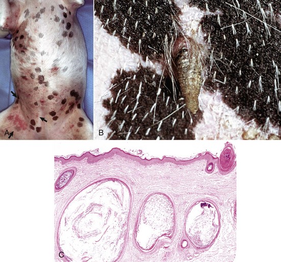
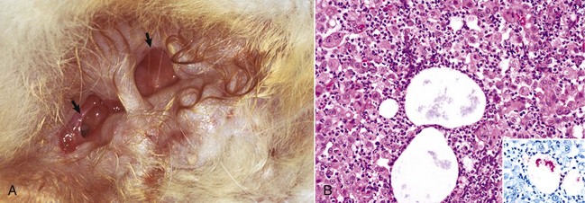
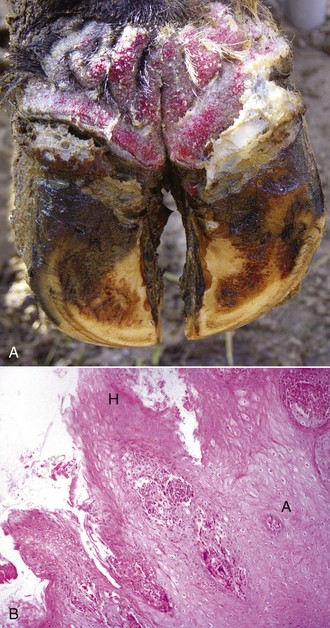
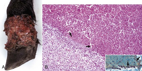
The macroscopic lesions follow the typical pattern for pox infections. Erythematous macules progress to papules, which may be firm. Sheeppox lesions have a variably prominent vesicular stage. The pustule stage is characterized by the formation of a thin crust. In severely affected animals, the lesions coalesce and form areas of edema, hemorrhage, necrosis, and induration, involving all layers of the skin and subcutis (Fig. 17-42). These areas correspond to the development of vasculitis described later with microscopic lesions (Fig. 17-42, B). Highly susceptible animals often develop hemorrhagic mucosal papules early in the course of the disease, and ulcerative lesions in the gastrointestinal and respiratory tracts develop later. Approximately one-third of animals develop multiple pulmonary lesions that comprise foci of pulmonary consolidation. The kidneys have multifocal, circular, and fleshy nodules throughout the renal cortices.
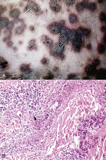
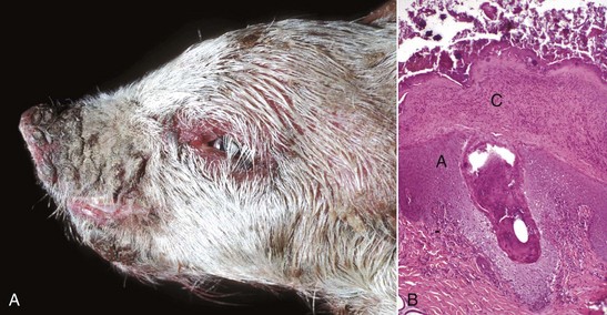
Fig. 17-42 Capripoxvirus, skin, lamb.
A, The clinical lesions are multifocal coalescing macules and plaques that are indurated, hemorrhagic, and necrotic likely a result of vasculitis. B, Note necrosis of vessel wall with fibrin deposition, red blood cells, and neutrophils and lymphocytes in the bordering dermis (vasculitis) (arrow). H&E stain. (A courtesy of Foreign animal diseases, ed 7, 2008, United States Animal Health Association. B courtesy Dr. A.M. Hargis, DermatoDiagnostics. Photographed from slides provided by Division of Animal Medicine, Animal Technology Institute Taiwan. From AFIP WSC October 8, 2008, Conference 5, Case III.)
Healing of the skin lesions is slow, taking up to 6 weeks, and a scar may remain. In the milder form of the disease, seen in endemic areas, the full range of pox lesions does not develop. Instead, epidermal proliferation produces papules covered by scale-crust, which heal with desquamation in a few days. Such lesions often occur on the ventral surface of the tail.
Sheeppox lesions have the typical microscopic poxviral epithelial lesions, including intracytoplasmic inclusion bodies. The lesions affect both surface epithelium and that of the hair follicles. There are also marked dermal lesions reflecting the systemic route of cutaneous involvement and possibly implicating immune-mediated lesions in addition to those caused by direct viral damage. The initial dermal lesions, corresponding to the macroscopic erythematous macule, are marked edema, hyperemia, and neutrophilic exocytosis. During the papular stage, large numbers of mononuclear cells accumulate in the increasingly edematous dermis. These mononuclear cells are called sheeppox cells and are characteristic of the disease. The nuclei of sheeppox cells are vacuolated and have marginated chromatin. The vacuolated cytoplasm contains single, occasionally multiple, eosinophilic intracytoplasmic inclusion bodies. Sheeppox cells are virus-infected monocytes, macrophages, and fibroblasts, but not endothelial cells. Approximately 10 days postinfection and corresponding with the most prominent epithelial lesions and peak of skin infectivity, severe necrotizing vasculitis develops in arterioles and post-capillary venules (Fig. 17-42, B). Virus particles have not been identified in endothelial cells, and the vasculitis may be the result of immune-complex deposition. Ischemic necrosis of the dermis and overlying epidermis follows. The pulmonary lesions are proliferative alveolitis and bronchiolitis with focal areas of caseous necrosis. Alveolar septal cells contain intracytoplasmic inclusion bodies. Additional histologic lesions, characterized by the accumulation of sheeppox cells, may involve heart, kidney, liver, adrenal gland, thyroid gland, and pancreas.
The course and outcome of sheeppox depend not only on the usual host-virus relationship but also on the nature and location of secondary infections. The virus itself may cause death during the febrile, eruptive phase of the disease. Secondary bacterial infection and even septicemia and pneumonia can be the cause of death because animals are susceptible to fly strike and while the skin lesions are in the healing stage.
Goatpox: Goatpox, caused by goatpox virus, occurs in the previously described geographic distribution, and a benign form of goatpox occurs in California and Sweden. The clinical signs of goatpox vary in different geographic areas. The disease is generally milder than sheeppox with a low mortality rate (5%), although generalized eruption with mortality rates approaching 100% may occur, with a course of disease similar to that of sheeppox infections in sheep. The cutaneous lesions have a predilection for the same areas as for sheeppox. In nursing kids, lesions may appear on the buccal mucosa or anterior nares. In animals with higher levels of resistance, the lesions may be confined to the udder, teats, inner aspects of thighs, or ventral surface of the tail.
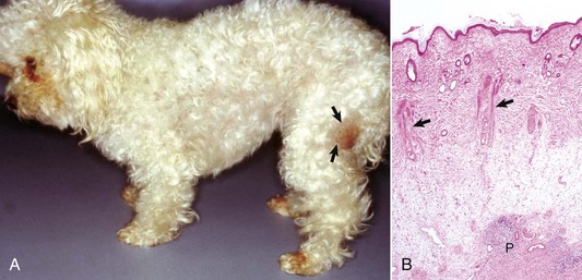
Contagious Ecthyma: Contagious ecthyma (contagious pustular dermatitis, orf, sore mouth) is a common localized infection of young sheep and goats caused by a parapoxvirus with worldwide distribution. Less commonly, humans, cattle, wild ungulates, and dogs are infected. Morbidity in lambs is usually great, and although mortality is usually low, it can approach 15% in lambs. Lesions are initiated by abrasions from pasture grasses or forage, begin at the commissures of the mouth, and spread to the lips (Fig. 17-43), oral mucosa, eyelids, and feet. Lambs can transfer the virus to the teats of ewes, and the lesions can spread to the skin of the udder. Contagious ecthyma is economically important as the result of weight loss in lambs that are reluctant to eat because of the pain associated with oral and perioral lesions. Pathogenesis of lesion formation and gross and microscopic features are consistent with the typical poxvirus lesions (see previous discussion and Fig. 17-31), except that the vesicle stage is very brief, the ulcer and crust stage persists and is clinically prominent, and the epidermis is markedly hyperplastic. Inclusion bodies are only briefly detectable histologically at the vesicular stage.
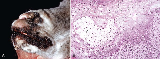
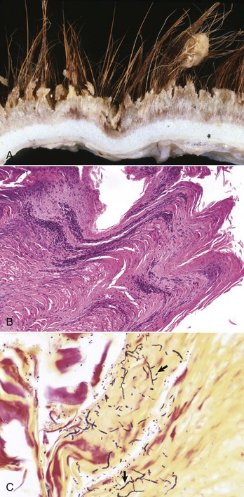
Fig. 17-43 Contagious ecthyma, skin, lamb.
A, Note crusts around nose and lips. These lesions are the late stage of the disease, are formed after rupture of vesicles and pustules, and are responsible for the common name scabby mouth. B, Note the epidermal hyperplasia (acanthosis), ballooning degeneration, vesicle (V), and neutrophils accumulating in the vesicle, which subsequently results in the formation of a pustule. Epidermal hyperplasia, upward movement of the pustule, and rupture of the vesicle or pustule contribute to crust formation as seen in A. H&E stain. (A courtesy Dr. M.D. McGavin, College of Veterinary Medicine, University of Tennessee. B courtesy Dr. Ann M. Hargis, DermatoDiagnostics.)
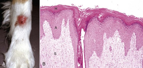
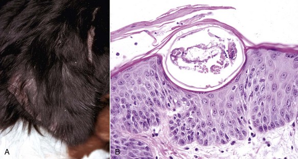
Swinepox: Pox lesions in pigs are caused by the host-specific poxvirus Suipoxvirus (swinepox). Normally, swinepox is transmitted by contact, although transplacental infection has not been ruled out. The sucking louse Haematopinus suis often acts as a mechanical vector and assists infection by causing skin trauma. The virus persists in dried crusts from infected animals. The pathogenesis of lesion formation and morphology of the gross and histologic lesions are consistent with the typical pox infection. The gross lesions typically affect the ventral and lateral abdomen, lateral thorax, and medial foreleg and thigh. Occasionally, lesions on the dorsum predominate. Lesions can be generalized and rarely involve the oral mucosa, pharynx, esophagus, stomach, trachea, and bronchi. The erythematous papules usually transform into umbilicated pustules without a significant vesicular stage (Fig. 17-44). The inflammatory crust eventually sheds to leave a white scar. The disease occurs worldwide and is endemic to areas of intensive swine production. The disease affects young, growing piglets and is mild with very low mortality.
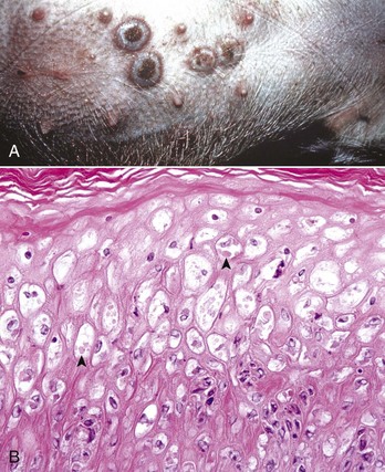
Fig. 17-44 Swinepox, skin, piglet.
A, Note the four umbilicated pustules in the abdominal skin. B, Note keratinocytes with ballooning degeneration and eosinophilic cytoplasmic inclusion bodies (arrowheads). Ballooning degeneration develops before vesicle formation. H&E stain. (A courtesy Dr. M.D. McGavin, College of Veterinary Medicine, University of Tennessee. B courtesy Dr. A.M. Hargis, DermatoDiagnostics. Photographed from slides provided by Department of Veterinary Pathology, Western College of Veterinary Medicine, University of Saskatchewan. From AFIP WSC January 21, 1998, Conference 15, Case IV.)
Herpesviruses: Herpesviruses are DNA viruses that only occasionally produce cutaneous lesions (see Table 17-7). Cutaneous lesions have rarely been reported in nondermatotropic herpesvirus infections, such as infectious bovine rhinotracheitis (bovine herpesvirus 1) and equine coital exanthema (equine herpesvirus 3), and in cats with FHV-1 infection. Two dermatotropic herpesvirus infections with economic importance are bovine herpesvirus 2 and bovine herpesvirus 4. Herpesviruses can be latent, with inactive virus persisting in tissue such as the trigeminal nerve ganglia. It is speculated that up to 80% of adult cats recovered from FHV-1 infection as kittens or young cats have latent herpesvirus 1 infections. During times of stress, the virus is reactivated and lesions can recur. Herpesviruses infect epithelial cells and replicate in the nucleus, leading to lysis of nuclear contents. As immature viral particles enter the cytoplasm, there is degeneration of cytoplasmic organelles, accumulation of cytoplasmic lipid, and precipitation of protein. Death of keratinocytes leads to spread of virus to neighboring cells, leading to rapid necrosis of focally extensive areas of the epidermis. Gross lesions consist of vesicles that rupture to form ulcers that are then covered by crusts. Microscopic lesions in herpesvirus infections depend on the stage, but early degenerative changes include ballooning and reticular degeneration, the sequelae of degeneration of epidermal cells and acantholysis. Syncytial cells may be seen. Intranuclear inclusions develop, but because of rapidly developing necrosis may not be found except at the margins of ulcers. The appearance of the viral inclusions varies with the specific herpesvirus. Some herpesviruses produce large, hyaline amphophilic inclusions that fill the nucleus (FHV-1), whereas others (bovine herpes mammillitis) produce typical Cowdry type A inclusions, which are also intranuclear but smaller and eosinophilic.
Bovine Herpesvirus 2: Bovine herpesvirus 2, a dermatotropic virus (Allerton virus), can cause generalized disease (pseudo–lumpy skin disease) or as seen in the US, localized infection of the teat called bovine ulcerative mammillitis (bovine herpes mammillitis). Mammillitis is inflammation of the teat or nipple. Localized infection occurs more commonly in lactating dairy cows but can develop in beef cows, pregnant heifers, and suckling calves. Trauma is implicated in the pathogenesis because normal skin is resistant to viral penetration. The pathogenesis of lesion formation was discussed earlier. Bovine ulcerative mammillitis is economically important because of decreased milk production and secondary bacterial mastitis. Lesions develop on the teats and skin of the nearby udder or occasionally the perineum. Suckling calves develop lesions on the muzzle (nose).
Bovine Herpesvirus 4: Bovine herpesvirus 4 (bovine herpes mammary pustular dermatitis) causes a similar but milder disease than the localized form of bovine herpesvirus 2.
Feline Ulcerative Facial Dermatitis and Stomatitis: Feline herpesvirus 1 is an uncommon cause of ulcerative, often persistent, facial dermatitis or stomatitis in cats of various ages and sexes. Glucocorticoid therapy or stresses, such as overcrowding, are thought to play a role in lesion development. Most lesions develop under circumstances suggesting reactivation of latent herpesvirus infection. The pathogenesis is typical of that described for herpesviruses in the previous section. Gross lesions are ulcerative and crusted (see Table 17-3). Histologically, there is extensive necrosis of the epidermal, follicular, and sometimes sebaceous gland epithelium accompanied by prominent mixed dermal inflammation that frequently includes numerous eosinophils. Follicular epithelium can be destroyed, and free keratin in the dermis is associated with eosinophils and foci of eosinophil degranulation bordering collagen fibers and collagen degeneration. Large amphophilic or hyaline intranuclear inclusions are present in the surface and adnexal epithelium. Inclusion bodies are often easily overlooked, variable in number, and sometimes present in small rafts of epithelial cells surrounded by necrotic debris. The lesions are different from those previously reported in domestic cats in that they persist and are limited to the skin of the face or oral mucosa and often have significant eosinophilic inflammation. The inflammation in feline herpesvirus dermatitis overlaps with that of the hypersensitivity reactions, including mosquito bite hypersensitivity, and also with that of eosinophilic ulcers, thus warranting close scrutiny of eosinophilic necrotizing cutaneous lesions for intranuclear inclusion bodies.
Papillomaviruses: Papillomaviruses are typically species- and site-specific pathogens that infect the squamous epithelium and cause benign proliferative masses and less commonly malignant tumors. As more precise methods of papillomavirus identification have been developed, including in situ hybridization, PCR, and DNA sequencing, increasing numbers of papillomaviruses have been identified. Papillomaviruses reproduce exclusively in keratinocyte nuclei. They gain access through defects in the epithelium and enter the basal layer epithelial cells. Once in the cell, there are three possible outcomes: (1) the virus can remain within the basal cell nucleus outside of the chromosomes in a circular DNA episome where the virus replicates synchronously with the host cell causing a latent infection without morphologic changes in keratinocytes; (2) as the basal cells mature, the virus can convert from latent to productive infection with the formation of complete infectious virions and with morphologic changes recognized as viral cytopathology, including epithelial hyperplasia, keratinocytes with clear cytoplasm and pyknotic nuclei, and sometimes with cytoplasmic or intranuclear inclusion bodies; or (3) the virus can become integrated into the genome of the host cell, resulting in malignant transformation and the morphologic changes of neoplasia. Malignant transformation occurs because the viral genes that remain after integration into the host cell are those associated with cellular regulation. These viral genes promote keratinocyte cell growth by inactivating tumor-suppressor proteins such as p53 and pRb. These events lead to uncontrolled cell proliferation, inability to repair DNA damage, and eventual malignant transformation.
All domestic animals are affected by one or more papillomaviruses (see Table 17-7), and some cross-species infections have been detected, particularly with the bovine papillomaviruses. Recently, a papillomavirus in an exophytic papilloma on the dorsal surface of the nose of a cat had 98% similarity to human papillomavirus type 9. The specific type or types of papillomaviruses involved in some infections have yet to be determined. Papillomavirus infections cause diverse clinical and histologic lesions ranging from papillomas, epidermal pigmented plaques, and fibropapillomas, including sarcoids. Papillomavirus DNA has also been identified within in situ and invasive squamous cell carcinomas in animals, but further work is necessary to prove a cause and effect relationship.
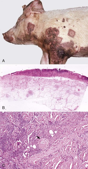
A common type of cutaneous papillomavirus infection (papilloma, wart, cutaneous papillomatosis) consists of clinical lesions that may be exophytic (proliferating to the exterior) (Fig. 17-45) or endophytic (inverted) papilliferous benign masses. Other papillomas may be flat, plaque-like lesions that lack the prominent papilliferous projections. Histologically, stratified squamous epithelium is covered by thickened orthokeratotic or parakeratotic keratin, is acanthotic, and in the exophytic and endophytic papillomas, has elongated epidermal-dermal interdigitations that project outwardly or inwardly, depending on the type. In some papillomas, keratinocytes, especially of the upper stratum spinosum, are swollen, have clear cytoplasm or a perinuclear halo, and a pyknotic nucleus; these keratinocytes are termed koilocytes (meaning hollow or concave). Keratohyalin granules are irregular. Also, pale basophilic intranuclear inclusion bodies, located in degenerating cells in the outer layers of the stratum spinosum and granulosum in which virion production is taking place, occur in some but not all papillomas. Many papillomas spontaneously regress, and in regressing stages, there is reduced epidermal hyperplasia, increased proliferation of fibroblasts, deposition of collagen, and infiltration of T lymphocytes at the epidermal dermal interface and in the epithelium. Some papillomas, such as those on the ears of horses and some affecting the teats of cattle, often do not regress.
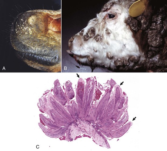
Fig. 17-45 Cutaneous papillomas, skin.
A, Chin, horse. Note multiple, small, verrucous masses arising in the skin. B, Head, cow. Note multiple, irregular, alopecic, verrucous papillomas. C, The papillary projections (arrows), often called fronds, are composed of hyperkeratotic epidermis covering a collagenous core. H&E stain. (A courtesy Dr. David Duclos, Animal Skin and Allergy Clinic. B and C courtesy Dr. M.D. McGavin, College of Veterinary Medicine, University of Tennessee.)
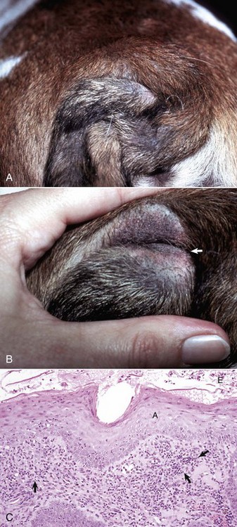
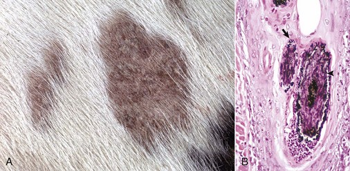
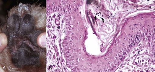
Viral plaques have been described in dogs and cats. Canine pigmented viral plaques are associated with papillomavirus infection in certain breeds of dogs (miniature schnauzers, pugs, and Shar-Peis) or in other breeds of dogs that are immunosuppressed. Novel papillomaviruses have been detected in some of these plaques. Clinical lesions occur most commonly on the ventral abdomen, groin, ventral thorax, or neck and consist of variably irregular, pigmented macules, or plaques. Histologically, the lesions are sharply demarcated, hyperkeratotic foci, or plaques with pigmented, acanthotic epidermis, and large keratohyalin granules. These pigmented epidermal plaques do not regress, are slowly progressive, and occasionally develop into a squamous cell carcinoma. Feline viral plaques are usually multiple, ovoid, slightly raised pigmented or nonpigmented, and slightly scaly and rough plaques less than 8 mm in greatest dimension. Papillomavirus has been detected in the lesions but not specifically identified to type. Histologically, there is an abrupt transition between normal epithelium and the plaque, which consists of epidermis thickened by hyperkeratosis, hypergranulosis, and acanthosis. Keratohyalin granules may be enlarged, and koilocytes (keratinocytes with clear cytoplasm and pyknotic nuclei), and cytoplasmic pseudoinclusions may be present. Malignant transformation commonly occurs; lesions resemble the in situ bowenoid carcinoma (see next paragraph).
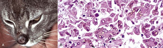
Papillomavirus infection also has been implicated in the development of another syndrome in cats and less often in dogs termed multicentric squamous cell carcinoma in situ (Bowen’s-like disease, in situ bowenoid carcinoma). Although not all the factors contributing to lesion formation have been documented, papillomavirus DNA has been identified in these lesions in cats, suggesting a nonproductive infection promotes the epithelial hyperplasia characteristic of this disease. The papillomaviruses identified in some of these lesions have homology with human papillomaviruses. Multicentric squamous cell carcinoma in situ clinically consists of sharply demarcated single, or more often multiple, scaly verrucous or irregular plaque-like lesions 0.5 to 3.0 cm in diameter. Histologically, the epidermis and follicular infundibular epithelium are thickened by proliferation of basal keratinocytes that tend to stream together, providing a “windblown” appearance to the epidermis. Nuclei are often varied in size with hyperchromatic nuclei, large nucleoli, and numerous mitoses, some of which are located above the basal layer. The basement membrane, at the time of histologic examination, is intact. Most lesions remain as “in situ” carcinomas indefinitely, but an occasional lesion has progressed to invasive basal cell carcinoma or invasive squamous cell carcinoma.
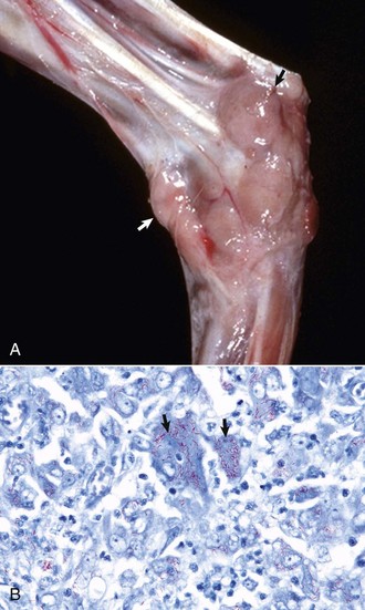
Some types of papillomaviruses, particularly bovine papillomaviruses 1 and 2, can infect fibroblasts and cause fibropapillomas, flat, verrucous, or nodular masses in which the proliferation of dermal fibroblasts is the prominent feature, often surpassing that of the epidermal hyperplasia (Fig. 17-46). Fibropapillomas occur in horses, mules, donkeys, cattle, sheep, and cats. These lesions in horses are called sarcoids, which are thought to represent a nonproductive infection by distinct variants of bovine papillomavirus. Equine sarcoids are locally aggressive, nonmetastatic fibroblastic skin tumors of horses, mules, and donkeys. They are the most common skin tumor of horses, accounting for up to 30% of tumors, and occur in any breed, sex, or age. Young adult horses 3 to 6 years of age are most commonly affected. Sarcoids frequently develop in areas subjected to trauma or at sites of wounds 6 to 8 months after wound healing and develop anywhere but are most common on the head, legs, and ventral trunk. They can be single or multiple. The tumors are classified according to their gross appearance as verrucous, fibroblastic, mixed, or occult. The verrucous type is a small wartlike growth, usually measuring less than 6 cm in diameter, with a dry, rough surface and variable alopecia. The fibroblastic type of sarcoid is more variable in appearance and can range from a well-circumscribed firm nodule with intact surface to a large mass, greater than 25 cm in diameter, with an ulcerated surface prone to hemorrhage, and resembling exuberant granulation tissue. The mixed type is a transitional form in which a verrucous sarcoid becomes a fibroblastic type as a result of trauma or biopsy. The occult form consists of a slow-growing, slightly thickened area of skin with slight surface roughening and alopecia that remains static for a long period.
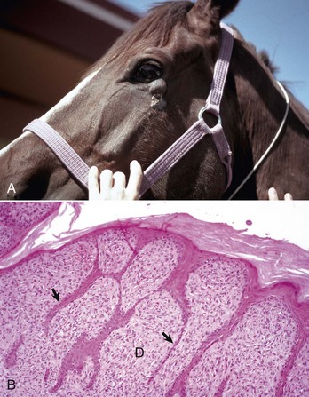
Fig. 17-46 Sarcoid, skin, horse.
A, Equine sarcoid, face. The irregular multinodular mass is present on the ventrolateral periocular skin, especially below the eye. B, Sarcoid. The sarcoid consists of an epidermal and dermal component. The hyperplastic epidermis has thin rete pegs that extend into the dermis (arrows). The dermis (D) is thickened by proliferating fibroblasts and collagen. H&E stain. (A courtesy Dr. Helen Power, Dermatology for Animals. B courtesy Dr. Pamela E. Ginn, College of Veterinary Medicine, University of Florida.)
Histologically, sarcoids are typically biphasic tumors composed of both epidermal and dermal components; however, the epidermal component may be minimal or absent in some tumors, especially those with extensive ulceration. When the epidermis is intact, hyperkeratosis, parakeratosis, and acanthosis with thin rete pegs extending deep into the dermis are common features. The dermal component consists of fibroblasts and collagen in various proportions. The fibroblasts have plump nuclei, and nucleoli may be prominent. The mitotic index is usually low. Fibroblasts at the dermal-epidermal junction are frequently oriented perpendicular to the basement membrane in a “picket fence” pattern, which is a distinctive histologic feature seen in most sarcoids. The cells are arranged in whorls, interlacing bundles, or haphazard arrays of variable density. Tumor margins are typically indistinct and adequacy of excision is frequently difficult to determine. Spontaneous remission can occur after several years in up to 30% of cases. The tumors are characterized by a high rate of recurrence, up to 50%, after surgical excision. Feline fibropapillomas (also called sarcoids) are similar morphologically to the equine lesion, and also likely represent a nonproductive infection with papillomavirus with similarities to bovine, ovine, deer, and European elk papillomaviruses. Affected cats often live in rural areas and have had exposure to cattle. Bovine fibropapillomas are caused by bovine papilloma virus 1 (teats, penis) or bovine papillomavirus 2 (head, neck, shoulder, legs, and teats) and occur in young animals. The lesions generally spontaneously regress within 1 to 12 months.
Papillomaviruses, some as yet to be identified by type, have been found in canine and feline invasive squamous cell carcinoma. However, the presence of the virus in the tumors does not allow differentiation between actual induction of the tumor and mere infection of the tumor. Further work to characterize the types of papillomaviruses and the role the viruses play in epidermal hyperplasia and neoplasia is necessary.
Other Viruses: Cutaneous lesions are seen with foot-and-mouth disease (picornavirus), vesicular stomatitis (rhabdovirus), swine vesicular disease (picornavirus), vesicular exanthema (calicivirus), and malignant catarrhal fever (herpesvirus) (see Table 17-7; see also Chapters 4 and 7). Feline leukemia virus (FeLV) can cause the development of lymphoma and fibrosarcoma. A few cats with FeLV infection have developed dermatitis characterized by epidermal and follicular acanthosis with epidermal giant cells, dyskeratosis, necrosis, and ulceration. In addition, a few FeLV-infected cats have also developed one or more localized areas of marked compact hyperkeratosis of the pawpads that clinically resemble “horns” (cutaneous horns). In immunoperoxidase-stained sections, the hyperplastic epidermis in cases with epidermal giant cells is strongly positive for FeLV. Both FeLV and feline immunodeficiency virus (FIV), because of their immunosuppressive capabilities, can cause cats to be susceptible to chronic skin infections, including abscesses, paronychia, and demodicosis.
Feline calicivirus, usually the cause of upper respiratory infection and oral ulcers with high morbidity but low mortality, rarely produces cutaneous ulcers. More recently, virulent systemic strains of feline calicivirus with mortality rates between 30% and 60% have been described. These strains cause alopecia, cutaneous ulcers, and subcutaneous edema. The pathogenesis involves lysis of epithelial cells of the epidermis, follicles, oral mucosa, bronchioles, alveoli, and exocrine pancreas, in addition to lysis of endothelial cells. The lysis of epithelial and endothelial cells leads to necrosis, edema, fibrin exudation, and thrombosis from vascular injury and release of inflammatory mediators from damaged cells. Clinical lesions include ulcers of the nose, lips, pinnae, feet, and oral mucosa, variable alopecia of limbs and ventrum, and subcutaneous edema, especially of the limbs and face. Systemic signs of fever, anorexia, icterus, red and swollen conjunctival mucosa, and nasal or ocular discharge are present. The most consistent microscopic lesions are epithelial necrosis with subsequent ulceration of the skin and oral and nasal mucosa. Vascular injury consists of edema, microthrombosis, and fibrin exudation. Bronchointerstitial pneumonia and necrosis of the liver, pancreas, spleen, and lymph nodes are present in some cats. In contrast to infection with the more common and less virulent field strains of feline calicivirus, the systemic virulent strains cause more severe disease in adult cats than kittens.
The portals for entry of bacteria into the skin include pores (follicular openings), hematogenous spread, or direct entry through damaged skin. Cutaneous bacterial infections vary in location (e.g., epidermis, dermis, subcutis, adnexa, or systemic), morphology (e.g., pyogenic, granulomatous, or necrotizing), distribution (e.g., focal, multifocal, regional, mucocutaneous, haired skin, or interdigital), and severity (e.g., mild and asymptomatic to severe with systemic signs). The variation is caused by the specific organism involved, predisposing or coexisting factors, and host-immune response. The so-called superficial and deep bacterial infections are frequently pus-producing infections (pyogenic) and are thus referred to as pyodermas. In contrast, bacterial granulomas are characterized by an abundance of macrophages and are usually caused by traumatic implantation of bacteria that generally are saprophytes of low virulence. Systemic bacterial infections or localized infection with toxin-producing bacteria are often most severe because of vascular damage or the presence of endotoxins or exotoxins that have systemic consequences. The most common bacterial infections of the skin are listed in Box 17-7.
Bacterial skin disease is seen much more frequently in dogs than other domestic species, possibly the result of the thin stratum corneum with small amount of lipids, lack of a protective lipid seal at opening of the canine hair follicles, and the relatively high pH of canine skin. Until recently, Staphylococcus intermedius has been considered the cause of most cases of pyoderma in dogs. However, recent molecular studies involving multilocus gene sequencing have revealed that bacterial isolates phenotypically consistent with Staphylococcus intermedius consist of three separate species (considered to be the Staphylococcus intermedius group), which include S. intermedius, Staphylococcus pseudintermedius, and Staphylococcus delphini. Staphylococcus pseudintermedius is now considered to be responsible for most cases of pyoderma in dogs. Coagulase-positive staphylococci are also the most common bacteria isolated from pyoderma in horses (S. aureus, S. intermedius), in cattle and sheep (S. aureus), and in goats (S. aureus). Molecular studies will likely be required to identify the species involved in some of these infections, particularly those of the Staphylococcus intermedius group. Staphylococcus hyicus causes exudative epidermitis in piglets and has been associated with superficial pyoderma in several other species. Many other bacteria can cause skin infections. Dermatophilus congolensis is responsible for superficial pyoderma in many species. Many Gram-negative bacteria are opportunistic pathogens that can invade already diseased or compromised skin.
Superficial Bacterial Infections (Superficial Pyodermas): Superficial bacterial infections (superficial pyodermas) involve the epidermis and the upper infundibulum of hair follicles, usually heal without scarring, and usually do not involve the regional lymph nodes. Gross lesions include erythema, alopecia, papules, pustules, crusts, and peripheral expanding rings of scale also called “epidermal collarettes” (see Table 17-3). The early microscopic feature of superficial bacterial infection that involves the epidermis is intraepidermal pustular dermatitis. The intraepidermal pustules are fragile and can rupture, leading to crust and superficial scale formation. The major microscopic feature of superficial bacterial infection that involves the follicles is superficial suppurative luminal folliculitis. The cellular infiltrate in and around hair follicles plus dermal congestion and edema correspond to the clinically evident papules and follicularly oriented pustules. Follicular injury leads to alopecia. Although Gram-positive cocci, such as Staphylococcus sp., are usually the cause of the superficial bacterial infections, the bacteria are not always microscopically demonstrable. Predisposing factors, such as allergy, seborrhea, and immune deficiency, and other causes of follicular inflammation or dysfunction often play a role.
Superficial Pustular Dermatitis: Superficial pustular dermatitis, typically caused by staphylococci, encompasses several syndromes including impetigo in a variety of animal species, exudative epidermitis in pigs, and superficial pyoderma in dogs. Pathogenicity may correlate with various proteins and toxins produced by the bacteria and thought to act as virulence factors. One of the factors that has come under recent scrutiny includes the exfoliative toxins. These toxins have been isolated from strains of Staphylococcus aureus in humans with impetigo, an acute contagious superficial bacterial skin infection that usually affects children and is characterized by vesicles and pustules that form yellowish crusts. In addition, similar exfoliative toxins have been identified as a source of another skin condition usually affecting infants and children, termed staphylococcal scalded-skin syndrome. In impetigo and staphylococcal scalded-skin syndrome, it has been shown that virulent forms of S. aureus produce exfoliative toxins that cause the loss of adhesion of keratinocytes in the superficial epidermis. These toxins are glutamate-specific serine proteases that cleave a single peptide bond in desmoglein 1, present in the extracellular protein core of the desmosome. The separation of these superficial keratinocytes results in intraepidermal splitting and initiation of lesion development in these infections. In impetigo, the S. aureus that produce exfoliative toxins can be isolated from the intact pustules. In contrast, in staphylococcal scalded-skin syndrome, there are generalized blisters and superficial exfoliation of the stratum corneum, but cultures from intact vesicles usually are negative for exotoxin producing S. aureus. It appears that the exfoliative toxins are produced in a distant area of infection and reach the skin via the bloodstream (a process called toxemia) where they cause the vesicular and exfoliative lesions. Investigative studies in two domestic species (pigs and dogs) suggest that a similar pathogenic mechanism involving exfoliative toxins may play a role in development of superficial pustular dermatitis caused by staphylococci. For example, it has been shown that Staphylococcus hyicus, which causes exudative epidermitis in piglets, produces an exfoliative toxin that can cleave swine desmoglein 1, and produce cutaneous exfoliation similar to that in pigs with exudative epidermitis. Similarly, an exfoliative toxin isolated from strains of Staphylococcus intermedius (pseudintermedius) from dogs with pyoderma has caused cutaneous exfoliation when injected into the skin of dogs. In addition, the exfoliative toxin gene has been identified in Staphylococcus intermedius and Staphylococcus pseudintermedius isolated from skin, wound, and ear infections in dogs suggesting a role for the toxin in pathogenicity. Although the exfoliative toxins in canine pyoderma have not been fully characterized, these findings suggest that some strains of Staphylococcus sp. in dogs and Staphylococcus hyicus in pigs may cause superficial bacterial infections via a pathologic mechanism involving exfoliative toxins. Further studies are necessary to determine if exfoliative toxins or other virulence factors contribute to the development of superficial pyoderma in other species.
Impetigo: Impetigo is observed most commonly in cows, ewes, does, and dogs and is usually caused by coagulase-positive Staphylococcus sp. Predisposing factors, such as cutaneous abrasions, viral infections, increased moisture, and poor nutrition, may contribute. Lesions of impetigo in cows, does, and ewes, occur predominantly on the ventral abdomen, perineum, medial thigh, vulva, ventral tail, teats, and udder. In dogs, lesions are largely in nonhaired ventral skin. Prepubescent puppies are usually healthy otherwise, but older dogs with impetigo often have underlying disease, including immunosuppression associated with hyperadrenocorticism. Gross lesions consist of nonfollicular pustules that develop into crusts. The microscopic lesion is a nonfollicular neutrophilic subcorneal pustule. In bullous impetigo, a more severe condition occurring in older dogs with underlying disease, the lesions are large interfollicular flaccid pustules (bullae) that when ruptured lead to more extensive loss of the superficial epidermis. Acantholytic cells may be present in the pustules, probably the result of the cleavage of desmoglein 1 by the exfoliative toxin, thus requiring differentiation between impetigo and pemphigus foliaceous (see Fig. 17-15). The presence of coccoid bacteria within intact pustules can help provide support for a bacterial origin of the lesions. Perivascular to interstitial neutrophilic to mixed mononuclear dermal inflammation is present.
Exudative Epidermitis of Pigs (Greasy Pig Disease): Exudative epidermitis, usually caused by Staphylococcus hyicus, is an acute, often fatal, dermatitis of neonatal piglets, but a mild disease in older piglets. Predisposing factors include cutaneous lacerations and poor nutrition. In piglets, brownish exudates develop around the eyes, pinnae, snout, chin, and medial legs and spread to the ventral thorax and abdomen, giving the animal an overall “greasy” appearance (Fig. 17-47). The lesions rapidly coalesce and become generalized, resulting in greasy, malodorous exudates covering an erythematous skin. If piglets survive, the exudate hardens, cracks, and forms fissures. Subacute disease develops gradually in older piglets, and lesions are generally localized to the skin of the face, pinnae, and periocular regions. Grossly, the epidermis is thickened with scaling. The early histopathologic lesion is subcorneal pustular dermatitis, which extends to the hair follicle, resulting in superficial suppurative folliculitis. In the fully developed lesion, the epidermis is hyperplastic and has thick crusts of keratin, microabscesses, and cocci. The term exudative epidermitis is descriptive of this condition as the inflammatory changes largely involve the epidermis, and there is an accumulation of exudates on the surface. The dermis is congested and edematous. In the early stages, the dermatitis is superficial and perivascular with neutrophils and eosinophils, and in the later stages is perivascular and mononuclear.
Fig. 17-47 Exudative epidermitis, Staphylococcus hyicus (hyos), skin, piglet.
A, Head. Exudative epidermitis is also called greasy pig disease. The skin in this pig is lichenified and has fissures. Greasy exudate focally has adhered to the hair and the surface of the skin. B, Note the epidermal hyperplasia (acanthosis [A]) and suppurative exudate within the lumen of a hair follicle and on the surface of the epidermis. The exudate has dried to form a crust (C) that is fragmenting superficially. H&E stain. (A courtesy Dr. M.D. McGavin, College of Veterinary Medicine, University of Tennessee. B courtesy Dr. Pamela E. Ginn, College of Veterinary Medicine, University of Florida.)
Canine Superficial Pyoderma (Superficial Spreading Pyoderma): Superficial spreading pyoderma is a common, often pruritic, superficial bacterial infection in dogs caused by Staphylococcus pseudintermedius. Clinical lesions are most frequently recognized in the glabrous ventral thoracic and abdominal skin but can affect the haired skin of the dorsal and lateral trunk as well. Early clinical lesions include erythematous macules, papules, and transient pustules. Older clinical lesions include epidermal collarettes, crusts, alopecia, and hyperpigmentation. Early microscopic lesions are superficial, spongiotic epidermal pustules that rapidly crust and form basophilic debris, often with cocci, on the surface of the epidermis. This basophilic debris can dissect peripherally (laterally) between the epidermis and stratum corneum and is thought to form the rim of scale that clinically represents the epidermal collarette. In this way, the lesions “spread” outwardly from the initial lesion. Occasionally, superficial spreading pyoderma can originate from superficial folliculitis where follicular pustular formation is minor and epidermal collarette formation more prominent. Dermal lesions include superficial perivascular to interstitial accumulations of neutrophils, eosinophils, and mixed mononuclear cells. Some dogs have neutrophilic vasculitis involving superficial venules possibly caused by immune-complex deposition, a feature suggesting a hypersensitivity response to bacterial antigens. Dermal congestion and edema are usually present.
Mucocutaneous Pyoderma: Mucocutaneous pyoderma is a putative bacterial infection of mucocutaneous junctional skin in dogs. Antibiotic responsiveness suggests that bacteria contribute, however, the etiology is likely more complex and may involve immunologic factors as well. A variety of breeds are affected, but the German shepherd breed is thought to be predisposed. The pathogenesis is unknown. Lesions consist of erythema, swelling, and crusting, and in severe cases, ulceration. Depigmentation may develop in chronic cases. Lesions are most common on mucocutaneous skin of the lips, but mucocutaneous skin of prepuce, vulva, and anus can be affected. Histologic lesions include a dense band of lymphoplasmacytic inflammation with variable numbers of neutrophils at the epidermal dermal junction (lichenoid inflammation), typically without basal cell degeneration. Other features include spongiosis and cellular exocytosis into the epidermis, neutrophilic pustular crusts, and folliculitis of adjacent follicles. Over time, pigmentary incontinence develops. Although classic cases are said not to have basal cell degeneration, apoptotic keratinocytes above the basal layer can be present. In addition, there can be interface inflammation obscuring the epidermal-dermal interface. These features prevent definitive histologic differentiation from discoid lupus erythematosus.
Dermatophilosis (Streptothrichosis): Dermatophilosis, caused by Dermatophilus congolensis, is characterized by crusty cutaneous lesions (Fig. 17-48) and occurs in horses, cattle, and sheep more often than goats, pigs, dogs, or cats. The bacterium is transmitted by carrier animals and is more common in tropical and subtropical climates and during wet weather, thus the layman’s term rain rot. Lesions tend to develop on the dorsum of the back and distal extremities and after epidermal irritation from ectoparasites, trauma, or prolonged wetting of the skin, hair, or wool, which allows penetration of the damaged epidermis by the Dermatophilus “zoospore.”
Fig. 17-48 Dermatophilus congolensis infection, skin, haired, cow.
A, The hair is matted by a crust composed of dried exudate, stratum corneum, and bacteria. B, Note thick laminated crust formed of alternating layers of hyperkeratotic/parakeratotic stratum corneum and degenerate neutrophils. H&E stain. C, The stratum corneum contains filamentous bacteria (arrows). These bacteria subdivide longitudinally and transversely and can result in a “railroad track” appearance (not evident here). Brown and Brenn stain. (A courtesy Dr. F. Lozano-Alarcon. B courtesy Dr. M.D. McGavin, College of Veterinary Medicine, University of Tennessee. C courtesy Dr. Ann M. Hargis, DermatoDiagnostics.)
The bacterium grows in the outer root sheath of the hair follicle and superficial epidermis and produces Gram-positive filamentous branches that subdivide longitudinally and transversely (see Fig. 17-48). These bacteria stimulate an acute inflammatory response in which neutrophils migrate from superficial vessels into the dermis and through the epidermis to form intraepidermal microabscesses. The inflammation inhibits further penetration of the bacterium. However, residual bacterial organisms subsequently invade the newly regenerated epidermis. Thus repeated cycles of bacterial growth, inflammation, and epidermal regeneration result in the formation of the multilaminated pustular crusts. Grossly, lesions consist of papules, pustules, and thick crusts that can coalesce and mat the hair or wool (see Fig. 17-48). The microscopic lesions consist of hyperplastic superficial perivascular dermatitis with multilaminated crusts of alternating layers of keratin and neutrophils covering the skin surface. Samples of crusts obtained by biopsy are necessary to identify organisms and make a definitive diagnosis.
Ovine Fleece Rot: Ovine fleece rot is a superficial bacterial dermatitis usually caused by excessive moisture (usually in the form of rain) that penetrates the fleece (wool), wets the skin, and causes proliferation of Pseudomonas spp. Approximately 1 week of continual wetting is usually sufficient to cause marked proliferation of the bacteria on the skin and in the fleece. This is followed by an acute inflammatory response with serum exudation and matting of the fleece. The fleece is also discolored because of production of pigments (chromogens) by the Pseudomonas bacteria and has a rotten odor. Microscopic lesions include epidermal pustular dermatitis and superficial folliculitis. Ovine fleece rot is important economically because the malodor attracts flies, predisposing to myiasis (infestation of tissue by the larvae of dipterous flies), and the value of the affected wool is reduced.
Deep Bacterial Infections (Deep Pyodermas): Deep pyodermas are bacterial infections of the hair follicle, dermis, and/or subcutis. They are caused by a variety of bacteria, including Staphylococcus sp., Streptococcus sp., Corynebacterium pseudotuberculosis, Pasteurella sp., Proteus sp., Pseudomonas sp., and Escherichia coli. Staphylococcus pseudintermedius is the primary pathogen of canine skin. Organisms typically enter the skin via natural pores such as hair follicles or any form of wound, including direct penetration of the skin. Deep bacterial infections of hair follicles often have predisposing causes, such as immune suppression, demodicosis (dogs), or disorders associated with follicular hyperkeratosis (callus or comedo formation), and also originate as a sequela to superficial bacterial folliculitis. Deep bacterial infections are less common than superficial infections and develop most frequently in dogs.
Bacterial Folliculitis and Furunculosis (Table 17-8): Infection of the hair follicle can be superficial or deep. Superficial infections involve the follicular infundibulum but can spread to involve the deeper portions of the follicle (the infundibulum and below). Mild-to-moderate folliculitis without furunculosis can resolve completely with appropriate antibiotic therapy. However, untreated or severe folliculitis can progress to involve deeper aspects of the follicle and result in follicular distention with rupture (furunculosis) and release of follicular contents (hair, follicular stratum corneum, and bacteria) into the dermis and sometimes subcutis. The bacteria proliferate in the deep dermis and subcutis and can reach draining lymph nodes. Draining tracts often develop as a foreign body response to extruded follicular contents (e.g., hair and follicular stratum corneum). Thus the infection and severe inflammation spread into the surrounding dermis and subcutis, resulting in the need for intensive long-term treatment and increasing the potential for systemic infection and local scarring.
TABLE 17-8
Bacterial Folliculitis and Furunculosis
*Pastern (proximal interphalangeal articulation).
†Fetlock (metacarpophalangeal articulation).
Numerous bacteria can cause folliculitis and furunculosis, but staphylococcal bacteria are the most commonly involved. Staphylococcal folliculitis and furunculosis develop most commonly in the dog (see Figs. 17-26 and 17-27), frequently affect the horse, sheep, and goat but are uncommon in the cow, pig, and cat.
In horses, lesions develop most commonly in association with tack, especially on the skin of the saddle area, the tail, or the caudal aspect of the pastern (proximal interphalangeal articulation) or fetlock (metacarpophalangeal articulation). When the skin of the pastern or fetlock is involved, the condition is called equine pastern folliculitis (also known as grease heel, scratches). Equine pastern folliculitis is a complex disorder in which secondary staphylococcal folliculitis plays a role. Predisposing factors are numerous and include excessive moisture, trauma, and contact dermatitis. Also, many other conditions affect the pastern skin in horses, including immune-mediated diseases (pemphigus foliaceous, vasculitis, or photosensitization), other infections (dermatophilosis, dermatophytosis), and mite infestation (Chorioptes sp.), necessitating an early thorough clinical evaluation and sometimes microbiologic or histopathologic evaluation to differentiate staphylococcal folliculitis of the skin of the pastern from these other disease processes. In severe chronic lesions, initiating causes may no longer be identifiable.
In adult sheep, lesions develop on the face, especially around the eyes, or on the limbs or teats. In otherwise healthy lambs, mild lesions develop most commonly on the lips and perineum and usually spontaneously regress. In goats, the face, pinnae, distal limbs, and glabrous areas of the udder, ventral abdomen, medial thighs, and perineum are most commonly affected.
In dogs, lesions are localized or generalized and develop on the dorsal nose, pressure points, interdigital areas, and chin. Other cutaneous areas can also be affected, especially if predisposing conditions (e.g., follicular dysplasia, cornification disorders, or demodicosis) are present. Deep pyoderma of adult German shepherd dogs (German shepherd folliculitis, furunculosis, and cellulitis) is a unique deep pyoderma with an apparent genetic predisposition. Lesions are located on the dorsal lumbosacral, ventral abdominal, and thigh areas. Hypersensitivity to the bites of fleas or alterations in immune or neutrophil function have been proposed as predisposing causes, but most of these potential causes have been discounted. Deep folliculitis and furunculosis, especially on the cheek area or neck of some large-breed dogs (golden and Labrador retriever, Saint Bernard, and Newfoundland), can clinically resemble superficial pyotraumatic dermatitis (acute moist dermatitis), belying the deep nature of the lesions.
Postgrooming furunculosis is an uncommon but acute, severe, and painful form of furunculosis in the dog that is believed to be associated with grooming. A variety of bacteria have been cultured from lesions, including Staphylococcus intermedius (pseudintermedius), Pseudomonas aeruginosa, Escherichia coli, and Proteus sp.
Grossly the lesions of superficial folliculitis include papules, crusted papules, pustules, epidermal collarettes, and alopecia. Deep folliculitis can have similar lesions plus hemorrhagic bullae, nodules, and draining tracts. The microscopic patterns include superficial or deep luminal folliculitis, pyogranulomatous furunculosis, draining sinuses, and occasionally, panniculitis. Microscopic lesions include suppurative luminal folliculitis with follicular distention often in conjunction with furunculosis. Pyogranulomatous dermatitis in response to release of follicular contents is often severe, may efface the dermal architecture, extend into the deep dermis and panniculus, and form tracts that drain to the surface. Scarring can lead to loss of adnexal structures and permanent alopecia localized to affected skin.
Subcutaneous Abscesses: Subcutaneous abscesses are localized collections of purulent exudate located within the dermis and subcutis. Abscesses are common in cats because of the frequency of bacterial contamination of puncture wounds. Abscesses also are common in large animals. In addition to puncture wounds, other predisposing causes include foreign bodies, injections, and shearing and clipping wounds. Granulation tissue or mature fibrous connective tissue borders the exudate. Subcutaneous abscesses frequently rupture and drain spontaneously, and heal by scarring. A wide variety of bacteria can cause subcutaneous abscesses. Commonly isolated bacteria include Pasteurella multocida (dog and cat bite wounds), Corynebacterium pseudotuberculosis (horses, sheep, and goats), and Arcanobacterium pyogenes (sheep, goats, cattle, pigs). Other frequently isolated bacteria include β-hemolytic streptococci, Fusobacterium sp., Peptostreptococcus sp., Bacteroides sp., Staphylococcus sp., and Clostridium sp.
Cellulitis: Cellulitis, in contrast to an abscess, is a poorly defined suppurative bacterial infection of the dermis and subcutis that dissects and spreads through surrounding soft tissues. The affected skin is often swollen, erythematous, and warm and may become devitalized and slough. The bacteria can cause a foul odor and some, such as Clostridium sp., can produce subcutaneous gas bubbles (subcutaneous emphysema). Cellulitis may be accompanied by fever and enlargement of regional lymph nodes. Histologic lesions consist of poorly defined areas of purulent to pyogranulomatous inflammation that may include hemorrhage, necrosis, and thrombosis. Bacteria may be visible histologically. As with subcutaneous abscesses, the source of the infection is usually a penetrating wound in the area of infection. A variety of bacteria, including those found in subcutaneous abscesses, can cause cellulitis. A rare but particularly severe subtype of cellulitis, termed necrotizing fasciitis has been described most commonly in dogs with Streptococcus canis infection (see later discussion of infection with toxin-producing bacteria).
Bacterial Granulomatous Dermatitis (Bacterial Granulomas): Bacterial granulomatous dermatitis is usually caused by traumatic implantation of bacteria, which are generally saprophytes of low virulence. Causative organisms usually stimulate a strong cell mediated-immune response by persisting as an antigen in the tissue. Grossly, lesions are slowly progressive, nodular or diffuse, and can ulcerate and drain through the surface of the skin via fistulas. Microscopic lesions consist of mixed populations of inflammatory cells, especially macrophages, thus lesions are granulomatous to pyogranulomatous. Multinucleated giant cells and caseous necrosis are present in some lesions. Causal agents can be present in macrophages, exudate, or in clear spaces or fat vacuoles within tissue but are often in such low numbers that they are difficult to identify in histologic sections.
Mycobacterial Granulomas: Mycobacterial organisms produce granulomatous to pyogranulomatous dermatitis and panniculitis in many species of animals, particularly cats and less frequently dogs and cattle. The majority of mycobacteria are intracellular pathogens that are able to persist in tissue by entering macrophages. Many are able to survive and replicate within the macrophages by inhibiting fusion with lysosomes. Tissue destruction results from persistence of antigen in the tissue and a cell-mediated inflammatory response. Infection occurs with obligate pathogens that require a vertebrate host to multiply and saprophytes in the environment that occasionally cause opportunistic infections. Infection occurs with the tuberculosis group considered to be obligate pathogens (Mycobacterium tuberculosis, Mycobacterium bovis), the leprosy group considered to be obligate pathogens (Mycobacterium lepraemurium), and the opportunistic group considered to be saprophytes or facultative pathogens (subdivided based on growth rate and pigment production). Rapid-growing opportunistic organisms (Mycobacterium fortuitum, Mycobacterium smegmatis, Mycobacterium chelonae, Mycobacterium abscessus, and Mycobacterium thermoresistible) and slow-growing opportunistic organisms (Mycobacterium avium-intracellulare complex, Mycobacterium kansasii, and Mycobacterium ulcerans) are inhabitants of soil, water, and decomposing vegetation, and infection tends to occur via wound contamination or traumatic implantation. To avoid confusion in terminology, by convention, infections caused by Mycobacterium tuberculosis and Mycobacterium bovis are referred to as tuberculosis. In contrast, infections caused by other mycobacterial agents are referred to as mycobacteriosis, which is sometimes further defined by the group of agents involved (e.g., atypical, opportunistic, or avian).
Mycobacterial infection is more common with the rapidly growing opportunistic mycobacteria (also called atypical mycobacteria), and infections are more common in cats, in which lesions are characterized by recurrent nodules, with draining sinuses frequently located in the dermis and subcutis of the inguinal area. The microscopic lesions are characterized by pyogranulomatous inflammation. Organisms are more often found extracellularly in clear spaces sometimes lined by neutrophils (Fig. 17-49). Infections with the slow-growing, opportunistic mycobacteria are more commonly disseminated (not limited to the skin) and resemble those caused by Mycobacterium tuberculosis.
Fig. 17-49 Atypical mycobacteriosis (opportunistic mycobacterial infection), rapidly growing Mycobacterium sp., pyogranulomatous panniculitis, skin, abdomen, cat.
A, Note the draining sinuses (arrows) that overlie areas of nodular pyogranulomatous inflammation in the dermis and panniculus. B, Note pyogranulomatous inflammation (neutrophils and macrophages) surrounding a vacuole containing bacteria. In atypical Mycobacterium sp. infections of this type, the mycobacterial organisms are extracellular. H&E stain. Inset, Pyogranulomatous inflammation with a vacuole containing acid-fast bacilli that are stained red. Fite’s method for acid-fast organisms. (A courtesy Dr. David Duclos, Animal Skin and Allergy Clinic. B and Inset courtesy Dr. Pamela E. Ginn, College of Veterinary Medicine, University of Florida.)
In cattle, cutaneous infections with opportunistic mycobacterial organisms, historically called skin tuberculosis, occur as single or multiple nodules 1 to 8 cm in diameter in the dermis and subcutis, particularly of the lower legs. But lesions can spread to the thighs, proximal forelimbs, shoulders, and abdomen through skin lymphatics. The skin of the udder is sometimes involved. The lymph nodes are unaffected. The causative organisms are thought to be saprophytic atypical mycobacteria that probably enter through cutaneous abrasions. In most of these infections, the specific mycobacteria have not been identified by culture, but Mycobacterium kansasii has been identified in a few cases. A more appropriate name for this condition is bovine cutaneous opportunistic mycobacteriosis. Clinical lesions are either firm or fluctuant nodules connected by thin cords of tissue that represent inflamed lymphatic channels (lymphangitis). The firm nodules consist of pyogranulomatous inflammation with fibrosis and sometimes mineralization. The fluctuant nodules are thick-walled abscesses that can ulcerate, rupture, and drain thick, tan exudate. Small lesions can spontaneously resolve, but larger lesions are persistent. This disease became apparent during the time of intense tuberculosis eradication efforts because infection with these opportunistic mycobacterial organisms can cause false-positive reactions to bovine tuberculin tests. Bovine cutaneous opportunistic mycobacteriosis is much less commonly identified in current times, partly because the prevalence of and thus testing for bovine tuberculosis has been reduced.
Feline leprosy, caused by Mycobacterium lepraemurium and probably other mycobacterial organisms (see later discussion) develops in cats living in cold, wet areas of the world, including the northwestern US and Canada. Mode of transmission is not known, but bites of cats or rodents, soil contamination of cutaneous wounds, or possible transmission via biting insect vectors may be involved. Mycobacterium lepraemurium does not grow in culture using standard techniques but has been identified by PCR with DNA sequencing. These molecular techniques have resulted in the identification of other, potentially causative, mycobacterial agents (i.e., Mycobacterium visibilis) in cutaneous lesions clinically consistent with feline leprosy. Additional mycobacterial species will likely be identified in lesional tissue in the future. Lesions develop most commonly on the head, neck, and limbs but can occur anywhere (Fig. 17-50). Histologically, two distinct morphologic patterns of inflammation are present. In one, there is diffuse granulomatous inflammation without necrosis and with large numbers of intracellular acid-fast bacilli; some of these infections have been caused by Mycobacterium visibilis. In the other pattern, there are granulomas with central necrosis surrounded by a zone of lymphocytes. Few to moderate numbers of acid-fast bacilli are generally limited to the areas of necrosis. Some of these infections have been caused by Mycobacterium lepraemurium.
Fig. 17-50 Feline leprosy syndrome, Mycobacterium lepraemurium (and sometimes other Mycobacterium sp. such as Mycobacterium visibilis), nodular granulomatous panniculitis, subcutis, cat.
A, Leg. Note multiple grouped nodules (arrows) consisting of granulomatous inflammation. In feline leprosy, slowly growing nodules are present in the skin or subcutis, especially of the face, forelegs, or trunk. B, Macrophages contain numerous mycobacteria that are stained red (arrows). Ziehl-Neelsen stain. (A courtesy Dr. Charles Leathers, Washington State University. B courtesy Dr. Ann M. Hargis, DermatoDiagnostics, Edmonds, Washington.)
Rarely, a nodular granulomatous dermatitis caused by acid-fast bacilli develops on the head, dorsal pinnae, or other distal extremities in dogs, often with short-hair coats (canine leproid granuloma syndrome). Saprophytic mycobacterial organisms transmitted via the bites of flies are thought to be the cause of the syndrome. The dogs are healthy otherwise, and cultures are negative.
Cutaneous infections caused by Mycobacterium tuberculosis and Mycobacterium bovis are rare; alimentary and pulmonary infections are more common, but skin infections can develop alone or in combination with disseminated infection. Tentative diagnosis of mycobacterial infections is made by considering the animal species affected, clinical lesion appearance and location, and cytologic or histopathologic detection of acid-fast bacilli. In the past, culture was required for definitive identification of the organism involved. The acid-fast bacilli can be rare in tissue sections, especially with the saprophytic opportunistic agents, and some organisms, such as those in feline leprosy and canine leproid granuloma syndrome, are exceedingly difficult to grow on culture media; thus diagnosis is challenging. Fortunately the need for cultural identification is being reduced by use of immunohistochemistry and PCR techniques that can identify the organisms or their genetic material in tissue and can be completed within a few days. The use of genetic techniques is enhancing studies of mycobacterial diseases in humans and animals. It is likely that taxonomy of mycobacterial diseases will be refined, based on the use of the genetic techniques.
Bacterial Granulomatous Dermatitis Caused by Other Bacteria: Botryomycosis is a term for a granulomatous dermatitis caused by nonfilamentous bacteria, typically Staphylococcus spp., Streptococcus spp., Pseudomonas aeruginosa, Actinobacillus lignieresii, and Proteus spp. In botryomycosis, these bacteria form small yellow “sulfur” granules, which consist of centrally located bacterial colonies surrounded by radiating club-shaped bodies of homogeneous eosinophilic material termed Splendore-Hoeppli material. This material is considered to be antigen-antibody complexes. Clinically, the lesions are progressive nodular masses located in cutaneous or subcutaneous areas that are composed of granulomatous inflammation with the embedded bacterial colonies bordered by the Splendore-Hoeppli material. Histologic differential diagnoses of botryomycosis include infections with filamentous bacteria that cause similar nodular masses (actinomycotic mycetomas) and nodular masses caused by fungi (eumycotic mycetomas).
Filamentous bacteria also cause bacterial granulomatous dermatitis with granules bordered by Splendore-Hoeppli material and are differentiated from botryomycosis by Gram staining and culture. The bacteria are introduced through traumatic injury; are Gram-positive, filamentous, and branching; and include various species of Nocardia and Actinomyces. Other actinomycetes (e.g., Actinomadura, Streptomyces) can also contribute. The granules contain mycelial filaments that are 1 µm or less in diameter. Nocardia spp. have a limited tendency to clump together; thus they typically do not form granules. The clinical lesions are progressive nodular cutaneous and subcutaneous masses, often with draining sinuses, which can extend into and involve underlying bone. These nodular masses are called actinomycotic mycetomas. Histologic lesions are nodular areas of granulomatous inflammation with abundant fibrosis and embedded bacterial colonies bordered by Splendore-Hoeppli material. Histologic differential diagnoses include botryomycosis and mycetomas caused by fungi (see the discussion of eumycotic mycetomas in the section on Subcutaneous Mycoses). A classic example of actinomycotic mycetoma in cattle is the so-called lumpy jaw, wherein the infection begins via traumatic implantation of Actinomyces bovis into the mandibular mucosa (rather than skin), which progresses to involve mandibular bone (see Chapter 16).
Dermal Lesions Secondary to Systemic Bacterial Infections or Infection with Toxin-Producing Bacteria: Systemic bacterial infections can cause skin lesions in animals by bacterial embolization to the skin during sepsis, toxin production, direct infection of vascular endothelial cells, or precipitation of immune-complex disease. In some infections, more than one mechanism is involved. Lesions often reflect vascular damage, specifically vasculitis and thrombosis.
Cutaneous lesions caused by Erysipelothrix rhusiopathiae (erysipelas) in pigs are a result of bacterial embolization to the skin during sepsis. Lesions consist of square to rhomboidal, firm, raised, pink to dark purple areas (Fig. 17-51) and are caused by vasculitis, thrombosis, and ischemia (infarction). The rhomboidal shape likely represents the area of skin supplied by the thrombosed vessel.
Fig. 17-51 Erysipelothrix rhusiopathiae infection, skin, haired, pig.
A, The reddish rhomboidal lesions in the skin are infarcts secondary to thrombosis, from the embolization of septic emboli. B, The epidermis and dermis are necrotic from infarction. The only normal dermis and epidermis are at the extreme left. H&E stain. C, Note the thrombosis of the vessel (arrow). H&E stain. (A and B courtesy Dr. M.D. McGavin, College of Veterinary Medicine, University of Tennessee. C courtesy Dr. Pamela E. Ginn, College of Veterinary Medicine, University of Florida.)
Septicemic salmonellosis causes cyanosis of the external ears and abdomen because of capillary dilation, congestion, and thrombosis. The thrombosis leads to necrosis of distal extremities. The mechanism of vascular damage involves endotoxin-induced venous thrombosis. Systemic infection with Pasteurella multocida can cause similar lesions in pigs. Escherichia coli production of Shiga toxin 2e (verotoxin 2e) causes edema disease of pigs. The Shiga toxin damages the endothelium and tunica media of small arteries and arterioles in various areas of the body, causing vascular degeneration, necrosis, and edema. Gross lesions of the skin in edema disease consist of accumulation of clear fluid (edema) in the subcutis of the snout, eyelids, submandibular area, ventral abdomen, and inguinal areas. Histologically, the subcutis is edematous, and sometimes, edema, hemorrhage, smooth muscle necrosis, and hyaline degeneration of the tunica media of vessels may be evident in the skin and other areas of the body.
Recently, conditions that resemble toxic shock syndrome in humans have been described rarely in dogs and more rarely in other domestic species. In humans, toxic shock syndrome is an acute febrile illness that results in hypotension, shock, an extensive cutaneous rash, and involvement of three or more visceral organ systems. The pathogenesis involves the release of bacterial exotoxins (e.g., toxic shock syndrome toxin-1 and enterotoxins) produced by certain strains of Staphylococcus aureus that usually cause minor or occult infections. The exotoxins act as superantigens, thus do not need to be processed by antigen-presenting cells to cause T lymphocyte activation and result in activation of large numbers of T lymphocytes in a short period of time. The T lymphocytes are stimulated to produce cytokines, including TNF-α, IL-1, and interferon, which are thought to cause the tissue damage and signs of toxic shock syndrome. Less commonly, group A streptococci are the cause of toxic shock syndrome. However, in contrast to the minor or occult infections associated with Staphylococcus aureus toxic shock syndrome, the streptococcal toxic shock syndrome is usually associated with bacteremia and severe necrotizing fasciitis (inflammation of the subcutaneous fat and fascial planes). The involved streptococci produce pyrogenic exotoxin A that has similarities to toxic shock syndrome toxin-1 and is thought to contribute to the development of the syndrome.
As in humans, two types of toxic shock–like syndrome occur in dogs. In one, Streptococcus canis is usually the cause of a severe localized infection in the skin or another site (e.g., lung or urogenital tract) with presumed release of bacterial toxins that cause severe secondary systemic shock. Clinically, the skin lesion, termed necrotizing fasciitis, is painful, hot, and swollen. The subcutaneous fat, fascia, and overlying skin can become necrotic and can slough. The swelling is caused by necrosis of fat and exudate accumulating between the fascial planes (fasciitis and/or cellulitis). Histologic lesions include edema, hemorrhage, necrosis, suppurative inflammation, and thrombosis. Occasionally, vasculitis and colonies of cocci are seen. The condition can rapidly lead to sepsis, multiorgan failure, and death if not treated early and aggressively. Fever or shock-like symptoms are present. With the exception of the area with necrotizing fasciitis, the skin is not otherwise affected.
The second type of toxic shock–like syndrome has been described in dogs without necrotizing fasciitis. This syndrome has distinct clinical and histologic similarities to the staphylococcal toxic shock syndrome in humans, but the site of infection and production of exotoxin have not yet been documented. The dogs are depressed, febrile, and anorectic. The clinical skin lesions include generalized macular erythema predominantly involving the trunk and legs. Some dogs also have edema of the limbs. Vesicles or pustules are seen in some dogs and can progress to crusts. Ulcers may be seen in advanced lesions. The histologic lesions are identical to those seen in the human staphylococcal toxic shock syndrome. The dogs have superficial to mid-dermal perivascular to periadnexal and interstitial neutrophilic to mixed cellular dermatitis with dermal congestion, edema, and sometimes hemorrhage. A unique feature is the presence of apoptotic keratinocytes in the epidermis and superficial follicular epithelium bordered by neutrophils or occasional eosinophils. Superficial epidermal pustules and crusts may be seen. Apoptosis may become confluent, resulting in full-thickness necrosis and ulceration of the epidermis. The lesions can be fatal without early therapy with appropriate antibiotics.
Bacterial infections can also develop from direct extension of bacterial infections of deeper tissue, such as clostridial myositis and cellulitis. Clostridium novyi can cause severe cellulitis, toxemia, and death in young rams whose heads have been traumatized by butting during the breeding season. Spores in the soil gain entrance through cutaneous lacerations at the base of the horns, germinate, produce toxins, and result in cellulitis and toxemia. Swelling of the head and neck result in the common term big head or swelled head. C. chauvoei is a secondary invader of wounds, where spores can germinate, proliferate, and produce necrotizing and hemolytic exotoxins leading to extensive necrosis of the skin and underlying tissue (gas gangrene).
Rocky Mountain spotted fever, the most important rickettsial disease associated with cutaneous lesions, is caused by Rickettsia rickettsii, an organism that infects endothelial cells. This organism is transmitted by ticks, mainly Dermacentor andersoni and Dermacentor variabilis. The disease is seasonal, corresponding with the increased activity of ticks and contact with ticks. In addition to systemic signs, affected dogs have cutaneous, ocular, genital, and oral erythema with petechiae, edema, necrosis, and ulceration as a result of the direct endothelial cell damage and vasculitis caused by the rickettsia.
Digital Infections of Horses and Ruminants (Bacterial Pododermatitis) (Table 17-9):
Proliferative Pododermatitis (Canker): Proliferative pododermatitis in horses, also known as canker, is a painful, proliferative, and inflammatory condition of the hoof. The cause of proliferative pododermatitis in the horse is unknown, but the disease appears to be polymicrobial in nature. The condition has been associated with the presence of a variety of Gram-positive and Gram-negative bacteria and, in some cases, with tissue colonization by an as-of-yet unidentified species of spirochetes. Bacteroides sp. and Fusobacterium necrophorum have been isolated in some cases. The pathogenesis of lesion formation is not yet known but moisture and unclean environments often predispose to the condition. It most often affects the rear feet of draft horses but can affect any foot or multiple feet of any breed of horse and even those kept in dry, clean environments. Initial lesions consist of a focal raised pink lesion resembling granulation tissue that bleeds easily and is surrounded by a gray or brown zone located in the frog. This lesion will progress to excessive, soft, white filiform papillomatous-like proliferations emanating from the frog, bars, sole, and sometimes hoof wall of the affected foot. Some cases will be foul smelling and have surface collections of caseous white exudate. Microscopically, areas of marked papillary epidermal hyperplasia associated with hyperkeratosis and neutrophilic infiltrates of the epidermis are present. The outer stratum spinosum may have areas of marked ballooning degeneration. The dermis contains superficial neutrophilic to lymphoplasmacytic infiltrates. A mixed population of Gram-positive and Gram-negative bacteria can be identified on the surface epithelium, but the organisms are not consistently associated with the areas of inflammation. In some cases, spirochetes have been identified within the proliferative epidermis.
Necrotizing Pododermatitis (Thrush): Necrotizing pododermatitis of the horse, commonly known as thrush, is a painful, necrotizing condition of the frog and central and lateral sulci of the hoof. It is caused by the anaerobic bacterium, Fusobacterium necrophorum. Trapping of moist and bacterial-ridden materials such as manure and mud lead to softening of the tissue of the frog and allow colonization by the bacterium. The condition most often affects the hind feet but can affect all hooves. Initial lesions consist of black discoloration and softening of the frog accompanied by a very foul odor. Over time, the black discoloring and softening spread to involve deeper tissues and more areas of the frog. The lesions consist of foul-smelling black exudate and loss of frog tissue. In chronic severe cases, the distal limb can be swollen and the frog becomes spongy and ragged, easily shreds, and can bleed. There can be long-term atrophy of the frog as tissue disintegration occurs. Gross characteristics are usually diagnostic, but microscopic lesions consist of degeneration, necrosis, and suppurative inflammation of the frog epithelium and sometimes of deeper tissues. Bacterial colonization of the tissues is usually present.
Papillomatous Digital Dermatitis: Papillomatous digital dermatitis, also known as foot warts or hairy heel warts, is a painful, contagious dermatitis of the feet of cattle. It occurs worldwide. The cause of papillomatous digital dermatitis is unknown, but the disease appears to be polymicrobial, with spirochetes belonging to the genus Treponema playing a predominant role. The condition is also usually associated with management conditions in which the feet of cattle remain wet for prolonged periods. The pathogenesis of lesion formation is not yet known. Papillomatous digital dermatitis most commonly affects the skin proximal and adjacent to the interdigital space at the caudal (plantar) aspect of the hind feet. Early gross lesions are well-circumscribed, round-to-oval, red plaques up to 6 cm in diameter with a moist granular surface prone to bleeding and with a very strong, pungent odor. Lesions are partially to completely alopecic and can be bordered by hypertrophied hairs two to three times longer than normal. Microscopically, this corresponds to areas of mild epidermal hyperplasia with foci of erosion, necrosis, ballooning degeneration, and microabscesses. The dermis contains minimal perivascular inflammation. Mixed bacteria can be present in the outer necrotic debris, but only spirochetes are present in the deeper viable epidermis. The lesions become progressively more proliferative and less painful with time. Mature lesions are irregular wartlike growths or filamentous papillae that measure 0.5 to 1.0 mm in diameter and 1 mm to 3 cm in length and are pale yellow, gray, or brown. Histologically the older lesions are composed of frondlike projections or plaques of markedly hyperplastic epidermis with parakeratosis and hyperkeratosis. Foci of necrosis and hemorrhage, ballooning degeneration, and aggregates of neutrophils are scattered throughout the hyperplastic epidermis (Fig. 17-52). At this later stage, inflammation is more intense in the dermis and plasma cells can be numerous. Lesions are painful, forcing the animal to shift its weight to the toe of the affected foot, which results in a smooth contour to the toe (clubbing) and atrophy of the bulbs of the heels.
Fig. 17-52 Papillomatous digital dermatitis, skin, cow.
A, Note the moist, irregular, and reddened alopecic areas on the bulbs of the heel. The lesions are of several weeks to a few months duration. B, Note the papillated epidermal hyperplasia (also see Fig. 17-11). The epidermis is thickened by hyperkeratosis (H) and acanthosis (A). Most of the acanthotic cells have ballooning degeneration. The hyperplastic epidermis covers dermal papillae, which contain congested vessels and foci of mixed inflammatory cells. H&E stain. (A courtesy Dr. Jan Shearer, College of Veterinary Medicine, University of Florida. B courtesy Dr. Pamela E. Ginn, College of Veterinary Medicine, University of Florida.)
Papillomatous digital dermatitis is economically important because it frequently causes moderate to severe lameness that results in weight loss, decreased milk production, and poor reproductive performance. The vast majority of cases are in dairy cows, but the infection has also been reported in beef cattle. Although the disease occurs in cattle of all ages, the highest incidence appears to be in replacement dairy heifers.
Necrobacillosis of Cattle: Necrobacillosis (foul-in-the-foot) of cattle occurs secondary to trauma to the interdigital skin and is caused by Fusobacterium necrophorum and Bacteroides melaninogenicus. There is lameness with an interdigital dermatitis and cellulitis with fissures and necrosis that may extend into the deeper structures of the foot such as the distal phalanx, distal sesamoid bone, distal interphalangeal joint, and tendons.
Contagious Foot Rot: Contagious foot rot in cattle and sheep develops when predisposing factors, such as moisture and trauma, damage the interdigital epidermis and allow colonization by a variety of microorganisms, including Fusobacterium necrophorum from skin or feces. If the obligate anaerobic bacteria Dichelobacter nodosus is present in interdigital skin, foot rot will develop. Contagious foot rot is a synergistic bacterial infection principally involving D. nodosus (elaborates proteases and growth-enhancing factors, which aid bacterial penetration of the epidermis and bacterial growth) and F. necrophorum (responsible for most of the necrosis and inflammation). Contagious foot rot in sheep occurs in virulent and benign forms, whereas foot rot in cattle occurs in a benign form. The virulent form of foot rot in sheep is caused by more virulent D. nodosus that produces significantly more proteolytic enzymes (proteases including elastase), allowing more bacterial penetration of the epidermis. The proteases in the virulent form also tend to be more heat stable. Virulent foot rot is more persistent (and can last for more than 1 year if not treated), affects a high percentage of sheep, affects more than one foot, and can result in death of sheep because of emaciation as a result of severe pain and reluctance to graze. Early lesions of virulent foot rot begin in the interdigital axial (inner) bulbar notch, affect both digits, and consist of red, moist, and swollen eroded skin. The infection spreads around the bulb of the heel to the epidermal matrix of the hoof and results in a malodorous exudate that separates the horn from the interdigital skin. Lesions progress to the bulb and sole and finally to the axial and abaxial (outer) surfaces of the hoof wall. The germinal epithelium is not destroyed and regeneration is attempted, but the new horn is destroyed. In chronic infections, hooves can become long and misshapen. Benign foot rot in sheep (foot scald) and benign foot rot in cattle are mild, confined to interdigital skin, and can have slight separation of the horn of the heel; also the hoof can overgrow.
Necrobacillosis of Sheep: Necrobacillosis of the foot in sheep includes ovine interdigital dermatitis and foot abscesses. Ovine interdigital dermatitis is an acute necrotizing dermatitis that clinically is similar to benign foot rot. Ovine interdigital dermatitis is differentiated from foot rot by the failure to demonstrate Dichelobacter nodosus in smears or cultures of exudate. Foot abscesses include heel abscesses (infective bulbar necrosis) and toe abscesses (lamellar abscesses). Foot abscesses are more common in wet seasons and in heavy adult sheep. In addition to Fusobacterium necrophorum, Arcanobacterium pyogenes (Actinomyces pyogenes) may be isolated from the lesions.
Contagious Ovine Digital Dermatitis: Contagious ovine digital dermatitis (CODD) is a newly documented and severe infection of the ovine hoof reported in the United Kingdom and rarely in Ireland. The disease is of major animal welfare concern. The cause of this form of digital dermatitis in sheep is not yet completely defined but thought to be polymicrobial, with Dichelobacter nodosus and possible spirochetes belonging to the genus Treponema playing a role. The condition differs from typical contagious foot rot in sheep in that the lesions have an acute onset, are more severe, and are characterized by ulcerative lesions of the coronary band and hoof wall in some cases (contagious foot rot lesions affect the heel and interdigital region). CODD also fails to respond to conventional treatment and is not preventable by vaccines. The pathogenesis of lesion formation is not yet known. Early gross lesions consist of ulcers at the coronary band and progress to loosening, and possible shedding, of the hoof wall. Interdigital lesions are not reported. Microscopic lesions have not been described.
Mycotic infections have been classified into four basic categories: superficial, cutaneous, subcutaneous, and systemic (Box 17-8). Ability to mount an inflammatory response is paramount to clearing the infection. Mycotic infections tend to occur more often in animals with compromised resistance because of debilitating systemic diseases, such as diabetes mellitus or neoplasia, or in animals treated with glucocorticoids or other immunosuppressive agents or with long-term, broad-spectrum antibiotics.
Superficial Mycoses: Superficial mycoses are infections restricted to the stratum corneum or hair with minimal or no dermal reaction. Piedra is a rare superficial mycosis caused by Trichosporon beigelii and has been reported in horses and dogs. Lesions consist of minute swellings restricted to the extrafollicular portion of the hair shaft.
Cutaneous Mycoses: Cutaneous mycoses (also included as superficial mycoses by some authors) are infections of keratinized tissue, including hair, claws, and epidermis. The fungi are usually restricted to the cornified layers and only very rarely are found in the dermis or subcutis, but tissue destruction and host response can be extensive. Infections in animals include dermatophytosis, cutaneous candidiasis, and Malassezia dermatitis.
Dermatophytoses: Dermatophytoses are fungal infections of the skin, hair, and claws of animals caused by taxonomically related fungi known as dermatophytes. Pathogenic genera include Epidermophyton, Microsporum, and Trichophyton. Dermatophytosis occurs worldwide, is the most important cutaneous (superficial) mycosis, and is common in humans and animals, especially cats. Superficial and cutaneous mycoses (dermatophytosis) are acquired by contact with infected animals or by contact with scales shed from infected animals. Dermatophytes are able to colonize the cornified structures (hair, claws) and the stratum corneum and cause disease without ever entering living tissue. Clinical disease in a dermatophyte infection is the result of the host’s reaction to the organism and its by-products. Dermatophytes are more contagious than other fungal infections, are more common in hot, humid environments, and young animals are more susceptible than adults. Animals kept in overcrowded, dirty, or damp areas and those with inadequate nutrition are also more susceptible. Fungal species that more commonly infect domestic animals are included in the genera Microsporum and Trichophyton. Epidermophyton is adapted to humans (anthropophilic) and rarely infects animals. Zoophilic dermatophytes (e.g., Microsporum canis) are primary animal pathogens but can infect humans. Microsporum canis is so well adapted, especially in long-haired, purebred cats that inapparent infections occur. Geophilic dermatophytes (e.g., Microsporum gypseum) occur in soil as saprophytes, but under favorable conditions can infect humans and animals if the integrity of the skin is broken or the host immune system is compromised.
Dermatophytes invade cornified tissues (stratum corneum, hair shafts, and claws) by producing proteolytic enzymes, which help them penetrate the surface lipid coat. The fungal hyphae invade the cornified tissue, and the hyphae break into chains of arthrospores. The products elaborated by the dermatophytes cause dermal irritation and damage to the epidermis. The fungal products and cytokines released from damaged keratinocytes result in epidermal hyperplasia (hyperkeratosis, parakeratosis, and acanthosis) and dermal inflammation. Inflammatory cells arrive via the superficial vessels (superficial perivascular dermatitis) and, subsequently, migrate through the epidermal layers (exocytosis) to the invaded keratinized layers, forming intracorneal microabscesses. Exocytosis of inflammatory cells into follicular walls and lumens results in mural and luminal folliculitis and, if the follicular wall is destroyed, in furunculosis. Bacterial infection increases the severity of the folliculitis and furunculosis. Gross and microscopic lesions are highly variable and range from an asymptomatic infection to an eruptive nodular mass (kerion), to deep granulomatous nodular dermal and pannicular masses containing distorted fungal hyphae (pseudomycetoma), to discolored, malformed, friable, broken, or sloughed claws (onychomycosis).
Gross lesions in haired skin are often circular or irregularly shaped, scaly to crusty patches of alopecia (Fig. 17-53), which can coalesce to involve large portions of the body. Fungi tend to die in areas of inflammation in the center of lesions but are viable at the periphery, thus giving rise to the peripheral red ring and the term ringworm. Hair loss is caused by breakage of hair shafts and loss of hair shafts from inflamed follicles. Follicular papules and pustules can be present. In animals with severe furunculosis, the inflammation can extend into the deep dermis and subcutis leading to draining tracts. Microscopic patterns include perifolliculitis, luminal folliculitis, or furunculosis, and epidermal hyperplasia with intracorneal microabscesses. In many lesions, septate hyphae or spores are present in hair shafts and in the stratum corneum of the epidermis or follicles (Fig. 17-53, B).
Fig. 17-53 Dermatophytosis, luminal folliculitis, skin, haired.
A, Dermatophytosis, presumed to be Trichophyton verrucosum, cow. Note irregularly ovoid, hairless dark brown areas with mild surface crusting. B, Dermatophyte infection presumed to be Microsporum canis, involving hair follicle, dog. Note spores (arrow) along periphery and hyphae (arrowhead) within hair shaft. The hair loss is caused by breakage of hair shafts and mural and luminal folliculitis, which interfere with production of new hairs and cause increased loss of old hairs. Gomori’s methenamine silver nitrate–H&E counter stain. (A courtesy Dr. H. Denny Liggitt, University of Washington. B courtesy Dr. Ann M. Hargis, DermatoDiagnostics.)
Candidiasis: Candidiasis is a yeast infection caused by Candida sp., normal inhabitants of the skin and gastrointestinal tract (see Figs. 7-28 and 7-29). Infection occurs when host resistance is compromised. Infections with Candida sp. are rare in domestic animals and usually occur on mucous membranes and at mucocutaneous junctions. Gross lesions consist of exudative and pustular to ulcerative inflammation of the lips (cheilitis), oral mucosa (stomatitis), and external ear canal (otitis externa). Microscopic lesions consist of spongiotic neutrophilic pustular inflammation, parakeratosis, and ulceration with exudation. The yeast organisms are present in the superficial exudates.
Malassezia Dermatitis: Malassezia dermatitis is caused by Malassezia pachydermatis (Pityrosporum canis), a lipophilic yeast isolated from the normal external ear canal and skin. This yeast proliferates and causes clinical disease when the microclimate or host defenses are altered. Lesions can be regional (interdigital, otic, perianal, or intertriginous) or more generalized (Fig. 17-54). Grossly the lesions are erythematous, often hyperpigmented, lichenified, alopecic, and scaly. Microscopic lesions consist of hyperkeratosis, focal parakeratosis, variable spongiotic pustular dermatitis, acanthosis, and the presence of Malassezia pachydermatis within the surface keratin. Because Malassezia organisms can be lost during tissue processing, cytology is often a more reliable method of assessing the number of yeast present. Low numbers in histologic sections with lesions compatible with Malassezia dermatitis are often considered significant, especially with concurrent characteristic gross lesions (see Fig. 17-54).
Fig. 17-54 Interdigital dermatitis (Malassezia pachydermatis), skin, dog.
A, In this dog with atopic dermatitis, the interdigital skin is erythematous, moist, and mildly lichenified indicating chronicity. B, Haired skin. Stratum corneum contains numerous Malassezia pachydermatis yeast (arrows), which are bilobed (“peanut”-shaped). The dermis is mildly edematous—note the mild separation of the collagen bundles by nonstaining to lightly amphophilic extracellular fluid. Gomori’s methenamine silver stain–H&E counter stain. (A courtesy Dr. David Duclos, Animal Skin and Allergy Clinic. B courtesy Dr. Ann M. Hargis, DermatoDiagnostics.)
Subcutaneous Mycoses: Subcutaneous mycoses are caused by fungi that, after traumatic implantation, invade cutaneous and subcutaneous tissue. Some infections remain localized, but others spread to the lymph vessels. Diseases in this category include eumycotic mycetomas, dermatophyte pseudomycetoma, subcutaneous phaeohyphomycosis, subcutaneous hyalohyphomycosis, sporotrichosis, subcutaneous entomophthoromycosis, and oomycosis (pythiosis and lagenidiosis, not true fungi). The gross appearance of subcutaneous mycoses and deep granulomatous infections caused by bacteria are similar, usually one or more ulcerative nodules, sometimes with draining sinuses. Microscopically the lesions of subcutaneous mycoses consist of nodular to coalescing, suppurative, pyogranulomatous, or granulomatous inflammation.
Eumycotic Mycetomas: Eumycotic mycetomas develop most often in horses and dogs and are rare fungal infections resulting in progressive cutaneous and subcutaneous nodular enlargements of granulomatous inflammation that can have draining sinuses and that resemble botryomycosis and actinomycotic mycetomas. The portal of entry is through traumatic injury into the dermis or subcutis, and most of the fungi involved in these infections are saprophytes. Curvularia geniculata is the most commonly isolated fungus in animals; other fungal genera include Madurella, Acremonium, Pseudoallescheria, and Phaeococcus. Histologic lesions are nodular masses of granulomatous inflammation with fibrosis and exudate in which there are embedded granules composed of masses of septate, branching fungal hyphae measuring 2 to 4 µm in diameter. The granules vary in size, shape, color, and texture and are bordered by Splendore-Hoeppli material. Culture identifies the organism involved.
Dermatophytic Pseudomycetoma: Dermatophytic pseudomycetoma is a rare, deep dermal and subcutaneous infection, usually caused by Microsporum canis, that develops predominantly in Persian cats, suggesting the possibility of a specific genetic deficit in innate or adaptive immunity in this breed. It is presumed that follicles rupture, releasing dermatophytes into the subfollicular dermis. Gross lesions are similar to other subcutaneous mycoses. Microscopic lesions are in the subfollicular dermis or subcutis and consist of a granulomatous inflammatory response and intermixed aggregates of fungal hyphae with irregular dilations. Hair shafts within adjacent follicles contain Microsporum hyphae and spores.
Phaeohyphomycosis: Phaeohyphomycosis is a mycotic infection caused by species of pigmented fungi (dematiaceous) of a variety of genera that have dark-walled, septate hyphae. Genera include Alternaria, Drechslera, Exophiala, Phialophora, and others. These fungi are plant pathogens, soil saprophytes, or in some instances, normal flora that enter the skin at sites of trauma. Most of these infections remain localized to the skin and subcutaneous tissue, but they can spread to other tissue via lymphatic drainage in immunocompromised hosts. Grossly, lesions consist of alopecic or haired cutaneous nodules that can ulcerate and drain (Fig. 17-55). Microscopically, lesions consist of foci of granulomatous, pyogranulomatous, or lymphocyte-rich granulomatous inflammation containing pigmented fungal organisms. Culture is necessary for specific identification of the fungus involved. Subcutaneous phaeohyphomycosis occurs in horses, cattle, cats, and rarely dogs. Hyalohyphomycosis (paecilomycosis) is similar to phaeohyphomycosis except that the fungal hyphae in tissue are nonpigmented (nondematiaceous). Organisms include Pseudoallescheria sp., Acremonium sp., Fusarium sp., Paecilomyces sp., and Geotrichum sp.
Fig. 17-55 Cutaneous opportunistic fungal infection, phaeohyphomycosis, granulomatous dermatitis, skin, cat.
A, Infection of nasal planum and dorsum of muzzle. There is nodular ulcerative and granulomatous dermatitis primarily on the planum nasale. An ulcer is on the dorsum of the muzzle. B, Granulomatous dermatitis. Note macrophages containing yeastlike pigmented (dematiaceous) fungi (arrows). The presence of the pigmented fungi indicate that the condition is phaeohyphomycosis. The specific fungal organism was not cultured. H&E stain. (A courtesy Dr. Alexander Werner, Valley Veterinary Specialty Service. B courtesy Dr. Ann M. Hargis, DermatoDiagnostics.)
Sporotrichosis: Sporotrichosis, caused by Sporothrix schenckii, is an uncommon mycosis that occurs in cutaneous, cutaneolymphatic, and disseminated forms in horses, mules, cattle, cats, and dogs. Sporothrix schenckii is a saprophytic dimorphic fungus found in moist organic debris, and entry into the body is by traumatic implantation. Ulcerated cutaneous nodules and fistulas develop at the site of inoculation and along lymph vessels (lymphangitis), but visceral dissemination is uncommon. Deep dermal to subcutaneous pyogranulomatous inflammation develops. Organisms are ovoid to elongate (cigar-shaped) bodies, which are often sparsely distributed and difficult to find in histologic sections. Immunohistochemistry or culture or both may be required to document infection. The exudate containing organisms is infectious to humans if introduced into cutaneous wounds.
Oomycosis (Pythiosis and Lagenidiosis): Oomycosis refers to dermal and subcutaneous infection by Pythium insidiosum or Lagenidium sp., which are both aquatic dimorphic water molds and members of the Oomycetes in the kingdom Stramenopila. Pythiosis most often affects the skin of the limbs and trunk of horses, cattle, dogs, and cats. Lagenidiosis has only been reported in dogs. Many infections develop in conjunction with exposure to freestanding water. Contamination of minor skin wounds is thought to be necessary for infection to occur. Infections are more common in tropical or subtropical climates, including the Gulf Coast of the US, and are characterized clinically by erythematous, sometimes necrotizing, nodular lesions that ulcerate and drain (Fig. 17-56). There can be extensive tissue destruction by inflammation and necrosis. A unique gross feature of pythiosis in the horse are yellow, friable fragments of necrotic tissue and hyphae, which can be dislodged from the lesions. Pythiosis in the dog is a rapidly progressive, debilitating, and often fatal disease seen most often in young, large-breed dogs. Although cutaneous pythiosis is most common, gastric pythiosis also occurs in dogs of this same signalment. Lagenidiosis in the dog is also a very aggressive disease, and dogs may have lesions in organs other than the skin and lymph nodes. Histologically, hyphae or hyphal-like structures are in areas of eosinophilic to pyogranulomatous dermal or subcutaneous inflammation. Organisms may not be readily visible in H&E-stained sections; thus special stains, such as Gomori’s methenamine silver stain, may be required to identify the organisms. Pythiosis, lagenidiosis, and entomophthoromycosis (see next discussion) cannot be reliably differentiated from one another via examination of histologic sections. PCR assays to detect oomycotic DNA and immunoblot analyses are required to differentiate these diseases from one another. Special collection and culture techniques are also necessary.
Fig. 17-56 Cutaneous pythiosis, Pythium insidiosum skin, horse.
A, Distal leg. Note the extensive ulcer with multinodular coalescing granulomatous and exudative dermatitis. Infection with Pythium insidiosum results in extensive inflammation and tissue damage. Necrotic tissue and inflammatory debris exude from the ulcerated surface. B, Note margin of necrotic debris (top right half of figure) and granulomatous and eosinophilic dermatitis (bottom left corner of figure). Hyphal-like structures of Pythium insidiosum do not stain well with H&E but may be visible as irregular clear hyphal-like spaces embedded within the necrotic debris (arrows). H&E stain. Inset, Numerous Pythium insidiosum organisms stained black (arrows). Gomori’s methenamine silver stain. (A courtesy University of Florida Clinical Dermatology Service. B and Inset courtesy Dr. Pamela E. Ginn, College of Veterinary Medicine, University of Florida.)
Entomophthoromycosis (Zygomycosis): Entomophthoromycosis refers to dermal and subcutaneous infections caused by Basidiobolus sp. and Conidiobolus sp., which are saprophytic fungi that gain entry to the body by inhalation or traumatic implantation by wounds or insects. Most Basidiobolus sp. infections have been seen in the horse. Infections with Conidiobolus sp. have been seen in horses, llamas, sheep, and dogs. Systemic dissemination of Conidiobolus sp. has developed in the sheep and dog. As in oomycosis, infections are more common in tropical or subtropical climates. Clinical and histologic features are similar to those of oomycosis. Differentiation between entomophthoromycosis and oomycosis requires culture and other techniques, such as PCR assays and immunoblot analyses, and is therapeutically important because infections with Zygomycetes (true fungi) can be responsive to antifungal treatment, whereas infections with Oomycetes are not.
Systemic Mycoses: The respiratory tract, especially the lung, is almost invariably the primary portal of entry and infection in the systemic mycoses, but cutaneous and subcutaneous infections can occur as part of the disseminated disease or by direct implantation of fungi by trauma. Systemic mycoses include Blastomyces dermatitidis, Coccidioides immitis, Cryptococcus neoformans, and Histoplasma capsulatum. Infections with these fungi can occur in animals with apparently normal immune function but are more extensive in immunocompromised animals. Grossly, one or more nodular areas in the skin can ulcerate and have draining sinuses. Histopathologically, there are nodular areas of granulomatous or pyogranulomatous inflammation in the dermis and possibly subcutis. Cryptococcus neoformans can cause a granulomatous response, but generally the inflammation is less severe than with the other fungi. The cryptococcal organisms have a mucinous capsule that does not stain with H&E. When inflammation is mild, the capsules of the numerous organisms in a lesion give the tissues a multicystic appearance microscopically. Cytology or microscopy is required for diagnosis. The morphologic features of the organisms (including mucicarmine-positive capsule) are sufficient for diagnosis.
Protothecosis is a rare infection of animals caused by achloric (colorless) alga of the genus Prototheca. Prototheca inhabit sewage, animal waste, and slime flux of trees. They enter the body via ingestion of contaminated water or food (see Chapter 7) or traumatic implantation. The organisms are usually of low pathogenicity, but severe or even disseminated infections occur in immunologically compromised hosts. Cell-mediated immunity is considered vital to control or eliminate the infection. Infection is most often reported in the dog and cat. Grossly, the lesions are nodular, and the microscopic pattern is nodular to diffuse granulomatous dermatitis and panniculitis. The organisms can be identified in tissues by the characteristic endospores especially when stained with Gomori’s methenamine silver stain or with immunohistochemical techniques. Prototheca sp. can also be identified by culture.
Ectoparasites include mites and ticks (which have eight legs), and lice, fleas, and flies (which have six legs) (Box 17-9). The presence of these ectoparasites is called an infestation. Endoparasites causing cutaneous lesions include nematodes, trematodes, and protozoa, and their presence is called an infection. Parasites cause a number of untoward effects including damage to hides and predisposition to secondary infection. Arthropod parasites (jointed limbs) also serve as vectors of bacterial, spirochetal, helminthic, rickettsial, protozoal, and viral infections. The cutaneous reaction to parasites varies with parasite number, location, feeding habits, and host immune response. The cutaneous reaction is often mediated in part by immune mechanisms (hypersensitivity).
Mites: Mite infestations can cause serious cutaneous lesions in domestic animals and economic loss in food animals. Sheep in the US are free of mite infestation except for Demodex sp. Cattle, however, can be infested with a variety of mites, including Sarcoptes, Psoroptes, and Chorioptes genera, which are reportable diseases. Mite infestations are rare in horses, except for Chorioptes sp., which produce dermatitis of distal limbs in heavy breeds. Mite infestations can also cause serious cutaneous diseases in dogs (Demodex canis, Sarcoptes scabiei, Otodectes cynotis), cats (Demodex cati, Demodex gatoi, Otodectes cynotis, Notoedres cati), and pigs (Sarcoptes scabiei). In Sarcoptes scabiei infestation, mites can be difficult to find, except for infestation of the skin of the external ears of pigs.
Most species of Demodex mites live their entire life cycle in the lumens of hair follicles or sebaceous glands as part of the normal fauna of the skin of most mammals. It is only when the normal equilibrium between the host and the parasite is changed to favor proliferation of the mite that skin lesions of demodectic mange are produced. Thus identification of large numbers of adult mites or an increased number of immature mites in skin scrapings or biopsy samples is required for diagnosis of demodicosis. Demodicosis is caused by host-specific mites; it is a major problem in dogs but is uncommon in other animals.
Demodicosis: Demodectic mange is rare in the horse. Demodex caballi is commonly present in pilosebaceous units of eyelids and nose, generally without producing lesions. In contrast, Demodex equi is distributed over the body. Clinical lesions are rare, but when present, develop on the face, neck, shoulders, or forelimbs, and consist of localized to diffuse alopecia and scaling or of papules, nodules, and pustules.
Demodicosis in cattle (Demodex bovis, Demodex tauri, and Demodex ghanaensis) and goats (Demodex caprae) is of little clinical significance, but extensive infection can damage hides by development of multifocal nodules in the skin of shoulders, neck, and face or in a more generalized distribution. Nodules correspond to follicular cysts that are filled with mites and keratinaceous material. Rupture of the cysts leads to severe granulomatous dermatitis and damage to the hide.
Sheep have two species of mites. Demodex ovis is located in hair follicles or sebaceous glands distributed over the body and can cause alopecia, erythema, scaling, pustules, and matted fleece. Lesions develop on the face, neck, shoulders, and back, but the ears, limbs, and coronary bands can also be affected. Demodex aries is located in sebaceous glands of the vulva, prepuce, and nostrils and can cause papular, rarely pustular, or nodular lesions.
Demodex phylloides of pigs causes scale-covered papules progressing to nodules that are filled with keratinaceous debris and mites and that damage the hide. Lesions develop in the ventral body skin, eyelids, and snout.
Demodicosis is one of the most common skin disorders of dogs in North America. Several different demodectic mites have been identified in dogs, Demodex canis (most common), Demodex injai (rare), and Demodex cornei (rare), a short-bodied mite. Demodex canis and Demodex injai live in hair follicles and can be found in sebaceous glands. Demodex cornei is found on the skin surface. Mixed infections with Demodex canis and Demodex injai and Demodex canis and Demodex cornei have been reported. Demodex injai has been associated with generalized demodicosis and a clinically greasy hair coat. Demodex cornei has been associated with generalized and localized demodicosis. Most cases of canine demodicosis are caused by Demodex canis, and occur in two clinical forms, localized and generalized, both of which are more common in juvenile dogs. Transmission from mother to offspring occurs via close skin contact, as occurs during suckling. Purebred dogs of many breeds are predisposed to infestation, suggesting an inherited basis for the disease related to a primary deficit in cell-mediated immunity. Research studies suggest the defect is one of T lymphocyte helper dysfunction, resulting in damage by cytotoxic T lymphocytes. Active lesions of demodicosis result in lymphocytic mural folliculitis with lymphocyte-mediated damage to the keratinocytes of the follicular wall. It is speculated that follicular keratinocytes express altered self-antigens or Demodex antigens, which leads to immune-mediated destruction of the follicular wall. Secondary immunodeficiency, caused by T lymphocyte suppression, is also associated with demodicosis, particularly if a secondary Staphylococcus intermedius (pseudintermedius) infection is present. The secondary immunodeficiency improves as the demodicosis resolves. Results of studies conflict as to whether the secondary immunodeficiency is caused by the accompanying bacterial infection or the mite infestation. Demodicosis occurs in adult dogs with underlying metabolic disorders (hypothyroidism, hyperadrenocorticism) or that are given drugs (glucocorticoids or cytotoxic drugs) that can compromise the immune system. Idiopathic cases also occur.
Gross lesions of localized demodicosis in the dog consist of one to several small scaly, erythematous, alopecic, areas on the face or forelegs (see Fig. 17-25). Canine generalized demodicosis usually involves large areas of the body; lesions consist of larger coalescing patches of erythema, alopecia, comedones, scales, and crusts. The early microscopic lesions include epidermal hyperkeratosis, perifolliculitis, and lymphocytic interface mural folliculitis, including mild degeneration of follicular basal cells, follicular pigmentary incontinence, and intraluminal mites (see Fig. 17-25). Follicles can become plugged with large numbers of mites, keratin, and sebum. Secondary bacterial infection leads to neutrophilic folliculitis that in conjunction with mite proliferation and follicular hyperkeratosis, progresses to follicular rupture. Mites, bacteria, keratin, and sebum spill into the dermis, stimulating a granulomatous to pyogranulomatous dermatitis. Perifollicular granulomas with portions of mites are often seen. Gross lesions in dogs with severe secondary bacterial infection include papules, pustules, edema, and draining tracts. In severe demodicosis, inflammation and organisms spread into the subcutis, and lymphadenitis and septicemia can develop. Severe chronic lesions consist of dermal fibrosis with effacement of adnexal structures.
In cats, demodicosis is rare and is caused by two species of mites, one (Demodex cati) lives in follicles and sebaceous glands, and the other (Demodex gatoi) resides on the skin surface within the stratum corneum. Unless the immune response is compromised, lesions associated with Demodex cati are usually localized to the chin, eyelids, head, or neck. When the immune response is compromised, as in feline retroviral infections, generalized lesions of erythema, scaling, alopecia, pustules, and crusts develop. Histologically, cats with Demodex cati have epidermal and follicular hyperkeratosis and follicular atrophy. Inflammation is minimal. The most common sign associated with the presence of Demodex gatoi is pruritus, resulting in excessive grooming and symmetric alopecia. Demodex gatoi is contagious between cats, and there is an asymptomatic carrier state.
Scabies: Scabies is caused by Sarcoptes scabiei. This highly contagious and zoonotic mite is the most important ectoparasite of pigs, is common in dogs, and is uncommon to rare in horses, cattle, sheep, goats, and cats. The mites burrow in tunnels in the stratum corneum and cause intense pruritus principally as a result of hypersensitivity reactions, although irritation from secretions also plays a role. Lesions begin on the external ears, head, and neck and can become generalized. Early gross lesions include erythematous macules, papules, crusts, and excoriations. Chronic lesions are scaly, lichenified, and hairless (Fig. 17-57). Microscopically, early lesions consist of superficial perivascular dermatitis with eosinophils, mast cells, and lymphocytes. Mild focal spongiosis can be seen. Small parakeratotic crusts can develop as spongiotic lesions age. Chronic lesions are associated with epidermal acanthosis with marked rete ridge formation, compact hyperkeratosis, parakeratosis, crusting, and perivascular dermatitis with eosinophils, mast cells, and lymphocytes. In areas of excoriation, neutrophils—and with time, dermal scarring—may be evident. Mites, mite eggs, or feces may be found in tunnels in the stratum corneum (see Fig. 17-57, B) but are not commonly seen in tissue sections because of small numbers of mites.
Fig. 17-57 Sarcoptes scabiei infestation, skin, dog.
A, Ear. Note the alopecia, erythema, and scaling along the margin of the ear. B, Note the section of a mite in the stratum corneum. The epidermal hyperkeratosis, acanthosis, and rete ridge formation are in response to both the mite itself and also to self-trauma caused by intense pruritus. H&E stain. (A courtesy Dr. Ann M. Hargis, DermatoDiagnostics. B courtesy Dr. M.D. McGavin, College of Veterinary Medicine, University of Tennessee.)