Notoedric Mites: Notoedric mite infestation is caused by Notoedres cati. This mite infests cats, rabbits, and occasionally foxes, dogs, and humans. It is a rare but highly contagious pruritic disease characterized initially by an erythematous papular rash followed by scales, crusts, and alopecia, and when chronic, with lichenification. Lesions begin on the neck and pinnae and extend to the head, face, and paw and can become generalized. Microscopic lesions consist of a hyperplastic, perivascular eosinophilic dermatitis with mild spongiosis and crusts. In cats, mites are readily found in the stratum corneum in tissue sections or in skin scrapings.

Otodectic Mites: Otodectic mite infestation caused by Otodectes cynotis occurs in the external ear canals of dogs and cats and occasionally can be present on other parts of the body. The mite lives on the skin surface and can be seen by direct visualization. Because Otodectes can be present in areas of the body other than ears, it is important to differentiate it from Sarcoptes and Notoedres mites, which can be identified in microscopically examined skin scrapings or sometimes in tissue sections.

Psoroptic Mites: Psoroptic mite infestation in horses, cattle, sheep, goats, rabbits, and other animals is caused by several species of host-specific mites. Psoroptes cuniculi live on the surface of the skin, feeding on lipids and later on serous and hemorrhagic crusts that exude from the traumatized skin. It infests the external ear canals of horses, sheep, goats, and rabbits. Psoroptes equi infests the base of the mane and tail, and skin under the forelock of horses. Psoroptes ovis causes serious disease in cattle and sheep, producing parasitic lesions of thickened skin and dry scales and crusts that begin on the withers and spread because of persistent self-inflicted trauma. In sheep, psoroptic mite infestation is called sheep scab. Lesions develop on the withers and sides. The wooled areas are chiefly involved with crusts that become adherent to the matted fleece and in time expand and coalesce. Damage is the result of self-inflicted trauma, caused by the pruritus associated with irritation and hypersensitivity reactions. The microscopic lesion is a spongiotic, hyperplastic, hyperkeratotic, or exudative superficial perivascular dermatitis with eosinophils. Self-trauma leads to erosions, ulcers, and exudation of serum and leukocytes. No cases of Psoroptes ovis have been reported in sheep in the US since 1970.

Chorioptic Mange: Chorioptic mange, caused by Chorioptes bovis, affects horses, cattle, goats, and in some countries, sheep. The mite is not host specific. Mites on the skin surface cause irritation and pruritus leading to self-trauma and the gross lesions of erythematous, papular, crusted, scaly, hairless, thickened skin on the lower limbs and tail of horses; lower hind limbs, scrotum, tail, perineum, udder, and thigh of cattle; scrotum and lower hind limbs of sheep; and lower limbs, hindquarters, and the abdomen of goats. Microscopic lesions are similar to those seen in other surface-dwelling mite infestations.

Cheyletiellosis: Cheyletiellosis, caused by infestation with Cheyletiella sp., occurs in dogs, cats, rabbits, wild animals, and humans. The mite lives on the surface of the skin and induces hyperkeratosis. In dogs and cats, lesions consist of hyperkeratosis manifested as dry, white, scaly dandruff along the dorsal midline of the back. Some infestations are asymptomatic. Cats can have focal, multifocal, or generalized red papules or crusts, characterized microscopically by superficial perivascular dermatitis with eosinophils. The diagnosis requires identification of the mites via skin scrapings, acetate tape, or brush techniques because mites are not usually seen in tissue sections.

Psorergatic (Psorobic) Mites: Psorergatic mite infestation, caused by Psorergates (Psorobia) ovis, occurs in sheep in Australia, New Zealand, South Africa, and Argentina. Sheep in the US have been free of these mites since 1973. Suspected cases of psorergatic mange should be reported to the state veterinarian. The infestation initially results in papules and scales along the trunk. Over time, the fleece becomes ragged as severe pruritus leads to secondary crusts, lichenification, and hyperpigmentation with alopecia.

Trombiculiasis: Trombiculiasis is infestation by larvae of trombiculid (harvest) mites also known as chiggers. Eutrombicula (Trombicula) alfreddugesi (North American chigger), and Eutrombicula (Trombicula) splendens are some of the species implicated in trombiculiasis in horses, dogs, and cats. Neotrombicula (Trombicula) autumnalis, the European harvest mite, attacks most domestic species. Eutrombicula (Trombicula) sarcina, an Australian species known as the leg-itch mite, is an important parasite of sheep, although its principal host is the gray kangaroo. The larvae tunnel into the epidermis and inject saliva that gels to form a characteristic stylostome used to obtain digested tissue fluids. Grossly, small red papules or crusts containing several orange to red larvae develop on parts of the skin in close contact with plants or the ground. Lesions are intensely pruritic. The microscopic lesions are a hyperplastic, superficial perivascular dermatitis with eosinophils, mast cells, and intraepidermal mites. Identification of a stylostome microscopically is pathognomonic.

Ticks: Ticks comprise two families, Ixodidae (hard ticks that contain a scutum, a hard chitinous plate on the anterior dorsal surface) and Argasidae (soft ticks that lack the scutum). Most of the pathogenic ticks are in the family Ixodidae. An exception is Otobius megnini, the spinose ear tick, which is parasitic to all domestic animals and causes severe otitis externa. Heavy tick infestations, particularly by adult argasid ticks that engorge repeatedly, can cause anemia. As obligate bloodsucking ectoparasites, ticks also serve as vectors for many potentially severe blood-borne diseases such as Rickettsia rickettsii (Rocky Mountain spotted fever), Borrelia burgdorferi (Lyme disease), Anaplasma marginale (anaplasmosis), and African swine fever. Tick bites also cause direct damage to the skin at the site of attachment, which predisposes to secondary bacterial infection leading to abscesses or septicemia and to myiasis. Adverse reactions to ticks depend in part on the content of salivary secretions. Tick saliva has been shown to contain factors that are antihemostatic, antiinflammatory, and immunosuppressive. These factors are thought to facilitate feeding and the transmission of tick-borne diseases. In addition, salivary secretions of several species of ixodid ticks (e.g., Dermacentor andersoni and Dermacentor variabilis in North America) contain neurotoxins that can cause an acute ascending lower motor neuron paralysis of the host. If the tick is removed, symptoms disappear rapidly.

The severity of local cutaneous reactions varies not only with salivary secretions but also with host resistance. In experimental studies, it has been shown that in nonsensitized hosts, the inflammatory response to tick mouth parts embedded deeply in the dermis develops in the immediate site of the bite, is composed largely of neutrophils, and is minor even about 2 days after the tick attaches. In contrast, previously sensitized hosts develop more rapid and intense local reactions (as early as 1 hour postattachment). Cutaneous lesions are present a greater distance from the site of attachment, and basophils, eosinophils, and neutrophils are present in the epidermis and dermis. Cutaneous basophil hypersensitivity, a form of delayed-type hypersensitivity, plays an important role in immunity to ticks.

In naturally occurring cases, gross lesions include red papules that progress to circular erythematous areas up to 2 cm in diameter. Lesions progress to foci of necrosis, erosions, ulcers, crusts, and in some animals, nodules. Lesions heal with scarring and alopecia. Histologic lesions include congestion, edema, and sometimes hemorrhage with an intradermal cavity below which the tick mouthparts may be present. Inflammation consists of perivascular to diffuse accumulations of neutrophils, eosinophils, and basophils. Later-developing lesions include epidermal and dermal necrosis, the granulocytic leukocytes of more acute lesions plus accumulations of lymphocytes and macrophages at the margin of the necrotic dermis. In a vertical section of skin (from epidermis to panniculus), these lesions can be triangular with the apex at the panniculus. Some lesions comprise granulomas (arthropod bite granulomas) in which the inflammatory cells efface the tissue architecture and lymphoid follicles form.

Lice: Pediculosis is infestation with lice and is caused by two orders of lice: Mallophaga (biting lice) and Anoplura (blood-sucking lice). Infestations are relatively host specific, are spread by direct contact, and are relatively easy to control because the life cycle takes place entirely on the host. Pediculosis occurs more commonly in winter when temperatures are cooler, the wool or hair coat is longer, animals are congregated, and the plane of nutrition is lower. Thus heavy infestations are usually an indication for underlying problems, such as overcrowding, poor sanitation, or poor nutrition. Generally, pediculosis is not a significant threat to the host, and animals with low infestations may not have clinical signs or lesions. Most problems are related to skin irritation and resultant pruritus. However, the Anoplura have piercing mouth parts and suck blood, thus heavy infestations can cause anemia. In addition, Haematopinus suis, a sucking louse that parasitizes pigs, is economically important because the lice transmit Eperythrozoon suis and the viruses of swinepox and African swine fever. The Mallophaga cause less severe signs as they feed on epithelial cellular debris. Primary lesions caused by lice are few, and most are secondary to scratching, rubbing, or biting. The cause of the pruritus is not known, but is thought to be a result of more than mechanical irritation alone. Gross lesions consist of papules, crusts, excoriations, and self-induced damage to hair and wool. Lice and eggs are visible on hair or wool. Animals infested with sucking lice can be anemic. Weight loss and reduced production of milk can result from the constant irritation associated with some infestations.

Fleas: Flea infestation is principally a problem in dogs and cats. Ctenocephalides felis is the most common flea causing infestation, and it also transmits Dipylidium caninum. Infestation can occur with Ctenocephalides canis and less commonly with fleas that parasitize other mammals and birds. Fleas can cause severe skin irritation because of frequent biting and release of enzymes, anticoagulants, and histamine-like substances; hypersensitivity reactions to saliva; and secondary host-inflicted trauma from scratching and biting. Severe infestations can cause blood loss (anemia), especially in puppies, kittens, or small debilitated adults. Lesions occur over the dorsal lumbosacral region (Fig. 17-58), caudomedial thighs, ventral abdomen, flanks, and the neck area in cats and consist of multiple red papules and secondary excoriations (see the section on Insect Bite Hypersensitivity).

image

Fig. 17-58 Insect bite hypersensitivity, skin, acute moist dermatitis (pyotraumatic dermatitis), dog.
A, Flea bite hypersensitivity. The hair has been clipped to allow better visualization of the lesions. Self-inflicted trauma is largely the source of the erosion, moist exudation, and crusting in the skin of this dog with flea bite hypersensitivity. B, Insect bite. The serocellular crust on the epidermal surface covers a defect in epidermis, beneath which in the dermis is a vertical zone of necrosis infiltrated by eosinophils (arrow). Hypersensitivity reactions to insect bites can be pruritic and initiate scratching and result in acute moist dermatitis. H&E stain. (A courtesy Dr. Ben Baker, Washington State University. B courtesy Dr. Ann M. Hargis, DermatoDiagnostics.)

Flies: Cutaneous reactions caused by fly bites range from minor to severe and are caused by bites from adult flies and myiasis by larvae. Reactions to the bites of flies vary and include irritation, anemia, direct toxicity, and hypersensitivity. Biting flies include Haematobia irritans (horn fly), Stomoxys calcitrans (stable fly), and horse flies, deer flies, black flies, biting gnats, mosquitoes, and the sheep ked (Melophagus ovinus), which is a common wingless fly that sucks blood. Lesions of biting flies are due to local irritation and include wheals and papules centered around a puncture wound that can bleed. Such lesions can persist with hair loss, scales, hemorrhagic crusts, erythema, and secondary excoriations because of self-inflicted trauma, especially if the animals are hypersensitive to the bites. Such hypersensitivity occurs with Culicoides sp. in horses (Queensland itch, sweet itch; see the discussion on Culicoides hypersensitivity in the section on Selected Hypersensitivity Reactions) and mosquitoes in cats (see the discussion on mosquito bite hypersensitivity in cats in the section on Selected Hypersensitivity Reactions). Microscopic lesions associated with fly bites vary, depending on the fly involved. Dermal hemorrhage and edema with a central area of epidermal necrosis are early lesions seen with bites of some flies. Hemorrhagic crust covers areas of necrosis, and perivascular neutrophilic, eosinophilic, and mixed mononuclear inflammation can be seen. Intraepidermal eosinophils, including eosinophilic pustules, are sometimes identified, and eosinophilic folliculitis and furunculosis can be present in reactions to mosquito bites. Epidermal hyperplasia, hyperkeratosis, parakeratosis, and crusting are associated with self-trauma.

Myiasis is infestation of tissues by the larvae of dipterous flies (flies with two wings or winglike appendages) and is a disease of neglect. Lesions develop in skin kept moist and soiled by urine, feces, or body secretions. Flies are attracted by the odor of such areas. Sheep, largely because of ovine fleece rot (see the section on Superficial Bacterial Infections) are most commonly affected. In myiasis caused by blow flies (Calliphoridae) and flesh flies (Sarcophagidae), eggs are deposited in wounds or on soiled hair or wool. Gross lesions consist of matted hair or wool and multiple irregular cutaneous holes or ulcers with an offensive odor. Secretion of proteolytic enzymes by larvae causes lesions to spread. Death can result from septicemia or toxemia.

In Cuterebra myiasis, eggs of Cuterebra sp. are deposited on stones or vegetation near the burrows of rabbits and rodents, the natural hosts. Less often, cats or dogs become infested. The eggs hatch to first-stage larvae on the vegetation, and when the host contacts the vegetation, larvae attach to the hair coat and move to the skin. Once on the skin, larvae move to natural body openings such as the nares, where they penetrate the mucosa. Other portals of entry are direct penetration of the skin or ingestion by the host during grooming. The larvae migrate to the subcutis, produce a cystlike subcutaneous nodule in which the larvae mature and cut a hole in the skin for respiration. The larvae feed on tissue debris. Wounds heal slowly after larvae are removed or released, but secondary bacterial infection can develop.

In hypoderma myiasis, larvae of Hypoderma lineatum and Hypoderma bovis penetrate the skin of the legs of cattle and less frequently horses and migrate proximally in the subcutis of the leg. The larvae can be found in many areas of the body. After weeks to months, first-stage larvae reach the esophagus (Hypoderma lineatum) or vertebral canal (Hypoderma bovis) where they develop into second-stage larvae. These second-stage larvae then migrate to the subcutis of the back, become established in subcutaneous nodules similar to those of Cuterebra sp. with an opening for respiration, and mature to third-stage larvae (Web Fig. 17-6). Microscopically, these larvae are located in a cavity filled with fibrin and a few eosinophils and bordered by granulation tissue containing clusters of eosinophils.

image

Web Fig. 17-6 Myiasis, skin, subcutis.
A, Hypodermal myiasis, Hypoderma sp. larva, cow. Multiple nodules each contain a single larva. One nodule (right) has been incised to expose a larva (arrow). B, Cuterebra myiasis, Cuterebra sp. larva, dog. Note a portion of a subcutaneous cystic nodule that contains a segment of a larva (arrow). The panniculitis is largely comprised of eosinophils. H&E stain. (Courtesy Dr. Ann M. Hargis, DermatoDiagnostics.)

Screwworm myiasis is caused by two species of Diptera larvae, Cochliomyia hominivorax (Coquerel), the New World screwworm, and Chrysomyia bezziana (Villeneuve), the Old World screwworm. Cochliomyia hominivorax occurs in tropical and semitropical regions of the Western hemisphere, including Central and South America and some Caribbean islands. It has been eradicated in the US and Mexico. Chrysomyia bezziana (Villeneuve) is found in tropical and semitropical regions of the eastern hemisphere, including Africa and Southern Asia. Screwworm flies deposit eggs in wounds or near mucocutaneous junctions of living animals. The eggs develop into first-stage larvae that move into the wound. Larvae have sharp, pointed mouth hooks that tear living tissue. Larvae feed on tissues liquefied by secretions of proteolytic enzymes. Screwworm myiasis is an important disease in domestic and wild animals because screwworm larvae destroy viable tissue. Grossly, malodorous wounds contain larvae, shreds of tissue, and copious amounts of reddish-brown fluid. Once an animal is infested, death is almost inevitable unless the larvae are removed. When it is necessary to differentiate screwworm myiasis from cutaneous myiasis caused by other flies, larvae can be preserved in 70% alcohol and submitted for identification. Screwworm myiasis is a reportable disease in some countries, including the US.

Larvae of the tropical warble fly, Dermatobia hominis, cause cutaneous myiasis in numerous species of mammals, most commonly and importantly in cattle in South and Central America. Adult Dermatobia hominis attaches its eggs to the legs of other insects that then transport the eggs to the mammalian hosts. While the insects feed, the eggs are deposited on the skin, hatch into larvae, and quickly penetrate the skin of the host mammal. The larvae grow in subcutaneous nodules similar to those of Cuterebra sp., with an opening on the skin surface for respiration, after which they leave the nodule and drop to the ground to complete their life cycle. Dermatobia myiasis is of economic importance because it causes condemnation of hides at slaughter and predisposes the skin to myiasis by other flies.

Helminths: Cutaneous infections with helminths are generally not life threatening but can be unsightly and irritating in companion animals and cause hide damage in food animals. Infections are caused by migration of helminth larvae, which live in noncutaneous sites as adults, or by filarial infections (filarial dermatitis), in which adults or microfilaria spend some time in the skin or subcutis.

Helminth Larval Migrans:

Cutaneous habronemiasis: Cutaneous habronemiasis (summer sores) occurs in horses and is caused by infection with the larvae of Habronema sp. or Draschia sp. deposited on the skin by house or stable flies. Larval deposition and lesions occur on parts of the body where the skin is either traumatized, such as the legs, or moist and soft, such as the prepuce and medial canthus of the eye (Fig. 17-59). Larvae are unable to penetrate normal skin, but fly bites cause sufficient damage to allow larval penetration. Grossly, single or multiple, proliferative, ulcerated red to brown, nodular masses are present that on section have small, yellow to white, gritty foci. The microscopic lesion is a nodular dermatitis with eosinophils, epithelioid macrophages, and sometimes, giant cells bordering larvae or necrotic debris (Fig. 17-59, B). Granulation tissue infiltrated by neutrophils is present on the ulcerated surface.

image

Fig. 17-59 Cutaneous habronemiasis, Habronema sp., skin, horse.
A, Face. Multiple coalescing nodular granulomatous and ulcerated areas are present on the skin of the medial canthus, skin immediately ventral to the eye, and the skin of the lateral surface of the face. Lesions of cutaneous habronemiasis develop in areas of the skin that are traumatized (often the legs) or in soft moist skin (around the genitalia or eyes). In this case, moisture from ocular secretion (tears) may have predisposed to bites of house or stable flies, with subsequent emergence and migration of Habronema sp. larvae into the dermis. B, Note sections of larvae within necrotic debris bordered by macrophages and mixed inflammation including eosinophils. H&E stain. (A courtesy Dr. Valerie Fadok, College of Veterinary Medicine, University of Florida. B courtesy Dr. Pamela E. Ginn, College of Veterinary Medicine, University of Florida.)

Hookworm dermatitis: Hookworm dermatitis is caused by cutaneous migration of the larvae of Ancylostoma or Uncinaria sp. Red papules that coalesce and develop into lichenified alopecic areas occur on the feet of dogs and less frequently, on other areas in contact with an unsanitary environment contaminated by hookworm larvae. Pawpads can become soft, the keratinized portion can separate, and secondary bacterial dermatitis and paronychia can develop. Hyperplastic spongiotic perivascular dermatitis with eosinophils, serocellular crusts, and migration tracks are the microscopic lesions. Parasitologic evaluation of fresh tissue may allow larval identification.

Other helminth parasites associated with cutaneous larval migration include Pelodera, Necator, Strongyloides, Gnathostoma, and Bunostomum. Schistosome cercariae, especially of birds, can cause similar lesions.

Filarial dermatitis: Onchocerciasis is a filarial dermatitis principally affecting horses. Adult parasites are located in nodules in connective tissue and can be asymptomatic. Microfilariae are located in the dermis, particularly of the ventral midline, and are the source of the major lesions. Intermediate hosts, such as the Simuliidae (black flies, gnats) and Ceratopogonidae (biting midges), transmit the microfilariae. Not all horses with microfilariae have clinical signs or lesions. In those horses with cutaneous inflammation attributed to microfilariae, dead or dying microfilariae induce the most intense inflammation, and inflammation can be enhanced by microfilaricidal therapy. Differences in lesion severity between horses may reflect different degrees of hypersensitivity to microfilariae, different degrees of hypersensitivity to the bites of intermediate hosts, or possibly other factors. Recent evidence in human filarial disease has revealed that the acute inflammatory response in two important diseases, elephantiasis and river blindness, may largely be a result of the endosymbiotic bacteria (Wolbachia) harbored within the filarial parasites and released into the blood by living parasites or following death or damage of the adults or microfilariae. The inflammatory stimulus is thought to be induced by proinflammatory and chemotactic cytokines and depends on pattern recognition receptors known to contribute to innate immunity. Wolbachia have been identified in a number of filarial parasites in animals, including Onchocerca gutturosa, Onchocerca lienalis, Onchocerca cervicalis, Dirofilaria immitis, and Dirofilaria repens. However, further research is needed to determine the role of these bacteria in filarial parasitic diseases in animals. In equine onchocerciasis, clinical lesions related to microfilariae develop on the head, neck, medial forelimbs, ventral thorax, and abdomen and consist of patchy to diffuse alopecia, erythema, scaling, crusting, and pigmentary changes. Some horses have a characteristic, variably pigmented, circular area of dermatitis on the forehead. Keratitis, conjunctivitis, and uveitis are observed in some horses. Microscopic cutaneous lesions vary from none to superficial and deep perivascular to interstitial dermatitis with eosinophils, lymphocytes, and microfilariae. Fibrosis is seen in older lesions.

Stephanofilariasis, a filarial dermatitis of cattle, buffalo, and goats, is transmitted by flies and caused by six species of parasites of the genus Stephanofilaria. Each species of Stephanofilaria causes lesions in a different body location. Cutaneous lesions are caused by a reaction to the parasites free in the dermis, to the bites of the flies serving as the vector, and self-inflicted trauma. Stephanofilaria stilesi occurs in cattle in the US and causes lesions along the ventral midline that consist initially of small (1 cm) circular patches with moist erect hairs, foci of epidermal hemorrhage, and serum exudation. Such foci expand and coalesce into a large area covered by crusts, which, on healing, consist of thickened hairless plaques as large as 25 cm in diameter (Fig. 17-60). Microscopic lesions consist of superficial and deep perivascular dermatitis with eosinophils, epidermal hyperkeratosis, parakeratosis, acanthosis with spongiosis, eosinophilic microabscesses, and crusts. Adult parasites and microfilaria can be seen. Other causes of filarial dermatitis include Elaeophora sp., Parafilaria sp., Suifilaria sp., and rarely Dirofilaria sp. or Acanthocheilonema sp.

image

Fig. 17-60 Stephanofilaria dermatitis, Stephanofilaria stilesi, skin, cow.
A, Ventral abdomen. Note the thickened plaque-like area of alopecia and lichenification. B, Note longitudinal and cross sections of adult parasite. The adult parasites usually live in a cystlike space at the base of a hair follicle (arrow) and can destroy follicles. Note the marked infiltrate of mixed mononuclear cells around the cystic space and base of the follicle (perifolliculitis). H&E stain. (A courtesy Dr. M.D. McGavin, College of Veterinary Medicine, University of Tennessee. B courtesy Dr. Pamela E. Ginn, College of Veterinary Medicine, University of Florida.)

Protozoa: In the US, cutaneous protozoal infections develop as part of systemic infections, principally with members of the genus Leishmania. Rarely, cutaneous infection with Caryospora sp., Neospora sp., Sarcocystis sp., or Toxoplasma sp. has been reported. Leishmaniasis occurs in humans, horses, dogs, cats, and other mammals, but severe disease is usually seen in humans and dogs. Members of the Leishmania donovani complex, an intracellular parasite of the mononuclear-phagocytic system, cause most infections. Sandflies serve as the vector for infection of animals. Dogs, cats, and rodents serve as reservoirs of infection for humans. Leishmaniasis is endemic in Mediterranean countries and some parts of Africa, India, and Central and South America. Leishmania infantum infection is endemic in Foxhounds throughout the US. It is thought that importation of infected Foxhounds to the US led to the breed-specific endemic status of leishmaniasis. It is also speculated that in this breed there is vertical transmission and probable genetic susceptibility to infection. Infection and clinical disease do not always correlate. Clinical disease in this breed is often precipitated by other stressors such as pregnancy or concurrent disease. The disease, which can occur in cutaneous, mucocutaneous, or visceral forms, is otherwise rare in animals in the US except in endemic areas in Oklahoma, Texas, and Ohio. However, visceral leishmaniasis has been identified recently in about 22 states in the US and in 2 provinces of Canada, indicating an increase in the prevalence of this disease. Infections in animals in the US also have been associated with foreign travel of humans and their animals.

The skin is one of the main organs affected in systemic leishmaniasis in dogs. Resistance to infection depends on a CD4+ TH1 lymphocyte immune response, and lesion severity and character vary with host immune response and concurrent disease. Dogs with the alopecic form of the cutaneous disease have fewer organisms and a more robust cellular immune response, including larger numbers of antigen-presenting Langerhans’ cells, MHC II–positive keratinocytes, and infiltrating T lymphocytes. In contrast, dogs with the nodular form of cutaneous disease have fewer antigen-presenting cells and greater numbers of macrophages and larger numbers of organisms. Therefore it has been suggested that the clinical and histologic lesions can be useful in establishing a prognosis for remission, in that the character of the lesions reflects immune competence. Cutaneous lesions in dogs consist of generalized alopecia with silvery white scales or more severe lesions of nodules and ulcers. Lesions occur chiefly in anatomic areas in which sandflies feed–around the muzzle (nose), ears, and eyes. Paronychia and deformed claws have also been reported. Microscopically, lesions include hyperkeratosis, parakeratosis, crusts, and granulomatous nodules in the dermis, including periadnexal regions. Accumulations of macrophages, lymphocytes, and plasma cells can efface sebaceous glands. Leishmania sp. are most commonly identified within macrophages; however, they can occasionally be found within other leukocytes, endothelial cells, or fibroblasts. In areas of necrosis, the organisms can be free within the interstitium. Leishmania sp. can be distinguished from other protozoa via light microscopy by recognizing the kinetoplast oriented perpendicular to the nucleus. Cytology, histopathology, immunohistochemistry, PCR, and serologic testing can help confirm the diagnosis.

Immunologic Skin Diseases

Immunologic diseases are classified as either hypersensitivity (allergic) or autoimmune. Hypersensitivity is a mild-to-severe reaction that develops in response to normally harmless foreign compounds, including antiserum, pollen, and insect venoms. In contrast, autoimmune diseases develop when antibodies or T lymphocytes react against self-antigens, rather than against foreign antigens. Autoimmune disease in this section is a general term referring to a spectrum of diseases in which autoimmune mechanisms appear to participate in lesion production. Four basic immune reactions, types I, II, III, and IV, mediate the tissue damage in both hypersensitivity and autoimmune diseases (Table 17-10). Most cutaneous hypersensitivity reactions are mediated either by type I or type IV reactions, or by a combination of one or more of the four reactions. Autoimmune mechanisms tend to be mediated by type II or III reactions, although more than one mechanism can be involved. Hypersensitivity reactions are common in dogs and horses, less common in cats, and are uncommon in food animals. Autoimmune diseases with cutaneous manifestations are uncommon in domestic animals, accounting for 1% to 2% of dermatoses in most species. Of the cutaneous autoimmune disorders, pemphigus foliaceous is the most prevalent, followed in incidence by discoid and systemic lupus erythematosus. In domestic animals, certain breeds of dogs, horses, and cats seem to be predisposed to develop particular autoimmune diseases.

TABLE 17-10

Mechanisms of Tissue Damage in Hypersensitivity or Autoimmune Diseases

image

image

IFN-γ, Interferon-γ; Ig, immunoglobulin; IL, interleukin; MHC, major histocompatibility complex; NK, natural killer.

Adapted from Janeway CA, Travers P, Walport M et al: Immunobiology: the immune system in health and disease, ed 5, New York, 2001, Garland Publishing.

Mechanisms of Tissue Damage in Hypersensitivity Reactions

Type I Hypersensitivity Reactions: Type I hypersensitivity reactions are mediated by preformed or newly synthesized pharmacologically active substances released by mast cells and basophils after reaction between foreign antigen and specific antibody (usually IgE) bound to high affinity IgE receptors on the membrane of the mast cells or basophils (see Table 17-10). Preformed substances released from mast cells include histamine, factors chemotactic for eosinophils and neutrophils, prostaglandins, serine esterases, and TNF-α. Substances synthesized on mast cell stimulation include leukotrienes, cytokines, and platelet-activating factor. Type I hypersensitivity can be systemic (anaphylaxis), localized to the skin, or both. In the skin, the reaction results clinically in pruritic, circumscribed wheals with raised, erythematous borders. The reaction occurs in two phases, immediate (15 to 30 minutes) and late (6 to 12 hours), and is generally referred to as an immediate hypersensitivity reaction. The eosinophil products, major basic protein and eosinophil cationic protein, are toxic to epithelial cells and contribute to tissue damage in the late-phase reaction. The production of IgE is genetically controlled, and therefore inherited predispositions to type I hypersensitivity occur. Cutaneous type I hypersensitivity reactions include atopic dermatitis (most common), urticaria, angioedema, hypersensitivity resulting from bites of flies such as Culicoides sp. and mites such as Sarcoptes sp., the presence of gastrointestinal parasites, and ingested dietary components (e.g., proteins, grains, preservatives). This type of reaction is characterized microscopically by mast cell degranulation, capillary dilation, edema, and infiltrates of eosinophils.

Type II Hypersensitivity Reactions: Type II hypersensitivity reactions, also called cytotoxic reactions, depend on IgG antibodies formed against either normal or altered cell membrane antigens. The reaction engages Fc receptor and complement-mediated effector mechanisms. Cell damage occurs by either complement-mediated lysis, antibody-dependent cell-mediated cytotoxicity, or antibody-directed cellular dysfunction (see Table 17-10). The first two mechanisms are most common in type II reactions affecting the skin. Examples include deposition of autoantibody to desmoglein 1 and desmoglein 3, transmembrane proteins found in desmosomes that provide physical connections between keratinocytes and are present in pemphigus (uncommon), and autoantibody to bullous pemphigoid antigen 2, a 180 kD hemidesmosomal transmembrane molecule in bullous pemphigoid (rare). The lesions vary with the location of the target antigen. In pemphigus foliaceous, desmosomal damage leads to formation of superficial epidermal vesicles that rapidly become pustules and crusts, whereas in bullous pemphigoid the deeper hemidesmosomal damage leads to formation of subepidermal vesicles that rapidly become ulcers.

Type III Hypersensitivity Reactions: Type III reactions are mediated by soluble immune complexes mostly of the IgG class formed in the circulation or in tissues. The antigen can be exogenous (e.g., bacterial) or endogenous (organ specific as in systemic lupus erythematosus). Immune complexes are often deposited in vessel walls and result in complement fixation and in the generation of cytokines and leukotactic factors, leading to vasculitis (see Table 17-10). Tissue damage results from lysosomal enzymes released from neutrophils, activation of complement and coagulation systems, platelet aggregation, and free oxygen radicals. Immune-complex vasculitis is believed responsible for the purpura seen in infections in horses with Streptococcus equi and is responsible for some of the lesions of systemic lupus erythematosus. Clinical lesions associated with immune-complex vascular damage include hemorrhages and edema with serum exudation. In severe cases, vascular damage leads to ischemic necrosis and ulceration of the skin. Microscopic lesions consist of the vascular wall disrupted by neutrophils (neutrophilic vasculitis), perivascular edema, hemorrhage, and fibrin exudation.

Type IV Hypersensitivity Reactions (T Lymphocyte–Mediated Hypersensitivity Reactions): Type IV reactions are mediated by antigen-specific effector T lymphocytes. These include sensitized CD4+ lymphocytes (TH1 or TH2), or CD8+ lymphocytes (cytotoxic T lymphocytes) (see Table 17-10). The reaction mediated by sensitized CD4+ TH1 lymphocytes develops after contact with a specific persistent or nondegradable antigen (such as tuberculin), causing the release of cytokines and recruitment of other lymphocytes and macrophages. The reaction largely depends on IFN-γ or other cytokines, including IL-2. IFN-γ activates macrophages that work to eliminate the targeted antigen. In reactions mediated by CD4+ TH2 lymphocytes (T-helper lymphocytes), contact with soluble antigen bound to MHC II results in inflammatory responses in which eosinophils predominate. This type of reaction is believed to participate in the pathogenesis of atopic dermatitis. In the cytotoxic reaction, the CD8+ T lymphocytes kill the targeted host cell directly. This is the mechanism of damage associated with allergic contact dermatitis associated with antigens such as poison ivy and can also participate in graft-versus-host disease. Type IV reactions take many hours to develop and are initiated by antigens bound to host cell major histocompatibility molecules. Type IV reactions mediated by CD4+ TH1 lymphocytes are used in the diagnosis of diseases such as tuberculosis, histoplasmosis, and coccidioidomycosis. The skin reaction typically develops 24 to 48 hours after exposure to the specific antigen and consists of perivascular mononuclear cell accumulations and dermal edema.

Combination Hypersensitivity Reactions: The strict categorization of hypersensitivity reactions is an oversimplification. Categories and lesions can overlap, and there are species differences. Hypersensitivity to fleas, ticks, Staphylococcus sp., hormones, and drugs are mediated by a combination of types I, II, III, or IV reactions. Therefore histopathologic examination may provide the general category of inflammatory reaction pattern and rule out other disease processes but may not lead to a diagnosis of the specific type of hypersensitivity present.

Selected Hypersensitivity Reactions

Urticaria and Angioedema: Urticaria (hives) and angioedema occur most commonly in horses and dogs, and consist of multifocal or localized areas of edema. In urticaria, the edema involves the superficial dermis, whereas in angioedema, the edema involves the deep dermis and subcutis. There are immunologic (foods, drugs, antisera, and insect stings) and nonimmunologic (heat, exercise, and stress) stimuli. Immunologic mechanisms involve type I and type III hypersensitivity reactions. A unique form of urticaria has been described in Jersey and Guernsey cattle because of a type I hypersensitivity reaction to casein in their milk. Urticarial lesions are wheals that typically arise suddenly and remain a few hours, although chronic urticaria (lasting weeks or longer) has been described. In some animals, serum oozes from the wheals matting the hair coat. Angioedema is a localized or generalized area of extensive deep dermal and subcutaneous edema. Histologic lesions in urticaria and angioedema are subtle and consist of vascular dilation and edema with or without perivascular eosinophilic to mixed mononuclear dermatitis. Occasionally, the intercellular epidermal edema (spongiosis) progresses to epidermal vesicles, serum exudation, and serous crusting.

Atopic Dermatitis (Atopy, Allergic Inhalant Dermatitis): Atopic dermatitis is defined as a genetically predisposed inflammatory and pruritic allergic skin disease with characteristic clinical features associated most commonly with IgE antibodies to environmental allergens. It is an example of a type I hypersensitivity reaction, although type IV mechanisms also participate. The skin is the major target organ in horses, dogs, and cats. There is increasing evidence that the major route of allergen exposure is percutaneous and that epidermal barrier dysfunction contributes to the development of atopic dermatitis. A discussion of the pathogenesis of atopic dermatitis can be found in the section on Host Defense Mechanisms Against Injury, disease example of barrier dysfunction.

The predominant clinical sign of atopy is pruritus. Horses can have pruritus of the head, pinnae, ventrum, legs, and tail head, or recurrent urticaria. Pruritus causes horses to bite themselves, rub against objects, stomp their feet, and switch their tails. In dogs, pruritus is often manifested as face rubbing (see Web Fig. 17-2) and paw licking. Severely affected dogs may be restless and may not be able to sleep because of frequent scratching. Pruritus may affect the face, distal extremities, ears, and ventrum or may be generalized. Pruritus of the distal extremities and otitis externa are frequent features of dogs with atopic dermatitis. Clinical signs of feline atopy vary and include pruritus of the face, neck, or ears, or generalized pruritus manifested as self-induced trauma or alopecia. Miliary dermatitis, eosinophilic granuloma complex, and symmetric alopecia are often features of feline atopic dermatitis. Primary clinical lesions are rare, and most lesions are the result of self-inflicted trauma such as excoriations, erythema, and alopecia. More chronic lesions include lichenification and hyperpigmentation. Microscopic lesions collected from nontraumatized skin in atopic dermatitis consist of superficial perivascular accumulations of lymphocytes, mast cells, variable numbers of eosinophils, and cells with a histiocytic morphologic appearance. Immunohistochemistry of atopic dog skin has revealed that the lymphocytes are T lymphocytes and the cells with the histiocytic appearance are dendritic antigen-presenting cells. Some animals, particularly horses, have deep perivascular inflammation as well. Eosinophils are seen less frequently in dogs than other species but are the predominant inflammatory cell in horses and cats, which can also develop eosinophilic folliculitis. The epidermis in atopic dermatitis is hyperplastic, and sometimes epidermal intercellular edema (spongiosis) progresses to small foci of parakeratosis (see Web Fig. 17-2, B). In dogs, epitheliotropic mononuclear cells identified as T lymphocytes and Langerhans’ antigen-presenting cells are present. Langerhans’ cells can occur in clusters. Uncommonly, intraepidermal eosinophils and subcorneal eosinophil microabscesses have been observed in dogs. Lesions of self-trauma, such as excoriations, and secondary infections with staphylococci and Malassezia sp. with the accompanying perivascular inflammation and exocytosis of leukocytes into the epidermis sometimes associated with epidermal or follicular pustules, can mask mild lesions of atopic dermatitis. Diagnosis of atopy is based on clinical signs, physical examination, intradermal skin testing, and the use of the radioallergosorbent test (RAST) and the ELISA for elevated allergen-specific IgE.

Insect Bite Hypersensitivity:

Culicoides Hypersensitivity: Culicoides hypersensitivity in horses is a common worldwide pruritic dermatitis, caused principally by type I and type IV hypersensitivity reactions to salivary antigens from bites of Culicoides sp. Signs can be seasonal or nonseasonal depending on climate and thus the prevalence of Culicoides. Signs usually develop in horses more than 2 years of age and in the geographic areas in which Culicoides live. Gross lesions depend on the stage of disease and severity of pruritus. Initial lesions are papules. Later-developing lesions are pustules and nodules. Self-trauma can cause excoriations, erosions, and sometimes ulcers, crusts, alopecia, and lichenification. Common sites are the tail base, withers, and head. Microscopic lesions include superficial and usually deep perivascular dermatitis with numerous eosinophils. Some horses also have eosinophilic folliculitis, intraepidermal pustules, crusts, erosions or ulcers, and eosinophilic granulomas. Older lesions have epidermal hyperplasia, hyperkeratosis, cellular crusts, and dermal fibrosis, usually the result of ulceration or folliculitis and furunculosis.

Flea Bite Hypersensitivity: Flea bite hypersensitivity is the most common hypersensitivity dermatitis in dogs and cats. It is mediated by type I and type IV reactions, including cutaneous basophil hypersensitivity. Flea bite hypersensitivity is pruritic. In dogs, cutaneous lesions occur principally along the dorsal lumbosacral area (see Fig. 17-58), ventral abdomen, caudomedial aspects of the thighs, and flanks. In cats, lesions occur around the neck but can be generalized, especially in highly sensitive animals. Secondary lesions are caused by self-inflicted trauma. Grossly, there is a papular dermatitis with secondary excoriations. Chronic lesions include lichenification (see Table 17-3), and some dogs develop multiple firm alopecic nodules (fibropruritic nodules) in the dorsal lumbosacral area. Microscopically, flea bite hypersensitivity in dogs consists of superficial perivascular dermal accumulations of mast cells, basophils, eosinophils, lymphocytes, and histiocytes. Occasionally, small foci of epidermal necrosis and eosinophils called nibbles are seen, which strongly suggest flea bite hypersensitivity. Fibropruritic nodules consist of a core of coarse thick collagen bundles covered by a hyperplastic epidermis; the end result of chronic inflammation associated with flea bites. In cats, the inflammatory lesions are in the superficial and deep perivascular dermis, and eosinophilic folliculitis and furunculosis also can be a feature. The overlying epidermis is acanthotic.

Mosquito Bite Hypersensitivity in Cats: Mosquito bite hypersensitivity develops in cats hypersensitive to mosquito antigens, presumably present within the injected mosquito saliva. Experimental studies using intradermal skin tests and Prausnitz-Kustner tests in cats indicate that these lesions are initiated by a type I hypersensitivity reaction. A delayed hypersensitivity reaction also may occur but has not been fully characterized. Mosquito bite hypersensitivity develops primarily on the haired skin of the nose, but lesions can involve nasal planum, periocular skin, pinnae, and less commonly the flexor surface of the carpi and margins of pawpads. Lesions begin as erythematous papules and progress to crusts, erosions, ulcers, and alopecia (Fig. 17-61). Inactive lesions can be hypopigmented or hyperpigmented, presumably from damage to or regenerative hyperplasia of melanin-containing cells in the epidermis. Histologic lesions include extensive superficial and deep, perivascular and interstitial eosinophilic to mixed dermatitis, occasionally with foci of degranulated eosinophils (flame figures) and eosinophilic folliculitis and furunculosis. The epidermis is acanthotic with foci of erosion, ulceration, and cellular crusting (see Fig. 17-61).

image

Fig. 17-61 Resolving mosquito bite hypersensitivity dermatitis, skin, face, cat.
A, Alopecia, erythema, and erosions are present. Note the mosquito that is biting the skin. The two red depressions nearest the mosquito are healing biopsy sites collected previously during a more active stage of the disease. Mosquitoes have been kept away from this cat for one week, allowing the active lesions of hemorrhagic crusting to resolve. B, The dermis under the ulcer is heavily infiltrated with eosinophils, lymphocytes, and plasma cells. H&E stain. (A courtesy of Dr. Kenneth V. Mason, Animal Allergy and Dermatology Service, Springwood, Queensland, Australia. B courtesy Dr. Ann M. Hargis, DermatoDiagnostics.)

Allergic Contact Dermatitis: Allergic contact dermatitis, an example of a type IV hypersensitivity reaction, is primarily the result of contact with chemicals such as aniline dyes in carpets, plant resins, chemicals in shampoo, and historically to plastics in food dishes. These chemical substances contain low molecular weight haptens that require binding to cell-associated proteins before they are recognized by cytotoxic T lymphocytes (CD8+). Lesions develop on reexposure to the antigen. The lesions are pruritic, result in self-inflicted trauma, vary in severity, and are located in regions in contact with the antigen, typically in areas of glabrous (smooth and bare or hairless) skin unless the antigen is a liquid or aerosol. Grossly, lesions consist of erythema, papules with or without vesicles, and exudates that develop into crusts. Chronic lesions consist of lichenification, hyperpigmentation, and alopecia. Early microscopic lesions are spongiotic superficial perivascular dermatitis with lymphocytes, macrophages, and usually, infrequent eosinophils. However, some lesions have many perivascular eosinophils and eosinophilic epidermal pustules. More chronic lesions, often those identified in biopsy samples, have acanthosis and foci of parakeratotic cellular crusts. Lesions associated with self-induced trauma can also be seen.

Hypersensitivity Reactions to Drugs: Hypersensitivity reactions to drugs are uncommon in dogs and cats, are rare in other domestic animals, and can result from any of the four types of hypersensitivity reactions. The drugs most commonly associated with hypersensitivity reactions include penicillins and trimethoprim-potentiated sulfonamides, but many drugs can cause a hypersensitivity reaction. Gross and microscopic lesions vary greatly. Microscopic lesions have different histopathologic patterns and include perivascular dermatitis, interface dermatitis, epidermal necrosis, vasculitis, vesiculopustular dermatitis, necrotizing dermatitis, perforating folliculitis (i.e., furunculosis), or panniculitis.

Selected Autoimmune Reactions

Reactions Characterized Grossly by Vesicles or Bullae as the Primary Lesion and Histologically by Acantholysis: Pemphigus represents a group of diseases clinically characterized by transient vesicles or bullae and histologically by acantholysis (Table 17-11). This group of diseases is caused by a type II response and involves autoantibodies produced against proteins responsible for keratinocyte cell-to-cell adhesion (desmosomes, in particular desmoglein-1 and desmoglein-3). Desmosomes are the sites where cells of the stratum spinosum attach to each other, and during fixation and processing for microscopic examination, the cells of the stratum spinosum contract, except for the desmosomal attachments, which provide the appearance of “spines” or intercellular bridges (see the discussion on epidermis in the section on Morphology of the Skin).These desmosomal protein antigens are found in various stratified squamous epithelia, including skin, mucocutaneous junctions, oral mucosa, esophagus, and vagina. Damage to desmosomes results in acantholysis, which leads to the formation of vesicles or bullae within varying levels of the epidermis according to the location of the target antigen. The pathogenesis of acantholysis is not fully understood and is an active area of investigation. However, the autoantibodies themselves induce acantholysis and thus are considered pathogenic. The pathogenesis of acantholysis may differ between the superficial form of the disease (pemphigus foliaceous) versus the deeper form (pemphigus vulgaris). For both pemphigus foliaceous and pemphigus vulgaris, accumulating experimental evidence suggests that antibody binding to specific desmoglein proteins initiate acantholysis by triggering intracellular signaling pathways, which indirectly results in loss of desmoglein-mediated binding. The signaling pathways involve p38MAPK (mitogen-activated protein kinase) and RhoA (a small protein known to regulate the actin cytoskeleton). In pemphigus vulgaris, plakoglobin (a protein in the desmosomal plaque that is thought to be part of a receptor complex required to transfer the signal from autoantibody-bound desmoglein-3 into the cell) also contributes. In addition, in pemphigus vulgaris direct inhibition of desmoglein-3 binding and desmoglein-3 depletion from desmosomes, as well as activation of other signaling pathways (involving the transcription factor c-Myc, phospholipase C, protein kinase C, and others) appear to contribute to the pathogenesis of acantholysis, but their role in pemphigus foliaceous is uncertain. Other factors, such as desmoglein proteolysis, mechanical stress, and secondary changes such as altered extracellular calcium concentration, may also play a role in acantholysis.

TABLE 17-11

Immune-Mediated Dermatoses in Which Bullae Form within or below the Epidermis

image

image

BPAG, Bullous pemphigoid antigen; Dsg, desmoglein; IgA, immunoglobulin A; LAD-1, leukocyte adhesion deficiency-1; SLE, systemic lupus erythematosus.

*The level of bulla formation depends on subtype of lupus erythematosus and antigen targeted, typically subepidermal with basal cell degeneration. Bullous SLE subtype is similar to EBA.

The severity of clinical disease is in part related to the depth of the formation of the vesicles. The more severe signs are caused by separations deep within the epidermis and or oral mucosa, and the more mild signs are caused by separations in the subcorneal epidermis. The reasons that blisters develop in the superficial versus deeper epithelium are controversial. One theory, the compensation theory, suggests that different histologic sites of blister formation in superficial versus deep forms of pemphigus are the result of different distribution patterns of desmoglein-1 and desmoglein-3 within the skin and mucous membranes and the ability of these desmoglein proteins to compensate for the loss of each other in regard to the adhesion of keratinocytes. However, other studies using mouse models and in vitro systems have suggested that this compensation theory does not fully explain the difference in depth of blister formation and that various signaling pathways required for the maintenance of desmosomal adhesion in the specific epidermal layers may play a role, but these pathways have not been fully elucidated.

Pemphigus Foliaceous: Pemphigus foliaceous (PF) is the most common and milder form of pemphigus and in domestic animals has been reported in the horse, goat, dog, and cat. The disease develops spontaneously and in dogs and cats, as an adverse reaction to drug therapy. In humans, PF autoantibodies recognize the desmosomal protein, desmoglein-1, which is expressed predominantly in the upper layers of the epidermis. This expression pattern of desmoglein-1 in conjunction with expression patterns of other intercellular adhesion molecules and signaling pathways appear to play a role in location of the vesicles and the anatomic distribution pattern of lesions. Antibodies targeting desmoglein-1 cause cutaneous, rather than oral lesions, and the acantholytic process occurs at a superficial level in the epidermis, producing clinical lesions that are typically exfoliative (Fig. 17-62). PF in animals is thought to be similar to that in humans. In fact, autoantibodies to desmoglein-1 have been identified in the serum of a small percentage of dogs with PF. In addition, serum autoantibodies, largely of the IgG4 isotype, that target intercellular autoantigen(s) in the stratum granulosum or less commonly autoantibodies that bind cytoplasmic basal antigen(s) have been identified in dogs with PF. Other studies have revealed IgG autoantibodies directed against intracellular keratinocyte antigens in various layers of the epidermis, nuclear epidermal antigens, and rarely basement membrane antigens. It also has been shown using immunoprecipitation-immunoblotting that serum autoantibodies of canine PF target desmosomal proteins, including desmoglein-1 and other as yet to be identified proteins localized to desmosomes. Thus it appears that PF is immunologically heterogeneous and that autoantibodies, including those directed toward desmosomal proteins, contribute to the pathogenesis of canine PF. Autoantibodies involved in the development of PF have not been studied in horses, goats, or cats.

image

Fig. 17-62 Schematic diagram of the development of acantholysis in pemphigus foliaceous.
In humans and about 6% of dogs with pemphigus foliaceous, acantholysis develops when autoantibodies bind to desmoglein-1, a glycoprotein within the extracellular core of the desmosome. A, Desmosomes provide physical connections between keratinocytes and consist of keratin intermediate filaments, an intracytoplasmic plaque region in which the intermediate filaments insert, and an extracellular core region. Proteins in the plaque region consist of desmoplakins, plakoglobin, and others. Proteins in the core region consist of desmogleins-1 and -3 and desmocollins-1 and -3. B, Autoantibody attaches to desmoglein-1, which is located predominantly in desmosomes of the upper layers of the epidermis. Simple binding of the autoantibody to desmoglein-1 induces keratinocyte detachment. Experimental evidence suggests that antibody binding to desmoglein-1 initiates acantholysis by triggering intracellular signaling pathways, which indirectly result in loss of desmoglein-mediated binding. Other factors, such as desmoglein proteolysis, mechanical stress, and secondary changes, including altered extracellular calcium concentration, may also play a role in acantholysis. C, An intraepidermal vesicle containing acantholytic keratinocytes is the result of loss of desmosomes between keratinocytes in the upper layers of the epidermis. PF, Pemphigus foliaceous; PV, pemphigus vulgaris. (Adapted from Rubin E, Farber JL: Pathology, ed 3, Philadelphia, 1999, Lippincott-Raven; and Lin MS, Mascaro JM Jr, Liu Z et al: Clin Exp Immunol 107 suppl 1:9-15, 1997.)

The gross lesions are similar in all species. The primary lesions consist of transient vesicles that rapidly become pustules, which can be localized to specific areas of the skin (nose, pinnae, periocular skin, pawpads, claw beds, and coronary bands), or can be more generalized and symmetric. The pustules are in the superficial epidermis, covered by only a small amount of stratum corneum or a few epidermal cells. Because the pustules are fragile, they quickly rupture from minor mechanical pressure on the surface, and this leads to secondary crusts, scales, alopecia, and superficial erosions (see Fig. 17-15). In horses, lesions often begin on the face or distal extremities or can be localized to the coronary bands. Most horses have multifocal to generalized crusting, scaling, and alopecia of the face, neck, trunk, and extremities. Some horses have been depressed and lethargic. In goats, pustules, crusts, scales, and alopecia develop on the face, abdomen, limbs, perineum, and tail, and in females, udder and teats. In most dogs, lesions are bilateral and symmetric and appear first on the dorsal muzzle, planum nasale, periocular skin, and ears. Pawpads are frequently involved, and claws may be affected and may slough. In more than half the cases, lesions become generalized. Mucosal lesions are rarely seen in dogs with PF. Pruritus is present in about one-fourth of affected dogs. Systemic signs (anorexia, depression, fever, and weight loss) usually are seen in dogs with more generalized and erosive lesions. In the cat, lesions are similar to those in the dog and occur on the face, ears, and feet and consist of erosions and crusts, as pustules are exceptionally transient. Skin around the nipples may be affected. Pustular exudate and crusting may be seen in the skin of the claw folds. In any species, the lesions can become generalized.

Microscopically, lesions in all species are similar. Subcorneal and intragranular acantholysis result in the formation of very transient “vesicles” (in animals the vesicle stage is not a major clinical feature as it is in humans because the vesicle stage in animals very rapidly progresses to the pustular stage). Pustules contain neutrophils, less often eosinophils, and acantholytic keratinocytes. The acantholytic keratinocytes may “cling” to the roof of the pustules and occur in clusters. In dogs, cells resembling apoptotic keratinocytes have been noted, but significance is unknown. Pustules are often large and bridge multiple follicles. The pustules may affect the follicular infundibular epithelium. Pustules progress to crusts. In the horse, subcorneal or intragranular pustules are observed, but in the dog, pustules may occur in the stratum spinosum. Crusts should be included in the biopsy sample, especially if well-developed pustules are no longer present because laminated crusts with acantholytic cells can help establish the histologic diagnosis. The laminated crusts are composed of multiple layers of dried pustules, one on top of the other. The dermis contains perivascular to interstitial accumulations of mixed inflammatory cells. Eosinophils are the predominant inflammatory cell in about one-third of the canine and equine cases. Deposition of IgG at intercellular bridges in all layers of the suprabasilar epidermis or in the superficial epidermis demonstrated by immunofluorescence (IF) or immunohistochemistry (IHC) is a feature of PF but is not specific for PF. With immunostaining, there are frequent false negative results (poor lesion selection or prior glucocorticoid or immunosuppressive therapy) and false positive results (chronic skin lesions with plasma cells and secondary immunoglobulin diffusion into the epidermis), thus immunostaining must be interpreted carefully and in conjunction with clinical and histologic findings. Newer techniques that detect more specific antigens, such as desmoglein, and use of better substrates for indirect immunostaining may improve diagnostic accuracy of superficial forms of pemphigus in the future.

Pemphigus Vulgaris: Pemphigus vulgaris (PV) is a very severe form of pemphigus and has been reported in the dog and cat (see Table 17-11). In the dog, autoantibodies are formed against desmoglein-3, one of the prominent desmosomal proteins involved in adhesion of basal cells of the epidermis and mucosal epithelium. In addition, autoantibodies to other proteins involved in intercellular adhesion have been reported in some dogs with PV, including desmoglein-1. It is thought that the distribution patterns of desmoglein-3 in conjunction with desmoglein-1 in skin and oral mucosa in combination with antibodies to other proteins involved in intercellular adhesion result in vesicular lesions deep in the epidermis, oral mucosa, or both. Thus some forms of PV largely affect the oral mucosa (mucosal-dominant PV) whereas others affect the skin and oral mucosa (mucocutaneous PV). The deep vesicular lesions lead to formation of vesicles or secondary erosions and ulcers in the oral mucosa, at mucocutaneous junctions, and or skin subject to mechanical stress such as in the axilla or groin. Animals can be febrile, depressed, and anorectic and have leukocytosis. Drooling is often a presenting complaint, as involvement of the oral mucosa is almost always present. Microscopic lesions consist of separation of keratinocytes of the lower epidermis owing to loss of intercellular attachments. However, basal cell keratinocytes remain attached to the basement membrane, resulting in a suprabasilar vesicle leaving a row of basal cells attached to the basement membrane (“row of tombstones”) (see Fig. 17-16). There usually is accompanying superficial perivascular to interface mixed inflammation. Direct IF or IHC reveal immunoglobulin and sometimes complement in the intercellular epidermis. Indirect IF has revealed circulating antikeratinocyte antibodies, typically toward desmoglein-3.

Paraneoplastic Pemphigus: Paraneoplastic pemphigus (PNP) is a rare, aggressive form of pemphigus associated with solid or hematopoietic neoplasms (see Table 17-11). PNP has been documented in humans and dogs. Cutaneous lesions can precede detection of the neoplastic process, and are resistant to treatment. Lesions consist of severe mucosal and mucocutaneous blistering and erosions. Histologically, lesions have a pattern of combined erythema multiforme and suprabasilar acantholysis resembling PV. Lymphohistiocytic cell–rich interface dermatitis with apoptosis of keratinocytes is present. In addition, lymphocytes border apoptotic keratinocytes (this is often called lymphocytic satellitosis). Labeling of intercellular bridges is detected by IHC or IF. In humans, immunoprecipitation reveals a typical set of five protein bands with molecular weights of 250, 230, 210, 190, and 170 kD. These protein bands have been identified as desmoplakin I and II, bullous pemphigoid antigen 1, envoplakin and periplakin. In dogs, immunoprecipitation has revealed the main antigen targets are 210 kD (envoplakin), 190 kD (periplakin), and a 130 kD protein that likely represents desmoglein-3. In addition, in one dog, immunoprecipitation also revealed other proteins, including 250 kD (desmoplakin-I), 210 kD (desmoplakin-II, a superposition of envoplakin), and 230 kD (bullous pemphigoid antigen I), whereas in another dog, immunoprecipitation revealed a 170 kD transmembrane molecule. Thus PNP in dogs is similar to that in humans.

Pemphigus Subtypes: Subtypes of pemphigus include pemphigus erythematosus, pemphigus vegetans, and “facially prominent” PF. Pemphigus erythematosus occurs in dogs and cats and is considered to be a variant of PF with a facial lesion distribution. Currently, there is insufficient clinical, histologic, immunologic, or prognostic evidence to clearly separate pemphigus erythematosus from facially predominant PF.

Pemphigus vegetans has very rarely been reported in dogs. Original designations of pemphigus vegetans in dogs were based on similarities to pemphigus vegetans in humans, a mucocutaneous condition in which pustules evolve into hyperplastic verrucous (vegetating) cutaneous lesions in conjunction with mucosal suprabasilar acantholysis as seen in PV. Antibodies to desmoglein-3 are identified in human patients, and sometimes, circulating autoantibodies to other proteins involved in intercellular adhesion have been identified. Some of the dogs diagnosed with pemphigus vegetans have not had oral lesions. In another dog with lesions suggestive of pemphigus vegetans antibodies to desmoglein-1 rather than desmoglein-3 were identified, thus the rare cases of pemphigus vegetans diagnosed in dogs to date are not directly comparable to those in the human.

Panepidermal pustular pemphigus (PPP) refers to a form of pemphigus in the dog that has some of the features of PF, pemphigus vegetans, and pemphigus erythematosus. It appears to represent a variant of PF. The term was originally developed when a facially predominant form of PF was identified in Akitas, Chows, and a few other breeds of dogs, and there appeared to be a need to reconsider the classification of pemphigus subtypes. The principal diagnostic feature used to distinguish dogs with PPP from those with PF is based solely on histopathology: the presence of acantholytic cell–containing pustules located in all layers of the epidermis and the follicular infundibular outer root sheath (e.g., pustules located in deeper epidermis than typical for PF). An explanation for the difference in pustule depth may simply reflect the regional variation in various antigens targeted by autoantibodies in different anatomic locations of canine skin. For example, it has been shown that desmoglein-1 can be identified on keratinocytes in all layers of the epidermis in skin from the dorsal muzzle, pinna, and pawpads, whereas desmoglein-1 is only detected in the upper layers of the epidermis in skin from the shoulder, groin, or abdomen. Thus the difference in desmoglein expression could influence pustule location. Further classification of subtypes of pemphigus require in-depth studies, including immunopathology, as well as the results of therapeutic trials.

Reactions Characterized Grossly by Vesicles or Bullae as the Primary Lesion and Histologically by Vesicles or Bullae within the Basement Membrane (Bullous Dermatoses): Bullous dermatoses are a rare group of disorders caused by autoantibodies directed toward one or more antigens within the basement membrane zone (see the discussion on the basement membrane zone in the section on Morphology of the Skin and Table 17-11) and characterized clinically and histologically by vesicles and bullae. The disorders include bullous pemphigoid (autoantibodies against bullous pemphigoid antigen 2 [BPAG2; BP180], which is type XVII collagen, a hemidesmosomal transmembrane molecule), epidermolysis bullosa acquisita (autoantibodies against type VII collagen in the anchoring fibrils), linear IgA bullous dermatosis (autoantibodies against the 120-kD linear IgA bullous dermatosis antigen [LAD-1] in the upper lamina lucida), mucous membrane pemphigoid (laminin-5, collagen XVII, and integrin alpha-6 beta-4), and bullous systemic lupus erythematosus (autoantibodies against the noncollagenous amino terminus of type VII collagen) reported in one dog. Bullous pemphigoid is described as it affects the greatest number of species. Basic features of the other subepidermal bullous dermatosis are listed in Table 17-11.

Bullous Pemphigoid: Bullous pemphigoid (BP) is caused by autoantibodies directed against hemidesmosomal proteins. In humans, the autoantibodies are directed toward BPAG1, a 230-kD intercellular antigen, and BPAG2, also called type XVII collagen, a 180-kD hemidesmosomal transmembrane molecule. In animals, only BPAG2 has been identified. BP has been reported in the horse, Yucatan minipig, dog, and cat. Clinical lesions are similar between species and consist of vesicles, erosions, ulcers, and crusts. The location and severity of clinical lesions vary. In horses, lesions are severe and associated with systemic signs; involve the oral mucosa, the squamous lining of the esophagus, and the stomach in some cases; and are generalized in the skin (Fig. 17-63). In Yucatan minipigs, lesions are usually limited to the skin of the back and rump. Dogs are usually mildly affected and have cutaneous lesions in the skin of the abdomen and axillae, concave pinnae, or mucocutaneous junctions. Oral lesions occur in about half the dogs. Cats usually have few lesions limited to the face and oral mucosa. Separation of the hemidesmosomes of the basal layer cells from the upper lamina lucida of the basement membrane leads to the microscopic lesions of vesicles and bullae, often with eosinophils and neutrophils in the superficial dermis or within the subepidermal vesicles. Lesions in dogs and pigs have more inflammatory cells than those in horses and cats. Direct immunofluorescence staining most commonly reveals IgG and in some dogs, complement linearly distributed at the dermoepidermal junction. Evaluation of salt-split epithelial substrates with indirect IF reveals staining on the epithelial side of the artificial split, helping to differentiate BP from epidermolysis bullosa acquisita and mucous membrane pemphigoid that are located in the sublamina densa or lower lamina lucida, respectively. The presence of eosinophils is considered to be suggestive of bullous pemphigoid.

image

Fig. 17-63 Bullous pemphigoid-like dermatitis, skin.
A, Bullous pemphigoid-like dermatitis, face, horse. Severe ulceration and hemorrhage are present in the skin, especially lateral to and above the eye, on the nose, and on the chin. Both cutaneous and mucocutaneous sites of the body lined by stratified squamous epithelium were affected. The epidermis had separated easily from the underlying dermis, leading to the formation of vesicles, bullae, and areas of ulceration (arrows). B, Subepidermal bullous dermatosis, dog. Note subepidermal vesicle formed when the intact epidermis, including the stratum basale (arrows) separated from the dermis (D). H&E stain. (A courtesy Dr. S. Terrell, College of Veterinary Medicine, University of Florida. B courtesy Dr. Ann M. Hargis, DermatoDiagnostics.)

Reactions Characterized Grossly by Depigmentation, Pleomorphic Papular to Macular Eruptions, or Ulceration and Histologically by Basal Cell or Keratinocyte Degeneration:

Systemic Lupus Erythematosus: Systemic lupus erythematosus (SLE) is a multiorgan disease of dogs and rarely cats and horses. Factors involved in development include genetic predisposition, viral infections, hormones, and UV light. SLE is a disease of immune dysregulation, with abnormalities in both cellular and humoral immunity, including defective T lymphocyte suppressor function and cytokine dysregulation. The defective T lymphocyte suppression function may be caused by anti–T lymphocyte antibodies or a primary suppressor T lymphocyte deficiency. The defective T lymphocyte suppression function results in B lymphocyte hyperactivity and in the formation of autoantibodies to a variety of membrane and soluble antigens, including nucleic acids. Antibodies are also directed to organ-specific antigens, clotting factors, and cells (e.g., erythrocytes, leukocytes, and platelets). The antinuclear antibody titer should be positive. Although the autoantibodies can damage tissue, the principal mechanism of injury in SLE occurs via antigen-antibody binding (i.e., immune-complex formation), and deposition of the antigen-antibody complexes in a variety of tissues, including skin. The deposition of these immune complexes, which in the skin occurs at the basement membrane and in the walls of dermal blood vessels, results in a type III hypersensitivity response. Lesions are intensified by exposure to UV light. The enhanced damage may occur via UV-induced expression of nuclear antigens on the keratinocyte surface, autoantibody binding to the newly expressed antigens with resultant keratinocyte damage, and release of keratinocyte cytokines (e.g., IL-1, IL-6, and TNF-α). UV light may also act by inducing the expression of adhesion molecules, thus facilitating trafficking of leukocytes to the epidermis. Systemic signs are variable but can include polyarthritis, fever, anemia, proteinuria (from glomerulonephritis), and thrombocytopenia.

Cutaneous lesions are highly variable, can be localized or generalized, but commonly involve the face, pinnae, and distal extremities. Lesions consist of erythema, depigmentation, alopecia, scaling, crusting, and ulceration. Stomatitis or panniculitis can be present. Microscopic lesions include lymphohistiocytic interface dermatitis with basal cell apoptosis, pigmentary incontinence, and the presence of subepidermal vacuolization. The basal cell degeneration and subepidermal vacuolization can lead to formation of subepidermal vesicles, which can rapidly ulcerate and crust. Basement membrane thickening caused by accumulation of immune complexes, and immune-complex vasculitis of small dermal vessels can also be seen.

Discoid lupus erythematosus (DLE), also called localized cutaneous lupus erythematosus and photosensitive nasal dermatitis, is seen most commonly in the dog but is rare in the horse. Historically, DLE has been considered to be a mild variant of SLE in which there is no involvement of other organ systems and the antinuclear antibody titer is negative. However, with the recent recognition of another disease, “mucocutaneous pyoderma” (see the section on Superficial Bacterial Infections), and the clinical and histologic overlap between localized cutaneous lupus erythematosus and mucocutaneous pyoderma, the classification of DLE as a distinct entity is being revisited. Investigative studies are needed to better define these two conditions. Clinical lesions of DLE consist of depigmentation, erythema, scaling, erosion, ulceration, and crusting, and generally occur in the skin of the nasal planum, dorsal surface of the nose, and less commonly, the pinnae, lips, periocular region, and rarely in the oral mucosa. The nasal planum may lose the normal surface architecture and become atrophic, scarred, and bleed easily when traumatized. DLE can be exacerbated by sunlight. Microscopic lesions include accumulations of lymphocytes and plasma cells at the epidermal dermal interface. In early cases the infiltrate can be sparse, but in some cases the lymphocytes and plasma cells are arranged in a dense bandlike pattern that obscures the epidermal-dermal interface. In addition, there are apoptotic basal cells resulting in loss of epidermal pigment that is phagocytosed by dermal macrophages (pigmentary incontinence). As with SLE, the basal cell degeneration can, in more severe cases, lead to subepidermal vesicles, loss of the epidermis, and ulceration and crusting. Other forms of lupus erythematosus also uncommonly occur in dogs.

Exfoliative cutaneous lupus erythematosus is formerly known as lupoid dermatosis of the German shorthair pointer. Lesions develop in German shorthair pointers between 3 months and 3 years of age. Clinical lesions consist of scaling and crusting first seen on the face, ears, and back. Lesions then become generalized. The lesions persist but wax and wane. Fever and lymphadenopathy can be present. Rarely, there is a positive antinuclear antibody titer. Histologic lesions consist of lymphocytic interface dermatitis with hydropic degeneration of basal cells and apoptosis of keratinocytes. Interface inflammation also affects the basilar epithelium of follicles and sebaceous glands, resulting in sebaceous gland atrophy.

Vesicular cutaneous lupus erythematosus is a disorder formerly known as ulcerative dermatosis of the collie and Shetland sheepdog. Lesions develop in middle age to older dogs. The Shetland sheepdog and collie appear predisposed to lesion development. Dogs with this form of lupus have a negative antinuclear antibody titer, but some have antibodies to extractable (soluble) nuclear antigens (e.g., Ro/SSA and La/SSB). Clinical lesions develop most commonly in the groin and axillary areas but may also occur in the mucocutaneous junctions around eyes, mouth, external genitalia, and anus. Lesions consist of vesicles and bullae that progress to ulcers. Lesions can be cyclic, and worsen in association with estrus. Lesions also tend to occur in spring and summer; season plus location in less-haired areas has suggested a possible role for sunlight in the pathogenesis of lesions. Histologic lesions include interface lymphocytic dermatitis with hydropic degeneration of basal cells, keratinocyte apoptosis, and extensive vesicles and bullae at the epidermal-dermal junction that progress to ulcers. Mixed inflammation is present in ulcerated lesions and subepidermal fibrosis may be extensive.

Lupus panniculitis is a rare manifestation of lupus erythematosus and is seen in dogs. Clinical lesions consist of nodules occurring predominantly in the subcutis of the trunk and proximal aspects of the legs. Histologic lesions consist of nodular masses of lymphoplasmacytic and histiocytic inflammation often with fat necrosis. Vasculitis can also be present. In addition, there may be apoptotic basal cell degeneration, pigmentary incontinence, and thickening of the basement membrane.

Immunostaining in cases of lupus erythematosus may reveal the presence immunoglobulin and sometimes complement or both at the basement membrane zone.

Erythema Multiforme, Stevens-Johnson Syndrome, and Toxic Epidermal Necrolysis: Erythema multiforme (EM), Stevens-Johnson syndrome (SJS), and toxic epidermal necrolysis (TEN) are uncommon to rare conditions affecting the skin and sometimes mucous membranes. They have been reported in humans, horses, cattle, dogs, and cats. They have been studied most extensively in the human and less so in the dog. Until recently, the conditions were considered to represent different ends of a spectrum of disease from mild (EM) to severe (TEN). However, in-depth studies scrutinizing the character and extent of clinical and histologic lesions in correlation with the clinical history have prompted a modification. Currently, EM in humans is considered to be a separate entity usually associated with infection, particularly with herpesviruses. Stevens-Johnson syndrome and TEN most often represent adverse reactions to drug therapy. Classification of EM, SJS, and TEN in animals is controversial. The results of a multicenter study aimed at better defining these conditions in animals suggested SJS and TEN were more likely to be associated with drug exposure, whereas EM was unlikely to be associated with drug exposure, but the cause or causes of EM in animals remains unclear. A variety of causes for EM (bacterial infection, diet, drugs, and neoplasia) have been proposed, but usually not proved. One dog with EM-like lesions had parvovirus infection. Drugs (sulfonamides, cephalexin, levamisole, and others) appear to be the main cause of SJS and TEN in animals, but drug causation is not usually proved because of the unwillingness to purposely reexpose a patient to the suspected offending drug. The pathogenesis in EM is thought to involve a cell-mediated (type IV) immune response directed against antigens (foreign peptides that are components of infectious agents, drugs, or others) expressed on the surface of keratinocytes. A main effector cell is the cytotoxic T lymphocyte (CD8+ lymphocyte) that recognizes and binds to the foreign peptide-MHC I complex on the surface of keratinocytes (see Table 17-10) resulting in apoptosis. In EM the apoptosis of keratinocytes is patchy, whereas in SJS and TEN, it is more extensive to confluent. The pathogenesis for the more extensive apoptosis in SJS and TEN is unclear, but in humans, it is thought to involve the binding of a transmembrane receptor (called FasL) to its corresponding receptor (Fas) expressed on the surface of keratinocytes. Soluble FasL is increased in serum of patients with TEN, and the binding of FasL to Fas causes keratinocyte apoptosis via activation of an intracellular enzyme cascade. The inability to remove the drug metabolites probably also contributes.

In animals, EM has been studied most extensively in dogs and is characterized clinically initially by polymorphous, coalescing circular areas of erythema. The erythema disappears from the center of the lesions, producing targetlike lesions that are most common on the trunk, axillae, and groin. Papules, vesicles, ulcers, erosions, and serpiginous erythematous lesions also can be seen (see Fig. 17-12). EM can occur in minor or major forms, depending on the extent of clinical involvement. In EM minor, mucosal surfaces are usually not involved or mucosal involvement is restricted to one site. In contrast, more extensive mucous membrane and cutaneous involvement are seen in EM major, referred to as Stevens-Johnson syndrome (SJS) by some pathologists. Histologically, individual keratinocytes in all layers of the epidermis undergo apoptosis (see Fig. 17-12) and are surrounded by lymphocytes (lymphocytic satellitosis). Apoptotic keratinocytes can coalesce, leading to the clinically visible erosions and possibly ulcers. There are perivascular mononuclear cells in the dermis with minor obscuring of the epidermal dermal interface.

In animals, TEN is seen principally in dogs and cats, is a much more serious condition than EM, and may overlap in the spectrum of gross and histologic lesions with SJS. TEN is a life-threatening, severe ulcerative disorder of the skin and oral mucous membranes that begins clinically as widespread erythematous macules that coalesce. Later there is full-thickness epidermal detachment that results in large subepidermal bullae and widespread ulceration as the confluently apoptotic (necrotic) epidermis is sloughed. Lesions are often present on the face, mucocutaneous junctions, and pawpads, but can be more widespread. Histologically there is full-thickness coagulative necrosis of the epidermis, with separation of the epidermis from the dermis forming bullae, which detach and leave the dermis denuded. Dermal inflammation in the acute lesions is minimal. The lesions in acute TEN are distinguished from thermal burns, by the lack of dermal necrosis in TEN. The diagnosis of EM and SJS requires both clinical, as well as histologic, findings because it is not possible to distinguish these conditions by histopathology alone. The extent of clinical involvement including presence of absence of mucosal lesions is paramount.

Reactions Characterized Grossly by Hemorrhage, Edema, Necrosis, Ulceration, Infarction, or by Alopecia and Scarring and Histologically by Vasculitis or Thrombosis: The diagnosis of cutaneous vasculitis is challenging because it can be difficult to distinguish between inflammatory cells targeting a vessel from inflammatory cells simply migrating through a vessel physiologically to reach an area of inflammation elsewhere in the epidermis or dermis. Disproportionate numbers of inflammatory cells in the vessel wall in comparison to the surrounding dermis suggest the vessel is a target of the inflammation. Vasculitis can be primary or secondary to systemic processes such as drug ingestion (e.g., sulfonamides), connective tissue disease (e.g., SLE), infections (e.g., Rickettsia rickettsii, Erysipelothrix rhusiopathiae), or it may be incidental to a local process such as ulceration or a thermal burn. In many instances, the cause of the vasculitis is unknown (idiopathic). Two principal mechanisms are thought to contribute to the pathogenesis of vasculitis; these include direct invasion of vessels by infectious agents (e.g., Rickettsia, herpesvirus) and immune-mediated mechanisms (e.g., allergic, antibody-mediated cytotoxic, immune complex, or cell mediated). The type of inflammatory cell may suggest the pathogenesis. For example, eosinophils may predominate in allergic reactions (arthropod bites or collagenolytic granulomas), neutrophils may predominate in immunologic reactions associated with immune complex deposition (lupus erythematosus, some drug reactions), and lymphocytes may predominate in cell-mediated immune responses (malignant catarrhal fever). However, the cell type may simply reflect stage of disease rather than the mechanism. In animals, type III hypersensitivity reactions (immune complex–mediated processes) are thought to contribute to many cases of immunologic vasculitis, but it is likely that multiple immunologic mechanisms contribute. Evidence for the role of immune-complex deposition is derived from experimental studies (Arthus phenomenon and serum sickness) and from identification of immune complexes in serum and tissues in patients with vasculitis caused by infectious agents and hypersensitivity reactions to drugs. Thus infectious agents can contribute to immune complex–mediated vasculitis. The immune complexes can form in the circulation, in the vessel wall, or both.

Small arterioles, capillaries, and postcapillary venules are most the most commonly affected vessels. Involvement of the deep vascular plexuses suggests a systemic component is contributing to the vasculitis. Clinical lesions include edema and hemorrhage and in severe cases in which thrombosis can develop, ischemic necrosis and infarction. Ulceration and sometimes sloughing of the skin can occur. In some cases, partial ischemia leading to alopecia and scarring are the main features. Histologic lesions may include the presence of variable numbers of intramural inflammatory cells, intramural or perivascular edema, hemorrhage, or fibrin exudation. Necrosis and fibrin exudation (fibrinoid necrosis) can occur but are rarely seen in small animals. Thrombosis may develop. There is often significant overlap in the clinical and histologic lesions of vasculitis, depending on the severity and stage of disease at the time lesions are examined. Vasculitis is most common in horses and dogs and is rare in cattle, sheep, pigs, and cats.

Vasculitis in Horses:

Purpura hemorrhagica: Purpura (from the Latin meaning purple) are red or purple macules or patches caused by hemorrhage in the skin or mucous membranes. Purpura hemorrhagica in the horse occasionally develops as a sequela to Streptococcus equi infection and more rarely is seen after other infections or vaccinations. The clinical lesions of subcutaneous edema and petechial and sometimes ecchymotic hemorrhages of the skin and mucous membranes develop as a consequence of immune-complex vasculitis. There may be serum exudation of distal extremities. Severely edematous areas may ooze serum and become necrotic and slough. Microscopic lesions consist of vascular wall disruption by neutrophils (neutrophilic vasculitis), perivascular edema, hemorrhage, and fibrin exudation.

Pastern leukocytoclastic vasculitis: Pastern leukocytoclastic vasculitis may represent a photoenhanced dermatosis; however, the cause and pathogenesis are unknown. Sun exposure appears to trigger lesion development in some horses, but lesions do not always resolve with removal from sun exposure. The disease is not considered a form of photosensitization because liver function is normal and exposure to photosensitizing chemicals has not been documented. Lesions typically develop in the white-haired legs, but rarely, similar lesions occur in legs covered with dark hair. Lesions initially consist of well-demarcated erythematous, moist, and crusted areas. More chronic lesions consist of plaques of epidermal acanthosis, hyperkeratosis, and crusting. Microscopically, lesions occur in small, thin-walled vessels of superficial dermal papillae. Early changes include vessel wall degeneration or necrosis and thrombosis. There is controversy regarding the presence of inflammation and true vasculitis. Although leukocytoclasia of neutrophils is described, the failure to demonstrate active vasculitis in many cases leads some veterinary dermatologists and pathologists to the preference of the term vasculopathy. Chronic changes include thickening and hyalinization of vessel walls. Epidermal changes include degeneration and hyperplasia, depending on stage of the disease. There may be mixed perivascular inflammation.

Vasculitis in Ruminants: Vasculitis is rare in cattle. It is seen with malignant catarrhal fever (see Chapters 4 and 7). Capripoxvirus causing lumpy skin disease in cattle causes damage to endothelial cells resulting in vasculitis that is central to the pathogenesis of lesions in this condition. Systemic infection with Salmonella dublin can also cause gangrene of the distal extremities, tail, and pinnae as a result of endotoxin-associated venous thrombosis.

Vasculitis in sheep and goats is also rare and is seen as part of systemic capripoxvirus infections (see the section on Viral Infections).

Vasculitis in Pigs: Vasculitis is uncommon to rare in pigs, and usually is seen in association with bacterial infection such as Erysipelothrix rhusiopathiae, and Gram-negative septicemias caused by Salmonella, Pasteurella, or Escherichia coli (discussed in the section on Bacterial Infections).

In addition, a condition called porcine dermatitis and nephropathy syndrome, predominantly affecting the vessels in the skin and kidneys, has been described. The incidence is usually low (less than 1%); however, epizootics where the incidence reaches 10% to 20% or higher have been described. Mortality is high (80% to 90%). The cause and pathogenesis are unknown, but the condition may be associated with infection with porcine circovirus 2, porcine reproductive and respiratory syndrome (PRRS) virus, or Pasteurella multocida. Immune complex deposition is thought to play a role. Immunoglobulin and complement have been detected in cutaneous vessel walls and glomeruli. Clinical lesions consist of cutaneous erythematous to hemorrhagic papules, macules, and plaques that are most severe on the hind limbs, ventral abdomen, flanks, and perineum. Necrosis and ulceration may develop. Histologic lesions are necrotizing vasculitis with hemorrhage, edema, and fibrin deposition affecting small and medium-sized arteries of the skin, kidney and other tissues.

Vasculitis in Dogs:

Dermatomyositis and similar disorders with cutaneous and vascular lesions (ischemic dermatopathy): Dermatomyositis is an inherited disease with variable expressivity that occurs in juvenile and adult onset forms in collies and Shetland sheepdogs (Fig. 17-64). Other breeds are occasionally affected. The pathogenesis involves vasculitis of skin, muscle, and sometimes other tissues. The vascular lesions are subtle and include mild thickening of the vessel wall, occasionally pyknotic cells in the vessel wall, and occasionally lymphocytes within the wall; these changes are termed cell-poor vasculitis. Circulating immune complexes have been identified and likely play a role. Dermatomyositis develops in puppies as early as 8 weeks of age. Early lesions include vesicular dermatitis of face, lips, and external ears, which progresses to involve the distal extremities, especially over bony prominences and the tip of the tail. Myositis and atrophy of muscles of mastication, distal extremities, and sometimes, of the esophagus develop after the dermatitis (see Fig. 17-64). The myositis is variably severe and multifocal, but more prevalent in peripheral anatomic locations. The muscle inflammation consists of lymphocytes, plasma cells, histiocytes, and fewer neutrophils or eosinophils. Perifascicular myofiber atrophy (atrophy at the periphery of muscle fascicles) occurs occasionally. The rostral and most superficial portion of the temporalis muscle is the biopsy site of choice to confirm the myositis. Dermatomyositis varies in severity. Mild skin lesions heal without scarring, but moderate skin lesions heal with permanent foci of alopecia, hyperpigmentation, and scarring. Hypopigmentation can also develop from damage to melanin-containing cells in the basal layer of the epidermis. Skin and muscle lesions in dogs with severe disease are progressive and disfiguring, the result of severe scarring of the skin and atrophy of muscle. Microscopic skin lesions include interface dermatitis with basal cell degeneration of the epidermis and follicular wall, variable epidermal vesicles and pustules, follicular atrophy, and dermal scarring. Cell-poor vasculitis, a major feature contributing to the lesions in dermatomyositis, is not always identified in small biopsy samples. The combination of interface dermatitis and mural folliculitis with follicular atrophy and cell-poor vasculitis has been considered to represent ischemic lesions, and is referred to as ischemic dermatopathy.

image

Fig. 17-64 Dermatomyositis, skin, dog.
A, Face. Chronic lesions of hair loss, hyperpigmentation, and scarring are present in the skin around the eye and on the lateral side of the face. Interface dermatitis, myositis, and vasculitis have resulted in ischemic follicular atrophy, muscle atrophy, and scarring. The scarring and possibly also some muscle atrophy have contributed to the contraction of the skin of the eyelid and the inability to close the eyelids fully at the medial canthus (arrows). B, Lip. Erosion is present on the surface of the lip skin at the far right. Atrophy of the adnexa, not present here, and dermis can predispose to injury of the epidermis and superficial dermis by minor trauma. Muscle atrophy (arrows) and scarring around the muscle fibers are present. If muscle is present in skin biopsy samples with features of dermatomyositis and if muscle atrophy or myositis are seen, the diagnosis of dermatomyositis is strengthened. H&E stain. (Courtesy Dr. Ann M. Hargis, DermatoDiagnostics.)

Skin and vessel lesions indistinguishable from those in dermatomyositis (e.g., ischemic dermatopathy) have developed in other ages and breeds of dogs sometimes in association with vaccination and have been organized into the following groups: (1) juvenile dogs other than collies and Shetland sheepdogs without known breed predilection to dermatomyositis, and sometimes with temporal association with vaccination; (2) dogs with localized reactions to subcutaneous injection of killed rabies and sometimes other killed vaccines; (3) dogs with more generalized postrabies vaccine–associated disease, and (4) dogs with generalized ischemic dermatopathy in which correlation with previous vaccination cannot be documented.

Rabies vaccine–induced ischemic dermatitis develops as a localized form limited to the site of vaccination and as a more widespread form, both developing in the months after rabies vaccination. Poodles, Yorkshire and Silky terriers, and other soft-coated breeds of dogs are predisposed to the localized form but it can occur in any breed. In the localized form, an alopecic, hyperpigmented patch of atrophic skin appears at the site of vaccination. Microscopically, in addition to lesions of ischemic dermatopathy, mild chronic lymphocytic cell-poor vasculitis, a mild diffuse increase in mononuclear cells throughout the dermis, and lymphocytic panniculitis are present. Rabies antigen has been detected in follicular epithelial cells and in vessels in affected skin. In the widespread form, lesions are present at the site of vaccination, ear margins, periocular skin, and skin over bony prominences, tip of tail, and pawpads. Lingual erosions and ulcers also occur. In addition, some dogs develop perifascicular muscle atrophy and perimysial fibrosis, with C5b-9 in the microvasculature. The microscopic lesions are similar to the localized form with the addition of possible muscle lesions. The development of lesions after vaccination and the identification of rabies-virus antigen in the vessels and follicular epithelial cells in dogs with the localized form of rabies vaccine–induced dermatitis have resulted in the speculation that lesions might be a result of an immunologic reaction to viral antigen in these sites in genetically predisposed dogs.

Familial vasculopathy of German shepherd dogs: Familial vasculopathy of German shepherd dogs appears to have a genetic basis, but the underlying cause and pathogenesis are unknown. Cutaneous and vascular lesions have similarities to ischemic dermatopathy. Puppies, about 1 to 2 months of age, are affected, and some puppies develop lesions after vaccination. The major clinical lesion is swelling of pawpads, and some puppies develop ulcers on the pawpads, ear margins, tail tip, and nasal planum with depigmentation of the nasal planum or nasal commissures. Histologically, early vessel lesions include neutrophil infiltration of small venules and arterioles, but more commonly, vascular lesions are subtle and consist of cell-poor vasculitis (mild thickening of the vessel wall with occasional pyknotic cells and lymphocytes within the wall). In addition, cutaneous lesions consist of mild interface dermatitis with pigmentary incontinence. The nodular lesions in pawpads are in the dermis and subcutis, and early lesions consist of focal collagen degeneration bordered by neutrophils and mononuclear cells. Chronic lesions have dermal and subcutaneous fibrosis sometimes accompanied by degeneration and fibrosis of skeletal muscle bundles.

Cutaneous and renal glomerular vasculopathy of the greyhound: Greyhounds with cutaneous and renal glomerular vasculopathy are typically from race track environments. The cause and pathogenesis are unknown; however, there is speculation that the disorder is similar to hemolytic-uremic syndrome in humans in which a verotoxin (Shiga-like toxin) damages vascular endothelium. Most racing greyhounds eat raw meats, which could contain the Escherichia coli–producing toxin. Clinical lesions include hemorrhagic macules that progress to deep ulcers of the tarsus, stifle, or inner thigh. Occasionally, lesions develop on the front legs, groin, or trunk. Lesions heal slowly (usually over 1 to 2 months) by fibrosis. Histologically, capillaries, venules, and arterioles in the dermis and occasionally the subcutis have degenerate walls with pyknotic or karyorrhectic nuclei, as well as occasional fibrinoid necrosis. Fibrin thrombi can result in cutaneous infarction. About 25% of the affected greyhounds also have systemic signs of renal failure because of glomerular arteriolar inflammation, necrosis, and thrombosis.

Cold Agglutinin Disease: Cold agglutinin disease is an autoimmune disease that has rarely been reported in dogs and cats but can be seen in other species and is caused by the presence of autoantibodies directed against erythrocytes. The autoantibodies precipitate at cool temperatures (0 to 37° C), and dissolve on warming. Clinical lesions include erythema, hemorrhage, cyanosis, necrosis, and ulceration of the distal extremities (pinnae, tip of tail, pawpads, and nasal planum). Histologic lesions include thrombosis, coagulation necrosis, ulceration, and later inflammation associated with secondary infection. Blood vessels may be filled with homogeneous eosinophilic material (cryoglobulin). Cryoglobulins sometimes develop in association with other diseases, including B cell lymphoma, myeloma, autoimmune and connective tissue diseases, and infection.

Vasculitis in Cats: Vasculitis is rare in cats. Multifocal cutaneous papules and nodules caused by granulomatous to pyogranulomatous vasculitis with leukocytoclasia has been reported in association with feline infectious peritonitis virus infection.

Virulent systemic strains of feline calicivirus infection have caused facial and limb edema, oral ulceration, and variable alopecia, crusting, and ulceration of the nose, lips, pinnae, and pawpads, in addition to lesions in visceral and internal organs. Virus has been identified in keratinocytes, mucosal and follicular epithelium, and endothelium of small dermal vessels where it causes epithelial cytolysis and endothelial injury. Virus-induced vascular injury is associated with edema and microthrombi and fibrin accumulation.

Disorders with Alopecia or Hypotrichosis

Cutaneous endocrine disorders are due to imbalances in hormones and generally are manifested as nonpruritic, bilaterally symmetric alopecia or hypotrichosis (Box 17-10). The remainder of the hair coat is dull, dry, easily epilated, and fails to regrow after clipping. The epidermis is often hyperpigmented. These lesions are referred to as endocrine alopecia. In disorders associated with alterations in sex hormones, the alopecia often begins in the perineal and genital areas and can extend cranially. However, it is not uncommon for a cutaneous endocrine disorder to have asymmetric alopecia and epidermal hyperpigmentation along with secondary pyoderma or seborrhea. Microscopically, uncomplicated endocrine disorders of the skin consist of hyperkeratosis of superficial epidermis and of hair follicles; normal, atrophic, or hyperplastic epithelium; dilated follicles from hyperkeratosis; increased numbers of catagen or telogen hair follicles; lack of hair shafts in follicles; and increased epidermal pigmentation. These general features support the diagnosis of an endocrine disease but often are not sufficiently specific to be diagnostic for an individual endocrine disorder. Also, inflammation caused by secondary seborrhea or pyoderma frequently complicates the diagnosis. Selected clinical and histologic features of individual endocrine disorders (e.g., clinical evidence of cutaneous and muscle atrophy and histologic evidence of mineral deposition in the case of hyperglucocorticoidism) in conjunction with clinical testing are used to establish a more definitive diagnosis. Cutaneous endocrine disorders are more common in dogs than in cats, food animals, or horses.

BOX 17-10   Conditions with Prominent Alopecia or Hypotrichosis

ENDOCRINE DISORDERS

Hypothyroidism

Hyperadrenocorticism

Hyperestrogenism

Hypersomatotropism

Hyposomatotropism

Cyclical flank alopecia (pineal gland)

NONENDOCRINE HAIR CYCLE ABNORMALITIES

Postclipping alopecia

Telogen effluvium

Chemotherapy-associated alopecia

DISORDERS ASSOCIATED WITH EXCESSIVE GROOMING

Feline psychogenic alopecia

Feline hypersensitivity reaction

FOLLICULAR DYSPLASIA

OTHER CONDITIONS ASSOCIATED WITH ALOPECIA

Alopecia X*

Acquired pattern alopecia

Trauma-associated alopecia


*Alopecia X (synonyms: adrenal sex hormone alopecia, castration responsive dermatosis, growth hormone responsive dermatosis) is a poorly understood syndrome. Hormonal association has not been proved.

Cutaneous Endocrine Disorders

Hypothyroidism: Deficiency of thyroid hormone develops most commonly in dogs and usually is caused by idiopathic thyroid atrophy and lymphocytic thyroiditis. Thyroid hormones play an essential role in normal growth and development of many organs, including the skin, and can result in a variety of systemic and cutaneous signs and lesions. In dogs, the hair follicle is considered to be an important target for thyroid hormones, where the hormones are thought to be necessary for the initiation of the anagen stage of the hair cycle. Clinical lesions of thyroid deficiency consist of a dull, dry, easily epilated hair coat that fails to regrow after clipping. Alopecia develops in areas of wear, including the tail, elbows, hips, around the neck (wear from the collar), and on the dorsal surface of the nose. Symmetric truncal alopecia is not as common as once thought. Microscopically, in areas of advanced alopecia, hair follicles are usually in the telogen stage of the hair cycle, generally without hair shafts (hairless telogen follicles). Follicular infundibular hyperkeratosis with plugging of the follicular opening is also present. Other histologic changes seen in some dogs with hypothyroidism include acanthosis of epidermis and follicular infundibular epithelium. Myxedema, an increase in dermal mucin resulting in dermal thickening, is a rare manifestation of canine hypothyroidism. Secondary staphylococcal infection can develop.

Hypothyroidism can also be the result of congenital iodine deficiency. Iodine deficiency develops in fetuses because of maternal ingestion of diets deficient in iodine or containing substances that interfere with production of thyroid hormones (goitrogens). These factors result in insufficient synthesis of thyroxine and reduced blood levels of thyroxine and triiodothyronine. The reduced levels of these hormones are detected by the hypothalamus and pituitary gland, stimulating secretion of thyrotropin and resulting in hyperplasia of the thyroid follicular cells. Regions of North America that are deficient in iodine include the Great Lakes Basin, the Rocky Mountains, the Northern Great Plains, the upper Mississippi Valley, and the Pacific Coast region. Paradoxically, maternal diets high in iodine can also result in congenital hypothyroidism. High blood iodine also interferes with one or more steps of thyroid hormone production, leading to low blood thyroxine levels, hypothalamic and pituitary stimulation, and secretion of thyrotropin. Congenital iodine deficiency can occur in any domestic animal but usually is seen in large animals; it is associated with the birth of dead fetuses or weak neonates. These neonates can have alopecia, and thyroid glands are usually enlarged because of the follicular cell hyperplasia.

Hyperadrenocorticism: Hyperadrenocorticism results in cutaneous lesions principally in dogs, less often in cats, and rarely in other domestic animals. It is usually caused by bilateral adrenal cortical hyperplasia secondary to a functional pituitary neoplasm, and less often by a functional adrenal cortical neoplasm or a functional nodule of cortical hyperplasia. Particularly in dogs, the administration of exogenous glucocorticoids is also a cause. In dogs, cutaneous lesions include endocrine alopecia that generally spares the head and extremities, thinning of the skin, comedones, increased bruising, poor wound healing, and increased susceptibility to infection (Fig. 17-65). Dystrophic calcification of the dermis of the dorsal cervical region, inguinal, and axillary areas can occur in dogs, particularly in iatrogenic hyperadrenocorticism (calcinosis cutis) (Fig. 17-66). Grossly, lesions of calcinosis cutis are firm, thickened, sometimes gritty, often ulcerated and alopecic crusted plaques or nodules (see Fig. 17-66). In cats, the dermal collagen fibers can be markedly thin and atrophic, resulting in extremely fragile skin that can tear with normal handling. Microscopically, the lesions of hyperadrenocorticism include epidermal, dermal, and follicular atrophy (see Fig. 17-65), and follicular hyperkeratosis. Hair follicles are in the telogen stage of the hair cycle and have lost their hair shafts (hairless telogen). In cats, the atrophic hair follicles often have brightly eosinophilic keratin (trichilemmal keratin) in the lumens, a feature considered highly suggestive of hyperadrenocorticism. Calcinosis cutis may develop in dogs (see Fig. 17-66). A foreign body reaction (granulomatous inflammation) and draining sinuses can develop in association with the calcium deposits.

image

Fig. 17-65 Truncal alopecia, hyperadrenocorticism, skin, dog.
A, Note the alopecia, distended abdomen, and thin skin in which blood vessels are faintly visible (arrow). The distended abdomen and visibility of blood vessels are a result of protein catabolism and loss of muscle and dermal collagen, respectively. The distended abdomen and thin skin with greater visibility of blood vessels in conjunction with symmetric alopecia suggest that a catabolic endocrine disease, such as hyperadrenocorticism, is likely. B, Atrophy of dermal collagen fibers is so severe that the collagen has almost disappeared, leaving the adnexa and arrector pili muscles readily visible. Hair follicles are in the telogen (resting) stage of the hair cycle. H&E stain. (A courtesy Dr. Alan Mundell, Animal Dermatology Service. B courtesy Dr. Ann M. Hargis, DermatoDiagnostics.)

image

Fig. 17-66 Calcinosis cutis, hyperadrenocorticism, skin, dog.
A, Dorsal neck. The skin is partially alopecic, ulcerated, and crusted. It is also palpably thickened. B, Subcutis. Mineral deposits are visible as white nodular foci. C, Skin at the margin of a plaque is thickened by dermal mineralization and granulomatous inflammation (left half). H&E stain. D, Higher magnification of dermal mineralization (arrow) and granulomatous inflammation. H&E stain. (A courtesy Dr. Alan Mundell, Animal Dermatology Service. B courtesy Dr. M.D. McGavin, College of Veterinary Medicine, University of Tennessee. C and D courtesy Dr. Ann M. Hargis, DermatoDiagnostics.)

Pituitary Dysfunction in the Horse: Tumors of the pars intermedia of the pituitary gland occur in older horses and can reach a large size, destroy the pituitary gland, and cause hypopituitarism and diabetes insipidus. The clinical signs in horses with pars intermedia pituitary tumors (polyphagia, polydipsia, polyuria, increased sweating, and an excessively long and thick hair coat) are largely mediated through dysfunction of the hypothalamus or neurohypophysis caused by an underlying expanding pituitary tumor. The long hair coat, also called hypertrichosis or hirsutism, is the result of failure to seasonally shed; cyclic shedding is mediated through the hypothalamus. Some tumors of the pars intermedia are functional and result in production of proopiomelanocortin (POMC), which is processed into high levels of various pars intermedia–derived peptides, including corticotropin-like intermediate lobe peptide, melanocyte-stimulating hormone, and beta endorphin, and much smaller levels of adrenocorticotropin. The combination of hypothalamic dysfunction and differential expression of pars intermedia–derived peptides over that of adrenocorticotropin result in a unique syndrome of hyperpituitarism in horses that differs from functional pituitary tumors in dogs and cats, which usually are associated with high levels of adrenocorticotropin. Some horses with large pituitary tumors also develop insulin-resistant hyperglycemia, which may be the result of downregulation of insulin receptors secondary to chronic polyphagia and hyperinsulinemia. Insulin resistance is associated with an increased prevalence of laminitis.

Hyperestrogenism: Hyperestrogenism can develop in male and female dogs. In females, the estrogen originates from ovarian cysts, rarely an ovarian neoplasm, or from estrogen administration. In males, elevated serum concentrations of estrogen are usually derived from a functional testicular Sertoli cell tumor. Iatrogenic estrogen administration has also caused hyperestrogenism in male dogs (Fig. 17-67). In addition to endocrine alopecia, female dogs have an enlarged vulva and abnormalities of the estrus cycle. Male dogs can develop gynecomastia, pendulous prepuce, or an enlarged prostate because of squamous metaplasia of prostatic ducts. Cutaneous microscopic lesions include orthokeratotic hyperkeratosis, follicular hyperkeratosis, and telogen follicles without hair shafts (hairless telogen) (see Fig. 17-67).

image

Fig. 17-67 Symmetric alopecia and hyperpigmentation, hyperestrogenism (iatrogenic from diethylstilbestrol therapy), skin, dog.
A, Note the symmetric alopecia and hyperpigmentation over the caudal dorsal trunk and caudolateral hind legs. In male dogs, the symmetric alopecia in conjunction with enlargement of nipples, pendulous prepuce, and attraction of other male dogs suggest the possibility of hyperestrogenism. B, Note epidermal orthokeratotic hyperkeratosis (arrowhead), follicles dilated with keratin (F), and small atrophic follicles (arrows) in the telogen (resting) stage of the hair cycle. H&E stain. (Courtesy Dr. Ann M. Hargis, DermatoDiagnostics.)

Hypersomatotropism: Hypersomatotropism rarely occurs in adult dogs and is a result of excess levels of growth hormone (somatotropin). The excess growth hormone can arise from acidophil tumors of the anterior pituitary gland, injection of pituitary gland extracts, administration of progestins, or with the metestrus (luteal) phase of the estrous cycle in intact female dogs. Elevated levels of growth hormone result in increased production of connective tissue, bone, and viscera. Clinical lesions consist of acromegaly (enlargement of parts of the skeleton, especially distal extremities) and thick, folded myxedematous skin over the head, neck, and extremities. The hair coat can be long and thick, and the claws can be thick and hard. Histologic lesions include thickened dermis caused by increased production of glycosaminoglycans and collagen by dermal fibroblasts. Myxedema is present in about a third of cases.

Hyposomatotropism: Deficiency of growth hormone in dogs younger than 3 months of age is usually the result of failure of the normal development of the pituitary gland leading to cyst formation. Deficiencies of thyroid, adrenal, and gonadal hormones are frequent accompanying problems. Pituitary deficiency results in failure to grow, retention of the puppy hair coat, and development of endocrine alopecia. Microscopically, the features are consistent with endocrine alopecia (e.g., hyperkeratosis of superficial epidermis and of hair follicles; normal or atrophic epithelium; follicular dilation from hyperkeratosis; increased numbers of telogen hair follicles; lack of hair shafts in follicles; and increased epidermal pigmentation). The numbers of dermal elastic fibers are reduced in the skin of some dogs.

Cyclical or Seasonal Flank Alopecia (Idiopathic Flank Alopecia): Cyclical or seasonal flank alopecia develops more commonly in dogs living in northern latitudes. The cause of cyclical flank alopecia is not known, but changes in the photoperiod and thus melatonin released from the pineal gland might play a role. Many breeds are affected, but English bulldogs, boxers, and Airedale terriers are among the more commonly affected breeds. Alopecia develops seasonally or cyclically in the skin of the flank, usually bilaterally (Fig. 17-68). Histologically, follicles are in the telogen stage of the hair cycle, generally without hair shafts (hairless telogen), and primary follicles are markedly dilated and distorted by follicular hyperkeratosis. The follicular hyperkeratosis also distends the openings of secondary follicles as they enter the primary follicle, giving the follicles the distorted appearance of an upside-down “footprint” (see Fig. 17-68, B). The condition, as the name suggests, is often transient but can be recurrent.

image

Fig. 17-68 Cyclical flank alopecia (seasonal flank alopecia), skin, dog.
A, Alopecia and hyperpigmentation are present in the skin of the flank of a boxer dog. The breed of dog, location of the alopecia, and often cyclic nature of the condition suggest that cyclical flank alopecia is a likely diagnosis. Hair is regrowing in the central area of alopecia (brown area within hyperpigmented zone), indicating that lesions are resolving. B, Note the distorted follicle with dilations at the base. The follicle resembles an upside-down “footprint.” This histologic feature, in conjunction with clinical history of cyclical flank lesions in the predisposed breeds, supports the diagnosis of this condition. H&E stain. (A courtesy Dr. David Duclos, Animal Skin and Allergy Clinic. B courtesy Dr. Ann M. Hargis, DermatoDiagnostics.)

Cutaneous Nonendocrine Disorders Associated with Hair Cycle Abnormalities

Postclipping Alopecia: Postclipping alopecia is a failure of the hair to regrow in apparently normal dogs after close clipping. The condition usually occurs in long-haired or heavily coated (plush-coated) breeds of dogs such as the Chow Chow. It is thought that heavily coated breeds of dogs have a prolonged telogen stage of the hair cycle, possibly to conserve energy by avoiding frequent cycles of shedding. Thus clipping when the hair coat is in a prolonged inactive stage of the hair cycle would result in lack of quick regrowth of the hair coat. The hair coat may not regrow until there is another significant growth phase, which may take 6 to 12 months. Most affected dogs regrow hair coat after they go through a cycle of heavy shedding. Histologic lesions consist of normal epidermis, dermis, and sebaceous glands and hair follicles in the telogen stage of the hair cycle, with prominent trichilemmal keratin resembling flame follicles but in which the hair shafts are retained.

Telogen Effluvium: Telogen effluvium develops when an animal is systemically ill or is severely stressed such as occurs with high fever, pregnancy and lactation, or anesthesia and surgery. The illness or stressful event triggers a sudden termination of the growth stage of follicles. This causes a majority of the hair follicles to synchronously enter the catagen and then telogen stages of the hair cycle. Gross lesions do not develop until about 1 to 3 months after the systemic illness or stress resolves, and the telogen (or inactive) follicles synchronously enter into the anagen stage of the hair cycle. When the new anagen hair shafts emerge from the follicle, the old telogen hair shafts are suddenly shed. The alopecia resolves as the new hair shafts emerge from the follicles, and the new hair coat becomes visible. Histologic changes vary with the time of biopsy sampling. In late stages (during the stage of excessive shedding), the skin and follicles are essentially normal and consist of the majority of follicles in the anagen stage of the hair cycle. In early stages (before shedding), telogen follicles predominate.

Chemotherapy-Associated Alopecia: Chemotherapy-associated alopecia is best documented in humans and occurs when there is a severe injury to the anagen (or growing) hair bulbs, and is thus called anagen effluvium. It is usually diagnosed clinically by examination of pulled hairs, so scalp biopsy sampling is rarely performed. It is the result of an injury that interrupts the mitotic activity of the epithelial cells in the hair bulb, which have the greatest proliferative activity. The abrupt cessation of mitotic activity is thought to lead to weakening of the partially keratinized proximal portion of the anagen hair shaft, which subsequently breaks at its narrowest point within the hair canal. The consequence of anagen effluvium is hair shedding that usually begins within a week or two after initiation of chemotherapy and is complete by 1 to 2 months after therapy. Because about 90% of human scalp hair is in the anagen phase, the hair loss is usually significant and alopecia obvious. Diagnosis can be made early in the course of hair loss by gently pulling out the damaged anagen hairs, which have narrow or pointed ends when examined microscopically. Diagnosis can also be made late in the course of hair loss after anagen hairs have been lost. Because telogen hair follicles are immune to this injury, they remain intact. Thus examination of hairs pulled during the late stage of hair loss reveals a vast majority of telogen hairs, essentially confirming the presence of anagen effluvium. Chemotherapy-associated alopecia also occurs in dogs and cats, but has not been well studied. In dogs, it has been reported most commonly with doxorubicin therapy and occurs in longer coated breeds such as poodles and old English sheepdogs, and also in some terrier breeds. The prolonged anagen hair phase in some of the longer haired breeds may explain why they are predisposed. The degree of hair loss varies substantially and depends on drug, dose, method of administration, and treatment schedule, as well as animal variables (dogs with long versus short anagen hair cycle phases). Hair loss may begin within 7 to 10 days, and is usually apparent within 1 to 2 months. The hair loss may be complete or partial (generalized thinning of the hair coat or loss of primary versus secondary hairs) and can affect different regions of the body (head and site of intravenous injection of the drug; skin of ventral trunk and medial legs; somewhat symmetrical involvement of facial skin). Hair loss can include vibrissae in both dogs and cats. With doxorubicin therapy, cutaneous hyperpigmentation is also present. Hair growth resumes after therapy is terminated, but the color or texture of the haircoat may be altered. Diagnosis is based on clinical history, physical examination, and examination of pulled hairs. Histopathologic examination has rarely been done and is not considered to be diagnostic. It has revealed a prominence of telogen follicles, which is more consistent with an early stage of telogen effluvium. The reason for this discrepancy is unknown, but may reflect stage of hair loss at time of biopsy sampling (late in the course of hair loss) or differences in hair follicle cycling, response to chemical injury, or regenerative capabilities between dogs and humans.

Alopecic Disorders Associated with Normal Follicles

Excessive grooming, particularly in cats, can result in symmetric alopecia or hypotrichosis that clinically resembles endocrine dermatoses. Excessive grooming can be the result of pruritus (usually associated with cutaneous hypersensitivity reactions) or allegedly from psychogenic problems such as boredom or stress (feline psychogenic alopecia). Thus it is important to determine if the alopecia or hypotrichosis is the result of excessive grooming, and if so, what the underlying stimulus is.

Feline Psychogenic Alopecia: Psychogenic alopecia occurs in cats of the more emotional breeds, including Siamese and Abyssinian, and possibly others. A partial alopecia is the result of the breaking of hairs from gentle but persistent licking. Linear or symmetric areas of alopecia are found along the caudal dorsal midline or in the perineal, genital, caudomedial thigh, or abdominal areas. Microscopically, the skin is generally normal, but there may be trichomalacia (twisted or broken hair shafts within hair follicles). The principal differential diagnosis is alopecia resulting from hypersensitivity (see next discussion). Endocrine-associated alopecia is rare in the cat.

Alopecia Caused by Hypersensitivity Reactions in the Cat: Clinical signs of alopecia caused by hypersensitivity reactions are often identical to those of feline psychogenic alopecia. Pruritus typically is the result of hypersensitivity reactions of a variety of causes (food allergy, parasitism, or atopy). Histologically, there is perivascular dermatitis, usually with eosinophils, mast cells, and lymphocytes. The inflammation helps to distinguish alopecia associated with hypersensitivity from that of feline psychogenic alopecia.

Disorders Associated with Dysplastic Follicles

Follicular Dysplasia Syndromes: Follicular dysplasia syndromes, defined as incomplete or abnormal development of follicles and hair shafts, comprise a group of generally poorly characterized disorders recognized most commonly in dogs but occasionally in horses, cattle, and cats. Clinical lesions are alopecia or hypotrichosis, and consequently, there is clinical resemblance to the endocrine disorders. Some of the more common follicular dysplasia syndromes are associated with coat color dilution (color dilution follicular dysplasia, color mutant alopecia) or occur in black versus white haired areas (black hair follicle dysplasia) (see Fig. 17-23). Microscopic features of follicular dysplasia vary with the syndrome. The conditions associated with coat color dilution or those that occur in black-haired areas have densely clumped melanin pigment in the hair bulb, hair shafts, and also in the basal layer of the epidermis (see Fig. 17-23). Lesions in other conditions include lipid distention of hair matrix cells, abnormally formed follicles or hair bulbs, and abnormal hair shafts. The microscopic features help to differentiate syndromes of follicular dysplasia from the endocrine dermatosis.

Other Conditions Associated with Alopecia

Alopecia X: Alopecia X (adrenal sex hormone alopecia, castration responsive dermatosis, growth hormone responsive dermatosis) is seen most often in breeds of dogs with plush hair coats (e.g., Pomeranian, Chow Chow, Samoyed, Keeshond, and Alaskan malamute). Toy and miniature poodles and sporadically other breeds of dogs are also affected. Dogs with this condition (or conditions) are grouped together by having in common (1) plush hair coats in the normal state (e.g., when not affected with this condition), (2) alopecia-sparing head and distal extremities, (3) normal thyroid and glucocorticoid levels, and (4) skin biopsy samples with telogen follicles that retain their hair shafts (haired telogen), and often prominent flame follicles (follicles with prominent trichilemmal keratin that forms spikes into the follicular stratum spinosum). The alopecia often develops at 1 or 2 years of age in otherwise healthy dogs of either sex. The alopecia is symmetric and involves the perineum, caudal thighs, ventral abdomen and thorax, neck, and trunk. The head and distal extremities are spared. Hyperpigmentation is usually present (Fig. 17-69). Thyroid function testing, adrenocorticotropic hormone (ACTH) response test, low-dose dexamethasone suppression test, and serum chemistry results are normal. Although abnormalities in a number of hormones have been detected, the cause of this condition remains unknown. Microscopic lesions include haired telogen follicles and prominent and diffuse formation of flame follicles (Fig. 17-69, B). Prominent diffuse flame follicle formation is suggestive of alopecia X, but flame follicles can be seen in other endocrine dermatoses (hyperestrogenism and hyperadrenocorticism, particularly in plush-coated breeds), and in follicular dysplasia of the Siberian husky. Follicles similar to flame follicles but with less exaggerated trichilemmal keratin spikes and with hair shafts retained are seen in normal plush-coated breeds of dogs and in post clipping alopecia. Epidermal hyperpigmentation and dermal atrophy are variable.

image

Fig. 17-69 Alopecia X, skin, dog.
A, Alopecia X in Chow Chow. Note the partial alopecia and hyperpigmentation of trunk. The alopecia is not diagnostic for a specific condition. The breed suggests that alopecia X should be considered as one of the differential diagnoses. B, Flame follicle, haired skin. The hair follicle is in the telogen stage of the hair cycle and has excessive trichilemmal keratinization resembling the spikes (arrows) of a flame and consistent with a “flame follicle.” H&E stain. (A courtesy Dr. Alan Mundell, Animal Dermatology Service. B courtesy Dr. Ann M. Hargis, DermatoDiagnostics.)

Acquired Pattern Alopecia (Pattern Baldness): Pattern alopecia develops in selected toy breeds of dogs (Dachshund, Boston terrier, Chihuahua, Italian greyhound, and Whippet). Breed predilections suggest a genetic basis. Generally, before 1 year of age, these dogs gradually develop a bilaterally symmetric thin hair coat in specific areas of the body such as the pinnae, skin caudal to the pinna, caudal thighs, perineal skin, or ventral neck, chest, and abdomen. The dogs are otherwise healthy. Histologic findings reveal miniaturized hair follicles and small (vellus) hair shafts.

Trauma-Associated Alopecia: Traction alopecia in dogs and posttraumatic alopecia in cats are presumed a result of interference with the local blood supply to follicles and adjacent skin. Traction alopecia develops in long-haired breeds of dogs in which rubber bands or other devices are used chronically to apply tension to the hair. Lesions usually occur on the top of the head or on the ears, the location where the traction devices are usually applied. Posttraumatic alopecia has been reported in cats that have suffered traumatic pelvic fracture. Alopecic lesions develop in the lower back several weeks after the fracture. The gross lesion in both conditions is alopecia, which is long term and generally permanent. Histologically, in both traction alopecia and posttraumatic alopecia, follicles are in hairless telogen, are atrophic, and may be partially or completely replaced by dermal fibrosis. Adnexal glands are also usually atrophic and may be absent. In traction alopecia some follicles may contain damaged hair shafts (malacic hairs) or melanin pigment debris indicating previous traction and fracture of hairs. In posttraumatic alopecia, the shearing force is severe and abrupt and results in degeneration of pannicular adipose tissue and more extensive dermal and superficial subcutaneous scarring.

Disorders Related to Nutrient Imbalances, Deficiencies, or Altered Metabolism

Malnutrition

Malnutrition can be primary (dietary deficiency of proteins, fats, carbohydrates, vitamins, minerals, amino acids, or fatty acids) or secondary (diet is adequate, but malnutrition results from nutrient malabsorption, impaired nutrient use, increased nutrient loss, or increased nutrient needs such as pregnancy, neonatal growth, disease states, or cold weather). Inadequate diets can involve imbalances between dietary nutrients (e.g., copper deficiency and molybdenum excess, and vitamin E deficiency and excess dietary fatty acids). Diets inadequate in one nutrient may be inadequate in multiple nutrients, or there may be a greater deficiency in one nutrient relative to the others (e.g., starvation, the diet may be inadequate in protein, fats, and carbohydrates and vitamins and minerals, or there may be a greater deficiency in protein relative to other nutrients). A variety of nutritional deficiencies result in similar cutaneous lesions. The lesions heal when the animal is fed a balanced diet. Deficiencies affecting the skin include protein-energy malnutrition (starvation), fatty acid deficiency, vitamin deficiencies (A, C, E), and deficiencies of riboflavin, pantothenic acid, biotin, niacin, iodine, cobalt, copper, and zinc. A few selected deficiency conditions are described.

Protein-Calorie Malnutrition

Protein-calorie malnutrition results in a range of clinical syndromes from inadequate dietary intake of protein and calories to meet the needs of the body. Often diets are deficient in multiple components including protein, carbohydrates, and fats, which together are responsible for the total caloric intake. Deficiency of calories per se results in weight loss (adults) or retarded growth (young growing animals) and reduced subcutaneous fat and muscle, producing a thin, emaciated appearance. Increased energy demands, such as pregnancy or cold weather, can worsen the severity of the malnutrition, leading to ketosis in pregnant sheep, birth of weak neonates or dead fetuses, lack of estrus cycles, and death. A greater deficiency of protein relative to calories also results in malnutrition, referred to herein as protein deficiency. In early stages, protein deficiency malnutrition resembles that from deficiency of calories and results in weight loss and reduced production (e.g., milk) in adults and reduced growth in young animals. In addition, prolonged protein deficiency also results in edema caused by the reduction in the concentration of the serum protein, albumin. Because of the requirement of protein for the production of hair coat, the hair coat of malnourished animals (protein-calorie malnutrition and protein deficiency) is thin and dull, with failure to shed or to complete the normal hair cycle. The epidermis, dermis, and adnexa are atrophic with reduced subcutaneous fat and muscle, and surface scales (hyperkeratosis) can be present. In long-standing severe protein deficiency, dermal and subcutaneous edema is present.

Zinc Deficiency

Zinc deficiency occurs chiefly in pigs and dogs and is of less importance in ruminants. It results from diets containing high concentration of phytic acid (binds zinc), low concentration of zinc, or high concentration of calcium (reduces absorption of zinc), or from inherited defective absorption or metabolism.

Dietary Zinc Deficiency in Ruminants: Dietary zinc deficiency has been reported in cattle, sheep, and goats. Cutaneous lesions include alopecia, scaling, and crusting of the skin of the face, neck, distal extremities, and mucocutaneous junctions. In uncomplicated cases, microscopic lesions consist of parakeratosis and sometimes hyperkeratosis.

Hereditary Zinc Deficiency in Calves: Hereditary zinc deficiency (lethal trait A-46, hereditary parakeratosis, hereditary thymic aplasia) is an autosomal recessive inherited form of zinc deficiency that has been reported in young calves (Friesian and Black Pied Danish cattle of Friesian descent in Europe, and shorthorn cattle in the US). The disease is caused by intestinal malabsorption of zinc, and lesions resolve with zinc supplementation. The disease is multisystemic. Skin lesions usually begin at 1 to 2 months of age, and without zinc supplementation, calves usually die within a few months from secondary infections associated with immune dysfunction. Skin lesions begin on the nose and spread to periocular areas, pinnae, intermandibular space, and distal extremities, including coronary bands. Ventral abdominal, flank, and perineal skin can also be affected. Lesions consist of erythema, exudation, crusting, scaling, and a rough hair coat that fades to lighter color. The calves have thymic hypoplasia, reduced humoral and cell-mediated immunity, and secondary infections. The major cutaneous histologic lesion is marked diffuse parakeratotic hyperkeratosis (parakeratosis). There also can be perivascular edema and dermatitis with neutrophilic exocytosis forming crusts colonized by cocci.

Zinc Deficiency in Pigs: Zinc deficiency, although once common in pigs, occurs infrequently today because of dietary supplementation. Gross lesions are generally symmetric, circumscribed, reddened macules that develop first on the ventral abdomen and medial thighs and spread to the lower limbs, especially over joints, periocular areas, pinnae, snout, scrotum, and tail. The macular lesions progress to papules and plaques that become covered with scales and crust. The crusts thicken and develop fissures filled with debris, including soil and bacteria. The fissures provide a route of entrance for bacteria that can cause infection, including the development of subcutaneous abscesses. Microscopically, the lesions are parakeratosis, hypergranulosis, acanthosis, and pseudocarcinomatous hyperplasia. Secondary bacterial invasion results in epidermal pustular dermatitis and folliculitis. The fissures filled with debris can become infected by mixed populations of bacteria and lead to the development of subcutaneous abscesses.

Canine Zinc-Responsive Dermatosis: Canine zinc-responsive dermatosis occurs in two forms. One form occurs principally in Siberian huskies and Alaskan malamutes, but other large-breed dogs can be affected. Alaskan malamutes have an inherited reduced ability to absorb zinc from the intestine. Scaling and crusting develop in the skin around the mouth, chin, eyes (Fig. 17-70), external ears, pressure points, and pawpads. The second form of zinc deficiency occurs in rapidly growing pups of large-breed dogs fed diets low in zinc or high in calcium or phytates, which can interfere with zinc absorption. Clinically dogs with this form have scaly plaques located on those areas of the skin subjected to repeated trauma (e.g., elbows and hocks), the pawpads, and planum nasale. Microscopically, there is marked diffuse parakeratosis (see Fig. 17-70, B) that extends into the hair follicles, and an accompanying superficial perivascular lymphocytic and sometimes eosinophilic dermatitis. Another disorder, generic dog food dermatosis, a largely historic disease that occurred in the 1980s in dogs fed generic dog foods, has clinical and histologic lesions similar to those of canine zinc-responsive dermatosis. However, dogs with generic dog food dermatosis had a more rapid onset of lesions and also had systemic signs such as fever, depression, lymphadenopathy, and pitting edema of the dependent areas. The acute onset and systemic signs suggested that more than zinc deficiency played a role in generic dog food dermatosis.

image

Fig. 17-70 Zinc-responsive dermatosis, skin, dog.
A, Siberian husky. Periocular skin is thickened, alopecic, pigmented, and covered by tightly adherent scale. In Siberian huskies and Alaskan malamutes in particular, scaling and crusting develop in the skin around the mouth, chin, eyes, external ears, pressure points, and pawpads. B, Note the papillary epidermal hyperplasia (H) with marked parakeratosis (P). The parakeratotic hyperkeratosis and acanthosis form the thickened adherent scale. Although epidermal hyperplasia and parakeratosis are features of zinc-responsive dermatosis, they also occur in other conditions (such as superficial necrolytic dermatitis, chronic surface trauma, and nasal parakeratosis). Therefore breed, lesion distribution, and other features in the clinical history or clinical chemistry analysis are important in differential diagnosis. H&E stain. (Courtesy Dr. Ann M. Hargis, DermatoDiagnostics.)

Lethal Acrodermatitis of Bull Terriers: Lethal acrodermatitis is an autosomal recessive inherited disease of defective zinc metabolism in white bull terriers. The exact cause or pathogenesis of the disorder is not known. Although defective zinc metabolism and/or absorption are thought to play a role, affected dogs do not respond to oral or parenteral zinc supplementation. The concentrations of serum zinc and copper are low in affected bull terriers compared with those of control dogs, suggesting that copper deficiency might contribute. Lesions generally begin between 6 and 10 weeks of age. Most affected dogs are dead by 15 months of age, usually because of bronchopneumonia. The thymus is small or absent, and T lymphocytes are deficient in lymphoid tissues, likely contributing to immunodeficiency and increasing the potential of infection. Cutaneous lesions begin between the digits and on pawpads and progress to involve mucocutaneous areas, especially of the face. Severe interdigital pyoderma, paronychia (inflammation of the skin around the claws), and villous thickening and fissuring of pawpad keratin ensue. Exfoliative dermatitis can also develop on pinnae, external nose, elbows, hocks, and in some dogs, can become more generalized, with crusting, ulceration, and secondary pyoderma. Microscopically, the principal lesions are extensive diffuse parakeratotic hyperkeratosis, responsible for the exfoliative dermatitis, and accompanying acanthosis. Lesions of secondary infection consist of epidermal pustular dermatitis and folliculitis.

Copper Deficiency

Copper is an essential component of tyrosinase, an enzyme critical in melanogenesis. Animals with copper deficiency or depressed tyrosinase activity have depigmented hair or wool. Copper deficiency can be the result simple deficiency or secondary to excessive dietary sulfate and molybdenum, which interfere with absorption. This pigmentary disorder is seen primarily in cattle and sheep. Affected cattle with normally black coats become rusty-brown and develop “spectacle” lesions of depigmented hair around the eyes. Black sheep develop intermittent bands of light-colored wool corresponding to periods of restricted availability of copper. The deficiency of copper also affects the physical nature of the wool or hair. In sheep, the wool has less crimp, prompting the colloquial name of “string” or “steely” wool. The straightness of the wool is the result of inadequate keratinization, probably caused by imperfect oxidation of sulfhydryl groups in prekeratin, a process that involves copper.

Vitamin E Deficiency

Cats fed diets containing an excess of dietary fatty acids, such as canned red tuna, can develop inflammation of the subcutaneous and abdominal fat (steatitis). This condition develops when the diet is high in fat and when food processing or oxidation inactivate vitamin E. Vitamin E has a number of functions that contribute to its role as an antioxidant that stabilizes lysosomes. Grossly, the subcutaneous fat contains firm, nodular, yellow to orange masses. Microscopic lesions consist of fat necrosis that stimulates a lobular to diffuse neutrophilia followed by granulomatous inflammatory response. Macrophages and multinucleated giant cells contain ceroid pigment, which is responsible for the yellow to orange color of the affected fat.

Vitamin A–Responsive Dermatosis

Vitamin A–responsive dermatosis is a rare disorder primarily occurring in cocker spaniels, although a few other breeds of dogs have been affected. Because lesions respond to vitamin A therapy and relapse when treatment is withdrawn, vitamin A plays a role in the pathogenesis. However, vitamin A deficiency is not the cause of the lesions, as plasma concentrations of vitamin A are within the normal range. Vitamin A might contribute to lesion resolution by influencing epithelial differentiation. Gross lesions consist of generalized scaling, dry hair coat, and hyperkeratotic plaques with large “fronds” of stratum corneum extending from distended follicular openings (large open comedones). The plaques are most prominent in the ventral and lateral thorax and abdominal skin but can also occur on the face and neck. Microscopic lesions consist of mild orthokeratotic hyperkeratosis, mild irregular epidermal hyperplasia, and follicles markedly distended by hyperkeratosis.

Disorders of Epidermal Growth or Differentiation

Predominant Epidermal Hyperkeratosis (Scale)

Primary Idiopathic Seborrhea: Primary idiopathic seborrhea is a disorder of epidermal hyperproliferation that results in increased production of corneocytes and visible scale. It occurs most commonly in dogs and less commonly in horses and cats. Most experimental work has been performed in cocker spaniels. The pathogenesis of the disease involves hyperproliferation of the epidermis, hair follicle infundibulum, and sebaceous glands. The basal cell labeling indices are 3 or 4 times higher in cocker spaniels with seborrhea than in normal dogs. The hyperproliferation results in reduction in the epidermal turnover time to about one third (e.g., from 22 days to 8 days in the cocker spaniel). In the cocker spaniel the disorder appears to be the result of a primary cellular defect in the keratinocyte, as the epidermal cells remain hyperproliferative when grown in culture and after being grafted onto the dermis of normal dogs. However, the molecular basis of the defect has not been studied. In seborrhea, quantitative studies on sebum production have not been performed, but it is known that there is a relative increase in free fatty acids and a relative decrease in diester waxes on the surface of the seborrheic skin of various breeds. In addition, there is a change from nonpathogenic resident bacteria to pathogenic, coagulase-positive staphylococci. Clinically, two forms of seborrhea have been described, a dry form (seborrhea sicca), with dry skin and white-to-gray scales that exfoliate (see Fig. 17-9), and a greasy form (seborrhea oleosa), with scaling and excessive brown to yellow lipids that adhere to the surface of the skin and hair. An animal can have seborrhea sicca in some areas of the body and seborrhea oleosa in others. Microscopic lesions include marked hyperkeratosis of the epidermis and follicular epithelium. The epidermis has a papillary appearance caused by widening of follicular ostia by the follicular hyperkeratosis (see Fig. 17-9). Comedones (follicles dilated with a plug of follicular stratum corneum and sebum) are present in some animals. At the edges of follicular ostia, foci of parakeratosis form over a spongiotic epidermis containing a few scattered leukocytes. The superficial dermis is congested and edematous.

Ichthyosis: The ichthyoses are a heterogeneous group of inherited skin disorders seen principally in cattle and dogs. In severe forms of ichthyosis, the skin is thickened by marked scaling and can crack into plates resembling fish scales, thus the disease is named “ichthys” from the Greek word meaning fish. Recently, advances in molecular diagnosis have improved the understanding of the defects in some of these disorders. In humans, most of the ichthyoses are associated with defects in the epidermal barrier, including the intercellular lipid layers, cornified envelope, and keratin proteins. These defects result in increased production of stratum corneum (scaling) characteristic of the disease and can result in an increased prevalence of secondary infections. Recently, molecular defects similar to those in the human ichthyoses have been identified as a cause of some forms of ichthyoses in cattle and dogs.

In cattle, two forms of the disease have been described; both are thought to have an autosomal recessive mode of inheritance. One (ichthyosis fetalis) is lethal, and most calves are stillborn or die within days of birth. Ichthyosis fetalis markedly resembles harlequin ichthyosis in human infants. Defects in a gene (ABCA12, a member of the adenosine triphosphate [ATP]-binding cassette family) have been identified as a cause of harlequin ichthyosis. The ABCA12 gene is involved in the production of a protein necessary for lipid transfer in lamellar granules, a process required for formation of intercellular lipid layers and epidermal barrier structure and function. Because of barrier dysfunction, infants with harlequin ichthyosis develop excessive loss of fluids (dehydration) and life-threatening infections in the first few weeks of life. A loss of functional ABCA12 protein disrupts the normal development of the epidermis, resulting in the hard, thick scales characteristic of harlequin ichthyosis. Recently, a mutation in ABCA12 has been identified in Chianina cattle, one of the breeds of cattle known to develop ichthyosis fetalis, confirming the similarity of the genetic defect for the disease in cattle and humans. Grossly, the skin in affected calves is covered by thick cornified plaques separated by fissures. Fissuring of the skin can lead to exudation of protein, and secondary bacterial and fungal infections that often lead to death or euthanasia. The ears may be small, and there may be eversion of the eyelids, lips, and other mucocutaneous junctional areas. Microscopically, the epidermis is thickened by marked compact hyperkeratosis with variable parakeratosis. Follicular infundibular epithelium is also affected, and stratum corneum surrounds entrapped hairs. In the less severe form of ichthyosis in cattle (ichthyosis congenita), the defect has not been identified molecularly. Lesions may be mild at birth and progress with age. The skin becomes thickened, folded, and covered by platelike scales separated by shallow fissures and in which hairs are entrapped. More severe lesions are seen where hair is shorter, particularly on the limbs, abdomen, and nose. Microscopically, the epidermal surface is wrinkled, variably thickened by acanthosis, and covered by prominent laminated orthokeratotic hyperkeratosis.

Ichthyosis in dogs is usually divided into two basic subtypes, epidermolytic and nonepidermolytic, based on the presence or absence of vacuolization of the keratinocytes of the superficial stratum spinosum and granulosum in conjunction with hyperkeratosis. The molecular basis of canine ichthyosis rarely has been investigated; however, recently, transglutaminase 1-deficient recessive lamellar ichthyosis, an autosomal recessive inherited disorder in Jack Russell terrier dogs, has been described. The disease is nonepidermolytic and resembles lamellar ichthyosis in humans, which is associated with defects in the cornified cell envelope and is caused by mutations in the transglutaminase 1 gene. Transglutaminases catalyze cross-linking of proteins that form the cornified envelope. Clinical lesions in the Jack Russell terriers include generalized adherent and loosely attached scales, as well as large, adherent white or tan scales in sparsely haired skin (Fig. 17-71). Pawpads are moderately hyperkeratotic, and claws are soft. Secondary infection with coccoid bacteria and yeast are common, likely the result of the epidermal barrier defect. Histologic lesions consist of laminated to compact hyperkeratosis of the epidermis and follicular infundibulum without epidermolysis. Secondary infections result in inflammation. Ultrastructurally, many layers of corneocytes are present. Corneocytes have irregular margins and linear or oval lamellar inclusions and in comparison to control animals, thin or less prominent cornified envelopes.

image

Fig. 17-71 Ichthyosis, skin, dog.
A, Abdomen. The skin is covered with adherent plates of scale (arrows), and the skin surface is wrinkled. B, Compact hyperkeratosis is present. Plates of stratum corneum are separating from each other and are lifting off the surface (arrows). H&E stain. (A courtesy Dr. Diane Lewis, College of Veterinary Medicine. University of Florida. B courtesy Dr. Ann M. Hargis, DermatoDiagnostics.)

Perhaps the most common form of nonepidermolytic hyperkeratosis in dogs occurs in young, otherwise healthy Golden retrievers. The mode of inheritance is uncertain, but an autosomal recessive mode of inheritance is suspected. Clinical lesions vary in severity and consist of large flat scales on the surface of the skin and within the haircoat. Pawpads and nasal planum are not affected. Histologic lesions consist of mild-to-moderate compact orthokeratotic hyperkeratosis without epidermal acanthosis, epidermolysis, or dermal inflammation. Ultrastructurally, affected dogs have more cohesive corneocytes and more numerous corneodesmosomes, suggesting that the disorder may be caused by delayed degradation of corneodesmosomes.

The molecular basis of an autosomal recessive inherited form of epidermolytic hyperkeratosis recently has been described in Norfolk terrier dogs that have a mutation in the gene that encodes for keratin 10 (KRT10). Similar histologic lesions have been seen in a few other breeds of dogs (Fig. 17-72). This condition is similar to epidermolytic hyperkeratosis in humans, caused by defects in keratin proteins 1 and 10. Keratin intermediate filaments are important structural proteins in the epidermis, and defects can be associated with irregular keratin filament aggregation and loss of strength, resulting in disruption of keratinocytes, especially in association with trauma. In Norfolk terrier dogs, the clinical lesions include scaling and epidermal fragility, and the superficial epidermis can slough after mild mechanical trauma. Pigmented scaling, especially in intertriginous areas, is present. Pawpads, claws, and hair are normal. Histologically, there is papillary epidermal hyperplasia with minimal-to-moderate hyperkeratosis, large keratohyalin granules, and disruption (epidermolysis) and clefting of granular cell keratinocytes (see Fig. 17-72). Ultrastructurally, keratinocytes in the upper stratum spinosum and granulosum have a reduction of tonofilaments and abnormal filament aggregation.

image

Fig. 17-72 Epidermolytic hyperkeratosis, skin, dog.
A, Lateral thorax of Rhodesian ridgeback. There are fronds of keratin adherent to hairs. B, Papillary epidermal hyperplasia with disruption and clefting of the granular layer and large keratohyalin granules. The papillary hyperplasia and hyperkeratosis contribute to the accumulation of keratin on the hairs in this breed of dog. H&E stain. (Courtesy of Dr. Ann M. Hargis, DermatoDiagnostics.)

Sebaceous Adenitis: Sebaceous adenitis, inflammation of sebaceous glands, occurs most commonly in dogs and rarely is seen in cats and horses. The cause and pathogenesis are uncertain, but an inherited component of the disease is proposed for two breeds of dogs (Standard poodle and Akita) in which the disease has been studied more extensively. Clinical lesions are variable between breeds of dogs. In general, there is multifocal to coalescing or diffuse alopecia and scaling with fronds of keratin adherent to hair shafts. Similar lesions are reported in cats. Progressive patches of nonpruritic scaling, crusting, alopecia, and leukoderma are reported in the horse. The reason scaling and alopecia develop in association with loss of sebaceous glands is speculative. Microscopic lesions include lymphocytes, neutrophils, and macrophages that efface sebaceous glands (see Fig. 17-29), and in some dogs, extensive orthokeratotic hyperkeratosis. Chronically affected dogs have no remaining sebaceous glands, but mild residual inflammation and fibrosis are present in the perifollicular dermis near the isthmus (site normally occupied by sebaceous glands).

Hyperkeratosis of Nasal Planum or Pawpads in Dogs: Nasal and/or digital hyperkeratoses have a variety of underlying causes, including infectious disease (e.g., canine distemper [see Chapter 14], leishmaniasis), immune-mediated disorders (e.g., PF and lupus erythematosus), familial or inherited disorders (e.g., idiopathic seborrhea, familial pawpad hyperkeratosis of Irish terriers and Dogue de Bordeaux, ichthyosis, nasal parakeratosis of the Labrador retriever, and acrodermatitis of bull terriers), metabolic or nutritional disease (e.g., superficial necrolytic dermatitis, zinc-responsive dermatosis), adverse reaction to drug therapy, and neoplasia (e.g., cutaneous lymphoma) (Box 17-11). In some cases, an underlying cause is not determined, thus the condition is considered to be idiopathic (occurs most commonly in old dogs). Some of the disorders in which nasal or digital hyperkeratosis is a feature also have skin lesions in other sites, and systemic disease can be present. Gross lesions on the pawpads or nasal planum include a dry, thick, irregular and rough surface in which crusts, fissures, or erosions can develop (see Figs. 17-10 and 17-15). The edges of the pawpads and non–weight-bearing pads are more severely affected because friction on weight-bearing surfaces wears through some of the excessively thick stratum corneum. Histologic lesions of nasal and/or digital hyperkeratoses may reflect the underlying cause (e.g., infectious, immune mediated, metabolic, or neoplastic). In the idiopathic nasodigital hyperkeratosis of old dogs, irregular epidermal hyperplasia with marked orthokeratotic to parakeratotic hyperkeratosis is present. In familial nasal parakeratosis of Labrador retrievers, there is variable parakeratotic hyperkeratosis with intraepidermal serum and leukocytic exocytosis. The dermis has perivascular to interface or interstitial mixed inflammation. In familial pawpad hyperkeratosis, there is moderate-to-extensive epidermal acanthosis and marked diffuse orthokeratotic hyperkeratosis in which the surface stratum corneum forms many papillary projections.

BOX 17-11   Hyperkeratosis of the Nasal Planum or Pawpads in Dogs

Immune Mediated

Pemphigus foliaceous

Lupus erythematosus

Drug reaction

Infectious

Canine distemper

Leishmaniasis

Metabolic

Superficial necrolytic dermatitis

Zinc-responsive dermatosis

Inherited

Familial pawpad hyperkeratosis (may be a form of ichthyosis)

Ichthyosis

Nasal parakeratosis of the Labrador retriever

Acrodermatitis of bull terriers

Idiopathic

Idiopathic seborrhea

Idiopathic nasodigital hyperkeratosis

Neoplastic

Cutaneous lymphoma

Predominant Follicular Hyperkeratosis (Comedones)

Comedones (see Table 17-3) occur in numerous skin disorders, including those associated with surface trauma (callus, solar dermatosis), endocrine dermatosis (especially hyperadrenocorticism), nutritional or inherited disorders of cornification (primary seborrhea, vitamin A responsive dermatosis), and in some disorders associated with follicular infection (especially demodicosis). In addition, comedones are prominent in two conditions wherein the comedones are considered a major feature of the disease.

Schnauzer Comedo Syndrome: Schnauzer comedo syndrome affects some miniature schnauzers and probably has an inherited basis. Gross lesions develop on the dorsum of the back and consist of comedones, papules, and crusts. Histologic lesions consist of follicles distended with a plug of follicular stratum corneum and sebum (comedones). Because the follicular opening is connected to the epidermis, the dilated follicles can contain coccoid bacteria. The dilated follicles can rupture (furunculosis) and release contents into the dermis, resulting in a foreign body response and bacterial infection.

Acne: Feline acne develops in the skin of the chin, lower lip, and less commonly upper lip. Cats of a variety of ages, sexes, and coat lengths are affected. The cause and pathogenesis are unclear, but defects in follicular cornification and poor grooming habits have been suggested. Gross lesions consist of comedones that can progress to papules, crusts, nodules, and diffuse swelling. Histologic lesions begin as mild follicular hyperkeratosis and progress to comedones, which can become secondarily infected by bacteria, result in folliculitis, follicular rupture (furunculosis), and localized to diffuse dermatitis, panniculitis, and cellulitis.

Canine acne is a chronic disorder that develops in the skin of the chin and lips of young dogs, usually with short hair coats. The cause of the disorder is not known, but a follicular cornification disorder has not been definitively documented. Early lesions consist of follicular papules and comedones that with time enlarge to nodules that can ulcerate and drain. Histologically, early lesions consist of moderate-to-marked follicular hyperkeratosis (the papules and comedones) and later of folliculitis, furunculosis, and draining sinuses (the nodular, ulcerated, and draining lesions).

Predominant Epidermal Hyperplasia (Lichenification or Crusts)

Equine Coronary Band Dystrophy: Equine coronary band dystrophy is a condition of unknown etiology and pathogenesis. Clinically, the coronary band (coronary border of hoof) is thickened, crusty, and scaly. Cracks and fissures can lead to lameness. The chestnuts and ergots (cornified protuberances considered to be vestiges of the first, second, and fourth digits) are similarly affected and can be ulcerated. Usually all four limbs are affected; however, the lesion may not involve the entire coronary band. Histologically, the epidermis of affected areas has marked papillary epidermal hyperplasia (see Fig. 17-11) and marked orthohyperkeratosis to parakeratotic hyperkeratosis. In some areas, there is ballooning degeneration of keratinocytes. Dermal inflammation is minimal unless secondary infection is present. The diagnosis is made by ruling out the various differential diagnoses, including PF, hepatocutaneous syndrome, bacterial or fungal infection, selenium toxicosis, mite infestation, and eosinophilic exfoliative dermatitis. The condition is chronic and treatment palliative. Although the condition affects adult horses of any breed, draft breeds are considered predisposed.

Porcine Juvenile Pustular Psoriasiform Dermatitis (Pityriasis Rosea): Porcine juvenile pustular psoriasiform dermatitis (pityriasis rosea) develops in suckling and young pigs (3 to 14 weeks of age), usually resolves spontaneously by 4 weeks of onset, and is thought to be inherited. A few piglets in the litter or entire litters can be affected. Lesions are symmetric and develop on the abdomen, groin, and medial thigh and begin as small papules covered by brown crusts. The lesions coalesce and spread and develop into umbilicated plaques with white centers and erythematous, scaly borders that can progress into mosaic patterns (Fig. 17-73). These clinical lesions resemble those of dermatophytosis, swinepox, and dermatosis vegetans, from which they need to be differentiated, but otherwise the clinical lesions are of no significance. Microscopically, the early histologic lesions are superficial and deep perivascular neutrophilic, eosinophilic, and mixed mononuclear dermatitis. Epidermal spongiosis and leukocytic exocytosis result in spongiform pustules. Later, lesions consist of marked psoriasiform epidermal hyperplasia (regular epidermal hyperplasia with epidermal ridges of uniform length and width) and parakeratotic cellular crust.

image

Fig. 17-73 Porcine juvenile pustular psoriasiform dermatitis (pityriasis rosea), skin, pig.
A, Abdomen. Note the circular to serpiginous (wavy) lesions with distinct raised and reddened border and the adjacent scale. These lesions need to be differentiated from those of dermatophytosis, swinepox, and dermatosis vegetans. B, Note the epidermal hyperplasia (acanthosis with elongated rete ridges) (H) and intraepidermal pustules (arrow). The dermis contains diffuse accumulations of neutrophils and mixed mononuclear inflammatory cells. The disease receives its name from the juvenile age of onset, the formation of epidermal pustules, and the exaggerated regular epidermal hyperplasia (psoriasiform hyperplasia). H&E stain. (A courtesy Dr. M.D. McGavin, College of Veterinary Medicine, University of Tennessee. B courtesy Dr. Pamela E. Ginn, College of Veterinary Medicine, University of Florida.)

Secondary Seborrhea: Secondary seborrhea is not a primary disorder of cornification; however, it clinically resembles the primary cornification disorders (dry exfoliative or greasy adherent scales) and thus needs to be differentiated from them. Secondary seborrhea is common and is caused by a variety of unrelated cutaneous disorders such as allergy, ectoparasitism, fungal infection, dietary deficiency, endocrine disease, and internal diseases. The lesions of secondary seborrhea resolve completely if the underlying disease is eliminated. Microscopic changes include epidermal and follicular hyperkeratosis with or without parakeratosis plus the lesions associated with the underlying disease.

Disorders of Pigmentation

Melanin pigments are responsible for the coloration of the hair, skin, and eyes and also play an important role in photoprotection. Melanin is synthesized by melanocytes, which are dendritic cells originating as melanoblasts in the neural crest. Melanoblasts develop in the neural crest and migrate to peripheral sites, including the basal and lower spinous layers of the epidermis, hair follicles, and dermis. Melanoblasts differentiate into melanocytes and synthesize melanosomes and melanin. Tyrosinase, a copper-containing enzyme, plays a critical role in the synthesis of melanin. Genetic mutations affecting any of the steps in the formation of melanin can lead to hereditary hypopigmentation. Many types of exogenous influences, such as inflammation, UVR, endocrinopathies, autoimmune diseases, and nutritional status, can affect melanocytes in the skin, resulting in acquired hypopigmentation or hyperpigmentation. Examples of some of the disorders include Chédiak-Higashi syndrome, color mutant (dilution) alopecia, the Maltese dilution of cats, leukoderma and/or leukotrichia of Doberman pinschers and Rottweilers, periocular leukotrichia in Siamese cats, and Arabian fading syndrome. Immune-mediated disorders associated with depigmentation include discoid lupus erythematosus and uveodermatologic syndrome (Vogt-Koyanagi-Harada [VKH]–like disease).

Hypopigmentation

Disorders associated with reduced pigment can (1) be inherited or acquired, (2) involve skin or hair, (3) be generalized or localized, or (4) be idiopathic or linked with other diseases. Reduction in pigmentation of the skin is leukoderma and of the hair is leukotrichia. Leukoderma and leukotrichia can occur independently. They can result from a decrease in melanin (hypomelanosis), a complete absence of melanin (amelanosis), or from a loss of existing melanin (depigmentation). These events result from either an absence of the pigment-synthesizing melanocytes or from a failure of melanocytes to produce normal amounts of melanin or to transfer it to adjacent keratinocytes. Because copper is a component of tyrosinase, production of melanin pigment depends on copper, thus copper deficiency can result in reduced pigmentation.

Inherited Hypopigmentation: Hereditary hypopigmentation can be divided into melanocytopenic hypomelanosis, characterized by the absence of melanocytes in affected areas, and melanopenic hypomelanosis, in which melanocytes are present but defective. Hypopigmentation can be localized, focally extensive, or generalized.

Syndromes analogous to the human Waardenburg-Klein syndrome have been reported in horses, dogs, and cats. In this melanocytopenic hypomelanosis, there is failure of melanoblasts to migrate from the neural crest into the skin or failure to survive in the skin. Affected animals typically have white coats and blue or heterochromatic irides and are deaf. In dogs, this syndrome has been described in breeds such as the Dalmatian, bull terrier, Sealyham terrier, collie, and Great Dane. In horses and dogs, the condition is inherited as an autosomal dominant trait with incomplete penetrance. In the cat, the inheritance is autosomal dominant with complete penetrance for the loss of pigmentation and an incomplete penetrance for the inner ear degeneration.

Overo lethal white foal syndrome, analogous to human Waardenburg type-4 (Hirschsprung’s disease), has been reported in American Paint horses in which white foals from the breeding of two Overo spotted paint horses are born with aganglionic colons. The condition has autosomal recessive inheritance and is the result of a genetic mutation in the endothelin signaling pathway, which is critical for correct development and migration of neural crest cells. Neural crest cells give rise to melanocytes and enteric neurons. These foals develop colic from greatly distended colons and die shortly after birth.

Piebaldism is also a form of genetic melanocytopenic hypomelanosis resulting in multifocal white patches in which there is an absence of melanocytes because of either a congenital failure of melanoblasts to migrate from the neural crest to the skin, or by their inability to survive and proliferate in the skin. Piebaldism has been seen in many species, including horses, cattle, dogs such as the Dalmatian, and cats.

The various forms of albinism are examples of melanopenic hypomelanosis. In albino animals and humans, melanocytes are present and normally distributed but are defective in function and fail to synthesize melanin. The extent of the biochemical defect varies, so that albinism covers a spectrum from amelanosis, oculocutaneous albinism, through graded pigmentary dilution. Oculocutaneous albinisms and pigment dilutions are inherited as autosomal recessive traits. In albino animals with white hair and skin and translucent irides, there is a mutation in the tyrosinase gene resulting in no residual enzyme activity.

Chédiak-Higashi syndrome in humans; Hereford, Brangus, and Japanese Black cattle; Persian cats; and various other animal species is an example of partial albinism and is inherited as an autosomal recessive trait. While melanin is produced, there is a mutation of the beige gene, which plays a major role in generating cellular organelles. This results in a membrane defect leading to the formation of giant melanosomes that are transferred with difficulty to the keratinocytes. The clumping of these giant melanosomes produces the color dilution effect. Chédiak-Higashi syndrome is discussed with the hematopoietic system (see Chapter 13).

Cyclic hematopoiesis (cyclic neutropenia), a lethal hereditary disease of collie dogs, is caused by an autosomal recessive gene with a pleiotropic effect on coat color dilution. Affected dogs are silver-gray. The abnormal hair pigmentation results from the diminished formation of melanin from its precursor tyrosine rather than from pigment clumping. The normal collie coat color is partially restored in animals receiving bone marrow transplants to correct cyclic hematopoiesis. The hematologic aspects of this disease are considered in the discussion on the hematopoietic system in Chapter 13.

Coat color dilution has been reported in many species. It occurs in many breeds of horses, cattle, dogs, and cats but is particularly common in Siamese cats. The pale coat coloration is caused by clumping of large melanin granules in hair shafts, hair matrix cells, and sometimes in the epidermis. In cats, dilute coat color is thought to be a result of an autosomal recessive trait (Maltese dilution).

Acquired Hypopigmentation: Acquired hypopigmentation follows damage to the epidermal melanin unit by various insults, including trauma, inflammation, radiation, contactants, endocrinopathies, infections, nutritional deficiencies, and neoplasia. In general, the severity of the injury determines whether an insult will result in hypopigmentation or hyperpigmentation. Mild injury results in pigmentary incontinence and epidermal hypopigmentation from death of melanin-containing keratinocytes. However, hyperpigmentation can occur, possibly from release of melanocyte-stimulating factors from surviving keratinocytes and subsequent increase in production of melanosomes. It is thought that these factors are preset in normal epidermis, but their level or activity is increased in response to stimulation or keratinocyte stress. In contrast, severe injury results in the death of melanocytes, which do not regenerate, and thus there is no repigmentation.

Vitiligo is a melanocytopenic hypomelanosis of humans and animals, which is characterized by gradually expanding pale macules that are often symmetric or segmental in distribution. The immediate cause of vitiligo is the destruction of melanocytes. Theories regarding the pathogenesis of this disease include autoimmune destruction of melanocytes, a neurogenic theory involving release of a neurochemical from peripheral nerves that inhibits melanogenesis, a self-destruction theory that involves failure of protection of melanocytes against the toxic effects of melanin precursors, or a combination of factors. Vitiligo has been described in horses, cattle, dogs, and cats. The condition is best characterized in Belgian Tervuren dogs. The depigmentation in this breed occurs chiefly on the pigmented skin and mucous membranes of the face and mouth in young adult dogs. Histologic examination of affected skin shows an epithelium devoid of both pigment granules and DOPA-positive cells. Electron microscopy confirms the lack of melanocytes in the lesions, their place being taken by Langerhans’ or indeterminate dendritic cells.

Uveodermatologic syndrome (Vogt-Koyanagi-Harada[VKH]–like syndrome) is a rare syndrome of histiocytic interface dermatitis and granulomatous uveitis in dogs, particularly Akitas, Chow Chows, Samoyeds, Alaskan malamutes, and Siberian huskies. The strong breed associations suggest there is an inherited basis for this disease. In fact, in the Akita, specific DLA class II gene alleles appear to predispose to the development of this disease. The pathogenesis is thought to involve an immune-mediated attack against melanin or melanocytes, but there are conflicting results regarding the role of humeral versus cell-mediated immune responses. Ocular lesions usually develop before cutaneous lesions. Clinical lesions consist of symmetrical patchy to diffuse depigmentation of the skin of the nose, lips, eyelids, scrotum or vulva, anal skin, ears, and pawpads. Lesions are occasionally more widespread. Leukotrichia of adjacent hair can be present. Uncommonly, lesions are more severe and consist of erosion, ulceration, and crusting. Fully developed histologic lesions are cell-rich interface inflammation, primarily of histiocytic cells containing melanin pigment (pigmentary incontinence). The inflammation occurs parallel to the epidermal surface but usually does not obscure the interface and may extend around adnexa. Basal cell degeneration is uncommon.

Cutaneous depigmentation in horses and dogs can result from contact with rubber. The monobenzene ether of hydroquinone, a common ingredient in rubber, inhibits melanogenesis. In horses, lesions result from contact with equipment such as rubber bit guards, crupper straps, or with feed buckets (lips, buttocks, and face). In dogs, lesions result from contact with rubber dishes or toys (lips or nose).

In dogs, hypopigmentation can occur in immune-mediated diseases targeting the epidermal-dermal interface, such as lupus erythematosus and dermatomyositis, and in association with neoplastic conditions, such as epitheliotropic lymphoma (mycosis fungoides). The hypopigmentation develops from injury and subsequent loss of the melanin-containing keratinocytes or melanocytes. Leukotrichia (depigmentation of hair) can be seen in the healing stage of alopecia areata, an immune-mediated condition characterized clinically by alopecia, and microscopically by lymphocytic inflammation of the hair bulb.

Hyperpigmentation

Secondary Hyperpigmentation: Hyperpigmentation can result from inflammation, irritation, and metabolic disorders. Consequently, hyperpigmentation is seen in all species with epidermal melanin pigment. Hypermelanosis results from an increased rate of melanosome production, an increase in melanosome size, or an increase in the degree of melanization of the melanosome. It is usually associated with an accelerated melanocyte turnover with an increased number of melanosomes, as occurs after trauma.

Acanthosis Nigricans: Primary idiopathic acanthosis nigricans is considered a genodermatosis (genetically determined skin disorder) of young dachshunds. The disease is manifested by bilateral axillary hyperpigmentation, lichenification, and alopecia, which can involve large areas and include secondary seborrhea and pyoderma. Histologic examination reveals hyperplastic dermatitis with orthokeratotic and parakeratotic hyperkeratosis, acanthosis, and rete ridge formation. All layers of the epidermis are heavily melanized. Spongiosis, neutrophilic exocytosis, and serous crusts can also be present. The dermal inflammatory reaction is mild, pleomorphic in cell type, and superficial perivascular in location. The term acanthosis nigricans has also been used to encompass a variety of chronic inflammatory and pruritic disorders that in their chronic form are manifested by axillary or more diffuse lichenification, alopecia, and hyperpigmentation. Consequently, the diagnosis of primary acanthosis nigricans requires clinical correlation together with the histologic findings to support the diagnosis in a young dachshund with compatible lesion distribution.

Miscellaneous Skin Disorders

Disorders Characterized by Infiltrates of Eosinophils or Plasma Cells

Disorders characterized by infiltrates of eosinophils or plasma cells are listed in Box 17-12. In addition to the syndromes discussed herein, eosinophils are often a prominent feature of hypersensitivity or parasitic dermatoses, especially in cats and horses, and are also often a feature in feline herpesvirus facial dermatitis.

BOX 17-12   Disorders Characterized by Infiltrates of Eosinophils or Plasma Cells

Eosinophilic plaques

Eosinophilic granulomas

Nasal eosinophilic folliculitis and furunculosis in dogs

Hypereosinophilia syndromes with systemic signs or lesions

Multisystemic eosinophilic epitheliotropic disease in the horse

Feline hypereosinophilic syndrome

Eosinophilic dermatitis with edema in the dog

Feline plasma cell pododermatitis

Hypersensitivity and parasitic dermatitis (see the section on Mechanisms of Tissue Damage in Hypersensitivity Reactions)

Feline herpesvirus facial dermatitis (see the section on Herpesvirus)

Eosinophilic Plaques: Eosinophilic plaques are common lesions of the skin of cats that occur on the abdomen and medial thigh and are thought to be associated with hypersensitivity reactions. Lesions consist of raised, variably sized erythematous, pruritic, and eroded to ulcerated plaques. Microscopically, epidermal lesions include acanthosis, variable spongiosis, erosion, and ulceration, accompanied by superficial and deep, perivascular to diffuse, predominantly eosinophilic dermatitis.

Eosinophilic Granulomas (Collagenolytic Granulomas): Eosinophilic and granulomatous lesions with brightly eosinophilic, granular to amorphous material bordering collagen fibers and somewhat obscuring the fiber detail (flame figures) occur in horses, dogs, and cats. The causes of these syndromes are poorly understood. The tinctorial change can develop in any lesion with large numbers of eosinophils such as reactions to parasites, foreign bodies (including hair), or in mast cell tumors. Eosinophils congregate and degranulate near collagen bundles causing the tinctorial change. Eosinophil degranulation results in release of a wide range of toxic granule proteins (e.g., major basic protein), enzymes (peroxidase, collagenase), cytokines (IL-3, IL-5, granulocyte macrophage colony-stimulating factor [GM-CSF]), chemokines (IL-8), and lipid mediators (leukotrienes and platelet-activating factor) augmenting an inflammatory response. Gross lesions include papules, nodules, plaques (sometimes linear), and ulcers in the skin (see Fig. 17-21). Nodular or ulcerated lesions can also develop in the oral mucosa of dogs and cats and in the pawpads of cats. Microscopically, nodular dermatitis (or stomatitis) consists of an inflammatory response with a prominence of eosinophils, flame figures, and macrophages, some of which are multinucleated (see Fig. 17-21). Collagen lysis develops in some lesions, likely a secondary event caused by the proteolytic enzymes (e.g., collagenase). Some indolent ulcers on the upper lip of cats have areas of flame figures and granulomatous inflammation and are considered to be eosinophilic granulomas.

Nasal Eosinophilic Folliculitis and Furunculosis in Dogs: Nasal eosinophilic folliculitis and furunculosis develops primarily on the dorsal and lateral surfaces of the nose of young dogs and is thought to be a result of arthropod bites (bees, wasps, spiders). Lesions develop acutely and are often painful swollen areas that rapidly ulcerate and can drain bloody fluid. Lesions can progress to involve the periocular, pinnal, and sometimes the glabrous ventral abdominal skin. Because lesions develop rapidly and appear clinically severe, biopsy samples are typically collected early in the course of the disease. At this time, microscopic lesions consist of ulceration, superficial and deep interstitial eosinophilic to mixed inflammation with eosinophilic folliculitis and furunculosis.

Hypereosinophilic Syndromes with Systemic Signs or Lesions:

Multisystemic Eosinophilic Epitheliotropic Disease in the Horse: Multisystemic eosinophilic epitheliotropic disease is a generalized, exfoliative dermatitis of horses that is of unknown etiology; however, one case report documents the coexistence of an intestinal T cell lymphoma and postulates a role for tumor cell overproduction of IL-5, a powerful eosinophilopoietin. Initial cutaneous lesions include dry scales and serous exudates of the epithelium of the skin of the head, coronary bands, and oral mucosa. The lesions progress to generalized excoriations with ulceration and alopecia. Secondary infections are common. Histologically, there is superficial and deep, perivascular to interstitial, eosinophilic lymphoplasmacytic, and sometimes granulomatous dermatitis with irregular epidermal hyperplasia and orthokeratotic and parakeratotic hyperkeratosis. Eosinophils, lymphocytes, and apoptotic keratinocytes can be prominent in the epidermis, and eosinophilic folliculitis, furunculosis, and flame figures are occasionally seen. The dermatitis is accompanied by a similar inflammatory response with fibrosis in other organs, including the alimentary tract, pancreas, liver, uterus, and bronchial epithelium. Clinically, the horses lose weight and become progressively debilitated.

Feline Hypereosinophilic Syndrome: Feline hypereosinophilic syndrome is a rare multisystemic disorder of unknown cause that is associated with moderate-to-marked peripheral eosinophilia and infiltrates of mature eosinophils in multiple organ systems, sometimes including the skin. Middle-aged female cats are more often affected. Gross lesions of the skin include erythema and excoriations associated with severe pruritus. Histologically, there is superficial and deep, perivascular dermatitis with prominent eosinophils. Clinical signs include anorexia, diarrhea, weight loss, and vomiting.

Eosinophilic Dermatitis with Edema in the Dog: Eosinophilic dermatitis with edema is a newly described condition affecting adult dogs of a variety of breeds, although Labrador retrievers may be overrepresented. The cause is not known, but a hypersensitivity reaction to medications, arthropod bites, or other antigens is suspected. Gross lesions consist of extremely erythematous macules that progress and coalesce into arciform and serpiginous plaques. Facial or generalized pitting edema is often seen. Lesions involve the pinnae, ventral abdomen and thorax, and less often the extremities. Histologic lesions consist of diffuse, predominantly eosinophilic dermatitis, vascular dilation, and edema. Eosinophil aggregation and degranulation are seen in some lesions. Depression, hypoproteinemia, and pyrexia are present in some dogs.

Plasma Cell Pododermatitis: Feline plasma cell pododermatitis is an uncommon condition of undetermined cause or pathogenesis. Immunohistochemical staining with a polyclonal anti-Mycobacterium bovis antibody cross reactive to a broad spectrum of bacteria and fungi, and PCR for a variety of potential feline pathogens, including Bartonella spp., Ehrlichia spp., Anaplasma phagocytophilum, Chlamydophila felis, Mycoplasma spp., Toxoplasma gondii, and feline herpesvirus 1 (FHV-1) have been negative. However, some cats have tested positive for FIV. Affected cats have hypergammaglobulinemia and a response to immunomodulating therapy leading to the hypothesis that feline plasma cell pododermatitis is an idiopathic immune-mediated disease. It is characterized clinically by soft, painless swelling of multiple pawpads that can lead to collapse of the pawpad and ulceration, hemorrhage, and lameness. Histologically, the skin of the pawpad is heavily infiltrated by plasma cells with variable numbers of Russell body plasma cells, neutrophils, and lymphocytes. This condition is sometimes accompanied by plasmacytic stomatitis, immune-mediated glomerulonephritis, or renal amyloidosis.

Nodular Granulomatous Inflammatory Disorders without Microorganisms

Nodular granulomatous inflammatory disorders without microorganisms are listed in Box 17-13. The diseases in this category have traditionally been considered to be sterile because no microorganisms have been identified by microscopic examination, including with special stains for organisms, by electron microscopic examination, by cultures, or by cytologic evaluation for organisms. However, newer techniques, including the PCR that detects minute amounts of DNA, suggest the potential for microbial participation in the pathogenesis of some of these seemingly sterile inflammatory disorders, especially in humans. It is possible, for instance, for an abnormal immune response to an as yet unidentified microbial antigen to initiate a macrophage-dominated inflammatory response. Defective downregulation of the immune response to the organism could lead to a persistent granulomatous inflammatory process. Currently, this issue remains unresolved, but as more of these lesions are probed for microbial agents, a better understanding of these so-called “sterile” inflammatory disorders will hopefully develop.

BOX 17-13   Nodular Granulomatous Inflammatory Disorders without Microorganisms

Juvenile sterile granulomatous dermatitis and lymphadenitis

Sterile pyogranuloma syndrome (idiopathic sterile granuloma and pyogranuloma)

Canine reactive histiocytosis

Canine Langerhans’ cell histiocytosis

Feline dendritic cell histiocytosis

Idiopathic sterile nodular panniculitis

Xanthoma (xanthogranuloma)

Equine generalized granulomatous disease (see the section on Vetch Toxicosis and Vetch-Like Diseases)

Feline nutritional pansteatitis (see the section on Vitamin E Deficiency)

Juvenile Sterile Granulomatous Dermatitis and Lymphadenitis (Juvenile Cellulitis, Juvenile Pyoderma, Puppy Strangles): Juvenile sterile granulomatous dermatitis and lymphadenitis, also known as juvenile cellulitis, juvenile pyoderma, or puppy strangles, is a disorder of unknown cause that occurs in pups younger than 4 months (Fig. 17-74), with one or more of the pups of a litter developing pustular and nodular dermatitis and edema of the face, ears, and mucocutaneous junctions. The pustular and nodular lesions tend to rupture, drain, and crust. Microscopically, early lesions consist of multifocal granulomatous or pyogranulomatous perifolliculitis and dermatitis (see Fig. 17-74). Early lesions are adjacent to but do not involve follicles; however, lesions typically progress to folliculitis, furunculosis, panniculitis, cellulitis, and granulomatous to pyogranulomatous lymphadenitis. The lesions initially are considered to be sterile, but secondary bacterial infections develop and can lead to sepsis if not treated. About half of the puppies are lethargic, and anorexia, fever, and joint pain can also occur. This condition occasionally has been reported in adult dogs.

image

Fig. 17-74 Granulomatous dermatitis, skin, dog.
A, Juvenile sterile granulomatous dermatitis and lymphadenitis (juvenile pyoderma), dachshund puppy. The pustules on the muzzle are of 1-day duration. The mandibular lymph node (held between thumb and index finger) is enlarged. B, Juvenile sterile granulomatous dermatitis and lymphadenitis (juvenile pyoderma), dachshund puppy (same as in A). The lesions, of 12-days duration, have progressed to include alopecia, thickening of the skin from edema, and crusting. The mandibular lymph node (held between thumb and index finger) has at least doubled in size. C, Note the periadnexal mixture of macrophages and fewer lymphocytes, plasma cells, and neutrophils in the dermis. No microorganisms are present. H&E stain. (A and B courtesy Dr. David Prieur, College of Veterinary Medicine, Washington State University. C courtesy Dr. Ann M. Hargis, DermatoDiagnostics.)

Idiopathic Sterile Granuloma and Pyogranuloma (Sterile Pyogranuloma Syndrome): Idiopathic sterile granuloma and pyogranuloma, or sterile pyogranuloma syndrome, are seen most commonly in dogs and rarely horses and cats, are of unknown cause, and are characterized grossly by single or multifocal papules, plaques, or nodules most commonly in the skin of the head and extremities. Early microscopic lesions include periadnexal to coalescing nodular accumulations of leukocytes predominantly consisting of macrophages (histiocytes), neutrophils, and lymphocytes. Organized granulomas and pyogranulomas are present. Older lesions can efface adnexa and extend into the subcutis. Neither microorganisms nor foreign material are found microscopically, and cultures and cytology for organisms are negative. The lesions must be differentiated from those of the infectious granulomatous disorders and reactive histiocytosis in dogs.

Canine Reactive Histiocytosis: Canine reactive histiocytosis is a poorly understood disorder that occurs in cutaneous and systemic forms in dogs of a variety of ages and breeds. Cultures and special stains fail to reveal causative agents, and the disorder is thought to be the result of immune dysregulation. The disorder typically has a slowly progressive, waxing and waning course but can respond favorably, at least for a time, to immunomodulatory therapy. The lesions require long-term management and often lead to death, particularly if there is systemic involvement. The cutaneous form consists of single or multifocal, nonpainful plaques or nodules composed of histiocytic cells that are immunophenotypically identified as dermal perivascular activated dendritic antigen-presenting cells (CD1+, CD4+, CD11c+, MHC II+, and CD90+). Also intermixed with the histiocytic cells are CD3+, CD8+, TCRαβ+ T lymphocytes, and CD11b+ neutrophils. The systemic form is identical immunophenotypically but can also involve the nasal mucosa, eyelids, sclera, lung, spleen, liver, bone marrow, and multiple lymph nodes in addition to the skin. Gross lesions in the cutaneous form are restricted to the skin and subcutis, can be alopecic or haired, and are most often on the head, neck, perineum, scrotum, and extremities. Histologically, there are single or multifocal infiltrates of large, round-to-oval histiocytes mixed with lymphocytes and neutrophils that, in early lesions, are periadnexal to elongate and are oriented vertically. Later, the infiltrates coalesce into larger deep dermal and subcutaneous masses. Vessels are often surrounded and invaded by the infiltrates, which can result in thrombosis and infarction.

Canine Langerhans’ Cell Histiocytosis: Canine Langerhans’ cell histiocytosis is a rare condition in dogs resulting from progression of single or multiple persistent or recurrent canine cutaneous histiocytomas that spread to regional lymph nodes and subsequently to internal organs. The cell of origin is the Langerhans’ cell, a cell immunophenotypically identified as the intraepithelial dendritic Langerhans’ antigen-presenting cell (CD1+, CD11c+, CD90, CD4, MHC II+, and ICAM-1+, and usually E-cadherin+). Expression of E-cadherin may diminish as the Langerhans’ cells lose their connections with epidermal and follicular epithelial cells. The lesions begin with the development of one or more nodular, dome shaped often hairless masses (histiocytomas). In contrast to the majority of histiocytomas, the lesions fail to regress and become persistent or they may recur after excision. The masses extend more deeply into the subcutis. There is enlargement of regional lymph nodes, the result of spread of Langerhans’ cells to the nodes. With time, infiltrative nodular masses of Langerhans’ cells develop in internal organs.

Histologically, the initial lesion consists of one or more circumscribed but nonencapsulated dermal-to-superficial pannicular masses that are broader at the surface than at the base. The masses consist of cords and sheets of round to polyhedral cells with a rounded sometimes indented or folded nucleus (Langerhans’ cells). The epidermis may be acanthotic with exaggerated epidermal dermal interdigitations. Langerhans’ cells are often present in the epidermis. Ulceration and secondary bacterial infection can develop. In the persistent lesions, the cellular infiltrates extend more deeply into subcutis, become less well-differentiated, have increased mitotic index, and lack peripheral T lymphocyte infiltrates and foci of necrosis (features of regression of typical more common cutaneous histiocytomas). In addition, clusters of Langerhans’ cells are located within dermal lymphatic channels. These cells spread to efface architecture of regional nodes and form infiltrative nodular masses in internal organs. The condition has a poor prognosis. Immunomodulatory therapy is not effective and not recommended in cases of Langerhans’ cell histiocytosis.

Feline Dendritic Cell Histiocytosis: Feline dendritic cell histiocytosis is a rare condition in cats resulting in development of histiocytic masses. The cell of origin is immunophenotypically identified as a dendritic cell (CD 18, CD1+, MHC II+), but variable expression of E-cadherin in different cases prevents further identification as to whether this cell normally functions as an intraepithelial or dermal dendritic antigen-presenting cell. Gross lesions begin with the development of one or more dermal masses that may subsequently enlarge and coalesce into larger plaque-like areas that may remain limited to the skin. In some cases, there may be spread to regional lymph nodes. In addition, some masses may become poorly differentiated and develop invasive features of histiocytic neoplasia with spread to one or more internal organs.

Histologic lesions consist of circumscribed, but nonencapsulated masses in the dermis and panniculus that are broader at the surface than the base. The masses consist of large rounded to polyhedral cells with a large central vesicular nucleus. Mixed inflammatory cells, including numerous vacuolated macrophages, may be present. In some masses, the histiocytic cells are also present in the epidermis.

Idiopathic Sterile Nodular Panniculitis: Idiopathic sterile nodular panniculitis develops in dogs, cats, and rarely horses. These lesions are of unknown cause and are characterized grossly by single or multifocal plaques or nodules in the subcutis and occasionally deep dermis of any anatomic site. Lesions can rupture and drain, thus they involve the dermis secondarily. Microscopic lesions consist of discrete, coalescing, or diffuse accumulations of macrophages (histiocytes), neutrophils, lymphocytes, and occasionally other leukocytes. The lesions must be differentiated from those of the infectious granulomatous disorders, sterile pyogranuloma syndrome, and reactive histiocytosis in dogs.

Xanthomas (Xanthogranulomas): Xanthomas are rare, usually multifocal, light tan to yellow papules, plaques, or nodules located in the skin of cats and more rarely horses and dogs. The lesions take their name from the Greek “xanthos” meaning yellow. Some xanthomas are associated with abnormalities in triglyceride or cholesterol metabolism and are thus seen in animals with hereditary defects in lipid metabolism or with metabolic disorders such as diabetes mellitus, hypothyroidism, or hyperadrenocorticism. Histologically, xanthomas associated with abnormalities in triglyceride or cholesterol metabolism consist of sheets of macrophages filled with foamy cytoplasm, scattered giant cells, and interstitial areas of granular to amorphous lipid material and cholesterol clefts. The lipids in the lesions impart a yellow to tan color to the clinical lesions, which is responsible for the name. Rarely, xanthogranulomas also have developed in apparently healthy cats and dogs.

Other syndromes, including equine generalized granulomatous disease (see the section on Vetch Toxicosis and Vetch-Like Diseases), and nutritional pansteatitis (see the section on Vitamin E Deficiency), are also categorized as sterile granulomatous disorders.

Disorders of the Claw Bed and Lupoid Onychodystrophy

Onychodystrophy refers to abnormal formation of the claw (nail), onychomadesis to sloughing of claws, and paronychia to inflammation of the skin of the claw fold. These conditions are rare. Onychodystrophy and paronychia of multiple claws on multiple feet have a variety of causes, including infections (e.g., bacterial, fungal), immune-mediated disorders (e.g., pemphigus, lupus erythematosus), systemic disease (e.g., hyperadrenocorticism, disseminated intravascular coagulation), and disorders of unknown cause (e.g., lupoid onychodystrophy, idiopathic onychodystrophy). Diagnosis can require amputation of the third phalanx and the adjacent skin proximal to the claw fold for histopathologic evaluation. Lupoid onychodystrophy is probably the most common cause of onychomadesis that leads to onychodystrophy of multiple claws involving multiple feet in dogs. The condition affects many breeds of dogs of varying ages; the dogs are healthy otherwise. History includes sudden loss of claws, eventually involving all claws on all feet. There is partial regrowth of misshapen, friable claws that continue to slough. Paronychia is usually absent. Histologic lesions are more prominent on the dorsal aspect of the claw and claw bed skin and include interface lymphoplasmacytic inflammation with basal cell vacuolation and apoptosis and pigmentary incontinence. Secondary bacterial infection and osteomyelitis can develop.

Cutaneous Manifestations of Systemic Disorders

Laminitis

The term laminitis refers to inflammation of the laminar structures of the hoof, but laminitis is a complex disease in which inflammation is only a part of the disease process. Laminitis occurs when there is failure of the basal epithelial cells of the epidermal laminae (attached to the inner hoof wall) to adhere to the basement membrane and underlying dermal laminae (attached to the third phalanx), which results in loss of support of the distal phalanx within the hoof. In the severe form of the disease, the loss of support leads to a devastating, crippling disease. Laminitis can be seen in any hoofed animal but is of greatest importance in horses and cattle. Laminitis occurs in three phases (developmental or preclinical, acute, and chronic). By definition, chronic laminitis (also called founder) refers to the stage of laminitis associated with radiographic or physical evidence of rotational or vertical displacement of the third phalanx relative to the hoof wall. In severe laminitis, rotation can occur as early as 24 hours after the appearance of lameness. Laminitis develops as a consequence of three broad types of disease processes. These include systemic disease (e.g., carbohydrate overload, endotoxemia or septicemia, metritis, enterocolitis, or contact with black walnut shavings), metabolic disease (e.g., hyperadrenocorticism, insulin resistance, obesity, or glucocorticoid administration), and trauma (excessive contusion, excessive weight bearing on a single limb). The pathogenesis of laminitis is complex, not completely understood, and controversial. Various theories have been developed to explain how the hoof’s lamellar structure is initially damaged in laminitis. The vascular theory suggests that there are abnormalities in the hoof blood flow, including increased capillary pressure, flow in arteriovenous anastomoses, and venoconstriction, which deprive the laminae of oxygen/nutrients and can lead to ischemia. The enzymatic theory suggests that increased digital blood flow delivers cytokines or other laminitis-triggering factors to the digit where they activate matrix metalloproteinases, which are enzymes shown to contribute to the separation of the laminar epithelial cells from the basement membrane. Matrix metalloproteinases are present in the laminar tissue because these enzymes are important in hoof remodeling. The inflammatory theory suggests that in the early stages of laminitis local digital cytokine (IL-1-beta, IL-6, and IL-8) and adhesion molecule (ICAM-1 and E-selectin) gene expression is associated with infiltration of leukocytes (neutrophils) into laminar tissue, creating inflammation and tissue damage. Another theory, the metabolic theory, was developed because it was discovered that horses and ponies with metabolic disease, including obesity, insulin resistance, hyperinsulinemia, and hypertriglyceridemia, are at increased risk for developing laminitis. The reasons for this are unclear, but it has been shown that laminitis can be induced in healthy ponies by maintaining superphysiologic circulating concentrations of insulin. Thus it has been hypothesized that laminitis can be triggered in an insulin-resistant horse or pony by conditions that increase insulin resistance or hyperinsulinemia (e.g., diets high in carbohydrates, overfeeding, administration of glucocorticoids, or episodes of endotoxemia). Recent research suggests that there are interconnecting links between these various theories, so they probably are not mutually exclusive. Further research is necessary to unravel the sequences of events leading to laminar failure. Gross findings of the external foot in acute laminitis can be minimal. Swelling or edema of the coronary band can be seen. Extravasation of serum through the skin above the coronary band is indicative of severe laminitis. Chronic lesions are highly variable, ranging from minimal gross changes to a totally gangrenous foot. Common gross lesions include parallel circumferential hoof rings (ridges, founder rings), altered foot shape, separation of the wall from the epidermis at the coronet, depression of the coronary band, a flattened sole, and in some cases, penetration of the third phalanx through the sole.

The principal clinical sign of laminitis is pain manifested as lameness, abnormal stance, or reluctance to move. Diagnosis of laminitis is based principally on clinical, radiographic, and gross findings. Histopathology is used to facilitate understanding of the pathogenesis of laminitis. Regardless of the initial stimulus, the lesions of acute laminitis include degeneration and necrosis of epithelial cells of the laminae, separation of epithelial cells from the basement membrane, and loss of the basement membrane. If the epithelial and basement membrane damage is minor and patchy, regeneration of the damaged cells and basement membrane occurs, preserving structural integrity of the epithelial laminae and hoof, and the animal does not enter into the chronic stage of laminitis. If the epithelial and basement membrane damage is more severe and confluent, the stability created by the interdigitating epithelial laminae attaching the hoof wall to the dermis and third phalanx is disrupted and the structural integrity of the foot is weakened. In addition, the epithelial necrosis causes release of inflammatory mediators, such as cytokines, which result in congestion, edema, and an influx of a small-to-moderate number of neutrophils and mixed mononuclear cells. The edema adds to the soft tissue swelling and in the confines of the rigid hoof wall, further compromises digital perfusion. If the tissue damage is subtotal, remaining epithelial cells regenerate. The hyperplasia and increased cornification of epithelial cells of the primary and secondary laminae cause broadening and fusion of the laminae, which reduces the surface area of the laminae and weakens the structural support of the hoof wall. The weakened structure of the epithelial laminae and basement membrane as a result of degeneration, necrosis, and subsequent epithelial hyperplasia, combined with the weight of the animal on the hoof and pulling force of the digital flexor tendon, contribute to displacement of the third phalanx, the hallmark of chronic laminitis, and to the altered shape of the foot in chronic laminitis (Fig. 17-75). For example, the circumferential hoof rings develop because the heel growth exceeds that of the dorsal wall. The unequal growth rate of the heels compared with the dorsal wall plus the mechanical forces on the wall also lead to abnormal hoof shape, such as the concave profile of the dorsal hoof, often accompanied by long under-run heels. Depression of the coronary band and flattened or dropped sole are a result of displacement of the distal phalanx relative to the hoof wall, thus are an indication that collapse of the foot has occurred.

image

Fig. 17-75 Laminitis, hoof, horse.
A, Normal hoof, midsagittal section. Note that the dorsal surface of the third phalanx is parallel to the epidermal laminae of the inner surface of the hoof wall (arrows). No space is visible at this junction or at the junction of the ventral surface of the third phalanx and the internal (weight-bearing) surface of the hoof. B, Acute laminitis. Note that the dorsal surface of the third phalanx has separated from the epidermal laminae of the inner surface of the hoof wall (arrows), leaving a large gap. The tip of the third phalanx has rotated ventrally slightly, resulting in a space between the ventral aspect of the third phalanx and the internal surface of the horny sole (weight-bearing surface of the hoof). C, Severe chronic laminitis. The dorsal surface of the third phalanx is widely separated from the epidermal laminae of the inner surface of the hoof wall, and the tip of the third phalanx has rotated ventrally. This space is filled with proliferated epithelium, connective tissue, and areas of inflammation (arrows). The shape of the entire weight-bearing surface of the hoof, the external surface of the horny sole, has been altered, leading to turning up and irregular wear of the toe region and thickening of the heel of the horny sole. (A courtesy Dr. Pamela E. Ginn, College of Veterinary Medicine, University of Florida. B courtesy College of Veterinary Medicine, University of Illinois. C courtesy Dr. T. Boosinger, College of Veterinary Medicine, Auburn University; and Noah’s Arkive, College of Veterinary Medicine, The University of Georgia.)

Cutaneous Paraneoplastic Syndromes

Cutaneous paraneoplastic syndromes are rare dermatoses that occur in association with internal malignancies (Box 17-14). Confirmation of a dermatosis as a paraneoplastic syndrome requires strict adherence to established clinical, histopathologic, and in some instances, immunologic criteria. Conditions meeting these criteria currently recognized in animals include PNP (discussed in the section on Selected Autoimmune Reactions); paraneoplastic alopecia and internal malignancies in the cat; exfoliative dermatosis and thymoma in the cat, dog, and rabbit; and superficial necrolytic dermatopathy in the dog and cat. Dermatofibrosis in the dog, pancreatic panniculitis, and multisystemic eosinophilic epitheliotropic disease in the horse (see the section on Disorders Characterized by Infiltrates of Eosinophils or Plasma Cells) have also been associated with underlying neoplasia; however, they have not yet been proved to be true paraneoplastic syndromes. This list is exclusive of the endocrine dermatoses associated with functional tumors of endocrine organs. Many other syndromes are documented in humans, and it is likely more will be documented in animals in the future. The refractory nature of these syndromes and their significance as an indicator of systemic disease underscores the importance of their recognition.

BOX 17-14   Principal Cutaneous Paraneoplastic Syndromes

Paraneoplastic alopecia and internal malignancies in the cat

Exfoliative dermatitis and thymoma

Superficial necrolytic dermatitis

Pancreatic panniculitis (necrotizing panniculitis)

Nodular dermatofibrosis and renal or uterine tumors in dogs

Paraneoplastic pemphigus (see the section on Selected Autoimmune Diseases)

Paraneoplastic Alopecia Associated with Internal Malignancies in the Cat (Pancreatic Paraneoplastic Syndrome): Paraneoplastic alopecia associated with internal malignancies in the cat (pancreatic paraneoplastic syndrome) is a rapidly progressive, largely ventrally distributed, symmetric alopecia that develops in older cats with metastatic pancreatic or biliary carcinomas. The pathogenesis of this condition is not known. The alopecia typically affects the ventral abdomen, thorax, and legs. The ears and periocular skin are less frequently involved. Alopecic skin is smooth, soft, and often has a shiny or glistening appearance. The pawpads are dry with circular rings of scale. Histologically, affected skin has small inactive hair follicles with a reduction or absence of the stratum corneum. Some cats groom excessively, and it has been suggested that the smooth shiny appearance of the skin is caused by the absence of the stratum corneum. In other areas of the skin, there is variable orthokeratotic and parakeratotic hyperkeratosis in which Malassezia pachydermatis is sometimes identified. In addition to the alopecia, the cats have systemic signs of anorexia, weight loss, and lethargy.

Exfoliative Dermatitis and Thymoma: A generalized exfoliative dermatitis has been documented as a paraneoplastic syndrome of older cats with thymomas. More recently, the condition has been recognized in dogs and rabbits. T lymphocyte immune dysregulation probably plays a role in lesion development. Rarely, identical cutaneous lesions are recognized in cats without evidence of underlying neoplasia or internal disease, suggesting that the histologic lesions in this disease syndrome may represent a cutaneous reaction pattern associated with T lymphocyte immune dysfunction. Gross lesions begin as scaling and erythema of the head, neck, and ears and progress to generalized alopecia with scales, crusts, and ulcers. Histologically, the lesions include basal cell hydropic degeneration, lymphocyte exocytosis, and lymphocyte clustering around apoptotic keratinocytes of the epidermis and outer follicular root sheath. Sebaceous glands may be absent. The lesions are compatible with the diagnosis of erythema multiforme or a graft-versus-host-type reaction. Malassezia pachydermatis is sometimes identified. Cats with this syndrome may have clinical signs referable to an intrathoracic mass resulting in dyspnea.

Superficial Necrolytic Dermatitis (Diabetic Dermatopathy, Hepatocutaneous Syndrome, Necrolytic Migratory Erythema, Metabolic Epidermal Necrosis): Superficial necrolytic dermatitis (also known as diabetic dermatopathy, hepatocutaneous syndrome, necrolytic migratory erythema, metabolic epidermal necrosis) is an uncommon disorder reported primarily in older dogs with deranged nutrient metabolism associated with hepatic dysfunction, diabetes mellitus, or less commonly with a glucagon-secreting tumor usually within the pancreatic islets. Long-term anticonvulsant therapy and the rare ingestion of mycotoxins also have preceded the development of superficial necrolytic dermatitis. The disorder is rare in cats, and has been associated in some instances with pancreatic carcinoma and/or hepatopathy, but other conditions also have been present, including thymic amyloidosis and intestinal lymphoma. The pathogenesis of superficial necrolytic dermatitis is not completely understood and may vary with the underlying defect. When glucagon is elevated, persistent gluconeogenesis is thought to result in a negative nitrogen balance with protein degradation, including proteins in the epidermis. However, when glucagon is not elevated, as occurs in humans with some types of hepatic or malabsorptive disease and in dogs with diabetes and multinodular vacuolar hepatopathy, it is thought that deficiencies of certain essential fatty acids, zinc, and amino acids play a role. In dogs, clinical lesions of scales, thick adherent crusts, erythema, alopecia, erosions, and ulcers develop on the mucocutaneous junctions, genitalia, pinnae, skin subjected to trauma (elbows, hocks), and ventral thorax. Pawpad lesions consist of crusting and fissuring or ulceration (see Fig. 17-10) and result in lameness. In cats, alopecia and scaling of trunk and limbs have been seen; another cat had alopecia of the ventral trunk and medial thighs and ulceration and crusting of the oral mucocutaneous and interdigital regions. Microscopic lesions, when fully developed, are considered diagnostic and consist of trilaminar thickening of the epidermis in which the stratum corneum has marked parakeratosis, the upper stratum spinosum is pale with reticular degeneration, and the lower spinous and basal cell layers are hyperplastic (see Fig. 17-10). Secondary infections with bacteria or yeast frequently complicate lesions, and secondary infection with dermatophytes also has been seen.

Pancreatic Panniculitis (Necrotizing Panniculitis): Pancreatic panniculitis (necrotizing panniculitis) is an acute rare disorder that has developed in dogs with pancreatic neoplasia and pancreatitis. It is seen less frequently in cats in which it has been associated with pancreatitis. The lesions are thought to be a result of the release of pancreatic enzymes (e.g., lipases) either from damaged pancreatic exocrine cells or from neoplastic exocrine cells. The lipases enter the systemic circulation and subsequently locate in the panniculus. Gross lesions are mostly truncal and consist of multiple, frequently ulcerated and hemorrhagic nodules or poorly defined swellings within the subcutis. Lesions may drain purulent, oily material. Histologically, there is necrosis of adipose tissue (caused by the lipases) with fine basophilic granularity (caused by mineralization of the necrotic fatty tissue). Suppurative to pyogranulomatous inflammation occurs at the periphery of the necrotic foci. Hemorrhage and fibrin exudation may be evident, and lesions may extend into the dermis and rupture through the epidermis.

Nodular Dermatofibrosis and Renal Disease in the Dog: In nodular dermatofibrosis, multiple cutaneous nodules composed of excessive collagen coexist with renal cystadenomas, cystadenocarcinomas, hyperplastic epithelial cysts, or uterine smooth muscle tumors. Renal lesions are often bilateral and may not be detectable clinically for months or years after the appearance of the cutaneous nodules. The syndrome has been described most commonly in the German shepherd but has been seen in a few other purebred dog breeds and mixed-breed dogs and is thought to have an autosomal dominant mode of inheritance in the German shepherd. Whether the condition is a true paraneoplastic syndrome with the renal neoplasm inducing dermal fibrosis or the simultaneous occurrence of two independent conditions with a common hereditary linkage is undetermined. Gross lesions consist of firm dermal and subcutaneous nodules on legs, head, or ears. Histologic lesions consist of nodular dermal and subcutaneous aggregates of poorly cellular, mature dermal collagen bundles that are slightly thickened. In the dermis, the collagen bundles blend often imperceptibly with bordering collagen, but in the subcutis, the nodules are usually circumscribed. Adnexa are normal or hyperplastic. The cutaneous nodules are benign but serve as a marker for the more serious renal lesions.

Paraneoplastic Pemphigus: See the discussion on paraneoplastic pemphigus (PNP) in the section on Selected Autoimmune Reactions.

Cutaneous Neoplasia

The skin is a common site of neoplastic growth in most animals; the neoplasms are of ectodermal, mesodermal, and melanocytic origin (Box 17-15). Ectodermal neoplasms of the epidermis and adnexa are most often benign with the exception of the neoplasms of the apocrine sweat glands, apocrine glands of the anal sac, and neoplasms of the surface epithelium (squamous cell carcinomas).

BOX 17-15

Examples of Tumors of the Skin

image

image

image

image

image

image

image

image

image

image

SLE, Systemic lupus erythematosus; UV, ultraviolet; TNF-α, tumor necrosis factor-α.

*Courtesy Dr. Peter Ihrke, College of Veterinary Medicine, University of California-Davis.

Courtesy Dr. Pamela E. Ginn, College of Veterinary Medicine, University of Florida.

Courtesy Dr. M. Donald McGavin, College of Veterinary Medicine, University of Tennessee.

§Courtesy Dr. Helen Power, Dermatology for Animals.

imageCourtesy Dr. David Duclos, Animal Skin and Allergy Clinic.

All photographs courtesy Dr. Ann M. Hargis, DermatoDiagnostics, unless otherwise noted.

Benign neoplasms do not metastasize or invade adjacent tissue. In general, benign neoplasms are circumscribed, grow by expansion, and are composed of well-differentiated cells that closely resemble the cells or tissue of origin (see Box 17-15). Malignant neoplasms are locally invasive and often metastasize. They are more often composed of anaplastic cells with a high mitotic index that no longer resemble the cells of origin. Anaplastic cells are pleomorphic (vary in cell size and shape) and have a large, vesicular nucleus with increased size and number of nucleoli (see Box 17-15). Malignant cells develop surface alterations such as altered antigenicity, decreased numbers or altered location of receptors for adjacent cells, and increased receptors for components of the extracellular matrix. Changes such as these allow malignant cells to detach from the primary site of tumor growth, move through tissues, and in some cases escape detection by the host’s immune system. A specific example is the loss of E-cadherins (proteins responsible for epithelial cell-to-cell attachment) by some types of carcinomas. E-cadherins are partially responsible for the “contact inhibition” that leads to density control and inhibits uncontrolled proliferation of epithelial cells.

Neoplasms of the skin develop secondary to the same basic molecular changes, leading to the development of neoplasms of any tissue. The neoplastic transformation of a cell is the end result of a series of events causing damage to the cellular DNA. Most agents that are known to be carcinogenic target and damage DNA. Solar radiation, x-radiation, viral infections, and continued trauma are important contributors to neoplastic transformation of components of the skin. Continued trauma contributes to tumor development by increasing cell turnover, which in turn increases the chance of mutations. Not all factors contributing to the development of cutaneous neoplasms are known.

Four categories of genes encode for a large number of proteins responsible for regulation of cellular proliferation and differentiation. These categories are the tumor-suppressor genes, the protooncogenes, genes that regulate apoptosis, and genes that regulate DNA repair. Damage to these genes results in deranged cellular proliferation by the abnormal expression or function of proteins such as growth factors, growth factor receptors, signal-transducing proteins, cell cycle regulators, and nuclear transcription factors. The majority of malignant neoplasms have evidence of damage (mutation) of multiple genes within these categories. Mutations are often collected by cells in a stepwise manner that imparts increasing degrees of malignant potential. These molecular changes are known to correlate with morphologic changes and the clinical behavior of some neoplasms. For example, it is known that squamous cell carcinomas often develop in a stepwise manner and progress through several recognizable stages: hyperplasia (increased number of cells; no cellular atypia or tissue disorganization) → dysplasia (increased mitoses, cellular atypia, and tissue disorganization consisting of loss of polarity) → carcinoma in situ (increased tissue disorganization, mitoses, anaplastic nuclei, but no invasion of underlying basement membrane) → invasive squamous cell carcinoma (disruption of the basement membrane with dermal invasion by anaplastic carcinoma cells).

The progression of the disease from mere hyperplasia to an invasive carcinoma represents a series of molecular events whereby the population of cells harbors an increasing number of damaged genes belonging to the four categories of genes listed. This series of changes takes place over long periods of time, often years, before a tumor reaches full malignant potential. The process may be halted in the early stages if the agents causing continued genetic damage can be removed (e.g., exposure to UVR).

Most cutaneous neoplasms are primary because the skin is an uncommon to rare site for metastasis; however, the skin can be the site of secondary tumor growth. Examples include mammary gland neoplasms that invade into adjacent skin, feline pulmonary bronchogenic carcinomas that metastasize to multiple digits of the feet, and canine visceral hemangiosarcomas that can metastasize to the skin. Web Tables 17-3 to 17-6 provide a listing of the salient features of the common neoplastic-like lesions and neoplastic lesions in domestic animals.

WEB TABLE 17-3

Nonneoplastic Tumor-like Lesions, Cysts: Any Epithelial-Lined Saclike Structure Containing Secretions

*Differentiation of these two cysts may not be possible histologically. They arise in stratified squamous keratinizing epithelium either of epidermis or follicular infundibular epithelium.

WEB TABLE 17-4

Nonneoplastic Tumor-like Lesions, Hamartoma: Presumed Congenital Origin*

*Localized tumor-like malformation of mature cells and tissues that are normal components of the organ in which they arise, but that are disorganized, present in excess, and sometimes larger than normal (also referred to as nevi, usually one tissue element predominates).

Some of these lesions are likely congenital, but others may be secondary to trauma.

May more accurately be considered an inherited condition in the German shepherd dog.

WEB TABLE 17-5

Nonneoplastic Tumor-like, Hyperplastic Lesions

WEB TABLE 17-6

Major Cutaneous Neoplasms of Domestic Animals

*Trichoblastoma and solid-cystic apocrine ductal adenoma were previously considered to represent “basal cell tumors” in the older literature. Morphologic features and in some instances immunohistochemistry allow reclassification of these tumors.

Suggested Readings

Veterinary Dermatology

Scott, DW, Miller, WH, Jr., Griffin, CE. Muller & Kirk’s small animal dermatology, ed 6. Philadelphia: Saunders; 2001.

Scott, DW, Miller, WH, Jr. Equine dermatology. St Louis: Saunders; 2003.

Scott, DW. Color atlas of farm animal dermatology. Ames: Blackwell; 2007.

Veterinary Dermatopathology

Bettenay, SV, Hargis, AM. Practical veterinary dermatopathology for the small animal clinician. Jackson: Teton NewMedia; 2006.

Ginn, PE, Mansell, JEKL, Rakich, PM. The skin and appendages. In Maxie MG, ed.: Jubb, Kennedy, and Palmer’s pathology of domestic animals, ed 5, Philadelphia: Saunders, 2007.

Gross, TL, Ihrke, PJ, Walder, EJ, et al. Skin diseases of the dog and cat: clinical and histopathologic diagnosis, ed 2. Oxford: Blackwell Science Ltd; 2005.

Veterinary Neoplasia

Goldschmidt, MH, Hendrick, MJ. Tumors of the skin and soft tissues. In Mueten DJ, ed.: Tumors in domestic animals, ed 4, Ames: Iowa State University Press, 2002.

Human Dermatopathology

, Lever’s histopathology of the skin. Elder, DE, Elenitsas, R, Johnson, BL, Jr., et al, eds. ed 10. Philadelphia: Lippincott Williams & Williams; 2009.

Weedon, D. Weedon’s skin pathology, ed 3. London: Churchill Livingston Elsevier; 2009.

Web Glossary 17-1

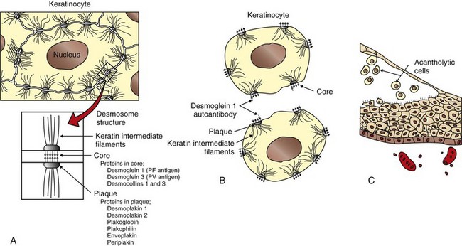
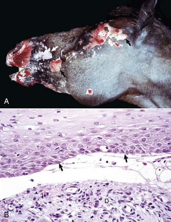
Acantholysis: loss of cohesion between keratinocytes caused by the breakdown of intercellular bridges.

Acanthosis: thickening of the spinous cell layer (stratum spinosum) of the epidermis.

Acral: distal parts of the extremities.

Alopecia: hair loss.

Anagen: phase of hair cycle in which hair synthesis takes place.

Anaplasia: lack of cellular differentiation and organization, a feature of neoplastic cells.

Angioedema: vascular reaction involving the deep dermis or subcutis and consisting of edema manifested as giant wheals and caused by dilation and increased permeability of capillaries (deeper version of urticaria).

Apoptosis: programmed cell death.

Atrophy: reduction in size of a cell, tissue, organ, or part.

Ballooning degeneration: marked intracellular fluid accumulation in the cells of the epidermis.

Blister (vesicle or bulla): localized collection of fluid usually in or beneath the epidermis.

Bulla: large blister (≥1.0 cm).

Carcinoma in situ: a malignant neoplasm of epithelial origin that has not invaded through the basement membrane.

Catagen: transition phase of the hair cycle between growth and resting phases.

Cellulitis: an acute bacterial infection of the dermis and subcutis that spreads to surrounding soft tissues and is characterized by erythema, warmth, swelling, and pain. The source of the infection is most often a penetrating wound in the area of infection. Cellulitis can also cause fever and enlarged lymph nodes.

Comedo (pl., comedones): plug of follicular stratum corneum and dried sebum in a hair follicle that leads to follicular distention.

Cornification: production of stratum corneum by terminal epidermal differentiation.

Crust: material formed by drying of exudate or secretion on the skin surface.

Cytokines: small molecular weight protein molecules (generally <30 kD) that are mediators of inflammation and growth.

Dematiaceous: naturally pigmented black or brown mycelium or conidium.

Dermatitis: inflammation of the skin.

Dermatophytosis: infection of the stratum corneum of the epidermis, hair, or claws with fungi of the genera Microsporum, Epidermophyton, or Trichophyton.

Dermatosis: noninflammatory lesion of the skin.

Dyskeratosis: abnormal, premature, or imperfect keratinization.

Dysplasia: abnormal development; term may be used in association with a congenital or inherited developmental anomaly or in association with an abnormality in maturation of cells within a tissue.

Effluvium: shedding of hair.

Elastosis: degeneration of dermal connective tissue leading to accumulation of elastotic fibers; sometimes seen with solar dermatitis.

Epidermal collarette: peripheral expanding ring of scale.

Epidermitis: inflammation of the epidermis.

Epidermolysis: separation of the epidermis from the dermis.

Epidermotropic/Epitheliotropic: having a predilection to enter the epidermis or other epithelial structures as seen with cutaneous T cell lymphoma (mycosis fungoides).

Erosion: loss of all or part of the thickness of the epidermis.

Eruption: rapid development of skin lesion associated with redness.

Erythema: redness of skin caused by congestion of capillaries.

Excoriation: superficial loss of epidermal layers caused by physical trauma (scratching).

Exfoliation: shedding of layers or scales.

Exogen: the stage of the hair cycle where old hairs are shed.

Exudate: fluid, cells, or debris from blood vessels deposited in or on other tissues.

Fissure: cleft or groove.

Folliculitis: inflammation of a hair follicle.

Furuncle: circumscribed, painful nodule (accumulation of pus) in the dermis secondary to follicular rupture.

Furunculosis: rupture of follicles usually caused by inflammation, distention, and/or trauma leading to entry of follicular contents into the dermis.

Genodermatosis: a genetically determined disorder of the skin.

Glabrous: smooth skin, hairless skin.

Hamartoma: a localized, tumor-like malformation of mature cells and tissues that includes normal components of the organ in which the hamartoma arises but that is disorganized, present in excess, and sometimes larger than normal. Usually, one tissue element predominates (e.g., follicular hamartoma, vascular hamartoma). A hamartoma is not a true neoplasm because it involves the proliferation of more than one cell type and often includes the development of complex structures such as arteries or follicles.

Hydropic degeneration: intracellular fluid accumulation in cells of the basal epidermis.

Hyperkeratosis: histologic term for thickening of stratum corneum.

Hyperplasia: increase in the number of normal cells.

Hypoplasia: incomplete development.

Hypotrichosis: less hair than normal.

Ichthyosis: congenital skin disorder in which the skin is thickened by scales (hyperkeratosis) that can crack into plates resembling fish scales.

Impetigo: bacterial dermatitis characterized by pustules.

Indolent: slow growing, a term applied to persistent ulcers on the lips of cats, and sometimes incorrectly called “rodent ulcer,” a term from the human literature used to refer to ulcerated basal cell carcinoma.

Indurated: Hardening of the skin as a result of inflammation or fibrosis.

Interface: inflammation arranged in a layer close to and often obscuring the epidermal-dermal junction (interface), and with vacuolated (hydropic degeneration) and sometimes apoptotic basal cells; the inflammation can be mild (cell poor) or extensive (cell rich).

Intertrigo: dermatitis that develops because of friction between apposing skin surfaces (e.g., adjacent folds).

Keratinocytes: the epidermal cells that synthesize keratin and comprise more than 90% of epidermal cells.

Keratosis (pl., keratoses): an uncommon to rare circumscribed papular, plaque-like, or linear focus of proliferative keratinocytes covered by thick stratum corneum; keratoses can be caused by sun exposure (solar or actinic keratoses) or can be idiopathic (lichenoid, linear, cannon [metatarsal bone] keratoses).

Kerion: an intense focal folliculitis usually caused by a dermatophyte infection.

Langerhans’ cells: intraepidermal dendritic antigen-presenting cells.

Lichenification: thickening of skin with accentuation of skin creases caused by marked acanthosis.

Lichenoid: confusing term that generally refers to a dense zone of dermal inflammation parallel to the epidermis usually without basal cell injury.

Lichenoid dermatosis (es): the conventional term for uncommon to rare, often idiopathic, single or grouped papules, plaques, or papillomatous foci covered by scale, and histologically composed of epidermal hyperplasia, lichenoid lymphoplasmacytic dermal inflammation, hyperkeratosis, and parakeratosis. The term dermatitis is probably better than dermatosis as inflammation is present in these lesions.

Macule: flat, circumscribed lesion of altered skin color.

Melanin: the dark granular pigment produced by melanocytes that is responsible for the brown coloration of hair, skin, and other tissues such as the iris and choroid of the eye.

Melanophage: macrophage containing ingested melanin.

Merkel cell: a neuroendocrine cell found in the stratum basale.

Mucin: glycosaminoglycan (GAG), a normal component of the intercellular ground substance of the dermis, consists of protein bound to hyaluronic acid.

Mycelium: a mass of hyphae.

Mycetoma: a slowly progressive infection of the cutaneous and subcutaneous tissue, fascia, and sometimes underlying bone caused by traumatic implantation of actinomycetes (actinomycotic mycetoma) or fungi (eumycotic mycetoma).

Myxedema: nonpitting edema of the skin because of abnormal deposits of mucin in the dermis.

Necrotizing fasciitis: an acute serious life-threatening subtype of cellulitis usually caused by streptococcal bacterial infection and toxin production, and located within the subcutaneous fat and fascial planes. The clinical lesions are painful, hot, and swollen areas with extensive exudation and necrosis. The condition can progress rapidly and result in systemic shock.

Nevus: circumscribed malformation of the skin assumed to be of congenital or inherited origin, and consisting of any component of the skin. The term “hamartoma” is preferred to nevus to avoid confusion with the pigmented nevus (mole) that arises in the skin of humans.

Nodule: a circumscribed, solid elevation of skin (≥1 cm).

Onychodystrophy: abnormal formation of the claw.

Onychomadesis: sloughing of claws.

Panniculitis: inflammation of subcutaneous adipose tissue.

Papule: circumscribed, solid elevation of skin (<1 cm).

Parakeratosis: retention of pyknotic nuclei in epidermal cells of the stratum corneum.

Paronychia: inflammation of skin around the claws.

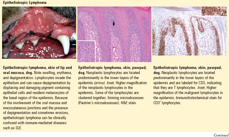
Pautrier’s microabscess: a localized intraepidermal collection of neoplastic lymphocytes characteristic of epitheliotropic lymphoma (mycosis fungoides).

Pemphigus: a group of cutaneous diseases associated with blistering.

Phaeohyphomycosis: mycotic disease caused by pigmented fungi (dematiaceous fungi) of a variety of genera and species that do not form sclerotic bodies or granules.

Pigmentary incontinence: melanin pigment within dermal macrophages or free in the dermis developing via injury to pigment containing basal layer cells.

Plaque: a flat-topped, solid elevation in the skin that occupies a relatively large surface area in comparison with its height (≥1 cm).

Pruritus: itching.

Pustule: small, circumscribed accumulation of pus within the epidermis or within a hair follicle.

Pyoderma: pyogenic (pus-producing) bacterial infection of the skin.

Rodent ulcer: a term used in human medicine to define an ulcerative basal cell carcinoma; sometimes used inappropriately in veterinary medicine to refer to an indolent ulcer affecting the lip of cats.

Scale: a thin, platelike accumulation of stratum corneum on the surface of skin.

Seborrhea: nonspecific term for clinical signs of scaling, crusting, and greasiness. Primary seborrhea is a more specific term applied to inherited cornification disorders.

Sebum: secretion of sebaceous glands.

Spongiosis: intercellular edema, which, by widening of the intercellular space and stretching of the “intercellular bridges,” creates a spongelike appearance to the epidermis.

Telogen: resting phase of the hair cycle.

Ulcer: loss of epidermis and at least the superficial portion of dermis.

Urticaria: usually transient vascular reaction in the upper dermis consisting of edema manifested clinically as wheals (hives); a more superficial version of angioedema.

Vesicle: small blister within the epidermis or at or below the dermal-epidermal interface (<1.0 cm) (see Fig. 17-17).

Vibrissa (pl., vibrissae): long, coarse hair located about the nose (sinus hair, tactile hair).

Vitiligo: acquired disorder characterized by circumscribed areas of depigmentation in the skin.

Wheal: smooth, circumscribed, slightly elevated area on skin caused by dermal edema.

Yeast: unicellular budding fungus.