SPLEEN

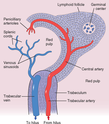
The spleen is an ingeniously designed filter for the blood and a site of immune responses to blood-borne antigens. Normally in the adult it weighs about 150 gm and is enclosed within a thin, glistening, slate-gray connective tissue capsule. Its cut surface reveals extensive red pulp dotted with gray specks, which are the white pulp follicles. These consist of an artery with an eccentric collar of T lymphocytes, the so-called periarteriolar lymphatic sheath. At intervals this sheath expands to form lymphoid nodules composed mainly of B lymphocytes, which are capable of developing into germinal centers identical to those seen in lymph nodes in response to antigenic stimulation (Fig. 13-39).

image

FIGURE 13-39 Normal splenic architecture.

(Modified from Faller DV: Diseases of the spleen. In Wyngaarden JB, Smith LH (eds): Cecil Textbook of Medicine, 18th ed. Philadelphia, WB Saunders, 1988, p. 1036.)

The red pulp of the spleen is traversed by numerous thin-walled vascular sinusoids, separated by the splenic cords or “cords of Billroth.” The endothelial lining of the sinusoid is discontinuous, providing a passage for blood cells between the sinusoids and cords. The cords contain a labyrinth of macrophages loosely connected through long dendritic processes to create both a physical and a functional filter. As it traverses the red pulp, the blood takes two routes to reach the splenic veins. Some flows through capillaries into the cords, from which blood cells squeeze through gaps in the discontinuous basement of the endothelial lining to reach the sinusoids; this is the so-called open circulation or slow compartment. In the other “closed circuit,” blood passes rapidly and directly from the capillaries to the splenic veins. Although only a small fraction of the blood pursues the “open” route, during the course of a day the entire blood volume passes through the cords, where it is closely examined by macrophages.

The spleen has four functions that impact disease states:

1. Phagocytosis of blood cells and particulate matter. As will be discussed under the hemolytic anemias (Chapter 14), red cells undergo extreme deformation during passage from the cords into the sinusoids. In conditions in which red cell elasticity is decreased, red cells become trapped in the cords and are more readily phagocytosed by macrophages. Splenic macrophages are also responsible for “pitting” of red cells, the process by which inclusions such as Heinz bodies and Howell-Jolly bodies are excised, and for the removal of particles, such as bacteria, from the blood.
2. Antibody production. Dendritic cells in the periarterial lymphatic sheath trap antigens and present them to T lymphocytes. T- and B-cell interaction at the edges of white pulp follicles leads to the generation of antibody-secreting plasma cells, which are found mainly within the sinuses of the red pulp. The spleen seems to be an important source of antibodies directed against platelets and red cells in immune thrombocytopenia purpura and immunohemolytic anemias, both discussed in Chapter 14.
3. Hematopoiesis. Splenic hematopoiesis normally ceases before birth, but can be reactivated in severe anemia. As we have seen, the spleen is also a prominent site of extramedullary hematopoiesis in myeloproliferative disorders, such as chronic myeloid leukemia.
4. Sequestration of formed blood elements. The normal spleen contains only about 30 to 40 mL of red cells, but this volume increases greatly with splenomegaly. The normal spleen also harbors approximately 30% to 40% of the total platelet mass in the body. With splenomegaly up to 80% to 90% of the total platelet mass can be sequestered in the interstices of the red pulp, producing thrombocytopenia. Similarly, the enlarged spleen can trap white cells and thereby induce leukopenia.

As the largest unit of the mononuclear phagocyte system, the spleen is involved in all systemic inflammations, generalized hematopoietic disorders, and many metabolic disturbances. In each, the spleen undergoes enlargement (splenomegaly), which is the major manifestation of disorders of this organ. It is rarely the primary site of disease. Splenic insufficiency due to splenectomy or autoinfarction (as in sickle-cell disease) has one major clinical manifestation, an increased susceptibility to sepsis cause by encapsulated bacteria such as pneumococcus, meningococcus, and Haemophilus influenzae. The loss of filtering and antibody production functions both contribute to the increased risk of sepsis, which may be fatal. All asplenic individuals should be vaccinated against these agents to reduce the risk of this tragic complication.

Splenomegaly

When sufficiently enlarged, the spleen causes a dragging sensation in the left upper quadrant and, through pressure on the stomach, discomfort after eating. In addition, enlargement can cause a syndrome known as hypersplenism, which is characterized by anemia, leukopenia, thrombocytopenia, alone or in combination. The probable cause of the cytopenias is increased sequestration of formed elements and the consequent enhanced phagocytosis by the splenic macrophages.

A list of the major disorders associated with splenomegaly is provided in Table 13-12. Splenomegaly in virtually all the conditions mentioned has been discussed elsewhere. There remain only a few disorders to consider.

TABLE 13-12 Disorders Associated with Splenomegaly

I. INFECTIONS
Nonspecific splenitis of various blood-borne infections (particularly infectious endocarditis)
Infectious mononucleosis
Tuberculosis
Typhoid fever
Brucellosis
Cytomegalovirus
Syphilis
Malaria
Histoplasmosis
Toxoplasmosis
Kala-azar
Trypanosomiasis
Schistosomiasis
Leishmaniasis
Echinococcosis
II. CONGESTIVE STATES RELATED TO PORTAL HYPERTENSION
Cirrhosis of the liver
Portal or splenic vein thrombosis
Cardiac failure
III. LYMPHOHEMATOGENOUS DISORDERS
Hodgkin lymphoma
Non-Hodgkin lymphomas and lymphocytic leukemias
Multiple myeloma
Myeloproliferative disorders
Hemolytic anemias
IV. IMMUNOLOGICAL-INFLAMMATORY CONDITIONS
Rheumatoid arthritis
Systemic lupus erythematosus
V. STORAGE DISEASES
Gaucher disease
Niemann-Pick disease
Mucopolysaccharidoses
VI. MISCELLANEOUS DISORDERS
Amyloidosis
Primary neoplasms and cysts
Secondary neoplasms

NONSPECIFIC ACUTE SPLENITIS

Enlargement of the spleen occurs in any blood-borne infection. The nonspecific splenic reaction in these infections is caused both by the microbiologic agents themselves and by cytokines that are released as part of the immune response.

Morphology. The spleen is enlarged (200–400 gm) and soft. Microscopically, the major feature is acute congestion of the red pulp, which may encroach on and virtually efface the lymphoid follicles. Neutrophils, plasma cells, and occasionally eosinophils are usually present throughout the white and red pulp. At times the white pulp follicles may undergo necrosis, particularly when the causative agent is a hemolytic streptococcus. Rarely, abscess formation occurs.

CONGESTIVE SPLENOMEGALY

Chronic venous outflow obstruction causes a form of splenic enlargement referred to as congestive splenomegaly. Venous obstruction may be caused by intrahepatic disorders that retard portal venous drainage, or arise from extrahepatic disorders that directly impinge upon the portal or splenic veins. All of these disorders ultimately lead to portal or splenic vein hypertension. Systemic, or central, venous congestion is encountered in cardiac decompensation involving the right side of the heart, as can occur in tricuspid or pulmonic valvular disease, chronic cor pulmonale, or following left-sided heart failure. Systemic congestion is associated with only moderately enlarged spleens that rarely exceed 500 gm in weight.

Cirrhosis of the liver is the main cause of massive congestive splenomegaly. The “pipe-stem” hepatic fibrosis of schistosomiasis causes particularly severe congestive splenomegaly, while the diffuse fibrous scarring of alcoholic cirrhosis and pigment cirrhosis also evokes profound enlargements. Other forms of cirrhosis are less commonly implicated.

Congestive splenomegaly is also caused by obstruction of the extrahepatic portal vein or splenic vein. This can stem from spontaneous portal vein thrombosis, which is usually associated with some intrahepatic obstructive disease, or inflammation of the portal vein (pylephlebitis), such as follows intraperitoneal infections. Thrombosis of the splenic vein can be caused by infiltrating tumors arising in neighboring organs, such as carcinomas of the stomach or pancreas.

Morphology. Long-standing splenic congestion produces marked enlargement (1000–5000 gm). The organ is firm, and the capsule is usually thickened and fibrous. Microscopically, the red pulp is congested early in the course but becomes increasingly fibrotic and cellular with time. The elevated portal venous pressure stimulates the deposition of collagen in the basement membrane of the sinusoids, which appear dilated because of the rigidity of their walls. The resultant slowing of blood flow from the cords to the sinusoids prolongs the exposure of the blood cells to macrophages, resulting in excessive destruction (hypersplenism).

SPLENIC INFARCTS

Splenic infarcts are common lesions caused by the occlusion of the major splenic artery or any of its branches. The spleen, along with kidneys and brain, ranks as one of the most frequent sites where emboli lodge. In normal-sized spleens, infarcts are most often caused by emboli that arise from the heart. The infarcts can be small or large, single or multiple, or even involve the entire organ. They are usually bland, except in individuals with infectious endocarditis of the mitral or aortic valves, in whom septic infarcts are common. Infarcts are also common in markedly enlarged spleens, regardless of cause, presumably because the blood supply is tenuous and easily compromised.

Morphology. Bland infarcts are characteristically pale, wedge-shaped, and subcapsular in location. The overlying capsule is often covered with fibrin (Fig. 13-40). In septic infarcts this appearance is modified by the development of suppurative necrosis. In the course of healing, large depressed scars often develop.

Neoplasms

Neoplastic involvement of the spleen is rare except in myeloid and lymphoid tumors, which (as already discussed) often cause splenomegaly. Benign fibromas, osteomas, chondromas, lymphangiomas, and hemangiomas may arise in the spleen. Of these, lymphangiomas and hemangiomas are most common and often cavernous in type.

Congenital Anomalies

Complete absence of the spleen is rare and is usually associated with other congenital abnormalities, such as situs inversus and cardiac malformations. Hypoplasia is a more common finding.

Accessory spleens (spleniculi) are common, being present singly or multiply in 20% to 35% of postmortem examinations. They are small, spherical structures that are histologically and functionally identical to the normal spleen. They can be found at any place within the abdominal cavity. Accessory spleens are of great clinical importance in some hematologic disorders, such as hereditary spherocytosis and immune thrombocytopenia purpura, where splenectomy is used as a treatment. If an accessory spleen is overlooked, the therapeutic benefit of removal of the definitive spleen may be reduced or lost entirely.

Rupture

Splenic rupture is usually precipitated by blunt trauma. Much less often, it occurs in the apparent absence of a physical blow. Such “spontaneous ruptures” never involve truly normal spleens but rather stem from some minor physical insult to a spleen made fragile by an underlying condition. The most common predisposing conditions are infectious mononucleosis, malaria, typhoid fever, and lymphoid neoplasms. They cause the spleen to enlarge rapidly, producing a thin, tense capsule that is susceptible to rupture. This dramatic event often precipitates intraperitoneal hemorrhage, which must be treated by prompt splenectomy to prevent death from blood loss. Chronically enlarged spleens are unlikely to rupture because of the toughening effect of extensive reactive fibrosis.

THYMUS

Once an organ buried in obscurity, the thymus has risen to a starring role in cell-mediated immunity (as detailed in Chapter 6). Here, our interest centers on the disorders of the gland itself.

The thymus is embryologically derived from the third and, inconstantly, the fourth pair of pharyngeal pouches. At birth it weighs 10 to 35 gm. It grows until puberty, when it achieves a maximum weight of 20 to 50 gm, and thereafter undergoes progressive involution to little more than 5 to 15 gm in the elderly. The thymus can also involute in children and young adults in response to severe illness and HIV infection.

The fully developed thymus is composed of two fused, well-encapsulated lobes. Fibrous extensions of the capsule divide each lobe into numerous lobules, each with an outer cortical layer enclosing the central medulla. Diverse types of cells populate the thymus, but thymic epithelial cells and immature T lymphocytes predominate. The cortical, peripheral, epithelial cells are polygonal in shape and have an abundant cytoplasm with dendritic extensions that contact adjacent cells. In contrast, the epithelial cells in the medulla are densely packed, often spindle-shaped, and have scant cytoplasm devoid of interconnecting processes. Whorls of medullary epithelial cells create Hassall corpuscles, with their characteristic keratinized cores.

As you know from the earlier consideration of the thymus in relation to immunity, progenitor cells of marrow origin migrate to the thymus and mature into T cells, which are exported to the periphery, but only after they have been educated in the “thymic university” to distinguish between self and non-self antigens. During adulthood the thymic production of T cells slowly declines as the organ atrophies.

Macrophages, dendritic cells, a minor population of B lymphocytes, rare neutrophils and eosinophils, and scattered myoid (muscle-like) cells are also found within the thymus. The myoid cells are of particular interest because of the suspicion that they play some role in the development of myasthenia gravis, a musculoskeletal disorder of immune origin.

Pathologic changes within the thymus are limited and will be described here. The changes associated with myasthenia gravis are considered in Chapter 27.

Developmental Disorders

Thymic hypoplasia or aplasia is seen in DiGeorge syndrome, which is marked by severe defects in cell-mediated immunity and variable abnormalities of parathyroid development associated with hypoparathyroidism. As discussed in Chapter 5, DiGeorge syndrome is often associated with other developmental defects as part of the 22q11 deletion syndrome.

Isolated thymic cysts are uncommon lesions that are usually discovered incidentally postmortem or during surgery. They rarely exceed 4 cm in diameter, can be spherical or arborizing, and are lined by stratified to columnar epithelium. The fluid contents can be serous or mucinous and are often modified by hemorrhage.

While isolated cysts are not clinically significant, neoplastic thymic masses (whatever their origin) compress and distort adjacent normal thymus and sometimes cause cysts to form. Therefore, the presence of a cystic thymic lesion in a symptomatic patient should provoke a thorough search for a neoplasm, particularly a lymphoma or a thymoma.

Thymic Hyperplasia

The term thymic hyperplasia is a bit misleading, since it usually applies to the appearance of B-cell germinal centers within the thymus, a finding that is referred to as thymic follicular hyperplasia. Such B-cell follicles are present in only small numbers in the normal thymus. Although follicular hyperplasia can occur in a number of chronic inflammatory and immunological states, it is most frequently encountered in myasthenia gravis, in which it is found in 65% to 75% of cases (see Chapter 27). Similar thymic changes are sometimes encountered in Graves’ disease, systemic lupus erythematosus, scleroderma, rheumatoid arthritis, and other autoimmune disorders. In other instances, a morphologically normal thymus is simply large for the age of the patient. As mentioned, the size of the thymus varies widely, and whether this constitutes a true hyperplasia or is merely a variant of normal is unclear. The main significance of this form of thymic “hyperplasia” is that it may be mistaken radiologically for a thymoma, leading to unnecessary surgical procedures.

Thymomas

A diversity of neoplasms may arise in the thymus—germ cell tumors, lymphomas, carcinoids, and others—but the designation “thymoma” is restricted to tumors of thymic epithelial cells. Such tumors typically also contain benign immature T cells (thymocytes).

The WHO has created a classification system based on histology for thymomas, but its clinical utility remains uncertain. We will instead use a classification that relies on the most important prognostic features, the surgical stage and the presence or absence of overt cytologic features of malignancy. In this simple system there are only three histologic subtypes:

Tumors that are cytologically benign and noninvasive
Tumors that are cytologically benign but invasive or metastatic
Tumors that are cytologically malignant (thymic carcinoma)

In all categories, the tumors usually occur in adults older than 40 years of age; thymomas are rare in children. Males and females are affected equally. Most arise in the anterior superior mediastinum, but sometimes they occur in the neck, thyroid, pulmonary hilus, or elsewhere. They are uncommon in the posterior mediastinum. Thymomas account for 20% to 30% of tumors in the anterosuperior mediastinum, which is also a common location for certain lymphomas.

Morphology. Macroscopically, thymomas are lobulated, firm, gray-white masses of up to 15 to 20 cm in size. They sometimes have areas of cystic necrosis and calcification. Most are encapsulated, but 20% to 25% of the tumors penetrate the capsule and infiltrate perithymic tissues and structures.

Noninvasive thymomas are most often composed of medullary-type epithelial cells or a mixture of medullary- and cortical-type epithelial cells. The medullary-type epithelial cells are elongated or spindle-shaped (Fig. 13-41A). There is usually a sparse infiltrate of thymocytes, which often recapitulate the phenotype of medullary thymocytes. In mixed thymomas there is an admixture of polygonal cortical-type epithelial cells and a denser infiltrate of thymocytes. The medullary and mixed patterns together account for about 50% of all thymomas. Tumors that have a substantial proportion of medullary-type epithelial cells are usually noninvasive.

image

FIGURE 13-41 Thymoma. A, Benign thymoma (medullary type). The neoplastic epithelial cells are arranged in a swirling pattern and have bland, oval to elongated nuclei with inconspicuous nucleoli. Only a few small, reactive lymphoid cells are interspersed. B, Malignant thymoma, type I. The neoplastic epithelial cells are polygonal and have round to oval, bland nuclei with inconspicuous nucleoli. Numerous small, reactive lymphoid cells are interspersed. The morphologic appearance of this tumor is identical to that of benign thymomas of the cortical type. In this case, however, the tumor was locally aggressive, invading adjacent lung and pericardium.

Invasive thymoma refers to a tumor that is cytologically benign but locally invasive. These tumors are much more likely to metastasize. The epithelial cells are most commonly of the cortical variety, with abundant cytoplasm and rounded vesicular nuclei (Fig. 13-41B), and are usually mixed with numerous thymocytes. In some cases, the neoplastic cells show cytologic atypia, a feature that correlates with a propensity for more aggressive behavior. These tumors account for about 20% to 25% of all thymomas. By definition, invasive thymomas penetrate through the capsule into surrounding structures. The extent of invasion has been subdivided into various stages, which are beyond our scope. With minimal invasion, complete excision yields a 5-year survival rate of greater than 90%, whereas extensive invasion is associated with a 5-year survival rate of less than 50%.

Thymic carcinoma represents about 5% of thymomas. Macroscopically, they are usually fleshy, obviously invasive masses, sometimes accompanied by metastases to sites such as the lungs. Microscopically, most are squamous cell carcinomas. The next most common variant is lymphoepithelioma-like carcinoma, a tumor composed of sheets of cells with indistinct borders that bears a close histologic resemblance to nasopharyngeal carcinoma. About 50% of lymphoepithelioma-like carcinomas contain monoclonal EBV genomes, consistent with a role for EBV in their pathogenesis. A variety of other less common histologic patterns of thymic carcinoma have been described; all exhibit cytologic atypia seen in other carcinomas.

Clinical Features.

About 40% of thymomas present with symptoms stemming from impingement on mediastinal structures. Another 30% to 45% are detected in the course of evaluating patients with myasthenia gravis. The rest are discovered incidentally during imaging studies or cardiothoracic surgery. In addition to myasthenia gravis, other associated autoimmune disorders include hypogammaglobulinemia, pure red cell aplasia, Graves’ disease, pernicious anemia, dermatomyositis-polymyositis, and Cushing syndrome. The basis for these associations is still obscure, but the thymocytes that arise within thymomas give rise to long-lived CD4+ and CD8+ T cells, and cortical thymomas rich in thymocytes are more likely to be associated with autoimmune disease. Hence, it seems likely that abnormalities in the selection or “education” of T cells maturing within the environment of the neoplasm contribute to the development of diverse autoimmune disorders.

REFERENCES

1 Dzierzak E, Speck NA. Of lineage and legacy: the development of mammalian hematopoietic stem cells. Nat Immunol. 2008;9:129.

2 Kiel MJ, Morrison SJ. Uncertainty in the niches that maintain haematopoietic stem cells. Nat Rev Immunol. 2008;8:290.

3 O’Malley DP. T-cell large granular leukemia and related proliferations. Am J Clin Pathol. 2007;127:850.

4 Mebius RE. Organogenesis of lymphoid tissues. Nat Rev Immunol. 2003;3:292.

5 Sagaert X, et al. The pathogenesis of MALT lymphomas: where do we stand? Leukemia. 2007;21:389.

6 Jost PJ, Ruland J. Aberrant NF-kappaB signaling in lymphoma: mechanisms, consequences, and therapeutic implications. Blood. 2007;109:2700.

7 Polo JM, Melnick A. B-cell lymphoma 6 and the molecular pathogenesis of diffuse large B-cell lymphoma. Curr Opin Hematol. 2008;15:381.

8 Dorsett Y, et al. A role for AID in chromosome translocations between c-myc and the IgH variable region. J Exp Med. 2007;204:2225.

9 Ramiro AR, et al. Role of genomic instability and p53 in AID-induced c-myc-Igh translocations. Nature. 2006;440:105.

10 Pasqualucci L, et al. Hypermutation of multiple proto-oncogenes in B-cell diffuse large-cell lymphomas. Nature. 2001;412:341.

11 Harris NL, et al. World Health Organization classification of neoplastic diseases of the hematopoietic and lymphoid tissues: report of the Clinical Advisory Committee meeting–Airlie House, Virginia, November 1997. J Clin Oncol. 1999;17:3835.

12 Szczepanski T. Why and how to quantify minimal residual disease in acute lymphoblastic leukemia? Leukemia. 2007;21:622.

13 Nabhan C, et al. Minimal residual disease in chronic lymphocytic leukaemia: is it ready for primetime? Br J Haematol. 2007;136:379.

14 Aster JC, et al. Notch signaling in leukemia. Annu Rev Pathol. 2008;3:587.

15 Mullighan CG, et al. Genome-wide analysis of genetic alterations in acute lymphoblastic leukaemia. Nature. 2007;446:758.

16 Greaves MF, et al. Leukemia in twins: lessons in natural history. Blood. 2003;102:2321.

17 Schultz KR, et al. Risk- and response-based classification of childhood B-precursor acute lymphoblastic leukemia: a combined analysis of prognostic markers from the Pediatric Oncology Group (POG) and Children’s Cancer Group (CCG). Blood. 2007;109:926.

18 Pfeifer H, et al. Kinase domain mutations of BCR-ABL frequently precede imatinib-based therapy and give rise to relapse in patients with de novo Philadelphia-positive acute lymphoblastic leukemia. Blood. 2007;110:727.

19 Calin GA, Croce CM. Genomics of chronic lymphocytic leukemia microRNAs as new players with clinical significance. Semin Oncol. 2006;33:167.

20 Bouley J, et al. New molecular markers in resistant B CLL. Leuk Lymphoma. 2006;47:791.

21 Endo T, et al. BAFF and APRIL support chronic lymphocytic leukemia B-cell survival through activation of the canonical NF-kappaB pathway. Blood. 2007;109:703.

22 Alinari L, et al. Alemtuzumab (Campath-1H) in the treatment of chronic lymphocytic leukemia. Oncogene. 2007;26:3644.

23 Tsimberidou AM, Keating MJ. Richter’s transformation in chronic lymphocytic leukemia. Semin Oncol. 2006;33:250.

24 Dave SS, et al. Prediction of survival in follicular lymphoma based on molecular features of tumor-infiltrating immune cells. N Engl J Med. 2004;351:2159-2169.

25 Kuppers R. Prognosis in follicular lymphoma—it’s in the microenvironment. N Engl J Med. 2004;351:2152.

26 Staudt LM, Dave S. The biology of human lymphoid malignancies revealed by gene expression profiling. Adv Immunol. 2005;87:163.

27 Abramson JS, Shipp MA. Advances in the biology and therapy of diffuse large B-cell lymphoma: moving toward a molecularly targeted approach. Blood. 2005;106:1164.

28 Parekh S, et al. BCL6 programs lymphoma cells for survival and differentiation through distinct biochemical mechanisms. Blood. 2007;10:2067.

29 Phan RT, et al. BCL6 interacts with the transcription factor Miz-1 to suppress the cyclin-dependent kinase inhibitor p21 and cell cycle arrest in germinal center B cells. Nat Immunol. 2005;6:1054.

30 Phan RT, Dalla-Favera R. The BCL6 proto-oncogene suppresses p53 expression in germinal-centre B cells. Nature. 2004;432:635.

31 Hemann MT, et al. Evasion of the p53 tumour surveillance network by tumour-derived MYC mutants. Nature. 2005;436:807.

32 Dave SS, et al. Molecular diagnosis of Burkitt’s lymphoma. N Engl J Med. 2006;354:2431.

33 Matsui W, et al. Characterization of clonogenic multiple myeloma cells. Blood. 2004;103:2332.

34 Peacock CD, et al. Hedgehog signaling maintains a tumor stem cell compartment in multiple myeloma. Proc Natl Acad Sci U S A. 2007;104:4048.

35 Anderson KC. Targeted therapy of multiple myeloma based upon tumor-microenvironmental interactions. Exp Hematol. 2007;35:155.

36 Terpos E, et al. Significance of macrophage inflammatory protein-1 alpha (MIP-1alpha) in multiple myeloma. Leuk Lymphoma. 2005;46:1699.

37 Hjertner O, et al. Bone disease in multiple myeloma. Med Oncol. 2006;23:431.

38 Bergsagel PL, Kuehl WM. Molecular pathogenesis and a consequent classification of multiple myeloma. J Clin Oncol. 2005;23:6333.

39 Terpos E, et al. Clinical implications of chromosomal abnormalities in multiple myeloma. Leuk Lymphoma. 2006;47:803.

40 Zhan F, et al. The molecular classification of multiple myeloma. Blood. 2006;108:2020.

41 Stewart AK, Fonseca R. Prognostic and therapeutic significance of myeloma genetics and gene expression profiling. J Clin Oncol. 2005;23:6339.

42 Rajkumar SV, et al. Proteasome inhibition as a novel therapeutic target in human cancer. J Clin Oncol. 2005;23:630.

43 Terpos E, et al. The effect of novel anti-myeloma agents on bone metabolism of patients with multiple myeloma. Leukemia. 2007.

44 Kyle RA, et al. Clinical course and prognosis of smoldering (asymptomatic) multiple myeloma. N Engl J Med. 2007;356:2582.

45 Kyle RA, et al. Prevalence of monoclonal gammopathy of undetermined significance. N Engl J Med. 2006;354:1362.

46 Kyle RA, et al. A long-term study of prognosis in monoclonal gammopathy of undetermined significance. N Engl J Med. 2002;346:564.

47 Bergsagel PL, Kuehl WM. Chromosome translocations in multiple myeloma. Oncogene. 2001;20:5611.

48 Chiarle R, et al. The anaplastic lymphoma kinase in the pathogenesis of cancer. Nat Rev Cancer. 2008;8:11.

49 Sun SC, Yamaoka S. Activation of NF-kappaB by HTLV-I and implications for cell transformation. Oncogene. 2005;24:5952.

50 Schmitz R, et al. Pathogenesis of classical and lymphocyte-predominant Hodgkin lymphoma. Annu Rev Pathol Mech Dis. 2009;4:151.

51 Kuppers R, Brauninger A. Reprogramming of the tumour B-cell phenotype in Hodgkin lymphoma. Trends Immunol. 2006;27:203.

52 Jungnickel B, et al. Clonal deleterious mutations in the IkappaBalpha gene in the malignant cells in Hodgkin’s lymphoma. J Exp Med. 2000;191:395.

53 Re D, et al. Molecular pathogenesis of Hodgkin’s lymphoma. J Clin Oncol. 2005;23:6379.

54 Joos S, et al. Classical Hodgkin lymphoma is characterized by recurrent copy number gains of the short arm of chromosome 2. Blood. 2002;99:1381.

55 de Bruijn MF, Speck NA. Core-binding factors in hematopoiesis and immune function. Oncogene. 2004;23:4238.

56 Scaglioni PP, Pandolfi PP. The theory of APL revisited. Curr Top Microbiol Immunol. 2007;313:85.

57 Kelly LM, et al. PML/RARalpha and FLT3-ITD induce an APL-like disease in a mouse model. Proc Natl Acad Sci U S A. 2002;99:8283.

58 Falini B, et al. Acute myeloid leukemia carrying cytoplasmic/mutated nucleophosmin (NPMc+ AML): biologic and clinical features. Blood. 2007;109:874.

59 Paschka P, et al. Adverse prognostic significance of KIT mutations in adult acute myeloid leukemia with inv(16) and t(8;21): a Cancer and Leukemia Group B Study. J Clin Oncol. 2006;24:3904.

60 Corey SJ, et al. Myelodysplastic syndromes: the complexity of stem-cell diseases. Nat Rev Cancer. 2007;7:118.

61 Campbell PJ, Green AR. The myeloproliferative disorders. N Engl J Med. 2006;355:2452.

62 Levine RL, et al. Role of JAK2 in the pathogenesis and therapy of myeloproliferative disorders. Nat Rev Cancer. 2007;7:673.

63 Goldman JM, Melo JV. Chronic myeloid leukemia—advances in biology and new approaches to treatment. N Engl J Med. 2003;349:1451.

64 Mullighan CG, et al. BCR-ABL1 lymphoblastic leukaemia is characterized by the deletion of Ikaros. Nature. 2008;453:110.

65 Druker BJ, et al. Five-year follow-up of patients receiving imatinib for chronic myeloid leukemia. N Engl J Med. 2006;355:2408.

66 Melo JV, Barnes DJ. Chronic myeloid leukaemia as a model of disease evolution in human cancer. Nat Rev Cancer. 2007;7:441.

67 Vannucchi AM, et al. Clinical profile of homozygous JAK2 617V > F mutation in patients with polycythemia vera or essential thrombocythemia. Blood. 2007;110:840.

68 Patel RK, et al. Prevalence of the activating JAK2 tyrosine kinase mutation V617F in the Budd-Chiari syndrome. Gastroenterology. 2006;130:2031.

69 Jelinek J, et al. JAK2 mutation 1849G > T is rare in acute leukemias but can be found in CMML, Philadelphia chromosome-negative CML, and megakaryocytic leukemia. Blood. 2005;106:3370.

70 Annels NE, et al. Aberrant chemokine receptor expression and chemokine production by Langerhans cells underlies the pathogenesis of Langerhans cell histiocytosis. J Exp Med. 2003;197:1385.

71 Fleming MD, et al. Coincident expression of the chemokine receptors CCR6 and CCR7 by pathologic Langerhans cells in Langerhans cell histiocytosis. Blood. 2003;101:2473.