Underlying molecular causes of arrhythmias have been reviewed in humans.1 Arrhythmias result from a combination of factors, including genetic predisposition, external stressors and subsequent myocardial remodeling, as well as iatrogenic contributions. Ischemia, electrolyte imbalances, activation of the RAAS and other systems, and pharmacologic therapy are among the triggers for arrhythmias.1 Inherited causes of arrhythmias are rare, but generally pathophysiology is straightforward, particularly if associated with mutations in ion channel genes. In contrast, arrhythmias associated with acquired heart disease are complex. Structural and electrical remodeling, hemodynamic changes, and neuroendocrine signals each influence ion channel function, intracellular calcium response, and intercellular communication and matrix composition.
Regardless of the source, all cardiac arrhythmias arise from two primary abnormalities: impulse initiation, which includes spontaneous automaticity and triggered activity, and impulse propagation—that is, conduction (generally manifested as reentrant arrhythmias). Impulse initiation establishes heart rate and is determined primarily by the rate of diastolic depolarization—that is, the slope of phase 4. In the normal heart the slope (heart rate) is autonomically controlled, being decreased by acetylcholine released from parasympathetic nerves and increased by norepinephrine released from the adrenal cortex. Stroke volume decrementally decreases as heart rate increases; accordingly, cardiac output may ultimately decrease with faster rates. The slope of phase 4 can be affected by a number of abnormal conditions. Enhanced automaticity occurs when the rate of spontaneous diastolic depolarization increases sufficiently to allow emergence of pathologically slowed or increased rates (e.g., sinus tachycardia). Ectopic foci (pacemakers that normally are latent) may emerge and may cause tachycardia if the frequency exceeds that of the sinoatrial node. Arrhythmias of initiation may also be triggered by an abnormal depolarization (phase 0), resulting in secondary upstrokes in the action potential. Two types of triggered arrhythmias occur. Delayed after-depolarization (DAD) occurs after a normal action potential and is followed by an overload of intracellular calcium. Examples include arrhythmias associated with myocardial failure, myocardial ischemia, adrenergic stress, and digoxin toxicity. Early after-depolarization (EAD) upstrokes occur during phase 3 repolarization and follow abnormally long cardiac action potentials. They generally result from abnormal inward sodium or calcium channel currents or exchange pumps and are associated with very slow heart rates or low extracellular K+ or in association with drugs that cause prolonged action potential duration.
KEY POINT 14-25
Arrhythmias reflect altered impulse initiation (automaticity) or impulse propagation (conduction).
Drugs that decrease automaticity do so through several mechanisms. Some decrease the rate (slope) of phase 4 spontaneous depolarization, suppressing the ectopic focus such that the sinoatrial node is allowed to resume its dominance. Automaticity might also be reduced by lengthening the time needed to attain threshold potential by increasing either the excitation threshold (more positive; e.g., Na+ or Ca2+ channel blockers) or the diastolic membrane potential (i.e., hyperpolarization, or more negative). Finally, spontaneous discharge might be reduced by prolonging the action potential duration, such as might occur with prolonged effective or absolute refractory periods.5 Triggered automaticity can be impaired by inhibiting the development of after-depolarizations or interfering with inward sodium or calcium channels. Shortening (rather than prolongation) of the action potential duration will inhibit EADs. Although the mechanism is not well known, magnesium will also inhibit EADs.5
KEY POINT 14-26
Automaticy can be reduced by prolonging the effective refractory period or increasing the time it takes for the membrane potential to reach threshold (phase 4).
Arrhythmias associated with conduction abnormalities are exemplified by reentrant arrhythmias. Reentrant arrhythmias may be either anatomic or functional. Functional reentrant arrhythmias are exemplified by pathologies such as ischemia that markedly slow conduction. Anatomic arrhythmias involve two or more pathways that travel to the same region of the heart but differ in electrophysiology (e.g., refractory period or conduction speed). Emergence of reentrant arrhythmias reflects the unique histologic make up of myocardial muscle. The geometric connection of myocardial cells to one another facilitates rapid movement of impulses throughout the myocardium such that the heart acts as a single large cell (Figure 14-8). One region might receive signals from several different pathways. Coordination of impulse conduction ensures that myocardial fibers are excited in a sequence that maximizes pump efficiency. Normally, retransmission of an impulse received in a specific region from an alternative (i.e., second) direction is prevented because either the signals arrive simultaneously, thus canceling one another, or the fiber remains refractory from the first impulse and cannot respond to the second. However, if the refractory period is abnormally short, or conduction of the reentering, alternative impulse is markedly slowed, the fiber will no longer be refractory from the first impulse. The impulse can be reinitiated and travels in the opposite direction, until it returns to the region again, generating a (circus) reentrant arrhythmia (see Figure 14-8). Altered membrane channel and ion movements with injured cardiac tissue channels slow movement through fast Na+ channels (phase 0) and slow Ca2+ channels. Conduction is accordingly slowed, potentially generating reentrant arrhythmias.
Figure 14-8 In the normal myocardium, electrical impulses travel down one or more paths of a bifurcated myocardial cell. Bifurcations allow coordination of contractions in all directions such that myocardial emptying is efficient and complete. Several mechanisms exist to ensure that impulses travel only in the proper direction. Those impulses that travel in opposite directions will be canceled by one another, precluding their transmission in the wrong direction. In the presence of a unidirectional block (i.e., resulting from damaged myocardium), transmission of one of the impulses will be prevented. However, although the impulse that normally would be canceled is now able to pass through the damaged tissue in the other direction, it will meet tissue that remains refractory from the first impulse. Consequently, it will not be transmitted any further. However, if conduction of the second unimpeded impulse is slowed, the myocardium will no longer be refractory when the impulse passes through the unidirectional block. Therefore the myocardium is receptive to the electrical impulse of this misdirected impulse, and the signal will be transmitted. Because of the combined effects of the unidirectional block and slowed conduction, the impulse may be perpetuated, resulting in a circus or reentrant arrhythmia. Drugs can reduce the arrhythmia by causing a bidirectional block, which prevents the second impulse from traveling through the damaged site, or by increasing the rate of conduction of the second impulse such that it reaches myocardial tissue while it is still in its refractory state from the first impulse.
Clinically relevant antiarrhythmics act to prevent reentrant arrhythmias by blocking specific ion channels or by targeting autonomic function, thus altering initiation, or conduction or action potential duration (thus refractory periods) of cardiac fibers. Initiation and thus automaticity can be suppressed through blockade of ion channels or drugs that facilitate adenosine or acetylcholine (thus increasing the maximum diastolic or resting potential) or antagonize adrenergic receptors (thus decreasing the slope of phase 4). Acceleration of conduction or prolongation of the ERP or APD causes the second impulse to reach tissue still refractory from the first impulse. Examples include Ca2+ channel blockers, β-adrenergic receptor antagonists, and digitalis glycosides. Slowed conduction caused by these drugs may increase the risk of functional reentrant arrhythmias. However, because reentrant arrhythmias respond best to drugs that prolong the refractory period, such drugs nonetheless remain effective for treatment of reentrant arrhythmias if the effective refractory period is prolonged. Examples of drugs that prolong ERP or APD might include drugs that delay recovery of sodium channels (particularly those that target inactivated channels) or potassium channel blockade (the delayed rectifier channels is particularly amenable to drug blockade), or drugs that prolong the APD. However, potassium channel blockade is also particularly conducive to causing arrhythmias, perhaps in part because of the importance of potassium channels in multiple phases of the action potential cycle.
In general, the complex action of antiarrhythmic drugs renders antiarrhythmic therapy generally inefficient and risky. All antiarrhythmic drugs are proarrhythmogenic. A major contributing factor to both inefficiency and risk is lack of knowledge regarding the particular electrophysiologic mechanism that underlies each arrhythmia or the drug. For example, the number of potassium channels, their differences in location, control, and role in the action potential complicate understanding, let alone predicting the impact of potassium channel blockade on cardiac function or rhythm. Increasingly, the arrhythmogenicity of drugs is likely to be understood as the differences in potassium channel impact by these drugs are understood. For example, blockade of delayed rectifier potassium current (Ik5) in the presence of high (β) adrenergic output predictably causes torsades de pointes in dogs.110a
Differences in clinical actions of antiarrhythmic drugs reflect in part different affinities of ion channel–blocking drugs for target receptors on the ion channel proteins, with some drugs targeting specific receptor subtypes. Affinity may change with conformation of the protein channel. As such, affinities are often state dependent (i.e., occurring only in the open, conducting state; in the closed, resting state; or during the inactivated, recovering state). Further, cardiac drugs often target more than one channel, and changes induced by a drug in one current generally influences the other currents. Direct and indirect (through the autonomic system) effects on cardiac contractility contribute to adverse effects.5 Finally, most antiarrhythmic drug target channels in both normal and abnormal tissues. Electrolyte abnormalities and hypokalemia in particular often predispose arrhythmogenicity and may increase adverse effects to antiarrhythmics.109,110 Indeed, a randomized human clinical trial that focused on the use of antiarrhythmic drugs for prevention of sudden death found that sudden death actually increased, primarily because of the proarrhythmic effects of drugs that target ion channels.111 Accordingly, antiarrhythmics should be considered dangerous,109,111 and their use should be pursued, whenever possible, under the guidance of clinicians with appropriate expertise (i.e., cardiologists).
KEY POINT 14-27
Reentrant arrhythmias can be decreased by increasing the rate of conduction or prolonging the effective refractory period.
Antiarrhythmic cardiac drugs fall into four main classes according to their dominant electrophysiologic effect on myocardial cells5,112 (Figure 14-9). Although this classification serves to couple electrophysiologic actions with antiarrhythmic effects, increasing emergence of the complex mechanisms of actions of these drugs complicates the classification, and care should be taken not to assume that drugs within classes behave the same way; indeed, most drugs have multiple effects that cross into multiple classes.
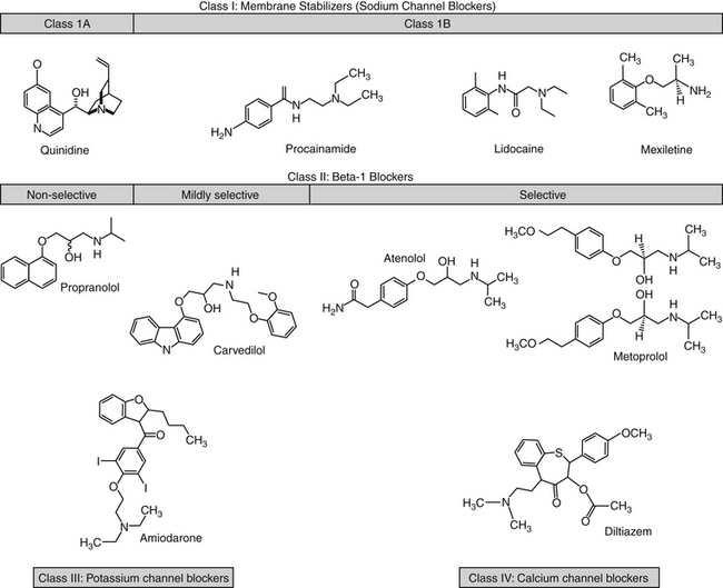
Figure 14-9 Structures of selected antiarrhythmic drugs. The drugs are classified by their mechanism of action, although much overlap occurs, particularly with potassium channel effects. Metoprolol exemplifies the chiral carbon present on most β blockers (some have two). Rotation of the four groups around the carbon yields enantiomers (stereoisomers that are non-superimposablemirror images). Although similar in structure, the enantiomers are likely to be different in both pharmacokinetic and pharmacodynamic properties. Most products are sold as racemic mixtures of both isomers, although only one may be active. The body is likely to handle the isomers differently. Class 1C drugs are not shown.
Class I agents comprise the standard membrane-stabilizing drugs such as lidocaine, quinidine, and procainamide. These agents work by selectively blocking the fast Na+ channels and depressing phase 0 of the action potential through the direct membrane-stabilizing or “local anesthetic” effect. Accordingly, class I drugs increase the threshold of excitability and decrease the rate of spontaneous phase 4 depolarization, thus reducing the emergence of ectopic foci (decreased automaticity). Although decreased phase 0 depolarization also decreases conduction velocity (thus prolonging conduction and increasing the risk of re-entrant arrhythmias), effects on automaticity and generation of ectopic foci appear to predominate over effects on conduction velocity. Some class I drugs also prolong action potential duration, especially the effective refractory period, and as such are particularly useful in treating reentrant arrhythmias.113 Class I agents can be further subdivided according to their effects on the refractory period and the rate of repolarization. Class 1A are intermediate blockers, including quinidine and procainamide. Class 1 B are rapid blockers, including lidocaine and mexiletine. Class 1C blockers are characterized by slow blocking kinetics and include flecainide, propafenone, and moricizine.
The risk of proarrhythmogenicity of antiarrhythmics is increased in dogs and cats by virtue of the dearth of scientifically based data supporting their use. However, regarding the data that do exist, when comparisons are made between dogs and humans, the concentrations at which antiarrhythmic effects of class I drugs are realized generally are similar. However, marked differences in pharmacokinetics exist; indeed, an exception to the generalization occurs for procainamide because of differences in its metabolism between dogs and humans.114
KEY POINT 14-28
All antiarrhythmics are proarrhythmic, and their use should be initiated cautiously and only if proper monitoring tools are available.
KEY POINT 14-30
Blockade of sodium channels might be expected to affect nondepolarizing cells more than depolarizing cells.
KEY POINT 14-31
The anticholinergic effects of quinidine –which initially result in an increased heart rate –contribute to its efficacy in the treatment of atrial fibrillation.
Class 1 A drugs increase the threshold for excitability and decrease automaticity, reduce the rate of phase 0 depolarization and thus slow conduction, prolong both the effective refractory period and the action potential duration, and delay repolarization. A shared arrythmogenic effect of sodium channel blockers with atrial flutter reflects slowed conduction. Subsequent slowing of atrial flutter may allow more signals to be transmitted through the atrioventricular node, causing the heart rate to increase.
Quinidine, derived from the bark of the cinchona plant, is a diastereomer of the antimalarial drug quinine. Quindine affects most types of cardiac muscles.5,112 Its efficacy against supraventricular and ventricular arrhythmias facilitates its classification as a broad-spectrum antiarrhythmic. Quinidine has both direct and indirect effects. For direct effects, quinidine blocks open Na+ channels, increasing the threshold for excitability and decreasing automaticity, thus suppressing ectopic pacemakers. Blockade of multiple K+ currents prolongs action potentials, particularly at slow heart rates. Electrophysiologically, quinidine increases the QRS complex; the QT interval may also be prolonged, causing torsades de pointes at therapeutic or subtherapeutic concentrations in some patients.5 Because it also prolongs the effective refractory period, especially in the atria, quinidine is particularly useful for treatment of reentrant arrhythmias such as atrial fibrillation.112,113,2,115 In the atria quinidine also has an indirect effect through antivagal (“atropine-like”) actions, contributing to undesirable side effects, and specifically, tachycardia.
Although given intravenously (which markedly increases the risk of cardiotoxicity) and intramuscularly, quinidine is most practically administered orally. Intramuscular injection is painful. Quninidine has been prepared as different salts to manipulate (prolong) oral absorption. Quinidine sulfate is absorbed rapidly after oral administration,5,112 whereas the gluconate form is absorbed more slowly. Despite marked (90%) binding to α glycoproteins, quinidine distributes rapidly to most tissues, resulting in a large volume of distribution. In states of high stress, it may be necessary to increase doses of quinidine to overcome increased binding associated with increased inflammatory and other α glycoproteins, although this may be an issue more commonly encountered in humans with myocardial infarction. Quinidine binds to tissue, including cardiac proteins. Hepatic metabolism is mediated by CYP3A and is extensive, with excretion of parent compound or metabolites in the urine. Variability in metabolism is marked among patients and can be influenced by other drugs, necessitating individualized therapy. The duration of action of quinidine may be shortened or lengthened by drugs that target CYP3A4. The half-life is about 6 hours, but the dosing interval may be prolonged with slow-release preparations.
Quindine disposition has been studied in Beagle dogs (n=4). An intravenous dose of 1 mg/kg distributed to a volume of 1.17 ± 0.40 L/kg and was cleared at a rate of 5.90 ± 0.40 mL/min/kg, yielding an elimination half-life of 3.46 ± 1.44 h and mean residence time (MRT) of 3.34 ± 1.25 h. After oral administration of the sulfate salt (100 mg orally), Cmax was 2162 ± 598 ng/mL and oral bioavailability was 73%.116
Quindine can cause or be affected by drug interactions, particularly by drugs that affect CYP 2D6 and 3A. It is a potent inhibitor of CYP 2D6, prolonging the elimination of selected drugs or preventing the formation of active drug (e.g., morphine from codeine).5 Its clearance is affected by other drugs that impair (e.g., cimetidine, verapamil) or induce (e.g., phenobarbital, phenytoin, rifampin) CYP 2D6. Clearance of quinidine was decreased in normal Beagles (n=4) by 50% after treatment with the CYP3A substrate inhibitor ketoconazole. The elimination half-life (hr) increased from 3.46 ± 1.44 to 6.78 ± 1.98; Cmax increased from 2162 ± 598 to 3295 ± 636 ng/mL.116 Quindine clearance also is prolonged by drugs that alkalinize the urine (e.g., carbonic anhydrase inhibitors, thiazide diuretics). Quindine decreases digoxin clearance and may compete with it for P-glycoprotein–mediated efflux. Competition at cardiac binding sites also may displace digoxin, further increasing plasma digoxin concentrations, which may exacerbate digoxin-induced cardiac arrhythmias.117
Quinidine is associated with cardiotoxicity-induced arrhythmias such as atrioventricular blockade or ventricular arrhythmias. Sudden death caused by syncope has been reported. The atropine-like (antivagal) effects of quinidine probably account for some potentially serious side effects. Loss of vagal tone, important to the control of conduction in the atrioventricular node, may increase impulse conduction to the ventricles, resulting in paradoxical acceleration, an undesirable increase in heart rate in the patient with supraventricular tachycardias (including atrial fibrillation). This loss of vagal tone also impacts drugs whose cardiac effect is based on enhanced vagal tone. In humans, digitalization prior to therapy is indicated. Quinidine is also an α-adrenergic blocking agent and can cause vasodilation and potential hypotension. Gastrointestinal symptoms (e.g., nausea, vomiting, diarrhea) may occur, particularly with the sulfate form.
As a broad-spectrum drug, quinidine may be effective for acute or chronic management of supraventricular and ventricular arrhythmias. However, its efficacy particularly targets atrial arrhythmias. Quinidine is contraindicated in the presence of complete A-V heart block. Daily doses range from 5 to 15 mg/kg; therapeutic concentrations are 2 to 6 μg/mL. The drug can be given intravenously, but only with extreme caution.
Procainamide differs from the local anesthetic procaine only by replacement of an ester with an amide (see Figure 14-9). Like quinidine, procainamide blocks open Na+ channels and outward K+ channels. Thus it decreases automaticity, increases refractory periods, and slows conduction as well as prolongs the action potential duration. Its N-acetyl metabolite (in which the dog is deficient) is active and accounts for a major proportion of activity in humans. Although the metabolite does not block Na+ channels, it does prolong the action potential duration (a K+-blocking effect).5 The metabolite is equal in efficacy but less potent than the parent compound in the control of ventricular arrhythmias.118 The effects of procainamide on automaticity, excitability, responsiveness, and conduction are similar to those produced by quinidine. Indirect effects (those affecting the autonomic nervous system) are significantly weaker, with no α-adrenergic blockade or paradoxical acceleration.5
KEY POINT 14-32
Because the acetyl metabolite of procainamide targets potassium channels, decreased efficacy can be anticipated in dogs compared to humans.
Procainamide is rapidly and almost completely absorbed after oral administration, although the rate varies with the preparation. Peak concentrations occur within 45 to 75 minutes with a capsule but take longer with tablets. Bioavailability in the dog approximates 85%.119 Only about 20% of the drug is protein bound to glycoproteins in humans.5 Distribution occurs to most tissues except the brain,5 yielding a large (1.44 L/kg in dogs) volume of distribution.119 Procainamide is extensively biotransformed by the liver to metabolites. In humans, N-acetylprocainamide is a major active metabolite, contributing significantly to antiarrhythmic effects. In dogs acetylation is deficient and therapeutic concentrations reflect principally the parent drug whereas for humans, it reflects both parent and metabolite. In dogs the mean concentration necessary to control arrhythmias in ouabain (a cardiac glycoside)-intoxicated dogs was 33.8 μg/mL, with a range of 25 to 48.5 μg/mL.120 This compares with a therapeutic range of concentration of 5 to 30 μg/mL for the combined parent and metabolite in humans. The elimination half-life of procainamide in the normal dog is 2.5 to 2.8 hours.
The fluoroquinolones decrease clearance of procainamide or its N-acetyl metabolite in humans. The extent and target (parent versus metabolite) varies with the fluoroquionlone drug. In humans ciprofloxacin decreases clearance of both by nearly 20%; competition appears to be occurring for renal tubular transport proteins.121 Toxicities of procainamide include cardiotoxicity, similar to that induced by quinidine; hypotension with rapid intravenous administration (bolus); and gastrointestinal signs (anorexia, nausea, vomiting, diarrhea). Cardiac toxicity is indicated by a 50% widening of the QRS complex or by bradyarrhythmias or tachyarrhythmias.
Procainamide is available as oral capsules, tablets, and sustained-release tablets. Intravenous preparations are available for acute or unstable situations. Intravenous infusion is a reasonable approach for the treatment of the acute patient and may be preferred to rapid bolus, which may be associated with hypotension. Procainamide also can be administered intramuscularly (15 to 20 mg every 2 hours [dog] or 8 to 16 mg every 3 to 6 hours [cat]). An advantage to procainamide over other antiarrhythmics is ease of converting from an intravenous to an oral preparation. The transition is best accomplished by stopping an infusion, and, at one elimination half-life, administering the first oral dose (125 to 500 mg every 6 to 8 hours, for a total of 33 mg/kg per day).
Procainamide is a broad-spectrum antiarrhythmic drug. In general, its effectiveness as a ventricular antiarrhythmic drug parallels or exceeds that of quinidine, and it is useful for patients who have failed quinidine therapy. Arrhythmias for which procainamide have proved useful include ventricular122 and, to a lesser degree, supraventricular types. Procainamide can suppress digitalis-induced toxicity but fatalities may occur.
The pharmacologic effects and spectrum of disopyramide are similar to those of procainamide and quinidine. Although it is effective in controlling supraventricular arrhythmias, its primary use is for ventricular tachyarrhythmias. Disopyramide has been studied in the dog.123 It is quickly absorbed after oral administration, but it undergoes rapid metabolism and clearance. Its half-life is less than 2 hours in the dog, necessitating multiple daily administrations. Like quinidine, disopyramide has potent antivagal effects and can therefore increase the ventricular rate dramatically in patients with supraventricular tachycardias. More important, disopyramide also has a negative inotropic effect on the heart and can be lethal for patients with preexisting myocardial disease. Clinical indications for disopyramide are probably limited by its potential adverse affects on the heart.124
Class IB drugs include agents such as lidocaine and its congeners and phenytoin. Lidocaine (see Figure 14-9), widely used as a local anesthetic, is the prototypic class IB drug. Increasingly, it is being used for a variety of conditions (discussed later). Lidocaine preferentially binds to open (phase 0) and inactivated (phases 1 through 3) channels. Therefore cardiac tissues with long action potential duration or tissues that are rapidly firing (because more time is spent in the inactivated state), such as ischemic tissues, are more affected by lidocaine. Because atrial tissues have a very short action potential compared with ventricles, lidocaine preferentially targets arrhythmias in the abnormal Purkinje system and ventricles. In contrast, it minimally affects the sinoatrial node, atria, or atrioventricular node, although it has been used to successfully treat supraventricular arrhythmias in a limited number of dogs.125 Additionally, abnormal ventricular tissues are preferentially affected compared to normal myocardial tissues. Lidocaine also minimally affects the autonomic system.
Lidocaine suppresses automaticity, increases the threshold, and hyperpolarizes (increases the resting membrane potential) of Purkinje fibers. Increased conduction velocity may facilitate inhibition of reentrant arrhythmias. The action potential duration is either unaffected or shortened. Lidocaine efficacy is dependent on potassium; hypokalemia will minimize its efficacy.
KEY POINT 14-33
Lidocaine targets inactive sodium channels, which are more prevalent in diseased, rapidly beating tissues, thus minimizing binding to normal or atrial myocardial tissues.
Lidocaine is well absorbed orally, but it is subject to first-pass metabolism, with only one third of the drug reaching systemic circulation. Rectal bioavailability ranges from 32% to 56%, depending on the preparation.126 Peak concentrations in dogs after intravenous administration of 6 mg/kg approximate 10 μg/mL, declining to 0.1 μg/mL by 3 hours, with the elimination half-life approximating 50 minutes.127 After intramuscular administration, absorption is complete, with peak concentrations at the same dose approximating 1.8 μg/mL at 30 minutes. Distribution is rapid to a volume of approximately 1.4 L/kg.127,128 About 70% of the drug is bound to protein (glycoprotein). Lidocaine is cleared by hepatic metabolism to active and inactive metabolites. Clearance in female dogs (n=4; dose of 2 mg/kg infused over 5 minutes) was 27.5 ± 6.0 mL/min/kg. As a flow-limited drug, systemic clearance of lidocaine should decrease in proportion with hepatic blood flow. Portosystemic shunting will increase oral bioavailability; experimentally induced portosystemic shunting in dogs increases it from 15% to 80%.129 Lidocaine is prepared for intravenous administration. Lidocaine can be administered intravenously as a rapid bolus or as a continuous intravenous infusion. It can also be given intramuscularly in emergency situations. Topical creams, salves, or gels and patches are intended for local anesthetic use only and will not provide antiarrhythmic efficacy.
As an antiarrhythmic, lidocaine is indicated for emergency treatment of ventricular arrhythmias. Pharmacologic effects occur rapidly. Lidocaine has several undesirable effects. Although lidocaine may decrease the risk of acute death associated with ventricular fibrillation, one study demonstrated decreased hospital survival, mitigating its routine use. The primary toxicity in the dog occurs in the central nervous system, with symptoms that range from drowsiness or agitation to muscle twitching and convulsions at higher plasma concentrations. Therapeutic and toxic lidocaine concentrations have been established experimentally. In an study of canine myocardial infarction, inhibition of electrically stimulated ventricular tachycardia responded to mean concentrations of 3.5 μg/mL (1 mg/kg intravenously followed by 80 μg/kg/min).130 Wilcke and coworkers131 compared effective and toxic concentrations in six dogs. Minimum effective concentrations for experimentally induced ouabain toxicity (outcome measure was eradication of ventricular tachycardia) ranged from 3.8 to 7.65 μg/mL (mean 6.25 μg/mL). The time necessary to eradicate the arrhythmia ranged from 0.3 to 1 hour after infusion of 1480 mg/hr. However, neurologic manifestations of toxicity appeared at 6.3 to 10.4 μg/mL (mean 8.21 μg/mL) (tonic extension) and became more exaggerated (cortical seizures) at 7.3 to 11.2 μg/mL (mean 9.58 μg/mL). Other neurologic manifestations of lidocaine toxicity include anxiety, sedation, and disorientation. The risk of toxicity with intravenous administration is greater in the cat compared with the dog; however, the cat s prone to cardiac toxicity. Cardiac suppression will occur at higher doses; exacerbation of heart failure has been reported in human patients with poor left ventricular function.5 Lidocaine can worsen first-degree or second-degree atrioventricular block and is contraindicated for patients with third-degree heart block because of suppression of ventricular automaticity. A large intravenous bolus may cause sinus arrest; cats are more prone to this adversity.
Drug interactions involving lidocaine are limited. Care should be taken when mixing lidocaine solution with other drugs to avoid pharmaceutic direct drug-drug interactions. Selected drug interactions reflect competition with other basic drugs for binding sites on glycoproteins, although their clinical relevance is not clear. Lidocaine increases hepatic blood flow: a 12-hour infusion of 76 μg/kg/min increased hepatic arterial blood flow 1.6- to 9.2-fold.128 Despite its flow-limited behavior, lidocaine appears to decrease its own clearance, with the impact being demonstrable following longer infusions compared with bolus administration.128,132 Changes were more profound in one study following a 24-hour compared with a 90-minute infusion period.132 Another study found that multiple administration of lidocaine (once daily) was associated with a decrease in hepatic intrinsic clearance from 1224 ± 859 to 285 ± 104 mL/min/kg.128 Lidocaine metabolism is inhibited by a number of drugs, including midazolam and thiamylal.133
Tocainide (no longer marketed in the United States) and mexiletine are class IB antiarrhythmic drugs that are similar in chemistry and mechanism of action to lidocaine but modified to reduce first-pass metabolism, thus allowing oral administration. For example, tocainide is 100% orally bioavailable. Using a model of canine myocardial infarction, tocainide prolonged conduction intervals and the effective refractory period by 26% to 31% in damaged tissue compared with 6% to 8% in the noninfarcted zone.134 Tocainamide has been used for long-term management of ventricular arrhythmias, particularly those that respond to lidocaine or fail to respond to procainamide. Arrhythmias that are refractory to lidocaine are not likely to respond to tocainide. Tocainide is prepared as a racemic mixture. Both the R and S enantiomers appear to have greater antiarrhythmic activity alone compared with the racemic mixture, whereas the racemic mixture appears more toxic than either enantiomer alone. Consequently, safety and efficacy might be improved if single enantiomer preparations become available.135 Tocainaide may be more likely than lidocaine to cause CNS and gastrointestinal adverse reactions. Bone marrow dyscrasias and pulmonary fibrosis have limited use of tocainide. Blood dyscrasias have occurred in dogs.110 Contraindications to lidocaine should be followed for tocainide.
After intravenous administration of tocainide, spontaneous premature ventricular complexes were reduced in dogs with experimentally induced myocardial infarction.136 In Doberman Pinschers (n=23) with cardiomyopathy, tocainide at 15 to 25 mg/kg every 8 hours reduced the number of ventricular premature complexes short term by at least 70% in 80% of treated dogs; ventricular tachycardia was corrected in 90% of affected dogs. Serum concentrations at 2 hours and 8 hours were 6.2 to 19.1 mg/L and 2.3 to 11.1 mg/L, respectively. Long-term control was more difficult in animals whose left ventricular shortening fraction was less than 17%. Although efficacious, tocainide therapy may be limited by side effects. Anorexia and gastrointestinal disturbances occurred in 35% of dogs, with peak concentrations being higher in afflicted dogs (14 μg/mL) compared with dogs not exhibiting toxicity (11 μg/mL). Serious adverse effects occurred in 58% of dogs treated longer than 4 consecutive months and included progressive corneal endothelial dystrophy; renal dysfunction occurred in 25% of the dogs.137
Mexiletine, like tocainide, is a structural analog of lidocaine, indicated for oral management of life-threatening ventricular arrhythmias. Like lidocaine, it decreases the the APD more than the ERP such that the ratio of ERP/APD increases (package insert). Normal cardiac tissue is minimally impacted. In humans and dogs, mexiletine is characterized by high oral bioavailability, low first pass metabolism, 50-60% bound to plasma protein, and a volume of distribution of 5-7 liters/kg. Clearance in humans is primarily hepatic via CYP2D6 metabolism (hence, major differences in metabolizer phenotypes may occur), with some CYP1A2; metabolites are largely inactive. extensive metabolizer phenotypes. In dogs, most of the drug may be renally excreted, although its plasma elimination half-life approximates 10-12 hours; liver disease may prolong clearance. Mexiletine exists as a racemic mixture; in humans, the S isomer is characterized by a higher area under the curve (mean R/S ration = 0.8). 137a In dogs, mexiletine has demonstrated stereoselective effects, with R-(-)-mexiletine being more potent in the prevention of ventricular tachycardia than S-(+)- mexiletine. 137b
In human clinical trials involving mexiletine, approximately 40% of study participants withdrew from studies because of adverse effects; this rate was similar to treatment groups receiving quinidine or procainamide. Rarely, mexiletine has been associated with liver disease, warranting monitoring in patients with abnormal tests indicative of liver disease. Although mexiletine does not cause heart block in normal animals, it occasionally exacerbates pre-existing conduction disturbances; its use is contraindicated in the presence of 2nd or 3rd degree heart block. Mexiletine is arrhythmogenic: sustained ventricular arrhythmias worsened in approximately 10% of human patients receiving mexiletine. Minimum therapeutic plasma concentrations appear to approximate 0.5 mcg/ml in humans (range 0.05 to 2 mcg/ml). Dosing intervals are designed to minimize fluctuation.
Studies of mexiletine in dogs are largely experimental in nature, supporting use in humans. Experimentally, including studies using canine models, it suppresses induced ventricular arrhythmias. Mexiletine (5 mg/kg) (or esmolol at 1.25 mg/kg or verapamil at 0.4 mg/kg) was limited in its success in preventing torsades de pointes in a canine model involving blockade of delayed rectifying current and β-adrenergic stimulation. 110a However, mexiletine (4.5 mg/kg load followed by 1.5 mg/kg/hr infusion) was able to partially prevent the proarrhythmic effects of β-adrenergic A-V blockade (sotalol 4.5 mg/kg load followed by 1.5 mg/kg/hr infusion) in diuretic (furosemide and hydrochlorthiazide)-induced hyopkalemic dogs. Mexiletine prevented sotalol-associated torsades de pointes that was electrically induced in the dogs. 110b Pharmacodynamic effects of melixetine were determined in three canine models of arrhythmias. The minimum effective plasma concentrations (μg/ml) of mexiletine necessary to suppress arrhythmias were 1.8 ± 0.6 when induced by digitalis, 3.7+0.9 for adrenaline, and 2.2 ± 0.4 for coronary ligation μg/mL.110c The plasma elimination half-life in dogs is 3 to 4 hr. Mexiletine may be more effective when used in combination with other drugs. For example, it was more effective at in preventing experimentally induced ventricular arrhythmias in dog myocardium when combined with quinidine. 110d
Like tocainide, mexiletine is more likely than lidocaine to cause central nervous system and gastrointestinal toxicity. Mexiletine induces seizures in dogs at 25 mg/kg; vomiting occurs at 15 to 30 mg/kg. However, the drug was well tolerated at 15 mg/kg for 13 weeks in normal dogs. 138b
Contraindications to lidocaine should be followed for mexiletine.
Phenytoin is an anticonvulsant with a limited spectrum of cardiac antiarrhythmic activity. Its mechanism and impact are similar to those of lidocaine. Its primary use in veterinary medicine has historically been management of digitalis-induced arrhythmias; phenytoin will shorten atrioventricular nodal and Purkinje refractory period in digitalized patients.
Class IC drugs cause effects similar to those of the class IB drugs except that they do not prolong the refractory period. Examples include encainide, flecainide, lorcainide, and propafenone. Conduction velocity is depressed. In addition to sodium channel blockade, flecainide also blocks (in vitro) delayed rectifier potassium channels and calcium channels. Action potential duration is shortened in Purkinje cells (as a result of blockade of late-opening sodium channels) but prolonged in ventricular tissues (because of potassium channel blockade). In contrast to most class 1B drugs, flecainamide affects atrial tissues, prolonging the action potentials proportionately with rates, rendering it effective for atrial arrhythmias. However, it affects normal as well as abnormal tissues. Flecainide studies in dogs have been largely experimental. In one study concentrations of 610 ± 111 ng/mL increased the electrically induced defibrillation threshold in dogs.138 The arrhythmogenicity of flecainamide can be lethal. Reentrant ventricular tachycardia may emerge when treating atrial arrhythmias, heart block may occur in conduction disturbances, and congestive heart failure can be exacerbated if ventricular performance is poor.5
Propafenone is a Na+ channel blocker that is similar to flecainide, including K+ channel blockade. It is prepared as a racemic mixture with the S-(+) isomer also exhibiting β-adrenergic antagonism in some (human) patients at concentrations at or above 1 μg/mL. Its efficacy is greater with atrial than with ventricular arrhythmias. It is eliminated through CYP2D6 metabolism as well as renal excretion, generally undergoing first-pass metabolism to an equipotent active metabolite. At least one other metabolite is active. Metabolism can be saturated with only small dose increases, resulting in disproportionately high plasma drug concentrations. Inhibitors of CYPD26 will contribute to high drug concentrations. It is available in slow-release preparations. Dosing is modified in human patients with liver disease.
Class II antiarrhythmic drugs are β-adrenergic receptor blocking agents. Although discussed here because they affect cardiac arrhythmias, β-blockers have a number of potentially positive effects, particularly on the failing heart. Abbott26 has reviewed β blockade in the management of systolic dysfunction. β-adrenergic stimulation causes the following effects, which are antagonized by β blockers: decreased magnitude and inactivation of calcium current, increased magnitude of potassium and chloride repolarizing currents, increased pacemaker activity, and increased DAD- and EAD-mediated arrhythmias.5,9 Adrenergic signals stimulate atrial chronotropic β receptors more so than ventricular receptors. As such, β-blocking drugs (propranolol, alprenolol, and metoprolol) are most effective in slowing atrial compared with ventricular rates and are more effective during states of adrenergic stress.139 Beta blockers decrease pacemaker current and thus sinus rate.5 Because β blockers increase atrioventricular nodal conduction and prolong atrioventricular nodal refractoriness, they are useful for reentrant arrhythmias associated with the atrioventricula node.5 In acutely ischemic myocardial tissue, β blockers increase the energy necessary to fibrillate the heart and thus may decrease mortality (in humans) if used in the first couple of weeks after myocardial infarct.5
A number of clinical trials have revealed the efficacy of β-blockade in the treatment of heart failure, reflecting a major effect on remodeling associated with the failing heart.140 The effects of β blockade in chronic heart failure have been described as protective. Effects considered protective include decreased heart rate, decreased energy consumption, and antifibrillatory effects. Prevention of adrenergic overactivation decreases myocardial cell necrosis. Beta blockers that induce an upregulation of β receptors improve contractility. These benefits tend to outweigh negative inotropic effects that might lead to deterioration of hemodynamics and decompensation, although care must be taken in patients experiencing acute failure. The success of the “paradoxical intervention” may not be obvious until 2 to 3 months after initiation of additional β blocker therapy.
KEY POINT 14-34
In addition to slowing the heart rate—particularly in the presence of high sympathetic tone—β blockers also provide cardioprotection and slowing of progressive myocardial failure.
Drug therapy with β blockers presents pharmacokinetic and pharmacodynamic challenges generally not encountered by many other drugs. Among the challenges are the potential sequelae of interactions between drugs and receptors. Pharmacodynamically, chronic exposure of receptors to agonists or antagonists may cause receptors to be internalized and destroyed, resulting in decreased response or desensitization. Desensitization of β receptors may accompany long-term β blockade therapy. However, initiation of therapy at low doses may attenuate desensitization, an approach that is recommended in human patients with CHF.7 Patients are monitored closely for acute adversities, and the dose is gradually increased only as low doses are tolerated. Low-dose metoprolol therapy simultaneously reduces vascular resistance and avoids reflex tachycardia, an advantage previously recognized only for carvedilol.141 Use of low doses may also decrease the rebound effect that has been documented in humans once β blockade is discontinued. Rebound is associated with worsening heart failure and arrhythmias. Down-titration (decreasing the dose as the drug is discontinued over a 2-week period) reduces the rebound effects.142 Use of β blockers in states of excessive adrenergic stimulation may allow unopposed α-adrenergic stimulation–mediated hypertension. Beta-blockers should be tapered rather than suddenly decreased to avoid the negative sequelae of receptor up regulation.
Beta blockers are not equal in efficacy. Protective effects of carvedilol and metoprolol were compared in progressive CHF in humans.143 Carvedilol performed better in preventing cardiovascular-related illness in a clinical trial (COMET) involving human patients with heart failure associated with ischemia or idiopathic cardiomyopathy (see later discussion). Pharmacokinetically, distribution into the central nervous system—which may be important for maximal response—varies with lipophilicity, with effects of water-soluble drugs (atenolol, nadolol) potentially muted compared with those of more lipophilic drugs (e.g., metoprolol). Distribution is complicated by binding to both albumin and α-glycoproteins, the latter a protein whose concentration increases with inflammation; this may be more relevant to acute myocardial infarction. Binding decreases concentration of active drug; however, because clearance of flow-limited drugs generally is not affected by protein binding, a compensatory increase in drug clearance should not be anticipated and the risk of drug interactions at the level of protein binding increases. Lipophilic β-adrenergic drugs generally are characterized by hepatic clearance that is flow-limited (exceptions are the water-soluble atenolol and nadolol, which are predominantly renally excreted). For flow-limited drugs, the rate of substrate delivery (hepatic blood flow) determines clearance. As such, oral administration is characterized by significant (if not total) hepatic extraction and first-pass metabolism. Beta blockers interact with P-glycoprotein, with carvedilol being an example; its impact is sufficient to warrant its therapeutic use for inhibition of drug transport, which otherwise would result in multidrug resistance. 143a The impact of disease and its successful treatment may impact response. Progressive cardiac disease or improvement thereof might profoundly alter clearance; disposition will be markedly altered in patients with liver disease associated with portosystemic shunting. Pharmacokinetics are further complicated by production of metabolites that are variably active. For example, for carvedilol, M4 and M5 are equipotent to the parent drug; M14 appears to be responsible for greater antioxidant effects compared with the parent. The hydroxymetabolite of metoprolol has 20% of the parent compound in dogs.144
Finally, most β-adrenergic blockers contain at least one chiral carbon, and thus are present as two isomers, each differing both kinetically and dynamically from the other.141 The stereometric character of β-blockers markedly complicates their use. Complicating interpretation, the isomers can be described according to their spatial orientation (S versus R) or the direction in which they rotate light (+ or −); the two are not necessarily related. Blockade of β-1 receptors is achieved predominantly by the S isomer for most drugs. An exception occurs for sotalol, for which the R isomer exhibits more β blocker activity. In the dog the (−) isomer (based on rotation of polarized light; not related to R or S terminology is almost twofold more potent than the (+) isomer of metoprolol.145 In contrast, non–β-1-blockade effects (e.g., blockade of α receptors, antiarrhythmic activity) generally are not stereoselective. All β blockers are marketed as a racemic mixture; exceptions include imolol (marketed as the S isomer), labetolol (which contains two chiral carbons), and nadolol (which contains three chiral carbons); all these are marketed as a mixture of four isomers. Absorption of β blockers does not appear to be stereoselective, although timolol may be stereoselectively metabolized by intestinal epithelial cells.141 Binding to proteins may be steroselective, with selectivity varying with the binding protein. Distribution to tissues does not appear to be stereoselective beyond that determined by differences in protein binding. However, β blockers appear to be stored at terminal nerve endings in a stereoselective manner, with preferential storage of the (−) isomer, particularly for water-soluble drugs such as atenolol. Metabolism of β blockers is very complicated. Each isomer may be stereoselectively metabolized to active metabolites, which maintain the chiral carbon; as such, metabolites may be stereoselectively active or further metabolized. For metoprolol and carvedilol (but not significantly for other lipid-soluble drugs), metabolism is stereoselective, resulting in stereoselective plasma drug concentrations. In humans concentrations of the (+) carvedilol isomer (the less active isomer) are approximately twice that of the (−) isomer with regard to Cmax and area under the curve; for metoprolol the concentrations of the isomers are almost equivalent, with the (–) isomer up to 30% higher than the (+) isomer. Renal clearance of water-soluble drugs (atenolol, nadolol) does not appear to be stereoselective. Drug interactions also may be stereoselective. In humans CCBs (verapamil and diltiazem) decrease first-pass metabolism. However, the inhibition is stereoselective for verapamil (but not diltiazem) toward the (+) R-isomer of propranolol. Cimetidine and quinidine also have exhibited stereoselective inhibition of β blockers. Finally, genetic polymorphism has been demonstrated for enzymes responsible for CYP-mediated drug metabolism in humans (and should be anticipated in dogs), although differences do not appear to be stereoselective.141
KEY POINT 14-35
Like many cardioactive drugs, enantiomers and active metabolites contribute to variability in disposition among patients.
Propranolol is the prototype β blocker. It is a competitive, nonselective β blocker of both β-1 and β-2 receptors. Like all β blockers, propranolol is most effective in the presence of elevated sympathetic tone. Its negative chronotropic effect is less likely in conditions not associated with elevated levels of catecholamines (e.g., less effective if associated with hypokalemia, fever, some heart diseases). It will slow ventricular rates in patients suffering from supraventricular arrhythmias, including those induced by digitalis toxicity, but is rarely able to convert a supraventricular arrhythmia to a normal sinus rhythm. As a β-1 blocker, propranolol is also a negative inotrope. This pharmacologic effect might be detrimental in the patient with small cardiac reserve (e.g., the patient with decompensated CHF) during acute treatment. Propranolol has been studied in euthryoid and hyperthyroid cats.146 Changes in disposition induced by hyperthyroidism suggest that a lower dose is indicated for oral administration because of increased bioavailability. Clinical indications of propranolol (β blockers) as an antiarrhythmic include reduction of ventricular rate in cases of supraventricular tachycardias, hypertrophic and other forms of obstructive heart disease, and hyperthyroidism. The effects of propranolol are dose, time, duration and route dependent; although oral administration may be associated with decreased bioavailability of the parent drug, formation of a more effective metabolite may cause better response with oral administration.146b
The toxic effects of propranolol are the result of nonselective β blockade and include bradyarrhythmias, hypotension, heart failure, bronchospasm, and hypoglycemia, particularly in diabetics. In an intact experimental canine model, insulin (4 IU/min intravenously with glucose) was shown to be superior to epinephrine for treatment of acute propranolol toxicity.147
Nadolol (5 to 10 mg orally every 6 to 12 hours [cat]; 40 to 60 mg orally every 6 to 12 hours [dog]) also is a nonspecific β blocker that is renally excreted. Side effects and contraindications typical of propranolol occur for nadolol.110 Sotalol is another non-specific β-blocker that also prolongs the APD and ERP of atrial and ventricular fibers (Class III).
Metoprolol is a relatively selective, lipophilic β-1 blocker. It is marketed as a racemic mixture, although the (−) isomer provides the predominant β blockade effect. The clinical pharmacology of metoprolol exemplifies the complexities associated with therapeutic use. Metoprolol undergoes hepatic clearance to several metabolites, of which the α-hydroxyl metabolite, the product of metabolism steroselective for the S(−) isomer is active. However, the proportion of this metabolite formed from the parent compound may not be clinically significant. O-Demethylation and N-dealkylation appear to be the major metabolites formed in dogs.148
The β-blocking effect of α-hydroxymetoprolol has been compared with that of metoprolol after intravenous administration in the dog. The dose–response relationship was linear for both compounds, but the metabolite required 10 times the plasma concentration of the metoprolol (5 times the dose) for therapeutic equivalence. The volume of distribution for the metabolite was 2 liters/kg compared with 3.5 liters/kg for metoprolol, whereas clearance was 3.5 mL/kg/min for the metabolite compared with 20 mL/kg/min for metoprolol. The net effect of these differences resulted in an elimination half-life for the α-hydroxy metabolite of 7 hours compared with 2 hours for the parent compound. Approximately 5% of an intravenous dose of metoprolol was metabolized to the active metabolite.144 In Beagles (n=4, 8 years old, all female) receiving 1.37 mg/kg metoprolol orally, the area under the curve (0-48 hours; μg/mL/hr) was 2 for α-hydroxymetoprolol, compared with 4.39 for metoprolol acid and 1.89 for metoprolol. Peak plasma concentrations (estimated from concentration versus time curve) of metoprolol and its metabolites were 400 ng/mL at 0.5 hour for metoprolol, compared with 100 ng/mL at 3 hours for α-hydroxymetoprolol. An intravenous dose of 2 mg/kg resulted in a decrease in heart rate in normal dogs by 35% at a concentration of 379 ± 24 ng/mL; the concentration of the α-hydroxyl metabolite was approximately 38 ng/mL. A similar intravenous dose of the metabolite (2 mg/kg) resulted in a 25% reduction in heart rate. These studies suggest that the active metabolite of metoprolol may not play a major role in cardiac response, although the impact on other effects (i.e., antiarrhythmic, cardioprotective) is not clear.
Stereoselective pharmacodynamic effects of metoprolol also have been described in dogs. The concentration (μg/mL) necessary to achieve 50% of the inhibitory effect for each isomer were as follows: for Vmax 250 ± 80 (R) versus 70 ± 30 (S), dP/dtmax: 450 ± 210 (R) versus 70 ± 40 (S) and heart rate, 520 ± 210 (R) versus 82 ± 27 (S). As such, the (S) isomer was more potent than the (R) isomer at a ratio of 3.7, 6.8, and 6.3 for Vmax, dp/dtmax, and heart rate, respectively.145 Stereoselectivity has also been reported for the disposition of metoprolol in anesthetized dogs, (although applicability to awake dogs is not clear). The peak times to maximum inhibitory effect of either metoprolol isomer occurred at 90 to 120 minutesBecause the isomers were not given intravenously, volume of distribution(Vd) and CLs could not be corrected for bioavailability (F); however, differences occurred in Vd/F, CLs/F, and area under the curve between the isomers;.145 Another investigator149 documented that hepatic clearance of metoprolol in dogs is slightly selective for the (S) isomer.
Metoprolol is prepared as an oral tartrate (Lopressor®), and slow-release succinate salt (Toprol XI®); the latter allows once-daily dosing in humans. The slow-release preparation does not appear to have been studied in dogs or cats. Indications of the slow-release preparation in humans include hypertension as well as treatment of stable CHF.
Verapamil (3 mg/kg) inhibited clearance of metoprolol in dogs approximately 50% to 70%. The effect is profound after oral administration, abolishing first-pass metabolism, with inhibition selective toward O-demethylation of the (S) isomer.149
A large multicenter clinical trial studied the use of metoprolol for treatment of acquired heart disease and particularly DCM compared with a placebo in dog.149a The drug did not appear to decrease mortality rates but did improve ventricular function as well as quality of life.
Carvedilol represents one of the more recently (third) generation of β blockers associated with potential antiarrhythmic, antihypertensive, and antiremodeling effects.150-152 Carvedilol is specifically approved to reduce cardiovascular mortality in human patients. Although it is characterized by β1 and β2 as well as α-adrenergic blockade, it is relatively (mildly) β1-selective in human patients. Vascular endothelium contains β1 and β2 receptors as well as α-1 receptors, each targeted by carvedilol. As such, it decreases total peripheral resistance and preload without compromise of cardiac output or causing reflex tachycardia. This advantage however, might be minimized by low-dose therapy of other (i.e., not carvedilol) β blockers.153Advantages compared with traditional selective β blockers such as metoprolol include reduced mortality in human patients with left ventricular failure, perhaps resulting from a more complete antagonism of sympathetic activation.140,142,154,155 Carvedilol benefits do not reflect a reduction in heart rate as much as improvement in left ventricular function. Additional advantages may include antioxidant and antiproliferative properties and inhibition of apoptosis in the heart.150-152,156 Finally, carvedilol may inhibit the synthesis of endothelin in coronary arteries.157 Carvedilol appears to protect against doxorubicin-induced cardiomyopathy.158 In a rabbit model of ischemia, carvedilol provided superior cardioprotection, probably because of antioxidant and antineutrophil effects.159 Similar effects were reported in a human patient receiving doxorubicin.160
KEY POINT 14-36
An advantage of carvedilol compared with other selective β blockers is α-adrenergic blockade, which may decrease afterload.
Carvedilol has been relatively well studied in dogs, It is well absorbed and undergoes extensive hepatic metabolism, including glucuronidation and subsequent biliary excretion.161 The kinetics and selected pharmacodynamics have been studied in anesthetized162 and awake163,164 dogs. In anesthetized dogs the elimination half-life was 54 minutes (compared with 2.4 hours in humans), and the volume of distribution was 2 L/kg. When studied at doses ranging from 10 μg to 630 μg/kg, heart rate did not decrease, although reports in awake dogs indicate otherwise. Pulmonary and systemic pressures decreased in treated animals but increased in control animals, consistent with the β blockade effect of the drug. The authors recommend an optimal plasma drug concentration of 100 ng/mL, achieved after intravenous infusion of 150 to 310 μg/mL. Disposition was characterized by marked variability. The median peak concentration (extrapolated) of carvedilol after intravenous administration of 1.75 μg/mL was 476 ng/mL (range 203 to 1920 ng/mL), elimination half-life (t1/2) was 282 minutes (range 19 to 1021 minutes), and MRT was 360 minutes (range 19 to 819 minutes). Volume of distribution at steady state was 2 L/kg (range 0.7 to 4.3 L/kg). After oral administration of 1.5 mg/kg, the median peak concentration was 24 μg/mL (range 9 to 173 μg/mL), time to maximum concentration was 90 minutes (range 60 to 180 minutes), t1/2 was 82 minutes (range 64 to 138 minutes), and MRT was 182 minutes (range 112 to 254 minutes). Median bioavailability after oral administration of carvedilol was 2.1% (range 0.4% to 54%). However, the bioavailability of active metabolites (M4 and M5, which are equipotent to parent drug in humans) was not determined, and it is not clear whether these metabolites are produced in dogs. On the basis of these data, monitoring should be considered to adjust dose. The half-life of 3 hours suggests 8-hour dosing. However, pharmacodynamic studies were also performed.164 Normal dogs were studied at baseline and after multiple-dose (>5 days) oral administration of carvedilol (1.5 mg/kg of body weight orally every 12 hours). Dogs were challenged with isoproterenol. Carvedilol had no effect on heart rate or blood pressure in six of eight dogs at baseline or study end, but heart rate reduced after multiple dosing in two of eight dogs. Carvedilol attenuated isoproterenol-induced changes in heart rate by 54% to 76% through 12 hours and by 30% at 24 hours. The effects of isoproterenol on blood pressure was attenuated by 80% to 100% through 12 hours. Based on normal dogs, an oral dose of 1.5 mg/kg was recommended every 12 hours. The magnitude of β-blockade response correlated strongly to peak plasma carvedilol concentration, suggesting that therapeutic drug monitoring may be clinically useful. Carvedilol also has been compared to bisopropol (see later discussion).
The efficacy of carvedilol and metoprolol for the treatment of chronic heart failure has been compared in humans. The efficacy of these drugs has also been compared with that of standard therapy in humans.154,165 No difference could be demonstrated in most outcome measures between carvedilol versus metoprolol treatment, although blood pressures were lower in carvedilol compared with metoprolol patients. Patients receiving either carvedilol or metoprolol significantly improved compared with those receiving standard therapy.154 More recently, the results of the COMET study, which compared carvedilol with metoprolol, have been reported. The COMET study was a randomized, double-blind, parallel comparison of carvedilol at approximately 0.3 mg/kg twice a day and metoprolol tartrate at approximately 0.7 mg/kg twice a day. Patients (n=3000) were studied for 58 weeks and had stable chronic heart failure, New York Heart Association (NYHA) functional class II to IV, with left ventricular dysfunction. Patients continued ACE inhibition and diuretics. Patients were randomly assigned to receive either carvedilol or metoprolol and followed for 58 months. Endpoints were cardiovascular events (which may be less relevant in dogs or cats), and the proportion of such events in each group (584 for carvedilol versus 667 for metoprolol), although statistically significant, may not be as clinically relevant.
In a study of myocardial perfusion in dogs receiving either carvedilol (2 mg/kg) or metoprolol (4 mg/kg) orally, carvedilol was associated with greater increase in myocardial perfusion and decrease in blood pressure, whereas metoprolol was associated with greater decrease in heart rate.166 Carvedilol appears to be an inhibitor of P-glycoprotein, at least as was demonstrated in vitro in cancer cells: the LD50 of doxorubicin in breast cancer cells increased by twentyfold (200 to 10 ng/mL)167 despite doxorubicin cardioprotection.160
Oyama and coworkers168prospectively failed to find a significant impact of carvedilol (0.3 mg/kg twice daily) in dogs with DCM (n=16; n=7 placebo) using a placebo-controlled, double-blinded randomized design. Endpoints for which significant differences were not documented included changes in ventricular function, activation of neurohumoral compensatory responses, or owner-perceived quality of life. However, animal death reduced sample size and the power of the study to detect a significant difference was not reported. Marcondes-Santos and coworkers169 found some beneficial effect of carvedilol when added to traditional therapy (digoxin, benazepril, codeine) in dogs (n=13) with chronic mitral valvular disease; 12 control dogs with disease did not receive carvedilol. Dogs were studied using a prospective blinded parallel study. A tendency for improvement occurred for quality of life and a reduction in SBP, and improved disease classification during the 3-month study period.
Atenolol (see Figure 14-9) is a selective β2 blocker. In humans its use is associated with greater mortality compared to non-atenolol β blockers. The difference presumably reflects a risk of ventricular fibrillation that is greater with atenolol compared to others. Compared to most other clinically used selective β blockers, atenolol is much less lipid soluble and thus less likely to penetrate the central nervous system; increased mortality may be related to the lack of centrally mediated vagal tone, which would otherwise counteract the risk of fibrillation.170 The implications of this difference among β blockers is not clear for dogs or cats. Little information is available regarding active metabolites or stereoisomers.
Atenolol is 90% bioavailable after oral administration in normal adult cats.171 Elimination half-life in normal adult cats is 3.44 ± 0.5 and 3.65 ± 0.39 hours after intravenous and oral administration, respectively. A dose of 3 mg/kg orally generates a peak plasma concentration of 0.48 ± 0.16 μg/mL and will block cardioresponsiveness to isoproterenol for 12, but not 24, hours, suggesting a 12-hour dosing interval. Using a prospective, randomized, crossover, blinded study, atenolol (6.25 mg every 12 hours) was studied in healthy cats as either an oral or transdermal preparation.172 Peak (2-hour) and trough (12-hour) concentrations were measured after 1 week of administration.Therapeutic concentrations (250 ng/mL) were reached in six of seven cats (579 ± 212 ng/mL) 2 hours after oral administration, but in only two of seven cats (177 ± 123 ng/mL) following transdermal administration. Trough plasma atenolol concentrations were 258 ± 142 ng/mL following oral administration and 62.4 ± 17 ng/mL following transdermal administration. The authors concluded Monitoring might be considered in animals that must receive atenolol transdermally. Atenolol is indicated for cats with HCM associated with outflow obstruction and respiratory distress. Henik and coworkers173 reported the efficacy of atenolol (1 to 2 mg/kg PO every 12 hours) for control of hypertension in hyperthyroid cats (n=20). Although heart rate was decreased, SPB was not well controlled, indicating the need for an additional vasodilator. Crandell and Ware174 described the successful use of atenolol for treatment of cardiac toxicity associated with phenylpropanolamine overdose in a dog.
KEY POINT 14-37
Transdermal delivery of atenolol in cats is not predictable and should be implemented only if monitoring is available.
Esmolol is a β1-selective blocker (S enantiomer) characterized by a very (ultra) short half-life owing to metabolism by erythrocyte esterases. Methanol is a metabolite of esmolol in humans, but its formation does not appear to be clinically relevant. Duration of effect is about 10 minutes; therefore its effects will rapidly dissipate once the drug is discontinued.5 It is administered intravenously and has proved useful in dogs for acute ventricular arrhythmias associated with inhalation anesthesia and surgical removal of hyperactive thyroid glands.110 In anesthetized dogs receiving a constant-rate infusion, steady-state concentrations occurred in 10 minutes, with duration of β blockade paralleling drug concentrations.175 Peak β blockade occurred within 15 seconds after loading with a 500 μg/kg constant-rate infusion and at 30 to 45 seconds after switching to a maintenance dose of 12.5, 25, or 50 μg/kg/min. Duration of β blockade was less than 15 minutes once drug was discontinued. Esmolol has been proved to be effective for treatment of cats with HCM and left ventricular outflow tract obstruction.176
Bisoprolol is among the β-1 selective blockers that have prolonged the life span of humans with cardiac disease. Among its distinguishing characteristics is less lipophilicity than other drugs; consequently, the parent drug is eliminated by both hepatic metabolism and renal excretion (approximately 50% each) in dogs.177 Bisoprolol is less lipophilic than other β blockers, including carvedilol; the implication is not clear, but pharmacodynamic data for bisoprolol apparently are not yet available in dogs. However, Beddies and coworkers177 compared the pharmacokinetics of carvedilol and bisoprolol (1 mg/kg either drug) after both intravenous and oral administration in 12 Beagles using a parallel nonrandomized study design (six dogs per group; oral drug was followed by intravenous drug). Intravenous administration of bisoprolol resulted in a Cmax (presumed to be extrapolated) of 408 ± 75 (presumed to be ng/mL) with a 3.9 ± 0.3 hour half-life. Volume of distribution was 2.4 ± 0.6 L/Kg, and clearance 0.42 ± 0.08 L/h/kg). After oral administration of bisoprolol, the Cmax was 322 ± 261 at 1.1 ± 0.7 hr. Oral bioavailability of bisoprolol was 91.4% compared with 14.3% for carvedilol. For carvedilol, after intravenous administration, Cmax (presumed to be extrapolated) for carvedilol was 788 ± 348 (presumed to be ng/mL) and half-life 1 ± 0.2 hour. Volume of distribution was 2.9 ± 0.6, and clearance 2.1 ± 0.5 L/hr/kg. After oral administration the Cmax of carvediolol was 51 ± 42 at 1.1 ± 07 hours.
Class III drugs prolong the cardiac action potential and refractory period by selective potassium channel blockade. As such, they have no effect on the fast Na+ conductance and prolong APD without slowing conduction velocity. They generally do not cause β-blockade; sotalol is an example exception. There are two members of this class: bretylium and amiodarone. Bretylium is used as an antifibrillatory drug in humans. It accumulates in sympathetic nerve terminals, where it blocks norepinephrine release, but only after an initial release of stored neurotransmitter. Bretylium is minimally effective in dogs, in part because it affects primarily the Purkinje fibers and ventricles, limiting its spectrum of activity. It is not used clinically in veterinary medicine but is used in human medicine for ventricular arrhythmias. It reportedly can cause defibrillation in cases of ventricular fibrillation in humans and has been investigated for similar effects in dogs.178 Because it causes the release of norepinephrine from adrenergic neurons, it may be associated with untenable, undesirable side effects.
Amiodarone is a structural analog of thyroid hormone; its mechanism may involve interaction with nuclear thyroid hormone receptors.5 It blocks activated sodium channels, calcium channels, and multiple potassium channels. Conduction velocity is slowed, the action potential duration is prolonged, and repolarization is delayed. Further, it noncompetitively blocks adrenergic receptors. It also inhibits cell-to-cell coupling, which may be important to its effects in diseased tissues. It is a powerful antiarrhythmic drug useful for both atrial and ventricular arrhythmias.179 In the normal canine heart, however, amiodarone causes negative inotropic effects.180 It also causes both α-blocking and nonselective β-blocking effects. Proarrhythmogenic effects are more likely in the presence of hypokalemia. Amiodarone is metabolized via CYP 3A4 to an active metabolite in dogs. Amiodarone, however, shows only moderate efficacy for the treatment of arrhythmias (supraventricular or ventricular) in dogs and cats,110 although it was more effective than bretylium in preventing sustained ventricular tachycardia or fibrillation in a canine model of reperfusion arrhythmia.178 Therapeutic concentrations of 0.5 to 2 μg/mL (1 to 2.5 μg/mL)181 have been recommended.5 Because it is very lipophilic, with lipid to plasma ratios may be as high as 300:1, amiodarone accumulates in cells (myocardial concentrations exceed plasma by 15 fold), and is slowly released, resulting in a lag time to onset and long duration of maximum effect. Generally, a loading dose is administered for several weeks, followed by a maintenance dose. Adverse effects tend to persist as the drug is eliminated over a period of weeks to months; one half- the peak effect is reached only after 21 days following drug discontinuation. Lipophilicity also limits oral absorption (bioavailability in humans is 30%). Adverse effects require long-term therapy, with the most serious being pulmonary fibrosis, which can be rapidly fatal. Other side effects include corneal deposits, hepatic dysfunction, neurologic dysfunctions (up to 40% in humans), including peripheral neuropathy, muscular weakness, and altered thyroid function (hyperthyroidism and hypothyroidism). Photosensitivity and blue discoloration of the skin have been reported. Amiodarone is associated with drug interactions.
Saunders and coworkers182 retrospectively studied the effect of amiodarone (median loading and following maintenance doses of 16.5 and 9 mg/kg/day) for treatment of atrial fibrillation in dogs (n=17). A variety of cardiac diseases were studied. Cardioconversion to normal sinus rhythm occurred in six dogs, and heart rate was decreased by at least 20% in 13 dogs. The drug was discontinued in five dogs because of adversities, including bradycardia and increased liver enzymes. Kraus and coworkers181 retrospectively studied amiodarone toxicity in Doberman Pinschers (n=22) with occult DCM. All dogs were simultaneously receiving either tocainamide or mexiletine; some dogs also were receiving other antiarrhythmics. All dogs had been dosed with a loading dose (9.0-12.1 mg/kg) followed by a maintenance dose (range 4.3 to 6.3 mg/kg). Serum amiodarone concentrations ranged within and between animals, in part because of dose changes, but roughly ranged from 1.5 to 3.7 μg/mL at doses that ranged from 200 to 400 mg/kg once (for higher concentrations) to (for lower concentrations) twice daily. Adverse events that emerged during loading or maintenance included anorexia and vomiting associated with increased liver enzymes in up to 45% of dogs. Dogs often responded to temporary discontinuation of the drug, but persistent hepatic involvement necessitated discontinuation of therapy for some dogs.
Hepatopathy associated with amiodarone therapy has been reported in a series of 4 cases (3 of which were Doberman Pinchers).182a Doses ranged from 8 to 10 mg/kg per day with duration as short as 6 weeks and as long as 8 months. Patients were receiving other drugs, including melixetine. Concentrations of amiodarone approximated 1.7 mcg/ml at the time of toxicity. Hepatopathy included increased serum liver enzymes in all dogs, and hyperbilirubinerma in 2 of the dogs. No risk factors were identified other than left ventricular dysfunction. Because the drug had been used by the authors in only 10 dogs, the incidence of hepatopathy was described as 40% in this small population of dogs. Hepatopathy resolved in 3 cases after amiodarone was discontinued despite continuation of other cardiac drugs (including melixetine); one dog died suddently.
Sotalol is a class III potassium blocking antiarrhythmic drug with nonselective β-blocking properties. Studies in dogs appear to be limited to experimental studies supporting its use in humans. For example, sotalol disposition was described in dogs (n=3). Oral absorption was rapid and oral bioavaiability 75 to 90%; elimination half-life was 4.5 ± 1 h.177a the volume of distribution is larger (by 4 fold) than total body water. 177b Excretion appears to be renal. Plasma concentrations necessary to achieve 50% and 100% of β-blockade after isoproterenol administration were 1-2 and 2.3 to 3.4 μg/ml, respectively.177b In an early canine model, the relative efficacy of β-blockade following isoproterenol stimulation was l-propranolol > propranolol >>sotalol >bunolol. 146b Sotalol is largely cleared in dogs by renal excretion; volume of distribution ranges from 1.1 to 1.6 L/kg. The half-life is shorter in dogs at 4 to 5 hrs compared to 7 to 18 hrs in humans. Experimentally, sotalol has been used to induce torsades de pointes in hyokalemic dogs (2.5 meg/L).
Class IV antiarrhythmic drugs are the CCBs. Those blockers particularly effective on the vasculature are discussed with the vasodilator drugs. Diltiazem and verapamil have been used in both dogs and cats for their effects on heart rate. Both drugs are also, however, characterized by negative inotropic effects, with verapamil being a more effective but less safe negative inotrope.
Because calcium entry into myocardial cells is regulated primarily by slow channels, the CCBs also affect the heart. Specialized tissues capable of automaticity and atrioventricular conduction tissues are particularly affected by CCBs. The differences in pharmacologic effects induced by these drugs often reflect their impact on recovery of slow calcium channel.2 CCBs that do not alter the rate of recovery (e.g., nifedipine and its congener amlodipine) will have little effect on conducting tissues. Drugs that do delay recovery of the channels can also delay conduction. For example, verapamil and diltiazem decrease the rate of recovery of calcium channels and thus not only decrease the magnitude of the cardiac action potential but also slow conduction through the atrioventricular node. The faster that atrioventricular nodal stimulation occurs, the more effective the atrioventricular nodal blockade is. Both drugs are useful for supraventricular arrhythmias. However, verapamil also has been shown to be useful in the treatment of experimentally induced ventricular tachycardias in dogs.183 Because of their effect on slow calcium channels, CCBs also decrease myocardial contractility.2 Verapamil also appears to provide cardioprotective effects in dogs with acute Chagas disease, which is characterized by destruction of sympathetic nerve terminals and alterations of β-receptor density.184 A proposed mechanism is increased adrenergic adenylyl cyclase activity.
KEY POINT 14-38
Calcium channel blockers active in the heart target cells normally capable of automaticity and thus are more useful for supraventricular arrhythmias.
Hypotension, bradycardia, and tachycardia (generally reflex) are the predominant clinical indicators of CCB overdose. In patients with poor myocardial reserve, exacerbation of CHF may result in peripheral or pulmonary edema
Diltiazem (see Figure 14-9) is the most commonly used CCB in veterinary medicine, in part because it has been studied in both dogs and cats. It exerts its greatest effects in the sinoatrial and atrioventricular nodes, tissues in which slow Ca2+ influx is largely responsible for phase 0 depolarization. Diltiazem slows sinus rate and atrioventricular conduction. The ventricular rate is reduced in patients with atrial fibrillation or flutter, but primary ventricular arrhythmias are generally unresponsive to diltiazem. Myocardial oxygen demand decreases in response to the effects of diltiazem. Cardiac side effects include hypotension, bradycardia, and various degrees of heart block. In human patients a therapeutic range of 50 to 300 ng/mL has been identified and can be used as a target for clinical response in animals.
The magnitude of the hemodynamic affects of the CCB reflects the route of administration. Bioavailability is reduced as a result of first-pass metabolism for nifedipine > verapamil > diltiazem, with bioavailability of diltiazem only 50% after oral administration. However, the extent of metabolism decreases with chronic administration, facilitating chronic oral therapy. Diltiazem is metabolized by acetylation, which is deficient in the dog but not in the cat; the impact of acetylation deficiencies in the dog on diltiazem disposition has not been studied.
In an attempt to identify a product that allows convenient (once to twice daily) dosing intervals, the disposition of several diltiazem products has been studied in cats. Comparison of intravenous, standard, and slow-release (CD) diltiazem preparations reveals a disappearance half-life of 120 minutes for the intravenous and oral preparations, but 460 minutes (7 hours) for the CD preparation. The bioavailabilities of the oral preparations are 71% (standard) and 36% (CD). The higher bioavailability of the standard preparation for cats compared with humans (30%) may reflect less first-pass metabolism, despite the fact that cats are efficient acetylators. Peak plasma concentrations for the standard preparation occurred at 45 minutes; peak steady-state concentrations of CD should occur at 1 to 2 days. Diltiazem is approximately 55% to 65% bound to serum proteins in cats. Maximum prolongation of the PR interval is less than 20% for either preparation, occurring at approximately 18 hours, when plasma diltiazem concentrations are between 50 and 100 ng/mL. The pharmacodynamic effects have not been studied in cats with HCM. However, on the basis of the pharmacokinetic data, the standard diltiazem product is administered at 1 mg/kg every 8 hours, but the CD product (prepared in gelatin as either 60-mg or 90-mg capsules) can be administered at 10 mg/kg every 24 hours. An extended-release diltiazem (Dilacor XR) also was studied in cats (n=13; 10 normal and 3 with HCM) after oral administration of either 30 or 60 mg (9.3 to 14.8 mg/kg, obtained as 60-mg tablets in 120-mg capsules; tablets were halved [with difficulty] to yield 30 mg fractions). Peak serum concentrations following 60 mg (8 cats; concentrations not measured until 6 hours) were markedly variable, ranging from approximately 71 to 1500 ng/mL (mean 787 ± 488 ng/mL); by 24 hours, concentrations were 196 ± 232 (range 5 to 920) ng/mL. At 30 mg (5 cats), peak concentrations were 448 ± 370 (20 to 800) at 6 hours and 43 ± 24 (20 to 88 ng/mL) at 24 hours. Variability in absorption indicates that monitoring, if available, would be a useful tool for targeting effective concentrations.185 The recommended dose is one half of one of the pellets given once or twice daily.
Diltiazem appears to be relatively safe in cats. A retrospective study of client-owned cats (n=25) with HCM treated with 60 mg diltiazem once daily, reported side effects evident at 60 mg per cat included lethargy, gastrointestinal disturbances (vomiting, diarrhea), and weight loss (36%). Clinical signs appeared in 1 to 7 days.185 Diltiazem has been studied in cats after single-dose transdermal administration. Therapeutic concentrations (50 to 200 ng/mL) were not acheived in any cat (n = 6).185a Administration as a transdermal gel is not recommended until multiple dosing has been documented to predictably achieve therapeutic concentrations.
Diltiazem has been studied in dogs.186,187 The PR interval is prolonged approximately 20% at plasma concentrations of 60 ng/mL. In dogs suffering from atrial fibrillation or CHF, diltiazem (0.5 to 1 mg/kg every 8 hours) can be used for its negative chronotropic effects to reduce heart rate in the presence of sustained supraventricular arrhythmias. Diltiazem can be used either alone or in combination with digoxin. Because of its negative inotropic effects, however, it must be used cautiously in patients with myocardial failure, and digitalization may be indicated before diltiazem therapy is begun. The drug can be used intravenously (0.2 to 0.4 mg/kg, followed by infusion of 4 to 8 μg/kg per minute), although extreme caution is recommended.
A single case of diltiazem toxicity has been reported in a dog that accidentally ingested between 95 and 109 mg/kg of a slow-release product.188 Clinical signs included cardiac arrhythmias, bradycardia, hypotension, mental depression, and gastrointestinal upset. The patient was treated with a temporary pacemaker. Costello and Syring189 reviewed the toxicity associated with CCBs in general. Therapeutic strategies discussed for treatment included calcium as the initial therapy to increase availability to cells. Response should be monitored by electrocardiogram and heart rate. Glucagon may improve or reverse bradycardia and hypotension through unclear mechanisms that increase cAMP. Experimental doses used in dogs were 0.2 to 0.25 mg/kg bolus followed by 150 μg/kg/min constant-rate infusion. Because calcium blockade may cause hypoinsulinemia (and hyperglycemia) at the level of insulin release from pancreatic β cells, insulin and dextrose may be indicated (4 U/min in 20% dextrose and potassium supplementation as needed), as is a pacemaker. Finally, drugs that act as direct agonists at calcium channels may be beneficial, although no drug is clinically available or described with this effect. Among the drugs studied is 4-aminopyridine.
Verapamil is similar to diltiazem in its actions. It undergoes first-pass metabolism after oral administration in humans. Although it is available in both intravenous and oral preparations, caution is recommended with intravenous use. It has been studied for the treatment of acute supraventricular tachycardia in the dog at a dose of 0.05 to 0.15 mg/kg up to 0.2 to 5 mg/kg.110,190 Supraventricular arrhythmias that do not respond to class IA drugs often respond to oral verapamil therapy.110 Among the CCBs used in small animals, verapamil is associated with the greatest negative inotropic effects and as such should be used cautiously in animals with ventricular myocardial dysfunction. Further, verapamil may be associated with more drug interactions than diltiazem.
Digoxin, a cardiac glycoside traditionally recognized as a positive inotrope used to improve cardiac muscle contractility in the failing heart, is also a negative chronotrope as a result of both its direct (inhibition of Na+,K+-ATPase pump) and indirect (cholinergic-like) effects. In fact, its most common use in the treatment of canine CHF is probably as a negative chronotrope rather than a positive inotrope. Vagomimetic effects inhibit atrioventricular nodal calcium currents and activate acetylcholine-mediated atrial potassium channels. Cardiac glycosides increase the slope of phase 4, particularly in the presence of low extracellular potassium. Indirectly, glycosides hyperpolarize the RMP and shorten atrial action potential durations but increase atrioventricular nodal refractory periods, thus enhancing activity against atrial reentrant arrhythmias.6 Although digoxin is less effective in slowing the heart rate in the presence of high catecholamine output compared with nonadrenergic stress conditions, only moderate decreases in heart rate are necessary to improve cardiac function. Use of digoxin as a negative chronotrope is discussed in greater depth along with its positive inotropic effects.
Dofetilide is a pure delayed rectifier K+ channel blocker. As such, it has no extracardiac adverse events. Its efficacy is in the maintenance of normal sinus rhythm in the patient with atrial fibrillation. It is renally excreted, with dose modification necessary with renal disease. The drug is available only through limited distribution. Ibutilide also is a pure delayed rectifier K+ channel blocker whose use is limited to intravenous administration because of extensive first-pass metabolism. It is metabolized by the liver, with a duration in humans ranging from 2 to 12 hours. Among the more common side effects is torsades de pointes.
Moricizine is a phenothiazine analog that blocks Na+ channels. It has been used to chronically treat ventricular arrhythmias but has been associated with increased mortality when used acutely to treat myocardial infection. Characterized by first-pass metabolism, its long duration of action in humans (many hours) probably reflects active metabolites.
Atropine is considered an antiarrhythmic by virtue of its blockade of vagally mediated cardiac slowing in some bradyarrhythmias. It is useful, however, only for short-term management. Longer-acting orally administered anticholinergics (e.g., propantheline) are indicated only rarely for bradyarrhythmias, with pacemaker placement being the preferred treatment.
Adenosine is an endogenous nucleoside. It must be administered as a rapid intravenous bolus. Adenosine can acutely block reentrant supraventricular arrhythmias and (rare) ventricular tachycardia associated with DAD events. Transient asystole is a not-uncommon adverse event that lasts less than 5 seconds because of rapid intracranial domination. Atrial fibrillation and bronchospasm are rare events. The effects of adenosine are ameliorated by methylxanthine derivatives.
Magnesium is an antiarrhythmic indicated in patients with torsades de pointes, probably by inhibition of ion flow responsible for EAD. When given intravenously, it has also been used for treating arrhythmias associated with digoxin toxicity. Chronic oral magnesium therapy has not been demonstrated to decrease arrhythmias.
A positive inotrope increases and a negative inotrope decreases myocardial contractility. Although intuitively, the use of a drug that increases cardiac contractility is reasonable in the patient with a failing myocardium, proof of clinical efficacy of positive inotropes has largely been lacking, and their use is controversial.191 The advent of newer positive inotropes, such as pimobendan (an inodilator), may provide more clear direction.
Positive inotropic agents increase cardiac force by mechanisms involving increased intracellular Ca2+-concentration. Most mechanisms increase the quantity of calcium available for binding, which in turn augments contractile protein interaction in the myocardial cell (see Figure 14-1). Mechanisms include increasing cAMP production by stimulating adenylyl cyclase; sensitizing myocardial cells (proteins) to calcium, decreasing cAMP degradation by inhibiting PDEs; altering the Na+,Ca2+-ATPase exchange pump; and, finally, directly stimulating the proteins in the cell membrane that control calcium channels (see Figure 14-1). However, increased intracellular calcium is also associated with adverse effects. It increases the risk of calcium overload in myocardial cells, which will decrease lusitropy (diastolic relaxation). Additional negative sequaelae include apoptosis, necrosis, and the development of tachycardia and arrhythmia. Further, the increase in contractility increases energy/ATP and oxygen consumption.192
KEY POINT 14-39
Inherent in the increased contractility associated with many positive inotropes is increased myocardial oxygen demand.
Digitalis was used as a cardiac drug as early as the 1200s,3,6,193 with its beneficial effects in the failing heart recognized by 1785. That its efficacy reflected increased myocardial contractility was described in 1958. In addition to improved hemodynamics, digoxin slows atrioventricular conduction and thus heart rate, a critical benefit in patients with atrial fibrillation. The popularity of digoxin reflected, in part, its use for treatment of tachycardia associated with rheumatic heart disease, a common cardiac malady of the mid twentieth century. Cardiac glycosides currently are used to increase circulation in patients with CHF and to slow the ventricular rate in the presence of supraventricular tachycardia (e.g., atrial fibrillation and flutter), with the latter negative chronotropic effect probably of more benefit. The therapeutic margin of the cardiac glycosides is narrow, but to date they have not been effectively replaced with an alternative safer drug. This is apt to change in the next decade with safer and more effective drugs that are in various stages of development (e.g., selective PDE inhibitors and calcium sensitizers) being identified and developed.
Digitalis refers to both digoxin and digitoxin. Digitalis is obtained from the dried leaf of the foxglove plant (Digitalis purpurea). The active component of cardiac glycosides is an aglycone, which is released from attached sugars by hydrolysis (Figure 14-10).3,6 Cardiac glycosides contain a steroidal nucleus containing a lactone ring (at C17) and one or more glycosidic residues (C3). Although the term digitalis has been used interchangeably with the term cardiac glycosides, current use is largely limited to digoxin in humans.
Cardiac glycosides are potent but reversible inhibitors of the α subunit of cell membrane–bound Na+,K+-activated adenosine triphosphate (Na+,K+-ATPase) “pump” (see Figures 14-1 and 14-11). The enzymatic activity is impaired, leading to like impairment of active transport, or exchange of sodium for potassium and a gradual increase in intracellular sodium. Cardiac fibers (both cell membrane and sarcoplasmic membrane) possess a second ATPase pump that exchanges Na+ for Ca2+; this pump is one of two ATP-ase pumps that extrude intracellular calcium after contraction (the second being a Ca2+-ATPase pump). As sodium increases in the cell in response to glycoside inhibition of the Na+,K+-ATPase pump, exchange of Na+ for Ca2+ is augmented, increasing calcium influx. Only a small increase in intracellular calcium occurs, but it is sufficient to cause a marked increased in Ca2+ release from the sarcoplasmic reticulum during systole (see Figures 14-1 and 14-2). Myocardial contractility improves, resulting in an increase in left ventricular function in both the normal and failing myocardium.6 In humans the increased contractility occurs at serum concentrations of 1.4 ng/mL.6
Figure 14-11 Direct effects of digoxin reflect blockade of the sodium/potassium ATPase pump. The resulting increase in intracellular sodium initiates the sodium/calcium ATPase pump. Increased intracellular calcium is sufficient to stimulate intracellular release of calcium, increasing contractility. Myocardial arrhythmias caused by digoxin reflect, in part, disruption in cell membrane fluxes of sodium, potassium, and calcium. ADP, Adenosine diphosphate; ATP, adenosine triphosphate.
At 1 to 2 ng/mL, digoxin also indirectly increases efferent vagal (cholinergic) tone and decreases sympathetic nervous activity. Increased vagal tone is associated with inhibiting Ca2+ currents; decreasing automaticity; increasing the diastolic RMP; increasing the ERP, and decreasing conduction velocity, particularly in atrial and atrioventricular nodal tissues. Paradoxically, in nonconducting atrial fibers, acetylcholine-mediated potassium currents are increased, which shortens atrial action potentials. Therefore digoxin may contribute to or cause atrial tachycardias, including reentrant arrhythmias such as atrial fibrillation. The risk is greater in the diseased heart. However, although more impulses are transmitted to the AV node by fluttering or fibrillating atrial tissues, the vagal effects of digitalis decrease the number of atrial impulses that are transmitted by the atrioventricular node to the ventricles. Consequently, digoxin is indicated for slowing of ventricular rates in patients with supraventricular tachycardias such as atrial fibrillation. Electrocardiographically, digoxin prolongs the PR interval and depresses the ST segment (the latter mechanism is not understood).6
KEY POINT 14-40
The direct effects of digoxin alter potassium, calcium and sodium flux across the myocardial cell membrane. The indirect effects increase vagal tone. Therefore digoxin is associated with a variety of cardiac arrhythmias.
In addition to increased vagal tone, the indirect effects of digoxin on heart rate also reflect decreased sympathetic nervous system activity.6 Digoxin decreases neurohumoral activation associated with heart failure.6 Inhibition of the sodium pump in neuronal cells, particularly in the baroreceptor, results in stimulation of parasympathetic and inhibition of sympathetic nerves. Digoxin appears to directly alter carotid baroreflex responsiveness to changes in carotid sinus pressure in animals with heart failure.6 Digoxin is less effective as a negative chronotrope for tachycardias associated with marked sympathetic drive such as occurs with CHF. However, even minor reductions in heart rate may markedly improve myocardial function. Heart rate will be slowed, particularly in patients with a rapid heart rate, with effects generally occurring at serum digoxin concentrations of 1.3 ng/mL.6 Digitalis has only minor indirect effects in ventricular tissue.
The effects of cardiac glycosides on myocardial oxygen demand depend on the net effect on cardiac function. Mechanistically, energy utilization shifts from one ATPase pump to another, which might minimize the increase in oxygen needs. In normal hearts oxygen demand increases proportionately with increased contraction.3 If ventricular volume decreases and systolic wall stress declines, however, as would be expected in an animal with myocardial failure, improved cardiac function and subsequent decreased fiber length will compensate for the increased oxygen demand caused by improved contractility.3,191
KEY POINT 14-41
The efficacy of digoxin probably reflects its negative chronotropic effects more than its positive inotropic effects.
Cardiac glycosides can cause peripheral arterial and venous vasoconstriction, but these effects are more likely in the normal animal.6 Pulmonary vasoconstriction is induced by all cardiac glycosides except digoxin. Effects in the vasculature are more common after intravenous administration and occur in less than 15 minutes; indeed, with oral administration vasodilation predominates. Thus oral digitalization with digoxin will minimize the risk of adverse effects caused by cardiac glycoside–induced vasoconstriction.191
Detrimental effects on myocardial remodeling are a potentially important deterrent to cardiac glycoside therapy. Ouabain (a largely experimental cardiac glycoside) induces mRNA and subsequent production of IL-1b, IL-6, and TNF-α in human peripheral blood mononuclear cells. In an experimental model of myocardial failure, high doses of digoxin increased myocardial necrosis, cellular infiltration, intracardiac IL-1b, TNF-α, and mortality. However, no death occurred among control animals treated with 10 mg/kg.35
Digoxin and digitoxin are the two most widely used cardiac glycoside preparations. The disposition of both drugs have been studied in dogs194-197 and is variable among animals and preparations. Oral bioavailability of digoxin varies from 40% to 90%, depending on the preparation. Up to 90% of the alcohol (i.e., elixir) is absorbed, with peak concentrations occurring in 45 to 60 minutes.3 Variation in bioavailability of tablets is affected by differences in dissolution between products. Absorption is retarded by food. Compared with digoxin, oral absorption of the more lipid-soluble glycoside, digitoxin, is much more complete and thus predictable. Interestingly, in humans Eubacterium lentum, a normal microbial inhabitant, metabolizes digoxin to inactive metabolites in a small percentage of the population, leading to unexpected decreased bioavailability. Both digoxin and digitoxin are distributed slowly, in part because of a large volume of distribution that includes most body tissues but also reflects concentration in cardiac tissues. Although the volume of distribution of digoxin is large, it is distributed primarily to cardiac and skeletal muscle, indicating that dosing should be based on lean body weight.6 Only 25% of digoxin is protein bound, whereas most of digitoxin (90%) is protein bound. Accordingly, response to digoxin should be faster and the dose of digitoxin should be higher (to compensate for high protein binding). Digoxin is primarily eliminated unchanged in the kidneys. The half-life varies from 21 to 60 hours,3 with a working average of 1.7 days. Half-life is variable in the same animal, ranging from 46 to 154 hours in one study.3 Elimination half-life is strongly influenced by renal function. Because variability increases as renal function changes with the progression of cardiac disease or response to therapy, monitoring of both a peak and a trough is strongly recommended. For example, our laboratory has demonstrated an elimination half-life for digoxin to be as short as 9 hours in animals concurrently receiving an ACE inhibitor and diuretic therapy but longer than 48 hours in animals with presumed renal dysfunction.
KEY POINT 14-42
The half-life of digoxin is markedly variable, and monitoring of both peak and trough samples may be prudent, particularly in patients that have responded to therapy.
In contrast to digoxin, digitoxin is metabolized by the liver to digoxin and other metabolites. Its metabolism is affected by factors that alter the microsomal system but generally not hepatic disease. Despite binding to serum proteins and hepatic metabolism, a half-life of 8 to 12 hours in dogs (parent compound) is shorter than that of digoxin, which may necessitate more frequent dosing. Digitoxin undergoes a small enterohepatic cycle.
Population kinetic analysis has been used to describe the disposition of digoxin in a population (n=161; 32 studied prospectively) of dogs with naturally occurring heart disease.198 Covariable data collected included serum creatitine and potassium, body weight and body surface area, and formulation of digoxin administered. Dosing regimens (dose, intervals), formulation of digoxin, type and state of cardiac disease, and adjuvant medications and method of monitoring (time of sample collection, analytic technique) varied among animals. The rate of oral absorption was markedly variable. The slowest absorbers (n=19) were prospectively studied and as such had been admitted 2 hours before dosing, compared with the remaining 142 cases, in which dosing occurred at home. Based on the reported rate constant, the absorption half-life in the slow absorbers approximated 6.1 hours compared with 1 hour for the fast absorbers. The type of formulation (Lanoxin tablets versus elixir) did not statistically influence the absorption rate constant. These data suggest that the stress of admission may profoundly affect the rate of absorption and perhaps the peak plasma drug concentration of digoxin, indicating that patients ideally be dosed at home before monitoring. Elimination rate constants (and thus half-life) were not reported. Potassium was negatively correlated to volume of distribution corrected for oral bioavailability (Vd/F), potentially reflecting displacement of digoxin from its tissue binding sites (Na+/K+ ATPase) by potassium, resulting in a smaller volume of distribution. Creatinine was a poor predictor of changes in digoxin clearance even when corrected for bioavailability (Cl/F).
Digoxin is available for intravenous or oral administration. Intravenous administration results in pharmacologic effects in 5 to 30 minutes, with a maximal effect in 2 hours. Intravenous administration might (very cautiously) substitute for oral dosing in animals intolerant to the latter. Digoxin should not be given intramuscularly because it is erratically absorbed and causes pain and tissue necrosis. Oral administration results in pharmacologic effects in 1 to 2 hours, although peak effects will not occur until steady state is reached. Digoxin elixir is more orally bioavailable, and it may be necessary to decrease doses by 15%. If deemed necessary, oral digitalization is best accomplished with digoxin tablets. In the absence of a loading dose, steady-state concentrations can be achieved within 48 hours of oral dosing in most patients. If necessary, a prudent intravenous loading dose is administration of the oral daily maintenance dose over a 12-hour period.191 Doses should be calculated on lean body weight; toxicosis is likely to be minimized if dosing is based on body surface area (0.44 mg/m2). Daily doses should be divided if warranted on the basis of drug elimination half-life (as calculated from therapeutic drug monitoring) to minimize fluctuation in plasma drug concentrations.
The concurrent administration of quinidine increases plasma concentrations of digoxin; the mechanism may reflect displacement from tissue binding sites, although this mechanism is controversial.117,199 Quinidine also appears to decrease renal digoxin clearance by decreasing renal blood flow.200 Verapamil also increases concentrations of digoxin.191 Interactions between digitalis and diuretics have been reported and stem primarily from the effects on potassium (hypokalemia). Diuretics do not seem to alter the disposition of digoxin.201 Phenobarbital has been reported to increase (rather than decrease) digoxin concentrations; the clinical relevance of this report is not clear.201,202 Administration of β-adrenergic agonists increases the likelihood of arrhythmias. Amphotericin B may also cause hypokalemia and thus potentiate digitalis intoxication.
Inotropic response to digoxin is greatest in the initially depressed state of the failing heart. Stroke volume increases as ventricular emptying improves, with a reduction in end-diastolic volume.191 This positive effect is less likely if intrinsic compensatory mechanisms are maintaining cardiac output. The inotropic response to digoxin occurs before evidence of electrophysiologic changes. As a positive inotrope, withdrawal of digoxin in human patients with mild to moderate CHF and a normal sinus rhythm resulted in a significant (compared to placebo) worsening of clinical signs. In patients with severe CHF, fewer patients receiving digoxin died or were hospitalized as a result of CHF during the study period. Retrospectively, digoxin effects were best when concentrations were below 1 ng/mL. However, it is not clear if this effect is due to decreased heart rate or a positive inotropic effect: clinically, animals may have the greatest improvement immediately before accumulated glycoside toxicosis.191 Digoxin may also decrease myocardial remodeling: ouabain increased mediators associated with ventricular remodeling after myocardial infarction.35 Digoxin modulates neurohumoral and autonomic states.35 Currently, in humans digoxin therapy generally is reserved for patients with heart failure accompanied by atrial fibrillation or for patients with a sinus rhythm but who have not yet sufficiently responded to ACE inhibitors and adrenergic receptor antagonists.
Proof of efficacy of digoxin in the treatment of CHF in dogs has been difficult to establish, in part because of poorly designed clinical trials.191 Studies are complicated by failure to control variables and, most notably, adjunct therapy. Subclinical measures based on hemodynamic response often have not been included, making it more difficult to identify efficacy. Different levels of disease also influence outcome; response to digoxin may be less obvious in the terminal stages of congestive cardiomyopathy (shortening fraction of 20%).191 Clinical trials based on survival analysis similarly have been fraught with poor methodologies. Controversy also exists regarding the relative efficacy of digoxin versus digitoxin in the treatment of CHF in dogs.191 Early studies with dogs receiving either drug revealed greater clinical improvement in dogs with CHF treated with digoxin (85%) than digitoxin (55%) but a greater risk of toxicity (based on electrophysiologic changes in the PR interval.203,204 It is likely, however, that digitoxin was underdosed in these studies.191 Current consensus among cardiologists is that digoxin is the preferred drug on the basis of both pharmacodynamics and pharmacokinetics.
Knight191 offered a description of the canine patient most likely to respond to digoxin therapy. Such a patient is asymptomatic, with CHF characterized primarily by systolic ventricular dysfunction accompanied by supraventricular tachycardia, including sinus tachycardia, atrial flutter, and fibrillation. Chronic mitral regurgitation is included in the indications, although timing of the use of digoxin as treatment in this syndrome may be less clear unless myocardial failure is evident. Digoxin is not indicated for the compensated patient that is asymptomatic (including normal sinus rhythm; plus or minus diuretics). Aggressive treatment may be necessary to control heart rate. Whereas vagally mediated effects occur at subtoxic concentrations (50% of the toxic dose), the direct effects on atrioventricular nodal conduction occur only at full digitalization—that is, as toxic concentrations are approached.191 Such concentrations may be more necessary in patients with atrial flutter or fibrillation for which response might be dependent on atrioventricular nodal effects. The negative chronotropic effects are decreased in the presence of concurrent sympathetic stimulation, which is more likely as the severity and chronicity of heart failure increases. Thus sinus tachycardia is most likely to normalize if cardiac function improves sufficiently to minimize sympathetic stimulation. β-adrenergic blockers or CCBs may be useful adjunct therapies if heart rate remains unacceptably high in patients receiving digoxin.191
Controversy exists regarding whether digoxin should precede, accompany, or follow vasodilator therapy and, more specifically, ACE inhibitor therapy. Preference generally favors the latter. Digoxin is, however, indicated for symptomatic patients that cannot tolerate ACE inhibitors and for patients that presented with moderate to severe clinical manifestations of decompensation, particularly if the patient is no longer responsive to diuretic or vasodilator therapy.191
Monitoring recommendations for digoxin are extrapolated from human medicine. The traditional recommended therapeutic range of 0.8 to 2.0 ng/mL is intended to prevent toxic concentrations but not necessarily identify efficacy. Maximal contractile activity in humans generally occurs at concentrations between 0.5 and 1.5 ng/mL.205 Neurohormonal benefits may occur at concentrations of 0.5 to 1 ng/mL.6 Although inotropic effects will continue to increase as drug concentrations increase, peak effects are likely to be limited by toxicity. Because the risk of death increases at higher concentrations associated with maximal contractility, concentrations below 1 ng/mL are recommended for humans.
Therapeutic concentrations of digitoxin and digoxin differ. For digitoxin recommended concentrations are 1.4 to 2.6 ng/mL.3 The recommended range for digoxin is likely to vary with the laboratory but generally is 1 to 2 ng/mL or 1.5 to 2.5 ng/mL.195 Because the time needed for Cmax to decline to Cmin is one half-life, the dosing interval should not exceed patient half-life. The targeted concentration for either cardiac glycoside in the individual animal should be based on clinical signs, including response to therapy. Traditionally, single samples (generally mid trough) have been collected at mid interval for monitoring. However, based on 2 hr peak and 12 hr trough samples, the author has documented a shortening of digoxin half-life in a patient treated with ACE inhibitors (presumably due to increased glomerular filtration rate and thus renal digoxin clearance) or other therapy. In such patients plasma drug concentrations can fluctuate twofold during a dosing interval, causing the patient to become both toxic or subtherapeutic. For such patients design of a dosing regimen might best be based on both peak and trough concentrations (at 2 and 11.5 hours after oral administration at home). If only a single sample is possible, selection of the time of collection depends on the intent of monitoring. If toxicity is of concern, a peak sample should be collected 1 to 2 hours after administration at home; if efficacy is of concern, then a trough sample should be collected just before the next dose. However, neither situation will provide guidance regarding drug concentrations throughout the dosing interval.
Digitalis intoxication is not uncommon, although improper use plays a large role in the incidence of adversity. In addition, signs of toxicity are more easily recognized than are signs of efficacy, contributing to the perceived narrow therapeutic margin. It is likely that with proper use the risk–benefit ratio is not as narrow as perceived. Serious toxic effects of digitalis are due to altered electrical activity, which reflects changes in intracellular calcium, sodium, and potassium changes and thus the electric potential formed across the cell membrane. Digitalis causes an increase in automaticity and ectopic beats. As concentrations surpass 2 ng/mL, the risk of sinus bradycardia or arrest, prolongation of atrioventricular conduction, heart block, or increased sympathetic nervous activity with increased automaticity increases. The negative chronotropic effects of digitalis can be ameliorated with atropine.
KEY POINT 14-44
The risk of hypokalemia, which increases the risk of digoxin toxicity, is increased in the anorectic patient receiving potassium-wasting diuretics. Potassium supplementation may be indicated.
Digoxin binds preferentially to the phosphorylated form of Na+,K+-ATPase, whose concentration is decreased by extracellular potassium. Thus hypokalemia increases digoxin toxicity by facilitating binding to the target protein.6 Increased intracellular calcium also contributes to the arrhythmogenicity of digoxin.6 The increased calcium causes the cytoplasmic membrane to become unstable immediately after repolarization and cell membrane permeability to Na+, Ca2+, and K+ increases. Ion flow of Na+ and Ca2+ follows the concentration gradient, tending to hypopolarize the membrane (i.e., it moves toward 0 mV); flow of potassium is less important because concentration and electrochemical gradients for potassium tend to balance one another. Dysrhythmias tend to worsen as calcium increases as the risk of after-depolarization–mediated automaticity increases.5
Because the mechanism of arrhythmogenicity for digoxin is the same as the mechanism of efficacy (positive inotropic and negative chronotropic effects), it is not surprising that cardiac glycosides are characterized by a narrow safety margin. Any cardiac antiarrhythmia may be induced by digitalis. An electrocardiogram should be useful in diagnosing digitalis toxicity if compared with an electrocardiogram obtained before drug administration. Changes in sodium, calcium, or potassium increase the risk of arrhythmias associated with automaticity. In the atrium digoxin shortens the action potential, predisposing it to atrial fibrillation.5 The negative chronotropic effects of digoxin also can directly slow sinus nodal activity, leading to heart blockade. Arrhythmias include sinus bradycardia; disturbances of atrial rhythm; atrioventricular conduction, including complete atrioventricular block (third-degree heart block); and disturbances of ventricular rhythm, especially premature beats. Ventricular tachycardia and flutter may also occur. The likelihood and severity of toxicity are related to the severity of cardiac disease. Toxic effects with digitalis are frequent and can be lethal if allowed to persist. Dogs with severe cardiomegaly and CHF are probably at greater risk of developing ectopic ventricular arrhythmias. Other factors predisposing to digoxin toxicity include but are not limited to hypokalemia, hypercalcemia, hypomagnesemia, hypothyroidism, acid–base imbalances, and abnormal renal function.6 Combination therapy with selected drugs also predisposes the patient to toxicity. The cat is more sensitive to digoxin than the dog.
Digoxin also causes noncardiac toxicities as a result of impaired Na+-K+ pump activity on neuronal and secretory organs. Frequently, the earliest indications of digoxin toxicity are gastrointestinal adversities including anorexia; nausea; vomiting; and, less frequently, diarrhea. Vomiting also results from direct stimulation of the chemoreceptor trigger zone. Neurologic effects include malaise and drowsiness.
The most frequent cause of digoxin toxicity is probably overdosing, which includes failure to individualize dosing regimens. The potential for toxicity is increased with hypokalemia because binding to the Na+-ATPase pump is facilitated. This may occur, for example, if the patient is also receiving diuretic therapy that causes potassium loss (furosemide, thiazides, and other “nonsparing” diuretics). Digitalis toxicity can be diagnosed and the risk minimized by plasma drug concentration monitoring. Therapeutic concentrations of digitoxin and digoxin differ. For digitoxin concentrations greater than 3.4 ng/mL are considered toxic.3 For digoxin the risk of toxicosis is greater if concentrations exceed 2 to 2.5 ng/mL.195 Because of overlap between toxicity and efficacy, concentrations should be considered in the context of clinical signs.
The treatment of cardiac glycoside intoxication includes (1) discontinuation of digitalis therapy for at least one drug elimination half-life; (2) discontinuation of potassium-depleting diuretics; and (3) administration (as needed) of phenytoin, which blocks atrioventricular nodal effects of digitalis (bradyarrhythmias), lidocaine (for ventricular arrhythmias)206 (1-3 mg/kg intravenously), and oral potassium supplementation (e.g., potassium chloride), but only if hypokalemia exists.3 Atropine may be useful to treat sinus bradycardia and second- or third-degree heart block induced by cholinergic augmentation. Procainamide also has been shown experimentally to be useful for treatment of digoxin-induced ventricular arrhythmias in the canine heart when plasma drug concentrations approximate 8 to 12 ng/mL.207,208 Cholestyramine can be used as a binding agent to decrease absorption from the gastrointestinal tract (including drug undergoing enterohepatic circulation).
The large volume of distribution of digoxin prevents the use of techniques for increasing clearance as an approach to decreasing the risk of toxicity in the case of overdose. Purified ovine antibody fractions (Fab) to digoxin (DIGIBIND® or DIGIFAB®) have proved to be an effective antidote to life threatening digoxin toxicity (e.g, ventricular arrhythmias) following massive overdose in humans. Although they have been successfully used by the author to treat an accidental overdose of digoxin, their cost is likely to be prohibitive. The drug costs $700-800 per vial; an adequate dose may result in a cost of thousands of dollars. Dosing is complicated, being based on the total amount of digoxin in the body. This can be estimated on the basis of the amount ingested and the average bioavailability or on serum digoxin concentrations and the average volume of distribution.6 Toxicity can recur once the Fab has been eliminated 1 to 2 days after therapy, particularly in patients with impaired renal function.
The recent approval of pimobendan for use in dogs should result in a decline in the use of cardiac glycosides as positive inotropes. Clinical uses of digitalis have included restoration of adequate circulation in patients with CHF and reduction of the ventricular rate as a treatment of atrial fibrillation or flutter. Both syndromes require long-term treatment. If there is no urgency in treatment, the drug can be administered orally. Maximal effect is achieved in four half-lives. Digoxin is the cardiac glycoside drug of choice except for the patient with renal disease; digitoxin should then be administered. Calculation of digoxin doses should be based on lean body weight, and dosages should be reduced in the obese patient or in the presence of ascites. Electrolyte disorders should be corrected before dosage.
A disadvantage of drugs that increase intracellular calcium are the deleterious effects of intracellular Ca2+ overload, including cardiac arrhythmias, and cell injury that could ultimately lead to myocardial cell death.209 Additionally, positive inotropic effects generally are associated with increased myocardial oxygen consumption. Calcium sensitizers are positive inotropic drugs that increase cardiac contractility by direct effects on cardiac myofilaments or the cross-bridge interaction, without altering intracellular Ca2+-concentration.192 Calcium sensitizers target “downstream” calcium sites, particularly the cardiac excitation–contraction coupling process. Examples of targeted sites include Ca2+ binding to troponin C, thin filament regulatory sites and/or directly on the cross-bridge cycling. Because efficacy is not dependent on an increase in intracellular calcium, they do not induce the negative sequelae generally associated with increased intracellular calcium (arrhythmias, cell injury, and death) or does energy activation increase. Further, calcium sensitizers can potentially reverse myocardial contraction dysfunction that accompanies pathologic conditions such as acidosis. Three classes of calcium sensitizers have been described according to the type of interaction between calcium and cellular sites. Class I sensitizers target the interaction between troponin C and calcium, thus increasing the calcium sensitivity of troponin C (e.g., pimobendan and levosimendan). Class II sensitizers directly interact with the thin filaments, facilitating actin–myosin interaction such as might occur if the troponin C/Ca2+-complex is stabilized without altering the Ca2+-affinity of troponin C. Class III sensitizers directly interfere with activation steps of the cross-bridge-cycle.192 Although their efficacy is not certain, Ca2+ sensitizers are clinically more effective than the agents that are purely downstream regulators of calcium.209
Disadvantages of calcium-sensitizing agents may include the prolongation of myocardial relaxation and possible exacerbation of impaired diastolic function (decreased lusitropy). The risk is greatest if calcium sensitivity of myofilaments increases at low (diastolic) calcium concentrations. The risk is reduced by drugs that increase myofilament calcium sensitivity only during high-calcium conditions (i.e., systole), thus enhancing systole without changing diastolic function. Current calcium-sensitizing drugs generally are characterized by marked inhibition of PDE.36 For example, the impact of levosimendan and pimobendan on lusitropy ranges from no impact to improvement.192 The ideal calcium sensitizer would not inhibit PDE.
Selective PDE inhibition has yielded therapeutic options for treatment of cardiovascular disorders. At least five PDE isoenzymes have been described; affinity for cyclic nucleotides may account for differences in activity. For example, the affinity of PDE III (located in the heart and systemic smooth muscle vasculature) for cAMP is greater than that for cGMP, whereas PDE V (limited in location to the retina, corpus cavernosum, and cerebral and pulmonary vasculature) has a greater affinity for cGMP.210 PDE inhibitors, including amrinone, pimobendan, and vesnarinone, prevent the breakdown of cAMP, increasing intracellular cAMP concentration. In myocardial cells, PDE inhibition results in an increase in myocardial contractility. PDE inhibitors also inhibit nitrite accumulation, with pimobendan being the most and amrinone the least potent inhibitor.35
Methylxanthine derivatives have been classified as PDE inhibitors, but the mechanism of action is controversial. Their positive inotropic effects may actually reflect altered calcium fluxes or other mechanisms. Of the methylxanthines, theophylline is the most cardiopotent. The positive inotropic effects of these drugs are complex because they have a variety of pharmacologic actions. In addition to their cardiac effects, these drugs have significant central nervous system, renal, and smooth muscle effects. Thus their use for cardiac disease is limited.
Fatal toxicities can and often do occur during chronic oral or rapid intravenous administration of methylxanthines, probably as a result of cardiac effects. Tachycardia and central nervous system signs (restlessness, hyperexcitability, sensory disturbances) can be correlated with increased plasma concentrations. Plasma monitoring may be used to control toxicity. Local gastrointestinal irritation and nausea, vomiting, and diarrhea may occur with oral administration. These can be prevented by administration of the drugs with food. Therapeutic uses for the methylxanthines in cardiac disease are limited. In veterinary medicine theophylline has been used to treat CHF. Currently, these drugs should be used only in cardiac patients with respiratory disease.
Bipyridines are nonglycosidic, noncatecholamine positive inotropes that have cardiac effects similar to catecholamines. The mechanism of action of these drugs is probably inhibition of PDE and increased intracellular cAMP concentrations. However, unlike catecholamines, myocardial oxygen consumption does not increase and may actually decrease in patients with CHF. Differences in potency and toxicity when compared with theophylline may reflect selective PDE isoenzyme inhibition for each group of drugs. The bipyridines inhibit PDE III only, whereas theophylline may be a nonselective inhibitor of PDE. Amrinone was the first of this class of drugs to be used therapeutically, but side effects limited its use. Milrinone is more potent than amrinone (20 to 30 times) yet characterized by a toxic to therapeutic ratio of 100 in normal dogs. Milrinone is a selective PDE III inhibitor. It lowers pulmonary vascular resistance in patients with CHF and pulmonary hypertension. Peripheral vasodilation is another major therapeutic benefit of PDE III inhibitors. Milrinone increases renin secretion, presumably by increasing cAMP in juxtaglomerular cells.211 Side effects at higher doses limit long term use. Intravenous milrinone lowers pulmonary vascular resistance in human patients with CHF and pulmonary hypertension.210
Intravenous or oral administration of milrinone results in marked positive inotropic effects in patients with CHF. Effects are dose dependent. Contractility increases up to 100% at plasma concentrations of 200 mm/L after infusion of 10 μg/kg per minute in anesthetized dogs versus only 60% in patients receiving digitalis. An oral dose of 1 mg/kg increases contractility by 90% and decreases blood pressure by 10%. As with the cardiac glycosides, animals with very poor myocardial function may not be able to respond to the bipyridines. In contrast to cardiac glycosides, heart rate increases 40%. As with drug disposition, individual animal response to milrinone appears to be quite variable.212 Pilot studies indicate an elimination half-life of about 1.4 hours for milrinone in dogs, although this is likely to be quite variable.212 Oral bioavailability approximates 92%. Milrinone does not appear to be as effective a positive inotrope for people as it is for dogs. In clinical trials in dogs with CHF, approximately 80% of animals reportedly respond to milrinone.213 Survival data were not reported for dogs. Milrinone appears to be substantially safer than digoxin when given at an oral dose of 0.5 to 1 mg/kg twice daily. However, exacerbations of arrhythmias may occur in some animals. Ruptured chordae tendineae (4%) and sudden death (13%) were other complications reported in clinical trials with dogs with CHF.
Pimobendan is a benzimidazole–pyridazinone derivative that acts as a specific type III PDE inhibitor, causing positive inotropy. However, its efficacy may reflect actions as a calcium sensitizer. Pimobendan enhances calcium–troponin C interaction. Pimobendan increases sensitivity at low concentrations, increasing the risk of diastolic dysfunction resulting from decreased lusitropy (relaxation). However, neither pimobendan nor its congener, levosimendan, affect or improve myocardial lusitropy. In addition to its positive inotropic effects, pimobendan prolongs the action potential duration by enhancing calcium current through L-type calcium channels in the sarcolemma. However, prolonged action potential duration may increase the risk of QT syndrome. In contrast to the other Ca2+-sensitizers, both pimobendan and levosimendan cause vasodilation of both the arterial and venous vessels, reflecting activation of the Ca2+-dependent K+-channels (and ATP-sensitive K+-channels for levosimendan); As such, pimobendan is referred to as an inodilator. Additionally, pimobendan is associated with a reduction of proinflammatory cytokines that initiate or perpetuate myocardial remodeling associated with progressive CHF.35 As such, pimobendan appears to provide some level of cardioprotection. Finally, pimobendan also exerts antithrombotic effects. 192,209 In contrast to pimobendan, an advantage of levosimendan is that it appears to sensitize to calcium without influencing PDIII activity unless higher concentrations are achieved. As such, levosimendan may be the perfect “designer” drug for treatment of systolic dysfunction in patient with CHF. Treatment with levosimendan improved hemodynamics and patient survival in one study, although the study groups were too small to allow mortality assessment. Improvement occurred even in patients treated with beta-adrenoceptor blockers, which currently are associated with the best evidence of improved hemodynamics and survival in patients with chronic heart failure.209
KEY POINT 14-45
The potential efficacy of the inodilator pimobendan reflects not only its positive inotropic effects but also calcium sensitization, vasodilation, and cardioprotection.
Pimobendan is prepared as a mixture of stereoisomers. In humans, although the pharmacokinetics of the enantiomers is the same, the l-isomer is 1.5 times more potent in increasing strength of contraction than the d-isomer. In humans both enantiomers accumulate in red blood cells, with the respective (+)- and (−)-pimobendan ratio (red blood cell: plasma) being 5.8 and 8.4. Similar data could not be found in dogs. Food may slow or impair oral absorption. Oral bioavailability is 60 to 65%, In normal dogs, peak plasma drug concentration (Cmax) for the parent and metabolite following administration of 0.2 mg/kg orally was 3.09 ± 0.76 ng/mL and 3.66 ± 1.21 ng/mL, respectively. Pimobendan is highly (>90%) protein bound in dogs. The apparent volume of distribution at steady state (presumably of unbound drug) is 2.6 L/kg. In dogs pimobendan is demethylated to an active metabolite (UDCG-212) that contributes substantially to its pharmacodynamic effects. The metabolite is a more potent inhibitor of PDIII; however, its mechanism of calcium sensitization may be different from that of the parent compound. Metabolism appears to occur primarily by CYP1A2 (in humans), although its contribution to metabolism is variable, ranging from 18 to 76%; CYP3A4 accounts for less than 10% of elimination. The active metabolite is then excreted by sulfation or glucuronidation and eliminated through feces. Elimination half-life of pimobendan and its metabolite in dogs are 0.5 and 2 hours, respectively (which is similar to both isomers in humans).
A delay occurs between peak pimobendan concentration and peak left ventricular contractility response; further, response persists beyond elimination of drug. In both dogs and humans, effects are still evident at 8 hours, despite a short half-life in either species. In humans pharmacodynamic effects include increased ejection fraction, mean shortening velocity, cardiac index, and stroke volume index (each increased 50% to 60%). Left ventricular end-systolic dimension, SBP, and diastolic blood pressure are decreased 8% to 11% in humans. However, although pimobendan improves morbidity and the physical exercise capacity of human heart failure patients, decreased mortality has not yet been demonstrated.
Pimobendan has been used extensively in dogs with acquired cardiac disease including mitral valve insufficieny and dilated cardiomyopathy; its use should be considered in dogs with systolic dysfunction associated with primary myocardial disease or chronic volume loading. The labeled dose of pimobendan in dogs is 0.5 mg/kg administered every 12 hours and given at least 1 hour before food. Dosing is usually started at the low end of the dose range. Pimobendan can be combined with a variety of cardiac drugs, including diuretics, ACE inhibitors, or digoxin. However, the positive inotropic effects may be reduced when given in conjunction with calcium channel antagonists or β-adrenergic antagonists.
Pimobendan has been compared to levosimendan and milrinone in anesthetized dogs.213a All three drugs increased myocardial contractility, venous and arteriolar vasodilation, left ventricular -arterial coupling and mechanical efficiency; levosimendan increased myocardial efficiency. The clinical use of pimobendan in dogs has been reviewed.214-216a
In normal dogs pimobendan moderately reduced systemic and pulmonary vascular resistance, markedly reduced left ventricular end-diastolic pressure, and moderately increased heart rate and cardiac output. Myocardial blood flow also is increased. A lusitropic effect also has been demonstrated in the left ventricle of normal dogs.54 Efficacy in diseased dogs may be superior to that in humans. Summaries of the field (clinical) trial (n=355 dogs) supporting approval of pimobendan found efficacy to be equivalent to that of enalapril in dogs with grades II to IV CHF associated with either valvular disease or DCM. Dogs also received diuretics and, as needed, digoxin for supraventricular arrhythmias. Side effects were similar in both groups.
Kanno and coworkers219 described the effects of pimobendan (0.25 mg/kg twice daily) on cardiac, hemodynamic, and neurohumoral factors in dogs with mild mitral regurgitation. Dogs were treated for 4 weeks. Pimobendan was associated with a decrease in SBP and the degree of regurgitation, an increase in renal blood flow and glomerular filtration rate, decreased norepinephrine concentrations, and improved heart size. Using a randomized, placebo controlled, double-blinded study in dogs (10 English Cocker Spaniels, 10 Doberman Pinschers) with DCM, when added to standard therapies (i.e., furosemide, enalapril, digoxin), pimobendan (0.3 to 0.6 mg/kg/day) was demonstrated to improve heart failure class, and to prolong survival in Doberman Pinschers (mean of 329 days compared with 50 in placebo group).216 O’Grady and coworkers220 also found that the addition of pimobendan (0.25 mg/kg PO every 12 hr) to standard diuretic (furosemide) and afterload reduction (benazepril) therapy was associated with a longer time (130.5 days) to treatment failure in Doberman Pinschers with DCM and CHF compared with the placebo group (63 ± 14 days).
Pimobendan also has proven useful for treatment of degenerative mitral valvular disease. The QUEST study221 compared the impact of pimobendan (n=124) versus benazepril (n=128) on mortality in dogs from Europe, Canada, and Australia afflicted with myxomatous mitral valve disease. Of the dogs reaching an endpoint associated with decline the median time was 267 days for pimobendan compared to 140 for benazepril. Pimobendan (0.2 to 0.3 mg/kg twice daily) did not have a sustained positive effect on echocardiographic values of asymptomatic dogs (some were receiving ACE inhibitors) (n=24) with mitral valve disease using a randomized, blinded design. An initial increase in systolic function at 30 days did not persist to study end (6 months). However, a major limitation of the study was the sample size.222
KEY POINT 14-46
Pimobendan can be safely combined with other traditional drug therapies for congestive heart failure.
Other studies have compared pimobendan to ACE inhibitor therapy in an attempt to identify the most effective approach. Using a prospective, randomized, single-blinded parallel design, the clinical efficacy and safety of pimobendan was compared in a 6-month study to ramipril in client-owned dogs (n=43) with mild to moderate heart failure associated with mitral valve disease. Treatment was well tolerated in both groups; pimobendan-treated dogs were only 25% as likely to have an adverse outcome related to heart failure (odds ratio 4.09, 95% confidence interval 1.03 to 16.3, P=0.046). However, despite randomization, dogs receiving ramipril began with a higher overall score, suggesting this group had more severe disease.217 The efficacy of pimobendan (n=41) was also compared to benazepril as a positive control (n=35) in dogs with atrioventricular disease using a parallel, randomized, blinded multicenter clinical trial (VetSCOPE). Animals were assessed at 56 days, and long-term survival was determined.218 Scores in dogs receiving pimobendan improved compared with those of animals not receiving pimobendan; further, long-term survival was greater at 415 days compared with 128 for dogs not receiving pimobendan.
Timing of pimobendan administration in the progression of myocardial disease may be important. Discontinuation of pimobendan improved mitral regurgitation in two dogs57 led to a double blinded parallel clinical trial comparing the effect of either pimobendan or benazepril in dogs (Beagles; n=12; 6 per group) with asymptomatic, naturally occurring mitral valvular disease.223 Increased systolic function associated with a longer regurgitant jet characterized by a greater velocity was detected in the pimobendan group within 15 days of initiation of therapy. Histologic mitral valvular lesions were worse (moderate to severe) in three dogs at the end of the 512-day treatment period in the pimobendan group compared with the benazepril group (mild to slight in six dogs).
Evidence exists that pimobendan can be combined with standard cardiovascular drugs with no apparent adverse effects, as has been suggested by clinical trials assessing efficacy. In addition, Fusellier and coworkers224 failed to detect significant differences in adversities when pimobendan was combined with meloxicam in 10 Beagles using a randomized, crossover design. However, dogs were studied 4 times (placebo, meloxicam, pimobendan, and the combination), and the power to detect a significant difference was not addressed, indicating that an adverse reaction cannot be ruled out.
Pimobendan has been associated with histologic damage to the endocardium, myocardium and valves, particularly at high intravenous disease. 224a A randomized, blinded, controlled clinical trail in Beagles (n=12) with mitral valvular disease comparing the impact of 512 days of therapy with either benazepril and pimobendan found that pimobendan was associated with an increase in the maximum area and peak velocity of the regurgitant jet turbulence. Further, acute focal hemorrhages, endothelial papillary hyperplasia, and infiltration of chordae tendinae with glycosaminoglycans occurred in the pimobendan-treated dogs. These studies support avoiding pimobendan use unless indicated.
The β-adrenergic agonists include the catecholamines (norepinephrine, epinephrine, isoproterenol, dopamine, and dobutamine). Catecholamines increase contractility through β-adrenoceptor–mediated accumulation of cyclic AMP and subsequent phosphorylation of regulatory proteins, by protein kinase A (PKA). Proteins targeted include L-type Ca2+ channels, phospholamban (regulates the calcium pump), ryanodine receptors (a class of intracellular calcium channels), TnI and myosin-binding protein C (see Figure 14-1). These drugs are the most potent myocardial stimulants, each causing increased contractility. However, depending on the drug, potent peripheral vasomotor responses may limit their use clinically as positive inotropes but may also justify their use as pressor agents.
Dopamine is an endogenous catecholamine (norepinephrine) precursor with selective β1 activity. It is widely used as a cardiac stimulant. Because it stimulates the release of norepinephrine, however, it has α-receptor-, β2-receptor-, and dopaminergic-receptor–mediated actions as well. Its inotropic effects are due to β1-receptor stimulation in the heart. At low doses (4 μg/kg per minute) in dogs, dopamine increases stroke volume and cardiac output and stimulates renal dopaminergic receptors, causing increased renal blood flow and diuresis. This is useful during situations of systemic vasoconstriction (e.g., shock), during which it is important to maintain renal blood flow. At high doses, however, it causes α-adrenergic stimulation and vasoconstriction. This may potentially reduce renal blood flow. Vasoconstriction can be reversed with alpha-adrenergic blocking drugs (e.g, phenothiazines). Dopamine appears to increase systolic pressure without significantly affecting diastolic pressure.
Dopamine is not effectively absorbed orally. It is rapidly metabolized by the body by monoamine oxidize and catechol O-methyl transferase (COMT) and has a half-life of less than 2 minutes. Dopamine is most commonly marketed as a solution that is further diluted with saline or dextrose. The drug is administered intravenously. Because the pharmacologic effects of dopamine are short lived, it is usually administered by constant infusion, and rate of administration can be used to control the intensity of effects.
KEY POINT 14-47
Beta agonists markedly increase myocardial oxygen demand and thus are arrythmogenic.
Cardiac arrhythmias may occur following dopamine therapy due to α-adrenergic activity. Dopamine should not be used in the hypovolemic patient (in part because of the potential for enhanced vasoconstriction in response to α-adrenergic activity). Tissue sloughing may occur in the event of perivascular leakage. Indications include cardiogenic, or endotoxic, shock and oliguria. Dopamine has been compared to norepinephrine as a pressor agent in humans; although statistical difference in 28 day mortality could not be demonstrated between the two groups, dopamine was associated with more life-threatening arrhythmias.223a Dopamine can be diluted in a variety of fluids; an exception is fluids containing sodium bicarbonate or other alkaline infusions that will inactivate the drug. It will remain stable for at least 24 h after dilution.
Dopamine receptors are present in the cat, with D1 receptors identified in the feline renal cortex. The concentration appears to be lower than that in rats, dogs, and humans. The status of dopamine receptors in the feline renal vasculature is not clear. However, fenoldopam, a dopamine agonist, exhibits 300-fold greater affinity than dopamine for feline dopamine receptors, suggesting that failure of feline renal response to dopamine reflects a lack of dopamine receptors.225 This suggestion is supported by the observation that low-dose dopamine can provide effective diuresis in the dog but not the cat, yet higher doses of dopamine do increase diuresis and natriuresis in the cat.225,226 Fenaldopam is used for treatment of emergency hypertension and to stimulate systemic vasodilation, natriuresis, and diuresis in human patients with renal disease.227 Dopamine is likewise indicated in veterinary patients to induce diuresis in dogs with oliguric renal failure, in dogs and cats as a pressor agent in the presence of hypotension, and to provide inotropic support in the failing congestive heart.
Dobutamine is a synthetic drug that is similar to dopamine but with the addition of a large bulky molecule that reduces non-β1 effects. Dobutamine is a more effective inotrope than dopamine and is not associated with increased cardiac rates at lower doses. Therapeutic and pharmacologic ranges, respectively, are 0.1-10 μM; 10-100 μM.228 Dobutamine appears to increase cardiac contractility with less cardiac oxygen consumption than other catecholamines. Dobutamine does not dilate the renal vascular bed as does dopamine, although in a canine model of endotoxic shock it increased urine output and mesenteric blood flow at 5 and 10 μg/kg per minute, probably resulting from cardiac effects.229 Because of its greater selective effect on contractility as opposed to increasing heart rate, it is preferred to dopamine as a positive inotrope for treatment of CHF that is severe and eminently life threatening. Its arrhythmogenicity is less than that of epinephrine and is not likely to occur (in normal dogs or dogs with ventricular ectopic beats) until therapeutic doses have been exceeded.
Dobutamine is not effective orally and has a plasma half-life of approximately 2 minutes because of metabolism by COMT. It is therefore usually administered by constant-rate intravenous infusion. The drug is metabolized in the liver to inactive glucuronide conjugates. Like dopamine, dobutamine is prepared as a solution to be diluted with dextrose or variety of fluids, but is inactivated by alkaline solutions such as those containing sodium bicarbonate. It is stable for only 6 hours after dilution. The major indication for dobutamine is short-term therapy for refractory CHF. It is the preferred drug (e.g., compared with digoxin) because its short half-life reduces the potential for toxicity and the inotropic effects of dobutamine are greater. Treatment beyond 48 hours is discouraged, in part because of the development of tolerance. In people, however, a residual effect occurs for up to several months. Dobutamine and volume replacement are indicated for treatment of hemorrhagic shock.230 Likewise, in dogs with septic shock, dobutamine (5 to 10 μg/kg per minute) increases mesenteric blood flow and urine output when administered in conjunction with fluid therapy.229
The dose generally recommended is 2.5 to 20 μg/kg/min in dogs and 0.5-5 μg/kg/min in cats, starting with an initial dose of 2.5 to 5 μg/kg/min and increasing 1-2 μg/kg/min every 5 to 30 minutes. Improvement should occur within 30 minutes. The most common side effect is ventricular arrhythmias, which are less likely to occur if doses of 15 μg/kg per minute are not exceeded. Arrhythmias should not be treated with β-adrenergic blockers; rather, the dose should be decreased until arrhythmias resolve. Animals with very severe CHF and very poor myocardial function may not be able to respond to dobutamine because of little contractile reserve. Tachyphylaxis may occur within 72 hours of continuous treatment owing to downregulation of β1 - receptors.
Epinephrine is one of the most potent vasopressor drugs known. It causes an immediate rise in blood pressure as a result of (1) direct myocardial stimulation and a positive inotropic effect, (2) an increased heart rate or positive chronotropic effect, and (3) vasoconstriction in many vascular beds. As with the other catecholamines, its cardiac effects are due to direct interaction with β1-receptors and cells of the pacemaker and conducting tissues. Cardiac systole is shorter and more powerful. Because the formation of cAMP requires ATP, however, epinephrine causes the greatest increase in the rate of energy usage and myocardial oxygen demand. This increase in oxygen need may be detrimental to the failing heart. Increased work and myocardial oxygen consumption result in reduced cardiac efficiency.
Epinephrine is rapidly metabolized in the gastrointestinal tract and does not reach therapeutic plasma concentrations after oral administration. Absorption is more rapid after intramuscular versus subcutaneous administration because of local vasoconstriction. Epinephrine is rapidly metabolized by the body. The liver plays an important but not essential role in the metabolism of epinephrine. Two enzymes catalyze its degradation: COMT and monoamine oxidase.
Epinephrine is available in several forms of solution that can be used for intravenous, inhalation, and nasal administration. Because of the decreased efficiency of cardiac work, epinephrine is not used simply as a positive inotropic agent. Ventricular arrhythmias can be expected. In addition, central nervous system signs may occur. The primary indication for epinephrine in treatment of cardiac disease is acute cardiac life support (see the discussion of “crash cart” drugs) or for pressor support in patients insufficiently responsive to norepinephrine.
Isoproterenol is a nonspecific β-agonist that, like epinephrine, increases myocardial oxygen demand. Tachycardia and the potential for other arrhythmias tend to exclude its use for the cardiac patient.
Miscellaneous inotropic agents include calcium when given as a slow intravenous injection or infusion. Care must be taken with the administration of calcium because it can cause cardiac rigor and standstill at high doses; attention must be given to the amount of calcium per dosing unit (e.g., ml or oral dosing form) among the different oral or injectable salts (see Table 14-3). The gluconate form is preferred to calcium chloride. Glucagon is also a positive inotropic agent.
Phenylpropanolamine is used primarily to treat urinary incontinence, but toxicity is manifested predominantly in the cardiac system. Toxicity was reported in a 5-year-old, female Labrador Retriever receiving 48 mg/kg. Clinical signs included tachypnea, tachycardia, and ataxia. Cardiac arrhythmias were characterized by multiform ventricular tachycardia, left ventricular dilation with a focal dyskinetic region in the dorsal intraventricular septum, and elevations in creatinine kinase and cardiac troponin I. Diagnostic tests were attributed to myocardial necrosis following transient infarction or directed myocardial toxicity. All abnormalities resolved within 6 months.174
Coenzyme Q, also called ubiquinone, is a natural fat-soluble compound similar to vitamin K in structure and ubiquitous in plants and animals. It acts as an antioxidant to protect cell membranes from free radical activity. Coenzyme Q plays a role in the conversion of 95% of energy needs; organs with the highest energy needs have the highest coenzyme Q concentrations. The use of the compound has been in patients with severe CHF (150 to 225 mg/day), particularly for those whose endogenous concentrations fall below 2 μg/mL. The effects of coenzyme Q on myocardial cells is controversial. In cultured myocardial cells, coenzyme Q stimulates beating activity, probably by stimulating the formation of mitochondrial ATP.231 In humans undergoing valve replacement, coenzyme Q appeared to scavenge hydroxyl but not superoxide anions.232 Singh and coworkers233 demonstrated a protective effect of coenzyme Q in patients receiving the compound within 3 days after acute myocardial infarction. However, a lack of effect on ventricular function was reported in patients with CHF.234 Coenzyme Q may block apoptosis.235 Foods highest in coenzyme Q include beef, spinach, sardines, albacore tuna, and peanuts. Beta blockers, statin (cholesterol-lowering drugs) and other cardiovascular drugs can decrease coenzyme Q. Coenzyme Q is available as a dietary supplement, generally in capsules ranging in size from 10 to 60 mg.
Nutritional intervention with essential nutrients, including L-arginine and L-carnitine, also has been recommended as effective adjunctive therapy for prevention and control of cardiovascular disease.236
The negative inotropes most commonly used in clinical practice are those that block β-receptors (propranolol, which is nonselective, or atenolol, which selectively blocks β1-receptors) or CCBs (diltiazem). By virtue of their mechanisms of action, drugs in either of these categories also tend to act as negative chronotropes; often this action is desirable. Both groups of drugs have been previously discussed as antiarrhythmics. These drugs are further discussed under treatment of HCM.
The primary indication for the use of negative chronotropes in veterinary medicine is feline HCM, a cardiac disease characterized by a thickened cardiac muscle; poor distensibility and compliance, and thus poor cardiac filling; and, depending on the degree of ventricular hypertrophy, obstruction to cardiac outflow. Atrial fibrillation is not uncommon in this syndrome and generally causes a tachycardia that worsens this syndrome. Thus the negative chronotropic effects of these drugs are of benefit in cats suffering from hypertrophic cardiomegaly.
Chronic mitral valve insufficiency (CMVI; chronic valvular disease) results from endocardiosis of the mitral valve. It is the most common cardiovascular disorder in the dog, leading to death resulting from CHF in 7% of all dogs before the age of 10 years. The pathophysiology has been well reviewed.237 The initiating cause is unknown. However, because the leaflets are stiff and malformed, their motion is abnormal. Because they fail to accurately oppose one another during systole, blood regurgitates into the left atrium. The abnormal motion and regurgitant fraction contribute to sheer stress on the valve leaflets. Endothelial damage may result in an imbalance in local growth factors. The progression of disease reflects a number of factors, including the volume of forward (reduced) and backward (regurgitant) flow, the size of the left atrium, and the compliance of the atria and pulmonary arterial tree. The progression of disease and the manifestations of clinical signs reflect both the underlying disease and compensatory mechanisms. The decrease in forward flow and thus cardiac output activates neural, humoral, and renal compensatory mechanisms as well as remodeling (hypertrophy and dilation) of the left atrium and ventricles. Increased circulating blood volume and arterial resistance generally are sufficient to support cardiac output, even for a number of years. The compensatory mechanisms, including cardiac remodeling, that maintain forward flow do so, however, at the cost of worsening the regurgitant fraction. The regurgitant fraction that accompanies CMVI is determined by the degree of valvular deformity; the amount of afterload (systemic impedance) placed on the left ventricle; and, as disease progresses, the amount of dilation of the valvular annulus and misalignment of papillary muscles as the left ventricle enlarges.90 Mitral regurgitation becomes uncompensated (symptomatic) when the leaking valves are no longer able to keep pulmonary capillary pressures from exceeding the threshold associated with pulmonary edema or maintaining forward cardiac output.237 Decreased myocardial contractility will inevitably emerge, although progression will be slow. However, the point at which impaired contractility contributes significantly to pathophysiology and clinical signs is not clear.237
Clinical signs indicative of cardiac disease may reflect left atrial overfilling and decreased atrial compliance rather than myocardial dysfunction. The development of pulmonary congestion and edema reflects pressure in the left atrium, which in turn depends on volume of the left atrium and compliance in the left atrial wall. If the rate of increase of the regurgitant fraction is sufficiently slow, left atrial compliance can gradually increase, and pulmonary congestion does not develop until later in the disease. In contrast, a sudden increase in the regurgitant volume such as might occur with a ruptured chordae tendineae will cause a rapid rise in atrial and thus pulmonary capillary pressure and pulmonary edema.90 Coughing associated with pulmonary edema may reflect edema of the bronchial walls and the accumulation of excess mucus. Left atrial enlargement can be great, with two potential sequelae. Pressure on the mainstem bronchus or the recurrent laryngeal nerve may stimulate coughing. In addition, the enlarged atria are predisposed to atrial tachycardias (premature supraventricular beats, atrial flutter, atrial fibrillation). Ventricular dilation also can predispose the development of ventricular tachycardia. Any of the arrhythmias can contribute to the progression of disease. Occasionally, the left atrial wall can rupture, leading to hemopericardium and cardiac tamponade.90 Although not well described in dogs, remodeling as described in multiple animal models should be assumed to contribute to the progression of CHF in dogs.
Clinical signs associated with CMVI that require medical management are variable, in part because of variable progression of the disease and the different pathophysiologic sequelae, each characterized by its own set of overlapping clinical signs. Decreased forward flow may result in weakness, decreased stamina, or syncope; enlarged left atria with mainstem compression (or primary bronchomalacia240a) can present as coughing (which can be sufficiently paroxysmal as to cause syncope); elevated left atrial and pulmonary capillary pressures may result in respiratory distress (tachypnea, loss of sinus arrhythmia, or dyspnea), coughing (deep and resonant), wheezing, or orthopnea if increase is sufficiently slow or fulminating, pulmonary edema, ventricular fibrillation, and sudden death if rapid; or right heart failure characterized by pleural effusion and ascites.90 Among the difficulties in recognizing the need for treatment is distinguishing clinical signs associated with mitral regurgitation and its sequelae from similar clinical signs that may be associated with a host of illnesses unrelated to myocardial disease. As noted by Haggstrom and coworkers,237 the tendency is to overdiagnose pulmonary edema–associated CHF. The role of molecular or other markers of disease (e.g., brain natriuretic peptide [BNP] or canine-specific N-terminal brain natriuretic peptide [NT-proBNP]) in the the diagnosis of disease is being defined and may assist in the diagnosis of cardiac disease as a cause of respiratory distress.237a Clinical signs, coupled with sequential radiography, should help clinicians distinguish early signs of mitral regurgitation from other causes.
The goals of therapy for CHF associated with CMVI are to increase tissue perfusion in order to relieve symptoms such that quality of life is improved; stabilize the disease, thus reducing hospital admissions; and decrease mortality or prolong survival by either slowing the progression of or, ideally, reversing myocardial systolic or diastolic dysfunction associated with left ventricular myocardial remodeling. The severity of symptoms of CHF may vary dramatically, with acute episodes of cardiac decompensation often accompanying comorbidity.
The traditional approach to treatment of CHF has been in response to resolution of clinical signs. During the last 2 decades, therapy focused on minimizing the impact of compensatory neurohumoral endocrine mechanisms. These included volume overload and increased afterload. Targets of therapy include decreasing fluid volume with diuretics (spironolactone, furosemide) and ACE inhibitors; decreasing afterload (ACE inhibitors, arterial dilators); and minimizing cardiac arrhythmias. Arrhythmias requiring management, most commonly are supraventricular tachycardias (β blockers, CCBs, or digoxin) but occasionally include ventricular arrhythmias (e.g., procainamide, melixetine, sotalol). Because impaired myocardial contractility does not emerge until late in the progression, positive inotropes (digoxin, pimobendan) generally have been the last class of drugs to be added to the armentarium.
However, as in humans, treatment of CHF increasing will be oriented toward preventing myocardial remodeling that occurs in response to the compensatory changes. These treatments will include new applications of traditional drugs, as well as the addition of new drugs for traditional targets and new drugs for new targets.
Various pharmacotherapies in humans have been reviewed by Lonn et al.238 Those clearly associated with improved survival are the ACE inhibitors and β-blockers, whereas diuretics and digoxin improve clinical signs. In contrast, class I antiarrhythmics, CCBs (including class IV antiarrhythmics), and digoxin have been associated with increased mortality rates. Newer classes of drugs in use (e.g., calcium sensitizers) have not been available for a sufficient length of time for data to have been generated for review. Future classes of drugs may include drugs that target ANP, such as neutral endopeptidase inhibitors (e.g., ecadotril), or drugs that target myocardial inflammation and remodeling, including antioxidants and anticytokines.
Currently, the cornerstones of pharmacotherapy in dogs for CHF include diuretics (preload reduction), drugs that impair the renin–angiotensin–aldosterone axis, β-adrenoceptor antagonists, vasodilators (afterload reduction), and, in some cases, positive inotropic drugs. The standard of care for treatment includes, at a minimum, an afterload reducer, ACE inhibitor, a β blocker, and the addition of a diuretic as needed to treat congestion. Digoxin, an aldosterone antagonist, and an angiotensin receptor antagonist are added as indicated in selected patients (Table 14-2). Although these therapies are based on clinical trials that have demonstrated subsequent decreased mortality or morbidity with the addition of each agent, the actual proportion of patients responding to therapy has been small, indicating a need for improved medical management.238 Because the progression of CHF in dogs appears similar to that in humans, it is reasonable to assume that pharmacodynamic effects on the slowing of progression of myocardial disease may be similar in both species. 238a-e
Among the factors confounding treatment of CHF in dogs (or cats) is the lack of pharmacokinetic studies supporting the design of effective and safe dosing regimens. Enatiomers, active metabolites, drug interactions, and the impact of disease or its treatment on drug disposition are just some of the factors that complicate effective use of cardioactive drugs in dogs and cats. The lack of pharmacokinetic studies is consistent limitation for many human-approved drugs used in dogs or cats. Also among the difficulties associated with effective management of CHF is the point at which therapy is initiated. This is particulary problematic for the patient with mitral regurgitation which may be characterized by a long preclinical stage.240 Preferences among clinicians may reflect experience rather than scientific studies. Several considerations may guide choices. Animals with left atrial enlargement and mainstem bronchus compression may have normal myocardial function. Reduction of systemic resistance (afterload) may allow more blood to exit the aortic valve, thus decreasing the regurgitant fraction through the mitral valve and left atrium such that coughing is resolved. The size of the left ventricle also may decrease, which may reduce the size of the mitral annulus, further reducing the regurgitant fraction. ACE inhibitors initially, followed by amlodipine and/or hydralazine, are indicated to decrease systemic vascular resistance in early phase II CHF.237,239,240 Theophylline, hydrocodone, or butorphanol might be considered for control of cough in early phases of the disease. 237,240 In addition to their effects on afterload, in the patient with evidence of compensatory mechanisms, ACE inhibitors have the added advantage of decreasing sodium and water retention and thus blood volume, helping to reduce the regurgitant fraction. As disease progresses, ACE inhibitors may be favored over amlopdipine by virtue of their, the beneficial effects on myocardial remodeling. Although hydralazine might also support myocardial revascularization and impart cardioprotection its positive inotropic effects may not be desireable in early stages of insufficiency. However, it may be indicated in dogs coughing due to mainstem bronchi compression that do not sufficiently respond to ACE inhibitors and may be useful for reducing.241 Care should be taken with all afterload reducers to avoid hypotension and potential activation of the RAAS.
As clinical signs progress, diuretics are indicated for reduction of sodium and water retention. Treatment of patients with pulmonary (interstitial) congestion does not differ much from treatment of atrial enlargement. Pulmonary congestion implies that atrial compliance is high, resulting in increased left atrial pressures and pulmonary capillary pressure. Afterload reduction and diuretics are indicated in that order, depending on the severity of clinical signs; positive inotropes are indicated in the presence of myocardial dysfunction. Diuretics initially may play a more active role in the presence of pulmonary edema, but a decreasing need may be evident after several weeks of therapy. Although diuretics are among the first classes of drugs used to treat the sequelae of CMVI, caution is recommended. Overuse can lead to reduced cardiac output, hypokalemia, acid–base imbalances, and activation of the RAAS and its associated negative sequelae. As such, use might be reserved for late phase II or early phase III CHF.240 The need for diuretics might be offset by the use of ACE inhibitors; further, and once diuretic therapy is begun, ACE inhibitors may allow for a substantial (up to 50%) dose reduction.37,240 Spironolactone might be considered first not only because its mechanism targets aldosterone but also because it imparts cardioprotective effects.240,242 However, its efficacy as a diuretic is probably least, whereas fursosimide is the most effective. In the case of acute cardiogenic pulmonary edema (i.e., that which is life threatening), nitroprusside may be indicated for afterload reduction and nitroglycerin for pre-load reduction. Morphine may be helpful. Its central effects may reduce stress and anxiety and the negative sequelae of vascular responses to stress. Respirations may deepen and slow, resulting in improved ventilation. In addition, ventilation may result in pooling of blood in the splanchnic vasculature, reducing preload. The enlargement of the left atrium may be associated with supraventricular tachycardias. Ventricular rates greater than 180 beats per minute (bpm) may reduce cardiac output; therapy should target reduction to 150 to 160 bpm or less. Digoxin has been the preferred antiarrhythmic drug because of its impact not only on heart rate but also on baroreceptor function. Either diltiazem or selective β1-blockers or metoprolol are added if response is insufficient. However, recent revelations of the impact that β-blockers have on myocardial remodeling may support their initial use for treatment of supraventricular tachycardia. Cardiac decompensation may occur in some patients if the heart rate drops below 150 bpm; animals whose heart rate depends on sympathetic activity may be particularly sensitive to β-blockade.90
KEY POINT 14-50
As with vasodilators, overuse of diuretics may result in activation or worsening of neurohumoral compensatory mechanisms.
Until recently, digoxin has been the only orally bioavailable positive inotrope recommended for improved contractility.90 In the absence of afterload reducers, increased myocardial contractility actually might increase the regurgitant fraction. Thus its use should be limited to animals that have not responded to afterload reduction alone or in combination with diuretic therapy. The use of pimobendan for treatment of CMVI is increasing. Patients benefit from both positive inotropic effects of vasodilation; the American College of Veterinary Internal Medicine’s “Guidelines for the diagnosis and treatment of canine chronic valvular heart disease” recommends its use for management of both actue and chronic heart failure.242a The use of pimobendan in canine patients with heart disease was previuosly discussed and has recently been reviewed again.242b
Right-sided heart failure is described as a common complication of CMVI.90 Because the right ventricular wall is thinner than the left ventricular wall, it is more compliant and thus better able to adjust to the increased volume associated with tricuspid insufficiency. It does not, however, adjust as well to increases in pulmonary pressure. Right ventricular stroke volume can markedly decrease in the face of very small increases in pulmonary pressure, resulting in decreased delivery to the left ventricle and decreased cardiac output. The use of systemic vasodilators in the presence of pulmonary hypertension may further decrease cardiac output. Unfortunately, pulmonary hypertension often does not respond selectively to pharmacologic management (see the section on pulmonary hypertension).
Rupture of the chordae tendineae has been described as possibly the most frequently encountered complication of CMVI.90 The sequelae of rupture appear more dependent on the type (i.e., first, second, or third order) rather than the number of those that rupture. The negative sequelae generally reflect the inability of the left atrium to accommodate to the rapid increase in left atrial pressure. Patients may present in severe respiratory distress as pulmonary capillary pressure and pulmonary (interstitial and alveolar) edema develop. Right-sided heart failure (manifested as pleural effusion or ascites) also may be present. Treatment includes aggressive diuretic therapy (furosemide at 8 mg/kg intravenously every 6 hours). Rapidly acting positive inotropes (dobutamine or dopamine) may facilitate emptying of the left ventricle and thus decrease the size of the mitral annulus. Afterload reducing agents appear to be of no benefit and may be deleterious if cardiac output decreases. Preload reducing agents are probably less beneficial with right-sided failure.
Because a normally functioning left ventricle ensures forward movement of blood in the right ventricle, incompetence of the tricuspid valve generally does not lead to cardiac insufficiency unless accompanied by an underlying disease (e.g., pulmonary hypertension, heartworm disease, CMVI). Identification of any underlying disorder leading to tricuspid valvular insufficiency is critical to successful management of cardiac insufficiency. Loop diuretics (furosemide) or aldosterone antagonists (spironolactone) (or a combination thereof) may be indicated for management of ascites. Spironoloactone may be more effective for right-sided, compared to left-sided therapy. Digoxin may be helpful in the presence of myocardial failure, and antiarrhythmic drugs may be necessary to control tachycardia.90 Treatment of pulmonary hypertension associated with heart failure is discussed below.
The cause of DCM in dogs is not known, although a number of causes have been proposed (e.g., viral, nutritional, toxins, hereditary). Often, secondary changes cannot be distinguished from primary changes (i.e., which is cause and which is effect). Biochemical changes similar to those accompanying DCM in humans have been identified in dogs, including decreased myocardial carnitine or myoglobin concentration, decreased β-receptor–mediated cAMP (e.g., downregulation of receptors or decreased intracellular proteins), decreased intracellular regulatory proteins (e.g., light chains), or altered calcium release from the sarcoplasmic reticulum. Among these causes, decreased carnitine has received the most attention.243 Carnitine is responsible for the transport of fatty acids into mitochondria, where they are subjected to β-oxidation; carnitine deficiency then might be characterized by altered energy metabolism and lipid accumulation in the myocardium. Clinical trials have, however, suggested that carnitine deficiency is a secondary rather than a primary disorder of DCM. In contrast to the cat, taurine deficiency is not a common disorder accompanying DCM in dogs. Although decreased plasma concentrations of both taurine and carnitine have been reported in American Cocker Spaniels with DCM, amino acid supplementation alone does not appear to resolve the disease.190,243 A reversible taurine-deficient DCM has been described in related Golden Retrievers (n=5). Improvement occurred within 3 to 6 months of taurine supplementation. Substantial systolic function was maintained with cardiac drugs (but not taurine) discontinued in four of five dogs.245
All four cardiac chambers are enlarged in dogs with DCM, although left-sided enlargement predominates in some breeds (e.g., Boxers, Doberman Pinschers).190 The myocardial muscle generally is pale, thin (compared with chamber size), and flabby. The mitral annular ring is dilated, and papillary muscles are often atrophied. The primary physiologic dysfunction accompanying the pathologic changes is poor systolic ventricular function characterized by decreased rate of ventricular development, reduced fractional shortening, ejection fraction, and rate. Diastolic dysfunction is characterized by increased end-diastolic pressures in the ventricles, atria, and venous circulation.190 Valvular insufficiency, cardiac arrhythmias, and compensatory neurohumoral and renal mechanisms complicate therapy.
As with CMVI, a number of clinical signs develop in DCM, depending on the severity of dysfunction and the level of compensation by neurohumoral and renal mechanisms. In contrast to CMVI, myocardial dysfunction characterizes DCM at the outset, and positive inotropic support is the mainstay of therapy. In addition, the use of afterload reducers may offset the progression of disease, potentially prolonging the life of the animal.190 The variable clinical signs are similar among breeds, although their frequencies may vary among selected breeds. Giant breeds are more likely to present with clinical signs reflecting right-sided heart failure (ascites, weight loss, fatigue), whereas clinical signs of left-sided heart failure are more common in Boxers and Doberman Pinschers.190 In working dogs, exercise intolerance may serve as an indicator of dysfunction relatively early in the disease compared with nonworking dogs for whom the disease might present as a rapid progression of deterioration.
Although myocardial dysfunction characterizes DCM, many dogs are diagnosed before overt heart failure occurs. Syncope or episodic weakness may be the primary clinical sign in animals in which disease is characterized by cardiac arrhythmias. Variability in the cardiac dysfunction also occurs among breeds, and recognition of these differences may direct drug therapy. For example, up to one third of Boxers diagnosed with DCM are asymptomatic, with DCM characterized by ventricular arrhythmias but normal myocardial indices.190 Asymptomatic giant breeds may have atrial fibrillation with only mild changes in myocardial function. In contrast, disease in asymptomatic Doberman Pinschers generally is characterized by ventricular arrhythmias and marked impairment of myocardial function.190 Whereas atrial fibrillation is a common finding in giant breeds, sudden death in otherwise asymptomatic Doberman Pinschers and Boxers is more common than in other breeds because of the advent of fatal ventricular arrhythmias.190
Clinical signs of DCM that may direct drug therapy include weak arterial pulses, irregular pulses with pulse deficits (indicative of ventricular or atrial arrhythmias), pulsus alternans (alternating arterial pulse in the absence of cardiac arrhythmias, indicative of severe myocardial failure), lung sounds indicative of pulmonary interstitial edema, and clinical signs associated with right-sided heart failure (previously discussed). A number of changes in clinical pathologic parameters also may modify drug selection. Of concern is evidence of renal dysfunction or, particularly in patients with right-sided failure, hepatic dysfunction, which may lead to modification of the dosing regimen of cardiac drugs or might predispose the patient to worsening renal disease (e.g., use of ACE inhibitors or diuretics). Electrocardiography and echocardiography are important tools for the selection of myocardial drugs and subsequent monitoring of efficacy in the dog with DCM. An electrocardiogram should be used to confirm the type and severity of cardiac arrhythmia. Echocardiography can be used, in concert with clinical signs and an electrocardiogram, to assess prognosis or response to drug therapy. Assessment might include the degree of chamber dilation, mitral valve configuration and closure, and systolic performance. Ejection phase indices including left ventricular fractional shortening, ejection fraction, and velocity of circumferential shortening decline with decreasing systolic function.190
The best-case scenario to be expected when treating DCM has been improved quality and length of life. Therapy should be selected such that clinical signs are minimized; additionally, use of appropriate therapy may slow the progression of myocardial dysfunction. During the progression of diseases, diuretics, positive inotropes, vasodilators, ACE inhibitors, and antiarrhythmics are apt to play a role in drug management. They are used in a variety of combinations, depending on the nature and severity of clinical signs associated with the disease in the individual animal. Individualization of therapy is paramount to proper use of the drugs and thus to achieving the goals of drug management. Therapeutic drug monitoring is indicated when drugs are used for which monitoring is available (see Chapter 5).
Antiarrhythmic drugs may or may not be indicated for the patient with DCM associated with atrial or ventricular arrhythmias but without clinical signs of CHF. Despite the fact that sudden death may be a sequela of ventricular arrhythmias, no evidence exists that their control with antiarrhythmics prolongs life. Indeed, in human patients antiarrhythmic drugs may be proarrhythmic in some cases, thus increasing the risk of sudden death.246a The proarrhythmic effects of these drugs occur in dogs as well, although reports largely relate to experimental situations. Well-controlled clinical trials regarding the use of antiarrhythmic drugs for dogs with DCM are lacking. Among the drugs reported to cause variable levels of success in the treatment of ventricular arrhythmias are procainamide,246 tocainide,247 and propranolol.246 Historically, Sisson and Thomas247a recommend the use of procainamide to treat frequent ventricular premature depolarizations or ventricular tachycardias in dogs with DCM. Refractory arrhythmias can be treated with the addition of propranolol or a change to quinidine, tocainide, or mexiletine.
Antiarrhythmic therapy when supplemented with conventional therapy for CHF (ACE inhibitors, furosemide, and digoxin) is associated with a longer life span in Doberman Pinschers (n=19) with DCM and sudden ventricular tachycardia: median (and range) survival from the first episode increased from 11 (3 to 38) to 197 (78 to 345) days, respectively. Treatment consisted of tocainide (n=6; 15 to 20 mg/kg every 8 hours) or mexiletine (n=7; 5 to 8 mg/kg every 8 hours), with the addition of procainamide (15 to 20 mg every 8 hours), quinidine gluconate (8 to 9 mg/kg every 8 hours), amiodarone (10 mg/kg every 12 hours for 1 to 2 weeks, followed by 5 to 10 mg/kg every 24 hours) or β-adrenergic antagonists (propranolol 0.5 mg/kg every 8 hours, or metoprolol or atenolol at 0.5 mg/kg every 12 hours).253
KEY POINT 14-52
Control of ventricular arrhythmias in the patient with dilated cardiomyopathy with currently available drugs is controversial and evidence thus far does not clearly support benefits.
Atrial fibrillation is among the most common atrial arrhythmias associated with DCM. Digoxin is the antiarrhythmic drug of choice not only for its negative chronotropic effect (including atrioventricular nodal impulse suppression) but also for its positive inotropic effects (discussed previously in this chapter). Although only a weak positive inotrope, digoxin remains useful because it slows heart rate and normalizes baroreceptor function.240 However, the positive inotropic effects can be marked in some dogs with DCM.248,249 Negative chronotropic effects likewise will benefit most patients.250 Both pharmacologic effects act to reduce activation of the RAAS.249 Studies in human patients with DCM have detailed the reduction of clinical manifestations, improved capacity for exercise, and slowed progression of disease induced by digoxin therapy.189 The RADIANCE and DIG trials in humans showed that patients in heart failure denied digoxin had worsening of signs, quality of life, exercise tolerance, and hemodynamic status. Digoxin has been indicated for treatment of heart failure in dogs with supraventricular tachycardia. Use in dogs with normal myocardial function should be pursued cautiously; however, discretion must be used in treatment of these dogs because other drugs will control signs with less danger of toxicity. In general, digoxin should be instituted in late phase II in the presence of myocardial failure (to treat atrial fibrillation) and reserved for phase III (as diuretic therapy is initiated) in dogs without myocardial failure.240 Its use as a positive inotrope has largely been replaced by pimobendan.
Should digoxin fail to control the heart rate, a CCB (e.g., diltiazem) or a β-adrenergic blocking drug (propranolol, atenolol) is indicated. Either type of drug also can be used to treat persistent sinus tachycardias. Although no study has documented increased efficacy of one class of drugs over another for dogs, β-blockade apparently has improved survival rates for human patients with cardiac failure. Although the risk of sudden detrimental effects on myocardial function and cardiovascular effects has lead to a call for caution when β-blockers are used in the patient with severe heart failure, evidence regarding improvement in human patients suggests otherwise. Selected CCBs can have a similar effect; diltiazem may be the least likely to detrimentally affect cardiac function in patients with severe disease.190
The role of β-blockade in dogs with DCM is controversial. The role of β-blockers for treatment of heart failure is currently being investigated (see discussion under antiarrhythmics) as a means of decreasing the progression of myocardial disease. Metoprolol and carvedilol are the most promising.151
KEY POINT 14-53
Digoxin may be the drug of choice for treatment of atrial fibrillation associated with myocardial failure.
ACE inhibitors are indicated for dogs with DCM and clinical signs of cardiac failure. In a clinical trial of 110 dogs in 15 locations throughout the United States, when enalapril was used in combination with other drugs indicated for treatment of DCM, the mean number of days until treatment failure increased from 56.6 (placebo) to 143 (enalapril).88 Less clear is the indication in the absence of clinical signs. Both increased afterload and preload (sodium and water retention) may be contributing to the progression of disease without causing overt clinical signs of decompensation. The presence of an ACE inhibitor may reduce the necessary dose of other drugs used to control clinical signs (e.g., furosemide). ACE inhibitors have been the cornerstone of chronic heart failure therapy, being implemented as early as phase II. In addition to their effects on afterload reduction and reduction of electrolyte abnormalities, they may blunt pathologic remodeling such that the progression of heart failure is slowed. Both duration and quality of life may be improved with their use. When used in combination with diuretics, the diuretic dose should be reduced; The dose of the ACE inhibitor might be increased (doubled for enalapril) in patients that fail to respond; it is not clear if dose increase results in enhanced response.
Diuretics are indicated for patients with signs of congestion. For severe pulmonary edema, therapy should be aggressive (intravenous, high doses). Oral therapy is indicated for control and long-term management. Dogs should be closely monitored for the advent of dehydration, excessive preload reduction, and subsequent decreases in cardiac output or azotemia. Although not common, hypokalemia may occur. In patients that are refractory (e.g., pleural effusion or ascites) to furosemide (particularly those with right-sided heart failure in which oral absorption may be reduced), parenteral rather than oral therapy may improve response. Alternatively, butamide is more orally bioavailable than furosemide in humans and may likewise be better absorbed in dogs.190 Alternative managements include the addition of a thiazide diuretic to furosemide therapy, a venodilator, or an ACE inhibitor.
Extreme sodium restriction activates the RAAS and may contribute to renal dysfunction; use of ACE inhibitors may compound renal dysfunction. Thus diuretic therapy (and a marked dietary sodium restriction) should not be used indiscriminately. Some clinicians suggest that diuretic therapy should be implemented only in conjunction with ACE inhibitors.237 Combined use should allow the diuretic dose to be decreased by 50% or more in earlier phases of disease, although higher doses may be necessary to affect sufficient diuresis in later stages. Furosemide has been the cornerstone of therapy; potassium-sparing diuretics may be added for more effective therapy. However, spironolactone increasingly should be considered because of the importance of aldosterone-receptor blockade (including contributions to altered myocardial remodeling) in cardiovascular therapy.
The evidence for use of pimobendan to treat congestive heart failure associated with dilated cardiomyopathy was previously discussed whether to use it prior to the onset of clinical signs associated with heart failure is not clear; the PROTECT study currently in process aims to evaluate timing of pimobendan treatment in DCV.
Therapy also should target neurohormonal abnormalities.240 These include ACE inhibitors (as previously described), aldosterone antagonists, β blockers (e.g., carvedilol or metoprolol), which blunt adrenergic (sympathetic) responses; neutral endopeptidase inhibitors (e.g. ecadotril), which prevents the breakdown of ANP (thus moderating activation of the RAAS; and AGII receptor blockers (e.g. losartan). Antioxidants and anticytokine therapies will increase as more information becomes available.
Both milrinone and amrinone are used short term in human patients with heart failure. The vasodilating properties of these positive inotropic drugs are beneficial. In dogs with DCM, the positive inotropic effects of milrinone improve hemodynamic function and clinical signs.252 Long-term use of milrinone has been limited by evidence of increased mortality rates in human patients.190 The absence of coronary arterial disease in dogs may, however, minimizes the risk associated with the use of milrinone for long-term treatment of DCM. Despite its potential benefits, milrinone is not generally used to treat DCM. The positive inotropes dobutamine, dopamine, and amrinone are recommended less frequently than in the past, except for emergency therapy (rescue) of myocardial failure (phase IV). These drugs markedly increase oxygen demand, influence ion flow and may worsen arrhythmias (including tachycardia). Dobutamine is the preferred drug; vasodilators should be used simultaneously. Miscellaneous agents such as morphine (redistribution of blood volume), epinephrine, and calcium chloride should be used for specific indications in phase IV severe heart failure or as rescue drugs for cardiopulmonary rescuscitation.240
Nutritional agents may also have a role in treatment of myocardial disease. Taurine has essentially eliminated DCM in cats. The role of carnitine in dogs is less clear. Support for use includes treatment of DCM in Cocker Spaniels with taurine and carnitine. Other potential nutritional therapy includes supplementation with omega fatty acids (oil of fish origin), which may improve appetite and blunt cardiac cachexia.
Pentoxifylline therapy in human patients with DCM positively correlated to improved left ventricular function.24,252 The use of pentoxifylline as an adjunctive therapy for CHF in adult Doberman Pinschers with idiopathic DCM has been studied in a double-blinded, placebo-controlled clinical trial when added to conventional heart failure therapy (ACE inhibitor, diuretics, and digoxin).24
Treatment of catastrophic CHF includes use of sodium nitroprusside and dobutamine.
The recognition that DCM in cats was largely reversible with the administration of taurine has all but eliminated DCM in cats.244,254 Most commercial feline diets now contain sufficient taurine to prevent the syndrome. A history of unconventional foods or homemade diets will help identify the occasional cat with cardiac disease associated with taurine deficiency. The clinical presentation of DCM in cats is similar to that in dogs, although pleural effusions are more common. Systemic thromboembolism is another potential complication. Plasma taurine concentrations are often but not always low (<30 nM/mL). Systemic thromboembolism apparently can increase, and fasting can decrease, plasma taurine concentrations. Taurine supplementation largely reverses the clinical signs and electrocardiographic abnormalities associated with taurine deficiency. Most animals respond to taurine supplementation (250 to 300 mg/day orally) within 3 to 6 weeks of therapy. Mortality rates are highest, however, during the initial 2 to 3 weeks of therapy. Echocardiography is the preferred method for monitoring response to therapy because response times can markedly vary. Adjuvant therapy may be necessary until clinical response.
Diuretics (furosemide) can be used to manage pulmonary edema and pleural effusions; differences in dosing regimens for cats compared with dogs should be observed. Cats may appear to be dehydrated (based on skin turgor), but this is more likely to represent redistribution of fluids to the pleural cavity or lungs. Fluid may be necessary in the presence of azotemia associated with low-output failure and hypotension but should only be administered cautiously and slowly. Digoxin (0.0035 to 0.0055 mg orally every 12 to 24 hours) may be indicated until the response to taurine is evident. The use of pimobendan for treatment of poor myocardial contractility has not been reported in cats. Differences in response compared with that of dogs might be anticipated if cats do not metabolize pimobendan to an active metabolite. However, anecdotally, cats appear to respond to the positive inotropic effects. Emergency management of myocardial failure may require administration of dobutamine (2 to 5 μg/kg per minute); common side effects of dobutamine administration in cats include seizures and vomiting. Vasodilator therapy should be implemented. Afterload reduction might be accomplished with ACE inhibitors in the presence of compensatory neurohumoral mechanisms or amlodipine (or both). Renal function should be closely monitored, however, particularly in the patient with azotemia.
Alternative therapies include hydralazine (0.5 to 0.8 mg/kg every 12 hours) to control systemic resistance and 2% nitroglycerin topically (¼ inch topically every 8 to 12 hours) for (short-term management) preload reduction. Adjuvant therapy may be required to treat or prevent thromboembolism. Provided that a properly prepared diet is fed, most drugs and taurine supplementation generally can be discontinued within 2 to 3 months of therapy. Failure to respond, particularly in the presence of normal plasma taurine concentrations, may indicate an idiopathic DCM.
Feline aortic thromboembolism (FATE) is not an uncommon sequela of feline cardiomyopathy. Thrombus formation reflects the combined result of altered endocardial surface (exposed collagen, von Willebrand factor or tissue factor, hemostatic/inflammatory blood components [e.g., hypercoagulability] and blood flow). In the cat the underlying defect in blood components has not been identified, although vitamin B12 and arginine have been identified as deficient in one study (as reviewed by Smith and Tobias).255 Blood flow derangements usually accompany and may be correlated with the magnitude of left atrial enlargement. Cats with any type of cardiomyopathy are at risk to develop FATE. The pathophysiology and treatment were reviewed by Smith and Tobias,255 and use of selected antithrombotic agents for its treatment also is discussed in Chapter 15.
The goals of therapy include, in addition to rest, control of pain, improved systemic perfusion, (including prevention of further embolism), and supportive therapy as necessary. The latter should address treatment of primary cardiac disease. Newer NSAIDs preferentially target COX-2. As such, the inhibitory effects of prostacyclin will be subdued while the pro-platelet activity will not be. Accordingly, other than the use of aspirin, which is recognized for its unique antiplatelet effect, NSAIDs — particularly those known to be preferential for COX-2, — should be avoided in cats with FATE or cardiac disease in general, unless scientific studies support their use. The use of alternative antiplatelet drugs is discussed in Chapter 16. Opioids and tramadol are reasonable drugs for controlling pain. Resolution of embolism and reinstitution of perfusion are difficult. No controlled clinical trials have established appropriate therapy. Fluid therapy appropriate for the cardiac status is indicated. Scientific evidence regarding the efficacy of acepromazine (as an arterial dilator) is lacking, and the risk of contributing to hypotensive shock warrants avoidance. Smith and Tobias255 reviewed the use of tissue type plasminogen activator (see Chapter 15). The treatment is expensive, and although reperfusion may occur, the risk of mortality associated with FATE, hyperkalemia, and other complications is 50% to 70%.
HCM is a disease that principally affects cats but occasionally is diagnosed in dogs. It is characterized by hypertrophy of the nondilated left ventricular free wall not associated with any other disease that can cause cardiac hypertrophy. Baty256 and Abbott256a have reviewed feline HCM. Several etiologies of HCM are likely in cats, including altered muscle proteins, calcium transport, catecholamine physiology, or trophic factors.190 A familial basis probably contributes to risk.256 Hyperthyroidism and possibly hypertension are likely causes of HCM that may require drug management. The syndrome can be symmetric or asymmetric (disproportionate hypertrophy of the intraventricular septum, left ventricular free wall, or papillary muscles). Recognition of different forms of HCM may lead to different approaches to drug therapy. Regardless of the cause or type of HCM, the primary physiologic abnormality of HCM is diastolic dysfunction with systolic anterior motion of the mitral valve and increasingly recognized component, contributing to outflow obstruction and often accompanied by mitral regurgitation.256a Causes of the dysfunction include increased wall thickness, impaired ventricular relaxation, and ischemic myocardial fibrosis.190 Obstruction to systolic outflow may occur in some cats, but it is not clear if the obstruction is the cause or the effect of HCM, nor is the clinical relevance of the obstruction well described. Clinical manifestations of HCM may include manifestations of the possible sequelae of HCM, including pleural effusions and arterial thromboembolism. Diagnosis is based on echocardiography, with adjuvant diagnostics as indicated to identify the underlying cause. The role of increased circulating concentrations of cardiac troponin I (CTnI) in diagnosis and response to treatment is evolving.256
Treatment for HCM should be based on physical examination and radiographic, electrocardiographic, and echocardiographic findings. The goals of drug therapy, depending on the severity of disease, include improvement in diastolic ventricular filling and treatment or prevention of pulmonary edema and thromboembolism. Drug therapy may not be indicated in asymptomatic cats. Left atrial enlargement, outflow obstruction (left ventricular), or serious cardiac arrhythmias are, however, indications for drug management.190
Acute pulmonary edema requires both oxygen and diuretic therapy. Diuresis generally is accomplished with furosemide. In severe cases of acute edema, nitroglycerin ointment may be indicated. Improvement in diastolic function generally has been accomplished with either β-blockade or CCBs. Both act to decrease cardiac rate and myocardial contractility. Both propranolol (a β blocker) and diltiazem (a CCB) have been studied. Propranolol, a nonselective β blocker, was the original cornerstone of therapy, although no clinical trial provides evidence of its efficacy in cats.257 Selective β blockers are used long term to increase ventricular volume, decrease contractility, and reduce outflow tract obstruction. The most important effect of β blockade appears to be antagonism of catecholamine effects.257 Benefits in cats include decreased heart rate and reduced severity of left ventricular outflow obstruction. In human patients myocardial perfusion also is improved. Beta blockade may have a minimal effect on systolic function if used at proper doses.257 Note that the oral dose of propranolol should be reduced by 25% to 50% in cats with hyperthyroidism because of increased bioavailability.146
The choice of treatment for HCM may reflect cardiologist preference more than direction by clinical trials. Beta blockade may be preferred in the presence of left ventricular outflow obstruction. A test dose of esmolol may provide further support of a β-blocking drug.190 A disadvantage of nonselective blockade by propranolol is potential respiratory distress secondary to bronchoconstriction that may accompany blockade of β2-receptors of the smooth muscle of the bronchial tree. Both atenolol (atenolol [cats] 12.5 mg once daily orally, increase 50% or administer twice daily if heart rate does not slow sufficiently; treat on an individual basis), a selective β1-receptor blocker, and diltiazem will minimize the risk of bronchospasm. β blockers also are preferred by some cardiologists if the heart rate is increased. An added advantage of atenolol is once-daily therapy, which may improve owner compliance. Controversy exists regarding preference of propranolol or other β blockers versus diltiazem. Some consideration might be given to selection of β-blockers based on penetrability of the myocardium; atenolol may be characterized by the least penetrability. The role of carvedilol in treatment of feline HCM is currently unclear.
In cats diltiazem has proved efficacious for the treatment of HCM because of its both negative inotropic and chronotropic effects. Compared with propranolol, diltiazem may have the additional benefit of directly enhancing myocardial relaxation and dilating coronary vessels.258,259 Comparisons between propranolol and verapamil in human patients with HCM support the additional benefits of CCBs compared with β-blockade. Propranolol caused deterioration of systolic performance without improving diastolic function.
KEY POINT 14-54
The choice of diltiazem versus a β blocker for treatment of feline hypertrophic cardiomyopathy may largely be a matter of clinician preference.
Compared with verapamil, diltiazem can minimally affect the inotropic state of the heart or peripheral vasculature at doses that produce coronary vasodilation.258,259 A controlled clinical trial in 17 cats with HCM compared responses to diltiazem, verapamil, and propranolol. Cats receiving propranolol or verapamil in general did poorly, with so few surviving that data analysis was precluded. In contrast, all cats (12) receiving diltiazem improved to the point of becoming asymptomatic with no adverse effects at 1.75 to 2.5 mg/kg orally administrated every 8 hours. Diuretic and aspirin therapies were discontinued without the development of circulatory congestion or thromboembolism. The survival rate for the diltiazem-treated cats was threefold greater than for the propranolol cats. Thickness of the left ventricular wall improved in cats after therapy with diltiazem. Mean heart rate decreased in diltiazem-treated cats; however, heart rate increased during stressful conditions. This might be interpreted as reduction in the resting rate reflects improved cardiac performance rather than depression of electrical activity in the sinoatrial node, which is a more physiologically appealing approach to control of cardiac rate. The authors found that cats receiving diltiazem did not require the addition of propranolol for control of their disease. This might suggest that propranolol (or atenolol) be reserved for cats whose heart rate exceeds 270 bpm.258,259 Cats with HCM secondary to hyperthyroidism also appear to benefit from diltiazem therapy.
Two preparations of diltiazem (0.5 to 1.5 mg/kg orally [dog]; 1.75 to 2.45 mg/kg 2 to 3 times daily orally [cat]) are available to facilitate ease of administration. Diltiazem CD can be given at a rate of 10 mg/kg once daily orally. Diltiazem XR is prepared as a capsule that contains four 60-mg pellets (total 240-mg capsule). Cats can be dosed by removing one of the pellets and administering half of the pellet once daily. An intravenous preparation of diltiazem is also available for emergency life-threatening supraventricular tachycardias (0.2 to 0.4 mg/kg administered intravenously, followed by 0.4 mg/kg per minute). Response to therapy is manifested as clinical improvement, including resolution of pulmonary edema and reduction in heart rate.
The role of ACE inhibitors in cats with HCM is not clear. A retrospective study (as reviewed by Baty)256 found that enalapril might improve clinical signs and reduce outflow obstruction. In a clinical study in Switzerland, left ventricular wall thickness was reduced compared with baseline in cats with HCM (n=32) receiving standard therapy (diltiazem 9 mg/kg once daily, at 6 plus-minus aspirin) with benazapril (0.33-0.75 mg/kg once daily) but did not decrease in cats receiving standard therapy alone.100 In another study, as reviewed by Abott, outcome measures including echocardiograhic findings and diastolic function were not significantly improved by ramipril compared to placebo in Maine coon cats with HCM but without heart failure. Likewise, 4 months of spironolactone therapy did not improve echocardiograhic indicators of diastolic functioin in a similar colony of Maine coon cats. 100a However, limitations in sample size must be considered before these negative findings are interpreted as evidence against efficacy; assuming proper precautions are taken with use, either an ACE in hibitor or spironolactone might be considered in the treatment of HCM. In her review Baty256 also addressed a large clinical trial comparing atenolol, diltiazem (long acting), and enalapril in addition to furosemide in cats; results were not yet available at the time of publication. The use of these and other drugs known to reduce the impact of neurohumoral signals on myocardial remodeling in the treatment of hypertrophic cardiomyopathy is being defined.
In cats with HCM, only those cardiac arrhythmias that are symptomatic or life threatening should be treated with antiarrhythmic drugs. Examples include sustained or paroxysmal ventricular tachycardia.190 Prevention of arterial thromboembolism generally should be implemented as previously discussed. The use of aspirin for prevention of thromboembolism in cats with HCM, however, remains controversial. Aspirin does impair feline platelet activity when dosed at 25 mg/kg every 3 days. Collateral circulation also may improve.257 Yet no controlled studies provide proof of efficacy. One study reports a 75% incidence of recurrent thromboembolic disease in cats receiving aspirin therapy.257
A small number of cats are afflicted with intermediate or intergrade cardiomyopathies. This poorly categorized and poorly understood form of myocardial disease (often referred to as restrictive cardiomyopathy because of the presence of extensive endocardial fibrosis) is characterized by normal to modestly decreased systolic function, dilated atria, and normal to dilated ventricular chambers. Some cats have mild obstruction or mitral regurgitation. Diastolic dysfunction is generally more detrimental than systolic dysfunction. As with HCM, CHF, pleural effusion, pulmonary edema, and arterial thromboembolism are potential complications associated with these cardiomyopathies. Because of the variable manifestations of this form of myocardial disease, treatment likewise is variable and not clear. Furosemide is indicated for control of pulmonary edema and pleural effusions. Caution is recommended when a β-blocker or a CCB is used in the presence of decreased diastolic function. Nitroglycerin or ACE inhibitors may be indicated in some cases. Digoxin is indicated in the presence of atrial tachycardia or fibrillation associated with reduced systolic function. Supraventricular tachycardia that does not respond to digoxin may respond to propranolol, atenolol, or diltiazem.
Myocardial hypertrophy associated with hyperthyroidism reflects high volume overload, increased sympathetic tone, systemic hypertension, and the direct effect of thyroid hormones on myocardial contractile proteins. Both systolic and diastolic pressures are often elevated. Increased systemic pressures reflect in part increased vascular resistance caused by high sympathetic outflow, vascular remodeling, and potentially renal disease. A variety of cardiac arrhythmias (ranging from tachycardia to heart block) are associated with hyperthyroidism. Cardiac drugs generally are not necessary, however, if hyperthyroidism is not associated with CHF and in the absence of marked cardiomegaly. Propranolol and diltiazem are beneficial in the patient with HCM associated with hyperthyroidism; verapamil does not appear to be of benefit.258,259 In the presence of cardiac disease, diuretics, antithyroid medications, ACE inhibitors or other vasodilators (for hypertension), and digoxin (for DCM) should be used as previously described.
Systemic hypertension occurs in both cats and dogs, although more commonly in cats, and is associated with a number of underlying causes. The most likely are hyperthyroidism (23% to 87% of afflicted cats) and chronic renal disease. The pathophysiologic cause of hypertension in renal disease is not well known but may include abnormal salt excretion, activation of the sympathetic nervous system or RAAS, altered renopressor mediators, and anemia-induced increased cardiac output.43 Hyperthyroidism apparently causes hypertension by increasing β-receptor number and activity in the myocardium. A thyroid-hormone–specific adenylyl cyclase system mediates the cardiovascular response. Other causes of systemic hypertension include diabetes mellitus, acromegaly, and primary aldosteronism.190 Occasionally, the recent history may include therapy with steroids (glucocorticoids, progestogens, anabolic steroids).43 An underlying cause cannot, however, be identified for all cases of systemic hypertension.
Clinical signs of hypertension in the cat include signs related to the underlying disease or signs specific to hypertension. The most common sign specific to hypertension is blindness (83% in one study) associated with retinal detachment or hyphema. Less commonly, signs related to cerebral vascular accident (e.g., seizures, ataxia, sudden collapse) may occur. Diagnostic tests may reveal the underlying causes or associated abnormalities that may require medical management.43 Azotemia may not be evident despite chronic renal disease as the precipitating cause. Kidneys may be small. Systolic murmurs and gallop rhythms may be auscultated, but tachycardia is not common. Mild to moderate cardiomegaly may be evident radiographically, but pleural effusion and pulmonary edema are unusual; renal disease will increase the likelihood of abnormal fluid retention. Left ventricular hypertrophy is likely but should not be confused with HCM. Ocular changes are easy to monitor and should be used to establish a baseline before implementation of therapy. Hemorrhage may be present in any chamber of the eye. Retinal hemorrhage should be interpreted as an indication of hypertension. Cats with retinal detachment have a higher pressure than cats without detachment.43 Retinal arteries may be tortuous.
Proper measurement of blood pressure is paramount to successful management of systemic hypertension. Measurements should be taken after an animal has had proper time to acclimate to its surroundings and under conditions of minimal stress.43 Among the indirect methods used to measure systemic blood pressure in cats, the Doppler and photoplethysmographic methods are probably preferred.190 Care must be taken to follow proper techniques with the appropriate equipment when blood pressure is indirectly measured.43 Several readings should be taken over several days unless contraindicated by clinical signs of hypertension. With indirect methods, normal blood pressure (mm Hg) in the unsedated cat is 118 (Doppler leg method) to 123/81.2 (mean arterial pressure 96.8)43 and in dogs is 133/76 (Dinamap on the tail). Antihypertensive therapy is indicated if the indirect systolic pressure or diastolic pressure is greater than 170 or 100 mm Hg, respectively.43
KEY POINT 14-55
Effective treatment of hypertension is vitally dependent on valid measurement of blood pressure.
Returning systolic pressure to normal (i.e., 120 mm Hg) may be an unrealistic goal of therapy; targeting less than 170 mm Hg in cats is more reasonable.43 Response to therapy should be based on appetite and body weight, ophthalmic examinations, and monitoring of blood pressure. Management of hypertension associated with renal disease should include medical management of that disorder. Additional management includes dietary manipulation (low sodium); diuretics; propranolol; and arterial vasodilators, including α-receptor antagonists, CCBs (amlodipine), hydralazine, and ACE inhibitors.190 Dietary changes probably should be postponed until drug therapy is stabilized.
Amlodipine is generally the preferred medication for treatment of systemic feline hypertension, followed by a combination of amlodipine with either an ACE inhibitor or a β-blocker in refractory cases. Hydralazine should be reserved for cases that continue to fail to respond, in part because of the potential for activation of the RAAS.43 For cats with severe ocular manifestations or neurologic signs, therapy must be more aggressive. Sodium nitroprusside can be given as a constant-rate infusion, but the risk of adverse reactions to this arterial and venous dilator requires administration by an infusion pump with constant monitoring. Alternatively, hydralazine coupled with furosemide can be administered, with the addition of a β-blocker (propranolol or atenolol) if response is not sufficient within 12 hours.43
Long-term management should be based on repetitive monitoring of blood pressure (weekly until the animal is stable at a sufficiently low pressure) and body weight, as well as the underlying disease causing hypertension. If renal disease is the underlying cause, monitoring also should include an ocular examination and serum potassium concentrations. Once control is acceptable, monitoring should take place at 3-month intervals. Multiple drug combinations are more likely to be associated with adverse effects (including sleeping, ataxia, and anorexia).
Emergency treatment of hypertension might be accomplished orally, although a loading dose may be necessary for amlodipine. Nitroprusside tends to be the preferred treatment for intravenous administration because of ease of titration. Acepromazine can be used as well, although its longer half-life precludes titration and hypotension associated with higher doses may negatively affect renal function. Sedation may be an unwanted side effect.
Use of enalapril and amlodipine has been studied in dogs with experimentally induced myocardial infarction. Although both drugs preserved left ventricular volume and function during the healing process, enalapril more effectively limited hypertrophy.260 The role of carvedilol in the management of feline hypertension has not yet been well described.
The pulmonary vasculature is a low-resistance, low-pressure, high-capacitance system. It is influenced by right ventricular cardiac output, pulmonary venous pressure, and pulmonary vascular resistance. Pulmonary hypertension (sustained mean pulmonary arterial pressure [PAP] >30 mm Hg; normal 10 to 14 mm Hg) has been categorized by the World Health Organization, largely on the basis of underlying disease, into five categories: (pulmonary) arterial hypertension (I), (pulmonary) venous hypertension (II), chronic alveolar hypoxia (III), chronic thromboembolic disease (IV), and miscellaneous causes (V). Classes II, III, and IV are most relevant to dogs or cats. As reviewed by Johnson and coworkers261a in a retrospective study of pulmonary hypertension in dogs (n=53), increased venous pressure caused by myocardial disease, including PDA, represented 46% of cases, followed by alveolar hypoxia (pulmonary fibrosis, pneumonia, others; 23%), and thromboembolism (including that associated with heartworm disease; 20%), with 9% of cases associated with miscellaneous causes. Pulmonary vascular changes (in addition to thromboembolism) that influence hypertension include vasoconstriction, and proliferation of smooth muscle and endothelium.
A number of mediators influence pulmonary vascular response. Arachidonic acid metabolites balance one another by either inhibiting (e.g., prostacyclin, released from endothelial cells in response to prostacyclin synthase) or stimulating (e.g., thromboxane, mediated by thromboxane synthase) vasoconstriction, platelet aggregation, and vascular proliferation. Endothelin and serotonin cause potent vasoconstriction and pulmonary arteriolar smooth muscle proliferation and, for endothelin, potentially fibrosis. In contrast, NO and vasoactive intestinal peptides cause vasodilation and inhibit platelet activation and vascular smooth muscle proliferation. In human patients hypertension is associated with a decrease of vasodilatory mediators (e.g., prostacyclin, NO) and an increase in vasoconstrictive mediators (e.g., thromboxane, serotonin, endothelin). The role of mediators in pulmonary hypertension of dogs is not well defined, although endothelin has been demonstrated to be higher in dogs with heartworm disease compared with other diseases that might cause pulmonary hypertension. However, chronic pulmonary overcirculation is associated with pulmonary vascular remodeling in the dog characterized by pulmonary arterial hypertrophy and increased resistance that may exceed systemic pressure, resulting in right to left shunts. In contrast, pulmonary venous hypertension such as that associated with myocardial disease generally is reduced by pulmonary edema with alveolar hypoxia as a compensatory response that minimizes pulmonary perfusion to poorly ventilated airways. Chronic hypoxia also can lead to pulmonary vascular remodeling.
Treatment of pulmonary hypertension in dogs was recently reviewed.261b Treatment of pulmonary hypertension focuses on treatment of underlying disease. Oxygen therapy induces vasodilation and is the only therapy associated with decreased mortality in humans. Vasodilatory drug therapy is implemented if the underlying cause cannot be identified or effectively treated. Vasodilators associated with variable efficacy include hydralazine, CCBs, prostacyclin analogs (e.g., epoprostenol), endothelin receptor antagonists (e.g., bosentan) and PDE inhibitors (e.g., sildenfil). Response and justification for long-term therapy is based on the magnitude by which pulmonary arterial pressure is reduced.
KEY POINT 14-56
Vasodilators are variably effective in the treatment of pulmonary hypertension; treatment is best accomplished if the underlying cause is corrected.
PDE-V is located (in humans) in the pulmonary vasculature and the corpus cavernosum. Other locations may occur in dogs. Inhibition of PDE-V specifically results in accumulation of cGMP. Drugs that specifically targeted PDE-V include, but are not limited to, sildenafil and the longer-acting tadalafil. Sildenafil (2 mg/kg once or twice daily; range 0.5 to 3mg/kg orally every 8 to 24 hours) has been studied retrospectively in dogs (n = 13) with pulmonary hypertension PAP≥ 25 mm Hg at rest for a duration of 3 days to 5 months.262 Underlying causes included chronic pulmonary diseases, valvular heart disease, patent ductus arteriosus, and pulmonary thromboembolism; a cause was not identified in five dogs. Ten of the dogs were receiving concurrent mediations oriented toward treating underlying disease. In six of eight dogs for which PAPW as determined before and after treatment, PAP decreased from 4 to 37 mm Hg at approximately 2 days into therapy. However, systolic pressure also decreased a median of 33 mm Hg (beginning pressure was 135/90 mm Hg). A number of clinical signs resolved among treated dogs, including cardiogenic ascites, which resolved in one of two dogs. Median survival time was increased 91 days. Complications associated with therapy were difficult to discern in part because of the number of other drugs dogs were receiving; one dog died after discharge when treated with nitroglycerin. The reported use of tadalafil in dogs is limited to a case report in a Yorkshire Terrier with idiopathic hypertension.263 Treatment (1 mg/kg every 48 hours) resulted in a 20% reduction (105 to 80 mm Hg) in pulmonary arterial hypertension. However, systemic hypotension occurred, and the dog was euthanized 10 days after therapy began.
Pimobendan has been effective in reducing (but not significantly) the ratio of pulmonary to systemic vascular resistance in human patients with chronic emphysema263a However, pimobendan was associated with an actue reduction of tricuspid regurgitation flow velocity and NTproBNP in dogs with pulmonary hypertension associated with mitral insufficiency.237b
Hydralazine may induce preferential dilation of the pulmonary arterial tree compared with the systemic vasculature. Hydralazine decreases pulmonary vascular resistance in both normal lungs and in lungs with experimentally induced embolization.261 Because hypoxia can worsen pulmonary arterial vasoconstriction, oxygen therapy is critical to the treatment of pulmonary hypertension associated with CMVI. Aggressive diuretic therapy is indicated to reduce pulmonary edema as well as pulmonary hypertension. Bronchodilator therapy may facilitate bronchiolar smooth muscle spasm, facilitating air movement. In humans CCBs (e.g., diltiazem, nifedipine) are effective in only 10% of patients and only at high doses. A 20% reduction in pulmonary arterial hypertension supports long-term therapy.
Prostacyclin is both a systemic and pulmonary vasodilator. When administered by continuous intravenous infusion it improves mortality and exercise tolerance in human patients with primary pulmonary hypertension. Intravenous prostacyclin also improves right-sided heart structure and function.210 Prostacyclin analogs are characterized by half-lives longer than the endogenous compound but sufficiently short that constant infusion is necessary. Most problematic is the cost, which is likely to be prohibitively expensive. Examples include epoprostenol (intravenous) and treprostinil (subcutaneous). Two alternative route preparations are available in the U.S. Iloprost is an inhalant form that is administered 6 to 12 times daily; beraprost is an oral preparation administered every 6 hours. A low dose of a systemic PDE III inhibitor may enhance the cAMP-dependent pulmonary vasodilatory response to inhaled prostacyclin.210 Studies do not appear to have addressed their use in dogs or cats.
Endothelin-receptor antagonists include bosentan, which targets both ET-A and -B and is approved in the United States. Cost may be prohibitive. Drugs available outside the United States include sitaxentan and ambrisentan, both ET-A antagonists. As such, vasoconstriction is minimized (ET-A blockade), but ET-B remains responsive to endothelin, thus promoting its clearance and contributing to vasodilation.
Theophylline is a nonselective PDE inhibitor. Additional benefits include fair bronchodilation, as well as some antiinflammatory control. However, is can be associated with ventilation perfusion mismatching, particularly with intravenous administration. Therefore oxygenation is important. The impact of theophylline in mismatching in patients suffering from hypertension is not known.
L-arginine, a substrate of NO synthase, anecdotally has been associated with reduction in pulmonary arterial hypertension.
Unless hyperthyroidism is accompanied by renal insufficiency, the degree of hypertension associated with hyperthyroidism generally is mild. Because hyperthyroidism-induced hypertension is due to high adrenergic output (causing increased cardiac inotropy and rate), β blockers are preferred to calcium channel blockers. However, the addition of amlodipine may be indicated in hyperthyroid hypertensive cats with renal insufficiency.
Treatment of cardiac arrhythmias is not innocuous and should be pursued with caution. The greater the extent of myocardial disease, the greater the risk of cardiac arrhythmias, including sudden death. No clear guidelines have emerged among cardiologists regarding first-choice antiarrhythmics or which arrhythmias should be treated. However, a consensus does indicate that not all arrhythmias require treatment. Arrhythmias associated with sudden death (e.g., ventricular arrhythmias, severe bradyarrhythmias including third-degree heart block), syncope, or clinical signs reflecting myocardial failure such as weakness should be treated.
Ectopic foci that generate premature contractions in the region of the sinoatrial node, atria, atrioventricular node, or junctional tissue generally are not serious enough to cause clinical signs and, as such, require no medical therapy. If clinical signs are evident, therapy should be directed toward the underlying disease. If antiarrhythmic therapy is deemed necessary, drugs are selected for their ability to slow atrioventricular nodal conjunction (e.g., digitalis glycosides, β- blockers, or CCBs). Drugs associated with negative inotropic effects should be used cautiously if myocardial failure is associated with the supraventricular premature contractions.
Supraventricular tachycardia includes sinus, atrial, or junctional tachycardia; atrial flutter; and atrial fibrillation. Therapy should be considered for heart rates above 220 or 260 bpm in the dog and cat, respectively. The most likely underlying cause is atrial enlargement resulting from dilation. Sinus tachycardia is the most common type of supraventricular tachycardia in dogs and is generated by increased sympathetic tone. Treatment includes digitalis glycosides (if CHF is present), the β blockers (e.g., nonselective propranolol or the preferred selective blockers atenolol, metoprolol, or carvedilol), the class III antiarrhythmic amiodarone (less ideal), or the CCBs (e.g., diltiazem). The most appropriate drug should be based on the underlying cause of the tachycardia. Atrial tachycardia may reflect an autonomic dysfunction (generally not amenable to treatment) or, less commonly, a reentrant circuit. For reentrant causes, class IA antiarrhythmics (especially quinidine), digitalis, or a β blocker is indicated. Atrioventricular nodal or junctional tachycardias can be similarly treated.
Gelzer and Kraus264 reviewed the management of atrial fibrillation. Atrial fibrillation occurs in the presence of multiple reentering wavelets; its development is facilitated by a large atrium. The authors264 suggest that treatment is indicated when the average heart rate from a Holter recording exceeds 150 bpm in dogs and at any heart rate in cats with atrial fibrillation. The most efficacious antiarrhythmic drugs are those that increase the wavelength (distance traveled by the depolarization impulse during the refractory period). Although this might be accomplished by either increasing the speed of conduction or increasing the refractory period, the former is not a recognized mechanism of action of antiarrhythmic drugs. Treatment may be unnecessary if the ventricular rate is less than 150 bpm in asymptomatic dogs with no evidence of cardiac disease. Treatment of atrial fibrillation should focus on slowing the ventricular response such that cardiac filling improves and myocardial oxygen demand decreases. In symptomatic dogs the atria can be targeted with class IA drugs (quinidine or, less preferred, procainamide), or conduction through the atrioventricular node can be targeted with digitalis, glycosides, CCBs, or β blockers. Although quinidine prolongs wavelength, its paradoxical acceleration (induced by anticholinergic effects) may increase the ventricular response rate, leading to worsening clinical signs. Quinidine can be used to convert atrial fibrillation to a sinus rhythm in dogs. Successful conversion is, however, generally limited to large-breed dogs with no evidence of underlying cardiac disease and is not recommended in dogs with cardiac failure.
Among the drugs, digitalis probably is the preferred treatment for atrial fibrillation despite the fact that it facilitates the arrhythmia by decreasing the impulse wavelength.264 Although Gelzer and Kraus264 recommend monitoring a trough sample 3 to 7 days after starting therapy, for reasons previously discussed, both a peak and trough sample might be prudent to determine half-life at baseline and in response to therapy. Decreased conduction within the atrioventricular node will decrease the ventricular rate. Additionally, positive inotropic effects may benefit the patient with myocardial failure. Previously mentioned precautions should be followed when negative chronotropes that also are negative inotropes are used; CCBs and β blockers may need to be reserved for patients that have not sufficiently responded to digoxin. According to Gelzer and Kraus,264 a CCB is preferred in animals with a rapid ventricular rate resulting from enhanced sympathetic tone. An exception can be made for acute management, in which case digitalis is begun simultaneously with a CCB; β blockers are generally reserved for cases that do not respond to other drugs. Because of a potential protective effect on the diseased myocardium, however, β blockers might be indicated earlier and perhaps preferentially to diltiazem or other CCBs if chronic activation of the sympathetic system is contributing to cardiac failure.
KEY POINT 14-57
Digoxin is probably the preferred drug for treatment of atrial fibrillation. However, treatment in dogs may not be indicated unless the heart rate exceeds 150 bpm. Atrial fibrillation should be treated in all cats.
Emergency management of supraventricular arrhythmias is indicated in the presence of sustained arrhythmias in patients that are hemodynamically unstable. Beta blocker therapy includes propranolol (0.02-0.06 mg/kg, slow intravenous administration every 8 hours) or esmolol. Esmolol (0.05-0.1 mg/kg intravenously every 5 minutes up to 0.5 mg/kg) has a shorter half-life compared to propranolol. Failure to convert can be followed with CCB therapy with little risk of negative inotropic effects. Diltiazem (0.25 mg/kg intravenously over 2 minutes followed by 0.25 mg/kg every 15 minutes) can be implemented until conversion occurs, up to a maximum dose of 0.75 mg/kg. Verapamil also has been used (0.05 mg/kg, slow intravenous administration up to 0.15 mg/kg).
KEY POINT 14-58
Emergency management of supraventricular or ventricular arrhythmias is indicated in the presence of sustained arrhythmias in patients that are hemodynamically unstable.
Despite the traditional consensus that lidocaine is indicated only for ventricular arrhythmias, Johnson and coworkers125 described the impact of lidocaine at a standard dose in five dogs with complex supraventricular arrhythmias. In each case normal sinus rhythm was achieved and then maintained with mexiletine.
The importance of any ventricular arrhythmia depends on the ventricular rate, duration of the arrhythmia (tachycardia), and the severity of underlying cardiac disease. The clinical sequelae of a detrimentally rapid ventricular rate and insufficient ventricular filling include weakness, syncope, seizures, collapse, and clinical signs indicative of CHF.
Ventricular tachycardias are caused by a variety of underlying disorders, including but not limited to primary cardiac disease, metabolic disorders causing acid–base or electrolyte imbalances, infectious disorders, neoplasia, and trauma. Resolution of the underlying cause is paramount to successful therapy of ventricular tachycardias. Not all ventricular premature contractions require treatment. Generally, ventricular premature contractions that are multifocal, occur more frequently than 25 per minute, or occur in repetitive runs that result in a heart rate of 130 bpm or more should be medically managed. Those that are associated with clinical signs or those that occur in breeds at risk for sudden death (e.g., German Shepherd Dogs, Boxers) might also be treated, although this is controversial.
Ventricular arrhythmias that are considered life threatening should be managed with intravenously administered lidocaine. Generally, a slow intravenous bolus (4 to 8 mg/kg [dogs]; 0.5 to 1 mg/kg [cats]) is followed by a constant infusion (22 to 66 μg/kg per minute [dogs]; 10 to 20 μg/kg per minute [cats]). Once sufficient response has occurred, oral therapy can be phased in. Procainamide can be administered intravenously (2 to 20 mg/kg over 30 minutes followed by a constant infusion of 2 to 40 μg/kg per minute [dogs]; 1 to 2 mg/kg intravenous bolus followed by 10 to 20 μg/kg per minute infusion [cats]) or, in less critical patients, intramuscularly or orally (6.6 to 22 mg/kg every 2 to 6 hours [dogs]) in patients that do not respond to lidocaine. Quinidine also can be used (6.6 to 22 mg/kg orally, intramuscularly every 6 hours). Eradication of the ventricular arrhythmia may not be a reasonable expectation. Insufficient response should lead to confirmation of the diagnosis; evaluation of acid–base or electrolyte disturbance; and, if necessary, addition of a second class IA antiarrhythmic or β blocker (see discussion of precautions in myocardial failure). Ventricular pacing devices may be necessary for animals that continue to fail to respond to antiarrhythmic therapy.
Response (hemodynamic, antiarrhythmic, and adverse) to a single intravenous bolus dose of procainamide was demonstrated to not differ from a single intravenous dose of lidocaine in dogs with postoperative ventricular arrhythmias.265 Neither drug was associated with undesirable hemodynamic changes. Specific information regarding the study (e.g., doses) could not be found.
Bradycardia is defined as a heart rate of less than 70 bpm and usually results from sinus nodal or atrioventricular conduction disturbances. Both conduction and automaticity (decreased) disturbances cause bradycardias. In general, medical management of bradyarrhythmias is unreliable, and placement of a pacemaker device is the preferred method of management.
Sinus brachycardia usually is clinically asymptomatic, with the most common clinical sign being syncope or episodic weakness. For animals that are symptomatic, long-acting anticholinergic drugs (e.g., propantheline) might be helpful. Failure to respond will require pacemaker placement.
Atrioventricular nodal block can be first, second, or third degree, depending on the severity. With third-degree block, there is no conduction between the atria and the ventricles. The ventricular escape rhythm approximates 40 bpm. Pacemaker placement is the preferred method of treatment, but emergency cases can be treated with isoproterenol (constant-rate infusion). If the ventricular rate increases in response to atropine, an orally active long-acting (relative to atropine) anticholinergic such as propantheline might be effective. Diphenoxylate reportedly also has been useful.
Accumulation of fluid in the pericardial sac most commonly reflects a disorder of the pericardium but can also occur as a manifestation of myocardial failure.190,266 Causes include but are not limited to pericarditis (septic or foreign body), neoplasia, and idiopathic hemorrhage. If accumulation is severe, cardiac tamponade may develop. Increased intrapericardial pressure and diastolic collapse of the right atrium and potentially the right ventricle can result in reduced preload to the left ventricle, decreased cardiac output, and hypotension. Systemic compensatory mechanisms may be activated in an attempt to maintain cardiac output. Chronic disease ultimately can lead to pleural effusion and (in extreme cases) pulmonary edema.
Because pericardial effusion often does not respond sufficiently to pharmacologic management, pericardiocentesis and surgical alternatives such as pericardiectomy or balloon dilation should be anticipated if the underlying cause cannot be rapidly resolved. In cats with feline infectious peritonitis, high doses of glucocorticoids may decrease the accumulation of pericardial fluid. Glucocorticoids also have been recommended for dogs with idiopathic hemorrhagic pericardial effusion that has not responded to pericardiocentesis.