Mild to Moderate Pure Agonists

Codeine

Codeine is 60% bioavailable in humans after oral administration. Although effective concentrations reach circulation after oral administration, the potency of codeine as an analgesic is less than that of morphine. Codeine has a very low affinity for opioid receptors, and its analgesic effects are primarily due to metabolism (demethylation) to morphine.29 Only a small percentage (10%) of morphine is formed, however, and its antitussive effects probably reflect direct interaction with codeine receptors.29 In dogs, even less codeine appears to be converted to morphine. Its antitussive effects require lower plasma drug concentrations than expected for analgesia. Currently, its primary indication for small animals is as a cough suppressant (2.2 mg/kg [dogs]) or antidiarrheal. Outpatient use of this drug as an analgesic, particularly when combined with a non-narcotic analgesic (i.e., NSAIDs or acetaminophen) is increasing. Codeine is scheduled as class II.

The disposition of codeine (20 mg/kg) after subcutaneous administration was briefly described in dogs.121,122 The elimination half-life of the free form approximated 2.5 hours, and the conjugated form 4 hours. Urinary excretion was the major route of elimination (approximately 50% to 70% of total dose) as either the conjugated (approximately 50% of total dose) or free form (up to 11%). Morphine was not detected.

Methadone

Methadone is an MOP receptor agonist similar to morphine in action. Analgesia is largely due to the l-methadone isomer, which is eightfold to fiftyfold more potent than the d-isomer. The -d-isomer is characterized by antitussive activity.27 Its extended duration of action is useful for its suppressant effects on withdrawal in physically dependent humans. Further, effects persist with repeated administration. Its ability to block tolerance induced by morphine may reflect its mild antagonism of NMDA receptors, also a function of the d isomer.123 In humans it is characterized by an elimination half-life of 15 to 40 hours, resulting in its designation as a long-acting drug. Side effects are similar to those produced by morphine; tolerance develops to the gastrointestinal (including anorexia), sedative, miotic, and cardiac and respiratory effects but not to the constipative effects. Its overall abuse potential is similar to that of morphine in humans.

Methadone has been used as an analgesic in dogs. The disposition of methadone (0.5 mg/kg; 0.45 mg/kg free base) has been described in Greyhounds124 and Beagles.125 Interestingly, the disposition is different between the two breeds. A larger volume of distribution in Greyhounds (Table 28-4) was attributed by the authors to potentially reflect pH partitioning of methadone (pKa of 7.4) from plasma (pH 7.39) into muscles (pH 6.9). Methadone was well tolerated in Greyhounds at the dose, although dogs panted and defecated shortly after administration. Because clearance is twice the volume in Greyhounds compared with Beagles, elimination half-life is similar between the two breeds despite the larger volume of distribution in Greyhounds. The antinociceptive effects of methadone apparently have not yet been determined in dogs. The impact of the CYP and P-glycoprotein inhibitors ketaconazole and omeprazole on the disposition of oral methadone (2 mg/kg) was studied in Beagles.125 Oral methadone was not detected in any dogs, with the exception of one that was also receiving ketaconazole. Methadone (0.2 mg/kg subcutaneously) was compared with buprenorphine (0.02 mg/kg subcutaneously) and morphine (0.2 mg/kg) in cats (n = 8) subjected to mechanical and thermal stimulation.126 Analgesia associated with methadone was better than that associated with buprenorphine and compared favorably with that produced by morphine, although it was not quite as effective.

Miscellaneous Drugs

Oxycodone (Percodan, class II), hydrocodone (Hycodan, class III), and propoxyphene (Darvon, class IV) are used to varying degrees in veterinary medicine. Among them, hydrocodone is most commonly used for suppression of cough. Diphenoxylate (class II as a sole agent; when combined with atropine to control substance abuse as Lomotil, class V) is used to control diarrhea because of its effects on the gastrointestinal tract. Loperamide is similar to diphenoxylate, but it does not penetrate the blood–brain barrier as effectively and therefore is associated with no CNS side effects.

Mixed Agonists and Antagonists

Butorphanol

For decades, butorphanol had a nonscheduled designation by the Food and Drug Administration, but it has since been redesignated as class IV. Butorphanol (0.4 to 0.8 mg/kg intramuscularly, subcutaneously, intravenously every 3 to 6 hours [dogs, cats]; oral [antitussive in dog]: 0.5 to 1 mg/kg every 12 hours) is a KOP agonist and a MOP antagonist, although it may have moderate MOP agonistic actions. It is three to five times more potent (KOP agonist) than morphine and about 50 times less potent than naloxone as an antagonist. Butorphanol has been used as both a preanesthetic and a perioperative analgesic in dogs and cats. Historically, it has been one of the three most commonly used opioids for control of postoperative pain in small animals,127,128 the other two being buprenorphine and oxymorphone.23 However, the short duration of analgesia provided by butorphanol increasingly is limiting its use to pre-operative in nature. Butorphanol also is approved in dogs for use as an antitussive.129

The duration of analgesia after intravenous or subcutaneous administration in dogs is 30 to 45 min at 0.4 mg/kg130,131 although analgesic effects can last up to 4 to 6 hours in some animals. The duration of analgesia is shorter than sedation.130 Limited information is available regarding pharmacokinetics in dogs (see Table 28-4).132,133 In dogs the elimination half-life is 1.65 hours.132 At high doses butorphanol provides some relief of somatic pain. Butorphanol may be an effective analgesic for mild to moderate pain. For postsurgical pain, butorphanol should be administered 10 minutes before the end of surgery. In general, the efficacy of butorphanol as an analgesic in dogs does not compare favorably with that of NSAIDs. For example, butorphanol (0.2 mg/kg intravenously) was less effective than carprofen, etodolac, or meloxicam for control of pain associated with experimentally induced acute synovitis in Beagles despite the small sample size (four dogs per treatment group).134 In this study butorphanol was different from control only at 3 and 4 hours after treatment of the 6-hour study period. Butorphanol (0.4 mg/kg) was ineffective as an analgesic in dogs (n = 22) undergoing laparotomy or shoulder arthotomy.135 The intent of the study was to compare butorphanol with ketorolac or flunixin meglumine; oxymorphone was subsequently substituted.

KEY POINT 28-18

The short duration of efficacy of butorphonal limits its use as a primary analgesic.

The oral preparation of the drug has been dispensed for 1 to 2 days in patients released from the hospital; higher oral doses compared with parenteral doses are required because of its reduced bioavailability after oral administration (0.5 to 1 mg/kg every 12 hours). However, there is no evidence that this is an effective analgesic dose.

Butorphanol may act synergistically when combined with acetaminophen (dogs only) for control of pain.136 Although safe, epidural administration of butorphanol does not provide sufficient duration of action to be clinically useful.137,138 As such, the short duration of action (and mechanism of action of butorphanol) should be questioned with regard to efficacy in most cases of pain control.

An advantage of butorphanol is its MOP antagonistic effects. Butorphanol (0.4 mg/kg) can be used to partially reverse the sedative or respiratory depressant effects of oxymorphone88 (and presumably other pure opioid agonists). Some of the analgesic effects of the pure opioid will also, however, be reserved. An advantage of butorphanol as a reversal agent is its apparent efficacy as a reversal agent in cats, which is in contrast to naloxone. For example, the initial intensity of hydromorphone and, to a lesser degree, butorphanol analgesia was substantially reduced when the two drugs were used in combination. Side effects included dysphoria (butorphanol only) and vomiting (hydromorphone only).97Although butorphanol does cause respiratory depression, a ceiling apparently is reached beyond which additional dosing does not cause further depression (a MOP antagonist).139 Butorphanol causes less biliary spasm than does morphine, supporting the postoperative use of butorphanol.54

Butorphanol appears to be safe in cats if used cautiously, although again, its analgesic efficacy is questionable.140 Butorphanol has been studied in cats (n = 6) after intramuscular and buccal transmucosal administration of 0.4 mg/kg.141 The relative bioavailability of butorphanol transmucosally compared with intramuscularly was 38%. The duration of potential antinociception was quite variable at 155 ± 130 minutes based on the duration that concentrations were at or above 45 ng/mL. The latter was the target concentration based on a review of the literature. The antinociceptive potential of butorphanol was studied in cats (n = 6) at 0.2, 0.4, or 0.8 mg/kg.142 No dose–response relationship was detected, although this likely reflected the small number of cats in the face of small changes in dose. Analgesia began at 15 minutes and persisted for 90 minutes after injection. Mydriasis and dysphoria were frequent. Lascelles and Robertson96 demonstrated that hydromorphone prolonged the antinociceptive effects of butorphanol (or vice versa).

Johnson143 compared the antinociceptive potential of intramuscular butorphanol, buprenorpine, or the combination in cats (n = 6). Marked variability was recorded in terms of duration of response: 1 to 8 hours for butorphanol, 0.5 to 5 hours for buprenorphine, and 1 to 8 hours with the combination. This variability contributed to the lack of significant differences among the opioid treatment groups.

Buprenorphine

Buprenorphine (0.005 to 0.03 mg/kg intravenously, intramuscularly, subcutaneously, epidurally [dogs]) is a class IV thebaine derivative with potent analgesic effects 25 or more times greater than those of morphine.144 It is a KOP antagonist and a MOP partial agonist–antagonist.145 It is metabolized to norbuprenorhpine, which, as with buprenorphine, is then glucuronidated. The metabolite has approximately 1⁄50 the analgesic effect of the parent compound (in rats) inpart because of poor penetration of the blood–CSF or blood–brain barrier as well as low intrinsic activity compared with buprenorphine.146 Buprenorphine has a long duration of activity which reflects, in part, tight adherence to its target receptors.146

Buprenorphine is one of the three most commonly used opioids for control of pain in small animals,127,128,147 and it has been recommended as the most generally useful analgesic for controlling pain in laboratory animals (including dogs).148 Although its onset of action is longer than that of morphine, its effects last much longer (in humans). In dogs buprenorphine appears to have a 42-hour half-life.149 Because of its high lipophilicity, buprenorphine has a very high volume of distribution (33 L/kg) and appears to be sequestered in tissues. The long half-life may contribute to its longer duration of action compared with butorphanol. It is metabolized by CYP3A4.

KEY POINT 28-19

Buprenorphine may be the most generally useful analgesic in dogs and cats.

Several studies have addressed the disposition of buprenorphine via alternative routes of administration. Sublingual use has been noted as impossible because of local pH inactivation. However, buprenorphine is approved as a sublingual product in the United Kingdom; sublingual bioavailability in humans ranges from 16% to 94% compared with 40% to 90% after intramuscular administration.157 Greater doses are claimed to be associated with longer duration of analgesia with no increase in sedation, yet increased doses have been associated with lethal respiratory depression in humans. The intravenous disposition has been reported in cats158 and dogs by a number of investigators (see Table 28-4).159-161 Among them are oral and transmucosal routes. Abbo and coworkers160 described the disposition of buprenorphine (20 and120 μg/kg) in dogs (n=6) after oral transmucosal administration. The bioavailability was 38 ± 12 % and 47 ± 16%, respectively, for the two doses. Robertson and coworkers138 described the disposition of buprenorphine after oral transmucosal (transbuccal) administration (0.01 mg/kg administered as 0.033 mg/kg) in cats. Peak concentrations of 7.5 mg/mL were achieved at 15 minutes.

Like morphine, buprenorphine induces dose-dependent respiratory depression, which may be delayed in onset. Like butorphanol, a ceiling is reached in respiratory depression but not analgesia in humans.150 The differential effect may reflect full agonistic actions at analgesic receptors and partial agonism at receptors controlling respiratory depression. The differences may also reflect differences in receptor number. Although respiratory depression has not been a problem in human patients receiving the drug, it is noteworthy that these effects are not fully reversible with antagonists such as naloxone. The risk of respiratory depression associated with buprenorphine is markedly increased when used in combination with other CNS-active drugs. Adversities, including deaths, have been reported in humans simultaneously taking opioids (drug addicts taking buprenorphine as substitution therapy),151 fentanyl,152 or ketorolac as part of balanced analgesia and diazepam;153 only mild interactions have been reported with amitriptyline.154

Cardiovascular side effects of buprenorphine are limited. The cat may respond to buprenorphine with mydriasis and agitation at doses exceeding 0.2 mg/kg. An added advantage of buprenorphine is its ability to reverse opioid-induced sedation while maintaining analgesia. It has been recommended as the reversal agent of choice (i.e., instead of naloxone) for human patients receiving neuroleptanalgesics.

Naloxone, when combined with buprenorphine at a 1:4 ratio, has no antagonistic effect when administered sublinqually in humans.155 However, it causes withdrawal signs in humans physically dependent on opioids and is combined for such purposes to prevent abuse.

Buprenorphine has proved to be a very effective analgesic. For example, in humans buprenorphine (intravenously) was found to provide superior analgesia and improved hemodynamic indices compared with fentanyl (intravenously) in human patients undergoing spinal surgery.156 A plethora of anectodal information exists regarding the use of buprenorphine in animals.

Buprenorphine is the most popular opioid used in small animal practice in the United Kingdom.138 It may provide 6 hours of analgesia after intramuscular administration (0.01 mg/kg), although onset of action may take 2 hours.138 Robertson and coworkers138 a determined the analgesic threshold of buprenorphine in cats after intravenous or transmucosal administration of 20 μg/kg. Bioavailability of the transmucosal route was 113%. Thermal thresholds did not differ by route; peak effect occurred at 90 minutes. The authors concluded that transmucosal was as effective as intravenous administration, providing analgesia for 6 hours. Buprenorphine also has been studied in cats (n = 6) after administration as a transdermal patch.162 Peak buprenorphine was 10 ± 0.81 ng/mL, but thermal thresholds did not change throughout the 72-hour test period. Steagall163 evaluated the antinociceptive response of IV buprenorphine in cats using both thermal and mechanical stimulation. Cats (n = 8) were treated with 0.01, 0.02 or 0.04 mg/kg using a randomized three-way crossover design with a week washout period between treatments. Response among cats was markedly variable and was not correlated with coat color or group. Cats (1 in the middle dose and 2 in the high dose) exhibited marked euphoric behavior. Thermal antinociceptive response was noted within 15 minutes at any dose, lasting from 2 to 4 hours at the low and middle dose and 8 hrs at the high dose. The medium and high doses were associated with greater MT antinociception. Slingsby and coworkers166 compared the difference between a 4-hour 10 μg/kg dose or a second 6-hour 20 μg/kg postoperative dose of buprenorphine in dogs (14 dogs per group) undergoing castration; both groups received buprenorphine (10 or 20 mg/kg) preoperatively. Dogs were studied using a parallel randomized design. Pain scores were low for both groups, decreasing again with the second dose. A trend towards better analgesia occurred with the higher dose and longer interval. Sedation was also effective in both groups. Three dogs at the low dose and 1 dog at the high dose required carprofen rescue analgesia.

A number of prospective clinical trials have examined the analgesic efficacy of buprenorphine compared with other drugs. Changes in thermal threshold for buprenorpine (0.01 mg/kg) was compared with those for morphine (0.2 mg/kg), butorphanol (0.2 mg/kg), and placebo.46 For butorphanol the threshold increased at 5 minutes but decreased by 2 hours. The duration for morphine was 4 to 5 hours and for buprenorphine 4 and 12 hours. Johnson and coworkers143 compared the antinociceptive response of intramuscular buprenorphine, butorphanol, or the combination thereof to placebo in cats using a thermal threshold method. Each treatment provided analgesia greater than that in the control group, but no differences could be demonstrated among treatment groups (power of this study not addressed). Duration of analgesia was from 35 minutes to 5 hours for buprenorphine and 50 minutes to 8 hours for butorphanol or the combination. Steagall and coworkers126 also compared the analgesic efficacy of methadone (0.2 mg/kg subcutaneously), morphine (0.2 mg/kg subcutaneously), and buprenorphine (0.02 mg/kg) in cats (n=8). Cats were studied using a four-way crossover design; mechanical and thermal antinociception was studied. Response to buprenorphine provided the least analgesia, which was attributed in part to subcutaneous administration. Bosmans and coworkers164 compared the combination of tepoxalin and buprenorphine to buprenorphine alone during the 24-hour period after cruciate repair in dogs (n=20; 10 per group). Animals were studied using a parallel randomized, blinded design. Pain was assessed using visual analog scales and a multifactorial pain scale. No statistical differences were found in either control of pain or side effects. However, the ability to detect a difference is likely to have been limited by the small sample size. Shih and coworkers165 compared the analgesic efficacy of buprenorphine (0.02 mg/kg) alone, carprofen (4 mg/kg) alone, and carprofen combined with buprenorphine in dogs (n=20 per group) undergoing ovariohysterectomy. A randomized parallel design was used. Anesthetic protocols were the same for all animals. Efficacy was based on a dynamic visual analog scale, and wound swelling was assessed. The carprofen-treated group required more propofol for induction compared with the other two groups. All three treatments provided effective analgesia at 6 and 24 hours, but the pain and wound management scores were superior for the groups treated with carprofen. Buprenorphine also has been compared to local analgesics (e.g., bupivacaine) (see later discussion).

The partial receptor effects of buprenorphine have proved therapeutically beneficial in the detoxification and maintenance treatment of heroin and methadone addicts. The potency of buprenorphine complicates kinetic studies because the low concentrations associated with analgesia are difficult to detect and thus difficult to characterize. The role of buprenorphine as a reversal agent in dogs or cats apparently has not been studied.

Pentazocine

Pentazocine (2 to 3 mg/kg intramuscularly every 2 hours [dogs] or every 4 to 5 hours [cats]) induces analgesia that is one third as potent as that induced by morphine. It is not, however, associated with as severe cardiovascular and respiratory depression. It is effective only as a visceral analgesic. Its utility is limited by its short duration of activity (30 minutes) and its tendency to cause undesirable behavior.140

Nalbuphine

Nalbuphine is a nonscheduled opioid that was at one time used as an analgesic for small animals. It is an MOP receptor antagonist and KOP receptor agonist with minimal cardiovascular effects. Butorphanol and buprenorphine have largely replaced the use of this drug.

Narcotic Antagonists

Antagonists are used to provide quick reversal in the event of an overdose or serious respiratory depression or if ambulation is desirable after use of an opiate. Depending on the antagonist and the receptors with which it interacts, analgesia will be reversed along with the undesirable side effects. This is particularly true of pure antagonists. Like agonists, antagonists can be considered pure or partial in their effects. The use of butorphanol as an antagonist was previously discussed. Antagonists generally are not scheduled. The use of MOP opioid antagonists as behavior-modifying drugs is addressed in Chapter 26.

Naloxone

Naloxone (is a pure antagonist with 30 times the potency of nalorphine and 50 times the potency of butorphanol. It is approved for use in the dog but not the cat. As a pure antagonist (0.04 mg/kg intramuscularly, subcutaneously, intravenously every 2 hours or as needed [dogs, cats]; dilute dose in 10 mL of saline and administer to effect intravenously; give remaining dose subcutaneously), it is not regulated by the Controlled Substances Act. Its ability to block each of the opioid receptor types varies; indeed, receptor type can be based somewhat on response to naloxone. MOP receptors are the most sensitive to naloxone antagonism, sigma receptors the least. High doses of naloxone will reverse both DOP and KOP receptors. Reversal with naloxone also depends on the affinity for naloxone and the target receptor being greater than that of the drug to be reversed and on the receptor. Among the opioids, buprenorphine is characterized by a very high affinity for receptors that exceeds that of naloxone; thus buprenorphine is not reversible by naloxone.155

After successful reversal with naloxone, respiratory, sedative, and cardiovascular effects of opioids are reversed for 1 to 4 hours. Repeat administration may be indicated, depending on which opioid agonist was used. For example, the effects of oxymorphone may last longer than those of naloxone.89 Reversal of undesirable opioid side effects with naloxone can be accomplished while maintaining opioid analgesia by mixing 0.1 to 0.25 mL/kg of 0.4 mg/mL naloxone with 10 mL saline. The diluted solution is then administered at 1 mL/minute until unwanted affects are eliminated.167 Dosing may need to be repeated at 30-minute intervals.

KEY POINT 28-20

MOP receptors are most conducive to antagonism, and in dogs more so than in cats.

Naloxone should be administered slowly when given as an intravenous bolus because of possible cardiovascular stimulation that manifests as increased sympathetic nervous system activity.168 The increased sympathetic nervous system activity presumably reflects the sudden reversal of analgesia and thus the perception of pain. In human beings this increased sympathetic nervous system activity may be demonstrated as tachycardia, hypertension, pulmonary edema,169,170 and cardiac dysrhythmias (including ventricular fibrillation). Naloxone induces hyperglycemia in cats, similar to that caused by morphine.171 Naloxone antagonizes the effects of nonopiate depressants and alters dopamine and GABA actions in the CNS.56 It has also been studied for its ability to alter detrimental physiologic response in circulatory and septic shock; however, this therapeutic use, which was once considered a benefit, appears to provide no decrease in mortality rates and may cause detrimental effects. Diltiazem may impair the pharmacologic actions of naloxone (statement in abstracts of drug interactions with buprenorphine). Naloxone is not approved as a reversal agent in cats, and cats do not appear to react predictably to reversal with naloxone. Recovery and survival were not improved in kittens in which opioids were reversed.172

Nalorphine

Nalorphine is a partial agonist and as such is a class III drug. Sedation and analgesia are maintained, and CNS (including respiratory) depression is reduced.

Nalbuphine

Nalbuphine (Nubain) (1 mL of 20 mg/mL solution to 9 mL saline, as with naloxone) is discussed as a mixed agonist–antagonist. Like butorphanol and buprenorphine, nalbuphine has been used as a partial antagonist to induce reversal of CNS depression but not as an analgesic.

Naltrexone

Naltrexone is an orally bioavailable pure antagonist that has been used to treat lick granulomas in dogs (see Chapter 26).

Therapeutic use of Opioid Analgesics

Opioids are used in veterinary medicine to control pain; as adjuvant anesthesia, emetics (apomorphine), antitussives, and antidiarrheals; and for chemical restraint. In 1954 the optimum dose of opioids as defined in human medicine was described as “that which provided the desired therapeutic effects with a minimum of undesirable side effects”.173 Yet caution was encouraged when opioids were used to control pain because of their unpleasant, dangerous side effects and the risk of addiction. Hence a 4- to 6-hour dosing interval has largely been followed, even though subsequent studies in humans have shown this approach to be largely ineffective. For human patients suffering from pain, a sophisticated method of opioid self-administration (patient-controlled analgesia [PCA]) is available; interesting observations have been generated from users of PCA regarding the effectiveness of opioids to control pain; many of these observations can be applied to the control of pain in animals.173,174

In patients using PCA, the method preferred by patients, the dose varies eightfold to tenfold among those within a given age group. Additionally, the analgesic needs of any patient are rarely constant; rather, temporary increases in the dosage of opioids are necessary for “incident” pain such as that caused by ambulation and wound dressing. As expected, the dose declines each day after the surgery. The use of visual analogs reduces the risk of underdosing. The sedative effects of the opioid that occur at high doses appear to prevent the risk of self-overdose. Excessive sedation is an earlier and better indicator of respiratory depression; reduced respiratory rate is a late and unreliable indicator of overdosage. Older patients require less drug than younger patients. Indeed, age is a better predictor of the initial dose than is body weight, although this likely reflects similarities in weight among people, a characteristic that would be more variable in animals. Opioid addiction occurs very rarely when these drugs are used to control postoperative pain.173

Comparisons of opioids have been based on potency, with morphine generally serving as the prototypic drug for comparison. Interestingly, the thermal threshold for a variety of opioids in cats approximates 50 ng/mL.135 Factors determining potency include lipid solubility (and movement into and out of the CNS), affinity for receptor sites, and other factors. As such, fentanyl and its congeners and buprenorphine are among the most potent drugs and thus are dosed in μg/mL as opposed to mg/mL. In contrast to potency, efficacy is affected by relationship to receptors and pharmacokinetics that describe absorption, distribution (including rate and extent), and elimination. Therefore elimination half-life will play a role in the selection of the drug. Generally, lipophilic drugs move rapidly into the CNS, but duration is short because of rapid movement from the CNS.173 In human patients the plasma elimination of the opioids varies from twofold to fourfold. Uptake and elimination from the CNS also may vary. Thus differences among patients should be anticipated, underscoring the importance of a visual pain-scoring system when evaluating patient need for opioid dosing.

Pain is generally best controlled with sufficient fixed doses at short intervals (generally 2 to 4 hours).23,29 Intramuscular doses will usually prolong the duration of analgesia. However, a transdermal patch or CRI can be used to provide prolonged analgesia without peak and trough effects of marked fluctuation in the plasma drug concentration. Epidural administration achieves analgesia at doses that are a fraction of parenteral doses.

Indications of opioid analgesics include but are not limited to trauma, postoperative pain, and thermal injuries. Postoperative analgesia is best achieved if the drug is administered before anesthetic recovery; preanesthetic administration may be indicated if postoperative analgesia is anticipated. Control of chronic pain with opioids may be difficult in part because tolerance and physical dependence may complicate therapy. Although long-term opioid therapy may be very effective, alternative therapies such as local nerve blocks, acupuncture, and behavioral modification also are implemented for human patients. Other drugs that might be used include antidepressants and NSAIDs. NSAIDs should also be considered in combination with opioids, particularly for orthopedic pain associated with cancer. For example, butorphanol and acetaminophen appear to act synergistically for control of pain.136 Constipation should be anticipated with chronic use of opioids, and mild laxatives or stool softeners may be necessary.

Rotation of opioids remains a therapeutic option for control of pain in humans.175 Drugs most commonly used are morphine, fentanyl, 1-methadone, and buprenorphine. The most common indications for change included insufficient control of pain (most common at 43%) and intolerable side effects or both. Change is successful in controlling pain in 65% of patients for which initial opioid choice failed. Intolerable side effects tend to reflect individual differences in response rather than dose dependency. Unpredictable and incomplete cross-tolerance among the drugs requires careful titration whenever one opioid replaces another.175

Other Centrally Acting Drugs

Tramadol

Structure Activity Relationship

Tramadol (Ultram) is a synthetic analog of codeine currently marketed as a racemic (1:1) mixture of ± enantiomers. Tramadol appears to have multiple mechanisms of analgesia, with interaction among the pathways perhaps contributing to its efficacy. Opioid analgesia reflects agonistic interaction with MOP receptors; additionally, tramadol enhances spinal pain inhibitory pathways through inhibition of neuronal reuptake of serotonin (5-HT) and noradrenaline (NA) and release of 5-HT. As such, tramadol might be indicated for a broad array of conditions associated with pain, including chronic pain: Studies suggest that the analgesia associated with 5-HT1A receptor agonists increases with chronic or repeat administration.48

KEY POINT 28-21

Tramadol has multiple mechanism of analgesic efficacy. However, its metabolite may provide most of the efficacy, contributing to variability in response among animals.

Both tramadol stereoisomers are associated with analgesia, although receptor interactions appear to differ between the isomers. Tramadol is de-ethylated by CYP2D6 to an active metabolite, O-desmethyltramadol (ODT, M1). Opioid MOP receptors are bound by both tramadol (low affinity) and ODT (higher affinity). Studies in CYP2D6 deficient or inhibited states suggest that the majority of analgesic activity (at different receptor types) reflects ODT. In animal models ODT is 200 times as potent as tramadol in MOP-opioid binding and up to 6 times more potent as an analgesic (sponsor information). However, the potency of ODT is still 6000 times less than that of morphine. Differences in enantiomer and receptor interactions with opioid and nonopioid drugs have been described for both tramadol and ODT. (+)-Tramadol and ODT interact with opiate (MOP, KOP, and DOP) receptors, alpha (2) receptors, and (tramadol) serotonin reuptake and receptors, whereas (-)-tramadol and ODT interact with norepinephrine uptake and alpha (2) receptors.176 Although ODT may be responsible for the majority of analgesic activity, tramadol appears to act synergistically with ODT, which may explain its apparent efficacy in dogs despite the apparently small contribution of ODT to tramadol metabolism.176

Minimum effective plasma concentrations for tramadol and ODT are quite variable, ranging from 298 (± 171) to 590 (± 410) for tramadol and 39.6 (± 29.5) to 84 (± 34 ng/mL) for ODT in postoperative human patients.176 The efficacy of tramadol as an analgesic is dose dependent, with efficacy described as being between that of codeine and morphine and similar to that of meperidine (pethidine), or about 10% to 20% of that of morphine.

Disposition

The volume of distribution of tramadol in humans (and dogs; discussed later) is large, and in rat models the drug crosses the blood–brain barrier. Hepatic metabolism of tramadol produces a number of metabolites. As noted, hepatic demethylation of tramadol by CYP2D6 (an enzyme characterized by polymorphism in humans) yields ODT (M1), which also exists as a racemic mixture. Tramadol also is metabolized by CYP3A4 and CYP2B6, with parent drug and metabolites excreted renally.

The disposition of tramadol and its active metabolite ODT have been studied in normal Beagles;176 ODT was studied after both intravenous and oral administration of tramadol or ODT (1 mg/kg IV) (see Table 28-4). The proportion of tramadol metabolized to ODT in dogs is approximately 15% after intravenous administration. After an oral dose of 11 mg/kg, plasma Cmax (ng/mL) of the parent compound is 1403 ± 695 occurring at 1.04 ± 0.51 hours. Oral bioavailability of tramadol is of 65 ± 38%. Elimination half-life of tramadol is approximately 2 hours (1.8 ± 1.2). The volume of distribution of tramadol appears to be larger than that of ODT. However, clearance also is faster for the parent compound compared with that for the metabolite, resulting in an elimination or disappearance half-life that is similar for both. Although the volume of distribution of tramadol is similar in humans compared with dogs, the elimination half-life of both tramadol and ODT is shorter in dogs compared with humans (approximately 6 and 7 hours, respectively for humans), suggesting that a more frequent dosing interval is indicated for tramadol in dogs. Simulated oral dosing regimens based on kinetics determined in the dog indicate that 5 mg/kg every 6 hours or 2.5 mg/kg every 4 hours should yield tramadol and M1 plasma concentrations associated with analgesia in humans. After intravenous administration of ODT (1.1 mg/kg), nausea and sedation occurred in all dogs (n=3), probably reflecting the MOP 1 agonistic properties. However, even though the area under the curve for ODT after intravenous administration of 1.1 mg/kg approximated that achieved after tramadol at 11 mg/kg, no side effects to tramadol were reported.

The disposition of tramadol and ODT after either intravenous (2 mg/kg) or oral (5 mg/kg) administration of tramadol also has been described in cats (n=6 see Table 28-4).177 The area under the curve of active metabolites is equal to or surpasses that of the area under the curve of the parent compound compared with dogs, for which it reaches about 30% of the parent compound in dogs (see Table 28-4). The elimination half-life of both parent and ODT is approximately twofold higher in cats. Cats tolerated tramadol by either route, although they exhibited euphoria for several hours. A 5 mg/kg oral dose achieves for tramadol and well exceeds for ODT the minimum effective analgesic concentrations suggested in humans. It is not clear if a similar level of analgesia would be expected in cats, but based on the higher concentrations of ODT in cats, a dose of 2 mg/kg twice daily is a reasonable starting dose.

Adverse Events and Drug Interactions

Side effects of tramadol appear to be unusual and generally reflect either overzealous use or overdosage. The risk of overdosage is increased in both renal and hepatic disease owing to prolonged elimination of both parent and metabolite. Cirrhosis is associated with a threefold to fourfold increase in elimination time in humans (sponsor information); the impact in the dog—in which the half-life is shorter—is not known. Dosage adjustment is recommended if hepatic or renal function is significantly impaired; for dogs prolongation of the interval may be more appropriate than reduction of the dose. Gender and age differences do not appear to play a major role in overdosing. Overdosage in humans is associated with significant neurologic clinical signs, including seizures, coma, and respiratory depression. Internet resources addressing safety of tramadol for human use indicate that seizures generally reflect overzealous use of tramadol, are of short duration, and are easily treated. Inappropriate (overzealous) administration of naloxone (an appropriate antidote to toxicity) may increase the risk of seizures. CNS side effects may emerge when tramadol is combined with other drugs or compounds that increase serotonin (e.g., behavior modifying drugs, s-adenosyl methionine [SAMe] or silymarin; see also Chapter 26).177a Cardiovascular signs, even with overdosage, tend to be limited to mild tachycardia and hypertension. Respiratory depression is unusual, although the risk may be increased with renal disease, with retention of ODT implicated as a cause in review references. Gastrointestinal motility disturbances typical of opioids, such as postoperative ileus and constipation (including that associated with chronic use), generally are not associated with tramadol. The incidence of nausea and vomiting appears to be similar to that of opioids in humans. However, vomiting did not occur in Beagles (n = 6) receiving tramadol intravenously, whereas it did in those treated with ODT;176 ODT may have a greater presence in humans compared with dogs, thus explaining the potential lack of this side effect in dogs.

Despite its widespread use, clinical trials demonstrating efficacy of tramadol as an analgesic are lacking in dogs and cats. Steagall and coworkers178 studied the antinociceptive effects of tramadol (1 mg/kg subcutaneously) with or without acepromazine (0.1 mg/kg) in cats (n=8) subjected to thermal stimulation. Animals had only a limited response to tramadol, although the effect was increased when tramadol was combined with acepromazine.

N-methyl-d-aspartate Agonists

Ketamine

Ketamine is a noncompetitive antagonist of NMDA receptors in the spinal cord and consequently may help prevent or reduce neuropathic pains such as hyperalgesia or wind-up pain. Norketamine is an active metabolite that also binds to NMDA receptors.179 Three “levels” of ketamine use can be discriminated in dogs or cats: a high dose associated with anesthetic effects, a low dose that targets analgesia and prevention of wind-up pain (hyperalgesia), and a subanalgesic dose that, when combined with other analgesics, provides analgesic dose-sparing effects. Ketamine combined with opioids appear to provide superior analgesia compared with either drug alone.180 Ketamine and other NMDA-receptor antagonists may impart a neuroprotective effect after reperfusion of ischemic tissues. The local anesthetic effects of ketamine are described later.

After intravenous ketamine (15 mg/kg) administration in dogs,181 the volume of distribution of the central and peripheral compartments were 0.52 and 1.95 L/kg, respectively; clearance was 32 mL/min/kg. Elimination half-life was 61 minutes. The drug was 53% protein bound. Ketamine was converted to ketamine I (62%) and II (11%). The N–demethylketamine metabolites were measured and determined not to accumulate sufficiently to interfere with anesthesia.

A number of studies have examined the efficacy of ketamine as an analgesic when included in combination therapy. The targeted minimum anesthetic concentration is 3 μg/mL. The addition of ketamine or magnesium enhanced tramadol analgesia in humans undergoing major abdominal surgery.182 Subanesthetic doses of ketamine were useful in controlling complex regional pain syndrome in humans. Complete resolution of pain occurred in 76% of 33 patients; partial pain relief occurred in another 18%. Side effects included a feeling of inebriation, hallucinations, and light-headedness.183 The analgesic benefits of ketamine have been demonstrated in dogs. Dogs presenting for elective forelimb amputation associated with neoplasia and receiving fentanyl (2 μg/kg intravenous bolus followed by 2 μg/kg/hr intravenously) and ketamine by intravenous infusion (0.5 mg/kg before surgery and 2 μg/kg/hr at 12 and 18 hours postoperatively) had lower pain scores and more rapid return to activity postoperatively compared with dogs receiving fentanyl and a saline infusion.180 Administration of a subanesthetic dose of ketamine (2.5 mg/kg intramuscularly) preoperatively or postoperatively delayed the onset of postoperative wound hyperalgesia in dogs undergoing OHE compared with dogs receiving no ketamine.184 The use of subanesthetic doses offers the advantage of minimal side effects. The same is true when administered as a CRI (an initial loading dose of 0.25 to 0.5 mg/kg of ketamine, followed by an infusion of 10 mg/kg/min for dogs). Because ketamine can be given by essentially any route, including oral administration,185 consideration might be given to its oral use in the control of neuropathic pain. A variety of dosing regimens are described for ketamine when used in combination with other analgesics such as lidocaine, morphine, and fentanyl.

KEY POINT 28-22

Combination analgesia might reasonably include a drug that targets N-methyl-d-aspartate receptors.

Amantadine

According to its package insert, amantadine is an antiviral drug with an unknown mechanism of antiviral replication action. It is approved to treat influenza A virus, but it also is useful for treatment of Parkinson’s disease in humans and drug-induced extrapyramidal effects. Its mechanism of action in Parkinson’s disease is not known, but proposed mechanisms included increased extracellular concentrations of dopamine (increased release or decreased uptake) at presynaptic neurons, direct simulation of dopamine receptors, or increased sensitivity of the receptors. Additionally, at concentrations considered to be in the low range, amantadine inhibits NMDA-receptor–mediated stimulation of acetylcholine release (rat striatum; probably at the MK-801 site). Although a dose of 31.5 mg/kg in dogs (equivalent to an approximate human dose of 15.8 mg/kg based on body surface area conversions) is not associated with anticholinergic actions, it nonetheless does cause anticholinergic-like side effects, including dry mouth, urinary retention, and constipation.

The drug is well absorbed in humans, with peak concentrations of about 0.22 mg/mL occurring at 3 hours after an oral dose of 100 mg (about 1.4 mg/kg). In humans saturation kinetics occur at 200 mg; after 15 days at this dose, Cmax doubled in healthy human patients. Volume of distribution is large and variable in humans (3 to 8 L/kg), indicating tissue binding. The acetylated metabolite accounts for up to 80% of a dose in about 40% of humans, with the metabolite not occurring in the remainder; only 5% to 15% of this metabolite occurs in the urine. It is not known if this metabolite is active or toxic. Because the dog is deficient in metabolic reactions involving acetylation, extrapolation of human kinetics to dogs is questionable. The drug is cleared as either the parent compound or its metabolite in the urine. The half-life of the drug in adult humans is approximately 15 hours but 29 hours in elderly patients because of smaller clearance; the reason for the differences in clearance is not known. Renal clearance was also higher in human male patients compared with that in human female patients. Renal disease results in at least a proportional decrease in amantadine clearance. Acidification of urine pH is likely to increase renal clearance of amantidine.

Amantadine has been associated with lethal acute intoxication in humans, with the lowest reported acute lethal dose being 1 gram (approximately 14 mg/kg) in humans. However, anticholinergic effects do not occur in dogs receiving 31.5 mg/kg (package insert). Acute toxicity may be attributable to the anticholinergic effects of amantadine. Drug overdose has resulted in cardiovascular (arrhythmias, including tachycardia, and hypertension), respiratory, renal, or CNS (behavioral changes, seizures) toxicity. A less common but life-threatening manifestation is neuroleptic malignant syndrome, which is characterized by fever or hyperthermia, muscle rigidity, involuntary movements, and altered consciousness or other mental status changes. Other disturbances, such as autonomic dysfunction, tachycardia, tachypnea, hypertension, or hypotension may occur; clinical pathology changes include increases in serum creatine phosphokinase activity, leukocytosis, myoglobinuria, and increased serum myoglobin. Treatment of acute intoxication has included dopamine agonists (e.g., bromocriptine) and muscle relaxants (e.g., dantrolene), but their efficacy has not been scientifically demonstrated. Doses should be adjusted for both liver and renal disease. The drug should not be used in conjunction with other anticholinergics, and extra precautions should be taken when combining with any other CNS-active drugs. Anecdotally, amantadine has been used safely in both dogs and cats as part of combined analgesic therapy.

Using a randomized, placebo controlled, blinded design, Lascelles and coworkers186 prospectively studied the efficacy of amantadine (3 to 5 mg/kg once daily) when combined with meloxicam (0.2 mg/kg followed by 0.1 mg/kg per day orally) in clinical canine cases (n=31) of pelvic limb lameness associated with osteoarthritis. Animals had not sufficiently responded to NSAIDs alone. Amantadine resulted in more activity compared with NSAIDs alone.

Dextromethorphan

Dextromethorphan is a semisynthetic derivative of opium (the d-isomer of the codeine analog of methorphan) that lacks narcotic, analgesic, or addictive properties. Sedation is unusual after its use. Its antitussive mechanism is not certain. Its onset of action is rapid, being fully effective within 30 minutes after oral administration. Dextromethorphan, which is sold in over-the-counter cough preparations, also is an NMDA-receptor antagonist. It has increased the analgesic effects of opiates and NSAIDs.14 The elimination half-life of dextromethorphan in dogs is approximately 2 hours.187 After intravenous administration of 2.2 mg/kg, an approximate extrapolated Cmax (Co) of 800 ± 400 and 360 ± 150 occurred before and after the distributive phase, respectively. This compares to a Cmax of 89.8 ± 47.2 at approximately 1 hour after oral administration, yielding an oral bioavailability of 11% or less. Dextromethorphan is metabolized by CYP2D6 to dextrorphan, which is characterized by pharmacodynamics that are similar to, albeit less potent (25% to 30%) than, that of dextromethorphan. Dextrorphan is a metabolite produced in dogs, although it was detected only as the glucuronide metabolite by KuKanich and Papich.187 After intravenous administration of 2.2 mg/kg in dogs, adverse reactions to dextrorphan were consistent with NMDA antagonism, ranging from lateral recumbency to nonresponsiveness, including ataxia, sedation, muscle rigidity, urination, and ptyalism. Changes in cardiovascular indices did not occur. Clinical signs resolved within 90 minutes, at concentrations of approximately 100 ng/mL. Oral administration was associated with vomiting in one dog. The role of dextromethorphan as an NMDA antagonist in either dogs or cats is not clear but is worth consideration. Dodman and coworkers188 reported the potential usefulness of dextromethorphan (2 mg/kg orally) for 2 weeks in dogs (n=12) with chronic allergic dermatitis using a double-blinded crossover placebo-controlled design. The percentage of time abnormal behaviors (self-licking, chewing and biting) were observed and the overall pruritis score were significant lower than that in the placebo group; global assessment improved in 11 of 12 dogs.

Other Tranquilizers and Sedatives

Tranquilizers generally are not analgesic, but they alter animals’ response to pain. They are most commonly used in combination with opioid analgesics, with which they may act. Some also may provide muscle relaxation. The common tranquilizers and sedatives are the phenothiazine derivatives (which may also provide antiemetic effects), such as chlorpromazine, promazine, and acetylpromazine, and the benzodiazepine derivatives, such as diazepam and midazolam. Phenothiazines (see Chapter 26) should be used cautiously for hypotensive patients or for patients with cardiovascular disease. The benzodiazepines (see Chapter 27) are particularly useful for geriatric and debilitated animals. Agents from either group can be combined with opioid analgesics.

α2-Agonists

Canine alpha2a receptors in the brain appear to be closely related to those in humans.189 Alpha2 agonists such as xylazine and medetomidine warrant special consideration because they are potent analgesics at doses that do not cause sedation. (see Chapter 24). Xylazine’s duration of analgesia is short (0.5 hours), and it has profound cardiovascular effects. Its CNS-depressant effects, however, can be reversed with yohimbine or tolazoline. In addition, xylazine can be used in combination with opioid agonist–antagonists such as butorphanol and as an epidural just before surgery or surgical recovery. Newer α2 agonists such as medetomidine (0.75 mg/m2 intravenously or 1 mg/m2 intramuscularly [dogs]) are associated with fewer cardiovascular effects and longer duration of activity than xylazine. Medetomidine provides both sedation and analgesia and is labeled for use in dogs for clinical procedures that require short-term chemical restraint. The effects can be reversed with atipamezole, an α2-antagonist, and as such, the drug may be useful for incident pain, such as bandage change. Like xylazine, medetomidine can cause vomiting and cardiovascular suppression. Medetomidine has proved to be an equal or better analgesic than buprenorphine for control of pain in dogs. The safety of the two drugs has not, however, been compared.190

In dogs the DEX enantiomer of medetomidine appears to be largely responsible for analgesia, sedation, and cardiovascular side effects of medetomidine.191 Although the LEV enantiomer appears to be pharmacologically inactive, it may be involved in drug interactions. The sedative or analgesic (based on withdrawal) effects of medetomidine appear to be characterized by a ceiling effect.191 Plasma concentrations associated with analgesia vary, ranging from 1 to 5 ng/mL, although 9.5 ng/mL was not associated with pain-induced withdrawal in dogs.191 The combination of epidural medetomidine and morphine offered only minimal analgesic benefits compared with epidural morphine alone.86

Anticonvulsants and Behavior-Modifying Drugs

Anticonvulsants (see Chapter 27) such as carbamazepine, phenytoin, valproic acid, and clonazepam, and more recently, gabapentin and pregabalin have been used by humans to control selected neuralgias.5 TCAs (see Chapter 26) have also been used by humans for the treatment of chronic pain. Efficacy of TCAs, anticonvulsants, and antiarrhyhmics requires several weeks and generally requires dose titration.9 Amitriptyline and imipramine are considered first-line drugs, particularly for pain that is continuous and aching. Their use for pain control has not been documented in animals, although they have been used successfully for behavioral problems. The sedative and anticholinergic side effects of these drugs may be undesirable. Not all TCAs—and particularly the newer products—appear to have analgesic properties. Neuropathic, myofascial, and arthritic pains appear to be most conducive to control. These drugs are contraindicated for patients suffering from urinary retention, heart block, or narrow-angle glaucoma.

Gabapentin and Pregabalin

Both gabapentin and pregablin were initially developed as human antiepileptic drugs. Pregabalin was approved for use for treatment of neuropathic pain in humans, most recently that associated with fibromyalgia. Both are structurally related to GABA; gabapentin is a molecule of GABA covalently bound to a lipophilic cyclohexane ring (Figure 28-5).192 Although their mechanism of action was intended to be a GABA agonist, pharmacologically, they do not bind to any portion of the GABA receptor, nor do they appear to interfere with degradation or other aspects of GABA receptor activity. Rather, their mechanism of analgesic action appears to occur through binding to Cavα2d proteins on voltage-gated calcium channels.193 Decreased calcium influx prevents release of neurotransmitters otherwise stimulated by a variety of chemical signals.

image

Figure 28-5 The structure of anticonvulsant-analgesics and gamma-aminobutyric acid.

A Cochrane review of the ability of gabapentin to control pain covered 14 studies in human medicine, one of which was for management of acute pain.194 The remaining causes of pain included herpes, diabetic neuropathy, cancer, phantom limb, and spinal cord injury. Gabapentin offered no benefit for control of acute pain. However, for chronic pain 42% of participants improved compared with 19% of patients receiving placebo. Side effects necessitating withdrawal were not considered significant. A study comparing the impact of gabapentin on analgesia provided by morphine in humans found gabapentin to have no analgesic effects by itself (concentrations approximating 3.5 mcg/mL) compared to placebo, but morphine analgesia was markedly enhanced (at gabapentin concentrations of about 5.5 mcg/ml). The disposition of gabapentin was altered by morphine, with an increased in AUC by more than 40%. The effect was posulated to reflect increased oral absorption.194a Dosing for control of pain is instituted at approximately 1.25 mg/kg once to three times daily and gradually increased every 1 to 3 days to a dose of approximately 15 to 50 mg/kg total daily dose. The gradual increase is intended to allow accommodation to the drug. Doses were reduced in the presence of renal disease.

Wagner and coworkers194b were not able to detect a significant difference between placebo or gabapentin 10 mg/kg followed by 5 mg/kg bid PO for 3 days prior to forelimb amputation in dogs ( n = 30, 15 each group). Other analyses were also used, which along with a small sample size, limited detection of a significant effect.

KEY POINT 28-23

Increasingly, gabapentin and pregabalin are proving to be effective alternatives for long-term neuropathic pain control.

Local Anesthetics

Local anesthetics act by binding reversibly to a target receptor located in the pore of voltage-gated sodium channels in nerves. Ion movement is subsequently blocked, preventing conduction of the action potential in any nerve fiber. In general, sensation of pain disappears first, followed by loss of the sensations of temperature, touch, deep pressure, and finally motor function. In addition to effects in the periphery, selective central blockade of afferent evoked activity in the spinal cord also has been suggested as a mechanism of local anesthetics. Further, selected drugs (e.g., lidocaine) may interact with (activate) the endogenous opioid system (as reviewed by Joad and coworkers).9 Local anesthetics also bind to other membrane proteins, including potassium, but generally at higher concentrations.

Local anesthetic effects were recognized for cocaine, an ester of benzoic acid. Safety concerns led to the development of synthetic alternative drugs. Local anesthetics generally consist of a lipid-soluble, aromatic component; an amide (e.g., lidocaine) or ester (e.g., procaine) link; and a water-soluble amine (usually tertiary) component (Figure 28-6).195 Whereas esters tend to be rapidly metabolized by esterases, amides are more resistant to clearance and generally are characterized by longer duration of effect.

image

Figure 28-6 The structure of selected amide (left) and ester (right) local or topical anesthetics.

The potency, onset of action, and duration of local anesthetic actions depend on lipid solubility, pKa, and protein binding, respectively. Highly lipid-soluble molecules are more potent and have a longer duration of effect because of their ability to penetrate cell membranes, thus accessing the receptor while avoiding clearance. However, lipophilic drugs also tend to be associated with more side effects. Larger drugs tend to have a longer duration of effect because they are less able to leave the site of action, an important characteristic for rapidly firing cells. Bupivacaine is more lipid soluble and 10 times more potent than lidocaine. Likewise, tetracaine is more lipid soluble and 40 times more potent than procaine (an aromatic ring is added).

KEY POINT 28-24

The analgesic effect of local anesthetics vary with lipid solubility, pKa, and protein binding.

Drug pKa also influences drug movement by determining the amount of un-ionized and thus diffusible drug. Local anesthetics are weak bases with pKas of 7.7 to 9. In pharmaceutical preparations the pH of the solution tends to be acidic; thus most of the drugs are present in ionized form. Although penetration of tissues is decreased, once penetrated, it is the protonated (cationic) form that preferentially interacts with the receptor. Thus the higher the pKa, the more drug present in ionized form and the longer the onset of action.

Local anesthetics that are more highly protein bound tend to be attracted to receptors and remain within sodium channels longer. Thus bupivacaine, which is highly protein bound, has a longer duration of activity than procaine. Duration of activity is also affected by the effect of the drugs on local vasculature or the presence of vasoconstrictors (e.g., epinephrine; discussed later).

Local anesthetics should contact the tissue for at least 20 minutes to be effective. Methods include splash blocks or direct infiltration at the surgical site to enhance intraoperative or postoperative analgesia (orthopedic procedures, lateral ear resections or total ear canal ablation, dew claw removal, onychectomy, and ear trims), infiltration of nerves before transection during amputation, regional nerve blocks (e.g., intercostal); intraarticular filtration (analgesic effect may last up to 24 hours), intrapleural infiltration, and epidurals. Lidocaine and bupivacaine are the agents most commonly used (nonophthalmically) in dogs and cats. Lidocaine is characterized by a rapid (5- to 10-minute) onset but a short (1- to 2-hour) duration; the pharmacologic (analgesic) action appears longer than its half-life. The lidocaine dose should not exceed 4 to 7 mg/kg. Side effects at 11 mg/kg include restlessness, muscle tremors, cardiac depression, and seizures.

All local anesthetics cause vasodilation, which decreases the duration of action and prolongs the onset of action. Lidocaine is available as a commercial preparation combined with epinephrine designed to prolong anesthetic effects. In humans, after intraperitoneal administration, the time to maximum absorption of lidocaine varies in proportion to the amount of epinephrine (reviewed by Wilson and coworkers).196 Maximum concentrations occur in approximately 30 minutes when epinephrine is not included. When epinephrine is included, the time ranges from 45 to 60 minutes when epinephrine is added at 1:320,000 (epinephrine) to up to 3 hours when present at 1:500,000. Maximum lidocaine concentrations likewise vary with both the dose of lidocaine and epinephrine; in humans at 400 mg (approximately 3.8 mg/kg), Cmax ranged from 2.7 to 4.3μg/mL with no epinephrine but approximated only 1.9 μg/mL at 1:800,000 and 2.3 μg/mL at 1:320,000.

Lidocaine (2% lidocaine, with 1:200,000 epinephrine commercial preparation, diluted to a volume of 0.8 mL/kg) has been studied after local (incisional: 2 mg/kg, final epinephrine 1:400,000) and intraperitoneal (8 mg/kg; final epinephrine 1:200,000) administration in normal dogs undergoing ovariohysterectomy.196,197 Administration occurred during abdominal closure. The combined administration yielded peak concentrations of lidocaine (n-acetyl metabolite not studied) of 1.45 ± 0.36 μg/mL at approximately 40 minutes after administration; disappearance half-life was 1.17 ± 0.11 hour. No clinical signs of toxicity occurred.

The addition of sodium bicarbonate with lidocaine (9:1) has been recommended to minimize pain on injection (one part sodium bicarbonate [1 mEq/L] to nine parts 1% to 2% lidocaine). Bupivacaine likewise can be diluted at a rate of 0.5 mL bupivacaine (0.5%) to 0.025 mL bicarbonate. Alkalinization may affect local anesthetic effect, although impact is likely to vary with the drug. Local analgesia may also potentially be enhanced, perhaps because of either an increase in the un-ionized local anesthetic or accelerated conversion of local anesthetic from un-ionized to ionized form, with intracellular acidification caused by bicarbonate.198 However, the risk of precipitation of anesthetic also should be considered and products modified with alkalinizers carefully monitored for changes in anticipated efficacy.

Lidocaine can be applied topically for local effects. A cream approved for use in humans containing 2.5% of lidocaine and prilocaine (eutectic mixture of local anesthetics [EMLA] cream) was found to be effective in awake cats undergoing percutaneous jugular catheterization.199 The cream (1 mL at 1 g/mL, yielding a 5 mg/kg dose of each anesthetic for a 5 kg cat) was applied to a 2 × 5-cm region on the skin at the site of anticipated catheter placement and covered with an occlusive bandage for 1 hour. Six of ten cats were successfully intravenously catheterized without the addition of chemical restraint; struggling was attributed to fear rather than pain. Neither anesthetic was detectable in the plasma of any cat, and methemoglobinemia did not deviate from baseline. The disposition of lidocaine after administration of a liposomal gel (equivalent to 15 mg/kg lidocaine) was studied in cats (n = 6).200 Absorption was negligible in two cats. In the remaining four cats, peak concentrations reached approximately 150 ng/mL at a median time of 2 hours.

Lidocaine is available in a transdermal patch. Lidocaine is contained in a polymer matrix; the amount of drug moving into the system is proportional to the covered skin surface. Weil and coworkers201 reviewed their use in dogs or cats. In contrast to fentanyl patches, lidocaine patches can be cut to size. Weiland and coworkers202 described the pharmacokinetics of one 5% lidocaine patch applied to the lateral thorax of Beagles (n = 6). Dogs were either clipped or treated with a depilatory agent. Peak concentrations (ng/mL) of lidocaine were 63 ± 24 and 103 ± 34 respectively (well below the concentration recommended for antinociception, see later discussion), with or without the depilatory. The areas under the curve were 517 and 1128 ng/hr/mL, respectively. The time to maximum concentration was approximately 10 hours for both groups. Ko203 also reported the impact of two 5% lidocaine patches, each containing 700 mg of lidocaine, applied to the ventral abdominal area of dogs weighing between 18 to 23 kg. Concentrations (ng/mL) increased from 18.5 ± 29.4 at 12 hr to 72.8 ± 65.8 at 24 hrs. Concentrations remained at steady-state for another 24 hrs; at 48 hrs, concentrations declined, reaching to 21.4 ± 24.7 ng/mL by 60 hour and remained detectable (30 ± 26 ng/mL) at 78 hour but were not detectable by 80 hour. The concentrations approximated 1/10th of that achieved when dogs are given 2 mg/kg IV (as reviewed by Weil).201 Ko and coworkers204 also studied the impact of a 700-mg lidocaine patch applied to the lateral thorax of cats (n = 8). Cats were also given lidocaine intravenously at 2 mg/kg. The overall bioavailability of lidocaine in the patch was 6.3 ± 2.7%, with 56% of the patch dose being systemically absorbed. Lidocaine was detectable within 3 hours of patch application and reached steady-state concentration was 103 ± 0.037 (which was approximately 10% of the peak concentration reached with intravenous administration). Peak concentrations occurred at 64 ± 4 hours. Plasma concentrations were still at steady state when the patch was removed at 72 hours.

Toxicity associated with cream preparations has been reported in humans but generally reflects inappropriate administration that facilitates increased rate or amount of absorption. Actions to be avoided include application to a larger-than-recommended surface area, longer contact time, increased body temperature, or application to surfaces not protected by an intact stratum corneum (diseased skin, mucosal membranes).

The use of lidocaine for prevention of reperfusion injury and thus a systemic inflammatory response associated with multiorgan dysfunction syndrome has been reviewed.205 Lidocaine has received considerable attention for its efficacy as a general analgesic when administered systemically. Concentrations of 0.620 to 5.7 μg/mL are recommended for antinociception.206 In human patients 5 mg/kg/hr was used effectively to control cancer pain associated either with the tumor itself or its treatment (e.g., surgery, radiation therapy). Interestingly, general sensation of pain and dysaesthetic, preanesthesia, and nightly exacerbations of pain were significantly decreased in the majority (70% for most symptoms) of patients studied (n=10) for up to 2 weeks.9 Unfortunately, the clinical trial was not controlled, and a placebo effect was not assessed. Systemic lidocaine has decreased halothane MAC at antiarrhythmic concentrations in dogs.207 Serum concentrations of 3 to 6 μg/mL have decreased halothane requirements in dogs by 10% to 28%,208 as reviewed by Wilson.196 In another study209 lidocaine CRI (2 mg/kg intravenously followed by 50 μg/kg/min as low dose and 200 μg/kg/min as high dose) significantly reduced, in a dose-dependent manner, the MAC of isoflurane in dogs (n=10). The MAC was reduced 19% at the low dose. Lidocaine and metabolite concentrations were (μg/mL) 1.5 μg/mL (lidocaine), 0.11 (glycinexylidide), and 0.18 (monoethylglycinexylidide). For the high dose MAC was reduced 43% at concentrations of 1.5, 0.18, and 0.47 μg/mL for lidocaine and its metabolites, respectively. No significant cardiovascular effects were observed. Smith and coworkers210 prospectively compared the efficacy of systemic lidocaine (1 mg/kg intravenously followed by 0.025 mg/kg/min CRI) to morphine (0.15 mg/kg intravenously followed by 0.1 mg/kg/hr CRI) or saline placebo for control of postoperative ocular pain. Morphine rescue was necessary in all placebo-treated dogs and in 50% of the dogs treated with lidocaine and morphine. The incidence of measures of ocular inflammation did not differ among groups.

Lidocaine pharmacokinetics pharmacodynamics were determined in cats after intravenous administration of 2 mg/kg. Pharmacodynamics were then based on thermal antinociception.206 Lidocaine at 0.250 to 4.32 μg/mL did not affect the thermal threshold in cats. However, the authors noted that the power of the study was sufficient to detect a thermal difference of 3.75o C as opposed to other studies. Pypendop and Ilkiw211 also demonstrated that, when used to decrease isoflurane dose, lidocaine (dosed to achieve 3 to 11 μg/mL) was associated with unacceptable adverse cardiac effects in cats. The authors recommended that lidocaine not be used with the intent to lower isoflurane MAC. This may reflect, in part, marked changes in the disposition (particularly reduced clearance) of lidocaine associated with anesthesia compared with awake cats.212

Bupivacaine can be a very effective analgesic. However, in contrast to lidocaine, bupivacaine needs approximately 20 minutes to take effect but provides 4 to 6 hours of analgesia. The bupivacaine dose should not exceed 2.2 mg/kg. Side effects typical of lidocaine occur at 4 mg/kg.115 Diazepam appears to enhance the cardiodepressant effects of bupivicaine.213 When administered locally (around five intercostal nerves), it was equal to epidural morphine for control of pain associated with lateral thoracotomy in dogs.85 Another prospective study in dogs (n=26) undergoing intercostal thoracotomy compared buprenorphine (10 μg/kg intravenously every 6 hours) with bupivicaine (1.5 mg/kg of 0.5% intrapleurally, by slow injection through a pediatric feeding tube placed dorsally, every 4 hours). Dogs were treated for 24 hours, starting 10 minutes before tracheal extubation. Significant changes from preoperative values in pain scores and cardiovascular assessment occurred in the buprenorphine group but not the bupivicaine group.214

KEY POINT 28-25

Bupivacaine requires approximately 20 minutes to become effective.

Cardiotoxicity of bupivacaine led to the development of ropivacaine, a nonracemic product consistent of the less toxic S isomer compared with the R isomer. Like bupivicaine, it is long acting, but it is less cardiotoxic. The incidence of arrhythmias and death is lower in animals given toxic doses of intravenous ropivacaine than those given bupivacaine.

A toxicity study in dogs demonstrated better safety for ropivacaine in dogs. Intravenous lidocaine (8 mg/kg/min), bupivacaine (2 mg/kg/min), and ropivacaine (2 mg/kg/min) were administered intravenously. Two of the lidocaine-treated dogs developed lethal hypotension and respiratory arrest. All bupivacaine-treated dogs developed ventricular arrhythmias, with death occurring in five of these, whereas only two of six (one death) dogs developed arrhythmias in the ropivacaine group.215 The Cmax of drugs in nonsurvivors were 469, 70 ± 14, and 72 μg/mL for lidocaine, bupivacaine, and ropivacaine, respectively.

The use of ketamine as an NMDA antagonist was previously discussed. However, ketamine has local anesthetic effects by blockade of Na+ and K+ channels, stabilizing cellular membranes and reducing nerve transmission, similar to that of other local anesthetics. The alkalinization of 5 mL of 1% ketamine solution with 0.5 mEq bicarbonate produced a more consistent and longer-lasting local analgesia (sesamoid block) in horses compared with 1% ketamine solution alone.216 However, a greater amount of sodium bicarbonate was associated with precipitation.

Miscellaneous Agents

Conotoxins

Conotoxins represent a new class of natural compounds produced by the marine mollusks of the Conus genus. Investigations regarding their therapeutic use in a variety of diseases, including control of pain, are under way. More than 500 species of Conus exist, each capable of producing 100 or more conotoxins (conopeptides); thus more than 50,000 pharmacologically active compounds may be investigated for potential therapeutic use. In their natural environment, conotoxins are used for prey capture (paralysis of fish), defense, and competitor deterrence.217 The molecular targets of conopeptides are functionally diverse; some target G protein–coupled receptors, others neurotransmitter transporters; and others are characterized by enzymatic activity. Most of the currently described conopeptides are small, structured peptides that target either ligand-gated or voltage-gated ion channels. Targets identified thus far include Na+, K+, Ca2+ or nicotinic acetylcholine channels, serotonergic receptors, and norepinephrine transporters. Most conotoxin families target specific structures: for example, ω-conotoxins target Ca channels, whereas α-conotoxins target nicotinic receptors. Different conotoxins may target the same structure but generally at different sites (e.g., μ and δ conotoxins target different sites on sodium channels). In December of 2004, the Food and Drug Administration approved ziconotide (Prialt), a synthetic peptide that mimics a conotoxin produced by Conus magus. The venom is used by the sea snail to paralyze fish; its analgesic mechanism of action is not clear but appears to reflect blockade of N-type calcium channels. Use is currently limited to intrathecal administration and thus is largely indicted for relief of pain that does not respond to all other options. Currently, because of their ability to discriminate among molecularly similar ion channels, the conotoxins are also being studied for their potential to identify and characterize the role of sodium channels isoforms in diseases, including pain.218

Others

Nicotine (e.g., that available in over the counter smoking-cessation products) increasingly is being investigated for its ability to relieve pain. Its mechanism appears to target nicotinic acetylcholine receptors, and specifically heteropentameric ion channels activated by acetylcholine. Nicotine may act synergistically with opioids.218 Nicotine poisoning as a result of tobacco ingestion has been reported in dogs. Clinical signs in one report were primarily CNS in origin but also include hypersalivation, vomiting, diarrhea, tachycardia, tachypnea, hypertension, and hyperthermia. Animals may present in total collapse, with dilated pupils, slow and shallow respirations, hypotension, tachycardia, and a weak and irregular pulse.219

The analgesic effects of marijuana appear to reflect its interaction with cannabinoid (CB) receptors in the CNS, peripheral nervous system, or other tissues. Current efforts are focusing on methods whereby the psychoactive effects, reflecting interaction with CB1 receptors, are avoided without minimizing interaction with CB2 receptors (not present in the CNS) associated with acute, inflammatory, and neuropathic pain.218

Special Considerations for Control of Pain in Animals

Neuropathic Pain

The use of drugs for treatment of neuropathic pain are also addressed under preemptive analgesia. The most comprehensively studied class of drugs for treatment of neuropathic pain in humans are the TCAs, although anticonvulsants are probably most commonly used.220 These drugs interact with a number of transporters, receptors, and channels associated with pain. In addition to their impact on synaptic serotonin and norepinephrine, TCAs modulate both sodium and potassium channels and NMDA and ATP receptors. The efficacy of TCAs for treatment of neuropathic pain may reflect closing of sodium channels that inappropriately remain active in response to injury. However, drugs that increase serotonin or other neurotransmitters in the synapse also minimize neuropathic pain, suggesting that multiple mechanisms of action may be important.218

Other classes of drugs used to treat neuropathic pain in humans include mexiletine, capsaicin, NMDA inhibitors, clonidine, tramadol, and lidocaine patches. Each has been used with variable success.220

Gabapentin appears to target neuropathic pain by binding calcium channels, specifically at the alpha2 delta1 subunit. As a result, neurotransmitter release appears to be altered.218

Cancer Pain

The pathophysiology of cancer-induced bone pain (CIBP) has been reviewed and includes both inflammatory (nociceptive) and neuropathic pain.221 Among the drugs used to control CIBP are the opioids, the mainstay of analgesic control; the NSAIDS, therapeutically appealing in part because of their recently recognized antitumor effects; and the bisphosphonates, which were originally used to treat hypercalcemia.221 Bisphosphonates inhibit the recruitment and activation of osteoclasts, increase osteoprotegerin, induce apoptosis, inhibit cancer cell proliferation, and reduce cytokine production and metalloproteinase secretion. The analgesic effect is delayed several weeks to months.221

Pregnant, Neonate, and Pediatric Animals

In her review, Matthews167 indicates that opioids are the analgesic of choice in pregnant animals. However, care should be taken in that the fetal blood−brain barrier is permeable to these drugs (see previous discussion). Chronic use (beyond several weeks) is likely to have a negative impact on the fetus, causing lower birth weights and behavioral deficits. The latter may reflect decease nervous system plasticity as a result of opioid effects on neuronal development. In contrast, short-term use is not considered to present a significant problem. Methadone appears to be safe in humans. Buprenorphine also should be safe, at least with single dosing, because the placenta serves as a depot site from which less than 10% of the drug is transferred to the fetus. In contrast, fentanyl rapidly crosses the placenta. An advantage to opioid use for cesarean section is its reversibility and general responsiveness of neonates to reversal. Repetitive dosing (e.g., 30 minutes) may be indicated if the status of the neonate indicates opioid depression. Animals should respond to sublingual administration. Ketamine also rapidly crosses the placenta. It may contribute to uterine contraction and potentially to discomfort of the mother. The risk of increased uterine tone, respiratory depression in the mother, and altered muscle tone in the fetus warrants careful consideration regarding ketamine use in the pregnant animal. Opioids also are preferred for analgesic control in the nursing mother. Some accumulation should be anticipated in the milk, particularly for the more lipid-soluble drugs. As weak bases, drugs with higher pKa levels may be accumulated to a higher degree. Nursing might be coordinated just before administration of doses. In humans butorphanal concentrations in milk approximated those in serum, whereas little hydromorphone was distributed to milk. Neonates might be observed for evidence of opioid-induced sedation.

NSAIDs should be avoided in pregnant animals for several reasons (see Chapter 29). Fetal defects may occur with administration in the first trimester of pregnancy. The impact of NSAIDs on the reproductive system may adversely affect the pregnancy itself by altering fetal circulation. If administered during labor, they may cause its cessation. Further, the impact of prostaglandin inhibition on the developing fetus (e.g., fetal kidneys) is being elucidated. Postoperatively, a single dose of NSAIDs is acceptable,167 but unless such administration is demonstrated to offer advantages over opioids, alternative analgesics (e.g., opioids) should be considered in the nursing bitch or queen. Likewise, because of their diffuse effects in the reproductive system, NSAIDs are not recommended for breeding animals.

Analgesic therapy should not be avoided in pediatric patients for fear of adversities. Again, opioids are preferred, although lower doses should be anticipated, particularly in the first 5 weeks of life. Higher volumes of distribution may require higher doses; longer intervals should be anticipated because of both the larger volume of distribution and decreased clearance. Transdermal patches are generally not recommended because of the unpredictable disposition in pediatric patients. NSAIDs should not be used in animals younger than 3 to 6 months unless safety for that particular drug has been demonstrated (see Chapter 29). The package insert for firocoxib states that the drug is not tolerated in juvenile animals, and similar responses might be anticipated for other NSAIDs. Local anesthetics can be used in both neonatal and pediatric patients, although a lower dose is recommended in neonates because of the immaturity of the nervous system.167 Buffering procedures might be used to reduce pain associated with injection. Topically applied gels might be considered as well.

Geriatric Patients

Because opioids are active in the CNS, the geriatric patient is predisposed to adverse reactions to opioid analgesics.38,222,223 Cardiac disease increases the risk of toxicity to opioids or any other CNS-active drug because of increased drug delivery to the brain. On the other hand, decreased receptor sensitivity may result in a reduced response to opioid analgesics. The mentation status of the geriatric animal may complicate interpretation of clinical response to opioids; CNS depression may be difficult to identify. The opioid analgesics decrease central response to increased Pco2, which is already impaired in the geriatric patient. The opioid analgesics are eliminated from the body by hepatic clearance. Both hepatic blood flow and hepatic metabolism are decreased in geriatric patients. Thus changes in hepatic clearance of these drugs renders the geriatric patient more susceptible to toxicity. Increased response of human geriatric patients to opioid analgesics (who require 60% to 75% less drug than younger patients) has been attributed to changes in drug elimination. Use of opioids that cause minimal sedation (e.g., butorphanol, buprenorphine) should be considered. Alternatively, opioid analgesics that are reversible are indicated for geriatric patients. Buprenorphine essentially is not reversible, as are most other opioid analgesics.

Surgical Pain

Pain associated with surgery is acute, but if it is insufficiently controlled, it can lead to neuropathic pain. The use of preemptive analgesia has been reviewed (see earlier discussion).14 To effectively prevent neuropathic pain, effective analgesics should be administered as long as the cause of the pain remains. Surgical pain should be managed with a combination of local anesthetics, NSAIDs, opioids, and NMDA antagonists; routes should include local, systemic, and epidural. α2 agonists are also indicated, although sedative and cardiovascular effects limit their use to epidural administration or as part of combination therapy. General anesthesia alone may not be sufficient to control intraoperative analgesia.

Several opioid analgesics are useful for the control of surgical pain. For animals suffering pain before surgical induction, oxymorphone or butorphanol administered as part of a preanesthetic or anesthetic regimen can both control pain and reduce the amount of general anesthetic. Likewise, animals to be subjected to a surgical procedure that will induce pain that is not likely to be successfully controlled with general anesthetics will also benefit from presurgical administration. Meperidine is generally used as a preanesthetic to minimize the amount of general anesthetic needed for a geriatric or debilitated (e.g., poor cardiovascular system) animal rather than to control pain. In addition, a fentanyl patch can be applied the day before surgery. Opioids might also be given intraoperatively if cardiovascular signs indicate the perception of pain despite a general anesthetic. Agents that can be reversed or those with a ceiling effect should be used to minimize the risk of respiratory or cardiovascular depression.

Postoperatively, any of the opioids can be administered, and the selection should be based on the degree of analgesia desired balanced with the risk of sedation in the postoperative patient (see the section on assessing pain). Those most commonly selected by small animal practitioners include oxymorphone, buprenorphine, and butorphanol, although the indications for butorphanol should be limited.23,127,128 Severe pain indicates the need for pure agonists such as fentanyl, morphine, hydormorphone, or oxymorphone. Fentanyl transdermal patches can be applied postoperatively, but it is likely that the analgesic effects of the patch will need to be supplemented over the short term. Less severe pain may sufficiently respond to buprenorphine or (less ideal) butorphanol. Of the two, buprenorphine can be expected to consistently provide a longer period of analgesia (about 4 to 6 hours). Acepromazine can be combined with either butorphanol or buprenorphine for sedation. Pulse oximetry can be used to evaluate tissue oxygenation and the potential risk associated with a sedating drug, including a pure agonist opioid.

The sedative effects of the opioid may be desirable for some patients but undesirable for others. An advantage of these drugs is that the sedating effects can be reversed if necessary. Note, however, that repeated administration of the reversing agent may be necessary (e.g., naloxone). Alternatively, if a sedating opioid has been used and reversal of the sedating effects is desirable, butorphanol or buprenorphine can be used. A portion of the analgesic effect will also be reversed (because of antagonistic actions at MOP receptors for both of these drugs), but KOP receptors will mediate some analgesia.

NSAIDs can be very effective postoperatively; in addition to their peripheral antiinflammatory effects, NSAIDs may have central actions. Care should be taken with the use of NSAIDs; drugs that are selective for COX-2 are preferred (see Chapter 29). However, the impact of COX-2 inhibition on healing and other physiologic response must be addressed. The availability of several approved COX-1 protective drugs in the dog and meloxicam (COX-protective status unknown in cats) calls into question the wisdom of using alternative NSAIDs. Further, the efficacy of NSAIDs as preemptive analgesics coupled with their inherent risks mandates caution when used perioperatively or intraoperatively, particularly in the unhealthy patient. Fluid therapy during surgery is important to prevent renal complications, and excessive bleeding may occur as a result of the inhibition of thrombogenesis. Combinations of opioids (codeine, butorphanol) and NSAIDs or acetaminophen should be considered to avoid the side effects associated with NSAID use.

Critically Ill Patients

Stress that is too severe can complicate the care of the critically ill patient. Invasive procedures intended to support the patient produce pain that can be severe, requiring use of sedation, local analgesics, or central analgesics. Procedures that require some type of analgesic therapy include placement of urinary, nasal, and nasogastric catheters; bone marrow aspiration; and drainage or lavage of body cavities. Opioid sedation, or a combination of ketamine (100 mg/mL) and diazepam (5 mg/mL) mixed 1 to 1 and administered at 0.05 to 0.1 mL/kg may limit stress associated with these procedures.25,224

Among the opioids most commonly used to control pain beyond that associated with supportive procedures are morphine, oxymorphone, and butorphanol.23 Side effects of most concern include respiratory and cardiac depression. Most animals can, however, tolerate mild respiratory depression associated with opioids, and respiratory disease or trauma is not a contraindication for opioid use in animals. For example, morphine can be used for the patient with pulmonary edema secondary to heart failure. In addition to splanchnic pooling and decreased preload to the heart, sedation decreases struggling and oxygen use. Morphine can be given to effect at 0.1 mg/kg every 3 minutes until light sedation has been achieved. Oxymorphone may be the preferred opioid of choice for animals for which systemic hypotension can be life threatening because it is less likely to cause histamine release. Fluid therapy is indicated if hypotension occurs.25,224

The use of local anesthetics for the critically ill patient should not be ignored. The short duration of action of lidocaine leads to its usefulness for short invasive procedures. It has been used intravenously in human patients as a centrally acting analgesic, but in animals it should be administered in conjunction with another analgesic if used centrally (1 to 2 mg/kg loading dose followed by CRI at 25 to 40 μg/kg per minute). The longer duration of action of bupivacaine lends itself to control of pain associated with surgery or trauma. Bupivacaine can be infiltrated in the proximal intercostal nerves for surgical procedures or infiltrated through a chest tube (0.25% to 0.5%, up to 1 to 2 mg/kg every 6 hours) for control of thoracic pain. Sufficient drug can be absorbed to induce toxicity after local administration. Bupivacaine is more cardiotoxic than lidocaine, and cardiac depression is more likely with repeated administration. CNS reactions include depression and stupor, which may precede seizures. Epinephrine can be used to slow absorption of drug into systemic circulation, although this should be used only with extreme caution in critically ill patients.25,224

The use of NSAIDs to control pain in the critically ill is risky because of the gastrointestinal, hematopoietic, and renal and other (often unknown) side effects of these drugs. Until data are available to support their use, however, all NSAIDs, including COX-2 preferential drugs, probably should not be used in the critically ill patient. An exception might be made for patients suffering from endotoxic shock.25,224

Assessment of the patient with cranial trauma can be difficult, complicating the monitoring of analgesic use. Clinical signs vary with the site and extent of brain injury (e.g., concussion, laceration, contusion). Brain trauma can lead to extracranial or intracerebral hemorrhage days after the injury (with clinical signs varying depending on exactly where the hemorrhage occurs), as well as edema (intracellular, extracellular, interstitial, or vasogenic), hypoxia (or ischemia), and increased intracranial pressure. Marked neuronal ion flux (sodium influx, potassium efflux) can lead to anaerobic glycolysis and marked cerebral acidosis. Calcium influx may lead to production of inflammatory mediators; edema is among the consequences of inflammation. The lipid nature of brain tissue renders it prone to damage by oxygen radical generation, which can contribute markedly to neuronal damage. Decreased cerebral blood flow can cause cessation of normal homeostasis, leading to cellular swelling. Loss of autoregulatory mechanisms for blood flow can persist for several days, further contributing to ischemic damage.225 Cerebral ischemia increases with intracranial pressure. Cerebral arterial Pco2 increases and PO2 decreases, causing increased cerebral blood flow. Cerebral blood flow also increases with cerebral metabolism, such as that associated with response to excitement, fear, and pain.225,226

After cranial trauma the blood–brain barrier becomes permeable to small molecules normally excluded by the brain.225 Maximal permeability occurs several days after the injury. Not only is the patient further predisposed to damage induced by metabolites, but also greater drug distribution into the brain increases the risk of neurologic adverse drug reactions. Conditions that might exacerbate the pathophysiologic sequelae of cranial trauma include systemic hypoxia (associated with pneumothorax, aspiration pneumonia, and adult respiratory distress syndrome), hyperglycemia (increases neuronal damage), and hyperthermia (exacerbating neuronal damage).

Control of pain in the patient with cranial trauma is controversial.225,226 Whether or not injury to the brain causes the sensation or perception of pain, the physiologic consequences of pain or the release of endogenous opioids is not clear. Trauma to the brain alone may not be an indication for analgesic therapy. In contrast, the indication for analgesics in patients with other injuries in conjunction with cranial trauma is clear in part because the physiologic consequences of pain may worsen the cranial trauma. For example, hypertension may facilitate cranial bleeding, as might lowered arterial Pco2 induced by tachypnea. Thus analgesic therapy is indicated for injuries sustained beyond cranial trauma. Among the analgesics, for such injuries the opioid analgesics tend to be preferred because of their efficacy, reversibility, and (compared with the NSAIDs) safety. The physiology of the damaged brain is not, however, well elucidated. Current knowledge has caused neurologists to reconsider traditional therapies such as the use of glucocorticoids (risk of hyperglycemia) and mannitol (increased risk of hemorrhage) for patients with cranial trauma. Even standard therapies such as resuscitative fluid therapy (increased risk of cerebral edema) can worsen damage induced by cranial trauma, and it is likely that neither the positive nor the negative sequelae of analgesics on the traumatized brain have been fully described. Thus caution is recommended not only in the selection of the analgesic but also in the supportive care provided to the patient with cranial trauma.

A number of known disadvantages are associated with the use of opioids in patients with cranial trauma. Altered mentation induced by cranial trauma and neurologic dysfunction associated with damage to the respiratory and cardiovascular systems are the major concerns. Failure to stabilize the patient and masking of worsening mentation induced by trauma with a sedating drug can lead to life-threatening depression. Because of their sedative effects, opioids selected for control of pain in the patient with cranial trauma should be either minimally depressive or reversible if there is risk of life-threatening sedation or continued loss of mentation caused by trauma. The time to maximal detrimental (as well as beneficial) effects of an opioid varies with the route of administration, occurring in most cases within 10 minutes after intravenous administration but up to 30 minutes after intramuscular administration in human patients. Administration of opioids in low doses and titrating the dose to match the patient’s response to pain and physical status can minimize the risk of opioid-induced complications.225,226

Opioids have both direct and indirect effects on the brain. Opioids may directly increase cerebrospinal pressure, which may contribute to neurologic damage induced by trauma and its consequences. This may not be reversible with opioid antagonists, and thus increased CSF pressure should be avoided by reserving use of opioids until the CNS status of the patient has stabilized; repetitive, close monitoring is needed after drug administration. The blood–brain barrier is likely to be damaged in the patient suffering from cranial trauma, facilitating drug movement into the brain. Doses of drugs, including opioids, may need to be decreased. In addition, drugs that induce seizure activity, such as meperidine, are not recommended.225,226

All of the opioids are associated with some degree of CNS depression. Pure opioid agonists that act at the MOP receptors are associated with the greatest CNS depressant effect. Drugs predominantly active at KOP receptors (located usually in the spinal cord), such as butorphanol and buprenorphine, are characterized by less sedation. As such, these drugs may be more appealing for the patient with cranial trauma. Buprenorphine, however, has both MOP (supraspinal) agonistic and antagonistic effects, whereas butorphanol only antagonizes MOP receptors. In addition, buprenorphine is not fully reversible at MOP receptors and therefore should probably be avoided until the traumatized patient has been completely stabilized. Once the risk of degrading mental status is minimal, buprenorphine will be safer. Pure opioid agonists such as morphine and oxymorphone can be used, but these drugs are more likely to contribute to CNS depression induced by trauma. An advantage to either of these drugs, however, is their full reversibility by opioid antagonists. Should reversal be indicated, extra caution should be taken to ensure slow reversal, thus avoiding a hyperanalgesic response and its physiologic sequelae. In addition, care should be taken to assess the need for repeated administration of the reversal agent.225,226

KEY POINT 28-26

An advantage of pure opioids as choice drugs for control of pain in special-needs population is their reversibility in dogs.

Direct depression of the respiratory centers, coupled with decreased responsiveness to arterial Pco2, are likely to be exacerbated in the patient with cranial trauma. Lung volume decreases in the normal patient, and depression of the cough reflex (especially morphine) increases aspiration of accumulated secretions and development of atelectasis owing to retention of respiratory secretions. Respiratory rate, depth, and rhythm should be closely monitored during opioid administration. Blood gases should be monitored in patients at risk for respiratory depression. Although pulse oximetry can confirm tissue oxygenation, tissue Pco2 drives respiratory rate and cerebral vascular responses. Respiratory acidosis may develop despite normal tissue oxygenation. Increased Pco2 can lead to increased cerebral blood flow, which may exacerbate cerebral hemorrhage. In addition to effects of cerebral vasculature, the respiratory rate may be altered (slowed). In a patient whose respiratory center is threatened as a consequence of cranial trauma, further suppression of the respiratory center by an opioid can be life threatening. Drugs characterized by a ceiling effect on respiratory depression (e.g., butorphanol and buprenorphine) will decrease, but not exclude, the risk of opioid-induced CNS respiratory depression. The use of continuous epidural opiate infusions is recommended for human patients who have sustained multiple pulmonary trauma.225,226

Cardiovascular depression is also a concern in the patient with cranial trauma receiving opioid analgesics. Depression can be mediated through the central centers, directly on the myocardium, or through peripheral effects (particularly opioids such as morphine that mediate histamine release). Again, close monitoring of rate, rhythm, and pulse is indicated during the initial stages of opioid use. Oxymorphone and fentanyl, which are not associated with as much histamine as is morphine, might be preferred over morphine.225,226

Epidural Analgesia

Epidural analgesia was reviewed by Jones.227 Epidural administration of analgesics can be used to facilitate anesthesia for surgery or provide prolonged postoperative analgesia (see Table 28-3). The efficacy of epidural opioids and local anesthetics in the preemptive control of pain in humans also was reviewed (Jones227 and Møiniche and coworkers15). For analgesic effects, opioids, particularly at high doses, are most useful. Opioids cause selective spinal analgesia by binding to opioid receptors in the dorsal horn of the spinal cord segments. The processing of signals sent by nociceptors is modulated. Thus central effects are absent. Opioids most commonly administered epidurally include morphine, oxymorphone, buprenorphine, and fentanyl. Opioids must cross through the dura and pass into the dorsal horn to be effective with epidural use.227 Bernards228 reviewed the factors that govern the rate and extent of opioid redistribution from epidural and intrathecal spaces such that target opioid receptors are reached. The specific opioid to be used depends on the targeted region. Pelvic analgesia can be provided by a number of opioids; for abdominal or thoracic analgesia, a drug with a low lipid solubility (e.g., morphine) is indicated to allow more time for cranial diffusion (after lumbosacral administration) before the dura is penetrated. Morphine also has the longest onset of action (up to 90 minutes) but provides the longest duration of analgesia (up to 24 hours) (see Table 28-3). When used to control postoperative pain, it should be administered before surgery. Sibanda and coworkers229 demonstrated that the combined extradural administration of bupivacaine (up to 1.5 mg/kg) with morphine (0.1 mg/kg) in dogs reduced concentrations of measures of stress, including cortisol and acute phase proteins, in clinical animals undergoing surgical procedures.229 Fentanyl is very lipid soluble. Its analgesia is very rapid in onset but does not extend more than one to two spinal cord segments from the injection site. Its central effects are more common than those of other analgesics because of its high lipid solubility and rapid absorption into systemic circulation. It can be combined with morphine to provide analgesia as the morphine penetrates the dura.

Pruritus is a common side effect in human patients receiving opioids epidurally. Delayed respiratory depression is a complaint in a much smaller percentage of the human population. Neither of these side effects has been reported in animals. Because opioids cause no paralysis, there is little to no loss of skeletal muscle (motor) function. Weakness of the detrusor muscle and urine retention are not, however, unusual. Among the opioids, buprenorphine appears the least likely to cause urinary retention. Catheterization may be necessary in some patients.

Local anesthetics have been used epidurally in dogs.230 Local anesthetics provide direct anesthesia at any nerve root with which the drug comes in contact. This effect is spinal if the drug is administered into the CSF. Bupivacaine is more potent and lipid soluble than lidocaine yet has a slower onset of action and longer duration of effect. Motor blockade should be anticipated but can be minimized by use of dilute concentrations (0.0625% to 0.125%). An opioid can be used for diluting bupivacaine 1 to 1 up to 1 to 3 (by volume).

Epidural administration of analgesics is contraindicated for patients with neurologic deficits or injuries and coagulopathies, bacteremia, and severe systemic infections. Despite the lack of reported toxicity, drugs that contain preservatives (e.g., multiple-dose vials) should be avoided because they may contain preservatives that are neurotoxic. The risk of toxicity increases with multiple injections. Products containing sodium metabisulfite should not be administered intrathecally. The use of epinephrine in combination with local anesthetics and particularly bupivacaine also should be avoided because of very prolonged (24 to 48 hours) muscle weakness. Potential complications of epidural analgesia include epidural or intrathecal hemorrhage, spinal nerve root trauma, motor blockage (particularly with local anesthetics), central effects as the drug diffuses to the brain, and weakness or ataxia.

References

1. Pain terminology. International Association for the Study of Pain, IASP Council in Kyoto, November 29-30, 2007.

2. Lanser P., Gesell S. Pain management: the fifth vital sign. Healthc Benchmarks. 2001;8:68-70.

3 Stein C., Clark J.D., Oh U., Vasko M.R., et al. Peripheral mechanisms of pain and analgesia. Brain Res Rev. 2009;60(1):90-113.

4. Dubner R. The neurobiology of persistent pain and its clinical implications. Suppl Clin Neurophysiol. 2004;57:3-7.

5. Heinricher M.M., Tavares I., Leith J.L., et al. Descending control of nociception: specificity, recruitment and plasticity. Brain Res Rev. 2009;60(1):214-225.

6. Hulsebosch C.E., Hains B.C., Crown E.D., et al. Mechanisms of chronic central neuropathic pain after spinal cord injury. Brain Res Rev. 2009;60(1):202-213.

7. Christianson J.A., Bielefeldt K., Altier C., et al. Development, plasticity and modulation of visceral afferents. Brain Res Rev. 2009;60(1):171-186.

8. Hwang S.W., Oh U. Current concepts of nociception: nociceptive molecular sensors in sensory neurons. Curr Opin Anaesthesiol. 2007;20(5):427-434.

9. Joad A.S.K., Burad J., Mehta C. Intravenous lignocaine infusion for neuropathic pain in cancer patients-A preliminary study. Indian J Anesth. 2002;46(5):360-364.

10. Wawrzczak-Bargiela M.J., Osikowicz A., Makuch M., et al. Attenuation of morphine tolerance by minocycline and pentoxifylline in naive and neuropathic mice. Brain Behav Immun. 2009;23(1):75-84.

11. Kiefer W., Dannhardt G. Novel insights and therapetuical applications in the field of inhibitors of COX-2. Curr Med Chem. 2004;11:3147-3161.

12. Jain N.K., Ishikawa T.O., Spigelman I., et al. COX-2 expression and function in the hyperalgesic response to paw inflammation in mice. Prostaglandins Leukot Essent Fatty Acids. 2008;79(6):183-190.

13. Finnoff J. Differentiation and treatment of phantom sensation, phantom pain, and residual-limb pain. J Am Podiatr Med Assoc. 2001;91(1):23-33.

14. Ong C.K., Lirk P., Seymour R.A., et al. The efficacy of preemptive analgesia for acute postoperative pain management: a meta-analysis. Anesth Analg. 2005;100(3):757-773.

15. Møiniche S., Kehlet H., Dahl J.B. A qualitative and quantitative systematic review of preemptive analgesia for postoperative pain relief. Anesthesiology. 2002;96:725-741.

16. Hogan Q.H. No preemptive analgesia: is that so bad? Anesthesiology. 2002;96(3):526-527.

17. Hellyer P.W. Editorial: a new look at pain. J Vet Intern Med. 2004;18:461-462.

18. Phillips D.M. JCAHO pain management standards are unveiled. J Am Med Assoc. 2000;284:428-429.

19. Johnson J.A. The veterinarian’s responsibility: assessing and managing acute pain in dogs and cats, Part I. Comp Contin Educ Pract Vet. 1991;13:804-807.

20. Kitchell R.L. Problems in defining pain and peripheral mechanisms of pain. J Am Vet Med Assoc. 1987;191:1195-1199.

21. Sackman J.E. Pain. Part II. Control of pain in animals. Comp Contin Educ Pract Vet. 1991;13:181-192.

22. Paul-Murphy J., Ludders J.W., Roberson S.A., et al. The need for a cross-species approach to the study of pain in animals. J Am Vet Med Assoc. 2004;224:692-696.

23. Hansen B., Hardie E. Prescription and use of analgesics in dogs and cats in a veterinary teaching hospital: 258 cases (1983-1989). J Am Vet Med Assoc. 1993;202(9):1485-1494.

24. Cousins M.F. Management of postoperative pain. Int Anesth Res J. 1986;60:32-39.

25. Anthony C. Acute pain in the intensive care unit. In: Shoemaker W.C., Ayers S.M., Grenvic A., et al, editors. Textbook of critical care. ed 3. Philadelphia: Saunders; 1995:1486-1498.

26. Potthoff A. Pain and analgesia in dogs and cats. Comp Contin Educ Pract Vet. 1989;11:887-896.

27. Gutstein H.B., Akil H. Opioid analgesics. In Brunton L.L., Lazo J.S., Parker K.L., editors: Goodman and Gilman’s the pharmacological basis of therapeutics, ed 11, New York: McGraw-Hill, 2006. pp 547–590

28. Wright E.M., Marcella K.L., Woodson J.F. Animal pain: evaluation and control. Lab Anim. 1985:20-36. May/June

29. Reisine T., Pasternak G. Opioid analgesics and antagonists. In Hardman J.G., Limbird L.E., editors: Goodman and Gilman’s the pharmacologic basis of therapeutics, ed 9, New York: McGraw-Hill, 1996. pp 521–555

30. O’Brien C.P. Drug addiction and drug abuse. In Brunton L.L., Lazo J.S., Parker K.L., editors: Goodman and Gilman’s the pharmacological basis of therapeutics, ed 11, New York: McGraw-Hill, 2006. pp 607–627

31. Johnson E.E., Gibson H., Nicol B., et al. Characterization of nociceptin/orphanin FQ binding sites in dog brain membranes. Anesth Analg. 2003;97(3):741-747.

32. Lester P.A., Traynor J.R. Comparison of the in vitro efficacy of MOP, delta, KOP and ORL1 receptor agonists and non-selective opioid agonists in dog brain membranes. Brain Res. 2006;16:1073-1074. 290–296

33. Pan Z.Z. MOP-opposing actions of the KOP-opioid receptors. Trends Pharmacol Sci. 1998;19:94-98.

34. Schultz J.E.E., Gross G.J. Opioids and cardioprotection. Pharmacol Ther. 2001;89:123-137.

35. Roberto M., Siggins G.R. Nociceptin/orphanin FQ presynaptically decreases GABAergic transmission and blocks the ethanol-induced increase of GABA release in central amygdala. Proc Natl Acad Sci USA. 2006;103(25):9715-9720.

36. Dale O., Hjortkj Æ R. Nasal administration of opioids for pain management in adults. Acta Anaesthesiol Scand. 2002;46:759-770.

37. Kart T., Walther-Larsen S., Svejborg T.F., Feilberg V., Eriksen K., Rasmussen M. Comparison of continuous epidural infusion of fentanyl and bupivacaine with intermittent epidural administration of morphine for postoperative pain management in children. Acta Anaesthesiol Scand. 1997;41(4):461-465.

38. Cooper J.W. Reviewing geriatric concerns with commonly used drugs. Geriatrics. 1989;44:79-86.

39. Hofmeister E.H., Herrington J.L., Mazzaferro E.M. Opioid dysphoria in three dogs. J Vet Emerg Crit Care. 2006;16(1):44-49.

40. Robinson E.P., Faggella A.M., Henry D.P., et al. Comparison of histamine release induced by morphine and oxymorphone administration in dogs. Am J Vet Res. 1988;49(10):1699-1701.

41. Guedes A.G.P., Rude E.P., Rider M.A. Evaluation of histamine release during constant rate infusion of morphine in dogs. Vet Anaesth Analg. 2006;33:28-35.

42. Muldoon S., Otto J., Freas W., et al. The effects of morphine, nalbuphine, and butorphanol on adrenergic function in canine saphenous veins. Anesth Analg. 1983;62(1):21-28.

43. Robertson S.A. Managing pain in feline patients. Vet Clin Small Anim. 2005;35:129-146.

44. Schurig J.E., Florczyk A.P., Rose W.C., et al. Antiemetic activity of butorphanol against cisplatin-induced emesis in ferrets and dogs. Cancer Treat Rep. 1982;66(10):1831-1835.

45. Blancquaert J.P., Lefebvre R.A., Willems J.L. Emetic and antiemetic effects of opioids in the dog. Eur J Pharmacol. 1989;128(3):143-150.

46. Robertson S.A., Taylor P.M., Lascelles B.D., et al. Changes in thermal threshold response in eight cats after administration of buprenorphine, butorphanol and morphine. Vet Rec. 2003;153(15):462-465.

47. Anderson M.K., Day T.K. Effects of morphine and fentanyl constant rate infusion on urine output in healthy and traumatized dogs. Vet Anaesth Analg. 2008;35(6):528-536.

48. Xu Xao-Jun. Colpert F, Wiesenfeld-Hallin Z: Opioid hyperalgesia and tolerance versus 5-HT1A receptor-mediated inverse tolerance. Trends Pharmacol Sci. 2003;24(12):634-639.

49. Allen C.P., Zissen M.H. Mechanical allodynia and thermal hyperalgesia upon acute opioid withdrawal in the neonatal rat. Pain. 2004;110:269-280.

50. Sweitzer S.M., Allen C.P., Zissen M.H., Kendig J.J. Mechanical allodynia and thermal hyperalgesia upon acute opioid withdrawal in the neonatal rat. Pain. 2004;110(1-2):269-280.

51. Martell B.A., O’Connor P.G., Kerns R.D., et al. Systematic review: opioid treatment for chronic back pain: prevalence, efficacy, and association with addiction. Ann Intern Med. 2007;146(2):116-127.

52. Martin W.R., Eades C.G., Thompson J.A., et al. The effects of morphine- and nalorphine-like drugs in the nondependent and morphine-dependent chronic spinal dog. J Pharmacol Exp Ther. 1976;197(3):517-532.

53. Jacob J.J., Michaud G.M., Tremblay E.C. Mixed agonist-antagonist opiates and physical dependence. Br J Clin Pharmacol. 1979;7(Suppl 3):291S-296S.

54. Roebel L.E., Cavanagh R.L., Buyniski J.P. Comparative gastrointestinal and biliary tract effects of morphine and butorphanol (Stadol). J Med. 1979;10(4):225-238.

55. Vieira Z.E., Zsigmond E.K., Duarte B., et al. Double-blind comparison of butorphanol and nalbuphine on the common bile duct by ultrasonography in man. Int J Clin Pharmacol Ther Toxicol. 1993;31(11):564-567.

56. Branson K.R., Gross M.E., Booth N.H. Opioid agonists and antagonists. In: Adams H.R., editor. Veterinary pharmacology and therapeutics. ed 7. Ames, Iowa: Iowa State University Press; 1996:274-310.

57. Robertson S.A., Taylor P.M. Pain management in cats—past, present and future. Part 2. Treatment of pain—clinical pharmacology. J Feline Med Surg. 2004;6(5):321-333.

58. Robertson S.A., Taylor P.M., Sear J.W. Systemic uptake of buprenorphine by cats after oral mucosal administration. Vet Rec. 2003;152(22):675-678.

59. Napier L.D., Stanfill A., Yoshishige D.A., et al. Autonomic control of heart rate in dogs treated chronically with morphine. Am J Physiol. 1998;275(6 Pt 2):H2199-H2210.

60. Hakim T.S., Grunstein M.M., Michel R.P. Opiate action in the pulmonary circulation. Pulm Pharmacol. 1992;5(3):159-165.

61. Cullen L.K., Raffe M.R., Randall D.A., et al. Assessment of the respiratory actions of intramuscular morphine in conscious dogs. Res Vet Sci. 1999;67(2):141-148.

62. Luks A.M., Zwass M.S., Brown R.C., et al. Opioid-induced analgesia in neonatal dogs: pharmacodynamic differences between morphine and fentanyl. J Pharmacol Exp Ther. 1998;284(1):136-141.

63. Zuckerman A., Bolan E., de Paulis T., et al. Pharmacological characterization of morphine-6-sulfate and codeine-6-sulfate. Brain Res. 1999;842(1):1-5.

64. Osborne R., Thompson P., Joel S., Trew D., Patel N., Slevin M. The analgesic activity of morphine-6-glucuronide. Br J Clin Pharmacol. 1992;34(2):130-138.

65. Krotscheck U., Boothe D.M., Boothe H.W. Evaluation of transdermal morphine and fentanyl pluronic lecithin organogel administration in dogs. Vet Ther. 2004;5(3):202-211.

66. KuKanich B., Lascelles B.D.X., Papich M.G. Pharmacokinetics of morphine and plasma concentrations of morphine-6-glucuronide following morphine administration to dogs. J Vet Pharmacol Ther. 2005;28:371-376.

67. Jacqz E., Ward S., Johnson R., et al. Extrahepatic glucuronidation of morphine in the dog. J Pharmacol Exp Ther. 1986;117(1):117-125.

68. Garrett E.R., Jackson A.J. Pharmacokinetics of morphine and its surrogates. III: Morphine and morphine 3-monoglucuronide pharmacokinetics in the dog as a function of dose. J Pharm Sci. 1979;68(6):753-771.

69. Tasker R.A., Ross S.J., Dohoo S.E., et al. Pharmacokinetics of an injectable sustained-release formulation of morphine for use in dogs. J Vet Pharmacol Ther. 1997;20(5):362-367.

70. Dohoo S. Steady-state pharmacokinetics of oral sustained-release morphine sulphate in dogs. J Vet Pharmacol Ther. 1997;20(2):129-133.

71. Eliot L, Cato A, Geiser R, et al: Evaluation of two loading-dose regimens of Morphelan™ in healthy volunteers, Journal of Applied Research in Clinical and Experimental Therapeutics, 2000-2003. Accessed November 24, 2009, at http://www.jarcet.com/articles/Vol2Iss1/Eliot.htm.

72. KuKanich B., Lascelles B.D., Papich M.G. Use of a von Frey device for evaluation of pharmacokinetics and pharmacodynamics of morphine after intravenous administration as an infusion or multiple doses in dogs. Am J Vet Res. 2005;66(11):1968-1974.

73. KuKanich B., Borum S.L. Effects of ketoconazole on the pharmacokinetics and pharmacodynamics of morphine in healthy greyhounds. Am J Vet Res. 2008;69:664-669.

74. Aragon C.L., Read M.R., Gaynor J.S., et al. Pharmacokinetics of an immediate and extended release oral morphine formulation utilizing the spheroidal oral drug absorption system in dogs. J Vet Pharmacol Ther. 2009;32(2):129-136.

75. Barnhart M.D., Hubbell J.A., Muir W.W., et al. Pharmacokinetics, pharmacodynamics, and analgesic effects of morphine after rectal, intramuscular, and intravenous administration in dogs. Am J Vet Res. 2000;61:24-28.

76. Robinson T.M., Kruse-Elliott K.T., Markel M.D. A comparison of transdermal fentanyl versus epidural morphine for analgesia in dogs undergoing major orthopedic surgery. J Am Anim Hosp Assoc. 1999;35(2):95-100.

77. Yaksh T.L., Provencher J.C., Rathbun M.L., et al. Pharmacokinetics and efficacy of epidurally delivered sustained-release encapsulated morphine in dogs. Anesthesiology. 1999;90(5):1402-1412.

78. King F.G., Baxter A.D., Mathieson G. Tissue reaction of morphine applied to the epidural space of dogs. Can Anaesth Soc J. 1984;31(3):268-271.

79. Yaksh T.L., Provencher J.C., Rathbun M.L., et al. Safety assessment of encapsulated morphine delivered epidurally in a sustained-release multivesicular liposome preparation in dogs. Drug Deliv. 2000;7(1):27-36.

80. Ostrop N.J., Lamb J., Reid G. Intravaginal morphine: an alternative route of administration. Pharmacotherapy. 1998;18(4):863-865.

81. Novello L., Corletto F., Rabozzi R., et al. Sparing effect of a low dose of intrathecal morphine on fentanyl requirements during spinal surgery: a preliminary clinical investigation in dogs. Vet Surg. 2008;37:153-160.

82. Langguth P., Khan P.G., Garret E.R. Pharmacokinetics of morphine and its surrogates. XI: Effect of simultaneously administered naltrexone and morphine on the pharmacokinetics and pharmacodynamics in the dog. Biopharm Drug Dispos. 1990;11(5):419-444.

83. Lucas A.N., Firth A.M., Anderson G.A., et al. Comparison of the effects of morphine administered by constant-rate intravenous infusion or intermittent intramuscular injection in dogs. J Am Vet Med Assoc. 2001;218(6):884-891.

84. Day T.K., Pepper W.T., Tobias T.A., et al. Comparison of intra-articular and epidural morphine for analgesia following stifle arthrotomy in dogs. Vet Surg. 1995;24(6):522-530.

85. Pascoe P.J., Dyson D.H. Analgesia after lateral thoracotomy in dogs. Epidural morphine vs. intercostal bupivacaine. Vet Surg. 1993;22(2):141-147.

86. Pacharinsak C., Greene S.A., Keegan R.D., et al. Postoperative analgesia in dogs receiving epidural morphine plus medetomidine. J Vet Pharmacol Therap. 2003;26:71-77.

87. Stiles J., Honda C.N., Krohne S.G., et al. Effect of topical administration of 1% morphine sulfate solution on signs of pain and corneal wound healing in dogs. Am J Vet Res. 2003;64:813-818.

88. Dyson D.H., Doherty T., Anderson G.I., et al. Reversal of oxymorphone sedation by naloxone, nalmefene, and butorphanol. Vet Surg. 1990;19(5):398-403.

89. Copeland V.S., Haskins S.C., Patz J. Naloxone reversal of oxymorphone effects in dogs. Am J Vet Res. 1989;50(11):1854-1858.

90. Torske K.E., Dyson D.H., Conlon P.D. Cardiovascular effects of epidurally administered oxymorphone and an oxymorphone-bupivacaine combination in halothane-anesthetized dogs. Am J Vet Res. 1999;60(2):194-200.

91. Cone E.J., Darwin W.D., Buchwald W.F., et al. Oxymorphone metabolism and urinary excretion in human, rat, guinea pig, rabbit, and dog. Drug Metab Dispos. 1983;11(5):446-450.

92. KuKanich B., Schmidt B.K., Krugner-Higby L.A., et al. Pharmacokinetics and behavioral effects of oxymorphone after intravenous and subcutaneous administration to healthy dogs. J Vet Pharmacol Ther. 2008;31(6):580-583.

93. Smith L.J., Krugner-Higby L., Trepanier L.A. Sedative effects and serum drug concentrations of oxymorphone and metabolites after subcutaneous administration of a liposome-encapsulated formulation in dogs. J Vet Pharmacol Ther. 2004;27:369-372.

94. Guedes A.G.P., Papich M.G., Rude E.P., et al. Pharmacokinetics and physiological effects of intravenous hydromorphone in conscious dogs. J Vet Pharmacol Therap. 2008;31:334-343.

95. Wegner K., Robertson S.A., Kollias-Baker C., et al. Pharmacokinetic and pharmacodynamic evaluation of intravenous hydromorphone in cats. J Vet Pharmacol Ther. 2004;27(5):329-336.

96. Lascelles B.D., Robertson S.A. Antinociceptive effects of hydromorphone, butorphanol, or the combination in cats. J Vet Intern Med. 2004;18(2):190-195.

97. Duncan B., Lascelles X., Robertson S.A. Antinociceptive effects of hydromorphone, butorphanol, or the combination in cats. J Vet Intern Med. 2004;18:190-195.

98. Briggs S.L., Sneed K., Sawyer D.C. Antinociceptive effects of oxymorphone-butorphanol-acepromazine combination in cats. Vet Surg. 1998;27(5):466-472.

99. KuKanich B., Hogan B.K., Krugner-Higby L.A., et al. Pharmacokinetics of hydromorphone hydrochloride in healthy dogs. Vet Anaesth Analg. 2008;35(3):256-264.

100. Smith L.J., KuKanich B., Hogan B.K., et al. Pharmacokinetics of a controlled-release liposome-encapsulated hydromorphone administered to healthy dogs. J Vet Pharmacol Ther. 2008;31(5):415-422.

101. Robertson S.A., Taylor P.M., Sear J.W., et al. Relationship between plasma concentrations and analgesia after intravenous fentanyl and disposition after other routes of administration in cats. J Vet Pharmacol Ther. 2005;28:87-93.

102. Lee D.D., Papich M.G., Hardie E.M. Comparison of pharmacokinetics of fentanyl after intravenous and transdermal administration in cats. Am J Vet Res. 2000;61(6):672-677.

103. Sano T., Nishimura R., Kanazawa H., et al. Pharmacokinetics of fentanyl after single intravenous injection and constant rate infusion in dogs. Vet Anaesth Analg. 2006;33:266-273.

104. Committee on Drugs. Alternative routes of drug administration—advantages and disadvantages (subject review). Pediatrics. 1997;100:143-152.

105. Paut O., Lang P., Massias L. Pharmacokinetics of aerosolized fentanyl in healthy volunteers. Anesthesiology. 2003;99:A490.

106. Riviere J.E., Papich M.G. Potential and problems of developing transdermal patches for veterinary applications. Adv Drug Deliv Rev. 2001;50(3):175-203.

107. Grond S., Radburch L., Lehmann K.A. Clinical pharmcokinetcis of transdermal opioids. Clin Pharmacokinet. 2000;38:50-89.

108. Kyles A.E., Papich M., Hardie E.M. Disposition of transdermally administered fentanyl in dogs. Am J Vet Res. 1996;57(5):715-719.

109. Egger C.M., Duke T., Archer J., et al. Comparison of plasma fentanyl concentrations by using three transdermal fentanyl patch sizes in dogs. Vet Surg. 1998;27(2):159-166.

110. Davidson C., Pettifer G.R., Henry J.D. Plasma fentanyl concentrations and analgesic effects during full or partial exposure of transdermal fentanyl patches in cats. J Am Vet Med Assoc. 2004;224:700-705.

111. Glerum L.E., Egger C.M., Allen S.W., et al. Analgesic effect of the transdermal fentanyl patch during and after feline ovariohysterectomy. Vet Surg. 2001;30(4):351-358.

112. Kyles A.E., Hardie E.M., Hansen B.D., et al. Comparison of transdermal fentanyl and intramuscular oxymorphone on post-operative behaviour after ovariohysterectomy in dogs. Res Vet Sci. 1998;65(3):245-251.

113. Franks J.N., Boothe H.W., Taylor L., et al. Evaluation of transdermal fentanyl patches for analgesia in cats undergoing onychectomy. J Am Vet Med Assoc. 2000;217(7):1013-1020.

114. Hofmeister E.H., Egger C.M. Transdermal fentanyl patches in small animals. J Am Anim Hosp Assoc. 2004;40:468-478.

115. Paddleford R.R. Manual of small animal anesthesia, ed 2. Philadelphia: Saunders; 1999.

116. Barrueto F.Jr., Howland M.A., Hofman, et al. The fentanyl tea bag. Vet Hum Toxicol. 2004;46(1):30-31.

117. Pettifer G.R., Hosgood G. The effect of rectal temperature on perianesthetic serum concentrations of transdermally administered fentanyl in cats anesthetized with isoflurane. Am J Vet Res. 2003;64(12):1557-1561.

118. Carroll G. Personal communication. Texas A&M University; 1992.

119. Balmer T.V., Irvine D., Jones R.S., et al. Comparison of carprofen and pethidine as postoperative analgesics in the cat. J Small Anim Pract. 1998;39(4):158-164.

120. Millette V.M., Steagall P.V., Duke-Novakovski T., et al. Effects of meperidine or saline on thermal, mechanical and electrical nociceptive thresholds in cats. Vet Anaesth Analg. 2008;35(6):543-547.

121. Yeh S.Y., Woods L.A. Excretion of codeine and its metabolites by dogs, rabbits and cats. Arch Int Pharmacodyn Ther. 1971;191(2):231-242.

122. Woods L.A., Meuhlenbeck H.E., Mellett L.B. Plasma levels and excretion of codeine and metabolites in the dog and monkey. J Pharmacol Exp Ther. 1956;117(1):117-125.

123. Davis A.M., Inturrisi C.E. d-Methadone blocks morphine tolerance and N-methyl-D-aspartate-induced hyperalgesia. J Pharmacol Exp Ther. 1999;289(2):1048-1053.

124. KuKanich B., Borum S.L. The disposition and behavioral effects of methadone in Greyhounds. Vet Anaesth Analg. 2008;35(3):242-248.

125. KuKanich B., Lascelles B.D., Aman A.M., et al. The effects of inhibiting cytochrome P450 3A, p-glycoprotein, and gastric acid secretion on the oral bioavailability of methadone in dogs. J Vet Pharmacol Ther. 2005;28(5):461-466.

126. Steagall P.V., Carnicelli P., Taylor P.M., et al. Effects of subcutaneous methadone, morphine, buprenorphine or saline on thermal and pressure thresholds in cats. J Vet Pharmacol Ther. 2006;29(6):531-537.

127. Dohoo S.E., Dohoo I.R. Postoperative use of analgesics in dogs and cats by Canadian veterinarians. Can Vet J. 1996;37(9):546-551.

128. Hubbell J.A., Muir W.W. Evaluation of a survey of the diplomates of the American College of Laboratory Animal Medicine on use of analgesic agents in animals used in biomedical research. J Am Vet Med Assoc. 1996;209(5):918-921.

129. Christie G.J., Strom P.W., Rourke J.E. Butorphanol tartrate: a new antitussive agent for use in dogs. Vet Med Small Anim Clin. 1980;75(10):1559-1562.

130. Houghton K.J., Rech R.H., Sawyer D.C., et al. Dose-response of intravenous butorphanol to increase visceral nociceptive threshold in dogs. Proc Soc Exp Biol Med. 1991;197(3):290-296.

131. Sawyer D.C., Rech R.H., Durham R.A., et al. Dose response to butorphanol administered subcutaneously to increase visceral nociceptive threshold in dogs. Am J Vet Res. 1991;52(11):1826-1830.

132. Pfeffer M., Smyth R.D., Pittman K.A., et al. Pharmacokinetics of subcutaneous and intramuscular butorphanol in dogs. J Pharm Sci. 1980;69(7):801-803.

133. Vaugh D. Pharmacokinetics of albuterol and butorphanol administered intravenously and via buccal patch (Thesis). Texas A&M University; May 2003. p 86

134. Borer L.R., Peel J.E., Seewald W., et al. Effect of carprofen, etodolac, meloxicam, or butorphanol in dogs with induced acute synovitis. Am J Vet Res. 2003;64(11):1429-1437.

135. Mathews K.A., Paley D.M., Foster R.A., et al. A comparison of ketorolac with flunixin, butorphanol, and oxymorphone in controlling postoperative pain in dogs. Can Vet J. 1996;37(9):557-567.

136. Pircio A.W., Buyniski J.P., Roebel L.E. Pharmacological effects of a combination of butorphanol and acetaminophen. Arch Int Pharmacodyn Ther. 1978;35(1):116-123.

137. Troncy E., Cuvelliez S.G., Blais D. Evaluation of analgesia and cardiorespiratory effects of epidurally administered butorphanol in isoflurane-anesthetized dogs. Am J Vet Res. 1996;57(10):1478-1482.

138. Robertson S.A., Lascelles B.D., Taylor P.M., et al. PK-PD modeling of buprenorphine in cats: intravenous and oral transmucosal administration. J Vet Pharmacol Ther. 2005;28(5):453-460.

139. Hosgood G. Pharmacologic features of butorphanol in dogs and cats. J Am Vet Med Assoc. 1990;196:135-136.

140. Sawyer D.C., Rech R.H. Analgesia and behavioral effects of butorphanol, nalbuphine and pentazocine in the cat. J Am Anim Hosp Assoc. 1987;23:439-446.

141. Wells S.M., Glerum L.E., Papich M.G. Pharmacokinetics of butorphanol in cats after intramuscular and buccal transmucosal administration. Am J Vet Res. 2008;69(12):1548-1554.

142. Lascelles B.D., Robertson S.A. Use of thermal threshold response to evaluate the antinociceptive effects of butorphanol in cats. Am J Vet Res. 2004;65(8):1085-1089.

143. Johnson J.A., Robertson S.A., Pypendop B.H. Antinociceptive effects of butorphanol, buprenorphine, or both, administered intramuscularly in cats. Am J Vet Res. 2007;68(7):699-703.

144. Heel R.C., Brogden R.N., Speight T.M., et al. Buprenorphine: a review of its pharmacological properties and therapeutic efficacy. Drugs. 1979;17:81-110.

145. Romero D.V., Partilla J.S., Zheng Q.X., et al. Opioid peptide receptor studies. 12. Buprenorphine is a potent and selective MOP/KOP antagonist in the [35S]-GTP-gamma-S functional binding assay. Synapse. 1999;34(2):83-94.

146. Ohtani M., Kotaki H., Sawada Y., et al. Comparative analysis of buprenorphine- and norbuprenorphine-induced analgesic effects based on pharmacokinetic-pharmacodynamic modelling. J Pharamcol Exp Ther. 1995;272(2):505-510.

146a. Boas R.A., Villiger J.W. Clinical actions of fentanyl and buprenorphine. The significance of receptor binding. Br J Anaesth.. 1985;57(2):192-196.

147. Watson A.D., Nicholson A., Church D.B., et al. Use of anti-inflammatory and analgesic drugs in dogs and cats. Aust Vet J. 1996;74(3):203-210.

148. Flecknell P.A. The relief of pain in laboratory animals. Lab Anim. 1984;18(2):147-160.

149. Garrett E.R., Chandran V.R. Pharmacokinetics of morphine and its surrogates. X: analyses and pharmacokinetics of buprenorphine in dogs. Biopharm Drug Dispos. 1990;11(4):311-350.

150. Dahan A., Yassen A., Romberg R., et al. Buprenorphine induces ceiling in respiratory depression but not in analgesia. Br J Anesth. 2006;96(5):627-632.

151. Tracqui A., Tournoud C., Flesch F., et al. Acute poisoning during substitution therapy based on high-dosage buprenorphine, 29 clinical cases—20 fatal cases. Presse Med. 1998;27(12):557-561.

152. Zanette G., Manani G., Giusti F., et al. Paediatric respiratory depression following administration of low dose buprenorphine as postoperative analgesic after fentanyl balanced anaesthesia. Anaesth. 1996;6(5):419-422.

153. Gaulier J.M., Marquet P., Lacassie E., et al. Fatal intoxication following self-administration of a massive dose of buprenorphine. J Forensic Sci. 2000;45(1):226-228.

154. Saarialho-Kere U., Mattila M.J., Paloheimo M., et al. Psychomotor, respiratory and neuroendocrinological effects of buprenorphine and amitriptyline in healthy volunteers. Eur J Clin Pharmacol. 1987;33(2):139-146.

155. Orman J.S., Keating G.M. Buprenorphine/Naloxone: a review of its use in the treatment of opioid dependence. Drugs. 2009;69(5):577-607.

156. Tabuchi Y., Takahashi S. A comparison of fentanyl and buprenorphine in total intravenous anesthesia using propofol during spinal surgery. Masui. 2000;49(7):745-749.

157. Bullingham R.E., McQuay H.J., Dwyer D., et al. Sublingual buprenorphine used postoperatively: clinical observations and preliminary pharmacokinetic analysis. Br J Clin Pharmacol. 1981;12(2):117-122.

158. Taylor P.M., Robertson S.A., Dixon M.J., et al. Morphine, pethidine and buprenorphine disposition in the cat. J Vet Pharmacol Ther. 2001;24(6):391-398.

159. Andaluz A., Moll X., Abellán R., et al. Pharmacokinetics of buprenorphine after intravenous administration of clinical doses to dogs. Vet J. 2009;181(3):299-304.

160. Abbo L.A., Ko J.C., Maxwell L.K., et al. Pharmacokinetics of buprenorphine following intravenous and oral transmucosal administration in dogs. Vet Ther. 2008;9(2):83-93.

161. Krotscheck U., Boothe D.M., Little A.A. Pharmacokinetics of buprenorphine following intravenous administration in dogs. Am J Vet Res. 2008;69(6):722-727.

162. Murrell J.C., Robertson S.A., Taylor P.M., et al. Use of a transdermal matrix patch of buprenorphine in cats: preliminary pharmacokinetic and pharmacodynamic data. Vet Rec. 2007;160(17):578-583.

163. Steagall P.V., Mantovani F.B., Taylor P.M., et al. Dose-related antinociceptive effects of intravenous buprenorphine in cats. Vet J. 2009;182(2):203-209.

164. Bosmans T., Gasthuys F., Duchateau L., et al. A comparison of tepoxalin-buprenorphine combination and buprenorphine forpostoperative analgesia in dogs: a clinical study. J Vet Med A Physiol Pathol Clin Med. 2007;54(7):364-369.

165. Shih A.C., Robertson S., Isaza N., et al. Comparison between analgesic effects of buprenorphine, carprofen, and buprenorphine with carprofen for canine ovariohysterectomy. Vet Anaesth Analg. 2008;35(1):69-79.

166. Slingsby L.S., Taylor P.M., Waterman-Pearson A.E. Effects of two doses of buprenorphine four or six hours apart on nociceptive thresholds, pain and sedation in dogs after castration. Vet Rec. 2006;159(21):705-711.

167. Mathews K.A. Analgesia for the pregnant, lactating and neonatal to pediatric cat and dog. J Vet Emerg Crit Care. 2005;15(4):273-284.

168. Michalis L.L., Hicker P.R., Clark T.A., et al. Ventricular irritability associated with the use of naloxone. Ann Thorac Surg. 1974;18:608-614.

169. Flacke J.W., Flacke W.E., Williams G.D. Acute pulmonary edema following naloxone reversal of high dose morphine anesthesia. Anesthesiology. 1977;47:376-387.

170. Tanaka G.Y. Hypertensive reaction to naloxone. J Am Med Assoc. 1974;228:25-26.

171. Feldberg W., Pyke D.A., Stubbs W.A. Hyperglycaemia, a morphine-like effect produced by naloxone in the cat. J Physiol. 1983;340(1):121-128.

172. Faggella A.M., Aronsohn M.G. Anesthetic techniques for neutering 6- to 14-week-old kittens. J Am Vet Med Assoc. 1993;202(7):1040-1041.

173. Upton R.N., Semple T.J., Macintyre P.E. Pharmacokinetic optimisation of opioid treatment in acute pain therapy. Clin Pharmacokinet. 1997;33:225-244.

174. Tranquilli W.J., Fikes L.L., Raffe M.R. Selecting the right analgesics: indications and dosage requirements. Vet Med. 1989;84:692-697.

175. Kloke M., Rapp M., Bosse B., et al. Toxicity and/or insufficient analgesia by opioid therapy: risk factors and the impact of changing the opioid. A retrospective analysis of 273 patients observed at a single center. Support Care Cancer. 2000;8(6):479-486.

176. KuKanich B., Papich M.G. Pharmacokinetics of tramadol and the metabolite O–desmethyltramadol in dogs. J Vet Pharmacol Ther. 2004;27:239-246.

177. Pypendop B.H., Ilkiw J.E. Pharmacokinetics of tramadol, and its metabolite O-desmethyl-tramadol, in cats. J Vet Pharmacol Ther. 2007;31:52-59.

177a. Sansone RA, Sansone LA: Tramadol: seizures, serotonin syndrome and co-administered antidepressants, Psychiatry 6:17-21, 2009.

178. Steagall P.V., Taylor P.M., Brondani J.T., et al. Antinociceptive effects of tramadol and acepromazine in cats. J Feline Med Surg. 2008;10(1):24-31.

179. Ebert B., Mikkelson S., Thorkildsen C., et al. Norketamine, the main metabolite of ketamine, is a non-competitive NMDA receptor antagonist in the rate cortex and spinal cord. Eur J Pharmacol. 1997;333:99-104.

180. Wagner A.E., Walton J.A., Hellyer P.W., et al. Use of low doses of ketamine administered by constant rate infusion as an adjunct for postoperative analgesia in dogs. J Am Vet Med Assoc. 2002;221:72-79.

181. Kaka J.S., Hayton W.L. Ketamine is a non-competitive NMDA receptor antagonist. Biopharm Drug Dispos. 1990;11(5):419-444.

182. Unlügenç H., Gündüz M., Ozalevli M., et al. A comparative study on the analgesic effect of tramadol, tramadol plus magnesium, and tramadol plus ketamine for postoperative pain management after major abdominal surgery. Acta Anaesthesiol Scand. 2002;46(8):1025-1030.

183. Correll G.E., Maleki J., Gracely E.J., et al. Subanesthetic ketamine infusion therapy: a retrospective analysis of a novel therapeutic approach to complex regional pain syndrome. Pain Med. 2004;5(3):263-275.

184. Slingsby L.S., Waterman-Pearson A.E. The post-operative analgesic effects of ketamine after canine ovariohysterectomy-a comparison between pre- or post-operative administration. Res Vet Sci. 2000;69(2):147-152.

185. Anonymous: Committee for Veterinary Medicinal Products, Ketamine: summary report, The European Agency for the Evaluation of Medicine Products Veterinary Medicine Evaluation Unit, December, 1997.

186. Lascelles B.D.X., Gaynor J.S., Smith E.S., et al. Amantadine in a multimodal analgesic regimen for alleviation of refractory osteoarthritis pain in dogs. J Vet Intern Med. 2008;22:53-59.

187. KuKanich B., Papich M.G. Plasma profile and pharmacokinetics of dextromethorphan after intravenous and oral administration in healthy dogs. J Vet Pharmacol Ther. 2004;27:337-341.

188. Dodman N.H., Shuster L., Nesbitt G., et al. The use of dextromethorphan to treat repetitive self-directed scratching, biting, or chewing in dogs with allergic dermatitis. J Vet Pharmacol Ther. 2004;27(2):99-104.

189. Schwarz D.D., Jones W.G., Hedden K.P. Molecular and pharmaoclogical characterization of canine brainstem alphat 2A adrenergic rectpors. J Vet Pharmacol Ther. 1999;22:380-386.

190. Vainio O., Ojala M. Medetomidine, an alpha 2-agonist, alleviates post-thoracotomy pain in dogs. Lab Anim. 1994;28(4):369-375.

191. Kuusela E., Raekallio M., Anttila M., et al. Clinical effects and pharmacokinetics of medetomidine and its enantiomers in dogs. J Vet Pharmacol Ther. 2000;23:15-20.

192. McNamara J.O. Pharmacotherapy of the epilepsies. In Brunton L.L., Lazo J.S., Parker K.L., editors: Goodman & Gilman’s the pharmacological basis pf therapeutics, ed 11, New York: McGraw-Hill, 2006., pp 501-525

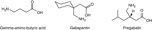
193. Taylor C.P. Mechanisms of analgesia by gabapentin and pregabalin-calciumchannel alpha2-delta [Cavalpha2-delta] ligands. Pain 142(1–2). 2009:13-16.

194. Wiffen P.J., McQuay H.J., Edwards J.E., et al. Gabapentin for acute and chronic pain. Cochrane database of systematic reviews. 2005. Issue 3. Art. No: CD005452. DOI:10.1002/14651858.CD005452

194a. Eckhardt K., Ammon S., Hofmann U., et al. Gabapentin enhances the analgesic effect of morphine in healthy volunteers. Anesth Analg. 2000;91(1):185-191.

194b. Wagner AE, Mich PM, Uhng SR, et al: Clinical evaluation of perioperative administration of gabapentin as an adjunct for postoperative analgesia in dogs undergoing amputation of a forelimb, J Am Vet Med Assoc 236:751-756, 2010.

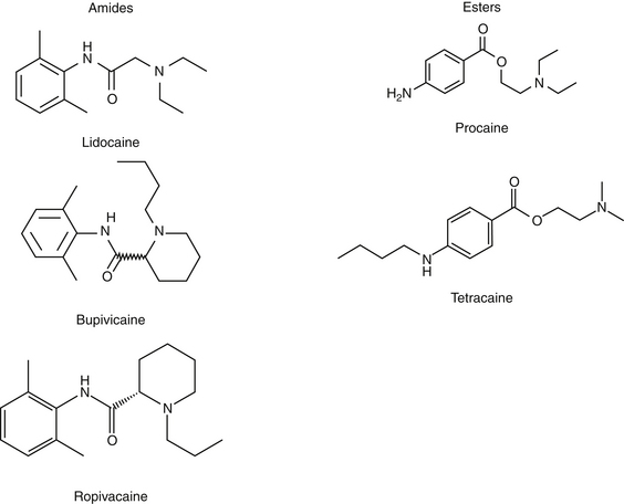
195. Catterall W.A., Mackie K. Local anesthetics. In Brunton L.L., Lazo J.S., Parker K.L., editors: Goodman & Gilman’s the pharmacological basis of therapeutics, ed 11, New York: McGraw-Hill, 2006. pp 369-386

196. Wilson D.V., Barnes K.S., Hauptman J.G. Pharmacokinetics of combined intraperitoneal and incisional lidocaine in the dog following overiohysterectomy. J Vet Pharmacol Ther. 2004;27:105-109.

197. Carpenter R.E., Wilson D.V., Evans A.T. Evaluation of intraperitoneal and incisional lidocaine or bupivacaine for analgesia following ovariohysterectomy in the dog. Vet Anaesth Analg. 2004;31:46-52.

198. Ibusuki S., Katsuki H., Takasaki M. The effects of extracellular pH with and without bicarbonate on intracellular procaine concentrations and anesthetic effects in crayfish giant axons. Anesthesiology. 1998;88(6):1549-1557.

199. Gibbon K.J., Cyborski J.M., Guzinski M.V., et al. Evaulation of adverse effects of eMLA (lidocaine/prilocaine) cream for the placement of jugular catheters in healthy cats. J Vet Pharmacol Ther. 2003;26:439-441.

200. Fransson B., Peck K., Smith J., et al. Transdermal absorption of a liposome-encapsulated formulation of lidocaine following topical administration in cats. Am J Vet Res. 2002;63:1309-1312.

201. Weil A.B., Ko J., Inoue T. The use of lidocaine patches. Compend Contin Educ Vet. 2007;29(4):208-210; 214–216.

202. Weiland L., Croubels S., Baert K., et al. Pharmacokinetics of a lidocaine patch 5% in dogs. J Vet Med A Physiol Pathol Clin Med. 2006;53(1):34-39.

203. Ko J., Weil A., Maxwell L., et al. Plasma concentrations of lidocaine in dogs following lidocaine patch application. J Am Anim Hosp Assoc. 2007;43(5):280-283.

204. Ko J.C., Maxwell L.K., Abbo L.A., et al. Pharmacokinetics of lidocaine following the application of 5% lidocaine patches to cats. J Vet Pharmacol Ther. 2008;31(4):359-367.

205. Cassutto B.H., Gfeller R.W. Use of intravenous lidocaine to prevent reperfusion injury and subsequent multiple organ dysfunction syndrome. J Vet Emerg Crit Care. 2003;13(3):137-148.

206. Pypendop B.H., Ilkiw J.E., Robertson S.A. Effects of intravenous administration of lidocaine on the thermal threshold in cats. Am J Vet Res. 2006;67(1):16-20.

207. Muir W.H.III, Weise A.J., March P.A. Effects of morphine, lidocaine, ketamine and morphine-lidocaine-ketamine drug combination on minimum alveolar concentration in dogs anesthetized with isoflurane. Am J Vet Res. 2003;64:1156-1160.

208. Himes R.S.Jr., DiFazio C.A., Burney R.G. Effects of lidocaine on the anesthetic requirements for nitrous oxide and halothane. Anesthesiology. 1977;47(5):437-440.

209. Valverde A., Doherty T.J., Hernández J., et al. Effect of lidocaine on the minimum alveolar concentration of isoflurane in dogs. Vet Anaesth Analg. 2004;31(4):264-271.

210. Smith L.J., Bently E., Shi A., et al. Systemic lidocaine infusion as an analgesic for intraocular surgery in dogs: a pilot study. Vet Anaesth Analg. 2004;31(1):53-63.

211. Pypendop B.H., Ilkiw J.E. Assessment of the hemodynamic effects of lidocaine administered IV in isoflurane-anesthetized cats. Am J Vet Res. 2005;66(4):661-668.

212. Thomasy S.M., Pypendop B.H., Ilkiw J.E., et al. Pharmacokinetics of lidocaine and its active metabolite, monoethylglycinexylidide, after intravenous administration of lidocaine to awake and isoflurane-anesthetized cats. Am J Vet Res. 2005;66(7):1162-1166.

213. Gerard J.L., Edouard A., Berdeaux A., et al. Interaction of intravenous diazepam and bupivacaine in conscious dogs. Reg Anesth. 1989;14(6):298-303.

214. Conzemius M.G., Brockman D.J., King L.G., et al. Analgesia in dogs after intercostal thoracotomy: a clinical trial comparing intravenous buprenorphine and interpleural bupivacaine. Vet Surg. 1994;23(4):291-298.

215. Feldman H., Arthur G., Pitkanen M., et al. Treatment of acute systemic toxicity after the rapid intravenous injection of ropivacaine and bupivacaine in the conscious dog. Anesth Analg. 1991;73:373-384.

216. López-Sanromán J., Cruz J., Santos M., et al. Effect of alkalinization on the local analgesic efficacy of ketamine in the abaxial sesamoid nerve block in horses. J Vet Pharmacol Ther. 2003;26(4):265-269.

217. Terlau H., Olivera B.M. Conus venoms: a rich source of novel ion channel-targeted peptides. Physiol Rev. 2004;84(1):41-68.

218. Livett B.G., Sandall D.W., Keays D., et al. Therapeutic applications of conotoxins that target the neuronal nicotinic acetylcholine receptor. Toxicon. 2006;48(7):810-829.

219. Vig M.M. Nicotine poisoning in a dog. Vet Hum Toxicol. 1990;32(6):573-575.

220. Freeman G., Entero H., Brem H. Practical treatment of pain in patients with chronic wounds: pathogenesis-guided management. Am J Surg. 2004;188(Suppl to July):31S-35S.

221. Urch C. The pathophysiology of cancer-induced bone pain: current understanding. Palliat Med. 2004;18:267-274.

222. Portenoy R.K., Farkash A. Practical management of non-malignant pain in the elderly. Geriatrics. 1989;43:29-47.

223. Wall R.T. Use of analgesics in the elderly. Clin Pharm. 1990;6:345-364.

224. Murtaugh R.J., Kaplan P.M. Veterinary emergency and critical care medicine. St Louis: Mosby; 1996.

225. Bullock R., Ward J.D. Management of head trauma. In: Shoemaker W.C., Ayers S.M., Grenvic A., et al, editors. Textbook of critical care. ed 3. Philadelphia: Saunders; 1995:1449-1456.

226. Dewey C.W., Budsberg S.C., Oliver J.E. Principles of head trauma management in dogs and cats: parts I and II. In: Drobatz K., editor. Emergency medicine in small animal practice: the compendium collection. Trenton, NJ: Veterinary Learning Systems; 1997:63-83.

227. Jones R.S. Epidural analgesia in the dog and cat. Vet J. 2001;161:123-131.

228. Bernards C.M. Recent insights into the pharmacokinetics of spinal opioids and the relevance to opioid selection. Curr Opin Anaesthesiol. 2004;17(5):441-447.

229. Sibanda S., Hughes J.M., Pawson P.E., et al. The effects of preoperative extradural bupivacaine and morphine on the stress response in dogs undergoing femoro-tibial joint surgery. Vet Anaesth Analg. 2006;33(4):246-257.

230. Hendrix P.K., Raffe M.R., Robinson E.P., et al. Epidural administration of bupivacaine, morphine, or their combination for postoperative analgesia in dogs. J Am Vet Med Assoc. 1996;209(3):598-607.