Dental anomalies
Michael J Aldred, Angus C Cameron, Nigel M King and Richard P Widmer

Introduction
The diagnosis and management of dental anomalies constitute important areas of paediatric dentistry. Although most dental anomalies present in childhood, many are misdiagnosed or left untreated, perhaps because of lack of experience or because the case is perceived to be ‘too difficult’. In some instances, genetic consultation is desirable, not merely to diagnose the condition but also to provide appropriate advice on the prognosis and the risk of recurrence in future generations. In many cases, the presence of an inherited dental disorder in one child would not stop a family from having additional children, but it is important to give parents and the affected children themselves appropriate information on which to base their decisions. Genetic services are usually available at most paediatric hospitals.
In this chapter, reference to particular inherited conditions is made to entries in OMIM (Online Mendelian Inheritance in Man). This online database is a catalogue of genetic disorders developed by Dr Victor McKusick of the Johns Hopkins University and the National Center for Biotechnology Information (see References and further reading, below).
Considerations in the management of dental anomalies
• Informing and supporting the child and parent.
• Interdisciplinary formulation of a definitive treatment plan.
• Provision of adequate function.
• Maintenance of occlusal vertical dimension.
• Use of intermediate restorations in childhood and adolescence.
Treatment planning for children with dental anomalies
Treatment planning should be multidisciplinary. Decision-making must involve the child and the parents and should consider the present and future needs and development of the child. Although children will cope with a range of appliances and treatments during childhood, early adolescence represents a period of social adjustment, as well as the transitional changes in the dentition. It is perhaps the most difficult time in which to formulate a long-term plan. Teenagers are most concerned about aesthetics, yet it may be too early to provide definitive restorations; extensive orthodontic treatment may be required or later orthognathic surgery. In institutions, various teams exist to treatment plan and/or manage these cases and a list is suggested below. Note the involvement of the child’s local general dental practitioner.
Dental anomalies at different stages of dental development
It is convenient to consider dental anomalies by the development stage at which they arise.
Dental lamina formation stage
Induction and proliferation
• Hypodontia/oligodontia/anodontia (which may be associated with other features of an ectodermal dysplasia).
• Double teeth (geminated or fused teeth).
• Odontomes (complex and compound).
• Odontogenic tumours, particularly the spectrum of ameloblastic fibroma/fibrodentinoma/fibro-odontome (dependent on differentiation and the presence and type of calcification within the lesion).
Morphodifferentiation
Eruption and root development
Formation of dental lamina
Hypodontia
Alternative terminology: Hypodontia, oligodontia, anodontia.
Hypodontia, oligodontia and anodontia are terms that can be interpreted to refer to progressive degrees of missing teeth, though the term hypodontia is preferred because it is inclusive of any number of missing teeth (Figure 11.1A). ‘Oligodontia’ refers to six or more missing teeth, and ‘anodontia’ to the complete absence of teeth. It is implicit in all cases that the teeth are missing because of failure of development. The term ‘congenitally missing teeth’ is a misnomer when applied to the permanent dentition because these teeth do not commence development until after birth (and with regard to the primary dentition one cannot usually determine this clinically at birth); ‘partial anodontia’ is a nonsense term. Some degree of hypodontia is not uncommon, occurring sporadically or with a hereditary component. The teeth most commonly absent are the last teeth in each series (i.e. the lateral incisor, the second premolar and the third molar). Clinically, it is less important to know how many, but rather which types of tooth are absent. It is particularly unusual for a patient to be missing central incisors, canines or first permanent molars. Multiple missing teeth in a child should lead to investigations to determine if there are other affected family members. The presence of a rudimentary or conical tooth may be associated with the absence of the same tooth on the opposite side of the arch. A common example of this is the peg lateral incisor. Furthermore, that lateral incisor itself may be absent in subsequent generations. Missing teeth are also a manifestation of many syndromes of the head and neck.
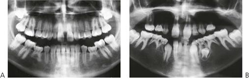
Figure 11.1 (A) The teeth most commonly missing are the last teeth in each series, namely the upper lateral incisors, the second premolars and the third molars. (B) Panoramic radiograph of a boy with autosomal dominant ectodermal dysplasia with absence of both primary and permanent teeth.
Frequency

Third molars > maxillary lateral incisors > second premolars > mandibular central incisors.
Major conditions manifesting hypodontia
Hypodontia is a major clinical feature of over 50 syndromes. These include:
Ectodermal dysplasias
Ectodermal dysplasia describes a group of developmental, often inherited, disorders involving the ectodermally derived structures, i.e. the hair, teeth, nails, skin and sweat glands. The most common is the X-linked hypohidrotic form (OMIM 305100, EDA1, Xq12-q13.1; short arm of X chromosome). In this condition the usual presentation is a male child with:
• Multiple missing teeth (Figure 11.1B).
• Fine, sparse hair (Figure 11.2A,B).
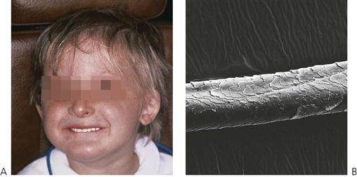
Figure 11.2 (A) Typical appearance of a boy with X-linked hypohidrotic ectodermal dysplasia (wearing a denture). The skin around the eyes is dry and wrinkled and may be pigmented (not shown here). (B) The hair is fine and sparse and often displays longitudinal grooves on the surface under the scanning electron microscopy.
• Dry skin (Figure 11.2A).
Teeth are small and conical, often with a large anterior diastema (Figure 11.4). Heterozygous females are often identified by dental examination and their manifestations may be limited to a single missing tooth or to a peg lateral incisor (see the Lyon hypothesis, below).
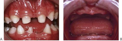
Figure 11.3 (A) This child is a heterozygous female with the X-linked form of ectodermal dysplasia and is less severely affected than her brother who has anodontia (B).
In the group of ectodermal dysplasias, autosomal dominant and recessive modes of inheritance are also seen. In such families, there will not be such a striking difference in the degree of the disorder between males and females compared with X-linked hypohidrotic ectodermal dysplasia (Figures 11.2A, 11.3). Mutations in the MSX1 gene (4p16.1) have been identified in families with missing third molars and second premolars with or without clefting, as well as in families with tooth-nail (Witkop) syndrome. PAX9 (14q12–q13) gene mutations have been found in other families with autosomal dominant missing teeth. More genes implicated in missing teeth and other anomalies continue to be identified.
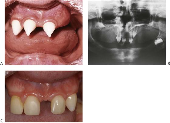
Figure 11.4 (A,B) Typical intra-oral and radiographic presentations in a boy with ectodermal dysplasia with multiple missing teeth; the teeth that are present are small and conical in shape. In the regions where teeth are absent, alveolar bone does not develop. (C) Composite resin build-ups of the conical teeth have provided improvement in the aesthetics; however, the problem of the diastema remains, given that there are only a few teeth for orthodontic anchorage.
In some countries, dental care (including prevention, orthodontics and prosthetics) for affected children may be provided under government-funded schemes.
Management
The aim of treatment is to provide adequate function, maintain the vertical dimension and restore aesthetic appearance. Ideally, for social reasons, treatment should begin at around 2–3 years of age. A first step is often the placement of composite restorations to mask the ‘fang-like’ appearance of the caniniform anterior teeth (Figure 11.4A). There is often considerable parental pressure to ‘normalize’ the appearance and later, steps may involve the provision of dentures to reduce the likelihood of teasing, often at about the time that the child starts school. This can begin as soon as the child allows adequate impressions to be taken. Often, however, the first denture is initially worn in the pocket(!), but as the child grows, there is often a desire to have a more ordinary appearance. With encouragement and positive reinforcement, most children will soon try their new appliances.
Treatment planning for children with hypodontia
Treatment planning should be multidisciplinary and should consider the present and future needs and development of the child, while being cognizant of the concerns of the individual and parents.
Treatment options
• Acid-etch retained, composite resin build-ups of conical teeth (Figure 11.6).
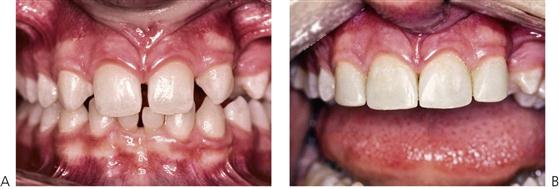
Figure 11.5 (A,B) Closure of an anterior diastema and reshaping of the canines with composite resin in a child with absence of the upper lateral incisors. The successful masking of upper canines to appear like lateral incisors is dependent very much on their size.
Figure 11.6 (A,B) Conical primary teeth are often associated with missing permanent teeth. This child had an autosomal recessive form of ectodermal dysplasia and was missing almost all of the permanent teeth. (C) These teeth have been built up with composite resin strip crowns. (D) Radiographic appearance of the same child at 15 years of age. Most of the primary teeth have exfoliated even in the absence of a permanent successor. There has also been loss of bone in the region of the tuberosity due to pneumatization of the sinus that will complicate implant placement.
• Composite resin or bonded orthodontic buttons can also be added to provide undercuts for denture clasps and retainers.
• Partial dentures: conventional or overdentures (Figure 11.7).
Figure 11.7 (A–C) Dentures for young children with ectodermal dysplasia. (A) Young children tolerate dentures extremely well and Adams’ cribs and ball retainers provide retention around primary molars. In this case, an overdenture covers two conical, widely spaced incisors. (B) A full upper denture for a child of 30 months will require periodic relining and re-making as the child grows. (C) Stock prosthetic teeth are sometimes difficult to obtain but paediatric denture teeth may be made freehand from acrylic and the palate can be customised.
• Surgical exposure of impacted teeth.
• Orthodontic management of spaces.
• Laboratory-fabricated composite resin veneers, crowns and bridges.
• Osseointegrated implants (usually after the cessation of growth).
The Lyon hypothesis (X chromosome inactivation)
During cellular differentiation, one of the two X chromosomes in each female somatic cell is inactivated. This means that in families with X-linked disorders, approximately 50% of the cells of heterozygous females will express the mutant gene disorder, whereas the remainder will express the normal gene. In the tissues affected by the condition, such females have a mosaic of affected and normal cells. This is of particular importance in X-linked forms of conditions such as haemophilia, hypohidrotic ectodermal dysplasia, vitamin D-resistant rickets and amelogenesis imperfecta. Thus, heterozygous females with X-linked hypohidrotic ectodermal dysplasia may have missing teeth, although they are invariably less severely affected than males. Similarly, in haemophilia A, heterozygous females do not usually have a clinical bleeding abnormality but this can occur if lyonization is severely skewed so that there is a preponderance of cells producing factor VIII under control of the mutant gene.
Dentoalveolar clefting
In patients affected by dentoalveolar clefting, disruption of the dental lamina at that site, there may be abnormal cellular induction or proliferation. This may give rise to either missing teeth, usually the maxillary lateral incisor, and/or supernumerary teeth adjacent to the cleft. However, it is extremely rare for the canine tooth to be affected in the same way.
Solitary median maxillary central incisor syndrome (OMIM 147250)
Solitary median maxillary central incisor syndrome (SMMCI) (Figure 11.8) is very rare. It presents with a midline symmetrical maxillary central incisor. The condition may also be associated with other midline disturbances such as cleft palate, choanal stenosis or atresia, imperforate anus or umbilical hernia and is probably part of the spectrum of the holoprosencephaly malformation complex. Of importance in some cases is the association with hypoplasia of the sella turcica, pituitary dysfunction, growth hormone deficiency and subsequent short stature. The syndrome is usually diagnosed on the basis of the dental manifestations. A mutation in the SHH gene (7q36) has been identified in one family but it is probable that there is genetic heterogeneity in the condition.
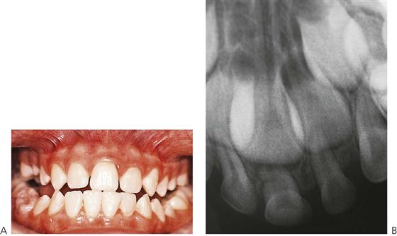
Figure 11.8 (A) Solitary median maxillary central incisor syndrome presenting with a symmetrical incisor in the midline. This child had a mild growth hormone deficiency, with his height on the 10th centile. (B) Periapical radiograph of the same patient in the primary dentition showing the single primary and permanent central incisors.
Ultimately, management of the dental anomaly is by orthodontic and prosthodontic therapy, determined by space considerations. In most cases, the single central incisor is moved to one side of the midline with either creation of space for a prosthodontic replacement, or the adjacent lateral incisors are recontoured.
Osseointegrated implants in children
There has been much controversy about the timing of placement of osseointegrated implants in young children. To date, there has been only limited published material about early placement and any long-term consequences. It is generally understood that implants act similarly to ankylosed teeth and do not move occlusally with the growing bone around adjacent natural teeth. Recent animal research has confirmed that most fixtures do become osseointegrated in growing jaws; however, there was no evidence from this research that the fixtures behaved like normal teeth during development. In the mandible, the fixtures came to lie lingual to the natural teeth; in the maxilla, they came to lie palatal and superior to the adjacent teeth and did not follow the normal downwards and forwards growth of this bone. This latter point is important when considering the placement of implants in the anterior maxilla. Furthermore, placement of fixtures retarded alveolar growth locally and changed the eruptive path of distally positioned tooth buds. Implants should, in most cases, not be considered before the cessation of growth. It should be noted, however, that in children with conditions such as ectodermal dysplasia, alveolar bone does not develop where teeth are not present. Consequently, it may be considered appropriate, particularly where there are multiple missing teeth, to place implants much earlier in these children than in those with a normal alveolus. Recent research suggests that in cases of anodontia, implants are best placed in the mandibular canine region at around 8–10 years of age (which is after the period of maximal mandibular transverse growth) to facilitate lower denture construction.
Disorders of proliferation
Supernumerary teeth (Figure 11.9)
• Supernumerary teeth arising as a result of budding of the dental lamina can occur sporadically or be inherited, as in cleidocranial dysplasia.
• The shape may resemble a tooth of the normal series (a supplemental tooth), in which case it can be incisiform, caniniform or molariform; otherwise it may be conical or tuberculate.
• Most often present as a result of failure of eruption of one or more permanent teeth. Usually appear as conical or tuberculate forms.
• Supernumerary teeth have been considered to be manifestations of a separate dentition (occurring between the primary and permanent dentitions), and consequently it may be possible to predict when and where supernumeraries may form (Jensen & Kreiborg 1990).
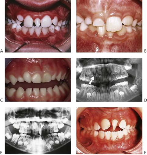
Figure 11.9 Common presentation of supernumerary teeth. (A) Conical teeth often erupt, except when inverted. (B) The late eruption of a permanent central incisor is most commonly caused by a supernumerary tooth. (C) Supplemental upper primary lateral incisor. (D) A panoramic radiograph is useful in determining the vertical orientation of the extra tooth (arrowed) and the degree of displacement of the permanent central incisor. In this case, after removal of the supernumerary, an upper denture was used as a space maintainer and the impacted tooth subsequently erupted into a normal position. (E,F) Dependent on the degree of displacement, given adequate space, most impacted incisors will normally erupt once an obstruction such as a supernumerary is removed. The rotation can be corrected later.
Alternative terminology
Mesiodens (a term restricted to supernumerary teeth in the midline of the maxilla), paramolar, distomolar, hyperdontia, polydontism, supplemental teeth.

Diagnosis
• Failed or eruption disturbance of permanent tooth (Figure 11.9B).
• Routine radiographic survey.
• As part of a syndrome such as cleidocranial dysplasia (Figure 11.10).
Management
• Conical teeth often erupt and are easily extracted (Figure 11.9A).
• Tuberculate and/or inverted conical teeth require surgical removal ((Figure 11.9D)) as early as possible to allow uninhibited eruption of the permanent teeth.
• It is essential to localize the position of the tooth to be removed prior to surgery. Periapical films using a tube-shift technique can be used to locate the tooth, however this is always open to errors and misinterpretation. Panoramic and standard maxillary occlusal films may be used in the same way.
• Digital imaging techniques using cone-beam tomography (CBCT) provide high definition, 3-dimensional imaging of the head and neck with much reduced radiation exposure than traditional computed tomography (CT) (see Figure 11.19, below).
• During surgical removal, care should be taken to avoid disturbing the developing permanent teeth.
• Before 10 years of age: if the unerupted central incisor is correctly aligned the treatment of choice is to remove the supernumerary surgically and allow normal eruption of the permanent tooth. Gingival exposure may be required later because of surgical scar formation that can inhibit final soft-tissue emergence. Some authorities recommend the simultaneous removal of primary canines to counteract this tendency. Inverted supernumeraries can be removed less traumatically if surgery is performed early, however, this needs to be done with caution to avoid damage to the adjacent tooth germs.
• After 10 years of age, or if the central incisor is malaligned: surgical exposure with or without bonding of orthodontic brackets or chains and subsequent traction may be required (Figure 11.11).
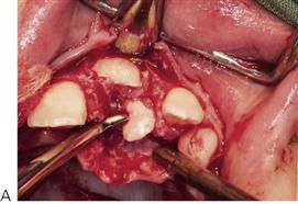
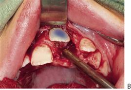
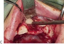
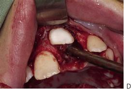
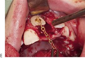
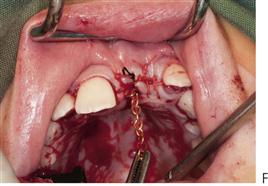
Figure 11.11 Surgical exposure and bonding of a gold chain to a central incisor which was impacted by a supernumerary tooth. (A) Elevation of the labial and palatal flaps and removal of the supernumerary. (B) Acid-etch applied to the labial surface of the upper left central incisor (C) Rinsing the acid etch gel from the tooth. (D) The appearance of the etch pattern on the prepared tooth. (E) Bonding of a gold chain attachment to the labial surface of the incisor. (F) The flap is closed and the chain is sutured to the gingiva with surgical nylon. The chain will be attached to an archwire and orthodontic traction will be applied to orthodontically align the tooth.
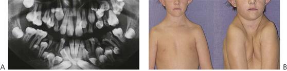
Cleidocranial dysplasia (Figure 11.10) (OMIM 119600)
This condition has an autosomal dominant mode of inheritance, with a high frequency of spontaneous mutations. The condition has been mapped to 6p21 with mutations found in the CBFA1 gene.
Manifestations
• Aplasia or hypoplasia of one or both clavicles (Figure 11.10B).
• Delayed ossification of fontanelles and sutures.
• Hypertelorism and maxillary hypoplasia.
• Wormian bones in cranial sutures.
• Multiple supernumerary teeth (Figure 11.10A).
Management
• Early diagnosis and documentation.
• Planned removal of non-resorbing primary teeth.
• Surgical removal of supernumerary teeth.
• Surgical exposure of permanent teeth.
• Orthodontic alignment and consideration of orthognathic surgery when growth complete.
Note that the simple extraction of a primary tooth will frequently guarantee the eruption of the impacted permanent tooth. A two-stage surgical procedure is usually required with an attachment placed on the permanent tooth followed by orthodontic traction. The first procedure involves exposure of the anterior segments with removal of the anterior primary teeth and any supernumeraries that may be present. The permanent teeth are surgically exposed, either with primary apically repositioned flaps or with bonded gold chains attached for orthodontic traction. The anterior teeth are then aligned orthodontically. The second stage involves extraction of the primary molars, surgical removal of remaining supernumerary teeth and exposure of the premolars and molars in the buccal segments. Definitive orthodontic therapy follows; orthognathic surgery may be required in cases with severe skeletal Class III malocclusion. Treatment obviously extends over many years and clinicians should be aware of the potential problems relating to the child’s compliance and the need for multiple surgical procedures.
Cherubism (OMIM 118400)
Cherubism is an autosomal dominant condition caused by mutations in the SH3BP2 gene at 4p16.3.
Patients may present in childhood with facial swelling and/or failure of eruption of teeth, typically the mandibular molars. Radiographs will reveal multilocular radiolucencies, typically involving the angles of the mandible (Figure 11.12). A biopsy will reveal multinucleate giant cells in a fibrous tissue stroma. Developing teeth in the affected area tend to be displaced and fail to erupt at the normal time. The maxillae can also be affected, as can the ribs. The facial swelling reflects the involvement of the underlying bone. In some patients the sclera in the lower part of the eyes may be exposed to give the cherubic or heavenward gaze that gives the condition its name. In some cases, there is no discernible facial swelling and the condition is identified as a result of routine radiographic studies such as for orthodontic treatment planning, or because of delayed eruption of teeth.

Figure 11.12 Dental panoramic radiograph showing almost symmetrical multilocular radiolucencies in the angles of the mandible of an 8-year-old boy. The displacement of developing molars and delayed eruption of teeth is usually seen in cherubism.
The condition progresses into adolescence and then tends to resolve, so that by the 3rd or 4th decade radiographic changes may no longer be found. In some families more affected males than females may be identified – this is a result of reduced penetrance in females and needs to be taken into account in genetic counselling. A subset of patients with cherubism is more severely affected with the multilocular radiolucencies affecting the whole of the mandible and maxillae. In mildly affected cases regular review may be all that is necessary, in more severely affected cases surgical reduction may be considered if the patient is distressed by their appearance.
Inflammatory follicular cysts
Some children may present with failure of eruption of a mandibular premolar associated with a radiolucency involving the roots of the primary molar and crown of the unerupted premolar (see Figure 7.2B and Figure 10.27). There is controversy as to whether such cases are due to radicular cyst formation associated with the roots of the primary tooth (which is considered by some to be a rare occurrence) or dentigerous cyst formation around the crown of the premolar. The common characteristics of such cases tend to be:
• Prior endodontic treatment of the primary molar.
• A radiolucency involving the roots of the primary molar and crown of the permanent successor.
• Displacement of the permanent successor away from the alveolar crest.
Histopathological examination tends to show intense acute and chronic inflammation of the curetted tissue which is lined by hyperplastic stratified squamous epithelium. Such cases have been designated ‘inflammatory follicular cysts’, with persistent inflammation from the endodontically treated primary molar leading to an inflammatory enlargement of the follicle of the underlying permanent tooth.
Odontomes (Figure 11.13)
Odontomes occur because of disordered differentiation and often present because of failure of eruption of a permanent tooth. In compound odontomes, multiplex of irregular denticles are found in a circumscribed soft-tissue stroma. Complex odontomes are disordered lesions with a discrete, haphazard mass of calcified tissue containing all dental elements. There is either a normal complement of teeth or the odontome replaces a tooth of the normal series.

Figure 11.13 Odontomes. (A) Compound odontome with multiple denticles causing displacement of the maxillary right central incisor. (B) Macroscopic specimen of a compound odontome from the anterior maxilla showing the numerous denticles surrounded by a well-defined capsule. (C) Complex odontome following elevation of a labial flap.
Odontogenic tumours (see Chapter 10)
The ameloblastic fibroma, fibrodentinoma and fibro-odontome are uncommon benign odontogenic mixed tumours. All are seen as altered differentiation of the tooth bud: in an ameloblastic fibroma no hard tissue is formed, in an ameloblastic fibrodentinoma only dentine-like tissue is recognizable and in an ameloblastic fibro-odontome enamel is also formed. The lesions tend to be well demarcated.
Odontogenic keratocysts (see Chapter 10)
Odontogenic keratocysts may arise in place of a tooth of the normal series or from the dental lamina in addition to a normal complement of teeth. They constitute 5–15% of odontogenic cysts.
Regional odontodysplasia (Figure 11.14)
Regional odontodysplasia is a sporadic defect in tooth formation with segmental involvement, usually localized to one, or part of one quadrant, but it may cross the midline to affect the contralateral central incisor. All dental tissues are involved in a bizarre dysplasia with hypoplastic teeth which are slow to erupt and which typically radiographically show a ghost-like appearance. The aetiology of the condition is unclear.
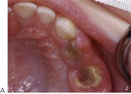
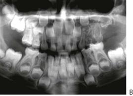
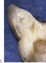
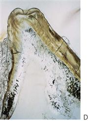
Figure 11.14 (A) Regional odontodysplasia presenting with abscessed primary molars in the maxillary left quadrant, soon after eruption. (B) The panoramic radiograph shows involvement of all the teeth in this quadrant including the permanent teeth. (C) Grossly abnormal enamel in an affected tooth and (D) the hard-tissue section demonstrates the disruption of odontogenesis. (Courtesy Dr N Pai, Sydney, Australia.)
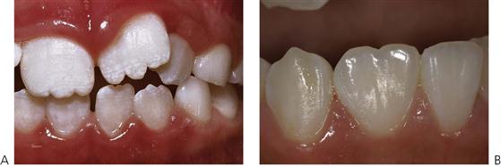
Figure 11.15 Morphological anomalies. (A) Mamelons, which are variations of normal anatomy. (B) Double tooth involving the right mandibular incisor, probably caused by fusion of the lateral and central incisor tooth germs.
• Usually presents initially with abscessed primary teeth before or soon after eruption.
• Some cases are associated with superficial vascular anomalies.
Alternative terminology
Ghost teeth.
Management
• In spite of attempts to restore teeth with stainless-steel crowns or composite resin, most affected teeth require extraction. Permanent successors of affected primary teeth are invariably affected, though sometimes to a lesser degree. There is no justification for bony excision at the time of tooth removal. There are reports of successful autologous tooth transplantation into the sites of removal of affected teeth.
Abnormalities of morphology
Macrodontia (Figure 11.16)
• Any tooth larger than normal for that particular tooth type.
• True macrodontia involving the whole dentition is extremely rare. More commonly, single teeth are abnormally large because of an isolated disturbance of development.
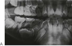
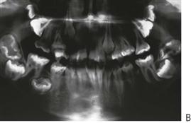
Figure 11.16 Macrodontia. (A) Isolated macrodontia in a mandibular second premolar tooth. (B) Generalized macrodontia associated with KBG syndrome. These children present with intellectual disabilities, broad facies, short stature and skeletal abnormalities.
Aetiology
• Unknown for a single tooth, but generalized macrodontia may be caused by a hormonal imbalance, as this has been described in pituitary gigantism. It should be remembered that an illusion of generalized macrodontia will occur if the jaws are small relative to the size of the teeth.
• May also be associated with hemifacial hyperplasia.
• True macrodontia should not be confused with the fusion or gemination of adjacent tooth units or a supernumerary to form a single tooth.
• Generalized macrodontia is also associated with KBG syndrome (the initials are taken from the surnames of the families first reported with the condition). These children present with short stature, intellectual disability, skeletal abnormalities, syndactyly, a broad face with microcephaly and other facial anomalies (Figure 11.16B).
Alternative terminology
Megadontia, megalodontia and gigantism.
Management
• Stripping to reduce tooth size; however, usually only a small change can be achieved.
• Can be combined with composite resin build-up of the antimere if only one tooth affected.
• Extraction and replacement by a prosthesis.
• Aesthetic adjustment of an isolated macrodont tooth by incisal edge ‘notching’ and the generation of a labial groove to break up light reflections may be helpful in some cases.
Microdontia
• One or more teeth that are smaller than normal for the tooth type.
• The most common form of microdontia affects only one or possibly two teeth; it is much rarer in the primary than in the permanent dentition.
• This anomaly most often affects the maxillary third molars and lateral incisors. It is noteworthy that the affected teeth are usually the ones that are also most often missing.
• Supernumerary teeth are frequently microdont.
• Patients with ectodermal dysplasia often present with microdontia.
True generalized microdontia
All of the teeth are of a normal morphological form but they are smaller than normal teeth. This condition is exceedingly rare but can occur in pituitary dwarfism.
Generalized relative microdontia
The teeth are of normal size but appear relatively small with respect to the jaws that are larger than normal.
Alternative terminology
Peg-shaped laterals.
Frequency
Most data are available only for maxillary lateral incisors.
| Primary dentition | <0.5% |
| Permanent dentition | ~2.0% (maxillary lateral incisors) |
More common in females.
Management
• Composite resin or (eventually) porcelain veneers to improve shape.
• The profile of the tooth is narrower at the gingival margin than a normal-sized tooth (emergence profile), and there is therefore a limit to how large the tooth can be enlarged with a restoration, without producing an overhang in the gingival region or an unsightly interdental shadow.
• Orthodontic alignment and extraction of the tooth may be required and other techniques such as autotransplantation and implants should also be considered.
Double tooth (Figure 11.17)
This anomaly is manifest as a structure resembling two teeth that have been joined together. In the anterior region, the anomalous tooth usually has a groove on (at least) the labial surface and a notch in the incisal edge. Although rarer in the posterior region, the cuspal morphology can be suggestive of two teeth that are joined together. Radiographs are necessary to determine if there is a union of the pulp chambers, and even then it may be speculative. If the ‘double tooth’ is present together with a normal complement of teeth in the same quadrant then it is presumed to have arisen as a result of gemination; if the number of teeth is reduced then fusion of tooth germs is assumed. If teeth have been extracted or exfoliated, the use of the neutral term ‘double tooth’ avoids the need to arbitrarily decide if this is due to gemination or fusion.
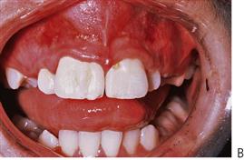
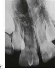
Figure 11.17 (A) Two double teeth formed by the fusion of each of the maxillary central incisors with supernumerary teeth. Two root canals were present in each tooth (B) Bilateral double teeth caused by gemnination. A single root canal was present in each tooth (C).
Alternative terminology
Fusion, gemination, connation, schizodontia, dichotomy.
Frequency
| Primary dentition | ~2.5% |
| Permanent dentition | ~0.2% |
Fusion, gemination or a ‘double tooth’ in the primary dentition should alert the clinician to the possibility of the same condition in the permanent dentition.
Fusion
Joining of two teeth of the normal series or a normal tooth and a supernumerary tooth by pulp and dentine. Two canals are usually present. The tooth has arisen from two tooth germs and so the number of teeth in the dentition is normally reduced by one unit. If, however, the normal tooth is fused to a supernumerary, the number of teeth in the arch will be normal. This fusion is assumed to occur between normal and supernumerary teeth because of the close proximity of the tooth buds.
Gemination
Budding of a second tooth from a single tooth germ. Usually one root canal is present.
Management
• The central groove on the labial and palatal surfaces of a double tooth is prone to dental caries; therefore early application of a fissure sealant is recommended.
• In the permanent dentition, surgical separation of fused teeth may be possible with subsequent orthodontic alignment and restorative treatment as needed to reshape the crown. The lack of cementum on the cut surface of the root may result in a periodontal defect.
• Reshaping or reduction of a double tooth with a single canal (geminated tooth) may be attempted by modifying the appearance of the labial groove and the use of composite resin but is often impossible and extraction may be the only alternative. Orthodontic treatment and/or prosthetic replacement is then required. Implants may be an option for adolescents.
• Deliberate extraction and surgical separation outside the mouth with replantation might also be considered, although this is not always successful because of resorption subsequent to reimplantation.
Clinical Hint – Fused and geminated teeth
Large geminated teeth present difficult management issues. It is essential to diagnose whether a single canal or separate canals are present. Plain radiographs are often of little benefit, especially when the abnormal tooth is in the central incisor region and there is superimposition of the lateral incisor which erupts palatal to the double tooth due to a lack of space. CT scans (Figure 11.18) and (better still) cone beam tomography (Figure 11.19) can be helpful to determine the morphology of the root canal.
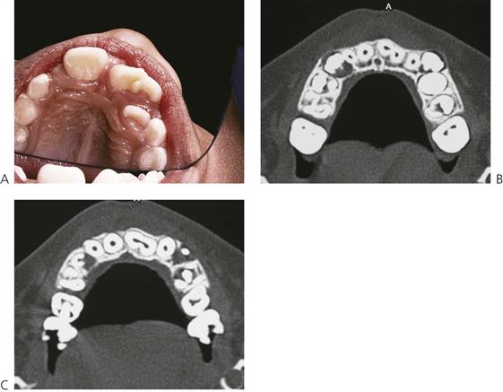
Figure 11.18 (A) It is often very difficult to determine the root canal anatomy of double teeth when using periapical films due to superimposition of the lateral incisor that has usually erupted palatally. CT can be used to determine the true internal anatomy of the root canal. (B) Apically, it appears that there are two distinct canals; however, a more coronal section shows that there is communication between the canals. (C) This contraindicates surgical separation of the roots. The diagnostic value of this radiography must be weighed against the radiation exposure.
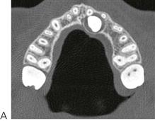
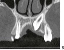
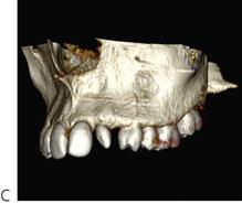
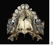
Figure 11.19 Further use of 3-dimensional CT to determine positioning and morphology of teeth. In these projections, the upper left canine is impacted. (A) The axial view shows the horizontal position in the palate in relation to the peg lateral incisor. (B) A coronal section details the root and the close association with the antrum. (C,D) 3-dimensional reconstructions are invaluable to the surgeon to determine the exact position in the mouth and the proximity of related structures.
Concrescence
Joining of two teeth, one of which may be a supernumerary, by cementum. Concrescence most commonly affects maxillary second and third permanent molars in older adults. Apart from when involving supernumeraries, this condition is rarely seen in children.
Dens invaginatus (Figures 11.20, 21)
Maxillary lateral incisors may have a developmental invagination of the cingulum pit with often only a thin hard-tissue barrier between the oral cavity and the pulp. Pulp necrosis often occurs soon after eruption of the affected tooth and may lead to a canine fossa abscess or cellulitis. This anomaly may occur in other teeth such as the maxillary central incisors and canines.
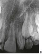
Figure 11.20 (A,B) Maxillary canine fossa cellulitis from an infected dens invaginatus. (B) Because of root canal morphology and the severity of the infection the tooth was removed. The patient required hospital admission, with high-dose intravenous antibiotics and surgical drainage of the abscess under general anaesthesia.
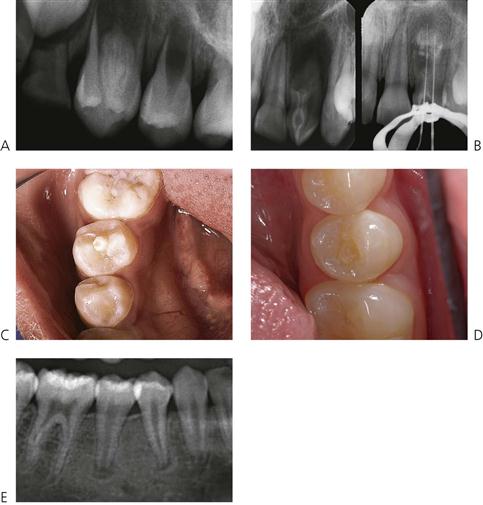
Figure 11.21 (A) Dens invaginatus in a maxillary first premolar tooth. (B) Ultimately, the prognosis is related to the ability to adequately instrument and obturate the canals of these teeth. Temporary endodontic dressings can be placed in such teeth to relieve symptoms and treat infection but it is almost impossible to obturate such canals. (C) Dens evaginatus. (D) Dens evaginatus where the tubercle has fractured off and the tooth has become necrotic. This child presented in acute pain with a facial cellulitis. The radiograph (E) shows the periapical area.
Alternative terminology
Invaginated odontome, dens in dente (used to describe the extreme variant, but is a misnomer), dilated odontome.
Management
• If newly erupted, the palatal fissures should be sealed as a preventive measure.
• If caries is evident, then place an acid-etched retained composite resin restoration with minimal preparation.
• If symptomatic and the root canal morphology is favourable, endodontic treatment can be undertaken.
• If the internal anatomy is complex and the root canal is not negotiable then, in the event of infection, extraction is necessary. The presence of this anomaly should be carefully considered during orthodontic treatment planning.
• The same tooth on the opposite side should be carefully assessed for the same problem.
Dens evaginatus (Figure 11.21C)
• An enamel-covered tubercle usually projecting from the occlusal surface of a premolar tooth.
• Usually bilateral and more common in the mandible.
• There is evidence of pulp tissue within the tubercle in ~50% of cases.
• Radiographs may show occlusal extension of the pulp chamber.
Alternative terminology
Leong’s premolar, tuberculated premolar, axial core type odontome, occlusal enamel pearl, composite dilated odontome, cone-shaped supernumerary cusp, evaginated odontome, interstitial cusp.
Frequency
| Primary dentition | Almost unknown |
| Permanent dentition | ~4% (Almost exclusively in people of Asian extraction) |
More common in females.
Management
• The tubercle can easily fracture because of occlusal interference, therefore grinding of the tubercle followed by fissure sealing can be of assistance. An alternative prophylactic measure is to support the sides of the tubercle with composite resin and then to recontour the occlusal surface to produce a central ridge. Ideally, this should be performed before the tooth comes into complete occlusion.
• If fractured or subject to attrition, pulp exposure occurs frequently. Because this exposure occurs soon after eruption, the apex of the tooth is often open and the long-term prognosis is less certain. Extraction of the tooth may be considered after orthodontic consultation. If the tooth is to be retained, a calcium hydroxide dressing is appropriate with an apexification procedure (see Chapter 9) to stabilize the tooth if orthodontic therapy is to commence later (and subsequent definitive endodontic treatment). More recently, revascularization has been proposed in the management of these teeth.
• If diagnosed early, an elective (Cvek) pulpotomy can be performed in an attempt to allow normal root formation.
Talon cusp (Figure 11.22)
This is a horn-like projection of the cingulum of the maxillary incisor teeth. It may reach the incisal edge of the tooth.
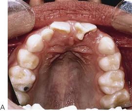
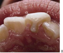
Figure 11.22 (A) Talon cusp. (B) T-cingulum. The cingulum cusp has pulp horns and removal of the cusp will often result in an exposure of the pulp.
Alternative terminology
T-cingulum, Y-shaped cingulum.
Management
• If there is no interference with the occlusion, no treatment is required.
• Fissure sealants to prevent caries in the grooves between the various parts of the tooth.
• If occlusal interference is present, small progressive reduction of enamel only to avoid pulp exposure, or elective pulpotomy, to allow root completion.
Taurodontism
Used to describe a molar tooth with a pulp chamber that is vertically enlarged at the expense of the roots. The distance from the cemento-enamel junction to the furcation of the root may be greater than the distance from the furcation to the apices. The tooth, therefore, has a long body and short roots, with a tendency towards a single root or apical displacement of the furcation. The anomaly appears to be caused by delay or failure of invagination of Hertwig’s epithelial root sheath. Taurodontism may have a genetic basis. Several syndromes and conditions such as ectodermal dysplasias, X-chromosome aneuploidies and some families with (autosomal dominant) amelogenesis imperfecta have this anomaly. The appearance may also be reflected in single-rooted teeth, with the pulp canals being wider than usual.
Frequency
Uncommon rather than rare. Enlarged pulp chambers may also be seen in:
Congenital syphilis
Although now very rare in most parts of the world, congenital syphilis presents with several important diagnostic dental manifestations. Both primary and permanent incisors have tapering crowns and central notching of the incisal edge. This tapering or screwdriver-like appearance is important in the differential diagnosis, as there are other causes of non-syphilitic notching of the incisal edge (e.g. trauma). This screwdriver morphology is also seen in Nance–Horan syndrome. The crowns of the molar teeth have a ‘cobbled’ or ‘mulberry’ appearance in congenital syphilis.
Developmental defects of enamel
Developmental defects of enamel can be acquired or inherited.
Chronological disturbances
Any severe systemic event during the development of the teeth (i.e. from 3 months in-utero to 20 years of age) may result in some dental abnormality. Many of these anomalies are subclinical and can only be observed in hard-tissue sections as changes in the incremental deposition lines. The neonatal line is manifest in all primary teeth, but unless there is a severe physiological disturbance or fetal distress the disturbance may not be clinically evident. Different teeth will show defects at different levels of the crown depending on the stage of crown formation at the time the disturbance occurred. The resulting enamel may be reduced in quantity (hypoplasia) and/or quality (usually hypomineralization).
A defect is described as localized when one or more teeth are affected, in an asymmetrical way, and generalized when there is a symmetrical disturbance on teeth of the same type on both left and right sides (and in both maxillary and mandibular teeth).
More than 100 aetiological agents have been reported to cause developmental defects of enamel. Those causing localized defects are listed in Table 11.1 and those causing generalized defects are listed in Table 11.2.
Table 11.1
Aetiological agents shown to produce developmental defects of enamel with a localized distribution
| Acute osteomyelitis | Gunshot wounds to jaws |
| Acute trauma to primary teeth | Irradiation |
| Ankylosis | Jaw fracture |
| Cleft palate | Laryngoscopy |
| Congenital epulis | Periapical infection of primary teeth |
| Electrical burn to mouth | Periodontal ligament injection |
| Extraction of primary teeth |
Table 11.2
Environmental aetiological agents shown to produce developmental defects of enamel and discolouration with a generalized distribution
Developmental defects of enamel can be considered according to their clinical appearance:
In general, the aims of management are to treat associated pathology and pain, provide adequate aesthetic appeal, maintain occlusal function and maintain the vertical dimension.
Tooth discolouration
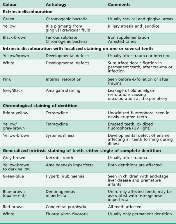
Tooth discolouration may be extrinsic or intrinsic in nature. Extrinsic staining is superficial and occurs after tooth eruption. Intrinsic discolouration may result from a developmental defect of enamel or internal staining of the tooth (Figure 11.23). Although such internal staining is manifest as a change in tooth colour, the intrinsic defect may affect the dentine primarily or exclusively. See Table 11.3 for the differential diagnosis of tooth discolouration.
Opacity
Opacities result from a defect in the quality of the enamel, affecting the translucency of the tissue. Hypomineralization results in a change in the porosity of the enamel, causing opacity. This may be located below the enamel surface, which otherwise remains intact.
Fluorosis (Figure 11.23C, see also Chapter 5)
In its mildest forms, fluorosis is manifest as hypomineralization of the enamel, leading to opacities. These can range from tiny white flecks to confluent opacities throughout the enamel, making the crown totally lacking in translucency. Hypoplasia occurs at higher concentrations of fluoride. When the tooth first erupts, the surface of even the most severely affected enamel may be intact; however, with wear, areas of enamel are lost and stains are taken up into the porosities. At 1 ppm of fluoride in public water supplies, up to 10% of the population will show very mild opacities attributable to fluorosis (though this may depend on individual water consumption); interestingly, this seems to be a minimum value and the proportion of opacities increases as fluoride levels either fall below 1 ppm or rise above 2 ppm. Severely affected cases may require microabrasion or restoration with composite resin, either in a localized or a more generalized manner, or porcelain veneers. Many opacities are incorrectly labelled as fluorosis without adequate justification or investigation of the patient’s fluoride history.
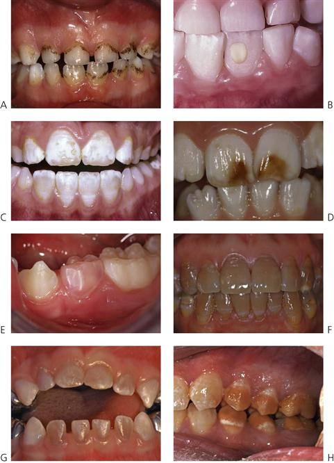
Figure 11.23 Tooth discolouration. (A) Brown-black superficial staining from chromogenic bacteria. (B) Localized enamel opacity caused by the root apex of a traumatized primary incisor. (C) Fluorosis – uniform opacity-type defects throughout the crown. Some of the hypomineralized enamel has been lost on the incisal edge revealing normal enamel underneath. (D) Brown discolouration due to incorporation of blood pigments into the enamel following trauma to the primary dentition. (E) Pink discolouration caused internal resorption of the tooth 74. (F) Tetracycline staining in a child from South-east Asia. Tetracycline containing liquid preparations are no longer routinely prescribed for children, however, tetracyclines may be included in some homoeopathic preparations in some countries. (G) Blue-brown appearance of dentinogenesis imperfecta. (H) Chronological discolouration of an unknown aetiology. There is a precise pattern to the hypomineralization and appearance of the posterior teeth, however, the anterior teeth, in particular, the canines that are developing at the same age are unaffected.
Management of stains and opacities
• Extrinsic stains can be removed with abrasives.
• Mild discolouration may be improved using peroxide-based bleaching agents.
• Intrinsic stains, if superficial, may be removed with microabrasion techniques.
Microabrasion
It must be understood that microabrasion techniques involve removal of the surface opaque layer of enamel. The opaque but often bright white layer of enamel is removed and children and parents are often disappointed at the appearance of the normal ‘yellow’ colour of the permanent crown. It is important to make an initial decision whether to attempt to alter the entire dentition to this darker colour, or to continue with the piecemeal adjustment of darker areas to match the overall ‘paper-white’ appearance. In present-day society, the latter decision may prove to be satisfactory or even desirable. Acid-based techniques may create more porosity in the enamel, which may accumulate more stains over time.
Hydrochloric acid – 18% (with or without the use of pumice)
Rubber dam should always be used and must be secured by ligation around individual teeth and sealing with copal-ether varnish or a proprietary sealer. Orabase® paste applied to the gingival margin prior to dam placement can be used to protect the soft tissues from any acid leakage. An aqueous slurry of sodium bicarbonate may be laid on the dam around the teeth to neutralize any inadvertent excess acid. An hydrochloric acid/pumice slurry is applied to the affected area using a slowly rotating rubber cup for 10 s only, and then rinsed thoroughly with water. Application is repeated a maximum of 10 times. This technique is potentially destructive to enamel and soft tissues, and must be used with caution.
Alternatives
• Abrasion with a mixture of pumice and 37% phosphoric acid (etchant).
• Polishing labial surfaces with a multi-fluted tungsten-carbide bur.
• Application of a 2% neutral sodium fluoride, followed after a short time by polishing with fine polishing paste (non-coloured toothpaste is suitable) in a rotating rubber cup is advisable after use of each of these techniques.
• Recent research has shown that mild fluorosis can also be remineralized and the opacity reduced by casein phosphopeptide-amorphous calcium phosphate (CPP-ACP) or casein phosphopeptide-amorphous calcium phosphate fluoride (CPP-ACPF). The enamel should be pretreated with sodium hypochlorite to denature any residual protein.
Deep intrinsic stains require removal of the affected enamel and rebuilding, usually with composite resin. Although localized marks may be dealt with by this method, treatment using composite resin or porcelain as full-face veneers, or crowns, should ideally be delayed in adolescents until the gingival attachment is established at the cemento-enamel junction. The longevity of hybrid composite resins has improved substantially, along with their colour stability, strength and translucency. These materials may be placed quickly and more cost-effectively than porcelain and other complex restorations such as crowns. Modern materials generally provide for densely white shades that make matching possible. It is important always to keep treatment options open. Involvement in contact sports may be another reason for delaying placement of complex restorations.
Enamel hypoplasia (Figure 11.24)
A defect in quantity that causes an altered contour of the surface of the enamel. This is usually caused by initial failure of the deposition of enamel protein, but the same clinical effect could also result if there is a mineralization defect that leads to loss of enamel substance after eruption. In the former case, the enamel is often hard and glassy, in the latter it will usually pit on probing. In some trauma cases, tissue may be lost after formation and is not regarded as a true hypoplasia. Examples of hypoplastic defects following trauma are shown in Figure 9.18.
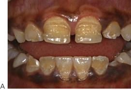

Figure 11.24 (A) Chronological enamel hypoplasia after a childhood illness from 11 months up to around 18 months of age. The incisal edges of the maxillary lateral incisors are affected. Fortunately, there is little hypomineralization of the teeth, making the dentition more easily restored with composite resin. (B) Chronological hypoplasia may affect the primary dentition, this child with a child suffered fetal distress and meconium aspiration at delivery. It is possible that the tips of the cusps of the first permanent molars are similarly affected. (C) Another form of chronological hypoplasia. Note that there is normal enamel at the cervical region and the primary tooth is not affected. (D) Localized hypoplasia of primary canines. These anomalies present as defects on the labial surface of primary canine teeth and often become carious. A minute area of hypoplasia is visible on the maxillary right canine, and all the other canine teeth are carious, while the remainder of the teeth are caries free.
Management
• Localized hypoplastic defects may be restored with composite resin. Pitting defects may need initial localized debris or stain removal with either rotary instruments or amine peroxide bleaching systems.
• It is important to maintain posterior support, and stainless steel crowns may be required to restore grossly hypoplastic molars. These teeth may be exquisitely sensitive to thermal and osmotic stimuli and treatment is made difficult by an inability to achieve good isolation of teeth that are only partially erupted. Glass ionomers may be used temporarily to restore hypoplastic occlusal defects and prevent caries.
• A realistic assessment of the likely longevity of affected first molars is important from an early age. Consideration should be given to the elective loss of these teeth as part of an occlusal developmental plan for the child. A pre-assessment accompanied by orthodontic advice not later than 8 years of age is recommended.
• Complex restorative treatment involving onlays, veneers and crowns should generally be delayed until late adolescence but the selective use of metal, adhesively retained onlays may provide a long-term solution in some molar cases.
Molar–incisor hypomineralization (Figure 11.25)
Molar–incisor hypomineralization (MIH) is a condition that, although recognized as a clinical entity for some time, is still a subject of considerable study. It presents as a qualitative change in enamel that, initially, is of normal thickness, ranging from localized opacity through opacity with discolouration and obvious poor quality to post-eruptive enamel breakdown. The cervical enamel appears to be normal in most affect first permanent molars. One or more first permanent molars may be affected in a quasi-chronological but inconsistent manner, together with (usually lesser) effects on one or more incisors. The presentation is puzzling since as few as one molar, or as many as all four may be affected. Often affected teeth are extremely sensitive, and this is often an indicator to poor ability to gain complete anaesthesia of the affected teeth. Use of low viscosity GIC sealants soon after eruption and also remineralizing agents such as CPP-ACP can help decrease sensitivity and reduce post-eruptive breakdown.
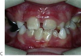
Figure 11.25 Molar incisor hypomineralization. Teeth may be variably affected in the one mouth. (A) Severely affected upper first permanent molar with loss of enamel and hypomineralization. (B) An otherwise intact dentition shows typical MIH. The incisors (palatal surface) are less severely affected than the molars. (C) Hypomineralization of the incisors associated with MIH and rampant caries in the primary dentition. Note that the crowns of the second primary molars and first permanent molars form at similar times, and this may explain the high rate of caries seen in the primary dentition in some of these patients. (Courtesy Dr E Alcaino.)
Many possible aetiological factors, especially those related to childhood disease and maternal illness during the 3rd trimester, have been suggested. Research is under way in many centres to define the aetiological factors, since a knowledge of these would permit early preventive and restorative interventions. The prevalence ranges widely, however, it is between 10% and 15% in many communities worldwide; with around 5% of the population being severely affected. It is believed that MIH may account for a large proportion of the restorative needs of children in many communities, and therefore is an important public health issue.
A familial tendency to the condition has been recognized by some authors. Irrespective of the exact aetiology of MIH, it is important to recognize that this condition represents a chronological disturbance in tooth formation between birth and 24 months of age. Enamel hypomineralization can affect any teeth in the permanent dentition, however, there is a very low prevalence compared with the first molars. It also affects second primary molars, with a prevalence of approximately 6%.
Management
• The ideal restorative approach for these cases has yet to be determined however if intracoronal restorations are planned, composite resin should be used. Some research indicates that pre-treating the enamel with 5% sodium hypochlorite after etching increases bond-strengths significantly. Stainless steel crowns are an option for severely affected teeth, however it needs to be explained to the child and parents that this is an intermediate phase of treatment, and further restorative work will be required at maturity. Trimming the length of the stainless steel crown for partly erupted first permanent molars so as to obtain a fit just apical to the maximum convexity of the crown makes such placement easier in many cases.
• There is a clear association between repeated, well-meaning attempts by practitioners to place ‘minimal’, adhesive or other treatments for these molars, without adequate local anaesthesia, and a real antipathy to dental treatment on the part of the affected children. Local analgesia should be used for the treatment of these cases but it should be noted that even under these conditions pain control will not always be adequate and treatment may be compromised. General anaesthesia is often required to provide high quality dental care for these children.
Amelogenesis imperfecta
The term amelogenesis imperfecta is usually applied to inherited defects of the enamel of both primary and permanent teeth (Figures 11.26–29). Although the definition implies a family history, for practical purposes it seems reasonable to extend this to include sporadic cases and also to those cases where the enamel defects are associated with extra-oral features, as found in some syndromes (i.e. focal dermal hypoplasia or the trichodento-osseous syndrome).
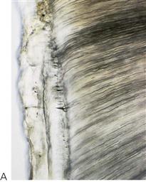
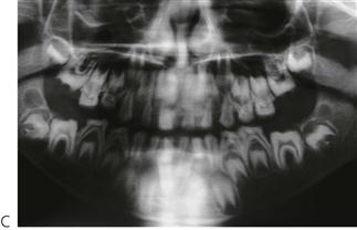
Figure 11.26 (A) Hard-tissue section of autosomal dominant amelogenesis imperfecta showing extremely thin enamel in this particular patient. (B) Scanning electron micrograph of a similar patient with abnormal etching pattern of enamel. (C) Panoramic radiograph showing absent or very thin enamel in this form of amelogenesis imperfecta.
Frequency examples
Few studies of prevalence have been carried out and there may be marked differences according to the gene pool involved.
Amelogenesis imperfecta variants with Mendelian inheritance
• X-linked (Xp22.3-p22.1) (OMIM 301200): The AMELX gene on the short arm of the X chromosome which codes for amelogenin, the major enamel matrix protein, has been shown to be mutated in several families with X-linked amelogenesis imperfecta.
• X-linked (Xq22-q28) (OMIM 301201): Another locus for X-linked amelogenesis imperfecta has been identified on the long arm of the X chromosome.
• Autosomal dominant (OMIM 104500): A number of genes in the 4q11–q21 region appear to be implicated in causing autosomal dominant amelogenesis imperfecta in some families. Mutations in the enamelin gene, which maps to the same region, have been identified. Families with autosomal dominant amelogenesis imperfecta with taurodontism (ADAIT) as part of the trichodentoosseous (TDO) syndrome have mutations in the DLX3 gene; one family with ADAIT without other features of TDO has also been found to have a mutation in the same gene, whereas in other ADAIT families mutations in the DLX3 gene have been excluded. With time, it is expected that this area will be better understood, as it is probable that as yet unknown genes are involved in the disorder in some families.
• Autosomal recessive (OMIM 204650): Mutations in the matrix metalloproteinase-20 (MMP-20) or kallikrein-4 (KLK-4) gene appear to cause autosomal recessive amelogenesis imperfecta.
Phenotypes
Phenotypes range from markedly hypoplastic (thin) enamel (Figure 11.27) (either uniformly with spacing between adjacent teeth or irregularly giving rise to pits or grooves) to varying degrees of hypomineralization (poorly formed enamel) with altered colour and translucency (Figure 11.28). In many cases, both hypoplasia and hypomineralization are seen together. The colour of the teeth is presumed to reflect the degree of hypomineralization of the enamel – the darker the colour the more severe the degree of hypomineralization.
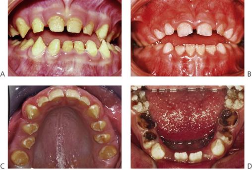
Figure 11.27 Different forms of predominately hypoplastic amelogenesis imperfect (AI). (A) Autosomal recessive with a rough hypoplastic phenotype. (B) Autosomal dominant with smooth hypoplasia with a marked anterior open bite. Note the open contact points in these two cases. (C) Hypoplastic AI teeth are yellow-brown with spacing between the teeth. (D) Rough or grossly pitted forms may be susceptible to caries as they are extremely difficult to keep clean.
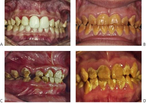
Figure 11.28 Different forms of predominately hypomineralized AI. (A–D) All these cases are characterized by varying degrees of hypomineralization. The enamel is so soft that it can be removed with an excavator. Note the discolouration and gross build-up of calculus on all tooth surfaces.
In X-linked amelogenesis imperfecta (Figure 11.29), females exhibit vertical bands of altered enamel (manifesting lyonization; see Lyon hypothesis, above). There may be vertical grooves (because of hypoplasia) and/or vertical bands of enamel of altered colour or lucency (because of hypomineralization) or a combination of the two. In such families, there will be no male-to-male transmission, whereas the heterozygous females may pass on the trait to children of either sex.
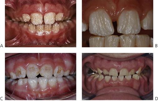
Figure 11.29 Different forms of AI with X-linked inheritance. (A,B) Typical appearance of vertical hypoplastic grooves in females. These represent enamel derived from different clones of ameloblasts that have undergone lyonization (X chromosome inactivation). (C) Pitting hypoplastic AI represents another clinical variety of X-linked AI. (D) X-linked AI in a male. All the enamel is affected uniformly.
In some patients affected by amelogenesis imperfecta, one or more teeth fail to erupt, presumably due to a more severe disturbance of the enamel organ, and may undergo resorption of their crowns. In some cases (up to 50%), a skeletal anterior open bite is seen.
Classification of amelogenesis imperfecta
There has been great controversy and confusion created with different nomenclature and classifications. Indeed, up to 14 different forms of the condition are described in some texts. It should be noted that all of these different manifestations are based on the clinical and radiographic appearance. It is essential that diagnosis and classification be based on the mode of inheritance and phenotype. Understanding the mode of inheritance is essential for genetic counselling. However, setting aside questions of inheritance for our present purposes, from a clinical treatment planning perspective, two clinically distinct basic forms are considered here.
Predominantly/exclusively hypoplastic forms (Figure 11.27)
• Lack of contact points between teeth.
• Enamel may be rough, smooth, or randomly pitted.
• Heterozygous females with X-linked amelogenesis imperfecta manifest lyonization (see above) with vertical banding of normal and abnormal enamel.
• Teeth may be delayed in eruption.
• Unerupted teeth may undergo resorption.
• Anterior open bite associated in about 50% of cases.
• Radiographically, it may be difficult to distinguish enamel from dentine if the former is extremely thin.
Predominantly/exclusively hypomineralized forms (Figure 11.28)
• May be normal thickness of enamel, at least initially.
• Enamel may be softer than normal, tends to chip and can be penetrated with an explorer. In severely hypomineralized cases, the enamel may be scraped away with a scaler.
• Teeth may erupt with enamel of normal thickness but it can be quickly lost, exposing highly sensitive dentine.
• Large masses of supragingival calculus may be present.
• Radiographically, can be difficult to distinguish between enamel and dentine because of decreased degree of mineralization of enamel.
• Unerupted teeth may undergo resorption; radiographic review is needed to monitor this.
Management
• Appropriate diagnosis, taking into account the mode of inheritance and phenotype.
• Continued commitment to and support of both children and families. These are disfiguring, painful conditions and children may be badly teased by their peers.
• Offer genetic counselling if appropriate.
• Early orthodontic assessment.
• Preservation of molar teeth with full coverage restorations to maintain vertical dimension.
• Overdentures may be an option in children with small, hypoplastic teeth (Figure 11.30C,D).
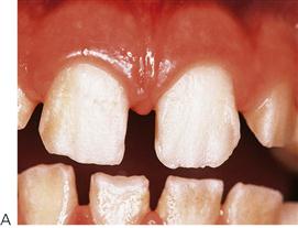
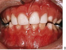
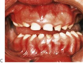
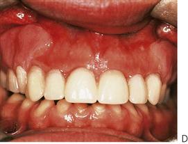
Figure 11.30 Different management options for amelogenesis imperfecta. (A,B) Composite bonding in a case with rough hypoplasia of the enamel. Etching times should be slightly longer than usual; however, the roughness of the enamel surface aids in mechanical retention. (C,D) A patient with autosomal recessive dystrophic epidermolysis bullosa with enamel defects. The posterior teeth failed to erupt and underwent spontaneous replacement resorption within the alveolus. Because of the small crown length and the prominent alveolus, an overdenture without a labial flange was constructed.
• Stainless steel crowns or gold onlays on molars (Figures 11.31 & 11.32) or laboratory-made composite resin crowns may be useful.
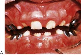
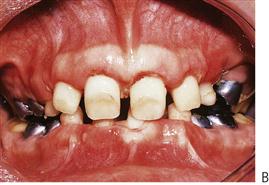
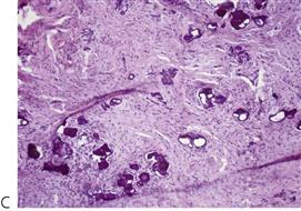
Figure 11.31 (A,B) A case of autosomal dominant amelogenesis imperfecta with failure of eruption of the anterior teeth. Many of the posterior teeth were unerupted and undergoing resorption. Initial surgical exposure of the anterior segments did not aid their complete eruption. The gingivae contained small islands of calcification which may be a significant factor in the failure of eruption (C). Periodontal surgery with apically repositioned flaps was used to fully expose the crowns.
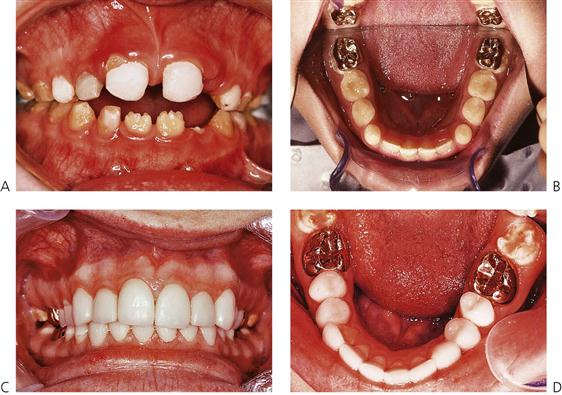
Figure 11.32 (A) Not all cases using composite resin are successful. With progressive eruption of the teeth it is difficult for the patient to keep the gingival margins clean and the restoration may therefore fail. (B) Cast gold onlays are useful to protect the occlusal surfaces. No preparation of the crown was performed. Onlays were cemented with composite luting cement. (C,D) Use of gold onlays and indirect composite resin veneers on anterior teeth and onlays on premolars. These composite restorations can provide good aesthetics during adolescence.
• Care is required when trial fitting crowns, because defective enamel can be easily scraped or flaked off the tooth in some cases.
• Composite resin veneers over anterior teeth for aesthetics. It is possible to bond composite resin successfully to hypoplastic and hypomineralized enamel (Figure 11.30A&B).
• Adequate margins may be difficult to achieve because of the poor quality of the enamel (Figure 11.32A).
• Ideally, delay definitive treatment with porcelain and precious metals until late adolescence. However, some middle-aged patients have commented that, had they known that their dentition was going to ‘fail’ at that stage, they would have preferred their practitioner to have been less ‘conservative’ in their teenage years and to have provided more conventional restorative care. Two points arise:
• Modern composite resins have improved greatly and ‘adolescent’ treatment now is hopefully more aesthetic and longer lasting than previously.
• There is evidence of a clear association between these conditions and lack of self-esteem. It is as important here as anywhere in dentistry to treat the whole patient, and not only the teeth.
• Orthodontic and possible orthognathic surgery to correct anterior open bite in hypoplastic forms.
Disorders of dentine
Dentinogenesis imperfecta (OMIM 125490) (Figure 11.33)
Dentinogenesis imperfecta is an inherited disorder of dentine, which may or may not be associated with osteogenesis imperfecta. The term ‘hereditary opalescent dentine’ is sometimes used for the isolated condition. Both osteogenesis imperfecta and dentinogenesis imperfecta are transmitted as autosomal dominant traits and are clinically indistinguishable dentally, although they have a different genetic basis. Osteogenesis imperfecta is caused by mutations in the type I collagen genes and dentinogenesis imperfecta to mutations in the dentine sialophosphoprotein I gene. Some individuals and families with osteogenesis imperfecta may have clinical evidence of dentinogenesis imperfecta but in other families there may be variable expression of the trait. Within these families, some individuals may have abnormal dentine, while others are clinically unaffected as far as the teeth are concerned. However, because of the same collagen defect, all such children with osteogenesis imperfecta may have abnormal dentine, albeit at a subclinical level. The possibility of osteogenesis imperfecta should be considered in children presenting with dentinogenesis imperfecta and investigated by measurement of bone density if necessary. The presence of blue sclera or a history of bone fractures should alert the clinician to osteogenesis imperfecta.
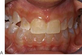
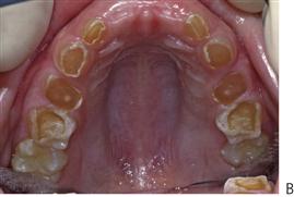
Figure 11.33 Manifestations of dentinogenesis imperfecta. (A) Dentinogenesis imperfecta associated with osteogenesis imperfecta. Dark discolouration of the crowns which appear normal in size and shape. (B) Severe attrition in the primary dentition in a case of dentinogenesis imperfecta.
Dental manifestations
• Amber, grey to purple-bluish discolouration or opalescence (Figure 11.33).
• Pulpal obliteration (Figure 11.34B).
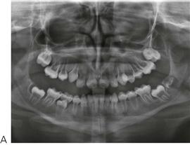
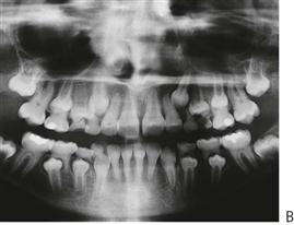
Figure 11.34 (A) Radiographic manifestations of dentinogenesis imperfecta showing short, bulbous crowns with wide-open root canals. (B) With further development, these teeth undergo pulpal canal obliteration, however, periapical pathology is fortunately rare.
• Enamel may be lost after tooth eruption, exposing the soft dentine, which rapidly wears. This is probably due to inherent weakness in the dentine rather than because of an enamel defect or abnormality at the dentinoenamel junction.
• Mantle dentine appears normal.
• Circumpulpal dentine has poorly formed dentine with abnormal direction of tubules. Small soft-tissue inclusions represent remnants of pulpal tissue.
Management
• Preservation of the vertical dimension of the occlusion.
• Continued commitment to and support of both children and families, providing adequate aesthetics and function through childhood and adolescence.
• Protection of posterior teeth from attrition using full coverage restorations.
• Provision of aesthetic appeal.
• Stainless steel crowns for posterior teeth.
• Initially composite resin to build up anterior teeth, possibly followed later by porcelain crowns. (These teeth will remain or even become increasingly brittle throughout life. Conventional crowns requiring tooth preparation may never be the treatment of choice, but see above, under ‘Amelogenesis imperfecta’.)
• Overdentures or even full dentures may be required in severe cases.
We have followed cases over many years into adulthood. The initial optimism over retaining these teeth for a life-time has been tempered by the eventual failure of complex restorative work and the loss of teeth in early adulthood. Clinicians must be sensitive to the implications of long-term failure and the aesthetic, functional and indeed financial legacy with which the patient is left.
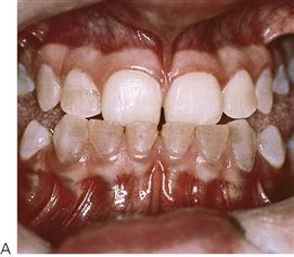
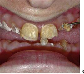
Figure 11.35 (A) It appears initially that the permanent dentition may not be as severely affected as the primary dentition in osteogenesis imperfecta. In this child, the severe skeletal Class III malocclusion and the posterior open bite required a surgical solution. (B) Clinical photograph of the same case at 30 years of age shows the destruction of the dentition over time resulting in the eventual loss of all of her teeth.
Osteogenesis imperfecta (OMIM 166200)
Osteogenesis imperfecta is caused by mutations in the collagen type I genes on chromosomes 7 (7q22.1) and 17 (17q21.3-q22). Previous classifications listed several variants of osteogenesis imperfecta with either autosomal dominant or autosomal recessive modes of inheritance. More recently it has been realized that most or all cases represent autosomal dominant osteogenesis imperfecta with variable expression. The essential features are:
• Bone fragility (Figure 11.36).
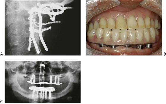
Figure 11.36 (A) The ‘Ransford Loop’ used to stabilize the vertebral column in a child with type IV osteogenesis imperfecta and basilar compression. This life-saving procedure is performed by an anterior approach through the pharynx, splitting the palate and sectioning the odontoid process of C2. The vertebral column is then wired to the occipital bone. This child initially presented with trigeminal neuralgia caused by pressure from C2 on the pons. (B) In spite of the bone pathology, osseointegrated implants can be successfully placed in patients with osteogenesis imperfecta. The same patient as in Figure 11.36A was rehabilitated with implant supported dentures following his surgery. (C) The panoramic radiograph shows the survival of the implants at 9-year follow-up.
Dentinal dysplasia – radicular dentinal dysplasia (Shields type I DD) (OMIM 125400)
• Originally described as rootless teeth, this appears to be a distinct entity from dentinogenesis imperfecta. Both dentitions are equally affected. The teeth may be lost early due to periapical infection or spontaneous exfoliation caused by the short roots.
• Autosomal dominant transmission.
• Teeth with very short or absent roots but clinically normal crowns (Figure 11.37).
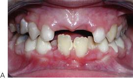
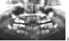
Figure 11.37 Radicular dentinal dysplasia. (A) The general appearance of the teeth is relatively normal. (B) While it appears that there is complete absence of the pulp chamber, a small horizontal band of pulp is evident at the beginning of root formation that is quite abnormal in appearance. (Courtesy Prof M-C Maniere, Strasbourg, France.)
• Total or partial obliteration of radicular pulp prior to eruption but with demilune of coronal pulp shown on the radiographs of molar teeth.
Dentinal dysplasia – coronal dentinal dysplasia (Shields type II DD) (OMIM 125420)
• The consensus now is that this is a variant of dentinogenesis imperfecta rather than a distinct entity. The primary teeth have a typical amber discolouration and undergo tooth wear associated with loss of the enamel and the appearance of ‘shell teeth’ radiographically.
• Varying degrees of pulp canal obliteration.
• Altered pulp morphology resembling a ‘thistle-shaped’ pulp chamber.
Management
Similar to the management of dentinogenesis imperfecta, however, some authors suggest that no treatment is required, as there are few sequelae. If there is enamel loss in the primary dentition, then full coverage restorations should be placed (stainless steel crowns). If the permanent dentition is clinically normal, then no special care may be needed.
Different classification of dentine anomalies
Many texts describe up to four different forms of dentinogenesis imperfecta. All these dentine anomalies are autosomal dominant in inheritance. Dentinogenesis imperfecta has been mapped to 4q13–21. Linkage studies of families with coronal dentine dysplasia (Shields type II DD) have shown that the candidate mutation occurs in a region on 4q that overlaps the most likely location of the dentinogenesis imperfecta locus. Further, a similar locus has been determined to that of dentinogenesis imperfecta Shields type III (Brandywine isolate – OMIM 125500). These results suggest that dentinogenesis imperfecta (Shields type II), Shields type III and DD type II (coronal dentinal dysplasia) are allelic or the result of mutations in tightly-linked genes. Subsequent studies have identified that the mutated gene in all of these phenotypes in the DSPP gene. Radicular dentinal dysplasia may be a separate entity.
X-linked vitamin D-resistant rickets (OMIM 307800) (Figure 11.38)
• Also termed X-linked rickets or hereditary hypophosphataemic rickets is due to a defect in a gene located at Xp22.
• X-linked disorder with rachitic changes in long bones associated with a failure of distal tubular reabsorption of phosphate in the kidneys. The rickets is unresponsive to vitamin D.
• Males severely affected, females may show milder features (typically short stature with bowing of legs), often not affecting the teeth.
Figure 11.38 (A) X-linked vitamin D-resistant rickets presenting with multiple abscessed teeth in the absence of caries. (B) Under polarized light, the hard-tissue section demonstrates globular dentine and a pulp horn that extends to the dentinoenamel junction, resulting in early exposure caused by attrition and subsequently pulpal necrosis.
Dental manifestations
• Attrition of incisal and occlusal enamel exposes elongated pulp horns, which often extend up to the dentinoenamel junction.
• Males (and some females), typically present with multiple abscesses in the absence of dental caries.
• Large pulp chambers and delayed apical root closure.
• May be reduced radiodensity of dentine on radiographs. Enamel may be spared or show some evidence of hypoplasia and/or hypomineralization.
• Patients may have repeated orthopaedic procedures and indwelling mechanics to promote bone straightening or lengthening. The avoidance of sepsis is essential. Treatment planning should include collaboration with orthopaedic colleagues and may demand the regrettable removal of infected teeth at times of particular infection risk.
Pre-eruptive intracoronal resorptive defects (Figure 11.39)
These defects are dentine lesions found on unerupted teeth, usually detected on routine dental radiographs. They have often erroneously been referred to as ‘pre-eruptive caries’ or ‘dentine cysts’. They are often located adjacent to the dentinoenamel junction in the occlusal aspect of the crown. There is evidence that these defects develop as a result of coronal resorption. On opening into the lesion, it is often empty or filled with an amorphous tissue comprising small particles of tubular dentine and crystalline material. Resorptive cells such as osteoclasts and macrophages may be found. When the tooth erupts, the lesion is likely to be rapidly colonized by oral flora and the lesion becomes similar to a carious lesion.
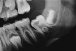
Management
The cavity should be restored conservatively. It has been proposed that these lesions may be responsible for many of the lesions that are clinically undetected in molar teeth that progress to rapid carious breakdown and, ultimately, loss of the tooth.
Dental effects of prematurity and low birth weight
| Normal birth weight for gestational age | >2500 g |
| Low birth weight | >1500–2500 g |
| Very low birth weight | <1500 g |
| Extremely low birth weight | <1000 g |
Problems in extreme prematurity
• Hyaline membrane disease and respiratory insufficiency.
• Hyperbilirubinaemia (Figure 11.40D).
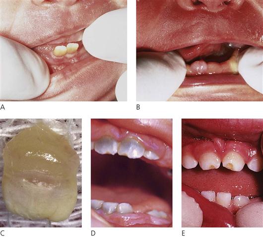
Figure 11.40 (A) Natal teeth in a 36-week premature infant. The teeth were extremely loose and the teeth were removed. (B) A newborn infant with two mandibular incisors soon to erupt. The reduced enamel epithelium has fused with the gingivae and teeth will probably erupt within a few days. (C) A neonatal tooth from a 27-week premature infant. Note the extent of the crown formation is consistent with the chronological age. (D,E) Dental effects of prematurity: hyperbilirubinaemia staining of the enamel (D) and hypoplastic defects on the incisal edges of the central incisors caused by laryngoscopy (E).
The limiting factor in survival is based on lung development and infants weighing less than 400 g at birth or those born before 24 weeks, rarely survive. Hyaline membrane disease is now treated with synthetic surfactant, although very young babies often develop pneumothoraces caused by the prematurity and fragility of the alveoli. Cerebral intraventricular bleeding and necrotizing enterocolitis with the resulting sepsis are common causes of mortality and morbidity. Surviving children may be left with problems of growth retardation, delayed cognitive development and a range of other abnormalities.
Dental implications
• Hypocalcaemia with reactive pseudohyperparathyroidism.
• Hyperbilirubinaemia, causing intrinsic staining of the teeth.
• Intubation trauma, causing enamel hypoplasia/hypocalcification. The maxillary central incisors are most often affected (Figure 11.40E). If the baby is intubated orally, palatal grooving may occur. Tooth eruption may be delayed, although it is often normal for the ‘corrected’ age after adjustment for prematurity.
Disorders of eruption (Figure 11.41)
Eruption of teeth is not always correlated with somatic development. Children with growth disturbances may exhibit delayed eruption or the delay may be due to other causes such as gingival overgrowth due to medication such as phenytoin. More importantly, premature exfoliation of teeth is invariably associated with severe systemic disease (see Chapter 10) and requires investigation.
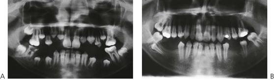
Figure 11.41 Teeth require guidance for normal eruption. (A) In this case the mandibular right first permanent molar was removed because of gross caries and pulpal necrosis. (B) While the second molar has drifted slightly mesially, the second premolar has rotated and drifted distally and impacted against the second molar. Had the second primary molar not been removed it is unlikely that this premolar would have drifted.
Delayed eruption of the primary dentition requires no treatment other than determining that all teeth are present. It is uncommon for children to require surgical exposure of the teeth in infancy. Parents should be reassured that there is considerable variability in the eruption of teeth (plus or minus 6 months for primary teeth, plus or minus 1 year for permanent teeth). In the permanent dentition, delayed eruption beyond this range should be investigated for the presence of supernumeraries and other pathology. Although the actual timing of tooth eruption is variable, evidence of progress in tooth crown and root development and the eruption sequence are of much more relevance. In contrast, the failure of eruption of a contralateral tooth more than 6 months after the appearance of its partner requires investigation.
Natal and neonatal teeth (Figure 11.40)
A natal tooth is present at birth, while a neonatal tooth is one that erupts within 30 days of birth. In almost all cases, this is simply the early eruption of a normal primary incisor tooth. The development of this tooth is consistent with the expected stage of development of a primary incisor at birth (i.e. only five-sixths of the crown is formed without any root being present). This lack of root development accounts for the mobility of the tooth. Babies with posterior natal teeth should be carefully investigated for other systemic conditions that may be associated with syndromes or other diseases.
Management
• The most important point to consider is whether the nursing mother can adequately establish breast-feeding. If either the nipples or the ventral surface of the infant’s tongue are being traumatized, the tooth should be removed.
• If the tooth is not excessively mobile it should be retained as it can become firm with time as the root continues to develop.
• If the tooth is excessively mobile, then it may spontaneously exfoliate; however, because of the theoretical risk of aspiration or ingestion it should be electively removed.
• If tooth removal is indicated, care should be taken to extract the entire tooth, as the crown only may be removed leaving behind the pulpal tissue. If this is the case, the dentine and a root will form subsequently; the root will then require removal at a later date.
• The permanent teeth should be unaffected by extraction of the primary tooth.
Infraoccluded (submerged) primary molars (Figure 11.42)
It is quite common for primary molars to become infraoccluded, however, it is controversial as to whether all teeth are truly ankylosed. The mechanism by which this occurs is unknown, but it has been suggested that a cessation of normal primary root resorption may stimulate healing and then ankylosis as the bone remodels. Some teeth never appear in the mouth and this infraocclusion may result from a failure or partial failure of teeth eruption, particularly secondary primary molars. If ankylosis occurs post-eruption, the tooth will appear to submerge into the alveolus (in fact, the tooth remains stationary while the alveolar bone grows around it and adjacent teeth erupt). The timing of the removal of an ankylosed tooth is based on the position of the first permanent molar and the extent of the resorption of the primary tooth.
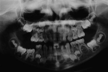
Figure 11.42 Failure of eruption of the mandibular right second primary molar. It is questionable whether these teeth are truly ankylosed. It is important to wait until the first permanent molar has erupted before surgical removal to avoid impaction of the second premolar. These teeth are often difficult to remove, especially if there is space loss and they should therefore be sectioned and elevated to minimize excessive bone loss.
Management
• If there is radiographic evidence of resorption of the roots, then removal should be delayed, as the vast majority of these teeth will exfoliate normally, because the tooth is probably not ankylosed.
• Orthodontic consultation (see Chapter 14).
• If the premolar is congenitally absent, early removal of the submerging primary molar might be indicated.
• Surgical removal of the ankylosed tooth is to be avoided before eruption of the first permanent molar as this latter tooth will migrate mesially and space loss is likely to occur together with impaction of the second premolar. Once the permanent tooth has erupted, the primary tooth may be removed and a space maintainer inserted.
• Retain space and use orthodontic treatment to align the permanent molar as required.
Failure of eruption of first permanent molars (Figure 11.43)
Failure of eruption of a first permanent molar is an uncommon finding, however, no one has yet been able to explain the mechanism as to why these teeth do not erupt. During surgical intervention it is invariably noted that these teeth are not ankylosed.
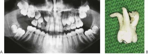
Figure 11.43 (A,B) Failure of eruption of first permanent molars is not uncommon. Surgical exposure of the crown may be sufficient to allow these teeth to erupt. Note that root development will continue in spite of a failure of tooth eruption and deviation of the roots will occur when the roots reach the cortical bone, in this case the antral floor. This root deviation may not be evident on radiographs, especially in the maxilla, and extraction of these teeth may prove extremely difficult.
Root development (Figure 11.44)
Just as enamel can be affected by systemic illness, so too can root development be delayed, altered or arrested by systemic disease. This is most commonly seen when radiotherapy causes shortening and tapering of the roots of premolars (see Chapter 10). Excessive orthodontic forces may also cause root resorption.
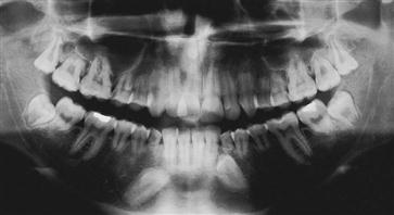
Figure 11.44 Arrested root development in a child who developed Stevens–Johnson syndrome at 10 years of age. Root development ceased at that time (probably as a result of the treatment as much as the disorder itself) and all teeth except the third molars were affected. It is interesting that these molars, which were not undergoing calcification, were unaffected.
Dental age (maturity) determination
The paediatric dentist is often asked to help in age assessment, either at necropsy or for orphaned children. It is important to take into consideration ethnicity and variation in somatic growth potential.
Tooth emergence may not be as important as tooth crown calcification and root development. The most widely used and accepted method is that developed by Demirjian (1978), based on the panoramic radiographic appearance of tooth calcification at different ages.
Although there remains little doubt that peak height velocity, skeletal development and sexual maturation are associated, dental development seems to be independent of general somatic development.
Loss of tooth structure
• From wear of one or more teeth in one arch against one or more teeth in the opposing arch.
• Exogenous from diet, habits or environment.
• Exogenous tooth substance loss from diet, habits or environment.
Enamel erosion
The prevalence of erosion in children and adolescents has been reported recently as very high, with over half of 14-year-olds in a UK population having moderate erosion, with an increased prevalence seen in lower socioeconomic groups. The aetiology of erosion in children and adolescents is varied and it has been suggested that the increased consumption of fruit juices and carbonated drinks is the most important factor, with the sale of soft drinks increasing by 56% over the past decade.
The erosive potential of acidic drinks is related to:
Carbonated soft drinks contain carbonic acid and often organic acids (commonly citric acid) are added to improve taste and ‘mouth feel’. Citrate ions strongly chelate calcium in both acidic and basic environments decreasing the amount of free ionic calcium available in both saliva and at the enamel surface and thereby enhancing demineralization. The erosive potential of ‘diet’ soft drinks is similar to that of sugared drinks; however, their potential to increase caries risk is decreased markedly. The method of drinking can also affect the extent of erosion, with the decrease in intraoral pH becoming greater as the beverage is held in the mouth or is drunk by ‘long sipping’; when the beverage is gulped, intra-oral pH does not decrease significantly.
The long-term and short-term consequences of dental erosion are marked, with the need for extensive and costly dental care and potential loss of teeth. The concomitant dental sensitivity can be severe and debilitating. It has been shown that even a few intakes of acidic drinks on a regular basis may be associated with considerable dental erosion. It is important to question the child and parent(s) carefully as to total family usage of such drinks (fruit squashes, fresh fruit juices, particularly citrus juices, carbonated drinks and colas) in the first instance. The taking of study models and the institution of an exclusion diet for 3 months may show a diagnostic ‘tide mark’ of unattacked tooth substance at a later review.
Prevention of erosion
• Cessation or restriction of exposure to the aetiological factor.
• Modification of beverage erosivity seems to have the most future potential for reducing tooth structure loss. Recent research has concentrated on the addition of calcium and/or phosphate and pH alteration of soft drinks.
• Families are bombarded with advice on healthy eating. They may find advice to limit intake of fruit juices confusing and careful explanation is required.
Gastro-oesophageal reflux (Figure 11.45A)
When loss of enamel by erosion cannot be explained by dietary factors, reflux must be considered. Children with reflux will show enamel erosion, which is a smooth loss of tooth structure, characteristically with any restorations standing proud. Some children have undiagnosed, asymptomatic reflux that presents first with enamel erosion.
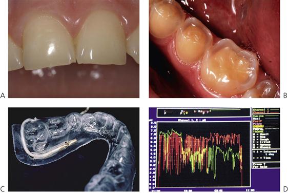
Figure 11.45 (A) Enamel erosion with asymptomatic gastro-oesophageal reflux. The first presentation of this child to a dentist was because of the erosion. Note the smooth, almost glassy appearance of the incisors. (B) Severe erosion in a 12-year-old boy showing the outline of enamel with exposed dentine. (C) A mouthguard containing a micro pH probe was worn in the mouth for a 24-hour period to measure the intra-oral pH. Another is placed in the lower oesophageal sphincter. (D) In some children an intra-oral pH of <1 has been recorded.
Diagnosis
• Barium swallows may not demonstrate reflux.
• 24-hour pH manometry is required to assess the extent of reflux (Figure 11.38D).
Management
• Diagnosis and treatment of reflux condition before definitive restoration of the teeth.
• Histamine blockers (H2 antagonists) such as ranitidine and cimetidine.
• Anti-emetics (prokinetic agents) such as metoclopramide.
• Composite resin, stainless steel crowns, glass ionomer cement coverings or onlays over the posterior teeth.
• Onlays on posterior teeth to protect occlusion and maintain vertical height.
• Professional application of fluorides. Nocturnal mouth guards with fluoride toothpaste as mechanical barriers against acid attack and fluoride to promote remineralization.
Further reading
Hypodontia
1. Bergendal T, Eckerdal O, Hallonsten A-L, et al. Osseointegrated implants in the oral habilitation of a boy with ectodermal dysplasia: a case report. International Dental Journal. 1991;41:149–156.
2. Fleming P, Nelson J, Gorlin RJ. Single maxillary central incisor in association with mid-line anomalies. British Dental Journal. 1990;168:476–479.
3. Freire-Maia N, Pinheiro M, eds. Ectodermal dysplasias: a clinical and genetic study. New York: Alan R Liss; 1984.
4. Hall RK. Congenitally missing teeth – diagnostic feature in many syndromes of the head and neck. Journal of the International Association of Dentistry for Children. 1983;14:69–75.
5. Hall RK, Bankier A, Aldred MJ, et al. Solitary median maxillary central incisor, short stature, choanal atresia/midnasal stenosis (SMMCI) syndrome. Oral Surgery, Oral Medicine, Oral Pathology, Oral Radiology, and Endodontics. 1997;84:651–662.
6. Lai PY, Seow WK. A controlled study of the association of various dental anomalies with hypodontia of permanent teeth. Journal of Pediatric Dentistry. 1989;11:291–295.
7. Nohl F, Cole B, Hobson R, et al. The management of hypodontia: present and future. Dental Update. 2008;35:79–80.
8. Rappaport EB, Ulstrom R, Gorlin RJ, et al. Solitary maxillary central incisor syndrome and short stature. Journal of Pediatrics. 1977;91:924–928.
Supernumerary teeth and other disorders
1. Hogstrom A, Andersson L. Complications related to surgical removal of anterior supernumerary teeth in children. Journal of Dentistry for Children. 1987;54:341–343.
2. Jensen BL, Kreiborg S. Development of the dentition in cleidocranial dysplasia. Journal of Oral Medicine and Pathology. 1990;19:89–93.
3. Jensen BL, Kreiborg S. Dental treatment strategies in cleidocranial dysplasia. British Dental Journal. 1992;172:243–247.
4. Omer RSM, Anthonappa RP, King NM. Determination of the optimum time for surgical removal of unerupted anterior supernumerary teeth. Pediatric Dentistry. 2010;32:14–20.
5. Papadaki ME, Lietman SA, Levine MA, et al. Cherubism: best clinical practice. Orphanet Journal of Rare Diseases. 2012;7(Suppl. 1):S6.
6. Von Arx T. Anterior maxillary supernumerary teeth A clinical and radiographic study. Australian Dental Journal. 1992;37:189–195.
Morphological anomalies
1. Nazif MM, Laughlin DF. Dens invaginatus in a geminated central incisor: case report. Journal of Pediatric Dentistry. 1990;12:250–251.
2. Rakes GM, Aiello AS, Kuster CG. Complications occurring resultant to dens invaginatus: a case report. Pediatric Dentistry. 1998;10:53–56.
3. Tsai SJJ, King NM. A catalogue of anomalies and traits of the permanent dentition of southern Chinese. Journal of Clinical Pediatric Dentistry. 1998;22:185–194.
Regional odontodysplasia
1. Aldred MJ, Crawford PJM. Regional odontodysplasia: a bibliography. Oral Pathology and Medicine. 1989;18:251–263.
Enamel hypomineralization
1. Cullen C. Erythroblastosis fetalis produced by Kell immunization: dental findings. Journal of Pediatric Dentistry. 1990;12:393–396.
2. Fleming P, Witkop CJ, Kuhlmann WH. Staining and hypoplasia caused by tetracycline. Journal of Pediatric Dentistry. 1987;9:245–246.
Enamel hypoplasia
1. Croll TP. Enamel microabrasion for removal of superficial dysmineralization and decalcification defects. Journal of the American Dental Association. 1990;129:411–415.
2. Eli H, Sarnat H, Talmi E. Effect of the birth process on the neonatal line in primary tooth enamel. Journal of Pediatric Dentistry. 1989;11:220–223.
3. Pendrys DG. Dental fluorosis in perspective. Journal of the American Dental Association. 1989;122:63–66.
Molar–incisor hypomineralization (MIH)
1. Beentjes VE, Weerheijm KL, Groen HJ. Factors involved in the aetiology of molar–incisor hypomineralisation (MIH). European Journal of Paediatric Dentistry. 2002;3:9–13.
2. Fayle SA. Molar–incisor hypomineralisation: restorative management. European Journal of Paediatric Dentistry. 2003;4:121–126.
3. Weerheijm KL. Molar–incisor hypomineralisation (MIH). European Journal of Paediatric Dentistry. 2003;4:114–120.
4. Weerheijm KL, Duggal M, Mejare I, et al. Judgement criteria for molar–incisor hypomineralisation (MIH) in epidemiologic studies: a summary of the European meeting on MIH held in Athens, 2003. European Journal of Paediatric Dentistry. 2003;4:110–113.
Amelogenesis imperfecta
1. Bäckman B, Ammeroth G. Microradiographic study of amelogenesis imperfecta. Scandinavian Journal of Dental Research. 1989;97:316–329.
2. Seow WK. Clinical diagnosis and management strategies of amelogenesis imperfecta variants. Pediatric Dentistry. 1993;15:384–393.
Dentine anomalies
1. Cole DEC, Cohen MM. Osteogenesis imperfecta: an update. Journal of Pediatrics. 1991;115:73–74.
2. Gage JP, Symons AL, Roumaniuk K, et al. Hereditary opalescent dentine: variation in expression. Journal of Dentistry for Children. 1991;58:134–139.
3. O’Carroll MK, Duncan WK, Perkins TM. Dentin dysplasia: review of the literature and a proposed sub-classification based on radiographic findings. Oral Surgery, Oral Medicine, and Oral Pathology. 1991;72:119–125.
4. Seow WK, Latham SC. The spectrum of dental manifestations in vitamin D-resistant rickets: implication for management. Pediatric Dentistry. 1991;8:245–250.
5. Seow WK, Brown JP, Tudehope DA, et al. Dental defects in the deciduous dentition of premature infants with low birth weight and neonatal rickets. Pediatric Dentistry. 1984;6:88–92.
Eruption disorders
1. Friend GW, Mincer HM, Carruth KR, et al. Natal primary molar: case report. Journal of Pediatric Dentistry. 1991;13:173–175.
2. Masatomi Y, Abe K, Ooshima T. Unusual multiple natal teeth: case report. Journal of Pediatric Dentistry. 1991;13:170–172.
3. Sauk JJ. Genetic disorders involving tooth eruption anomalies. In: Davidovitch Z, ed. The biological mechanisms of tooth eruption and root resorption. Birmingham: EBSCO Media; 1988;171–179.
4. Shaw W, Smith M, Hill F. Inflammatory follicular cysts. ASDC Journal of Dentistry for Children. 1980;47:97–101.
Erosion
1. Jarvinen V, Muerman JH, Hyvarinen H, et al. Dental erosion and upper gastrointestinal disorders. Oral Surgery, Oral Medicine, and Oral Pathology. 1988;65:298–303.
General
1. Demirjian A. Dental development: index of physiologic maturation. Medicine and Hygiene. 1978;36:3154–3159.
2. Hall RK. Pediatric orofacial medicine and pathology. London: Chapman and Hall Medical; 1994.
World Wide Web Database
1. Online Mendelian Inheritance in Man, OMIM (TM). McKusick-Nathans Institute for Genetic Medicine, Johns Hopkins University (Baltimore, MD) and National Center for Biotechnology Information, National Library of Medicine (Bethesda, MD). In: http://www.ncbi.nlm.nih.gov/omim/; 2000.