Infection and Inflammation of the Central Nervous System

The CNS may be affected directly by bacteria, viruses, fungi, parasites, and mycobacteria. Viruses causing acute and chronic CNS infections are listed in Table 17-10. The infecting microorganisms gain entry to the nervous system by (1) hematogenous spread through arterial blood or (2) direct extension from another site of infection.66 Neurologic infections produce disease by several mechanisms: direct neuronal or glial infection; mass lesion formation; inflammation with subsequent edema; interruption of cerebrospinal fluid pathways; neuronal damage, or vasculopathy; and secretion of neurotoxins (Figures 17-32, 17-33, and 17-34). Syndromes are acute and subacute bacterial meningitis, epidural and brain abscess, encephalitis, peripheral neuropathy, or neurosyphilis depending on the infecting microorganism. Signs and symptoms are produced because of (1) interference with the function of the nervous system tissue being invaded or compressed or (2) the inflammatory response produced by the body in response to infection. The cardinal signs of CNS infection are fever, head or spine pain, and generalized or focal neurologic dysfunction.67

Table 17-10

Viruses Causing Acute and Chronic CNS Diseases

image

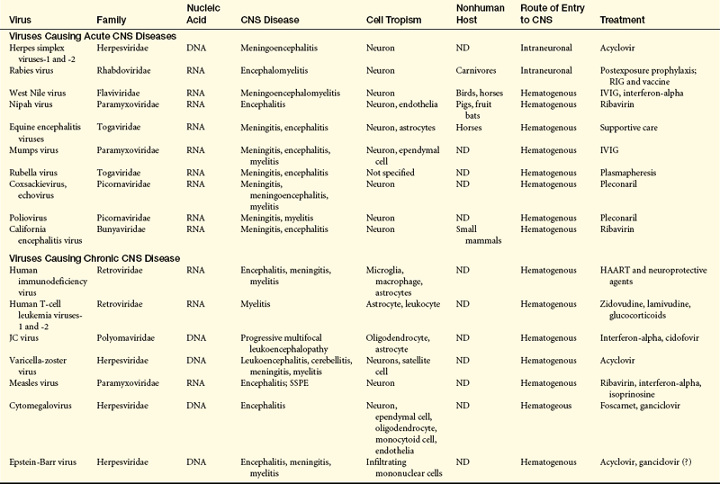
CNS, Central nervous system; DNA, deoxyribonucleic acid; HAART, highly active retroviral therapy; IVIG, intravenous immunoglobulin; JC, John Cunningham; ND, not determined; RIG, rabies immune globulin; RNA, ribonucleic acid; SSPE, subacute sclerosing panencephalitis.

From Lindquist L, Vapalahti O: Lancet 371(9627):1861-1871, 2008; Power C, Noorbakhsh F: Central nervous system viral infections: clinical aspects and pathogenic mechanism. In Gilman S, editor, Neurobiology of disease, pp 487-488, Burlington, MA, 2007, Elsevier.

image

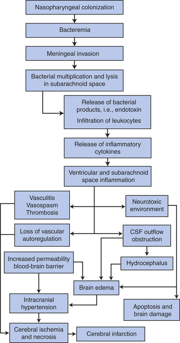
Figure 17-32 Pathogenesis of meningitis. (Adapted from Cohen J, Powderly WG: Infectious diseases, ed 2, Mosby, 2004, Edinburgh.)

image

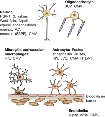
Figure 17-33 Viral infection in the central nervous system (CNS). Viruses infect specific cell types within the CNS depending on the specific properties of the virus together with individual cell membrane proteins expressed on permissive cell types. Normally the brain is protected from circulating pathogens and toxins by the blood-brain barrier. HIV, Human immunodeficiency virus; CMV, cytomegalovirus; HSV, herpes simplex virus; HTLV-1, human T-cell lymphotropic virus (causes T-cell leukemia); JCV, John Cunningham virus (a polyomavirus causing progressive multifocal leukoencephalopathy); SSPE, subacute sclerosing panencephalitis; VZV, varicella zoster virus. (Adapted from Power C, Noorbakhsh G: Central nervous system viral infections: clinical aspects and pathogenic mechanisms. In Gilman S, editor, Neurobiology of disease, p 488, Burlington, MA, 2007, Elsevier.)

image

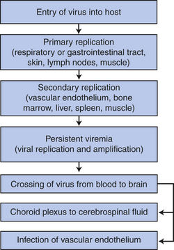
Figure 17-34 Hematogenous spread of viral pathogens to the central nervous system. (Adapted from Cohen J, Powderly WG: Infectious diseases, Mosby, 2007, Edinburgh.)

Meningitis

Meningitis (infection of the meninges) may be caused by bacteria, viruses, fungi, parasites, or other toxins. (The pathophysiology of infection is discussed in Chapter 9.) The infections are classified as acute, subacute, or chronic processes, and the pathophysiology, clinical manifestations, and treatment differ for each type of microorganism.

Bacterial meningitis is primarily an infection of the pia mater and arachnoid, the subarachnoid space, the ventricular system, and the CSF. A systemic or bloodstream infection or a direct extension from an infected area is the access route to the subarachnoid space. The bacterial infection originates in another part of the body. The incidence of bacterial meningitis is 2.5 to 3.5 cases per 100,000. The incidence is 20 per 100,000 annually for neonates, and 2 to 9 per 100,000 annually for those older than 60 years. The mortality is 25% in adults. Meningococcus (Neisseria meningitidis) and pneumococcus (Streptococcus pneumoniae) are the common causes of bacterial meningitis after the neonatal period. Pneumococcus and gram-negative enteric bacilli are the most common neonatal agents.

Meningococcus has been identified worldwide. Meningococcal meningitis occurs predominantly in men and boys and during the fall, winter, and spring. Epidemics of meningococcal meningitis occur in approximately 10-year cycles. Children and adolescents are affected predominantly; with pneumococcal meningitis, young persons and those older than 40 years are mostly affected.

Aseptic meningitis (viral meningitis, nonpurulent meningitis, lymphocytic meningitis) is an inflammation believed to be limited to the meninges. The most at-risk populations and the time of year when occurrences are seen depend on the virus. Aseptic meningitis produces a variety of symptoms and is caused by a variety of infectious agents, most of which are viruses. They include enteroviral viruses (the most common) (echovirus, coxsackievirus, and nonparalytic poliomyelitis), mumps, herpes simplex types 1 and 2, St. Louis encephalitis virus, West Nile virus, California encephalitis virus, Venezuelan equine encephalitis, Colorado tick fever, lymphocytic choriomeningitis virus, Epstein-Barr virus, and influenzavirus types A and B.68 Bacterial infections not adequately treated are another cause of aseptic meningitis.

Fungal meningitis is a chronic, much less common condition than bacterial or viral meningitis. The most common fungal infections of the nervous system are histoplasmosis, cryptococcosis, coccidioidomycosis, mucormycosis, candidiasis, and aspergillosis. Fungal meningitis most frequently occurs in persons with impaired immune responses or alterations in normal body flora. Fungal meningitis develops insidiously, usually over days or weeks. Syphilis, tuberculosis, and Lyme disease also are associated with chronic meningitis.

Tubercular meningitis, the most common and serious form of CNS tuberculosis, is again on the rise in the United States, especially in persons with acquired immunodeficiency syndrome (AIDS). Miliary tubercles form in the brain and meninges. At some point the tuberculomas erode the pia mater, and the mycobacteria enter the CSF, producing a hypersensitivity reaction that results in a purulent exudate involving the basal meninges, cerebrum, and spinal nerves. Cerebral ischemia and infarction occur from vasculitis. Symptoms include headache, low-grade fever, nausea and vomiting, irritability, difficulty sleeping, and fatigue. These signs and symptoms increase to confusion, stiff neck, significant behavioral changes, and seizures. Hydrocephalus and cranial nerve palsies or cerebral infarcts may occur. Recovery rate is 90% with early diagnosis and treatment with appropriate antituberculosis therapy.

PATHOPHYSIOLOGY The bacteria that commonly cause bacterial meningitis are common inhabitants of the nasopharynx, but a predisposing factor such as a prior upper respiratory infection must be present before the bacteria become blood-borne. Bacterial meningitis also may develop as a consequence of ear, dental, or paraspinal infections; impairment in the anatomic barrier from trauma or neurosurgery; and, rarely, when a brain abscess ruptures into the ventricular system or subarachnoid space.67 The method of CNS entry is through the choroid plexuses or areas of altered blood-brain barrier or by hematogenous spread. Bacteria multiply in the subarachnoid space. The bacteria or their toxins function as irritants and induce an inflammatory reaction by the meninges (pia mater and arachnoid), the CSF, and the ventricles. The meningeal vessels undergo change, becoming hyperemic and increasingly permeable. Blood cells (neutrophils) migrate into the subarachnoid space, producing an exudate that thickens the CSF and interferes with normal CSF flow around the brain and spinal cord (Figure 17-35). The exudate has the potential to obstruct arachnoid villi and produce hydrocephalus and interstitial edema. The amount of purulent exudate increases rapidly (especially around the base of the brain), causing further inflammation. The exudate extends into the sheaths of the cranial and spinal nerves and into the perivascular spaces of the cortex. Meningeal cells become edematous. The exudate and vasogenic edema increase ICP. The small and medium-sized subarachnoid arteries, veins, and choroid plexuses undergo inflammatory changes and become engorged, disrupting blood flow and potentially producing thrombosis. Secondary infection of the brain may occur. The cortical neurons also show some changes, including an increase in the number of microglia and astrocytes.

image

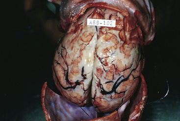
Figure 17-35 Acute leptomeningitis. The leptomeninges contain abundant creamy, purulent exudate, most prominently over the superior surface of the cerebrum. The underlying brain is swollen, and the vessels are congested. (From Kumar V, Cotran RS, Robbins SL: Robbins basic pathology, ed 7, Philadelphia, 2003, Saunders.)

Fungi in the nervous system usually produce a granulomatous reaction with formations of granulomas or gelatinous masses. These usually develop in the meninges at the base of the brain. Fungi also may extend along the perivascular sites in the subarachnoid space and into the brain tissue, producing arteritis with thrombosis, infarction, and communicating hydrocephalus. Meningeal fibrosis develops later in the inflammatory process. Cranial nerve dysfunction, caused by compression, often results from the granulomas and fibrosis.

CLINICAL MANIFESTATIONS The clinical manifestations of a bacterial meningitis can be grouped as (1) inflammation and irritation—generalized meningeal signs, throbbing headache that becomes more severe, photophobia that becomes more severe, nuchal rigidity, Kernig sign, and Brudzinski sign; (2) local tissue dysfunction—cranial nerve palsies, focal neurologic deficits (such as hemiparesis/hemiplegia, ataxia), and seizures; (3) mass effect—decreased level of consciousness, nausea, vomiting, and increased intracranial pressure; and (4) vascular compromise. The irritation and damage to the cranial nerves produced by the inflamed sheaths manifest as follows:

Cranial nerve II: papilledema, blindness

Cranial nerves III, IV, and VI: ptosis, visual field deficits, diplopia

Cranial nerve V: photophobia

Cranial nerve VII: facial paresis

Cranial nerve VIII: deafness, tinnitus, vertigo

Neck stiffness and pain, and possibly head retraction, reflect the irritability of spinal accessory and cervical spinal nerves. Often the vomiting center is irritated, causing projectile vomiting. Confusion and decreasing responsiveness are evidence of cortical involvement. In meningococcal meningitis, petechial or purpuric rash involving the skin and mucous membranes occurs. As ICP increases, papilledema may develop and delirium may progress to the point that the individual becomes unconscious.

The signs and symptoms of bacterial meningitis in neonates are subtle and nonspecific. Low-grade fever and mild behavioral changes with few meningeal signs may be the first signs. High fever, lethargy, irritability, hypothermia, seizures, bulging fontanels, poor feeding, vomiting, and respiratory distress may be present. Children may present with either a subacute infection that worsens over several days following an ear infection or upper respiratory infection, or as an acute fulminant illness that has developed rapidly over a few hours. Older adults often develop low-grade fever with confusion or other mild behavioral changes. Stupor or coma may appear later.

The clinical manifestations of aseptic meningitis are mild compared with those associated with bacterial meningitis. Mild generalized throbbing headache, mild photophobia, mild neck pain, stiffness, fever, and malaise are manifestations of aseptic meningitis.

Fungal meningitis develops slowly and insidiously. The first manifestations are often those of dementia or communicating hydrocephalus (see Chapter 16). The individual is characteristically afebrile.

EVALUATION AND TREATMENT Diagnosis of bacterial meningitis is based on physical examination, including skin rash, nasopharyngeal smear, and antigen tests. CSF is the gold standard for diagnosis. Bacterial meningitis and fungal meningitis are treated with appropriate antibiotic therapy, but resistant strains are an increasing problem. Other supportive measures may be needed. Aseptic meningitis is managed pharmacologically with antiviral drugs and steroids. Conjugate vaccines exist for meningococcal, pneumococcal, and hemophilic meningitis.69 Chemoprophylaxis for exposure to meningococcal meningitis is rifampin, ciprofloxacin, or ceftriaxone.70

Suppurative Cerebral Masses

Localized pus-filled masses can develop with the CNS. The mass may be an abscess within the brain or spinal cord, a subdural empyema with the infection between the dura and the subarachnoid space, or an epidural abscess with the infection between the dura and skull or vertebrae. The latter two occur less commonly but have similar predisposing conditions, infectious agents, and clinical manifestations. Treatments are also similar. Subdural empyemas and epidural abscess generally spread locally from infections in an adjoining structure.

Abscess: Abscesses are localized collections of pus within the parenchyma of the brain and spinal cord. The incidence of abscesses is about 1 per 100,000 hospital admissions. Men experience abscesses more frequently than women, with a 2:1 ratio. The median age for abscess formation is 30 to 40 years of age. Abscesses occur (1) after open trauma and during neurosurgery; (2) in association with a contiguous focus of infection, such as the middle ear, mastoid cells, nasal cavity, and nasal sinuses; (3) through metastatic or hematogenous spread from distant foci, such as the heart, lungs, pelvic organs, skin, tonsils, abscessed teeth, osteomyelitis in other than cranial bones, and dirty needles (especially in compromised hosts); and (4) cryptogenically, arising without other associated areas of infections. Streptococci, staphylococci, and bacteroids, often in combination with anaerobes, are the most common bacteria that cause abscesses; however, yeast and fungi also have been found in CNS abscesses. Toxoplasma gondii is producing an ever-increasing number of CNS abscesses in individuals with AIDS: 80% are located in the cerebrum and 20% are cerebellar. The frontal and temporal lobes are the most common sites (Figure 17-36). The abscesses are in more than one site in 5% to 20% of cases. The immunosuppressed are particularly at risk for abscesses.

image

Figure 17-36 Brain abscess. The abcess is sharply demarcated, indicating that it has been present for some time. Purulent exudate is visible in the center of the abscess. Because antibiotics penetrate very poorly into abscesses, surgical drainage is often necessary to treat such lesions. (From Kumar V, Cotran RS, Robbins SL: Robbins basic pathology, ed 7, Philadelphia, 2003, Saunders.)

Spinal cord abscesses are classified as epidural or intramedullary. Debilitated individuals with sepsis more frequently develop intramedullary spinal cord abscesses (those within the spinal cord).

PATHOPHYSIOLOGY Microorganisms gain entrance to the CNS from adjacent sites by direct extension from osteomyelitis or spread along the wall of a vein. Infective emboli carry the microorganisms from distant sites. Brain abscess evolves through four stages regardless of infecting microorganism except in the immunosuppressed host, where the process may be incomplete. The stages are as follows:

1. Early cerebritis (days 1 to 3): localized inflammatory process in which perivascular infiltration or inflammatory cells, composed of neutrophils, plasma cells, and mononuclear cells, surround a central core of coagulative necrosis; marked cerebral edema surrounds the area

2. Late cerebritis (days 4 to 9): necrotic center is surrounded by inflammatory infiltrate of macrophages and fibroblasts; rapid new blood vessel formation occurs around the abscess; a thin capsule of fibroblasts and reticular fibers gradually develops; the area is still surrounded by cerebral edema

3. Early capsule formation (days 10 to 13): necrotic center decreases in size; inflammatory infiltrate changes in character and contains an increasing number of fibroblasts and macrophages; mature collagen evolves forming a capsule

4. Late capsule formation (days 14 and longer): well-formed necrotic center surrounded by a dense collagenous capsule67

A free (nonencapsulated) abscess is associated with a higher mortality. A mature abscess has three layers: (1) a center of polymorphonuclear leukocytes, (2) a collagenous capsule, and (3) peripheral gliosis. Existing abscesses also tend to spread and form daughter abscesses.

Abscesses arising from the ear frequently are located in the middle or inferior temporal lobe or in the anterolateral cerebellar hemispheres. Abscesses originating from the oral and nasal area most commonly are located in the frontal and temporal lobes. Abscesses from distant foci often occur in multiple numbers in the distal portion of the middle cerebral arteries. In extradural abscesses, pus and granulation tissue accumulate in the extradural space.

CLINICAL MANIFESTATIONS Clinical manifestations of brain abscesses are associated with (1) intracranial infection, such as fever and increased sedimentation rate; or (2) an expanding intracranial mass, such as headache, nausea, vomiting, decreasing cognitive abilities, paresis, and seizures. Early clinical manifestations of brain abscesses are low-grade fever, headache, neck pain and stiffness with mild nuchal rigidity, confusion, drowsiness, sensory deficits, and communication deficits. Headache is the most common early symptom. Later clinical manifestations may include inattentiveness (distractibility), memory deficits, decreased visual acuity and narrowed visual fields, papilledema, ocular palsy, ataxia, and dementia. Symptoms depend on the location of the abscess. The development of symptoms may be very insidious, often making an abscess difficult to diagnose. Extradural brain abscesses are associated with localized pain, purulent drainage from the nasal passages or auditory canal, fever, localized tenderness, and neck stiffness; occasionally the individual experiences a focal seizure. Clinical manifestations of spinal cord abscesses have four stages: (1) spinal aching; (2) root pain, which is usually severe, accompanied by spasms of the back muscles and limited vertebral movement because of pain and spasm; (3) weakness caused by progressive cord compression; and (4) paralysis.

EVALUATION AND TREATMENT The diagnosis is suggested on the basis of clinical features and confirmed by CT. MRI is helpful when the CT scan does not show an abscess even though it is suggested by clinical features. Surgery is indicated if the diagnosis is in doubt or there are space-occupying problems. Aspiration through a burr hole or excision through craniotomy with antibiotic therapy may be used. Multiple or surgically inaccessible abscesses are treated with antibiotics, often in conjunction with steroid therapy to treat the cerebral edema. In addition, ICP may have to be managed. Because decompression is necessary, spinal cord abscesses are treated with surgical excision or aspiration. Antibiotic and support therapy also is instituted.

Encephalitis

Encephalitis is an acute febrile illness, usually of viral origin, with nervous system involvement. The most common encephalitides are caused by arthropod-borne (mosquito-borne) viruses and herpes simplex, almost exclusively herpes simplex type 1 in adults (Figure 17-37). Etiologic agents for viral encephalitis are presented in Table 17-10. Referred to as infectious viral encephalitides, encephalitis also may occur as a complication of systemic viral diseases such as poliomyelitis, rabies, or mononucleosis, or it may arise after recovery from some viral infection such as rubella or rubeola. Encephalitis also may follow vaccination with a live attenuated virus vaccine if the vaccine has an encephalitis component. Such vaccines include measles, mumps, and rubella. Typhus, trichinosis, malaria, and schistosomiasis also are associated with encephalitis. Toxoplasmosis may acutely reactivate in immunosuppressed hosts when the once-dormant parasite in cyst form disseminates in brain tissues (see p. 629).

image

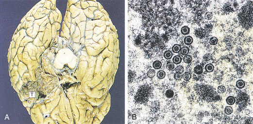
Figure 17-37 Herpes simplex encephalitis. In herpes simplex encephalitis (A) necrosis of the temporal lobes (T) is a typical development. Brain biopsy is useful in diagnosis when the virus can be seen by electron microscopy (B) as rounded particles with a dense core. Virus also can be identified by immunostaining or culture. In early cases, polymerase chain reaction (PCR) can be used to identify viral deoxyribonucleic acid (DNA) in cerebrospinal fluid samples. (From Stevens A, Lowe J: Pathology, ed 2, London, 2000, Mosby.)

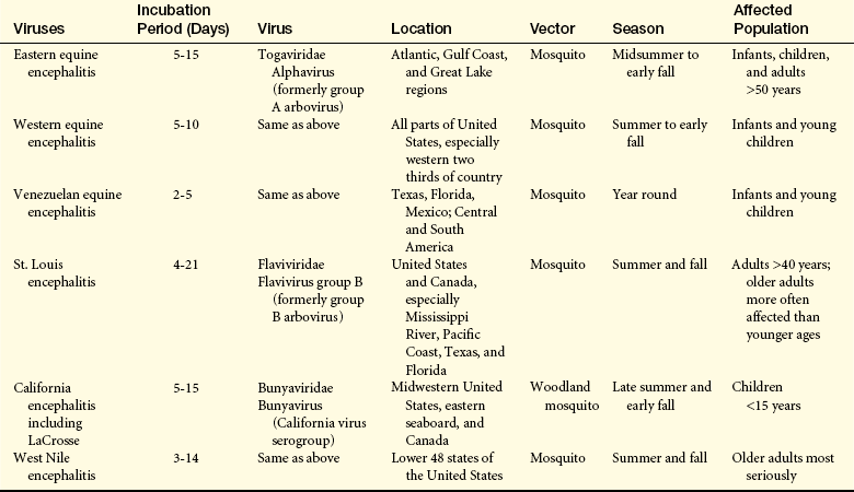
With the exception of the California viral encephalitis, which is endemic, the arthropod-borne encephalitides occur in epidemics, varying in geographic and seasonal incidence (Table 17-11). Eastern equine encephalitis is the most serious but least common of the encephalitides.67 West Nile virus is presented in Box 17-5.

Box 17-5   Emergence of West Nile Virus

West Nile (WN) virus, a flavivirus transmitted predominantly by the Culex mosquito, emerged in New York State in 1999. By the end of 2004, human cases had been found in the 48 contiguous states. Humans and horses, as well as other mammals, are incidental hosts. Birds and mosquitoes are life cycle hosts. Summer and fall are peak times of infection incidence. Besides mosquito transmission, WN virus can be transmitted through blood transfusions and organ transplants. Health experts believe transmission from mother to unborn child and through breast milk is possible.

Three clinical forms of West Nile have emerged: WN fever; WN encephalitis; and WN meningitis, although some individuals have other clinical manifestations. Some persons develop WN fever. The febrile illness of acute onset may be clinically unrecognizable as WN. About 20% of those infected have mild symptoms that last for 4 to 6 days that generally include fever and may include weakness, nausea, vomiting, headache, mental status changes, diarrhea, rash, and lymphadenopathy. WN encephalitis is marked by disorientation, stupor, coma, seizures, and movement disorders including tremor, ataxia, extrapyramidal signs, and paralysis. WN meningitis is characterized by meningeal signs of severe headache, high fever, and nuchal rigidity. Myelitis and polyradiculitis also may be present. Myocarditis, pancreatitis, and fulminant hepatitis are rare. Abnormalities in the thalamus, basal ganglia, and cerebellum are often seen on MRI in people with severe infection—about 1 in 150 develop nervous system involvement. Identifiable risk factors for this are advanced age and immunocompromise.

A preliminary diagnosis is made if IgM for the virus is found in serum or CSF. A rapid test became available in 2007. Plaque reduction neutralization assay (PRNA) is the confirmatory test. Interferon-alpha is used during treatment along with supportive care for those with severe infection. A new vaccine is being tested.

CSF, cerebrospinal fluid; IgM, Immunoglobulin M; MRI, magnetic resonance imaging.

Data from National Institute on Allergy and Infectious Disease, Department of Health and Human Services, National Institutes of Health: NIAID research on West Nile Virus, January 2008, available at www.niaid.nih.gov.

Table 17-11

Classification and Characteristics of Arthropod Viruses Causing Encephalitis

image

PATHOPHYSIOLOGY Viruses gain access to the CNS through the bloodstream or through an intraneuronal route from peripheral nerves.71 Evidence of meningeal involvement appears in all encephalitides. The arthropod-borne viral encephalitides cause widespread nerve cell degeneration. Edema and areas of necrosis with or without hemorrhage develop. Increased ICP develops and may progress to herniation. Large degenerative injuries are found in eastern equine encephalitis, whereas the other arthropod-borne viral encephalitides have microscopic areas of injury and degeneration.

Infectious encephalitis may result from a postinfectious autoimmune response to the virus or from direct invasion of the CNS by the virus. Herpes simplex type 1 has a tendency to infect the inferomedial surfaces of the temporal and frontal lobes and causes hemorrhagic necrosis.

CLINICAL MANIFESTATIONS Encephalitis may range from a mild infectious disease to a life-threatening disorder. The dramatic clinical manifestations of encephalitis are fever, delirium or confusion progressing to unconsciousness, seizure activity, cranial nerve palsies, paresis and paralysis, involuntary movement, and abnormal reflexes. Signs of marked ICP may be present.

EVALUATION AND TREATMENT Diagnosis is based on medical history and clinical presentation aided by CSF examination and culture, serologic examination, white blood cell (WBC) count, CT scan, or MRI. (Treatment available for the viral encephalitides is listed in Table 17-10.) Supportive therapy is initiated, and measures to control ICP are paramount.

Neurologic Complications of Acquired Immunodeficiency Syndrome

Approximately 40% to 60% of all persons with AIDS have neurologic complications (see Chapter 8 for the pathophysiology of AIDS). On postmortem examination, 75% have nervous system pathologic findings. The CNS pathologic findings result from (1) the primary human immunodeficiency virus (HIV) infection; (2) the immune dysregulation of early HIV infection and progressive immunosuppression in late HIV infections resulting in opportunistic infections, neoplasms, and systemic illness; and (3) complications of therapy.72

A variety of CNS complications of HIV exist (Box 17-6). Multiple CNS pathologic conditions may be experienced by one person. The most common neurologic disorder is HIV-associated dementia; others are peripheral neuropathies, vacuolar (spongy softening) myelopathy, opportunistic infections of the CNS, and neoplasms.

Box 17-6   Nervous System Complications of HIV-1 Infections

CNS Complications of HIV-1 Infection

Diffuse cerebral disorders

Minor cognitive and motor deficits (MCMDs)

HIV-associated dementia

Meningitis

Atypical aseptic meningitis

Acute: typical meningitis signs

Chronic: headache syndrome

Nonviral infection

Cryptococcus neoformans

Other fungal infections

Mycobacterial infections

Myelopathy

Spinal vacuolar myelopathy

Focal brain disorders

Opportunistic viral infections

Progressive multifocal leukoencephalopathy

Herpesviruses

Nonviral infections

Toxoplasma gondii

Bacterial infections

Neoplasms

Primary CNS neoplasms

Metastatic neoplasms

Cerebrovascular complications

Complications resulting from systemic HIV therapy

Peripheral Nervous System Complications of HIV-1 Infections

Distal symmetric peripheral neuropathy (polyneuropathy)

Inflammatory demyelinating polyradiculoneuropathy

Mononeuropathy multiplex

Progressive polyradiculopathy

Other causes of peripheral nervous system dysfunction

Herpes zoster radiculitis

Cranial neuropathies

CNS, Central nervous system; HIV, human immunodeficiency virus.

HIV-infected macrophages/monocytes in blood are attracted to the brain by up-regulation of proinflammatory mediators such as monocyte chemoattractant protein-1 (MCP-1), tumor necrosis factor-alpha (TNF-α), and adhesion molecules on endothelial cells. Up-regulation enables transendothelial migration of activated macrophages/monocytes.

Human Immunodeficiency Virus–Associated Dementia: HIV-associated dementia (HAD) (HIV-associated cognitive dysfunction, HIV encephalopathy, subacute encephalitis, HIV-associated dementia complex, HIV cognitive motor complex, AIDS encephalopathy, AIDS dementia complex, or AIDS-related dementia) may affect adults and children and is characterized by progressive cognitive dysfunction in conjunction with motor and behavioral alterations72 (Table 17-12). The syndrome typically develops later in the disease but may be an early or a singular manifestation. The syndrome is more prevalent in drug users with HIV.73

Table 17-12

Classification of HIV-Associated Cognitive Dysfunction

image

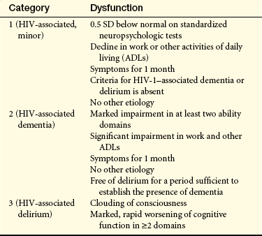
HIV, Human immunodeficiency virus; SD, standard deviation.

From Belman AL, Mirjana-Savatic M: Human immunodeficiency virus and acquired immunodeficiency syndrome. In Goetz CG, editor: Textbook of clinical neurology, ed 2, Philadelphia, 2003, Saunders.

At the time of primary HIV infection, circulating HIV-infected leukocytes (activated leukocytes), including macrophages and lymphocytes, adhere to the endothelial lining of blood vessels within the nervous system. These cells subsequently enter the brain parenchyma. HIV infects the perivascular macrophages, the microglial cells in the area, and, to a lesser degree, the astrocytes in the area.74 HIV preferentially affects the basal ganglia and deep white matter.74 Affected macrophages, macrophage-derived multinucleated cells, and microglia cause an immune-mediated demyelination process in white matter. Some viral replication occurs in some of the glial cells and occasionally within neurons. Multiple small nodules containing inflammatory cells are scattered throughout the white matter and in subcortical gray matter, such as the basal ganglia and thalami. Perivascular inflammation is present. Focal and diffuse demyelination of white matter and spongy changes of the spinal cord are present. Factors other than direct cell damage are involved, such as released toxins, lymphokines, or other substances. Elevation in CSF levels of quinolinic acid, a neurotoxic metabolite of tryptophan, is correlated with the degree of dementia. A decreased concentration of adenosine triphosphate (ATP) and inorganic phosphates in the CNS produces a decrease in metabolism.

HIV-associated dementia is insidious in onset and unpredictable in its course. Most individuals experience a steady progression of mental slowing characterized by abrupt accelerations of signs over several months to more than 1 year, although some experience an abrupt onset or an accelerated course. The triad of clinical manifestations are neurocognitive impairment, behavioral disturbance, and motor abnormalities.

Early clinical manifestations of HIV-associated dementia may be vague. Impaired concentration and short-term memory and retrieval deficits commonly occur. Apathy and lack of motivation, social withdrawal, irritability, and emotional lability may appear. Later, difficulties with language, spatial or temporal disorientation, and visual construction appear. Some individuals manifest an organic psychosis with agitation, inappropriate behavior, and hallucinosis.

Generalized cognitive system deficits occur later in the course of HIV-associated dementia, often accompanied by psychomotor slowing and decreased speech spontaneity and fluency. Progressive loss of balance, ataxia, spastic paraparesis or paralysis, and generalized hyperreflexia are common motor signs. Decreased writing ability, tremor, myoclonus, and seizure are less commonly seen.

Diagnosis is difficult, especially in early stages. The individuals medical history along with physical examination findings and supporting CSF, CT, and MRI data help establish the diagnosis. Specific screening tools have been developed to assist in diagnosis. Antiretroviral, protease inhibitors, reverse transcriptase inhibitors, and adjunctive agents may be effective along with supportive treatment. However, highly active antiretroviral therapy (HAART) protocols have shown little efficacy in reversing HAD because it does not target the cause.75

HIV Myelopathy: HIV myelopathy involving diffuse degeneration of the spinal cord may occur with HIV. Vacuolar myelopathy is believed to be a direct consequence of HIV. The lateral and posterior columns of the lumbar spinal cord are affected. A progressive spastic paraparesis with ataxia is the predominant clinical manifestation. Leg weakness, upper motor neuron signs, incontinence, and posterior column sensory loss may be present. Diagnosis is made on the basis of history, physical findings, and supporting data from diagnostic procedures. Vacuolar myelopathy is treated supportively and does not respond to antiretrovirals.

HIV Neuropathy: Some HIV-related peripheral neuropathies occur early, may coincide with seroconversion, are immune-mediated, and respond to standard immunotherapies. Other peripheral neuropathies primarily develop with advanced HIV infection and immunocompromised states and are facilitated by HIV replication, neurotoxicity from antiretroviral therapies, and coinfection with opportunistic pathogens.76

HIV neuropathy may have one or a combination of several presentations: a predominantly sensory neuropathy, an autonomic neuropathy, a mononeuritis multiplex, a Guillain-Barré–like syndrome, and a myopathy. The peripheral nervous system may sustain injury in HIV, manifesting as a peripheral neuropathy or radiculopathy. A progressive radiculopathy of predominantly the dorsal roots of the lumbar and sacral nerves may occur, involving severe myelin and axonal loss. HIV-associated distal symmetric polyneuropathy, a sensory neuropathy occurring late in the disease, is the most commonly occurring neuropathy with slowly progressive numbness and paresthesias and burning sensations in the feet.77

HIV has been isolated from peripheral nerves, so it is believed that the virus may directly infect nerves. Individuals experience painful, burning dysesthesias and paresthesias, typically in the extremities. Weakness and decreased or absent distal reflexes may be present. Diagnosis is established through history, physical findings, laboratory data, nerve conduction studies, EMG, and possibly biopsy. The most common myopathy is polymyositis; it may be present initially or develop later. The muscle fiber is infiltrated, initiating inflammation that leads to cellular degeneration and necrosis. The individual experiences muscle weakness of extremities with myalgia and fatigue. Steroids are used therapeutically in polymyositis.

Aseptic Viral Meningitis: Some people develop an acute aseptic meningitis at approximately the time of seroconversion. This may well represent the initial infection of the nervous system by the HIV. Symptoms include headache, fever, and meningismus. Cranial nerve involvement, especially of nerves V and VII, may appear, but the disease is self-limiting and requires only symptomatic treatment. Aseptic meningitis might occur at any point in the disease.

Opportunistic Infections: Opportunistic infections may be bacterial, fungal, protozoal, or viral in origin and produce nervous system disease. Typically bacterial infections are caused by unusual microorganisms. Cryptococcal infection is the most common fungal disorder and the third leading cause of neurologic disease with HIV. In Cryptococcus neoformans, small granulomas and cysts are found in the cerebral cortex and later may be present in deep cerebral tissues. The symptoms are vague, such as fever, headache, malaise, and meningismus. Herpes encephalitis and herpes varicella-zoster radiculitis may develop. Papovavirus (especially JC virus) in the immunocompromised person with HIV may produce a demyelinating disorder called progressive multifocal leukoencephalopathy (PML). This virus is found in 90% of healthy persons but is dormant. The virus reactivates to cause PML in 15% of HIV victims. Sensory and motor deficits, aphasia, and apraxia are common clinical manifestations. The condition is progressive.

Cytomegalovirus Infection: Cytomegalovirus encephalitis is common with AIDS but often not diagnosed while a person is alive. The encephalitis may be present as an acute illness with encephalitis features accompanied by nystagmus and cranial nerve signs. Retinitis is found in 50% of those affected.

Parasitic Infection: Toxoplasmosis is the most common opportunistic infection and occurs in one third of AIDS cases. CNS toxoplasmosis typically manifests as focal encephalitis. Toxoplasma gondii, a protozoan, is thought to reactivate from latent lesions to produce a well-demarcated necrotizing process. Inflammatory infiltrates, thrombotic lesions, and fibrinoid vascular walls are present at the necrotic edge. Marked edema is present adjacent to necrotic areas. Lesions may be multiple and exist throughout the cerebral hemispheres.

Clinical manifestations of CNS toxoplasmosis are focal but highly variable and include clumsiness to hemiplegia, aphasia, seizures, ataxia, cognitive changes, and constitutional symptoms. Fever and headache are common. Toxoplasmosis is difficult to diagnose but is treated effectively with pyrimethamine and sulfadiazine. Allergic response to sulfadiazine can be a problem, and other drugs can be substituted. Individuals with HIV may develop meningitis, encephalitis, or fungal, mycobacterial, and bacterial brain abscesses.

Central Nervous System Neoplasms: CNS neoplasms associated with HIV include primary CNS lymphoma, systemic non-Hodgkin lymphoma, and metastatic Kaposi sarcoma. The precise mechanism of lymphoproliferation is not known. Primary CNS lymphoma is a large-cell lymphoma that presents as rapidly developing and expanding multicentric intracranial mass lesions. The meninges are invaded and, possibly, the cranial nerves and spinal cord are invaded as well in systemic non-Hodgkin lymphoma. Metastasis of a Kaposi sarcoma to the CNS is uncommon.

Other Central Nervous System Complications: Individuals with HIV may develop multifocal ischemic infarctions, hemorrhagic infarctions, hemorrhage into tumors, subdural hematomas, and epidural hemorrhage. The precise mechanism of these cardiovascular complications is not yet known. Reported neurologic symptoms produced by HIV therapeutics include extrapyramidal movements, myoclonus, dysphasia, delirium, and acute myelopathy.

Lyme Disease

Lyme disease, a tick-borne spirochette bacterial infection, is a common arthropod-borne infection in the United States. It affects all age groups and involves the peripheral and central nervous systems. Lyme disease is caused by Borrelia burgdorferi introduced by tick bite, requiring about 36 hours of attachment.76 Transmission is most likely in the late summer or early fall. Infected ticks are endemic in the Midwest, western wooded and coastal areas, and the mid- to northeast Atlantic. The microorganism incubates for 3 to 32 days and then migrates to the skin, lymph nodes, and other body systems. The pathologic process progresses through three stages:

Stage I (acute localized): Within 1 month after the bite, the disease is characterized by a bull’s-eye–like (5 cm in diameter) burning centrifugally expanding erythema migrans rash followed by acute disseminated disease with general malaise, flulike symptoms (fever, muscle pain), stiff neck, and headache.

Stage II: With acute widespread dissemination of antibodies and immune complexes, cardiac and neurologic involvement predominates. About 10% of cases show cardiac signs and symptoms (palpitations, dizziness, shortness of breath, dysrhythmias, and first-degree heart block). Neurologic signs occur in 10% to 15% and include headache, chronic aseptic (lymphocytic) meningitis, Bell palsy, encephalitis, and radiculitis. Pathologically there is meningeal inflammation, perivascular inflammatory cell formation, and focal demyelination.

Stage III (chronic stage): The third stage may occur up to 2 years after the bite and involves arthritis and involvement of brain parenchyma with encephalitis, chronic neuropathy, and encephalopathy.

Treatment of choice is antibiotic therapy. Minor recurring symptoms are common in 50% of individuals.

Demyelinating Disorders

Demyelinating disorders are the result of damage to the myelin nerve sheath. They can occur in either the central (i.e., multiple sclerosis) or peripheral (i.e., Guillain-Barré syndrome) nervous system. Causes of the disorders include genetics, infections, autoimmune reactions, environmental toxins, and unknown factors.

Multiple Sclerosis

Multiple sclerosis (MS) is an autoimmune disorder diffusely involving degeneration of CNS myelin and loss of axons. The peripheral nervous system is not involved. MS and its variants are primary demyelinating disorders. In secondary disorders, CNS demyelination is caused by disorders other than multiple sclerosis.

MS is a diffuse and progressive CNS disease that affects white and gray matter.78 About 0.1% of the population is affected (400,000 persons in the United States and 2.5 million worldwide).79 Prevalence rates vary with geographic location (higher in temperate regions far above the equator) and racial groups (highest in whites, although it occurs in all races).

The onset of MS is usually between 20 and 40 years of age with a peak of age 30. Male:female ratio is about 1:2. MS is the most prevalent CNS demyelinating disorder and a leading cause of neurologic disability in early adulthood. Life expectancy is not greatly altered by MS and the disease course often extends over 30 years.80 Genetic and environmental factors and interactions are implicated in disease onset.81 Although the disorder does not exhibit a defined inheritance pattern, 15% of those with MS have an affected relative. Multiple genes affect risk of development of MS. A genetic link exists in the human leukocyte antigen (HLA) complex, a large cluster of genes responsible for many immune functions. Two genes, the interleukin [IL]-2 receptor gene (IL2RA) and IL7RA, have been identified.82 The first demyelinating event or “clinically isolated syndrome” (CIS), is a single episode of neurologic dysfunction lasting greater than 24 hours that can be a prelude to MS. Characteristic episodes include optic neuritis, solitary brainstem lesions, and transverse myelitis. MRI is used to assist diagnosis and assess prognosis although there are limitations to detecting the full extent of the lesions.83

PATHOPHYSIOLOGY MS is described as occurring when a previous viral insult to the nervous system has occurred in a genetically susceptible individual with a subsequent abnormal immune response in the CNS. The innate and adaptive immune systems are activated in the pathology of MS.84 Various mechanisms cause the irreversible tissue damage (inflammation, oligodendrocyte injury, demyelination, and axonal degeneration) that characterizes MS. These degenerative processes begin early in the course of the disease and continue to progress throughout a person’s life.80 Early inflammation and demyelination lead to irreversible axonal degeneration and scarring or sclerosis. Demyelinated axons are more fragile and susceptible to further damage, and when degeneration exceeds self-repair ability (remyelination), permanent disability results.85

Myelin destruction and axonal damage begin prior to symptom onset (early inflammatory demyelination). As the disease progresses, inflammatory changes in the CNS increase, and loss of brain volume progresses more rapidly.86 Box 17-7 describes the clinical courses of MS in relation to disease progression.

Box 17-7   Clinical Course of Multiple Sclerosis (MS)

Relapsing-Remitting (RR) MS

Clear relapses (called acute attacks or excerbations) with either full recovery, or with partial recovery and lasting disability. Between attacks there is no progression (or worsening) of disease. RR is the most common course of MS (90% of cases).

Primary Progressive (PP) MS

Steady progression (or worsening) from onset, with only occasional plateaus or minor recovery. This is a fairly uncommon disease course and one that may involve different brain and spinal cord damage than do other forms of MS.

Secondary Progressive (SP) MS

Begins with a pattern of clear-cut relapses and recovery but becomes steadily progressive over time with continued worsening between acute attacks (develops in two thirds of individuals eventually).

Progressive-Relapsing (PR) MS

A rare type that is steadily progressive from onset but also has clear acute attacks.

The immunopathology of MS involves:

1. CD8+ T-cell activation (autoreactive), cells cross the blood-brain barrier and enter CNS and attack myelin

2. IL-12 and IL-23 (proinflammatory cytokines)

3. Lack of IL-10 (an anti-inflammatory cytokine)

4. IL-17 (proinflammatory cytokine) production by T cells

5. Integrins expressed to facilitate adherence and passage of immune cells into the CNS

6. Chemokines promote the migration of immune cells and are up-regulated in MS

7. B lymphocytes and plasma cells contribute to the inflammatory response and directly damage myelin and axons; B cells are more active in chronic or progressive forms of MS (B cells produce autoantibodies, secrete inflammatory cytokines, and activate T cells by presenting antigen)87

8. Complement activation (promotes inflammation) during the acute phase; may be neuroprotective during relapse88

In addition, eosinophils, neutrophils, and macrophages are present. The demyelination disrupts sodium, calcium, and potassium ion channels and calcium influx is proinflammatory and neurotoxic in itself. Activated micro-glia and macrophages release nitric oxide and oxygen-free radicals. Activated immune cells also produce glutamate, a neurotoxin.89

MS is characterized not only by focal inflammatory changes but also diffuse injury throughout the CNS (MS lesions) (Figures 17-38 and 17-39). Four MS lesion pathologic patterns have been described (Box 17-8). MS lesions may occur anywhere in white or gray matter. In addition, other neurodegenerative processes that involve the entire CNS are taking place, including (1) changes in gray matter in the cortex, basal ganglia, brainstem, and spinal cord with substantial loss over time; (2) brain atrophy that begins early in the disease and is highly correlated with disability and progressive MS; and (3) direct dysfunction of or damage to oligodendrocytes that manufacture myelin.80,90 Normal-appearing white matter also is highly abnormal microscopically with the presence of wallerian degeneration, diffuse inflammation, extensive microglial activation, neurotoxic substances, and gene activation that disrupt cellular processes.80 In established disease the multifocal, multistaged feature of MS lesions gives rise to the aphorism that the lesions are “scattered in space and time.” Symptoms therefore are multiple and variable.

Box 17-8   Multiple Sclerosis: Pathologic Patterns

Pattern I

Cell-mediated

Macrophages, T cells

Remyelination occurs

Pattern II

Cell mediated

IgG and complement in areas of active myelin damage

Remyelination occurs

Pattern III

Cellular infiltrate

Macrophages, T cells, activated microglia

No IgG or complement

Oligodendrocyte apoptosis

No remyelination

Pattern IV

Cellular infiltrates

No IgG

Primary injury to oligodendrocytes?

No remyelination

IgG, Immunoglobulin G.


51 biopsies and 32 autopsy samples.

From Lucchinetti C et al: Ann Neurol 47(6):707-717, 2000.

image

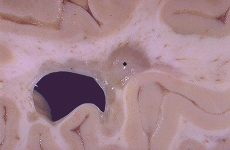
Figure 17-38 Multiple sclerosis, gross. Seen here in periventricular white matter is a large “plaque” (image) of demyelination that has a sharp border with adjacent normal white matter. Such plaques have a gray-tan appearance and are typically associated with the clinical appearance of transient or progressive loss of neurologic function in multiple sclerosis (MS). Because MS is often multifocal, and the lesions appear in various white matter locations in the central nervous sytem over time, the clinical course and findings can be quite varied. (From Klatt EC: Robbins and Cotran atlas of pathology, Philadelphia, 2006, Saunders.)

image

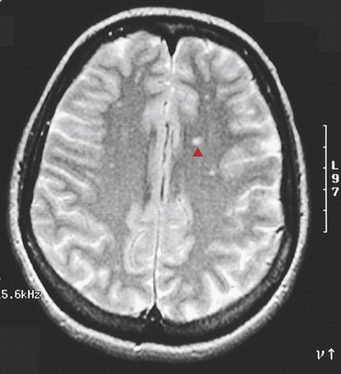
Figure 17-39 Multiple sclerosis (MS), MRI. This T2-weighted magnetic resonance image in axial view shows multiple bilateral small bright foci (image) that represents areas of demyelinating plaque formation in an individual with an exacerbation of MS. White matter anywhere within the brain and spinal cord can be involved. (From Klatt EC: Robbins and Cotran atlas of pathology, Philadelphia, 2006, Saunders.)

CLINICAL MANIFESTATIONS A variety of events (e.g., infection, trauma, or pregnancy) occurring immediately before the onset or exacerbation of symptoms are regarded as precipitating factors related to MS. Most of the pregnancy-related exacerbations occur 3 months postpartum, suggesting a relation to the stresses of labor and the increased fatigue during the postpartum period rather than to the pregnancy itself.

The major classifications of MS are relapsing-remitting, primary progressive, secondary progressive, and progressive-relapsing (see Box 17-7). Initially 90% of persons present with a relapsing, remitting course; 10% present with a primary progressive course; and 90% develop a progressive course in 10 to 20 years after onset of the disease. However, once walking problems develop, disease progression occurs quickly regardless of disease type. Usually persons with late MS have one of the established syndromes—mixed, spinal, or cerebellar (Box 17-9). The initial syndrome depends on the portion of the CNS that is most involved. After years, 50% of individuals appear to have established syndromes of mixed involvement.

Box 17-9   Established Syndromes of Multiple Sclerosis

Mixed or Generalized Type (50% of Persons)

Optic signs—optic neuritis

Brainstem signs—internuclear ophthalmoplegia, diplopia, vertigo (vomiting), nystagmus, dysarthria

Cerebellar signs—see page 633

Spinal Type (30% to 40% of Persons)

Spastic ataxia

Deep sensory changes in the extremities

Bladder and bowel symptoms

Cerebellar or Pontobulbar-Cerebral Type (5% of Persons)

Motor ataxia

Hypotonia

Asthenia

Amaurotic Form (5% of Persons)

Blindness

Mixed (General) Type: Twenty-five percent of persons initially experience retrobulbar or optic neuritis, the manifestations of optic nerve axonal loss.91 The condition usually evolves rapidly over hours to days and is highly suggestive of MS. Involvement may be unilateral or bilateral. Subjective symptoms are impaired central vision (blurring, fogginess, haziness) and impaired color perception. Signs are decreased central visual acuity; central or paracentral scotoma (area of diminished vision); acquired color vision deficit, especially to red and green; and defective pupillary reaction to light. A variety of field defects may occur. In the acute phase these symptoms may reflect optic papillitis (inflammation and swelling of the optic disc) or retrobulbar neuritis with a normal disc. One third of persons recover completely, and most others improve significantly. Later, pallor of the temporal half of the disc occurs from demyelination of a portion of the optic nerve. (Normal visual function is discussed in Chapter 15.)

The brainstem lesions involve cranial nerves III through XII at the root, nuclear, or corticobulbar (upper motor neuron) level. Internuclear ophthalmoplegia, nystagmus, and dysarthria are the most common brainstem symptoms, followed by deafness, vertigo and vomiting, tinnitus, facial weakness, and facial sensory deficit. Internuclear ophthalmoplegia is lateral gaze paralysis caused by involvement of the medial longitudinal fasciculus, the brainstem pathway that coordinates eye movement. Diplopia and eyeball pain are common complaints. Bilateral internuclear ophthalmoplegia in a young adult is virtually diagnostic of MS.

Cognitive dysfunction has been demonstrated to occur early in the disease course. The person experiences decreased short-term memory, recent memory impairment, decreased concentration, word-finding problems, and planning difficulties. Mood alterations are common in MS. Depression is far more common than euphoria.

Spinal Type: The spinal type of MS is the second most common type, chiefly involving the spinal tracts and dorsal column. Weakness, numbness, or both in one or more limbs are initial symptoms in 50% of cases of MS. Subjective corticospinal (upper motor neuron) symptoms (stiffness, slowness, weakness) are often unilateral and are a component of fatigability. Spinal signs are usually bilateral (symmetric), with lower limbs more often and more severely affected than upper limbs; spastic paraparesis is probably the most common single neurologic finding in MS.

Bladder and bowel symptoms occur with major spinal cord involvement. Urgency and hesitancy generally precede incontinence. Bladder dysfunction most often involves a small, spastic bladder, although occasionally a large, flaccid bladder may result with retention problems. Neurogenic impotence often occurs when sphincter symptoms are present. Bowel incontinence is rare, but constipation is common with severe disease. Subjective dorsal column symptoms are symmetric paresthesias (tingling and numbness) in an unpredictable pattern but with a predilection for lower extremities over upper extremities. Dorsal column signs are vibration, position, and two-point discrimination deficits. Sensory complaints often are not substantiated by objective physical findings but by further diagnostic tests.

Cerebellar Type: A nystagmus and ataxia presentation initially is not uncommon and reflects cerebellar and corticospinal involvement. Cerebellar deficits are usually symmetric, with all four limbs involved. With combined corticospinal and cerebellar involvement, the individual has a spastic ataxic gait and ataxia of the arms. Pure cerebellar symptoms are those of motor ataxia, hypotonia, and asthenia (weakness). Manifestations of motor ataxia are decomposition of movement, inability to perform rapid alternating movements (dysdiadochokinesia), and dysmetria. Charcot triad describes a combination of dysarthria, intention tremor, and nystagmus. Hypotonia is manifested by decreased resistance to passive movement, hypoactive deep tendon reflexes, and pendular knee jerk.

Short-lived attacks of neurologic deficits are the temporary appearance or worsening of symptoms. The mechanism of these attacks is complete reversible conduction block in partially demyelinated axons. Conditions that cause short-lived attacks include (1) minor increases in body temperature or serum Ca++ concentration and (2) functional demands exceeding conduction capacity. An increase in body temperature or serum Ca++ level increases current leakage through demyelinated neurons. Individuals with MS may become dramatically worse when body temperature is raised. Hypercalcemia induced by decreased serum pH may aggravate symptoms of MS. Physical and emotional stress imposes functional demands that may exceed conduction capacity of affected neurons.

Paroxysmal attacks are sensory or motor symptoms of abrupt onset and short duration (a few seconds or minutes). These symptoms include paresthesias, dysarthria and ataxia, and tonic head turning. The mechanism of paroxysmal attacks is nonsynaptic transmission in which nerve impulses are directly transmitted between adjacent demyelinated axons. These impulses arise focally and spuriously in the cervical portion of the spinal cord or in the brainstem. A common paroxysmal symptom, called Lhermitte sign, is the momentary paresthesia (shocklike or tingling sensation) that shoots down the trunk or limbs during active or passive flexion of the neck. Bending the neck evokes nonsynaptic impulses in demyelinated axons of the dorsal column in the spinal cord. A person with MS may have many paroxysmal attacks each day. Inciting events include sensory stimulation, voluntary movement, hyperventilation, and emotional stress. Paroxysmal attacks tend to persist for weeks or months and may be followed by progressive symptoms of MS.

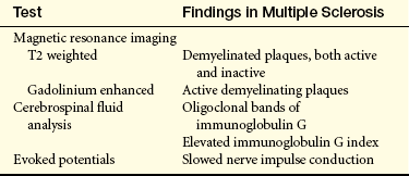
EVALUATION AND TREATMENT The diagnostic criteria for MS were revised in 2001 and are known as the McDonald criteria. Clinical examination in combination with MRI, to demonstrate MS lesions in time and space, and CSF findings are used to make the diagnosis earlier (Table 17-13) so that treatment may begin sooner. Persistently elevated CSF immunoglobulin G (IgG) index is found in about two thirds of individuals with MS, and oligoclonal bands of IgG on electrophoresis are found in more than 90% of persons. Evoked response (ER) studies aid diagnosis by detecting decreased conduction velocity in visual, auditory, and somatosensory pathways.

Table 17-13

Diagnostic Tests for Multiple Sclerosis

image

From Chisholm-Burns MA, et al: Pharmacotherapy principles and practices, p 434, New York, 2008, McGraw-Hill.

The treatment goal in MS is prevention of permanent neurologic damage. Acute relapses are treated with corticosteroids to speed recovery.92 Evidence supports that corticosteroids may improve symptoms in acute relapse situations.93 Disease-modifying drugs are used to decrease the number of relapses, prevent permanent CNS damage, and prevent disability.94 Evidence supports that in relapsing-remitting multiple sclerosis (RRMS) glatiramer acetate, intravenous (IV) immunoglobulins, and azathioprine or other drug combinations may reduce relapses.95 Interferon-beta and mitoxantrone may reduce relapses and disease progression but have serious adverse effects.2 Treatments are in development to prevent demyelination, promote remyelination and repair, and suppress selective B-cell and T-cell function.96,97

Symptom management for fatigue, weakness, vertigo, ataxia, tremor, heat intolerance, spasticity, bladder dysfunction, bowel dysfunction, sexual dysfunction, sensory sensations, pain, cognitive difficulties, depression, and psychosocial issues is essential but many treatments are only partially effective, particularly for fatigue and spasticity.2 Supportive and rehabilitative management is directed toward preventing the complications of immobility, especially pressure sores and infections of the pulmonary and genitourinary systems. Interdisciplinary inpatient rehabilitation may improve function in the short term.98

Neurodegenerative Disorders

Amyotrophic Lateral Sclerosis

Amyotrophic lateral sclerosis (ALS) (sporadic motor system disease, sporadic motor neuron disease, motor neuron disease) is a worldwide degenerative disorder diffusely involving lower and upper motor neurons resulting in progressive muscle weakness leading to respiratory failure and death, usually 2 to 5 years from symptom onset.99,100 There are no racial, ethnic, or socioeconomic boundaries. The prevalence rate is about 1 to 3 cases per 100,000,101 with 2 deaths per 100,000, and 5000 newly diagnosed cases per year in the United States. The term amyotrophic (without muscle nutrition or progressive muscle wasting) refers to the predominant lower motor neuron component of the syndrome. Lateral sclerosis, or scarring of the corticospinal tract in the lateral column of the spinal cord, refers to the upper motor neuron component of the syndrome. ALS differs from other motor neuron disorders in that upper and lower motor neurons are involved.

Classic ALS (Lou Gehrig disease) may begin at any time from the fourth decade of life; its peak occurrence is in the early 50s. Male/female ratio is 1:4 to 2:5,100 equalizing after menopause. In familial ALS, mutations have been found in the superoxide dismutase (SOD1) gene on chromosome 21.102 The defective SOD1 gene leads to an autosomal dominant pattern with age-dependent penetrance and accounts for 15% to 20% of the cases of familial ALS. The ALS2 gene is mapped to chromosome 2q33 and leads to a rare, recessively inherited ALS associated with deficiency of the gene product alsin and a juvenile form of ALS. The ALS4 gene (senataxin) is linked to locus 9q34 and produces a rare juvenile form of familial ALS inherited in an autosomal dominant pattern. Several genes also alter the risk of developing sporadic ALS.103

ALS presentations include crural ALS, proximal or shoulder girdle ALS, and hemiplegic (Mills) ALS. Of persons with ALS, 20% have a benign form of the disease.

PATHOPHYSIOLOGY The pathogenesis of ALS is not clear. The molecular dysfunction caused by the SOD1 is under study. Apoptotic factors, abnormal synthesis of filament units, defects in axonal transport, excitotoxicity and glutamate transports, oxidative stress, growth factors, mitochondrial dysfunction, and neuroinflammation are likewise under study for a possible contribution to the pathogenesis.

The principal pathologic feature of ALS is lower and upper motor neuron degeneration. The number of large motor neurons in the spinal cord, brainstem, and cerebral cortex (premotor and motor areas) is reduced, with ongoing degeneration in the remaining motor neurons. The nuclei of cranial nerves III, IV, and VI are not involved. Death of the motor neuron results in axonal degeneration and secondary demyelination with glial proliferation and sclerosis (scarring) along the corticospinal tract. Inclusion bodies containing the protein ubiquitin are found in surviving neurons. However, there also is widespread neural degeneration of nonmotor neurons in the spinal cord and motor cortices, as well as in the premotor, sensory, and temporal cortices.100 Altered astrocytes and microglial functions are suspected to exist.

Lower motor neuron degeneration denervates motor units. Adjacent, still-viable lower motor neurons attempt to compensate by a process of distal intramuscular sprouting, reinnervation, and enlargement of motor units. The initial symptoms of the disease may be related to lower or upper motor neuron dysfunction or both. Fifty percent of persons with ALS present with hand weakness or incoordination, dysarthria, or leg weakness or incoordination.104

CLINICAL MANIFESTATIONS Weakness may begin in any or all muscles of the body. Muscle weakness in ALS exhibits the following characteristics:

1. Paresis usually begins in a single muscle group.

2. Corresponding muscle groups are asymmetrically affected in a mottled distribution.

3. Gradual involvement occurs in all striated muscles except extraocular muscles and heart and progresses to paralysis with no remissions.

4. Flaccid and spastic paresis may coexist in a single muscle group; flaccid paresis may mask spasticity, which is usually mild.

5. Urethral and anal sphincter weakness is uncommon.

The lower motor neuron syndrome of flaccid paresis consists of weakness of individual muscles, progressing to paralysis, associated with hypotonia and primary muscle atrophy (i.e., atrophy caused by denervation). Hypotonia is manifested by (1) decreased resistance to passive movement, (2) hypoactive or absent deep tendon reflexes, (3) absent abdominal and cremasteric reflexes, and (4) absent Babinski sign. Primary atrophy is manifested by (1) severe, irreversible muscular wasting; (2) fasciculations; (3) metabolically related changes in the skin and appendages; and (4) specific EMG findings. Fasciculations, along with fibrillations, are prominent features of ALS. Metabolic changes include (1) thinning of the skin, (2) thickening of the nails, (3) loss of body hair, and (4) decreased perspiration.

The upper motor neuron syndrome of spastic paresis consists of weakness of movement patterns, progressing to paralysis, associated with spasticity and, in some cases, atrophy secondary to disuse. Spasticity is manifested by (1) clasp-knife phenomenon, evident with passive movement; (2) hyperactive deep tendon reflexes and clonus with severe spasticity; (3) absent abdominal and cremasteric reflexes; and (4) presence of Babinski sign. The coexistence of a dementia has been demonstrated to be higher than previously thought.

EVALUATION AND TREATMENT The diagnosis of the syndrome is based predominantly on medical history and physical examination.105 EMG and muscle biopsy verify lower motor neuron degeneration and denervation. Muscle biopsy usually is not needed to confirm the diagnosis. Riluzole (Rilutek), an antiglutamate, is the standard treatment for ALS, decreasing the risk of death by 35%.100 Treatment is also directed at symptom relief, prevention of complications, maintenance of maximal function, and maintenance of optimal quality of life.104 Special problems requiring preventive and symptomatic management are communication difficulty caused by dysmasesis and dysphonia, salivation problems with either thick saliva or excessively thin saliva (sialorrhea), and dyspnea caused by diaphragmatic and intercostal weakness. Ventilatory issues become prominent. Supportive and rehabilitation management is directed toward preventing complications of immobility. Psychologic support of the affected individual and the family is extremely important in this disorder.

PERIPHERAL NERVOUS SYSTEM AND NEUROMUSCULAR JUNCTION DISORDERS

Peripheral Nervous System Disorders

Neuropathies

The axons traveling to and from the brainstem and spinal cord neuronal cell bodies may be injured by a multitude of disease processes. Distinct anatomic areas of the axon may be injured or the spinal nerves may be affected at the spinal roots, at the plexus before peripheral nerve formation, or at the peripheral nerves themselves. Cranial nerves do not have roots or plexuses so are affected only within the nerves themselves. Autonomic nerve fibers may be injured as they travel within certain cranial nerves or emerge through the ventral root and plexuses to travel in the peripheral nerves of the body.

Neuropathies can be classified as (1) generalized symmetric polyneuropathies, (2) generalized neuropathies, and (3) focal or multifocal neuropathies. Generalized symmetric polyneuropathies are characterized by symmetric involvement of sensory, motor, or autonomic fibers although, with clinical signs, one type of fiber may predominate. Generalized symmetric polyneuropathies further subdivide into distal axonal polyneuropathy and demyelinating polyneuropathy. Distal axonal polyneuropathy affects peripheral axons and is the generalized peripheral neuropathy commonly seen. The clinical feature of distal axonal polyneuropathy is involvement of the longest nerve of the body, those going to the feet, first. Sensory impairment is greater than motor impairment. Symptoms are burning pain, tingling, and numbness of the feet. Small nerve fiber damage produces decreased pain and temperature sensation, as well as burning, numbness, and tingling. Large nerve fiber injury causes decreased light touch, vibration, and position sense. The two most common causes are diabetes mellitus and alcohol abuse; occasionally, neurotoxic therapeutic agents also are the cause. Within the classification of distal axonal neuropathies is another group of neuropathies called autonomic neuropathy.106 This neuropathy can involve virtually any sympathetic or parasympthetic nerve fiber with impairment of cardiovascular, gastrointestinal, urogenital, thermoregulatory, sudomotor, and pupillomotor autonomic function.107 Autonomic neuropathies have a progressive course and are usually reversible. The myelin or Schwann cells are affected in demyelinating polyneuropathy, which occurs far less frequently. Weakness is the predominant sign with far less sensory impairment. Acute and chronic inflammatory demyelinating neuropathies comprise this group, of which Guillain-Barré syndrome is the most widely recognized disorder (see p. 636).

Generalized neuropathies affect the cell body of only one type of peripheral neuron. The dorsal root ganglion cell is affected in sensory neuropathies, producing numbness that may begin in a focal or asymmetric distribution or in a distal symmetric pattern. Sensory neuropathies are seen in leprosy, some industrial solvent poisonings, some hereditary disorders, and chloramphenicol toxicity. The anterior horn in motor neuropathy is affected, causing weakness that may be symmetric or asymmetric. Motor neuropathies are caused by anterior horn cell disease, such as ALS or paralytic poliomyelitis.

Focal neuropathy or multifocal neuropathies affect sensory and motor fibers in one or more nerves as is seen in common compression neuropathies such as carpal tunnel syndrome (median nerve compression), ulnar nerve compression (at the elbow), peroneal nerve compression, or sciatic nerve compression. Focal neuropathies can involve one or more cranial nerves. Plexus injuries and radiculopathies also fall into this category.

PATHOPHYSIOLOGY Although distinct pathophysiologic processes are recognized in a neuropathy, these are not disease specific and may exist simultaneously in any one neuropathy. Wallerian degeneration, in which the axon and myelin distal to the site of axonal interruption degenerate, may be present (see Chapter 14). This type of degeneration is characteristic of a traumatic nerve injury in which the nerve is severed. In demyelinating neuropathies the axon may be spared and only the myelin degenerates. In axonal degeneration distal degeneration of the axon occurs first and is followed by degeneration of the myelin and the axis cylinder. Many pathologic processes may give rise to neuropathy, and one or more nerves may be involved.

CLINICAL MANIFESTATIONS When the axons are affected, muscle strength, muscle tone, and muscle bulk also are affected. Whole muscles or groups of muscles are paretic or paralyzed, and the muscles of the feet and legs often are affected first and more severely. These long, large axons are thought to (1) be more vulnerable to injury because of their size and length, (2) have more Schwann cells available to be injured, and (3) exhibit a “dying back” phenomenon caused by difficulty of the nerve cell body in maintaining the terminal portion of the axon. If unchecked, the pathologic process tends to involve the hands and arms because these have the next longest and largest axons.

Tone and the deep tendon reflexes in the affected muscles generally are decreased in a neuropathy. Atrophy is distributed according to the peripheral nerves involved. The degree and distribution of the atrophy probably depend on the extent of the injury. Fasciculation may be present, especially with associated ventral root or motor neuron changes or both, as in Guillain-Barré syndrome, diabetic neuropathy, and porphyric neuropathy. Mild fatigue may be experienced. A few disorders, notably Guillain-Barré syndrome, produce a pattern of paresis and paralysis that involves all limbs, the trunk, and the neck. Peripheral bifacial and other cranial nerve palsies may be seen with a variety of disorders. Tenderness of the nerve trunks and associated sensory alterations help to distinguish neuropathy from amyotrophy. These include paresthesias and dysesthesias as well as decreased or absent primary sensations (e.g., of temperature, touch, light pain, position, or vibration). Ataxia of gait or limb may arise from the loss of position and vibratory sensations (i.e., proprioceptive sensory loss) and may be enhanced by motor weakness.

Reflexes may be altered. Reflex-mediated autonomic nervous system functions, such as sweating and pupillary size, may be affected. Neuropathies associated with autonomic disturbances include diabetes mellitus, alcoholism and related nutritional neuropathies, amyloidosis, porphyria, Guillain-Barré syndrome, Riley-Day syndrome, and familial sensory neuropathy. In many chronic polyneuropathies the feet, hands, and spine become deformed. Metabolic changes may arise secondary to nerve dysfunction.

EVALUATION AND TREATMENT The diagnostic workup to determine the cause of a neuropathy is often extensive. Early diagnosis and treatment before irreversible neuronal cell damage ensues are of paramount importance. Although axonal regrowth and recovery of function may take months, many neuropathies can be reversed. The therapeutic management is directed first at elimination of the cause, if possible. At least the primary disorder, such as diabetes mellitus, should be controlled. Further damage to the axon must be prevented by avoiding (1) trauma from too-early demand for reuse of the nerve, (2) accidents that cause tissue damage, and (3) hypoxia and ischemia or other deprivation of essential substrates.

Guillain-Barré Syndrome: Guillain-Barré syndrome (GBS) (Landry-Guillain-Barré syndrome, idiopathic polyneuritis, acute inflammatory demyelinating polyradiculopathy, acute autoimmune neuropathy) is an acquired acute inflammatory demyelinating or axonal polyneuropathy with four subtypes. The subtypes and their clinical features are presented in Table 17-14. These subtypes occur throughout the world, affect children and adults of both genders and all age groups equally, and occur in all seasons of the year.

Table 17-14

Expanded Classification of Guillain-Barré Syndrome (GBS)

image

DTR, deep tendon reflex; GM, ganglioside GM 1; IgG, immunoglobulin.

Data from: Hughes RA, Cornblath DR: Lancet 366(947):1653-1666, 2005; Kuwabara S: Curr Neurol Neurosci Rep 7(1):57-62, 2008; Overell JR et al: Cochrane Database Syst Rev (1):CD004761, 2007; Yuki N, Koga N: Curr Opin Neurol 19(5):451-457, 2007.

The annual incidence rate is 1 to 2 per 100,000 with a 4% to 6% mortality rate, and a 5% to 10% morbidity rate (permanent disabling weakness, imbalance, or sensory loss).

PATHOPHYSIOLOGY GBS is considered to be an autoimmune disease triggered by a preceding bacterial or viral infection. It has been recognized that glycolipids, particularly gangliosides, are immune targets in the subtypes of GBS. Different gangliosides predominate in different locations in peripheral nerves and in different nerve fiber types. Table 17-14 presents the pathology and pathogenesis of the four axonal subtypes of GBS.108 The muscle innervated by the damaged peripheral nerves undergoes denervation and atrophy. If the cell body survives, regeneration of the peripheral nerve takes place and recovery of function is likely. If the cell body dies from intense root involvement in the inflammatory-degenerative process, no regeneration is possible. Collateral reinnervation from surviving axons and regenerating axons may take place. In this case, motor recovery is less complete and residual deficits persist.

CLINICAL MANIFESTATIONS Clinical manifestations may vary depending on subtype. Typical first manifestations are numbness, pain, paraesthesias, or weakness in the limbs. Motor signs manifest as an acute or subacute progressive paralysis. Proximal muscles may be involved earlier and more significantly than distal muscles. The paresis/paralysis may be present in an ascending pattern involving limbs, respiratory muscles, and bulbar muscles. Only bulbar muscles may be involved, resulting in dysphagia and dysarthria. Weakness usually plateaus or improves by the fourth week in 90% of cases. After weakness plateaus, strength improves over a period of days to months, with the majority of individuals reaching activity levels similar to their predisease state. If sensory symptoms are present in the subtype they may include paresthesias/dysthesias (tingling, burning, shocklike sensations, particularly in the limbs), pain (throbbing, aching, particularly in the lower back, buttocks and legs), and numbness. Position and vibratory sensations are more affected than superficial sensation. Respiratory muscle weakness leads to the need for ventilatory support in 10% to 30% of individuals. Cranial nerve weakness manifests as facial weakness and bulbar weakness involving chewing, swallowing, and cough. Autonomic dysfunction may manifest as tachycardia or, less frequently, bradycardia; hypotension or hypertension; and loss of or significant increase in sweating in those more severely affected. Persons may undergo a respiratory arrest or cardiovascular collapse.109 Hyponatremia caused by the syndrome of inappropriate antiduretic hormone (SIADH) is common, especially in ventilated individuals.

EVALUATION AND TREATMENT Clinical history helps diagnose GBS and its subtypes. The major diagnostic tests are the examination of the CSF, nerve conduction studies, and EMG. The CSF findings include an unusually high protein level (500 mg/dl) without cellular abnormality. Nerve conduction studies help identify the subtype. Ventilatory support and management of the autonomic nervous system dysfunction are two dominant aspects of the therapeutic management. Plasmapheresis or plasma exchanges within the first 2 weeks of onset of clinical manifestations may be indicated. Intravenous immunoglobulin is used as well as combination therapy. After the disorder begins to remit, aggressive rehabilitation should be instituted.110

Radiculopathies: As the spinal roots emerge from or enter the vertebral canal, they may be injured or damaged by compression, infection, inflammation, ischemia, or direct trauma whereby the roots are stretched or torn. Radiculopathies are disorders of roots of spinal nerves. Radiculitis (radiculoneuritis) refers to an inflammatory disorder of the spinal nerve roots. One or more roots may be affected.

PATHOPHYSIOLOGY Many different pathologic conditions may cause compression, inflammation, or tearing of nerve roots.111 Roots may be traumatized by a forceful tearing of a nerve, termed avulsion, often associated with injuries to the head and shoulders. An acute intervertebral disk prolapse (herniated disk), degenerative spondyloarthropathies, or a benign tumor may compress nerve roots. Metastatic tumors of the lung, breast, and gastrointestinal tract may produce a carcinomatous meningitis, causing compression and inflammatory changes in nerve roots. Other causes of inflammatory changes in nerve roots are chronic meningitis, neurosyphilis, sarcoidosis, and inflammatory arachnoiditis produced by myelography and lumbar punctures.

CLINICAL MANIFESTATIONS The strength, tone, and bulk of the muscles innervated by the involved roots are affected. The pattern and distribution of weakness and atrophy are similar to those of the amyotrophies. Tone and deep tendon reflexes are decreased, but rarely absent, because the involved muscles are usually innervated by two or more spinal roots. Fasciculations often are present, and mild fatigue may be experienced. Because pathologic processes usually affect the ventral as well as the dorsal roots, sensory alterations are common.

Diseases that involve spinal roots typically produce local pain; pain on local percussion; pain and paresthesias in the sensory root distribution (called radicular pain and radicular paresthesia); increased pain with movement, stretching of the root, and maneuvers that transiently increase CSF pressure; sensory loss in a radicular pattern; and spasms of the muscles surrounding the vertebral column (i.e., paravertebral muscle spasms).

EVALUATION AND TREATMENT Diagnostic measures may include spinal films, nerve conduction studies, EMG, lumbar puncture with CSF examination, myelography, and biopsy of tumor masses. Treatment is directed at the cause of the injury and may take the form of surgery, antibiotics, removal of the injurious agent, steroids, and radiation therapy and chemotherapy. Supportive management may include control of the discomfort, protection from further injury, prevention of complications, and rehabilitation when appropriate.

Plexus Injuries: Plexus injuries involve the nerve plexus distal to the spinal roots but proximal to the formation of the peripheral nerves. Such injuries may be caused by trauma, compression, or infiltration, or they may be iatrogenic, caused by positioning during surgery or by an intramuscular injection. Clinical manifestations include motor weakness, muscle atrophy, and sensory loss in affected areas. Paralysis can occur with complete plexus lesions.

The diagnosis is made on the basis of history and clinical manifestations. Therapeutic treatment is directed at removal of the cause, repair and approximation of nervous tissue, prevention of further injury, control of discomfort, prevention of complications, and rehabilitation when appropriate.

Neuromuscular Junction Disorders

Transmission of the nerve impulse at the neuromuscular junction requires the release of adequate amounts of neurotransmitter from the presynaptic terminals of the axon and effective binding of the released transmitter to the receptors on the membranes of muscle cells (see Figure 14-14). Four neuromuscular junction disorders (NMJD), whose pathogenesis is thought to be autoantibodies, are described: myasthenia gravis, muscle-specific kinase (MuSK) protein antibody-associated myasthenia gravis, Lambert-Eaton myasthenic syndrome, and acquired neuromyotonia. In addition, there are rare inherited (congenital) myasthenic syndromes that result from mutations in different key proteins for the acetylcholine receptor, ion channels, and motor end plates at the neuromuscular junction.112,113 Nutritional deficits, certain drugs (e.g., reserpine or methyldopa [Aldomet]), certain toxins (e.g., botulism); some snake, scorpion, or spider venoms; certain plant extracts; and some insecticides also affect the neuromuscular junction. The most common autoimmune NMJD is myasthenia gravis caused by the binding of autoantibodies to the acetylcholine receptor on the postsynaptic membrane. MuSK antibody-associated myasthenia gravis is also postsynaptic and is a severe form of the disease.114 Presynaptic autoimmune diseases include neurotonia with autoantibodies to the potassium channels and Lambert-Eaton myasthenic syndrome with autoantibodies to calcium channels (Figure 17-40 and Table 17-15).

Table 17-15

Antibodies and Mechanisms of Action at Neuromuscular Junction

image

AChR, Acetylcholine receptor; Ig, immunoglobulin; MuSK, muscle-specific kinase; NMJ, neuromuscular junction; VGCC, voltage-gated calcium channel; VGKC, voltage-gated potassium channel.

Data from Beeson D et al: Ann N Y Acad Sci 1132:99-103, 2008; Goodman BE: Adv Physiol Edu 32(2):127-135, 2008; Juel VC, Massey JM: Orphanet J Rare Dis 2:44, 2007; Vincent A: Myasthenia gravis and myasthenia syndromes. In Gilman S, editor: Neurobiology of disease, p 943, Burlington, MA, 2007, Elsevier.

image

Figure 17-40 Antibodies and myasthenia gravis. Acetylcholine receptor antibodies block the acetylcholine receptor and inhibit the stimulating effect of acetylcholine on the postsynaptic membrane. AChR, Acetylcholine receptor. (Data from Juel VC, Massey JM: Orphanet J Rare Dis 2:44, 2007; Mahadeva B, Phillips LH 2nd, Juel VC: Semin Neurol 28[2]:212-227, 2008; Shigemoto K: Acta Myol 26[3]:185-191, 2007.)

Marked weakness results from interference with neuromuscular transmission. The distribution of affected muscles is mainly in the bulbar, respiratory, and proximal muscle groups. Botulism toxin has a predilection for the cranial nerves. Lambert-Eaton syndrome affects limb musculature, whereas myasthenia gravis predominantly involves ocular, bulbar, and proximal upper extremity muscles. There is marked fatigability. Muscle tone may be slightly reduced, as may deep tendon reflexes, but the muscle cells are not denervated. Atrophy, if present, is only mild, probably because the small motor system is intact so that tone is maintained to a large degree. Fasciculations or sensory alterations are not present.

Myasthenia Gravis

Myasthenia gravis is a chronic autoimmune disease mediated by acetylcholine receptor (AChR) antibodies that act at the neuromuscular junction; it affects 20,000 to 70,000 people in the United States. The disease is characterized by exertional fatigue and weakness that worsens with activity, improves with rest, and recurs with resumption of activity. Myasthenia gravis is associated with an increased incidence of other autoimmune diseases, including systemic lupus erythematosus, rheumatoid arthritis, polymyositis, and thyrotoxicosis. (Autoimmune mechanisms are discussed in Chapter 8.) Transitory signs of myasthenia gravis are present in 10% to 15% of infants born to mothers with myasthenia gravis. The etiology of myasthenia gravis is unknown. Some persons have genetic susceptibility related to variants in AChR genes, as well as the major histocompatibility genes.

The subtypes of autoimmune myasthenia gravis are generalized AChR, ocular, and neonatal. Generalized AChR myasthenia gravis is further subdivided based on age and thymic pathology into:

1. Young persons, mostly female, with thymic hyperplasia

2. Older adults, both sexes, with normal or involuted thymus glands

3. Persons of both sexes with thymomas

In neonatal myasthenia, signs appear in 1 to 3 days after birth and persist for a few days to a few weeks. Myasthenia immunoglobulin is transferred from the mother to the neonate through the placenta. Ocular myasthenia, which is more common in males, involves weakness of the eye muscles and eyelids and may include swallowing difficulties and slurred speech as well. Generalized AChR myasthenia involves the proximal musculature throughout the body and has several courses: (1) a course with periodic remissions, (2) a slowly progressive course, (3) a rapidly progressive course, or (4) a fulminating course. Classification by disease severity is:

Grade I: ocular disease

Grade II: generalized mild weakness

Grade IIa: mild weakness

Grade IIb: moderate weakness

Grade III: severe generalized weakness

Grade IV: myasthenic “crisis” with respiratory failure115

About 10% to 15% of those with signs and symptoms of myasthenia gravis, often involving severe swallowing and breathing (bulbar) problems, do not have AChR antibodies on testing. Some of these have MuSK antibodies (an IgG4). This is now subtyped as MuSK antibody-associated myasthenia gravis.

PATHOPHYSIOLOGY Myasthenia gravis results from a defect in nerve impulse transmission at the neuromuscular junction. The main defect is the formation of autoantibodies (an IgG antibody) against receptors at the ACh binding site on the postsynaptic membrane. The autoantibodies block the AChR or cause complement-mediated loss of AChRs from the neuromuscular junction (see Figure 17-40).116 The cause of this autosensitization is not known. Eventually the destruction of receptor sites occurs, and the number of receptors on the plasma membrane is reduced. The destruction of receptor sites causes diminished transmission of nerve impulses across the neuromuscular junction. Muscle depolarization is incomplete or not achieved.

CLINICAL MANIFESTATIONS Myasthenia gravis typically has an insidious onset. Clinical manifestations may first appear during pregnancy, during the postpartum period, or in conjunction with the administration of certain anesthetic agents. The foremost complaint is muscular fatigue and progressive weakness. The individual often complains of fatigue after exercise and has a recent history of recurring upper respiratory tract infections. The muscles of the eyes, face, mouth, throat, and neck usually are affected first. The extraocular (eye) muscles and the levator muscles are most affected. Manifestations include diplopia, ptosis, and ocular palsies.

The muscles of facial expression, mastication, swallowing, and speech are the next most involved. The results are facial droop and an expressionless face; difficulty chewing and swallowing associated with dietary changes and weight loss; drooling; episodes of choking and aspiration; and a nasal, low-volume but high-pitched monotonous speech pattern.

The muscles of the neck, shoulder girdle, and hip flexors are affected less frequently. When these muscles do become involved, however, the person experiences fatigue requiring periods of rest, weakness of the arms and legs that improves with rest, and difficulty in maintaining head position. The respiratory muscles of the diaphragm and chest wall become weak, and ventilation is impaired. Impairment in deep breathing and coughing predisposes the individual to atelectasis and congestion. In the advanced stage of the disease, all the muscles are weak.

Myasthenic crisis occurs when severe muscle weakness causes extreme quadriparesis or quadriplegia, respiratory insufficiency with shortness of breath and a markedly decreased tidal volume and vital capacity, and extreme difficulty in swallowing. The individual in myasthenic crisis is in danger of respiratory arrest. Myasthenic crisis usually occurs 3 to 4 hours after the person takes medication.

Cholinergic crisis may arise secondary to drug overdose (anticholinesterase drug toxicity). The clinical picture resembles that of myasthenic crisis but the weakness occurs 30 to 60 minutes after taking anticholinergic medication. Other symptoms are also present. Intestinal motility increases and is associated with episodes of diarrhea and complaints of cramping; fasciculation, bradycardia, pupillary constriction, increased salivation, and increased sweating are present. These clinical manifestations are caused by the smooth muscle hyperactivity secondary to excessive accumulation of acetylcholine at the neuromuscular junctions and excessive parasympathetic-like activity. As in myasthenic crisis, the individual is in danger of respiratory arrest.

EVALUATION AND TREATMENT The diagnosis of myasthenia gravis is made on the basis of a response to edrophonium chloride (Tensilon), repetitive single-fiber EMG, and detection of AChR and MuSK antibodies. The antibodies are found in 80% of generalized autoimmune myasthenia and 70% of individuals with occular myasthenia. With intravenous administration of Tensilon, immediate improvement in muscle strength usually persists for 5 to 10 minutes The EMG is diagnostic in that the muscle fiber weakens readily. Mediastinal CT and MRI are used to determine whether a thymoma is present. The progression of myasthenia gravis is highly variable. In some individuals it is mild and spontaneously remits. There is usually a series of relapses, with symptom-free intervals ranging from weeks to months. Over time the disease can progress, leading to death. Ocular myasthenia has a very good prognosis.

Treatment of myasthenia gravis is individualized. Anticholinesterase drugs, steroids, immunosuppressant drugs, azathioprine, and cyclosporine are used to treat myasthenia gravis and myasthenic crisis.115 Plasmapheresis may be lifesaving during myasthenic crisis, before and after thymectomy, and at the start of immunosuppressant therapy. For individuals with cholinergic crisis, treatment is to withhold anticholinergic drugs until blood levels fall out of the toxic range while providing ventilatory support and preventing respiratory complications. Thymectomy is the treatment of choice for a thymoma.117

SUMMARY REVIEW

Central Nervous System Disorders

1. MVAs are the major cause of traumatic CNS injury. Traumatic injuries are classified as closed-head trauma (blunt) or open-head trauma (penetrating). Closed-head trauma is the more common type of trauma.

2. Different types of focal brain injury include contusion (bruising of the brain), laceration (tearing of brain tissue), extradural hematoma (accumulation of blood above the dura mater), subdural hematoma (blood between the dura mater and arachnoid membrane), intracerebral hematoma (bleeding into the brain), and open-head trauma.

3. Open-head trauma involves a skull fracture with exposure of the cranial vault to the environment. The types of open-head trauma (compound fracture, perforated fracture) are linear, comminuted, compound, and basilar skull fracture (in the cranial vault or at the base of the skull).

4. DAI results from the effects of head rotation. The brain experiences shearing stresses resulting in axonal damage ranging from concussion to a severe DAI state.

5. Spinal cord injuries occur most often in young men who sustain various kinds of injuries (recreational or travel related) and older adults because of preexisting degenerative vertebral disorders.

6. Spinal cord injury involves damage to vertebral or neural tissues by compressing tissue, pulling or exerting tension on tissue, or shearing tissues so that they slide into one another.

7. Spinal cord injury often causes spinal shock with cessation of all motor, sensory, reflex, and autonomic functions below any transected area. Loss of motor and sensory function depends on the level of injury.

8. Paralysis of the lower half of the body with both legs involved is called paraplegia. Paralysis involving all four extremities is called quadriplegia.

9. Return of spinal neuron excitability occurs slowly. Reflex activity can return in 1 to 2 weeks in most people with acute spinal cord injury. A pattern of flexion reflexes emerges, involving first the toes and then the feet and legs. Eventually reflex voiding and bowel elimination appear, and mass reflex (flexor spasms accompanied by profuse sweating, piloerection, and automatic bladder emptying) may develop.

10. Immobilization of the spine is the immediate intervention for a suggested or confirmed vertebral fracture.

11. The pathologic findings in DDD include disk protrusion, spondylosis, and/or subluxation and degeneration of the vertebrae (spondylolisthesis) and spinal stenosis.

12. Low back pain is pain between the lower rib cage and gluteal muscles and often radiates into the thigh.

13. Low back pain has a high prevalence, affecting 75% to 90% of the population at some time. Sciatica affects about 1% of those with low back pain.

14. Most causes of low back pain are unknown; however, some secondary causes are disk prolapse, tumor, bursitis, synovitis, DDD, osteoporosis, fracture, inflammation, and sprain.

15. Diagnosis of injury to the lower back is made on the basis of physical examination, EMG, myelography, CT, and MRI.

16. Treatment for low back pain includes bed rest, use of analgesics and NSAIDs, exercise, PT, education, and surgery.

17. Herniation of an intervertebral disk is a protrusion of part of the nucleus pulposus. Herniation most commonly affects the lumbosacral disks (L5-S1 and L4-L5). The extruded pulposus compresses the nerve root, causing pain that radiates along the sciatic nerve.

18. Clinical improvement occurs in most cases; only 10% have sufficient pain after 6 weeks to consider surgery. There is little evidence that drug treatments are effective.

19. Cerebrovascular disease is the most frequently occurring neurologic disorder. Any abnormality of the blood vessels of the brain is referred to as a cerebrovascular disease.

20. Cerebrovascular disease is associated with two types of brain abnormalities: (a) ischemia with or without infarction and (b) hemorrhage.

21. The most common clinical manifestation of cerebrovascular disease is a CVA (stroke syndrome).

22. CVAs are classified according to pathophysiology and include global hypoperfusion and thrombotic (arterial occlusions caused by thrombi), embolic (fragments that break from a thrombus outside the brain), hemorrhagic (intracranial hemorrhage), and lacunar strokes.

23. Aspirin, systemic anticoagulation, and thrombolysis improve outcomes with ischemic stroke. Antiplatelet therapy and statins decrease recurrence. Endarterectomy is effective if carotid stenosis is greater than 50%.

24. Intracranial aneurysms result from defects in the vascular wall and are classified on the basis of form and shape. They are commonly asymptomatic, but the signs vary according to the location and size of the aneurysm.

25. In cerebral aneurysms, surgical intervention is the treatment of choice before rupture.

26. An AVM is a tangled mass of dilated blood vessels. Although sometimes present at birth, AVM exhibits a delayed age of onset.

27. Clinical manifestations of AVM range from headache and dementia to seizures and ICH or SAH.

28. A SAH occurs when blood escapes from defective or injured vasculature into the subarachnoid space. When a vessel tears, blood under pressure is pumped into the subarachnoid space. The blood produces an inflammatory reaction in these tissues.

29. Clinical manifestations of a SAH include headache, changes in mental status, transient motor weakness, and numbness and tingling. Vasospasm and delayed cerebral ischemia are serious complications. Treatment of vasospasm includes use of calcium channel blockers to prevent or reverse vasospasm and augmenting cerebral perfusion by volume expansion and hemodilution.

30. Migraine is now viewed as a familial episodic disorder whose marker is headache. Migraine is classified as a headache with and without aura and is precipitated by a triggering event.

31. The clinical phases of a migraine attack are the premonitory phase, the migraine aura, the headache phase, and the recovery phase.

32. Cortical spreading depression is thought to be followed by a compensatory overactivity of the trigeminovascular system of the brain.

33. Cluster headaches occur in episodes several times during a day for a period of days at different times of the year. The pain is unilateral, intense, tearing, and burning. Associated symptoms include ptosis, lacrimation, reddening of the eye, and nausea. The cause of trigeminal activation is unknown. There is sympathetic nervous system underactivity and parasympathetic overactivity. The two forms are acute and chronic.

34. Chronic paroxysmal hemicrania is a cluster headache with more frequent daily attacks; it occurs primarily in women. It responds to treatment with indomethacin.

35. Tension-type headache is the most common type of headache. Both a central mechanism and a peripheral mechanism are associated with the etiology. The headache is bilateral, with the sensation of a tight band around the head. The pain may last for hours or days. There are acute and chronic forms.

36. Two main types of tumors occur within the cranium: primary and metastatic. Primary tumors are classified as intracerebral or extracerebral. Metastatic tumors can be found inside or outside the brain substance.

37. CNS tumors cause local and generalized manifestations. The effects are varied; local manifestations include seizures, visual disturbances, loss of equilibrium, and cranial nerve dysfunction.

38. The principal treatment for brain tumors is surgical or radiosurgical excision or decompression if total excision is not possible. Chemotherapy and radiation therapy also are used.

39. Spinal cord tumors are classified as intramedullary (within the neural tissues) or extramedullary (outside the spinal cord). Metastatic spinal cord tumors are usually carcinomas, lymphomas, or myelomas.

40. Extramedullary spinal cord tumors produce dysfunction by compression of adjacent tissue, not by direct invasion. Intramedullary spinal cord tumors produce dysfunction by invasion and compression.

41. The onset of clinical manifestations of spinal cord tumors is gradual and progressive, suggesting compression. Specific manifestations depend on the location of the tumor; for example, there may be paresis and spasticity of one leg with thoracic tumors, followed by involvement of the opposite leg.

42. Spinal cord tumors are treated by surgery, radiation therapy, chemotherapy, and hormonal therapy.

43. Infection and inflammation of the CNS can occur by bacteria, viruses, fungi, parasites, and mycobacteria. The resulting infection by bacteria is pus producing, or pyogenic.

44. Meningitis (infection of the meninges) is classified as bacterial, aseptic (nonpurulent), or fungal. Bacterial meningitis is primarily an infection of the pia mater and arachnoid and of the fluid of the subarachnoid space. Aseptic meningitis is believed to be limited to the meninges. Fungal meningitis is a chronic less common type of meningitis.

45. The meningeal vessels become hyperemic, and neutrophils migrate into the subarachnoid space with bacterial meningitis. An inflammatory reaction occurs, and exudation ensues and increases rapidly.

46. The variety of clinical manifestations depends on the type of meningitis and ranges from throbbing headache to neck stiffness and rigidity and decreasing responsiveness. Specific cranial nerve dysfunction is a common occurrence.

47. Bacterial meningitis and fungal meningitis are treated with appropriate antibiotic therapy; aseptic meningitis is treated with antibiotics, antiviral drugs, and steroids.

48. Brain abscesses often originate from infections outside the CNS. Microorganisms gain access to the CNS from adjacent sites or spread along the wall of a vein. A localized inflammatory process develops with exudate formation, thrombosis of vessels, and degenerating leukocytes. After a few days the infection becomes delimited, with a center of pus and a wall of granular tissue.

49. Clinical manifestations of brain abscesses include headache, nuchal rigidity, confusion, drowsiness, and sensory and communication deficits. Treatment includes antibiotic therapy and surgical excision or aspiration.

50. Encephalitis is an acute febrile illness of viral origin with nervous system involvement. The most common encephalitides are caused by arthropod-borne viruses and herpes simplex virus. Meningeal involvement appears in all encephalitides.

51. Clinical manifestations of encephalitis include fever, delirium, confusion, seizures, abnormal and involuntary movement, and increased intracranial pressure.

52. Most encephalitides are treated with an antiviral agent or an immune globulin.

53. The common neurologic complications of AIDS are HIV neuropathy, HIV myelopathy, opportunistic infections, cytomegalovirus infection, parasitic infection, and neoplasms. Pathologically, there may be diffuse CNS involvement, focal pathologic findings, and obstructive hydrocephalus.

54. MS is a relatively common degenerative disorder involving CNS myelin. Immune system dysfunction produces the pathology but there are four different patterns. The demyelination is thought to result from an immunogenetic-viral cause. A previous viral insult to the nervous system in a genetically susceptible individual yields a subsequent abnormal immune response in the CNS.

55. The clinical manifestations of MS involve different types: mixed or generalized, spinal, and cerebellar.

56. There is no cure for MS. Steroid and immune therapy is used to acutely manage relapses or reduce frequency of relapses.

57. ALS is a degenerative disorder diffusely involving lower and upper motor neurons. The pathogenesis of ALS is not fully known; however, lower and upper motor neuron degeneration occurs as well as degeneration of the nonmotor neurons in the cortices and spinal cord.

58. Clinical manifestations of ALS may include weakness in all muscles. Flaccid paresis progressing to paralysis is characteristic of the lower motor neuron syndrome. One treatment is available to alter the time course of the ALS syndrome.

Peripheral Nervous System and Neuromuscular Junction Disorders

1. Neuropathies are the syndromes that result when the peripheral nerves are affected. Axon and myelin degeneration may be present. Neuropathies are classified as generalized symmetric polyneuropathies, generalized neuropathy, and focal or multifocal neuropathies.

2. Therapy for the neuropathies is directed at the primary cause, such as diabetes mellitus. Axonal regrowth and recovery of function may take months, but many neuropathies can be reversed.

3. Guillain-Barré syndrome is an acquired, acute inflammatory demyelinating or axonal disorder caused by a humoral or cell-mediated immunologic response, or both, directed at peripheral nerves. Four subtypes have been identified and clinical manifestations depend on the subtype.

4. Radiculopathies are disorders of the roots of spinal cord nerves. The roots may be compressed, inflamed, or torn. Clinical manifestations include local pain or paresthesias in the sensory root distribution. Treatment may involve surgery, antibiotics, steroids, radiation therapy, and chemotherapy.

5. Autoimmune myasthenia gravis is a disorder of voluntary muscles characterized by muscle weakness and fatigability, and has three subtypes.

6. Myasthenia gravis results from a defect in nerve impulse transmission at the neuromuscular junction. Autoantibodies complement deposits, and membrane attack complex destroys the receptor sites causing decreased transmission of the nerve impulse across the neuromuscular junction.

7. Clinical manifestations of myasthenia gravis include weakness of the muscles of the face and throat and may involve muscles of the diaphragm and chest wall.

8. Treatment of myasthenia gravis involves symptom relief and immunotherapy. The progression of the disease is highly variable; in some individuals it is mild and spontaneously remits.

KEY TERMS

Amyotrophic lateral sclerosis (ALS) (sporadic motor system disease, sporadic motor neuron disease, motor neuron disease) 633

Arteriovenous malformation (AVM) 607

Aseptic meningitis (viral meningitis, nonpurulent meningitis, lymphocytic meningitis) 620

Autonomic hyperreflexia (dysreflexia) 695

Axonal degeneration 635

Bacterial meningitis 620

Basilar skull fracture 584

Blunt (closed, nonmissile) trauma 583

Brudzinski sign 609

Capillary telangiectasis 607

Cavernous angioma (malformation) 607

Cerebral vasospasm (CVS) 608

Cerebrovascular accident (CVA, stroke) 600

Cholinergic crisis 640

Chronic paroxysmal hemicrania (CPH) 611

Classic ALS (Lou Gehrig disease) 634

Classic cerebral concussion 590

Cluster headache 611

Compound fracture 584

Compressive syndrome (sensorimotor syndrome) 619

Contrecoup injury 585

Contusion 585

Coup injury 585

Degenerative disk disease (DDD) 596

Delayed cerebral ischemia (DCI) 608

Demyelinating polyneuropathy 635

Diffuse axonal injury (DAI) 588

Diffuse brain injury 588

Distal axonal polyneuropathy 635

Embolic stroke 601

Encephalitis 625

Ependymoma 616

Extradural brain abscess 625

Extradural hematoma 586

Extradural tumor 618

Extramedullary tumor 618

Focal brain injury 585

Focal neuropathy 635

Fungal meningitis 622

Fusiform aneurysm (giant aneurysm) 606

Generalized AChR myasthenia 639

Generalized neuropathy 635

Generalized symmetric polyneuropathy 635

Glioma 613

Guillain-Barré syndrome (GBS) (Landry-Guillain-Barré syndrome, idiopathic polyneuritis, acute inflammatory polyradiculopathy, acute autoimmune neuropathy) 636

Hemorrhagic stroke (intracranial hemorrhage [ICH]) 601

HIV-associated dementia (HAD) (HIV-associated cognitive dysfunction, HIV encephalopathy, subacute encephalitis, HIV-associated dementia complex, HIV cognitive motor complex, AIDS encephalopathy, AIDS dementia complex, or AIDS-related dementia) 627

HIV myelopathy 628

HIV neuropathy 628

Inflammatory arachnoiditis 637

Inherited (congenital) myasthenic syndrome 638

Intracerebral hematoma (intraparenchymal hemorrhage) 587

Intradural tumor 618

Intramedullary spinal cord abscess 624

Intramedullary tumor 618

Irritative syndrome (radicular syndrome) 619

Kernig sign 609

Lacunar stroke (lacunar infarct) 601

Lambert-Eaton myasthenic syndrome 638

Lyme disease 629

Mass reflex 595

Meningioma 616

Meningitis 620

Migraine 609

Mild concussion 590

Mild diffuse axonal injury 590

Moderate diffuse axonal injury 590

Multifocal neuropathy 635

Multiple sclerosis (MS) 630

Myasthenia gravis 639

Myasthenic crisis 640

Mycotic aneurysm 607

Neonatal myasthenia 639

Neurogenic shock 593

Neurotonia 638

Ocular myasthenia 639

Oligodendroglioma 615

Open (penetrating, missile) trauma 583

Penetrating injury 584

Plexus injury 638

Postconcussive syndrome 590

Radicular pain 638

Radicular paresthesia 638

Radiculitis (radiculoneuritis) 636

Radiculopathy 636

Saccular aneurysm (berry aneurysm) 606

Secondary spinal cord injury 592

Sensory neuropathy 635

Severe diffuse axonal injury (DAI) 591

Spinal cord abscess 624

Spinal shock 593

Spinal stenosis 598

Spondylolisthesis 598

Spondylolysis 598

Subarachnoid hemorrhage (SAH) 608

Subdural hematoma 586

Syringomyelic syndrome 620

Tension-type headache 611

Thrombotic stroke (cerebral thrombosis) 601

Toxoplasmosis 629

Transient ischemic attack (TIA) 601

Traumatic (dissecting) aneurysm 607

Traumatic brain injury (TBI) 583

Tubercular meningitis 628

Vacuolar myelopathy 628

Venous angioma 607

REFERENCES

1. Langlois, J.T., Rutland, W., Thomas, K.E. Traumatic brain injury in the United States: emergency department visits, hospitalizations, and deaths. Atlanta: Centers for Injury Prevention and Control; 2006.

2. BJM clinical evidence handbook. London: BJM Publishing Group Ltd; 2007.

3. Ivins, B.J., et al. Traumatic brain injury in U.S. army paratroopers: prevalence and character. J Trauma. 2003;55(4):617–621.

4. Evans, R.W., Wilberger, J.E. Traumatic disorders. In: Goetz C.G., ed. Textbook of clinical neurology. ed 2. Philadelphia: Saunders; 2003:1129–1153.

5. Gallagher, C.N., Hutchinson, P.J., Pickard, J.D. Neuroimaging in trauma. Curr Opin Neurol. 2007;20(4):402–409.

6. Hartl, R., et al. Effect of early nutrition on deaths due to severe traumatic brain injury. J Neurosurg. 2008;109(1):50–56.

7. Jordan, B.D. Genetic influences on outcome following traumatic brain injury. Neurochem Res. 2007;32(4-5):905–915.

8. Spinal Cord Injury Information Network: Facts and figures at a glance, June 2006. Available at www.spinalcord.uab.edu/show.asp?durki=21446. Accessed March 29, 2008.

9. Khastgir, J., Drake, M.J., Abrams, P. Recognition and effective management of autonomic dysreflexia in spinal cord injuries. Expert Opin Pharmacother. 2007;8(7):945–956.

10. Branco, F., Cardenas, D.D., Svircev, J.N. Spinal cord injury: a comprehensive review. Phys Med Rehabil Clin North Am. 2007;18(4):651–679.

11. Raj, P.P. Intervertebral disc: anatomy-physiology-pathophysiology-treatment. Pain Pract. 2008;8(1):18–44.

12. Chou, R., Huffman, L.H. Nonpharmacologic therapies for acute and chronic low back pain: a review of the evidence from an American Pain Society/American College of Physicians clinical practice guideline. Ann Intern Med. 2007;147(7):492–504.

13. Modic, M.T., Ross, J.S. Lumbar degenerative disk disease. Radiology. 2007;245(1):43–61.

14. Legrand, E., et al. Sciatica from disk herniation: Medical treatment or surgery? Joint Bone Spine. 2007;74(6):530–535.

15. American Heart Association. Heart disease and stroke statistics: 2009 update. Dallas: American Heart Association; 2009.

16. American Heart Association. Heart disease and stroke statistics: 2008 update. Dallas: American Heart Association; 2008.

17. Sacco, R.L., et al. Guidelines for prevention of stroke in patients with ischemic stroke or transient ischemic attack. Stroke. 2006;37:577–617.

18. Chung, C.S., Caplan, L.R. Neurovascular disorders. In Goetz C.G., ed.: Textbook of clinical neurology, ed 2, Philadelphia: Saunders, 2003.

19. Furie, K. Cardioembolism. In: Gilman S., ed. Neurobiology of disease. Burlington, MA: Elsevier, 2007.

20. Guercini, F., et al. Cryptogenic stroke: time to determine etiology. J Thromb Haemost. 2008;6(4):549–554.

21. Keep, R.F., Xi, G., Hoff, J.T. Intracerebral hemorrhage and intraventricular hemorrhage-induced brain injury. In: Gilman S., ed. Neurobiology of disease. Burlington, MA: Elsevier, 2007.

22. Wagner, K.R. Cerebral ischemic: molecular mechanism and protective therapies. In: Gilman S., ed. Neurobiology of disease. Burlington, MA: Elsevier, 2007.

23. Foley, N., Salter, K., Teasell, R. Specialized stroke services: a meta-analysis comparing three models of care. Cerebrovasc Dis. 2007;23(2-3):194–202.

24. Ovbiagele, B. Antiplatelet therapy in management of transient ischemic attack: overview and evidence-based rationale. J Emerg Med. 2008;34(4):389–396.

25. Blakeley, J.O., Llinas, R.H. Thrombolytic therapy for acute ischemic stroke. J Neurol Sci. 2007;261(1-2):55–62.

26. Chcon, M.R., et al. Neuroprotection in cerebral ischemia: emphasis on the SAINT trial. Curr Cardiol Rep. 2008;10(1):37–42.

27. Pantaz, G., et al. Early surgical treatment vs. conservative management for spontaneous supratentorial intracerebral hematomas: a prospective randomized study. Surg Neurol. 2006;66(5):492–501.

28. Sudlow, C. Preventing further vascular events after a stroke or transient ischaemic attack: an update on medical management. Pract Neurol. 2008;8(3):141–157.

29. Roquer, J., Campello, A.R., Gomis, M. Sex differences in first-ever acute stroke. Stroke. 2003;34(7):1581–1585.

30. Glader, E.L., et al. Sex differences in management and outcome after stroke, a Swedish national perspective. Stroke. 2003;34(8):1970–1975.

31. Athyros, V.G., et al. Statins for the prevention of first or recurrent stroke. Curr Vasc Pharmacol. 2008;6(2):124–133.

32. Baumann, F., Khan, N., Yonekawa, Y. Patient and aneurysm characteristics in multiple intracranial aneurysms. Acta Neurochir Suppl. 2008;103:19–28.

33. Palestrant, D., Connolly, S., Jr. Subarachnoid hemorrhage. In: Gilman S., ed. Neurobiology of disease. Burlington, MA: Elsevier, 2007.

34. Komotar, R.J., Mocco, J., Solomon, R.A. Guidelines for the surgical treatment of unruptured intracranial aneurysms: the first annual J. Lawrence Pool memorial research symposium—controversies in the management of cerebral aneurysms. Neurosurgery. 2008;62(1):183–193.

35. Brouillard, P., Vikkula, M. Genetic causes of vascular malformations. Hum Mol Genet. 2007;16(Spec No 2):R140–R149.

36. Topper, R., et al. Clinical significance of intracranial developmental venous anomalies. J Neurol Neurosurg Psychiatry. 1999;67(2):234–238.

37. Matschke, J., Lockemann, U., Schulz, F. Intracranial arteriovenous malformations presenting as sudden unexpected death: a report of 3 cases and review of literature. Am J Forensic Med Pathol. 2007;28(2):173–176.

38. Rothoeri, R.D., Ringel, F. Molecular mechanisms of cerebral vasospasm following aneurismal SAH. Neurol Res. 2007;29(7):636–642.

39. Edlow, J.A., Malek, A.M., Ogilvy, C.S. Aneurysmal subarachnoid hemorrhage: update for emergency physicians. J Emerg Med. 2008;34(3):237–251.

40. Ferrari, M.D., Goadsby, P.J. Migraine as a cerebral ionopathy with abnormal central sensory processing. In: Gilman S., ed. Neurobiology of disease. Burlington, MA: Elsevier, 2007.

41. Headache Classification Committee of the International Headache Society. The international classification of headache disorders, ed 2. Cephalagia. 2004;24(1):1–160.

42. Akerman, S., Goadsby, P.J. Dopamine and migraine: biology and clinical implications. Cephalagia. 2007;27(11):1208–1214.

43. Cutrer, F.M., Huerter, K. Migraine aura. Neurologist. 2007;13(3):118–125.

44. O’Neilck. Pain management. In: Chisholm-Burns M.S., et al, eds. Pharmacotherapy principles and practices. New York: McGraw-Hill, 2008.

45. MacGregor, E.A. Menstural migraine. Curr Opin Neurol. 2008;21(3):309–315.

46. Janus, T.J., Yung, K.A. Primary neurological tumors. In Goetz C.G., ed.: Textbook of clinical neurology, ed 2, Philadelphia: Saunders, 2003.

47. Persson, A.I., et al. Glioma. In: Gilman S., ed. Neurobiology of disease. Burlington, MA: Elsevier, 2007.

48. Fisher, J.L., et al. Epidemiology of brain tumors. Neurol Clin., 2007;25(4):867–890. [vii].

49. Sauvageot, C.M., Kesari, S., Stiles, C.D. Molecular pathogenesis of adult brain tumors and the role of stem cells. Neurol Clin. 2007;25(4):891–925.

50. Sanchez-Martin, M. Brain tumour stem cells: implications for cancer therapy and regenerative medicine. Curr Stem Cell Res Ther. 2008;3(3):197–207.

51. Piccirilli, M., et al. Extra central nervous system metastases from cerebral glioblastoma multiforme in elderly patients, Clinico-pathological remarks on our series of seven cases and critical review of the literature. Tumori. 2008;94(1):40–51.

52. Belda-Iniesta, C., et al. Molecular biology of malignant gliomas. Clin Transl Oncol. 2006;8(9):635–641.

53. Marosi, C., et al. Meningioma. Crit Rev Oncol Hematol. 2008;67(2):153–171.

54. Lamszus, K., Hagel, C., Westphal, M. Meningioma. In: Gilman S., ed. Neurobiology of disease. Burlington, MA: Elsevier, 2007.

55. Simon, M., Bostrom, J.P., Hartmann, C. Molecular genetics of meningiomas: from basic research to potential clinical applications. Neurosurgery. 2007;60(5):787–798.

56. Whittle, I.R., et al. Meningiomas. Lancet. 2004;363(9429):1535–1543.

57. Rockhill, J., Mrugala, M., Chamberlain, M.C. Intracranial meningiomas: an overview of diagnosis and management. Neurosurg Focus. 2007;23(4):E1.

58. Ferner, R.E., O’Doherty, M.J. Neurofibroma and schwannoma. Curr Opin Neurol. 2002;15(6):679–684.

59. Farrell, C.J., Plotkin, S.R. Genetic causes of brain tumors: neurofibromatosis, tuberous sclerosis, von Hippel-Lindau, and other syndromes. Neurol Clin. 2007;25(4):924–946.

60. Piersall, M.S., Gutmann, D.H. Neurofibromatosis 1. In: Gilman S., ed. Neurobiology of disease. Burlington, MA: Elsevier, 2007.

61. Nathoo, N., et al. Pathobiology of brain metastases. J Clin Pathol. 2005;58(3):237–242.

62. Well, J.R. Central nervous system metastasis. In: Gilman S., ed. Neurobiology of disease. Burlington, MA: Elsevier, 2007.

63. Benjamin, R.K., Das, A., Hochberg, F.H. Metastatic neoplasms and paraneoplastic syndrome. In Goetz C.G., ed.: Textbook of clinical neurology, ed2, Philadelphia: Saunders, 2003.

64. Mareel, M., Madani, I. Tumour-associated host cells participating at invasions and metastasis: targets for therapy? Acta Chir Belg. 2006;106(6):635–640.

65. Winson, B.M., Friedman, W.A. Linear accelerator sterotactic radiosurgery for metastatic brain tumors: 17 years of experience at the University of Florida. Neurosurgery. 2008;62(5):1018–1031.

66. Bleck, T.P. Bacterial and fungal infections of the nervous system. In: Gilman S., ed. Neurobiology of disease. Burlington, MA: Elsevier, 2007.

67. Roos, K.L. Nonviral infections. In Goetz C.G., ed.: Textbook of clinical neurology, ed 2, Philadelphia: Saunders, 2003.

68. Lee, B.E., Davies, H.D. Aseptic meningitis. Curr Opin Infect Dis. 2007;20(3):272–277.

69. Bilukha, O., Messonnier, N., Fischer, M. Use of meningococcal vaccines in the United States. Pediatr Infect Dis J. 2007;26(5):371–376.

70. Prasad, K., Karlupia, N. Prevention of bacterial meningitis: an overview of Cochrane systematic reviews. Respir Med. 2007;101(10):2037–2043.

71. Roos, K.L. Viral infections. In Goetz C.G., ed.: Textbook of clinical neurology, ed 2, Philadelphia: Saunders, 2003.

72. Belman, A.L., Maleti-Savatic, M. Human immunodeficiency virus and acquired immunodeficiency syndrome. In Goetz C.G., ed.: Textbook of clinical neurology, ed 2, Philadelphia: Saunders, 2003.

73. Anthony, I.C., Bell, J.E. The neuropathology of HIV/AIDS. Int Rev Psychiatry. 2008;20(1):15–24.

74. Power, C., Noorbakhsh, F. Central nervous system viral infections: clinical aspect and pathogenic mechanisms. In: Gilman S., ed. Neurobiology of disease. Burlington, MA: Elsevier, 2007.

75. Hult, B., et al. Neurobiology of HIV. Int Rev Psychiatry. 2008;20(1):3–14.

76. Twydell, P., Herrmann, D.N. Neuropathies associated with infections. In: Gilman S., ed. Neurobiology of disease. Burlington, MA: Elsevier, 2007.

77. Gonzalez-Duarte, A., Cikurel, K., Simpson, D.M. Managing HIV peripheral neuropathy. Curr HIV/AIDS Rep. 2007;4(3):114–148.

78. Stadelmann, C., et al. Cortical pathology in multiple sclerosis. Curr Opin Neurol. 2008;21(3):229–234.

79. Calabresi, P.A. Diagosis and management of multiple sclerosis. Am Fam Physician. 2004;70(10):1934–1944.

80. Costello, K. New insights into MS immunotherapy: implications for treatment and long-term outcomes. New York: Bioscience Communications; 2008.

81. Ebers, G.C. Environmental factors and multiple sclerosis. Lancet Neurol. 2008;7(3):268–277.

82. International Multiple Sclerosis Genetics Consortium, et al. Risk alleles for multiple sclerosis identified by a genomewide study. N Engl J Med. 2007;357(9):851–862.

83. Zivadinov, R., Cox, J.L. Neuroimaging in multiple sclerosis. Int Rev Neurobiol. 2007;79:449–474.

84. Dhib-Jalbut, S. Pathogenesis of myelin/oliogodendrocyte damage in multiple sclerosis. Neurology. 2007;68(22 Suppl 3):S13–S21.

85. Stadelmann, C., Bruck, W. Interplay between mechanisms of damage and repair in multiple sclerosis. J Neurol. 2008;255(Suppl 1):12–18.

86. Vollmer, T. The natural history of relapses in multiple sclerosis. J Neurol Sci. 2007;256(Suppl 1):S5–S13.

87. Oh, S., et al. B-cells and humoral immunity in multiple sclerosis: implications for therapy. Immunol Res. 2008;40(3):224–234.

88. Rus, H., et al. Complement activation in autoimmune demyelination: dual role in neuroinflammation and neuroprotection. J Neuroimmunol. 2006;180(1-2):9–16.

89. Prat, A., Antel, J. Pathogenesis of multiple sclerosis. Curr Opin Neurol. 2005;18(3):225–230.

90. Zeis, T., Schaeren-Wiemers, N. Lame ducks or fierce creatures? The role of oligodendrocytes in multiple sclerosis. J Mol Neurosci. 2008;35(1):91–100.

91. Plant, G.T. Optic neuritis and multiple sclerosis. Curr Opin Neurol. 2008;21(1):16–21.

92. Ryan, M. Multiple sclerosis. In: Chisholm-Burns M.A., et al, eds. Pharmacotherapy principles and practice. New York: McGraw-Hill, 2008.

93. Froman, E.M., et al. Corticosteroids for multiple sclerosis: I, Application for treating exacerbations. Neurotherapeutics. 2007;4(4):618–626.

94. Gold, R. Combination therapies in multiple sclerosis. J Neurol. 2008;255(Suppl 1):51–60.

95. Stuart, W.H. Combination therapy for the treatment of multiple sclerosis: challenges and opportunities. Curr Med Res Opin. 2007;23(6):1199–1208.

96. DeAngelis, T., Lublin, F. Multiple sclerosis: new treatment trials and emerging therapeutic targets. Curr Opin Neurol. 2008;21(3):261–271.

97. Racke, M.K. The role of B cells in multiple sclerosis: rationale for B-cell targeted therapies. Curr Opin Neurol. 2008;21(Suppl 1):S9–S18.

98. Boissy, A.R., Cohen, J.A. Multiple sclerosis symptom management. Exper Rev Neurother. 2007;7(9):1213–1222.

99. Pirko, I., Noseworthy, J.H. Demyelinating disorders of the central nervous system. In Goetz C.G., ed.: Textbook of clinical neurology, ed 2, Philadelphia: Saunders, 2003.

100. Maragakis, N.J., Rothstein, J.D. Amyotrophic lateral sclerosis: idiopathic and inherited. In: Gilman S., ed. Neurobiology of disease. Burlington, MA: Elsevier, 2007.

101. Logroscino, G., et al. Descriptive epidemiology of amyotrophic lateral sclerosis: new evidence and unsolved issues. J Neurol Neurosurg Psychiatry. 2008;79(1):6–11.

102. Valdmanis, P.N., Rouleau, G.A. Genetics of familial amyotrophic lateral sclerosis. Neurology. 2008;70(2):144–152.

103. Mitchell, J.D., Borasio, G.D. Amyotrophic lateral sclerosis. Lancet. 2007;369(9578):2031–2041.

104. Siddique, N., Sufit, R., Siddique, T. Degenerative motor, sensory and autonomic disorders. In Goetz C.G., ed.: Textbook of clinical neurology, ed 2, Philadelphia: Saunders, 2003.

105. Ferguson, T.A., Elman, L.B. Clinical presentation and diagnosis of amyotrophic lateral sclerosis. NeuroRehabil. 2007;22(6):409–416.

106. Reges, T. Gastroparesis, an autonomic neuropathy: recognition, diagnosis and treatment. AACN News. 2004;21(2):14.

107. Freeman, R. Autonomic peripheral neuropathy. Neurol Clin. 2007;25(1):277–301.

108. Teener, J.W., Albers, J.W. Acquired inflammatory demyelinating and axonal neuropathies. In: Gilman S., ed. Neurobiology of disease. Burlington, MA: Elsevier, 2007.

109. Hughes, R.A., Cornblath, D.R. Guillain-Barre syndrome. Lancet. 2005;366(947):1653–1666.

110. Burns, T.M. Guillain-Barre syndrome, Semin Neurol. 2008;28(2):152–167.

111. Tsao, B. The electrodiagnosis of cervical and lumbosacral radiculopathy. Neurol Clin. 2007;25(2):473–494.

112. Beeson, D., et al. Congenital mysathenic syndromes and the formation of the neuromuscular junction. Ann N Y Acad Sci. 2008;1132:99–103.

113. Newsome-Davis, J. The emerging diversity of neuromuscular junction disorders. Acta Myol. 2007;26(1):5–10.

114. Wolfe, G.I., Oh, S.J. Clinical phenotype of muscle-specific tyrosine kinase-antibody-positive myasthenia gravis. Ann N Y Acad Sci. 2008;1132:71–75.

115. Bartt, R., Shannon, K.M. Autoimmune and inflammatory disorders. In Goetz C.G., ed.: Textbook of clinical neurology, ed 2, Philadelphia: Saunders, 2003.

116. Vincent, A. Myasthenia gravis and myasthenic syndromes. In: Gilman S., ed. Neurobiology of disease. Burlington, MA: Elsevier, 2007.

117. Juel, V.C., Massey, J.M. Myasthenia gravis. Orphanet J Rare Dis. 2007;2:44.