TEACHING STRATEGIES

Food safety has become an important public health issue in recent years. Populations particularly at risk are older and younger persons, as well as immunosuppressed people.

Precautionary measures:

Wash hands with hot, soapy water before touching or eating food.
Cook meat, poultry, fish and eggs until they are well done.
If transporting food (e.g. picnic), make sure it is kept cold in a chilly bin/esky with plenty of ice.
Wash fresh fruits and vegetables thoroughly.
Do not eat raw meats or unpasteurised milk.
Do not buy or consume food that has passed the expiration date.
Cool foods quickly and refrigerate as soon as possible.
Keep foods properly refrigerated below 5°C or frozen below −18°C.
Wash dishes and cutting boards with hot soapy water.
Do not use the same cutting board or knives for meat/chicken and vegetable preparation without thorough washing.
Do not save leftovers for more than 2 days in refrigerator.
Wash dishrags, towels and sponges regularly, or use paper towels.
Clean the inside of refrigerator and microwave regularly to prevent microbial growth.

EVALUATION

Ask patient to state measures to prevent food-borne illnesses.

Observe the patient at home for safe practices, if making home visit.

Modified from Keithley JK, Swanson B 1998 Minimizing HIV/AIDS malnutrition. Med Surg Nurs 7(5):256.

Acute care

Patients who are NBM and receive only standard IV fluids for more than 7 days are at nutritional risk. In addition, nutritional problems commonly occur in conditions such as HIV infection, cancer, eating disorders, GI disease, critical illness, malabsorption problems, metabolic diseases, obesity, renal disease and diseases of the liver, pancreas and gallbladder, and following major surgical procedures.

Table 36-11 gives an overview of the immune system and how it relates to nutrient intake. The normal course of dietary advancement and a basic description of each type of therapeutic diet for hospitalised patients are summarised in Box 36-10.

TABLE 36-11 NUTRITION AND THE IMMUNE SYSTEM

IMMUNE/PHYSIOLOGICAL COMPONENT MALNUTRITION EFFECT VITAL NUTRIENT
Antibodies Decreased amount Protein, vitamins A, C, B12, B6, folic acid, thiamine, biotin, riboflavin, niacin
Gastrointestinal (GI) tract Translocation of bacteria to systemic bodily areas Arginine, glutamine, omega-3 fatty acids
Granulocytes and macrocytes Longer time for phagocytosis kill time and lymphocyte activation Protein, vitamins A, C, B12, B6, folic acid, thiamine, riboflavin, niacin, zinc, iron
Mucus Flat microvilli in GI tract, decreased antibody secretion Vitamins B12, B6, C, biotin
Skin Integrity compromised, density reduced, wound healing slowed Protein, vitamins A, B12, C, niacin, copper, zinc
T-lymphocytes Depressed T-cell distribution Protein, arginine, iron, zinc, omega-3 fatty acids, vitamins A, B12, B6, folic acid, thiamine, riboflavin, niacin, pantothenic acid

Modified from Grodner M and others 2007 Foundations and clinical applications of nutrition: a nursing approach, ed 4. St Louis, Mosby.

BOX 36-10 DIET PROGRESSION OF HOSPITALISED CLIENTS

CLEAR LIQUID

Broth, bouillon, coffee, tea, carbonated drinks, clear fruit juices, gelatin, ice blocks.

FULL LIQUID

As above with addition of smooth-textured dairy products, custards, refined cooked cereals, vegetable juice, pureed vegetables, all fruit juices.

PUREED

All of above with addition of scrambled eggs, pureed meats, vegetables, fruits, mashed potatoes and gravy.

MECHANICAL SOFT

All of above with addition of minced or finely diced meats, flaked fish, cottage cheese, cheese, rice, potatoes, pancakes, light breads, cooked vegetables, cooked or canned fruits, bananas, soups, peanut butter.

SOFT

All of above with addition of moist tender meat, poultry, fish, soft casseroles, lettuce, tomatoes, soft fresh fruit, cake, biscuits without nuts or coconut.

REGULAR

No restrictions, unless specified.

From Grodner M and others 2007 Foundations and clinical applications of nutrition: a nursing approach, ed 4. St Louis, Mosby.

ENTERAL TUBE FEEDING

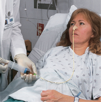
Enteral nutrition (EN) refers to nutrients given via the GI tract. EN is the preferred method of meeting nutritional needs if the patient’s GI tract is functioning because it provides physiological, safe and economical nutrition support. Enterally fed patients receive formula via nasogastric, jejunal or gastric tubes. Gastric feedings may be given to patients with a low risk of aspiration; however, if there is a risk of aspiration, jejunal feeding is preferred. Box 36-11 lists indications for tube feeding. Enteral tube feedings are easily given in the home setting by either the nurse or the family. Regardless of the setting, the principles in Skills 36-1, 36-2 and 36-3 for enteral feedings must be maintained.

BOX 36-11 INDICATIONS FOR ENTERAL AND PARENTERAL NUTRITION

ENTERAL NUTRITION

Cancer

Head and neck
Upper gastrointestinal (GI) tract

Critical illness/trauma

Neurological and muscular disorders

Brain neoplasm
Cerebrovascular accident
Dementia
Myopathy
Parkinson’s disease

Gastrointestinal disorder

Enterocutaneous fistula
Inflammatory bowel disease
Mild pancreatitis

Respiratory failure with prolonged intubation

Inadequate oral intake

Continuous feedings
Supine positioning
Cerebral vascular accident
Local trauma
Anorexia nervosa
Difficulty chewing, swallowing
Severe depression

PARENTERAL NUTRITION

Non-functional GI tract

Massive small bowel resection/GI surgery
Paralytic ileus
Intestinal obstruction
Trauma to abdomen, head or neck
Severe malabsorption
Intolerance to enteral feeding (established by trial)
Chemotherapy, radiation therapy, bone marrow transplantation

Extended bowel rest

Enterocutaneous fistula
Inflammatory bowel disease exacerbation
Severe diarrhoea
Moderate to severe pancreatitis

Preoperative total parenteral nutrition (TPN)

Preoperative bowel rest
Treatment for comorbid severe malnutrition in patients with non-functional GI tracts
Severely catabolic patients when GI tract non-usable for more than 4–5 days

SKILL 36-1 Inserting a small-bore nasoenteric tube for enteral feedings

DELEGATION CONSIDERATIONS

This task requires the problem-solving and knowledge-application skills of professional nurses. For this reason, delegation of this task to nurse assistants is inappropriate.

EQUIPMENT

Nasogastric or nasointestinal tube (8–12 Fr) with guide wire or stylet

60 mL or larger Luer-lock or catheter-tip syringe

Hypoallergenic tape and tincture of benzoin

pH indicator strip

Glass of water and straw

Emesis basin

Safety pin

Rubber band

Towel

Facial tissues

Clean gloves

Suction equipment in case of aspiration

Penlight to check placement in nasopharynx

Tongue blade

STEPS RATIONALE

1. Assess patient for the need for enteral tube feeding: NBM or insufficient intake for more than 5 days, functional GI tract, unable to ingest sufficient nutrients.

Identifying patients who need tube feedings before they become nutritionally depleted may help to prevent complications related to malnutrition.

2. Assess patient for appropriate route of administration:

Evaluates nares for patency.
 

a. Close each nostril alternately, and ask patient to breathe.

Nares may be obstructed. Assessment determines which naris to use.
 

b. Assess for gag reflex.

Identifies ability to swallow and risk of aspiration.
 

c. Inspect nares for any irritation or obstruction.


d. Review patient’s medical history for nasal problems and risk of aspiration.

Nurse may seek doctor’s order to change route of nutritional support or to place tube past the stomach into the intestine with increased risk of aspiration.

3. Review doctor’s order for type of tube and enteral feeding schedule.

Procedure and tube feedings require a doctor’s order.

4. Wash hands.

Reduces transfer of microorganisms.

5. Explain procedure to patient.

Reduces anxiety and helps patient to assist in insertion.

6. Stand on same side of bed as naris for insertion, and help patient into high-Fowler’s position unless contraindicated. Place pillow behind head and shoulders.

Allows easier manipulation of tube. Fowler’s position reduces risk of aspiration and promotes effective swallowing.

7. Place bath towel over chest. Keep tissues within reach.

Prevents soiling of gown. Insertion of tube may produce tears.

8. Determine length of tube to be inserted and mark with tape: measure distance from tip of nose to earlobe to xiphoid process of sternum (see illustration).

Length approximates distance from nose to stomach in 98% of patients. For duodenal or jejunal placement, an additional 20-30 cm is required.
 
image

Step 8 Determining length of tube to be inserted.

9. Prepare nasogastric or nasointestinal tube for intubation:


a. Plastic tubes should not be iced.

Tubes will become stiff and inflexible, causing trauma to mucous membranes.
 

b. Inject 10 mL of water from 30 mL or larger Luer-lock or catheter-tip syringe into the tube.

Aids in guide wire or stylet insertion.
 

c. Make certain that guide wire is securely positioned against weighted tip and that both Luer-lock connections are snugly fitted together.

Promotes smooth passage of tube into GI tract. Improperly positioned stylet can induce serious trauma.

10. Cut tape 10 cm long.


11. Put on clean gloves.

Reduces transmission of microorganisms.

12. Dip tube with surface lubricant into glass of water.

Activates lubricant to facilitate passage of tube into naris to GI tract.

13. Insert tube through nostril to back of throat (posterior nasopharynx). Aim back and down towards ear.

Natural contours facilitate passage of tube into GI tract; reduces gagging by patient.

14. Flex patient’s head towards chest after tube has passed through nasopharynx.

Closes off glottis and reduces risk of tube entering trachea.
Critical decision point: Encourage patient to swallow by giving small sips of water or ice chips when possible. Advance tube as patient swallows. Rotate tube 180 degrees while inserting.

15. Emphasise need to mouth-breathe and swallow during the procedure.

Helps facilitate passage of tube and alleviates patient’s fears during the procedure.

16. Advance tube each time patient swallows until desired length has been passed. Do not force tube. If resistance is met or patient starts to cough, choke or become cyanotic, stop advancing the tube and pull tube back.

Reduces discomfort and trauma to patient.

17. Check for position of tube in back of throat with penlight and tongue blade.

Tube may be coiled, kinked or entering trachea.

18. Perform measures to verify placement of tube:


a. Inject 30 mL of air into the tube, and aspirate GI contents with a syringe.

Gastric contents are usually cloudy and grassy green or tan to off-white; in contrast, intestinal fluid is usually deep golden yellow and is more clear than gastric fluid (see Figure 36-11). Fasting gastric pH is usually in a range of 1-4; only infrequently is it greater than 6. In contrast, intestinal sites usually have a pH of 7 or greater.
 

b. Measure pH of aspirated GI contents (see illustration).


c. X-ray verification of placement may be necessary.

 
image

Step 18b Comparing pH strip with colour chart.

 
Critical decision point: Auscultation is no longer considered a reliable method for verification of tube placement because a tube inadvertently placed in the lungs, pharynx or oesophagus can transmit a sound similar to that of air entering the stomach (Tho and others, 2011).

19. Apply tincture of benzoin or other skin adhesive to tip of patient’s nose and tube. Allow to dry.

Helps tape adhere better. Protects skin.

20. Remove gloves, and secure tube with tape, avoiding pressure on naris.

A properly secured tube allows the patient more mobility and prevents trauma to nasal mucosa.
 

a. Split one end of tape lengthwise 5 cm. Place the intact end of tape over bridge of patient’s nose. Wrap each of the 5 cm strips around tube as it exits nose (see illustration).

Securing tape to nares prevents tissue necrosis.
 

b. Fasten end of nasogastric tube to patient’s gown by looping rubber band around tube in slip knot. Pin rubber band to gown (see illustration).

Reduces traction on the naris if tube moves.
image

Step 20a Wrapping tape to anchor nasogastric tube.

image

Step 20b Pinning tube to gown.

21. For intestinal placement, position patient on right side when possible until radiological confirmation of correct placement has been verified. Otherwise, help patient into a comfortable position.

Promotes passage of the tube into the small intestine (duodenum or jejunum).
Critical decision point: Leave guide wire or stylet in place until correct position is ensured by X-ray. Never attempt to reinsert partially or fully removed guide wire or stylet while feeding tube is in place.

22. Obtain X-ray of abdomen.

Placement of tube is verified by X-ray examination.

23. Put on gloves, and administer oral hygiene (see Chapter 34). Clean tubing at nostril.

Promotes patient comfort and integrity of oral mucous membranes.

24. Remove gloves, dispose of equipment and wash hands.

Reduces transmission of microorganisms.

25. Inspect naris and oropharynx for any irritation after insertion.

If insertion was difficult, irritation of naris or oropharynx may have occurred.

26. Ask whether patient feels comfortable.

Evaluates patient’s level of comfort.

27. Observe patient for any difficulty breathing or gagging.

Malposition of the tube may cause these symptoms.
RECORDING AND REPORTING HOME CARE CONSIDERATIONS

Record and report type and size of tube placed, location of distal tip of tube, patient’s tolerance of procedure, pH value and confirmation of tube position by X-ray.

Placement may be verified on the basis of pH recordings. A small amount of water may be instilled via the feeding tube while the patient is carefully observed for coughing or gagging.

The patient and care provider should be instructed to report pH values that fall outside an established range and to report any difficulties that occur during the feeding.

SKILL 36-2 Administering enteral feedings via nasoenteric tubes

DELEGATION CONSIDERATIONS

This task requires the problem-solving and knowledge-application skills of professional nurses. For this reason, delegation of this task to nurse assistants is inappropriate.

EQUIPMENT

Disposable feeding bag and tubing or ready-to-hang system

30 mL or larger Luer-lock or catheter-tip syringe

Stethoscope

pH indicator strip

Infusion pump (required for intestinal feedings): use pump designed for tube feedings
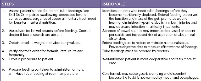
Prescribed enteral feedings

Gloves

Equipment to obtain blood glucose by finger-stick

STEPS RATIONALE

1. Assess patient’s need for enteral tube feedings: impaired swallowing, decreased level of consciousness, head or neck surgery, facial trauma, surgeries of upper alimentary canal.

Identify patients who need tube feedings before they become nutritionally depleted.

2. Auscultate for bowel sounds before feeding.

Absent bowel sounds may indicate decreased ability of GI tract to digest or absorb nutrients.

3. Obtain baseline weight and laboratory values. Assess patient for fluid volume excess or deficit, electrolyte abnormalities and metabolic abnormalities such as hyperglycaemia.

Enteral feedings are to restore or maintain a patient’s nutritional status. Provides objective data to measure effectiveness of feedings.

4. Verify doctor’s order for formula, rate, route and frequency. Laboratory data and bedside assessments, such as finger-stick blood glucose measurement, are also ordered by the doctor.

Tube feedings, laboratory tests and bedside tests must be ordered by doctor.

5. Explain procedure to patient.

Well-informed patient is more cooperative and at ease.

6. Wash hands.

Reduces transmission of microorganisms.

7. Prepare feeding container to administer formula:


a. Have tube feeding at room temperature.

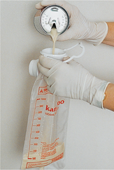
Cold formula may cause gastric cramping and discomfort because the liquid is not warmed by mouth and oesophagus.
 

b. Connect tubing to container as needed or prepare ready-to-hang container.

Tubing must be free of contamination to prevent bacterial growth.
 

c. Shake formula container well, and fill container and tubing with formula (see illustration).

Filling the tubing with formula prevents excess air from entering GI tract.

8. Place patient in high-Fowler’s position, or elevate head of bed 30 degrees.

Elevated head helps prevent aspiration.
image

Step 7c Pouring formula into container.

image

Step 9a Checking for gastric residual.

9. Determine tube placement:


a. Aspirate gastric contents to check for gastric residual (see illustration). Return aspirated contents to stomach unless the volume exceeds 150 mL

Presence of gastric secretions indicates that the distal end of the tube is in the stomach. Residual volume indicates whether gastric emptying is delayed. Delayed gastric emptying may be reflected by 150 mL or more remaining in the patient’s stomach.
 

b. Measure pH of aspirated GI contents.

Fasting gastric pH is usually equal to or less than 4; it is only infrequently greater than 6; in contrast, intestinal aspirates usually have a pH equal to or greater than 7.
 

c. Observe the aspirate’s appearance.

Gastric fluid is usually cloudy and grassy green or tan to off-white in colour; in contrast, intestinal fluid is usually deep golden yellow and is more clear than gastric contents (see Figure 36-11).
 

d. Consider the results from pH testing and the aspirate’s appearance together.

An aspirate with a low pH and a typical gastric fluid colour indicates a high probability of gastric placement; in contrast, a golden-coloured aspirate with a high pH indicates a high probability of intestinal placement.
Critical decision point: Auscultation is no longer considered a reliable method for verification of placement of tube because air in tube inadvertently placed in lungs, pharynx or oesophagus can transmit sound similar to that of air entering stomach.

10. Initiate feeding:


A. Bolus or intermittent feeding


(1)Pinch proximal end of the feeding tube.

Prevents air from entering patient’s stomach.
   

(2)Remove plunger from syringe and attach barrel of syringe to end of tube.


(3)Fill syringe with measured amount of formula. Release tube and hold syringe high enough to allow it to empty gradually by gravity, refill; repeat until prescribed amount has been delivered to the patient.

Gradual emptying of tube feeding by gravity from syringe or feeding bag reduces risk of abdominal discomfort, vomiting or diarrhoea induced by bolus or too-rapid infusion of tube feedings.
   

(4)If feeding bag is used, hang feeding bag on an IV pole. Fill bag with prescribed amount of formula, and allow bag to empty gradually over at least 30 minutes.


B. Continuous-drip method

Continuous feeding method is designed to deliver prescribed hourly rate of feeding. This method reduces risk of abdominal discomfort. Patients who receive continuous drip feedings should have residuals checked every 4 h and tube placement verified.
   

(1)Hang feeding bag and tubing on IV pole.


(2)Connect distal end of tubing to the proximal end of the feeding tube.


(3)Connect tubing through infusion pump (see illustration) and set rate.


11. Advance tube feeding gradually.

Tube feedings should be advanced gradually to prevent diarrhoea and gastric intolerance to formula.

12. When feeding is complete, flush the tubing with 30 mL water. Chart the amount of water on the fluid balance chart.

Reduces the risk of sediment forming and blocking the tubing.

13. When tube feedings are not being administered, cap or clamp the proximal end of the feeding tube.

Prevents air from entering stomach between feedings.

14. Administer water via feeding tube as ordered with diluted formula.

Provides patient with source of water to help maintain fluid and electrolyte balance.

15. Rinse bag and tubing with warm water whenever feedings are interrupted.

Rinsing bag and tubing with warm water clears old tube feedings and reduces bacterial growth.
 
image

Step 10B Connecting tubing through infusion pump.

16. Measure amount of aspirate (residual) every 4 hours.

Evaluates tolerance of tube feeding.

17. Monitor finger-stick blood glucose every 6 hours until maximum administration rate is reached and maintained for 24 hours. Monitor blood sugar level (BSL). Obtain medical direction for reportable BSL.

Alerts nurse to patient’s tolerance of glucose.

18. Monitor intake and output every 24 hours.

Intake and output are indications of fluid balance or fluid volume excess or deficit.

19. Weigh patient daily until maximum administration rate is reached and maintained for 24 hours; then weigh patient 3 times per week.

Weight gain is indicator of improved nutritional status; however, sudden gain of more than 900 g in 24 hours usually indicates fluid retention.

20. Observe return of normal laboratory values.

Improving laboratory values (e.g. albumin, transferrin and prealbumin) indicate an improved nutritional status.
RECORDING AND REPORTING HOME CARE CONSIDERATIONS

Record amount and type of feeding, patient’s response to tube feeding, patency of tube and any side effects. Report patient’s tolerance and adverse effects.

Ask patient or care provider about any symptoms or discomfort during enteral feedings. Reinforce instruction to contact nurse if symptoms or discomfort occur.

SKILL 36-3 Administering enteral feedings via gastrostomy or jejunostomy tube

DELEGATION CONSIDERATIONS

This task requires the problem-solving and knowledge-application skills of professional nurses. For this reason, delegation to nurse assistants is inappropriate.

EQUIPMENT

Disposable feeding container or ready-to-hang bag

30 mL or larger Luer-lock or catheter-tip syringe

Formula

Infusion pump: use pump designed for tube feedings

pH indicator strips

Stethoscope

Gloves

Equipment to obtain blood glucose by finger-stick

image image image

Tube feedings are typically started at full strength at slow rates of 20–50 mL/h. The hourly rate is increased by 25 mL increments every 12–24 hours if no signs of intolerance appear (nausea, cramping, vomiting, diarrhoea).

Studies have demonstrated a beneficial effect of enteral feedings compared with parenteral nutrition. Feeding by the enteral route may reduce sepsis, blunt the hypermetabolic response to trauma and maintain intestinal structure and function (Sudakin, 2006). EN has been used successfully within 24–48 hours after surgery or trauma to provide fluids, electrolytes and nutritional support. Gastric ileus may prevent nasogastric feedings, while nasointestinal or jejunal tubes allow successful postpyloric feeding where formula is placed directly into the small intestine or jejunum or beyond the pyloric sphincter of the stomach (Sudakin, 2006).

Aspiration of enteral formula into the lungs irritates the bronchial mucosa, resulting in decreased blood supply to affected pulmonary tissue. This then leads to necrotising infection, pneumonia and potential abscess formation. The high glucose content serves as a bacterial medium for growth, promoting infection. Acute respiratory distress syndrome (ARDS) is also an outcome frequently associated with aspiration of gastric contents (Baskin, 2006).

EN formulas vary in composition and nutrient density. General categories of EN formulas include standard whole-protein formulas, hydrolysed protein (elemental or peptide) and disease-specific formulas or crystalline amino acids (see Table 36-12).

TABLE 36-12 CLASSIFICATION OF ENTERAL NUTRITION PRODUCTS

CLASSIFICATION FORMULA CHARACTERISTICS
INTACT PROTEIN
Blenderised Isotonic; nutritionally complete; contain fibre; lactose-containing and lactose-free
Low-residue:  
 Standard protein Isotonic; nutritionally complete; lactose-free
 Intermediate–high protein Isotonic; nutritionally complete; lactose-free
 Concentrated Kilojoule-dense; lactose-free; nutritionally complete; hyperosmolar
Fibre-supplemented Isotonic; nutritionally complete; lactose-free; fibre ranges from 5 to 14.4 g/L; standard to intermediate protein
Disease-specific:  
 Renal Low and standard protein; low mineral, vitamin A and vitamin D content; lactose-free; kilojoule-dense; hyperosmolar
 Glucose-intolerant Low CHO, high fat; lactose-free; isotonic; contain fibre; nutritionally complete
 Pulmonary Low CHO, high fat; lactose free; nutritionally complete; hyperosmolar; kilojoule-dense
 Trauma/s tress High protein; isotonic to hyperosmolar; lactose-free; nutritionally complete; some contain fibre, hydrolysed protein, supplemental amino acids (branched-chain amino acids (BCAAs), arginine, glutamine) and beta-carotene; low to high fat
HYDROLYSED PROTEIN
Very low fat Nutritionally complete, lactose-free, less than 10% of the energy from fat, hyperosmolar, vary in peptide length, contain amino acids
Low fat Nutritionally complete, lactose-free, 11–30% of the energy from fat, hyperosmolar, vary in peptide length, contain amino acids
Moderate fat Nutritionally complete, lactose-free, 30–35% of the energy from fat (high in medium-chain triglycerides (MCTs)); hyperosmolar, vary in peptide length, contain amino acids
Disease-specific: trauma Nutritionally complete, lactose-free, intermediate to high protein content, 25–35% of the energy from fat (high in MCTs), vary in peptide length, contain amino acids, may contain supplemental arginine and beta-carotene
Crystalline amino acids Nutritionally complete, lactose-free, low fat (1–3% of the total energy), low to standard protein content, hyperosmolar
Disease specific: High BCAAs, nutritionally incomplete, lactose-free, low to moderate fat
 Hepatic failure Essential amino acids plus histidine, nutritionally incomplete, lactose-free, hyperosmolar, low fat
 Renal failure Essential and non-essential amino acids, nutritionally incomplete but contain water-soluble vitamins, hyperosmolar, lactose-free, low fat

Standard formulas are suitable for patients who do not have altered digestion or absorption; elemental and peptide formulas are used for patients who have impaired digestion or absorption; and disease-specific formulas have modifications in the content of specific nutrients or in caloric density. Nearly all tube-feeding formulas are lactose-free. Specialty enteral products tend to be very costly, and their use is generally reserved for specific indications (Sudakin, 2006). Research is examining nutrients such as glutamine, arginine, nucleotides, and omega-3 fatty acids for enteral formulas.

ENTERAL ACCESS TUBES

When the patient is unable to ingest food but is still able to digest and absorb nutrients, enteral tube feeding is indicated. Feeding tubes can be inserted through the nose (nasogastric or nasointestinal), surgically (gastrostomy or jejunostomy) or endoscopically (percutaneous endoscopic gastrostomy (PEG) or jejunostomy (PEJ)). If enteral therapy is for less than 4 weeks in total, nasogastric or nasojejunal feeding tubes may be used. Surgical or endoscopically placed tubes are preferred for long-term feeding (more than 4 weeks) to reduce the discomfort of a nasal tube and to provide a more secure, reliable access. Patients with gastroparesis (decreased or absent innervation to the stomach that results in delayed gastric emptying) or oesophageal reflux, at risk of aspiration or with a history of aspiration pneumonia require placement of tubes beyond the stomach into the intestine (Baskin, 2006).

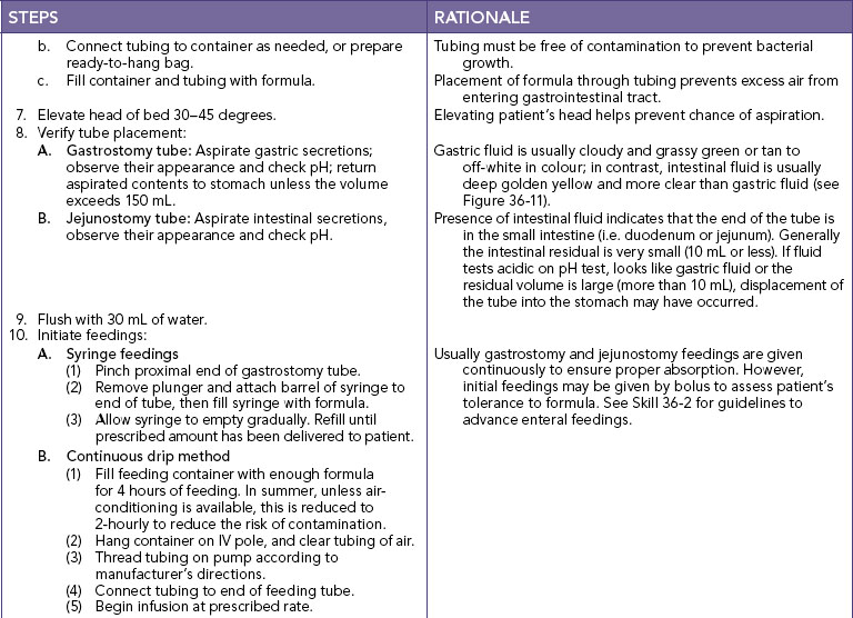
Nursing research has investigated the problems associated with nasoenteric tube placement, type of feeding instilled, rate of feeding and complications associated with tube feeding. Small-bore feeding tubes create less discomfort for the patient and are currently most often used (Figure 36-10). For the adult, most of these tubes are 8–12 French gauge (Fr) and 90–110 cm long. A stylet is often used during insertion of a small-bore tube to stiffen it. The stylet is removed when the correct position of the feeding tube is confirmed. Skills 36-1, 36-2 and 36-3 describe the procedures for inserting a small-bore nasoenteric tube and initiating enteral feedings.

image

FIGURE 36-10 Enteral tubes, small bore.

From Potter PA, Perry Ag 2013 Fundamentals of nursing, ed 8. St Louis, Mosby.

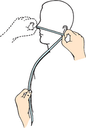
Upon insertion, the placement of small-bore feeding tubes is verified by X-ray examination. The tube should then be marked where it exits the nose and taped securely. Historically, feeding-tube placement was checked by injecting air through the tube while auscultating the stomach for a gurgling or bubbling sound, or asking the patient to speak (Tho and others, 2011). These methods have a high degree of inaccuracy. At present, the most reliable method is radiographical verification, which is cost-prohibitive for ongoing placement verification. Measurement of the pH of secretions withdrawn from the feeding tube may help to differentiate the location of the tube. For accurate pH measurements, 30 mL of air is injected into the tube before measurement to flush out formula, medications, flush solutions or other substances (Turgay and Khorsid, 2010). Addition of blue food colouring to enteral formula (0.2 mL/250 mL) assists with the detection of formula aspirated into the lung, presumably by staining the tracheobronchial secretions. However, the absence of blue-stained tracheobronchial secretions does not rule out pulmonary aspiration (Tho and others, 2011). Another method sometimes advocated for detecting pulmonary aspiration of enteral formula is testing the tracheobronchial secretions for the presence of glucose (using glucose reagent strips) (Baskin, 2006).

Checking samples of fluid withdrawn from newly inserted feeding tubes for acidic (gastric) or alkaline (intestinal) values prior to use and before intermittent feedings is perhaps the most sensitive bedside indicator of tube placement (Turgay and Khorsid, 2010). X-ray verification remains the ‘gold standard’. Concurrent use of acid-inhibitor medications alters gastric pH; however, in patients who have fasted for at least 4 hours, gastric pH continues to be ≤4 in slightly over half of cases. When acid inhibitors are used, fasting gastric pH is ≤6 in about three-quarters of the cases (Tho and others, 2011). Acid inhibitors have no effect on intestinal pH, which is usually ≥7. For a continuously tube-fed patient, gastric pH is expected to be higher because most enteral formulas have pH values close to 6. Thus, although pH is a helpful indicator of tube location, additional markers are needed to help differentiate between gastric and intestinal fluids.

Major complications of enteral nutrition are outlined in Table 36-13. Of special note, severely malnourished patients are at risk of electrolyte disturbances from refeeding syndrome, as ions such as potassium, magnesium and phosphate move intracellularly during enteral or parenteral nutrition therapy.

TABLE 36-13 ENTERAL TUBE FEEDING COMPLICATIONS

PROBLEM POSSIBLE CAUSE INTERVENTION
Pulmonary aspiration

Regurgitation of formula

Feeding tube displaced

Patient in supine position

Deficient gag reflex

Gastro-oesophageal reflux disease (GORD)

Check tube placement before feeding (every 4–8 hours during continuous)

Elevate head of bed 30–45 degrees during feedings and for 2 hours afterwards

Add sterile (blue) food colouring to formula for easier formula detection (See delayed gastric emptying below)

Diarrhoea

Delayed gastric emptying

Hyperosmolar formula or medications

Allergy to elixir ingredients (sorbitol)

Malnutrition/hypoalbuminaemia

Antibiotic therapy

Bacterial contamination

Malabsorption

Deliver formula continuously, lower rate, dilute or change to isotonic EN

Liquid medications are often sweetened with sorbitol, consider as possible cause

Albumin 25 g/L lessens oncotic pressure equilibrium

Antibiotics may destroy normal intestinal flora; doctor may change medication; treat symptoms with Lomotil, Kaopectate

Do not hang formula longer than 4–8 hours in bag, wash bag out well when refilling, change tube feeding bags every 24 hours and use aseptic practices

Check expiration dates

Check for pancreatic insufficiency, use low-fat, lactose-free formula and continuous feedings.

Constipation

Lack of fibre

Lack of free water

Medications

Inactivity

Select a formula containing fibre

Add water as needed as flushes*

Evaluate side effects; suggest stool softener or bulk-forming laxative

Monitor patient’s mobility; collaborate with doctor for activity order or physiotherapy

Tube occlusion

Pulverised medications given per tube

Insufficient tube irrigation

Sedimentation of formula

Reaction of incompatible medications or formula

Irrigate with 20 mL water before and after each medication per tube*

Dilute crushed medications if not liquid

Avoid crushed medications, if liquid available

Shake cans well before administering (read label)

Read pharmacological information on compatibility of drugs and formula

Tube displacement

Coughing, vomiting

Not taped securely

Replace tube and confirm placement before restarting tube feeding

With placement verification, check that tape is secure (nasoenteric)

Abdominal cramping, nausea/vomiting

High osmolality of formula

Rapid increase in rate/volume

Delayed gastric emptying

Lactose intolerance

Intestinal obstruction

High-fat formula used

Cold formula used

Suggest an isotonic formula, or dilute current formula

Lower rate of delivery to increase tolerance

Suggest use of lactose-free formula

Stop feeding with GI obstruction

Use greater proportion of carbohydrate

Warm formula to room temperature

Delayed gastric emptying

Diabetic gastroparesis

Prematurity

Serious illnesses

Inactivity

Consult with doctor regarding medication for increasing gastric motility (e.g. metoclopramide or cisapride)

Check for residual (see agency policy)

Consult doctor regarding advancing tube to intestinal placement (if gastric)

Monitor medications and pathological conditions that may affect GI motility

Monitor for increase of patient activity

Serum electrolyte imbalance

Excess GI losses

Dehydration

Cirrhosis

Renal insufficiency

Congestive heart failure, oedema

Diabetes mellitus

Monitor serum electrolyte levels daily.

Know of links with specific pathological condition

Monitor blood glucose level in diabetics or patients receiving insulin

Increased respiratory quotient Overfeeding of carbohydrates Balance kilojoule needs provided from fat, protein and carbohydrate with greater proportion of fat in formula (to decrease CO2 production)
Fluid overload

Refeeding syndrome in malnutrition

Excess free water or diluted (hypotonic) formula

Restrict fluids if necessary and use either a specialised formula or a diluted enteral formula at first

Monitor levels of serum proteins and electrolytes

Use a more concentrated formula with fluid volume excess without risk of refeeding syndrome

Hyperosmolar dehydration Hypertonic formula with insufficient free water Slow rate of delivery, dilute or change to isotonic formula

*Check first for fluid-restricted conditions that would affect volume of water given.

• CRITICAL THINKING

Sam Lang is a 22-year-old who sustained a closed head injury 3 days ago. He remains unconscious. Sam is to be started on enteral tube feeds. What are the potential complications of enteral tube feeding and the nursing interventions to minimise the occurrence of these?

PARENTERAL NUTRITION

Parenteral nutrition (PN) is a form of specialised nutrition support in which nutrients are provided intravenously. Safe administration of this form of nutrition depends on appropriate assessment of nutrition needs, meticulous management of the central venous catheter (CVC) and careful monitoring to prevent or treat metabolic complications. PN is administered in a variety of settings, including the patient’s home. Regardless of the setting, the nurse adheres to the same principles of asepsis and infusion management to ensure safe nutrition support.

image

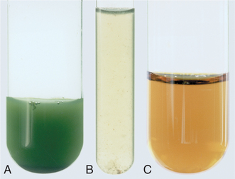
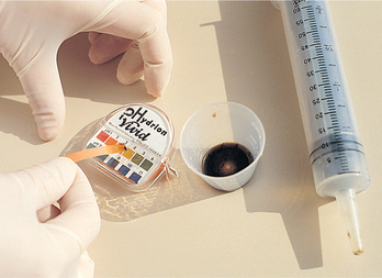
FIGURE 36-11 Gastric contents: A, Stomach. B, Stomach. C, Intestinal.

Courtesy Dr Norma Metheny, St Louis University School of Nursing.

Patients who are unable to digest or absorb enteral nutrition benefit from PN. Patients in highly stressed physiological states such as sepsis, head injury or burns are candidates for PN therapy. Box 36-11 outlines indications for PN.

Throughout PN therapy, clinical and laboratory monitoring by a multidisciplinary team is required (Table 36-14). The need for continued PN is constantly re-evaluated. The goal to move towards use of the GI tract is constant (Sudakin, 2006). Disuse of the GI tract has been associated with villus atrophy and generalised cell shrinkage. Translocation of bacteria from the local gut to systemic regions has been noted in relation to GI cell shrinkage, resulting in Gram-negative septicaemia (Baskin, 2006).

TABLE 36-14 MONITORING PARENTERAL NUTRITION (PN)*

PARAMETER BASELINE ROUTINE
Weight Daily Daily
Glucose (bedside) Every 6 hours Every 6 hours
Vital signs/temperature Once a shift when necessary (prn) Once a shift/prn
Intake/output Daily Daily
Electrolytes Daily first 3 days Biweekly
Creatinine, blood urea nitrogen (BUN) Baseline Biweekly
Albumin, prealbumin, transferrin Baseline Weekly
Cholesterol Baseline As ordered
Triglycerides Baseline As ordered
Liver enzymes (LFTs) Baseline Weekly
Full blood count (FBC) Baseline Weekly
Prothrombin time (PT)/partial thromboplastin time (PTT) Baseline Weekly
Platelets Baseline As ordered
Nitrogen balance (24-hour UUN) 1–2 days after PN started Weekly
Serum trace minerals and vitamins Baseline As ordered
Estimate energy requirements Baseline As needed

*Patients at home often adapt to less-frequent monitoring.

Give intramuscular vitamin K once weekly while on total parenteral nutrition and nil by mouth.

Modified from Grodner M and others 2007 Foundations and clinical applications of nutrition: a nursing approach, ed 4. St Louis, Mosby.

LIPID EMULSIONS

provide supplemental energy and prevent essential fatty acid deficiencies. These emulsions can be administered through a separate peripheral line, through the central line via Y-connector tubing (see Chapter 38) or as an admixture to the PN solution. The addition of lipid emulsion to the PN solution is called a total parenteral nutrition (TPN) mixture and is given over a 24-hour period. The admixture should not be used if oil droplets are observed or if an oil or creamy layer is observed on the surface of the admixture. This observation indicates that the emulsion has broken into large lipid droplets that can cause fat emboli if administered. Lipid emulsions are white and opaque; thus care should be taken to avoid confusing enteral formula with parenteral lipids.

INITIATING PN

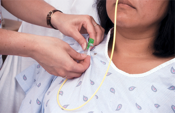
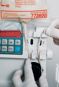
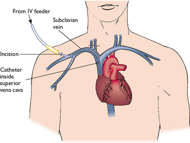
Solutions of less than 10% dextrose may be given in a peripheral vein in combination with amino acids and lipids. Peripheral solutions are not as calorically dense and are therefore usually temporary. Parenteral nutrition with greater than 10% dextrose requires a CVC that is placed into a high-flow central vein such as the superior vena cava by a doctor under sterile conditions (Figure 36-12). Nurses who have special training insert peripherally inserted central catheters (PICCs) that are started in a vein of the forearm and threaded into the subclavian or superior vena cava vein.

image

FIGURE 36-12 Central venous catheter placement.

Drawn by Rolin Graphics.

After CVC placement, the catheter is flushed with saline or heparin until the position is radiographically confirmed. The doctor sutures the catheter in place and covers the site with a sterile dressing. A chest X-ray examination identifies any complications.

BEGINNING AN INFUSION

Before beginning an infusion, the nurse compares the doctor’s order with the solution prepared by the pharmacy and checks the solution for particulate matter or a break in the lipid emulsion. An infusion pump is always used.

INFUSION FLOW RATE

Patients initially receive PN solutions at a moderate rate such as 40–60 mL/h. The rate is gradually increased until the target energy needs are being supplied. Too-rapid administration of hypertonic dextrose can result in an osmotic diuresis and dehydration (see Chapter 38). If an infusion falls behind schedule, the nurse should not increase the rate in an attempt to catch up. Sudden discontinuation of the solution can cause hypoglycaemia. Usually, 5–10% dextrose is infused when PN solution is suddenly discontinued. Diabetic patients are more at risk.

Patients who receive PN at home on a long-term basis are frequently acclimatised to a system of delivering 1–3 L of PN over 12 hours at night. The CVC is flushed and bunged (plugged with a sterile ‘bung’ or stopper) each morning for independent mobility during daytime hours. Nocturnal administration of PN is done by tapering the flow rate up at the beginning and down when ending. This individualised routine is tested during hospital stays for success.

PREVENTING COMPLICATIONS

Complications of PN include mechanical complications from insertion of the CVC, infection and metabolic aberrations (Table 36-15). Pneumothorax results from a puncture insult to the pulmonary system and culminates in accumulation of air in the pleural cavity with subsequent impaired breath. Pneumothorax is usually accompanied by symptoms of sudden sharp chest pain, dyspnoea and coughing, and occurs most often during CVC placement. Other complications of CVC insertion are cardiac dysrhythmias and, infrequently, rupture of major vessels.

TABLE 36-15 COMPLICATIONS OF PARENTERAL NUTRITION (PN)

PROBLEM SIGNS/SYMPTOMS INTERVENTION
Air embolism Tachypnoea, apnoea, wheezing, hypotension, cyanosis

Turn patient to left lateral decubitus position, instruct patient to perform Valsalva manoeuvre and lower head of bed

Cap open end of catheter or tape perforation in catheter wall

Administer oxygen; notify doctor

Maintain integrity of closed system, and have patient perform Valsalva manoeuvre when changing cap

Catheter occlusion No flow or sluggish flow through the catheter Temporarily stop infusion and flush with heparin; if effort to flush is unsuccessful, attempt to aspirate a clot; if still unsuccessful, follow protocol for use of thrombolytic agent (e.g. urokinase)
Catheter sepsis Fever, chills, glucose intolerance, positive blood culture

To prevent, change catheter site dressing if it becomes wet or contaminated, use aseptic technique when changing dressing or handling IV tubing, catheter caps or PN containers

Do not hang a single container of PN for more than 24 hours, or lipids more than 12 hours; use an inline 22-micron filter to remove bacteria*

Electrolyte imbalance Monitor Na, Ca, K, Cl, PO4, Mg and CO2 levels

See Chapter 39 for signs of deficiency/toxicity

Check TPN for supplemental electrolyte levels. Notify doctor of imbalances

Fatty liver LFT and bilirubin elevation, jaundice, upper abdominal pain

To prevent, do not overfeed carbohydrates, check history of hepatic dysfunction, chronic alcoholism, biliary disease

Reducing the energy to protein ratio and cycling TPN 12 hours on/off may help reduce elevated liver enzymes

Hypercapnia Increased oxygen consumption, increased CO2, respiratory quotient > 1.0, minute ventilation To prevent, ventilator-dependent patients are at risk; monitor parameters; provide 30–60% of energy requirements as fat
Hyperglycaemia Thirst, headache, lethargy, increased urination

Monitor blood glucose level daily until stable, then as ordered or prn

TPN is initiated slowly and tapered up to maximal infusion rate

Additional insulin may be required during therapy if problem persists (or if patient has diabetes mellitus)

Hyperglycaemic hyperosmolar non-ketotic dehydration/coma (HHNC) Hyperglycaemia (> 500 mg/dL), glycosuria, serum osmolarity > 350 mOsm/L, confusion, azotaemia, headache, severe signs of dehydration (see Chapter 39), hypernatraemia, metabolic-acidosis, convulsions, coma

To prevent, monitor blood glucose, BUN, serum osmolarity, glucose in urine and fluid losses; administer insulin as ordered; replace fluids as needed; maintain consistent infusion rate; and provide 30% of daily energy needs as fat

Patients at risk are hypermetabolic, receiving steroids, elderly, diabetic, have impaired renal or pancreatic function or are septic

Hypoglycaemia Diaphoresis, shakiness, confusion, loss of consciousness

To prevent, do not abruptly discontinue TPN but taper rate down to within 10% of infusion rate 1–2 hours before stopping

If hypoglycaemia is suggested, test blood glucose, administer IV bolus of dextrose per doctor’s order if necessary

Pneumothorax Severe dyspnoea, cyanosis, X-ray confirmation Complication that occurs upon catheter insertion, may evolve slowly afterwards. Monitor for first 24 hours for pulmonary distress. Notify doctor
Thrombosis of central vein Unilateral oedema of neck, shoulder and arm, pain Follow measures to prevent sepsis; repeated or traumatic catheterisations are likely to result in thrombosis

*With 3-in-1-admixture total parenteral nutrition (TPN), filtration is not possible due to large lipid molecules.

Air embolus can occur during insertion of the catheter or when changing the tubing or cap. Having the patient perform a Valsalva manoeuvre (holding one’s breath and ‘bearing down’) while assuming a left lateral position can prevent air embolus.

To avoid infection, the infusion tubing should be changed every 24 hours for a lipid infusion and every 48 hours when lipids are not infused. During CVC dressing changes, sterile mask and gloves are always used and insertion sites should be assessed for signs and symptoms of infection (see Chapter 29).

The TPN solution contains most of the major electrolytes, vitamins and minerals. Supplemental vitamin K must be given as ordered throughout therapy. Vitamin K can be synthesised by microflora found in the jejunum and ileum with normal use of the GI tract; since PN circumvents GI use, exogenous vitamin K must be administered.

Electrolyte and mineral imbalances may occur. Administration of concentrated glucose is accompanied by increases in endogenous insulin production, which causes ions (potassium, magnesium and phosphorus) to move intracellularly. Blood glucose levels are monitored and insulin is given if required. Liver function studies are monitored to determine the development of fatty deposits in the liver. In malnourished or cachectic patients, the resulting low serum (extracellular) levels of electrolytes and oedema may cause cardiac arrhythmias, congestive heart failure, respiratory distress, convulsions, coma or death. This has been called refeeding syndrome.

The goal is to move patients from parenteral to enteral and/or oral feeding. Once patients are meeting one-third to half their kilojoule needs per day enterally, TPN is usually decreased to half the original volume. Enteral feedings should then be increased to meet needs. When 75% of daily energy needs are consistently met with tube feeding, parenteral feeding may be discontinued. Patients who make the transition from PN to oral feedings typically have early satiety and decreased appetite. PN should be gradually decreased in response to increased oral intake. If oral intake is inadequate, small frequent meals may prove helpful. Kilojoule/protein counts are recommended when patients begin taking soft foods. When 75% of needs are being met by reliable dietary intake, PN therapy may be discontinued.

Medical nutrition therapy

Good nutrition is important in health and illness, but the specific dietary intake pattern that results in optimal nutrition must be modified for patients with particular diseases. Medical nutrition therapy (MNT) is the use of specific nutritional therapies to treat an illness, injury or condition. MNT may be necessary to assist the body’s ability to metabolise certain nutrients, correct nutritional deficiencies related to the disease and eliminate foods that may exacerbate disease symptoms. The following section provides a summary of MNT for a variety of diseases.

Restorative and continuing care

Some patients who are discharged from hospital with diet prescriptions will need dietary education to plan meals that meet specific therapeutic requirements. Restorative care includes both immediate postsurgical care and routine medical care, and therefore includes hospitalised and home care patients. The following sections cover nutritional interventions for some common disease states.

GASTROINTESTINAL DISEASES

Helicobacter pylori was first identified by Marshall and Warren in 1984 and is a bacterium that causes peptic ulcers. This is confirmed by laboratory tests and treated with antibiotics. Stress and overproduction of gastric HCl also contribute to peptic ulcer disease. Peptic ulcers are controlled with regular meals and medications such as ranitidine. Ranitidine is one of a class of drugs that are histamine-receptor antagonists, blocking secretion of hydrochloric acid (HCl). Patients are encouraged to avoid foods that increase stomach acidity, such as caffeine, decaffeinated coffee, frequent milk intake, citric acid juices and certain seasonings (hot chillies, chilli powder, black pepper). Smoking, alcohol and aspirin are also discouraged.

Inflammatory bowel disease includes Crohn’s disease and idiopathic ulcerative colitis. Treatment of acute inflammatory bowel disease may include elemental diets (formula with the nutrients in their simplest form, ready for absorption) or parenteral nutrition when symptoms such as diarrhoea and weight loss are prevalent. In the chronic stage of the disease a regular, highly nourishing diet is appropriate. Vitamins and iron supplements may be required to correct or prevent anaemia. Irritable bowel syndrome is managed by increasing fibre, reducing fat, avoiding large meals and avoiding lactose- or sorbitol-containing foods for susceptible individuals.

RESEARCH HIGHLIGHT
Research focus

Dietary treatment of children with behavioural conditions has been a topic of immense interest in recent times. Currently there is insufficient evidence to support dietary restrictions in children with behavioural conditions including autism and attention deficit hyperactivity disorder (ADHD). The focus of this paper was to review the evidence-based literature regarding dietary interventions and provide recommendations as to how nurses can assist families in their decision making regarding dietary choices.

Research abstract

Case studies were used together with a review of the literature to identify a number of different dietary approaches in the treatment of children with autism and ADHD. It was found that parents are increasingly dissatisfied with medication regimens for their children and were seeking alternative approaches to treatment for behavioural and developmental problems associated with autism and ADHD. The effectiveness of different diets in relation to behaviour modification were reviewed. The additive-free diet focused on the effect of artificial food colours on hyperactivity. The results from the meta-analysis found that artificial food colours do contribute to hyperactivity in children. The results from the sugar-elimination diets did not support the hypothesis that refined sugar increases hyperactivity and lessens attention span. Adding fatty acid supplements to the diet did not demonstrate an improvement in ADHD symptoms, although research is ongoing. Data from the gluten-free, casein-free diet, also ongoing, have mixed results with some studies suggesting this diet to be significant in its effect on autism, while other studies have not been able to replicate these results.

Evidence-based practice

Assessment of parents’ knowledge, and particularly their beliefs about the condition specific to their child, should be considered carefully.

Nurses, in helping parents to make informed choices, should be open to parents searching for alternative treatments and diets.

Nurses need to ensure sound knowledge of dietary options and effects to inform their educative and supportive role.

Reference

Cormier E, Elder JH. Diet and child behaviour problems: fact or fiction? Pediatr Nurs. 2007;33(2):138–143.

The treatment of malabsorption syndromes, such as coeliac disease, includes a gluten-free diet. Gluten is present in wheat, rye, barley and oats. Short-bowel syndrome results from extensive resection of bowel after which patients suffer from malabsorption due to lack of intestinal surface area. These patients may require lifetime feeding with either elemental enteral formulas or parenteral nutrition.

Diverticulitis is nutritionally treated with a moderate or low-residue diet until the infection subsides. Then a high-fibre diet is generally prescribed for chronic diverticulosis.

DIABETES MELLITUS

Type 1 diabetes mellitus requires both insulin and dietary restrictions for optimal control, beginning with diagnosis. In contrast, type 2 diabetes mellitus may initially be controlled solely by exercise and diet therapy. If these measures prove ineffective, it is common to add oral medications. Insulin injections may follow if type 2 diabetes worsens or fails to respond to these initial interventions. In both cases the diet is individualised according to the patient’s age, build, weight and activity level. Fats are moderately controlled (30% or less), and complex carbohydrates make up the majority (50–60%) of the diet, rather than simple carbohydrates. Protein comprises 10–20% of daily intake. Foods that contain soluble fibre are recommended, with a daily intake of 40 g of fibre.

Foods for dietary planning are classified into two exchange groups: the carbohydrate group and the meat and meat-substitute group. Foods from within the same group can be exchanged, but it is not recommended to exchange a carbohydrate food for a meat item. Each item has about the same nutrient value as other foods in the group (for more information, see Schlenker and Roth, 2011).

The strategies used for type 1 and type 2 diabetes are summarised in Table 36-16. The goal of treatment is normal glycaemic levels, and a haemoglobin A1c level of less than 7%, with resultant minimisation of complications of ophthalmic, vascular, renal and neuropathic damage (Grodner and others, 2007). Nurses also need to be aware of signs and symptoms of hypoglycaemia and hyperglycaemia.

TABLE 36-16 DIETARY STRATEGIES FOR TYPE 1 AND TYPE 2 DIABETES MELLITUS

DIETARY STRATEGY TYPE 1 (NON-OBESE) TYPE 2 (USUALLY OBESE)
Decrease energy intake (kilojoules) No Yes
Increase frequency and number of meals Yes Usually no
Regular daily intake of kilojoules, carbohydrates, protein and fat Very important Not as important as low average kJ intake
Consistent daily ratio of protein, carbohydrates and fat per meal Desirable Not necessary
Plan ahead for food to treat or prevent hypoglycaemia Very important Not necessary
Use extra food for exercise Yes Not usually necessary
Illnesses require small, frequent feeding of carbohydrates to prevent starvation ketosis Important Not usually necessary due to resistance to ketosis

From Schlenker E, Roth S 2011 Williams’ Essentials of nutrition and diet therapy, ed 10. St Louis, Mosby.

CARDIOVASCULAR DISEASES

Dietary therapy following an acute myocardial infarction includes initial reduction in energy, soft-textured foods and amounts of fat, sodium and cholesterol that conform to recommendations (see Box 36-12). Magnesium, folic acid and vitamin B6 appear to be important for primary prevention of coronary heart disease. Increases in folic acid are associated with a decrease in homocysteine, which is associated with greater risk of coronary artery disease (Lutz and Przytulski, 2011).

BOX 36-12 HEALTHY EATING

Eat a variety of foods, including vegetables, wholegrains, lean meats, oily fish, fruit, dairy products which are low-fat, reduced-fat or no-fat, and vegetable and seed oils. Remember to also include nuts, seeds and legumes.

Include 5 serves of vegetables and 2 serves of fruit every day. Choose wholegrain breads and breakfast cereals, and include pasta, noodles and rice.

Incorporate legumes and pulses like canned beans (e.g. baked beans, kidney beans and three-bean mix), dried peas (e.g. split peas), dried beans (e.g. butter beans and broad beans), chickpeas or lentils into two meals per week.

Consume reduced-fat, low-fat or no-fat milk, yoghurt, custard and desserts. Include small portions of cheese (1–2 slices or 20–40 g) up to four times a week.

Use spreads and margarines made from canola, sunflower or olive oil and dairy blends instead of butter. Choose healthier oils like canola, sunflower, soybean, olive, sesame and peanut oils when preparing food.

Eat 2–3 serves of oily fish (fresh or canned) a week.

Select lean meat and poultry (meat trimmed of fat and chicken without skin). Try to limit processed meats (e.g. sausages) and deli meats (e.g. salami).

A healthy balanced diet can include a serve of eggs (2 eggs) in 2–3 meals a week.

Try to limit take-away foods, such as pastries, pies, pizza, fried fish, hamburgers, hot chips and creamy pasta dishes, to once a week. Healthier take-away choices include sushi or sashimi, Asian stir-fries, tomato-based pasta dishes, grilled fish, chicken and lean meat.

Snack on plain, unsalted nuts and fresh fruit. Try to limit sugary, fatty and salty snack foods, such as crisps, cakes, pastries, biscuits, lollies and chocolate, to once a week.

It’s better not to add salt to food. If you want to add flavour, use herbs and spices. When shopping choose foods labelled ‘no added salt’, ‘low salt’ or ‘salt reduced’ where possible.

From National Heart Foundation of Australia (NHFA) 2009 Healthy eating and drinking. Online. Available at www.heartfoundation.org.au/SiteCollectionDocuments/Healthy-Eating-Tips-2009-05.pdf 30 Jun 2012. © 2009 National Heart Foundation of Australia. Reproduced with permission.

Nutritional therapy for hypertension includes energy reduction to promote weight loss as appropriate, decreased sodium intake, and potassium-rich foods if potassium-wasting diuretics are part of the treatment.

PULMONARY DISEASE

Macronutrients are digested and metabolised to form carbon dioxide and water. The ratio of carbon dioxide produced to oxygen consumed is the respiratory quotient (RQ). The RQ of carbohydrate digestion is 1.0, whereas for fat the RQ is 0.7. Patients with pulmonary disease generally maintain healthier RQs with a higher dietary intake of fat and less carbohydrate. This is relevant when weaning patients from ventilators in acute care settings, or counselling chronic obstructive pulmonary disease (COPD) patients for self-care.

RENAL DISEASE

Dietary treatment of acute glomerulonephritis depends on the patient’s symptoms and is designed to maximise nutritional intake. Fluid, salt and protein are not restricted unless indicated by symptoms such as oedema, uraemia or oliguria.

Acute renal failure (ARF) is related to low blood pressure or cellular damage within the nephron. Low blood pressure results from trauma, haemorrhage or shock and indirectly damages the nephrons. Other causes of ARF are nephrotoxic drugs, septicaemia or streptococcal infection that directly damages the nephrons. ARF usually consists of three stages: oliguric, diuretic and recovery. Dietary treatment of ARF changes according to these stages. The oliguric stage (from 7 to 21 days) requires that fluids be restricted to the patient’s output (typically 400–500 mL per day) plus an additional 400–500 mL per day. In contrast, diuresis (which ranges in duration from 7 to 14 days) requires large amounts of fluid replacement to make up for high urinary output. Protein is restricted to 0.6–0.8 g/kg/day, and parenteral amino acids may be required. A balanced mixture of essential and non-essential amino acids is provided along with concentrated dextrose and lipids. If the GI tract is functional, special enteral products are also available (Table 36-12). Finally, the recovery stage occurs over 3–12 months and is highly individual. Some residual kidney damage is common (Grodner and others, 2007).

Chronic renal failure treatment typically consists of a diet that restricts protein, potassium, phosphate, sodium and fluid. Adequate carbohydrates spare the use of protein for energy. As dialysis is begun, protein ingestion may increase. Calcium intake varies according to serum levels. There are no guidelines or specific renal diets recommended by any of the main Australian associations of nutritionists/dietitians. Renal diets are individually developed in consultation with the nephrologist, dietitian and patient. General renal nutrition information is available from the Australian Kidney Foundation.

Dietary treatment for renal calculi depends on the stone composition. For calcium phosphate stones, the diet is low in calcium and high in acid ash. For uric acid stones, the diet is low in purines. For calcium oxalate stones, the diet avoids all foods high in calcium and oxalates (Grodner and others, 2007).

CANCER AND CANCER TREATMENT

Malignant cells compete with normal cells for nutrients, increasing the metabolic needs of the patient. Most cancer treatments cause nutritional problems. Patients with cancer typically complain of anorexia and taste distortions. Malnutrition in cancer is associated with increased morbidity and mortality. Enhanced nutritional status may improve the patient’s quality of life.

Chemotherapy and radiation therapy are intended to destroy rapidly dividing malignant cells; however, other normal rapidly dividing cells, such as the epithelial lining of the GI tract, are often affected. These therapies can cause anorexia, stomatitis, severe diarrhoea, strictures of the intestine and pain. As well, chemotherapy often causes severe nausea and vomiting during the course of the treatment. Radiation treatment of the head and neck region can cause taste and smell disturbances, decreased salivation and dysphagia.

Nutrition management of the patient with cancer focuses on maximising intake of nutrients and fluids. The nurse should use creative approaches to manage alterations in taste and smell.

HUMAN IMMUNODEFICIENCY VIRUS (HIV)

Patients infected with HIV typically experience body wasting and severe weight loss. The wasting can be related to anorexia, stomatitis, oral thrush infection, nausea or recurrent vomiting, all resulting in inadequate intake. Factors associated with weight loss and malnutrition are severe diarrhoea, GI malabsorption and altered metabolism of nutrients. Systemic infection results in hypermetabolism from cytokine elevation. Often the medications taken to treat HIV infection cause side effects that alter nutritional status.

Restorative care of acquired immune deficiency syndrome (AIDS) malnutrition focuses on maximising kilojoules and nutrients. Each cause of nutritional depletion should be diagnosed and tackled in the care plan. Individually tailored nutrition support should progress in stages from oral to enteral, and lastly to parenteral. Good handwashing and food safety (see Box 36-9) are essential, including minimisation of exposure to Cryptosporidium in drinking water, lakes or swimming pools. Low-fat diets and small, frequent nutrient-dense meals may be better tolerated (Lutz and Przytulski, 2011).

EVALUATION

Care plans should reflect achievable goals. Nurses need to evaluate outcomes of nursing actions and be alert for signs that goals are being met. Adequate time should be allowed to test each nursing approach to a problem. Multidisciplinary collaboration remains essential in provision of nutrition support.

Patient care

Effectiveness of nutritional interventions is best measured by meeting the patient’s expected outcomes and goals of care (Figure 36-13). Nutrition therapy does not always produce rapid results. Ongoing comparisons may be made with baseline measures of weight, serum albumin or prealbumin, and protein and energy intake. Enteral nutrition therapy is frequently interrupted. Medications may produce unwanted side effects. If gradual weight gain is not observed, or if weight loss continues, the prescription may need to be increased. Changes in condition may also indicate a need to change the nutritional plan of care. Multidisciplinary members of the healthcare team should be consulted in an effort to better individualise the patient’s plan of care. The patient should be an active participant whenever possible. In the end, the patient’s ability to incorporate dietary changes into their lifestyle with the least amount of stress or disruption will ensure that outcome measures are successfully met.

image

FIGURE 36-13 Critical thinking model for nutrition evaluation phase.

• CRITICAL THINKING

Mrs Caine is 85 years old. She has been hospitalised for a fractured left hip and is now ready for discharge. She has always been active, and she lives alone. She has no family nearby, but a few close friends. What arrangements would you make to continue her nutritional intake at home while she recovers?

Patient expectations

Patients expect competent and accurate care. If ongoing nutritional therapies are not resulting in successful outcomes, patients expect nurses to recognise this fact and alter the plan of care accordingly. Expectations held by nurses may differ from those held by patients (Talbot and Verrinder, 2010). All patient teaching must have a clear understanding of the patient’s present knowledge and expectations. Successful interventions and outcomes depend on recognition of this concept in addition to nursing knowledge and skill. Working closely with the patient will enable the nurse to redefine those expectations that can be realistically met within the limits of the patient’s condition and treatment.

KEY CONCEPTS

Essential amino acids and essential fatty acids must be supplied by dietary intake since the body is unable to synthesise them from other ingested substances.

Through digestion, food is broken down into its simplest form for absorption. Digestion and absorption occur mainly in the small intestine.

Recommended dietary intakes provide a range of values that cover the needs of groups (estimated average requirement) and individuals (adequate intakes, recommended dietary allowances and tolerable upper intake level).

Guidelines for dietary change advocate reduced fat, saturated fat, sodium, refined sugar and cholesterol and increased intake of complex carbohydrates and fibre.

Age affects the requirements for essential nutrients. Periods of rapid growth increase the need for protein, vitamins and minerals.

Because improper nutrition can affect all body systems, nutritional assessment includes a review of total physical assessment.

Multidisciplinary collaboration is essential to optimal nutrition.

Nurses can improve food intake of patients by thoughtful attention to the preparation of the patient and environment before meals are served.

Tube feedings can be used for patients who are unable to ingest food but are able to digest and absorb food.

Enteral nutrition may protect intestinal structure and function and enhance immunity.

Total parenteral nutrition supplies essential nutrients in appropriate amounts to support life through the introduction of a concentrated nutrient solution into the superior vena cava near the right atrium of the heart.

Medical nutrition therapy is a recognised treatment for both acute and chronic disease states.

Special diets alter the composition, texture, digestibility and residue of foods to suit the patient’s particular needs.

Evaluation of the outcomes of nursing intervention in the area of nutrition support is essential to revise, update or continue nursing activities.

Nutritional research is a dynamic process. Results of studies and new recommendations by expert sources need to be followed for future changes in practice standards.

ONLINE RESOURCES

Australian guide to healthy eating, www.health.gov.au/internet/main/publishing.nsf/Content/health-pubhlth-strateg-food-guide-index.htm

Australian Breastfeeding Association, www.breastfeeding.asn.au

Australian Kidney Foundation, www.kidney.org.au

Department of Health and Ageing; nutrition and healthy eating resources, www.health.gov.au/internet/main/publishing.nsf/Content/health-pubhlth-strateg-food-index.htm

Diabetes Australia, www.diabetesaustralia.com.au

Diabetes New Zealand, www.diabetes.org.nz

HealthEd; healthy eating resources, www.healthed.govt.nz/health-topic/healthy-eating

Heart Foundation; healthy eatingAustralia, www.heartfoundation.org.au/healthy-eating/Pages/default.aspxNew Zealand, www.heartfoundation.org.nz/healthy-living/healthy-eating

Kidney Health New Zealand, www.kidneys.co.nz/Home

La Leche League New Zealand, www.lalecheleague.org.nz

Ministry of Health, New Zealand; food and nutrition guidelines, www.moh.govt.nz/foodandnutrition

New Zealand Breastfeeding Authority, www.babyfriendly.org.nz

Nursing Mothers Association of Australia, www.nmaa.asn.au

Royal New Zealand Plunket Society, www.plunket.org.nz/your-child/newborn-to-6-weeks/food-and-nutrition

REFERENCES

Abbott P, et al. Barriers and enhancers to dietary behaviour change for Aboriginal people attending a diabetes cooking course. Health Promot J Aust. 2010;21(1):33–38.

Al-Shammari K, et al. Risk indicators for tooth loss due to periodontal disease. J Periodontol. 2005;76(11):1721–1728.

Australian Breastfeeding Association (ABA). Strategic direction 2009–2012. Melbourne: ABA, 2009. Online Available at www.breastfeeding.asn.au/aboutaba/documents/direction/2009 4 Jul 2012.

Australian Bureau of Statistics (ABS). Breastfeeding in Australia, 2001. Cat. no. 4810.0.55.001. Canberra: ABS, 2003. Online Available at www.abs.gov.au/ausstats/abs@.nsf/Lookup/8E65D6253E10F802CA256DA40003A07C 20 Jun 2012.

Australian Institute of Health and Welfare (AIHW). Residential aged care in Australia 2008–09: a statistical overview. Aged care statistics no. 31. Cat no. AGE 62. Canberra: AIHW, 2010.

Australian Traditional Medicine Society (ATMS). Overview. Sydney: ATMS, 2011. Online Available at www.atms.com.au/index.php/overview 22 Jun 2012.

Baskin W. Acute complications associated with bedside placement of feeding tubes. Nutr Clin Pract. 2006;21:40–55.

Bocock M, et al. Defining malnutrition risk for older home care clients. Can J Dietetic Pract Res. 2008;69(4):171–176.

Food Standards Australia New Zealand (FSANZ). Australia, New Zealand Food Standards Code. Online. Available at www.foodstandards.gov.au/foodstandards/foodstandardscode.cfm, 2003. 21 Jun 2012.

Grant CC, et al. Nutrition and indigenous health in New Zealand. J Paediatr Child Health. 2010;46(9):479–482.

Grodner M, et al. Foundations and clinical applications of nutrition: a nursing approach, ed 4. St Louis: Mosby, 2007.

Hockenberry M, Wilson D. Wong’s Essentials of pediatric nursing, ed 8. St Louis: Mosby, 2009.

Hockenberry M, Wilson D. Wong’s Nursing care of infants and children, ed 8. St Louis: Mosby, 2007.

Holmes S. Barriers to effective nutritional care for older adults. Nurs Stand. 2006;21(3):50–54.

Lobo E, et al. Is there an association between low to moderate alcohol consumption and risk of cognitive decline? Am J Epidemiol. 2010;172(6):708–716.

Langstroth C, Wright C, Parkington T. Implementation and evaluation of a nutritional screening tool. Br J Midwif. 2011;19(1):15–21.

Lutz C, Przytulski K. Nutrition and diet therapy: evidence-based applications, ed 5. Philadelphia: FA Davis, 2011.

McDonald L. Florence Nightingale at first hand. London: Continuum, 2010.

National Health and Medical Research Council (NHMRC). Dietary guidelines for children and adolescents in Australia, incorporating the Infant feeding guidelines for health workers. Canberra: NHMRC, 2003. Online Available at www.nhmrc.gov.au/_files_nhmrc/publications/attachments/n34.pdf 25 Oct 2011.

National Health and Medical Research Council (NHMRC). Food for health: dietary guidelines for Australians. Canberra: NHMRC, 2005. Online Available at www.nhmrc.gov.au/_files_nhmrc/publications/attachments/n31.pdf 25 Oct 2011.

National Health and Medical Research Council (NHMRC), Ministry of Health (MOH). Nutrient reference values for Australia and New Zealand including recommended dietary intakes. Canberra: NHMRC; Wellington, MOH, 2006. Online Available at www.nhmrc.gov.au/guidelines/publications/n35-n36-n37 20 Jun 2012.

Neumark-Sztainer D, et al. Family meal patterns: associations with sociodemographic characteristics and improved dietary intake among adolescents. J Am Dietetic Assoc. 2003;103(3):317–322.

Nutbeam D, et alReview Team. Goals and targets for Australia’s health in the year 2000 and beyond: report prepared for the Department of Health, Housing and Community Services. Sydney: Public Health Department, University of Sydney, 1993.

Pagana K, Pagana T. Mosby’s diagnostic and laboratory test reference, ed 8. St Louis: Mosby, 2007.

Public Health Association of Australia (PHAA). Background paper on breastfeeding. Canberra: PHAA, 2007. Online Available at www.phaa.net.au/documents/policy/Breastfeedingbackgrounddpaper.pdf 30 Jun 2012.

Schaller C, James E. The nutritional knowledge of Australian nurses. Nurse Educ Today. 2005;25(5):405–412.

Schlenker E, Roth S. Williams’ Essentials of nutrition and diet therapy, ed 10. St Louis: Mosby, 2011.

Sudakin T. Supporting nutrition with TEN or TPN. Nursing (Lond). 2006;36(12):52–55.

Talbot L, Verrinder G. Promoting health: the primary health care approach, ed 4. Sydney: Elsevier, 2010.

Tan D, et al. Weight management in general practice: what do patients want? Med J Aust. 2006;185(2):73–75.

Tho P, et al. Implementation of the evidence review on best practice for confirming the correct placement of nasogastric tube in patients in an acute care hospital. Int J Evidence-Based Healthc. 2011;9:51–60.

Turgay A, Khorshid L. Effectiveness of the auscultatory and pH methods in predicting feeding tube placement. J Clin Nurs. 2010;19:1553–1559.