Chapter 39 Fluid, electrolyte and acid–base balance
Mastery of content will enable you to:
• Use knowledge of the distribution, composition, movement and regulation of body fluids and electrolytes to discuss physiological mechanisms that maintain fluid balance in all body compartments.
• Develop a beginning understanding of the meaning of changes in laboratory results associated with common fluid, electrolyte and acid–base imbalances.
• Demonstrate an ability to apply theoretical concepts to clinical practice in the determination of appropriate screening, prescription and monitoring of intravenous (IV) fluids and electrolyte administration in patients in ambulatory and acute care settings.
• Discuss reasons for nursing interventions for patients with fluid, electrolyte and acid–base imbalances.
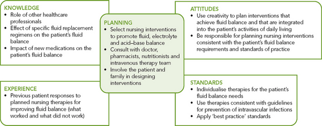
• Construct a plan of care (assessment, plan, implementation and evaluation) for maintaining fluid, electrolyte and acid–base balance:
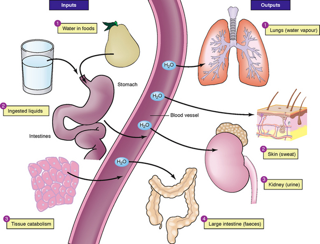
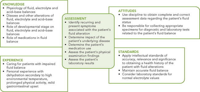
Fluid and electrolyte balance is where all compartments of the body contain the appropriate concentration of water and electrolytes. Nurses need to understand the distribution of body fluids, constituents of body fluids and processes used by the body to maintain fluid and electrolyte balance (Figure 39-1). Knowledge of these processes is essential in the early identification and understanding of factors contributing to fluid and electrolyte imbalances and in monitoring the effectiveness of appropriate management. Nurses must assume that each and every patient has a fluid and electrolyte imbalance until proven otherwise. Thus, the nurse must comprehensively assess fluid and electrolyte status for each and every patient and be able to articulate, at any time throughout the shift, the clinical indicators which prove the patient’s degree of fluid and electrolyte balance or imbalance.
Water is the largest single component of the body and a healthy, mobile, well-oriented adult can usually maintain normal fluid, electrolyte and acid–base balances because of the body’s adaptive physiological mechanisms.
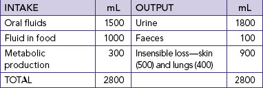
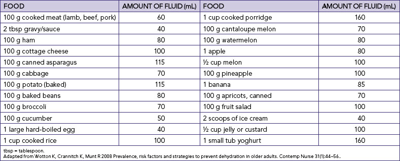
Nurses who are aware of their own fluid and electrolyte requirements and factors contributing to an imbalance in a range of different contexts will be better prepared to assess, identify and manage early indicators of fluid and electrolyte imbalance. For example, knowledge of average daily intake and output in healthy adults (Table 39-1) will assist in determining the amount of maintenance fluids required. The information in Table 39-1 can be used to demonstrate the risks to fluid and electrolyte balance for a person who decreases dietary intake and only consumes usual fluid intake. Students should also think about the impact on electrolyte balance if this person was only drinking water.
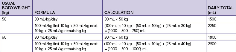
Nurses’ knowledge underpinning fluid and electrolyte balance needs to be more specific than described above. Many formulas exist for calculating the amount of fluid required per day. A general guide for the amount of fluid required in a healthy adult is 30 mL/kg/day (Eaton and others, 1999). If, for example, a student weighed 50 kg they would require 30 mL × 50 kg = 1500 mL fluid per day. There are also more-specific formulas for calculating the amount of fluid required per day. For example, fluid required per day for a healthy adult can be calculated as 100 mL/kg for the first 10 kg + 50 mL/kg for next 10 kg + 25 mL/kg for the remaining weight in kg (Austin, 1996). Table 39-2 demonstrates the difference between fluid requirements using the general formula and this more-specific formula for individuals weighing 50 kg and 60 kg.
Naturally, the daily fluid and electrolyte requirements depend on the person’s wellness, activity level and environmental conditions. Consider, for example, fluid and electrolyte loss and appropriate type and amount of fluid and electrolyte replacement for a person playing tennis in a temperature of 36°C, a bricklayer building an outside wall in summer in an environmental temperature of 39°C or a child with diarrhoea and vomiting with a temperature of 38.6°C.
As determining the appropriateness of urine output is important in the assessment of each of the situations described above, nurses should also be aware of the expected urine output based on gender and weight. The minimum urine output is the minimum amount of urine production required to ensure adequate renal function and to eliminate the body of waste products. A general rule is that urine output indicative of adequate renal function should be equal to, or more than, 30 mL/h. This is only a general guide and may lead to over- or under-estimation of the adequacy of urine output. A more specific formula for adequate urine output is 0.5 mL/kg for an adult male and 0.4 mL/kg for an adult female; infants and children weighing < 30 kg require 1 ml/kg/h. Thus, the expected urine output for a male weighing 65 kg is 34.5 mL/h and for a female weighing 65 kg is 26 mL/h.
Students should aim to develop the ability to use concepts covered in this chapter in developing knowledge of what is normal and why patients are at risk for fluid and electrolyte and acid–base imbalances, skills in comprehensive assessment, and knowledge of the appropriate fluid and electrolyte replacement therapy for different imbalances.
Total body water (TBW) is generally about 60% of bodyweight, with the following variations: 80% in newborns; 60% in adult males; 50% in adult females; and around 50% in obese people and 70% in lean people. Nurses need to understand these differences in usual TBW based on age, usual weight and gender. TBW calculations for an adult male are 0.6 × usual bodyweight = TBW (litres) and for an adult female 0.55 × usual bodyweight = TBW (litres) (Table 39-3).
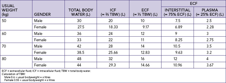
Body fluids are distributed in two distinct compartments, one containing intracellular fluids and the other extracellular fluids (Figure 39-2). Almost two-thirds of TBW is in the intracellular compartment (intracellular fluid, ICF); the other third is extracellular (extracellular fluid, ECF). Various semipermeable cell membranes separate TBW into these and further compartments, allowing each compartment to function relatively independently (Patton and Thibodeau, 2010).
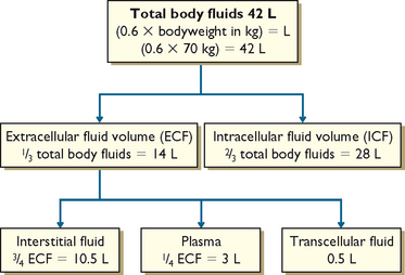
Intracellular fluid comprises all fluid within body cells. This fluid contains dissolved solutes essential to fluid and electrolyte balance and metabolism. In adult males, approximately 40% of bodyweight is ICF; in females it is 35% (Patton and Thibodeau, 2010). Extracellular fluid is all the fluid outside the cells. ECF is divided into two main smaller compartments: interstitial fluid and intravascular fluid. Interstitial fluid is the fluid between the cells and outside the blood vessels; intravascular fluid is blood plasma and lymph. Normally, about 25% of the ECF is in the intravascular compartment and the other 75% is interstitial fluid. Other extracellular fluids are transcellular fluid, cerebrospinal fluid (found in the ventricles of brain and surrounding the brain and spinal cord); synovial fluid (found inside synovial joint capsules); gastrointestinal tract fluids (saliva and gastric, pancreatic and intestinal juices); eye and ear fluids (vitreous and aqueous humour); pleural, pericardial and peritoneal fluids; and glomerular filtrate (Patton and Thibodeau, 2010).
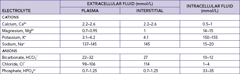
Body water contains substances that are sometimes called minerals or salts but are technically known as electrolytes. An electrolyte is an element or compound that, when dissolved in water, separates into ions and is able to carry an electrical current. An ion is an atom or molecule which possesses a positive or a negative charge. Positively charged ions are cations (sodium Na+, potassium K+, calcium Ca2+, magnesium Mg2+, iron Fe2+ and hydrogen H+). Negatively charged electrolytes are anions (chloride Cl−, bicarbonate HCO3−, sulfate SO42–, phosphate HPO43– and protein anions) (Table 39-4).
Electrolytes are measured in millimoles per litre (mmol/L). One mole (mol) is defined as the molecular (or atomic) weight of a substance, in grams. One millimole (mmol) is thus equal to one thousandth of a mole, in milligrams. In clinical practice, measurement of an amount of a substance is reported as the concentration of that substance in a particular volume; thus mass per unit of volume. The unit of measurement frequently used in the clinical setting is millimoles per litre (mmol/L). Millimoles per litre is a measure of the weight of a particular ion (the solute) dissolved in 1 L of a liquid (the solvent). The liquid formed by dissolving the solute in the solvent is called a solution.
Students should possess knowledge of the function of electrolyte anions and cations and their role in maintaining electrolyte balance in the human body in order to understand the patient’s maintenance and restorative dietary and fluid needs. If, for example, the patient has elevated levels of potassium (cation) then they are at risk for cardiac arrhythmias, whereas low potassium (cation) may result in paralysis. Decreased levels of calcium (cation) and magnesium (cation) may result in muscle spasms. Nurses must understand the reason why the consumption of just glucose or sodium solutions in individuals experiencing diarrhoea and vomiting will be insufficient to replace the electrolyte lost.
Electrolytes differ between ICF and ECF (Table 39-4). The body fluids and electrolytes in the ICF and ECF compartments are also in a constant state of mobility. Electrolyte distribution is a primary determinant of water concentration in a particular compartment.
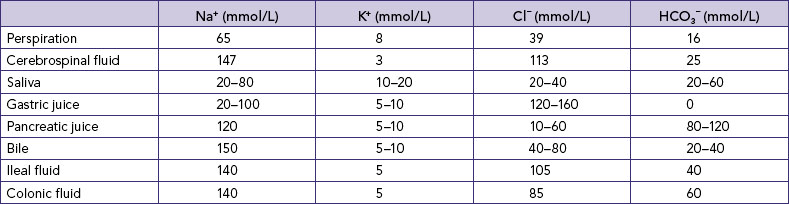
Knowledge of the electrolytes in different body fluids (Table 39-5) can assist nurses in identifying potential fluid and electrolyte imbalances when fluids are lost from particular sources, and therefore in understanding the appropriateness of the composition of fluids used to replace specific losses. Clearly there will be a difference in the electrolytes lost for a patient with a temperature of 39°C who is diaphoretic (major loss by perspiration), a patient who has an ileostomy (loss of ileal fluid) and a patient who is vomiting (loss of gastric juice)—and therefore a difference in the replacement fluids required.
• CRITICAL THINKING
Your next-door neighbour Mary calls you over to talk about what fluid she needs to give her 4-year-old child who has had acute diarrhoea with some vomiting for the past 24 hours. Respond to Mary’s question with a well-developed rationale based on your knowledge of fluid and electrolyte balance as to whether she should give either, both or none of the lemonade and Gatorade she has purchased.
Minerals (e.g. iron, zinc and magnesium; see Table 39-6) are constituents of all body tissues and fluids and are important in maintaining physiological processes. Minerals serve as cofactors in the thousands of enzyme-controlled body reactions; act as catalysts in nerve response, muscle contraction and metabolism of nutrients in foods; regulate electrolyte balance and hormone production; and strengthen skeletal structures (Patton and Thibodeau, 2010). Minerals do not work alone, but in balance with one another and with the metabolism of proteins, carbohydrates, fats and vitamins. An imbalance in one mineral can cause a chain reaction of deficiencies.
TABLE 39-6 MAJOR MINERALS AND THEIR FUNCTION
Fluids and electrolytes constantly shift from compartment to compartment to facilitate body processes such as tissue oxygenation, acid–base balance and urine formation. Cell membranes separating the body fluid compartments are selectively permeable, allowing water to pass through them easily. Most ions and molecules pass through them more slowly. Fluids and solutes move across these membranes by four processes: osmosis, diffusion, filtration and active transport.
Osmosis is the net movement of water molecules through a selectively permeable membrane down a water potential gradient; the water molecules move from an area of high water potential (low solute concentration, called hypotonic) to an area of low water potential (high solute concentration, called hypertonic) (Figure 39-3) (Haynie, 2001). The membrane is permeable to the solvent (water) but is impermeable to the solute. The rate of osmosis depends on the concentration of the solutes in the solution, the temperature of the solution, the electrical charges of the solutes and the differences between the osmotic pressures exerted by the solutions. The concentration of a solution is measured in osmols, which reflect the amount of a substance in solution in the form of molecules, ions or both.
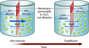
FIGURE 39-3 Osmosis through a semi-permeable membrane.
From Patton KT, Thibodeau GA 2010 Anatomy and physiology, ed 7. St Louis, Mosby.
Osmotic pressure is the drawing power for water in osmosis, and depends on the number of molecules in solution. If the concentration of the solute is greater on one side of the selectively permeable membrane, the rate of osmosis is quicker with a more rapid transfer of solvent across the membrane. This continues until equilibrium is reached. The osmotic pressure of a solution is called its osmolality, which is expressed in osmols per kilogram (Osm/kg) or milliosmols per kilogram (mOsm/kg) of the solution. The normal serum osmolality is 280–295 mOsm/kg.
Zinc is an essential trace element. It acts as a cofactor in numerous transcription factors and enzymes systems that augment auto-debridement and keratinocyte migration during wound repair. Zinc also assists the body to resist bacterial toxins. Zinc deficiency, whether caused by inadequate dietary intake or hereditary factors, can delay wound healing.
Oral zinc supplements may be beneficial in the treatment of patients with leg ulcers or slowly healing wounds. The therapeutic value of oral zinc supplements for faster healing of surgical wounds has not, however, been proven and requires further research. Topical application of zinc has been shown to be superior to oral zinc supplements in reduction of super-infections and necrotic material.
The action of topical zinc is thought to be from enhanced local defence mechanisms and increased collagenolytic activity. Sustained release of the zinc ions from topical applications is thought to stimulate the epithelialisation of wounds.
This review article suggests that topical zinc therapy is underappreciated even though clinical evidence emphasises its importance in autodebridement, anti-infective action and promotion of epithelialisation.
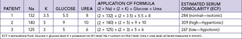
Osmolarity is the number of osmoles of solute in a litre of solution (Osm/L). Although the terms osmolality and osmolarity are often used interchangeably, the osmolality of a substance in serum is about 6% higher than its osmolarity because of proteins and lipids present in plasma. Debate exists on the accuracy of different calculations of serum osmolarity, but one which can be used at the bedside using the patient’s haematology results is (Erstad, 2003):
Table 39-7 shows the use of this formula for patients with normal, high and low osmolarity. Osmolarity is the measure used to evaluate serum and urine in clinical practice; if the ECF is too hypotonic, water will readily fill cells, increasing their volume and potentially lysing them (breaking down the cell wall, called cytolysis). As the osmolarity of ECF is approximately equal to that of ICF, serum osmolarity is a guide to intracellular osmolarity. Changes in extracellular osmolarity may result in changes in both ECF and ICF volume.
Solutions are classified as hypertonic, isotonic or hypotonic. A solution with the same osmolarity as blood plasma is called isotonic. A hypertonic solution (a solution of higher osmotic pressure) pulls fluid from cells; an isotonic solution (a solution of same osmotic pressure) expands the body’s fluid volume without causing a fluid shift from one compartment to another; and a hypotonic solution (a solution of lower osmotic pressure) moves fluid into the cells, causing them to enlarge (Figure 39-4). Each of these actions occurs through osmosis.
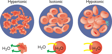
FIGURE 39-4 Hypertonic, isotonic and hypotonic solutions and their effect on cells.
From LadyofHats, Wikimedia Commons http://commons.wikimedia.org/wiki/File:Osmotic_pressure_on_blood_cells_diagram.svg
The process of osmosis within the body may be better understood by examining what would happen if a human cell was placed in a hypotonic, isotonic or hypertonic salt solution (Table 39-8).
TABLE 39-8 OSMOTIC RESPONSE TO HYPOTONIC, ISOTONIC AND HYPERTONIC SALINE SOLUTIONS
SALT SOLUTION IN WHICH THE CELL IS PLACED | DEFINITION | OSMOTIC RESPONSE |
---|---|---|
Hypotonic saline solution | Dilute solution, with a higher water concentration than the cell | Cell will gain water through osmosis |
Isotonic saline solution | Solution with exactly the same water concentration as the cell | No net movement of water across the cell membrane |
Hypertonic saline solution | Concentrated solution, with a lower water concentration than the cell | Cell will lose water by osmosis |
The osmotic pressure of the blood is affected by plasma proteins, especially albumin. Albumin exerts colloid osmotic or oncotic pressure, which tends to keep fluid in the intravascular compartment. Knowledge of the actions of colloid osmotic pressure is essential if nurses are to understand situations causing decreases in osmotic pressure and reasons for the use of intravenous (IV) colloids and crystalloids. Where patients lose plasma (plasma proteins), for example in burns or ascites, there will be insufficient osmotic pressure to maintain fluid in the intravascular compartment. The resultant increase in plasma proteins in the interstitial compartment will continue to draw fluid away from the intravascular compartment. Thus, although there are sufficient plasma proteins in the body they are in the incorrect compartment; ultimately, failure to correct insufficient circulating plasma proteins will result in circulatory collapse.
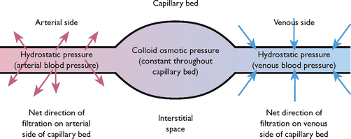
The net filtration pressure (NFP) is the difference between the hydrostatic pressure (pressure exerted by the fluid) and the oncotic pressure. Fluid leaves the capillary and enters the interstitial space when hydrostatic pressure is greater than oncotic. Fluid enters the capillary from the interstitial space when hydrostatic pressure is less than oncotic.
Fluid moves out of the arterial end of the capillary into the interstitial space because of a positive NFP of +5 mmHg, the difference between the hydrostatic pressure (30 mmHg) and the oncotic pressure (25 mmHg). Thus with this dominant hydrostatic pressure, fluid and diffusible solutes move out of the capillary into the interstitial space, and thus flow out of the circulation (Figure 39-5).
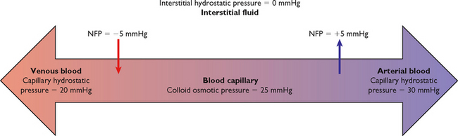
Fluid moves out of the interstitial space into the venous end of the capillary because of a negative NFP of –5 mmHg, the difference between the hydrostatic pressure (20 mmHg) and the oncotic pressure (25 mmHg). Oncotic pressure therefore dominates at the venous end of the capillary, and fluid and diffusible solutes move into the capillary from the interstitial space (Figure 39-5). The excess fluid and solutes remaining in the interstitial space are returned to the intravascular compartment by the lymph channels (Patton and Thibodeau, 2010).
A patient who has had a breast and lymph nodes removed because of breast cancer is at risk for the development of lymphoedema because of decreased return of proteins to the circulation (via the lymph) from the interstitial space. This results in increased fluid and proteins (increased oncotic pressure) in the interstitial space and the consequent development of localised oedema.
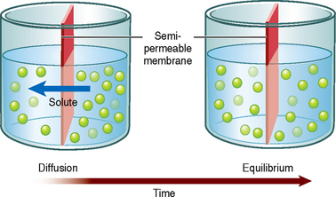
Diffusion is the movement of a solute (which can be a dissolved gas) in a solution across a semipermeable membrane from an area of higher solute concentration to an area of lower solute concentration (Figure 39-6) to ensure the maintenance of equilibrium and the even distribution of the solute in the solution. For example, if a small amount of red food colouring was added to a glass of water without stirring, over time the red food colouring molecules would evenly diffuse through the whole glass, giving an overall pink rather than a red appearance. A physiological example is the movement of oxygen and carbon dioxide between the alveoli and blood vessels in the lungs. The difference between the two concentrations is known as a concentration gradient.
Filtration is the process by which water and diffusible substances move together in response to fluid (hydrostatic) pressure. This process is active in capillary beds, where hydrostatic pressure differences determine the movement of water (Figure 39.7). When there is increased hydrostatic pressure on the venous side of the capillary bed, as occurs in congestive heart failure (CHF), the normal movement of water from the interstitial space into the intravascular space by filtration is reversed, resulting in an accumulation of excess fluid in the interstitial space, known as oedema.
Unlike diffusion, osmosis and filtration, active transport requires metabolic activity and expenditure of energy to move materials across cell membranes. This allows cells to admit larger molecules than they would otherwise be able to admit, or to move molecules ‘uphill’ from areas of lesser concentration to areas of greater concentration. An example of active transport is the sodium and potassium pump (Figure 39-8). Sodium is pumped out of the cell and potassium is pumped in, against the concentration gradient. This process makes it possible to keep a higher concentration of potassium in the ICF and a higher concentration of sodium in the ECF.
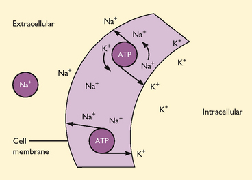
FIGURE 39-8 Sodium–potassium pump.
From Lewis SM and others 2008 Medical–surgical nursing: assessment and management of clinical problems, ed 6. St Louis, Mosby.
Active transport is enhanced by carrier molecules in a cell, which bind themselves to incoming molecules. For example, glucose is able to enter cells after it binds with the transport vehicle insulin. Active transport is the mechanism by which cells absorb glucose and other substances to carry out metabolic activities.
Body fluids are regulated by fluid intake, hormonal controls and fluid output. This physiological balance is termed homeostasis (Lewis and others, 2008). In health, a number of feedback mechanisms work together to maintain homeostasis (Figure 39-9).
Fluid intake is regulated mainly through the thirst mechanism. Thirst is the conscious desire for water and is one of the major factors that determine fluid intake (Patton and Thibodeau, 2010). The thirst-control centre is located within the hypothalamus in the brain. The thirst reflex is stimulated by three processes:
• a decrease in saliva results in dry mucosa in the mouth and pharynx
• an increase in blood osmotic pressure stimulates osmoreceptors in the hypothalamus
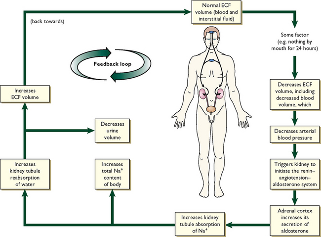
• a decrease in blood volume leads to the renin–angiotensin–aldosterone system (RAAS) stimulating the thirst centre in the hypothalamus (Figure 39-10). A decrease in blood volume can be due to hypovolaemia, a reduction in extracellular fluid volume that results in decreased tissue perfusion. Hypovolaemia can be caused by loss of either salt or water (e.g. with vomiting, diarrhoea or third space fluid shift). The term dehydration is more specific, and is the loss of only water; it results in imbalance between water and salts in the body and therefore leads to hypernatraemia.
The osmoreceptors continually monitor the solute concentration of the plasma. Osmoreceptors indirectly monitor ‘water balance’ and control the distribution of water between intracellular and extracellular fluid. Osmoreceptors respond to changes in osmotic pressure and tonicity caused by an excess or a deficit of water. When osmolality increases, the hypothalamus is stimulated. Increased plasma osmolality can occur with any condition that interferes with the oral ingestion of fluids, or it can occur with the intake of hypertonic fluids. The hypothalamus will also be stimulated when excess fluid is lost and hypovolaemia occurs, as in excessive vomiting and haemorrhage. If the thirst centre is activated, fluid deficit has already occurred to some extent. The response of the thirst reflex to fluid deficit is, however, diminished by older age, hypothalamic dysfunction, diminished cognitive function and in infants. Infants are at a higher risk than adults for dehydration because of a physiological inability of their renal tubules to concentrate urine, a higher metabolic rate, a larger body surface area and a poorly developed thirst mechanism.
Hormones regulate fluid intake through various mechanisms.
Antidiuretic hormone (ADH) is stored in the posterior pituitary gland. The osmoreceptors in the hypothalamus are stimulated to release ADH if there is an increase in blood osmolality. ADH works directly on the renal tubules and collecting ducts to increase their permeability to water. This in turn causes water to return to the intravascular space circulation, which dilutes the blood and decreases its osmolality. As the body attempts to compensate, the patient will experience a temporary decrease in urinary output. When the blood has been sufficiently diluted, the osmoreceptors stop the release of ADH and urinary output is restored.
Aldosterone is released by the adrenal cortex in response to increased plasma potassium levels or as a part of the RAAS to counteract hypovolaemia. It acts on the distal portion of the renal tubule to increase the reabsorption (saving) of sodium and the secretion and excretion of potassium and hydrogen. Because sodium retention leads to water retention, the release of aldosterone acts as a volume regulator (Patton and Thibodeau, 2010).
Renin, a proteolytic enzyme secreted by the kidneys, responds to decreased renal perfusion secondary to a decrease in extracellular volume. Renin acts to produce angiotensin I, which causes some vasoconstriction. However, angiotensin I almost immediately becomes reduced by an enzyme’s action to angiotensin II. Angiotensin II then causes massive selective vasoconstriction of many blood vessels and relocates and increases the blood flow to the kidney, improving renal perfusion. Angiotensin II also stimulates the release of aldosterone when the sodium concentration is low (Patton and Thibodeau, 2010).
Fluid output occurs through four organs: the kidneys, the skin, the lungs and the gastrointestinal (GI) tract. The kidneys are the major regulatory organs of fluid balance. They receive approximately 180 L of plasma to filter each day and produce 1200–1500 mL of urine daily (see Table 39-1).
Water loss from the skin is regulated by the sympathetic nervous system, which activates sweat glands. Water loss from the skin can be a sensible or an insensible loss. An average of 400–500 mL of sensible and insensible fluid is lost via the skin each day (Patton and Thibodeau, 2010). With fever, another 50 to 75 mL/day may be lost for each 1°C of temperature elevation above normal. Insensible water loss from the skin is continuous, not usually perceived by the individual, at around 0.4–0.5 mL/kg/h, and can increase significantly with pyrexia. Sensible water loss from the skin occurs through excess perspiration and can be perceived by the patient or by the nurse through inspection. The amount of sensible perspiration is directly related to the stimulation of the sweat glands.
The lungs expire about 400 mL of water daily. This insensible water loss may increase in response to changes in respiratory rate and depth. In addition, devices for administering oxygen can increase insensible water loss from the lungs.
The GI tract plays a vital role in fluid regulation. Approximately 3–6 L each day of isotonic fluid is moved into the GI tract and then returns to the extracellular fluid. Under normal conditions, the average adult loses only 100–200 mL of the 3–6 L each day through faeces. However, in the presence of a disease process, for example diarrhoea, the GI tract may become the site of a large amount of fluid loss. This loss may have a significant impact on maintaining normal fluid regulation.
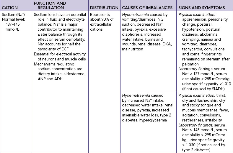
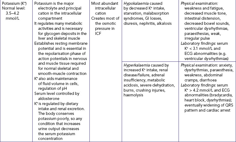
Major cations within the body fluids include sodium (Na+), potassium (K+), calcium (Ca2+) and magnesium (Mg2+). Cations interchange when one cation leaves the cell and is replaced by another. This occurs because cells tend to maintain electrical neutrality. Regulation of individual cations is included in Table 39-9.
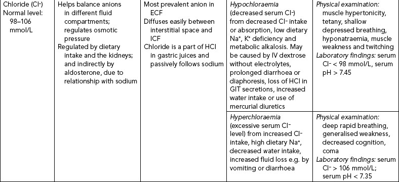
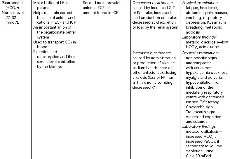
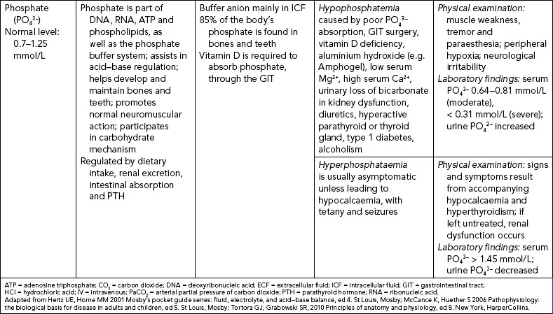
The three major anions of body fluids are chloride (Cl−), bicarbonate (HCO3−) and phosphate (PO43–) ions. Their regulation is included in Table 39-10.
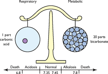
Metabolic processes maintain a steady balance between acids and bases to maintain optimal functioning of the cells. Arterial pH is an indirect measurement of hydrogen ion (H+) concentration: the higher the pH, the more acidic the solution (contains more H+ ions); a lower pH means a more alkaline solution (fewer H+ ions). Thus the pH is a scale for measuring the acidity or alkalinity of a fluid. A pH value of 7 is neutral, below 7 is acid, and above 7 is alkaline. Normal pH values in arterial blood range from 7.35 to 7.45.
The pH is regulated by two broad control systems, the chemical buffer systems (bicarbonate, phosphate and protein buffer systems) and the physiological buffer systems (the respiratory and renal response systems) (Patton and Thibodeau, 2010). A hydrogen ion buffer is a substance or a group of substances that can absorb or release H+ to correct an acid–base imbalance. Acid–base balance exists when the net rate at which the body produces acids or bases equals the rate at which acids or bases are excreted. This balance results in a stable concentration of hydrogen ions in body fluids, expressed as the pH value. Normal hydrogen ion levels are necessary to maintain cell membrane integrity and the speed of cellular enzymatic actions. Although buffers can raise the pH of body fluids, they do not remove the H+ from the body; this is left to the physiological buffers (Patton and Thibodeau, 2010).
Several buffering agents bind hydrogen ions to prevent or reduce changes in pH. Extracellular buffers include bicarbonate and ammonium ions; intracellular buffers include proteins and phosphate ions.
The most important chemical buffer in ECF is the carbonic acid and bicarbonate buffer system which reacts to imbalances within seconds (Figure 39-11). The equation below demonstrates how hydrogen ions (H+) and carbon dioxide (CO2) concentrations are directly related to each other, in that an increase in one causes an increase in the other. An increase in carbon dioxide concentration produces an increase in hydrogen ions; an increase in hydrogen ions results in increased carbon dioxide production (McCance and Huether, 2006). In the bicarbonate buffering system, carbon dioxide (CO2) moves reversibly through carbonic acid (H2CO3) to form hydrogen ions and bicarbonate ions (HCO3−):
A second type of chemical buffering occurs with the absorption or release of hydrogen ions by cells. This buffering occurs after the carbonic acid and bicarbonate buffering, and takes 2–4 hours. The positively charged hydrogen ion is exchanged with another positively charged ion, frequently potassium (K+). In conditions with excess acid, a hydrogen ion enters the cell and a potassium ion leaves the cell and enters the ECF, thus causing elevated serum potassium.
Another buffer is the haemoglobin–oxyhaemoglobin system. Carbon dioxide diffuses into the red blood cells (RBCs) and forms carbonic acid. The carbonic acid dissociates into hydrogen ions and bicarbonate ions. The hydrogen ions attach to haemoglobin, and the bicarbonate ion becomes available for buffering by exchanging with extracellular chloride (McCance and Huether, 2006).
The last buffer is the chloride shift within RBCs. When blood is oxygenated in the lungs, bicarbonate diffuses into the cells and chloride travels from the haemoglobin to the plasma to maintain electrical neutrality. The reverse occurs when carbon dioxide moves into RBCs in tissue capillary beds.
The two physiological buffers in the body are the lungs and the kidneys. The excretion of carbon dioxide resulting from metabolism is controlled mainly by the lungs, and the excretion of hydrogen and bicarbonate ions is controlled by the kidneys.
The lungs respond rapidly to acid–base imbalances to return the pH towards normal before the biological buffers act. Increased levels of hydrogen ions and carbon dioxide provide the stimulus for respiration. When the concentration of hydrogen ions is altered, the lungs react to correct the imbalance by altering the rate and depth of respiration. For example, when metabolic acidosis is present, respirations are increased, resulting in a greater amount of carbon dioxide being exhaled; this results in a decrease in the acidic level. Conversely, when metabolic alkalosis is present, the lungs retain carbon dioxide by decreasing the respirations, thereby increasing the acidic level (Madias and Adrogue, 2003).
Although the kidneys take from a few hours to several days to regulate acid–base imbalance, they are ‘the most powerful of the acid–base regulatory systems’ (Guyton and Hall 2006). In acidosis (excess acid):
• cells in the renal tubules reabsorb more bicarbonate ions from the tubule fluid
• cells in the renal collecting duct secrete more hydrogen ions and generate more bicarbonate ions
• there is increased formation of ammonia through the ammonia mechanism (see below).
In addition, the kidneys use a phosphate ion (PO43–) to excrete hydrogen ions by forming phosphoric acid (H3PO4); sulfuric acid (H2SO4) may also be excreted. The ammonia mechanism regulates acid–base balance by certain amino acids being chemically changed within the renal tubules into ammonia; this, in the presence of hydrogen ions, forms ammonium and is excreted in the urine, hence releasing hydrogen ions from the body (Roth and Chan, 2001).
In alkalosis (acid deficit), the kidneys excrete more bicarbonate ions by decreasing hydrogen ion secretion from the epithelial cells of the renal tubules, and lowering rates of glutamine metabolism and ammonium ion excretion.
Disturbances in electrolyte, fluid or acid–base balances seldom occur alone, and disrupt many other normal body processes. When there is a loss of body fluids because of dehydration, burns, illnesses or trauma or an increase of body fluids because of hypervolaemia, acute renal failure or CHF, the patient is also at risk of electrolyte imbalances. In addition, some untreated electrolyte imbalances (e.g. potassium loss) result in acid–base disturbances.
Hyponatraemia is a lower-than-normal concentration of sodium in the blood (serum), which can occur with a net sodium loss or net water excess (Table 39-9). It occurs frequently in seriously ill patients but can also occur in water intoxication, when someone drinks too much water too quickly, leading to haemodilution. There are multiple examples of this both in hospitals and in the community. Almond and others (2005) studied 488 runners in the 2002 Boston marathon; 13% of runners were found to have hyponatraemia (a serum sodium concentration of ≤135 mmol/L) and 0.6% critical hyponatraemia (≤120 mmol/L). In this situation hyponatraemia was associated with substantial weight gain, consumption of more than 3 litres of fluids during the race, female gender and low body mass index (BMI).
Nurses’ awareness of the effect of medications on serum sodium levels should increase early identification of hyponatraemia through regular appraisal of serum electrolyte results. An association has been found between hyponatraemia and prescription of antidepressant medication (Mannesse and others, 2010; Movig and others, 2002). Selective serotonin re-uptake inhibitors (SSRIs) were associated with an increased risk of hyponatraemia compared with other classes of antidepressant drugs. The risk of hyponatraemia increased in elderly patients also prescribed diuretics (Movig and others, 2002).
Clinical indicators and treatment depend on the cause of the hyponatraemia and association with normal, decreased or increased ECF volume (McCance and Huether, 2006). The usual situation is a loss of sodium without a loss of fluid, resulting in decreased ECF osmolality. The body initially adapts by reducing water excretion and thus sodium excretion to maintain serum osmolality at near-normal levels. As the sodium loss continues, the body continues to preserve the blood and interstitial (tissue) volume. As a result, the sodium in ECF becomes diluted.
Hypernatraemia is a greater-than-normal concentration of ECF sodium, caused by excess water loss or an overall sodium excess (see Table 39-9). When the cause of hypernatraemia is increased aldosterone secretion, sodium is retained and potassium is excreted. When hypernatraemia occurs, the body attempts to conserve as much water as possible through renal reabsorption.
Hypokalaemia is an inadequate amount of potassium in ECF (see Table 39-9). When severe, hypokalaemia can affect cardiac conduction and function. Because the normal levels of serum potassium are low, there is little tolerance for fluctuations. The most common cause of hypokalaemia is the use of potassium-wasting diuretics such as thiazide and loop diuretics.
Hyperkalaemia is a greater-than-normal amount of serum potassium. Severe hyperkalaemia produces marked cardiac conduction abnormalities (see Table 39-9). The main cause of hyperkalaemia is renal failure and thus decreased excretion of potassium.
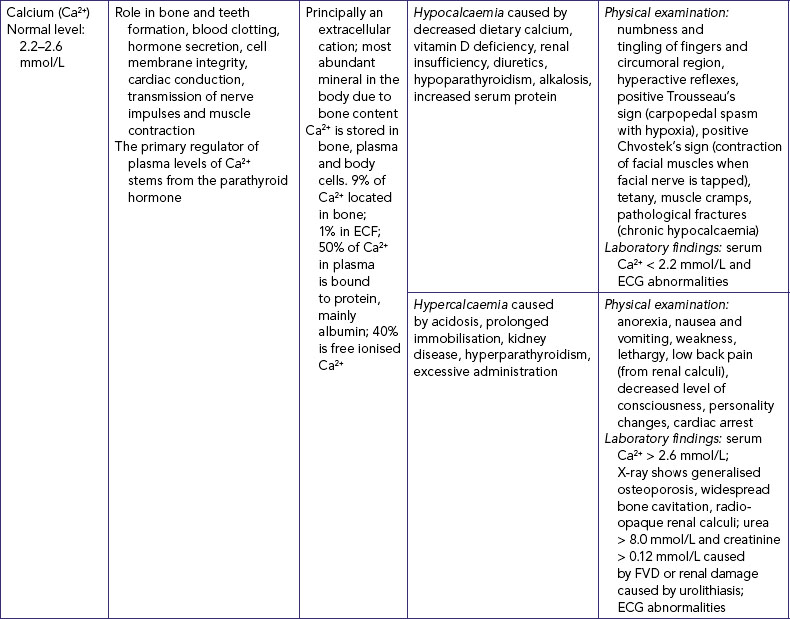
Hypocalcaemia represents a drop in serum and/or ionised calcium levels. It can result from illnesses which directly affect the thyroid and parathyroid glands (see Table 39-9) and from renal insufficiency (in which the kidneys’ inability to excrete phosphorus causes the phosphorus level to rise and the calcium level to decline). Signs and symptoms can be related to a diminished function of the neuromuscular, cardiac and renal systems.
Hypercalcaemia is an increase in the total serum concentration of calcium and/or ionised calcium. Hypercalcaemia is frequently a symptom of an underlying disease resulting in excess bone reabsorption with release of calcium.
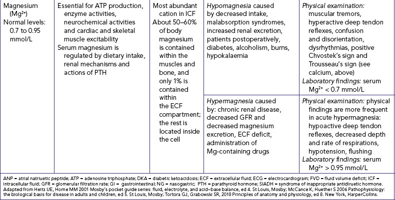
Disturbances in magnesium levels result in changes in neuromuscular excitability (see Table 39-9). Hypomagnesaemia, a decrease in serum magnesium, occurs with malnutrition and with malabsorption disorders. Signs and symptoms are directly related to the neuromuscular system. Hypermagnesaemia, an increase in serum magnesium levels, depresses skeletal muscles and nerve function. Hypermagnesaemia occurs during renal failure, aldosterone deficiency and hypothyroidism and by excess intake of antacids containing Mg2+. Hypermagnesaemia causes hypotension, muscle weakness, nausea, vomiting and altered mental functioning (Lewis and others, 2008).
Hypochloraemia occurs when serum chloride levels fall below normal (see Table 39-10). Vomiting or prolonged and excessive nasogastric or fistula drainage can result in hypochloraemia because of the loss of hydrochloric acid. The use of loop and thiazide diuretics also results in increased chloride loss, as sodium is excreted. When serum chloride levels fall, metabolic alkalosis results as the body adapts by increasing reabsorption of the bicarbonate ion to maintain electrical neutrality. Hyperchloraemia occurs when the serum chloride levels rise above normal (see Table 39-10), which usually occurs when serum bicarbonate levels fall or sodium levels rise.
Hypochloraemia and hyperchloraemia rarely occur as single disease processes, but are commonly associated with acid–base imbalance. There is no single set of symptoms associated with these two alterations.
The basic types of fluid imbalance are isotonic and osmolar. Isotonic deficit and excess exist when water and electrolytes are gained or lost in equal proportions. In contrast, osmolar imbalances are losses or excesses of only water, so that the concentration (osmolality) of the serum is affected. Table 39-11 lists the causes and symptoms of common fluid disturbances.
TABLE 39-11 FLUID DISTURBANCES
CAUSES | SIGNS AND SYMPTOMS |
---|---|
ISOTONIC IMBALANCES | |
Fluid volume deficit (FVD)—water and electrolytes lost in equal or isotonic proportions | |
Physical examination: postural hypotension, decreased pulse pressure, decreased MAP, tachycardia, dry mucous membranes, poor skin turgor, thirst, confusion, rapid weight loss*, slow vein filling, lethargy, oliguria, weak pulse Laboratory findings: urine specific gravity: 1.025; increased haematocrit level, 50%; and increased BUN level, > 8 mmol/L (haemoconcentration) |
|
Fluid volume excess (FVE)—water and sodium retained in isotonic proportions | |
Physical examination: rapid weight gain, peripheral oedema, hypertension, increased pulse pressure, increased MAP, polyuria (if renal mechanisms are normal), increased jugular venous pressure, increased venous pressure, bi-basal lung crepitations Laboratory findings: decreased haematocrit level, 35%; and decreased BUN level, > 3 mmol/L (haemodilution) |
|
OSMOLAR IMBALANCES | |
Hyperosmolar imbalance—dehydration | |
Hypo-osmolar imbalance—water excess | |
BUN = blood urea nitrogen; MAP = mean arterial pressure (of blood); SIADH = syndrome of inappropriate antidiuretic hormone.
*With third space fluid shift, there is either no change or an increase in weight
Arterial blood gas (ABG) analysis is the best way to evaluate acid–base balance. Measurement of ABGs involves analysis of six components: pH, PaCO2, PaO2, oxygen saturation, base excess and HCO3−. Deviation from a normal value will indicate that the patient is experiencing an acid–base imbalance.
pH measures hydrogen ion (H+) concentration in the body fluids. Even a slight change can be potentially life-threatening. An increase in H+ concentration makes a solution more acidic; a decrease makes the solution more alkaline. Normal pH values are 7.35–7.45 (acidic is < 7.35, and alkaline is > 7.45).
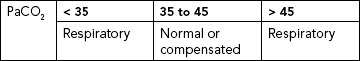
PaCO2 is the partial pressure of carbon dioxide in arterial blood, and reflects the depth of pulmonary ventilation. The normal range is 35–45 mmHg. A PaCO2 of < 35 mmHg is an indication of hyperventilation. As rate and depth of respiration increase, more carbon dioxide is exhaled and the carbon dioxide concentration decreases. Hypoventilation causes increased PaCO2, above 45 mmHg. As rate and depth of respiration decrease, less carbon dioxide is exhaled and more is retained, increasing the concentration of carbon dioxide.
PaO2 is the partial pressure of oxygen in arterial blood. It has no primary role in acid–base regulation if it is within normal limits. The normal range is 80–100 mmHg. A PaO2 of less than 60 mmHg (hypoxaemia) can lead to anaerobic metabolism, resulting in lactic acid production and metabolic acidosis. There is a normal decline in PaO2 in older adults. Hypoxaemia also may cause hyperventilation, resulting in respiratory alkalosis (McCance and Huether, 2006).
Oxygen saturation (SaO2) is the percentage of available haemoglobin bound to oxygen. Oxygen saturation can be affected by changes in temperature, pH and PaCO2. Normal range is 95–99%.
Base excess is the amount of blood buffer (haemoglobin and bicarbonate) that exists. The normal range is –2 to +2. A high value indicates alkalosis and can result from the ingestion of large amounts of sodium bicarbonate solutions (some antacids), citrate excess with rapid blood transfusions or intravenous infusion of sodium bicarbonate to correct ketoacidosis. A low value indicates acidosis and is usually the result of the elimination of too many bicarbonate ions. An example is diarrhoea and prolonged loss (vomiting or nasogastric suction) of deep GI contents that force the bicarbonate-containing fluid to be lost instead of being absorbed (Berry and Pinard, 2002).
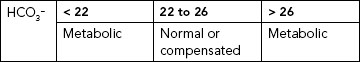
Serum bicarbonate (HCO3−) is the major renal component of acid–base balance, and is excreted and produced by the kidneys to maintain a normal acid–base environment. It is the principal buffer of the ECF of the body, and once bicarbonate is in the ECF it is maintained at a concentration of 20 times that of the fluid concentration of carbonic acid (Berry and Pinard, 2002). Normal range is 22–26 mmol/L. Less than 22 mmol/L usually indicates metabolic acidosis; more than 26 mmol/L indicates metabolic alkalosis.
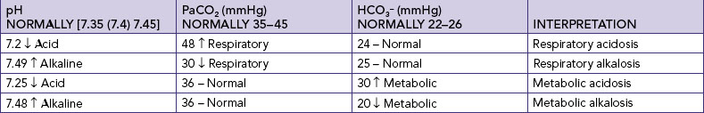
The four main types of acid–base imbalance are respiratory acidosis, respiratory alkalosis, metabolic acidosis and metabolic alkalosis (Table 39-12).
TABLE 39-12 ACID–BASE IMBALANCES
PaCO2 = partial pressure of carbon dioxide in arterial blood; PaO2 = partial pressure of oxygen in arterial blood;
Respiratory acidosis is marked by an increased arterial carbon dioxide concentration (PaCO2), excess carbonic acid (H2CO3) and an increased hydrogen ion concentration (decreased pH). With respiratory acidosis, the cerebrospinal fluid and brain cells become acidic, causing neurological changes. Hypoxaemia occurs because of respiratory depression, resulting in further neurological impairment. Electrolyte changes such as hyperkalaemia and hypercalcaemia may accompany the acidosis.
Respiratory alkalosis is marked by decreased PaCO2 levels and increased pH. Like respiratory acidosis, respiratory alkalosis can begin outside the respiratory system (e.g. anxiety with hyperventilation) or within the respiratory system (e.g. initial phase of an asthma attack).
Metabolic acidosis results from abnormal loss of bicarbonate or accumulation of excess metabolic acids causing high acid content of the blood (Guyton and Hall, 2006). Identification of the cause of metabolic acidosis requires an analysis of serum electrolytes to detect an anion gap. An anion gap reflects unmeasurable anions present in plasma, and is calculated by subtracting the sum of chloride and bicarbonate levels from the plasma sodium level (Table 39-13). The anion gap is thus an artefact of measurement, and not a physiological reality. Anion gap is an ‘artificial’ and calculated measure that is representative of the serum ions not routinely measured when testing serum electrolytes. For example, the anions (e.g. lactate, acetoacetate, sulfate) produced during metabolic acidosis are not measured as part of the usual laboratory profile.
TABLE 39-13 DIFFERENTIATION OF CAUSES OF METABOLIC ACIDOSIS FROM EXAMINATION OF NORMAL VS ELEVATED ANION GAP
Adapted from Heitz UE, Horne MM 2001 Mosby’s pocket guide series: fluid, electrolyte, and acid–base balance, ed 4. St Louis, Mosby; McCance K, Huether S 2006 Pathophysiology: the biological basis for disease in adults and children, ed 5. St Louis, Mosby; Tortora GJ, Grabowski SR, 2010 Principles of anatomy and physiology, ed 8. New York, HarperCollins; Wotton K, Crannitch K, Munt R 2008 Prevalence, risk factors and strategies to prevent dehydration in older adults. Contemp Nurse 31(1):44–56.
Mrs Sinclair was admitted with blood pressure 90/70 mmHg, pulse 126 beats per minute, respirations 32 breaths per minutes and a 2-day history of nausea and vomiting, excessive thirst and polyuria. She was recently diagnosed with type 2 diabetes. Her arterial blood gas results reveal metabolic acidosis; calculation of the anion gap using laboratory information reveals a high anion gap of 27.
Calculating the anion gap is useful in distinguishing between anion-gap and non-anion-gap metabolic acidosis, differentiating between causes of metabolic acidosis, assessing biochemical severity of acidosis, and monitoring response to treatment. If the laboratory report does not include an anion gap it can be calculated from the formula
Table 39-13 indicates reasons for normal and increased anion gap.
Metabolic alkalosis is marked by the heavy loss of acid from the body or by increased levels of bicarbonate. The most common causes of acid loss are vomiting and gastric suction. Other causes include the overcorrection of metabolic acidosis, loss of chloride, use of diuretics causing resorption of bicarbonate, hyperaldosteronism and excessive ingestion of alkali (Berry and Pinard, 2002).
• CRITICAL THINKING
Your friend meets with you for coffee after playing cricket (bowling and fielding) on a day when the air temperature was 38.9°C. He states he is not feeling very well, has a severe headache and would prefer just water. On asking what he has consumed during the day, he states he had around 4 litres of plain water.
Discuss the appropriateness of his intake throughout the day, using what you know about the fluid he would have lost and required replacement therapy.
Nurses should assume that every patient is at risk for fluid and electrolyte imbalances. Infants, severely ill adults, disoriented or immobile patients and older adults are often at greater risk because of an inability to independently respond to early signs and symptoms of haemodynamic instability. Over time, the body’s adaptive compensatory mechanisms can no longer adequately maintain fluid and electrolyte or acid–base balance, and the patient’s health becomes compromised. For example, kidney function, urine concentration, thirst sensation, aldosterone secretion and renin activity are all significantly lowered with age (Lancaster and others, 2003; Xiao and others, 2004). Prolonged or severe compromises may lead to irreversible chronic health problems that decrease the quality of life of the patient, caregiver(s), guardians, parents, families and/or friends (Box 39-1).
BOX 39-1 RISK FACTORS FOR FLUID, ELECTROLYTE AND ACID–BASE IMBALANCES
• 3 or more medical conditions
• Alterations in mental health status
• New admission to a residential-care facility/hospital
• Cardiovascular disease such as congestive heart failure
• Endocrine disease such as Cushing’s disease and type 1 diabetes
The proficiency of a nurse lies in his or her ability to observe, recognise, discriminate and interpret clinical evidence in order to reach a judgment about a patient’s fluid, electrolyte and acid–base balance. Nurses have a professional obligation to monitor the patient’s clinical progression and identify at an early stage changes in a patient’s fluid and electrolyte status (Wotton, 2011). Any delay in the identification of changes in haemodynamic status has direct consequences for the patient, their family and the healthcare institution.
Clinical reasoning involves the rational analysis and review of patient data, theoretical knowledge, inferences, assumptions and beliefs, conclusions and actions to optimise patient outcomes. The clinical reasoning process is not just one episode of thinking, but a cyclical process of data collection and analysis (data interpretation or re-interpretation and problem formulation or re-formulation) in the development of a progressively broader and deeper understanding of patients’ fluid and electrolyte status (Wotton, 2011). Successful critical thinking requires the nurse to challenge ideas, be open to different and alternative views, embrace the ambiguity and complexity of the reasoning process and resist the urge to come to hasty decisions without adequate evidence. During assessment (Figure 39-12), the nurse must consider all critical-thinking elements, as well as data about the specific patient, to make appropriate nursing diagnoses.
FIGURE 39-12 Critical thinking model for fluid, electrolyte and acid–base balances assessment phase.
• CRITICAL THINKING
Mrs Emanuele is an 81-year-old admitted to hospital with a 3-day history of vomiting and diarrhoea. She has only had ice chips since the first episode of vomiting and is now complaining of malaise and cramping muscles; she has a temperature of 38°C.
Nurses must possess a well-developed understanding of the physiology of fluid, electrolyte and acid–base balances in order to appropriately assess patients. To be able to effectively reason in the nursing domain, nurses require knowledge of their patient. This ‘knowledge’ is not only theoretical (e.g. relationship of cues to health status), but also refers to ‘knowing’ the patient. Knowing the patient means understanding the patient’s situation and responses to fluid, electrolyte or acid–base balance from a physiological, pharmacological, psychological and social perspective (Wotton and others, 2008). A comprehensive assessment which incorporates patient history and physical examination will assist nurses to identify patients at risk of fluid and electrolyte problems and identify all appropriate nursing diagnoses.
The nursing assessment begins with a patient history, which is designed to reveal any risk factors or pre-existing conditions that may cause or contribute to a disturbance of fluid, electrolyte and acid–base balances. The nurse explores with the patient any factors contributing to a disturbance and integrates the information with knowledge of fluid volume regulation, electrolyte concentration and acid–base regulation.
The nurse first considers the patient’s age. An infant’s percentage of total body water is greater than that of children or adults. As infants ingest and excrete a greater daily water volume relative to their weight than adults (McCance and Huether, 2006), they are at a greater risk of fluid volume deficits (FVDs) and hyperosmolar imbalance because body water loss is proportionately greater per kilogram of weight.
Children aged 2–12 years have less-stable regulatory responses to fluid and electrolyte imbalance, and in childhood illnesses they tend to operate within a narrow haemodynamic range with less tolerance for large changes. Children frequently respond to illnesses with pyrexia or hyperpyrexia, which increases the rate of insensible water loss.
Adolescents have increased metabolic processes and increased water production because of the major and rapid changes that occur in the anatomical and physiological process of adolescence. Changes in fluid balance are greater in adolescent girls because of hormonal changes associated with the menstrual cycle.
Older adults experience a number of age-related changes that can affect fluid, electrolyte and acid–base balances. The kidneys have a decrease in renal blood flow and functioning glomeruli, causing a decreased glomerular filtration rate (Ebersole and others, 2004). In the presence of fluid and electrolyte imbalances the older adult may be unable to maintain homeostasis and the imbalance is instead worsened. The changes in lung function that accompany ageing can lead to respiratory acidosis and the inability to compensate for metabolic acidosis. The older adult who has any condition involving renal function, fluid and electrolyte balance or acid–base balance is more likely than younger adults to experience more serious consequences. Thirst responses in the older adult can be impaired, which increases their vulnerability to dehydration (see Research highlight).
Recent surgery, head and chest trauma, shock and burns are conditions that place patients at high risk of fluid, electrolyte and acid–base alterations. The stress response experienced in acute illness may cause fluid-balance changes when aldosterone, glucocorticoids and ADH are increasingly secreted, causing sodium and chloride retention, potassium excretion and decreased urinary output.
The more extensive the surgery and fluid loss during the surgical procedure, the greater the body’s response to the surgical trauma. In addition, patients can exhibit acid–base changes after surgery. The patient who is reluctant to breathe deeply and cough may develop respiratory acidosis due to retained PaCO2. The patient with nasogastric suction may develop metabolic alkalosis due to the loss of gastric acid, fluids and electrolytes. As well, surgical patients may be dehydrated when they arrive at theatre because of the combination of nil by mouth (NBM) for protracted periods of time and delays in surgery.
The greater the body surface burned, the greater the fluid loss. The burned patient loses body fluids by one of five routes.
1. Plasma leaves the intravascular space and becomes trapped in the interstitial space. This is also called the plasma-to-interstitial fluid shift. This internal ‘loss’ is accompanied by a loss of serum proteins from the bloodstream into the interstitial space because of increased capillary permeability.
2. Plasma and interstitial fluids are lost externally as burn exudate.
3. Water vapour and heat are lost in proportion to the amount of skin that is burned.
4. Blood leaks from damaged capillaries, adding to the intravascular fluid volume loss.
5. Sodium and water shift into the cells, further compromising extracellular fluid volume (McCance and Huether, 2006).
Many alterations in respiratory function predispose the patient to respiratory acidosis. For example, inadequate gas exchange in pneumonia, sedative and narcotic overdose and exacerbated chronic airflow cause hypoventilation which interferes with the elimination of carbon dioxide. As the carbon dioxide continues to build up in the bloodstream, the body’s compensatory mechanisms can no longer adapt and the pH decreases. Likewise, hyperventilation associated with fever or anxiety causes respiratory alkalosis by the exhalation of too much carbon dioxide with the increased respiratory rate.
More than one-third of older adults have a reduced thirst sensation resulting in inadequate water consumption and increased risk of dehydration. Awareness of thirst is an important mechanism to increase water consumption.
Data were from three American surveys on national health and nutrition from 1994 to 2000 with a sample of over 8000 adults aged 60 years or older. Participants self-reported their daily dietary intake. Bodyweight was either self-reported or measured by an examiner. Findings revealed that 30% of participants did not meet the recommendation of 30 mL/kg for total water consumption. Additionally, those not meeting the recommendation drank approximately 2–3 times less plain water and consumed about 1.5 times less moisture from foods and beverages than did those meeting the recommendations.
• Assessment requires careful monitoring of fluid intake and types of fluids consumed, dietary intake and living arrangements.
• Management involves encouraging adequate intake of water, increasing intake of foods with high moisture content such as fruit and vegetables and compensating for the diuretic effect of caffeinated fluids such as coffee.
• There is a need to observe the effect of medication on hydration, to ensure rooms are not overheated and to avoid excesses of physical activity.
Head injury can result in cerebral oedema. Occasionally this oedema creates pressure on the pituitary gland and, as a result, ADH secretion is changed. Two alterations can occur. Type 2 diabetes occurs when too little ADH is secreted and the patient excretes large volumes of diluted urine with a low specific gravity. The second alteration is syndrome of inappropriate antidiuretic hormone (SIADH) with continued inappropriate secretion of ADH. This results in water intoxication characterised by fluid-volume expansion and hyponatraemia, and hypotonicity of fluids as a result of high urine osmolality and low serum osmolality (McCance and Huether, 2006).
As chronic disease (e.g. cancer, CHF, renal disease) can create fluid, electrolyte and acid–base imbalances, the nurse must review the normal course of such conditions to understand how fluid, electrolyte and acid–base status may be affected.
Fluid and electrolyte imbalances observed in a patient with cancer depend on the type and progression of the cancer. All electrolyte imbalances can occur in the patient with cancer; they are caused by anatomical distortion and functional impairment from tumour growth, and tumour-caused metabolic and endocrine abnormality. In addition, patients with cancer are at risk of fluid and electrolyte imbalances related to the side effects (e.g. diarrhoea and anorexia) of chemotherapeutic and radiological treatments.
In the patient with chronic cardiovascular disease, a diminished cardiac output reduces kidney perfusion, causing decreased urinary output. Sodium and water are retained, resulting in circulatory overload and the risk of pulmonary oedema. Fluid and electrolyte imbalances associated with heart disease can be controlled for a time with medications, and fluid and sodium restrictions. The goal of restricting fluid intake is to decrease left ventricular workload by reducing the excess circulating fluid volume.
Kidney disease alters fluid and electrolyte balance by the abnormal retention of sodium, chloride, potassium and water in the extracellular compartment. The usual renal compensatory mechanisms such as bicarbonate reabsorption are not available, so the body’s ability to restore normal acid–base balance is limited. The severity of fluid and electrolyte imbalance is proportional to the degree of renal failure. The plasma levels of metabolic waste products such as blood urea nitrogen and creatinine are elevated because the kidneys are unable to filter and excrete the waste products of cellular metabolism. This elevation is toxic to cellular processes. Metabolic acidosis results when hydrogen ions are retained due to decreased renal function. Occasionally, shock induced by acute kidney injury (AKI) or a decrease in extracellular fluid will be reversible. Although chronic renal failure is progressive, the patient may be treated successfully with dietary control of protein and salt intake, diuretic medications and fluid restrictions and, ultimately, haemodialysis.
Gastroenteritis and nasogastric suctioning result in a loss of fluid and potassium and chloride ions. Hydrogen ions are also lost, causing a disturbance in acid–base balance. Timely education of infant and child caregivers is necessary to prevent dehydration when the infant or child is experiencing diarrhoea. Gastrointestinal fistulas can also result in a loss of potassium, resulting in an increased risk of hypokalaemia. The loss of potassium also increases the risk of acid–base disturbances.
Regardless of the presence of any disease process, the nurse must determine how long the patient has suffered from that disease and the type of treatment currently prescribed. In addition to chronic health problems, the nurse determines if the patient has a history of new-onset acute illnesses such as diarrhoea, vomiting, ileostomy, nasogastric suctioning or intestinal drainage. Any condition that results in the loss of GI fluids predisposes the patient to dehydration and a variety of electrolyte disturbances.
The nurse should include certain environmental factors in the nursing history. Patients who have participated in vigorous exercise or who have been exposed to temperature extremes may have clinical signs of fluid and electrolyte alterations. Exposure to environmental temperatures exceeding 28–30°C results in excessive diaphoresis with weight loss. Loss of fluid from diaphoresis varies, and can reach a maximal rate of 4 L/hour (McCance and Huether, 2006). A bodyweight loss above 7% decreases the ability of the cooling mechanism to conserve water. Inadequate fluid replacement can lead to fluid-volume disturbances. High environmental temperatures plus humidity work together to disrupt fluid balance, especially in the elderly and young children.
A patient’s current dietary history is an important component of nursing assessment. Dietary intake of fluids, salt, potassium, calcium, magnesium and the necessary carbohydrates, fats and protein helps maintain normal fluid, electrolyte and acid–base status. Recent changes in appetite or the ability to chew and swallow can affect nutritional status and hydration. When nutritional intake is inadequate, the body tries to preserve its protein stores by breaking down glycogen and fat stores. When excess free fatty acids are released, metabolic acidosis can occur because the liver converts free fatty acids to ketones, a strong acid. However, after normal glycogen and fat stores are depleted, the body begins to destroy protein stores. When serum protein levels decrease below normal, hypoalbuminaemia occurs. Hypoalbuminaemia results in decreased serum colloid osmotic pressure with resultant fluid shifts from the circulating blood volume to interstitial fluid spaces (for example in the peritoneal cavity) and ultimately FVD.
Lifestyle factors should also be included in the nurse’s history. Pre-existing medical risks, such as smoking or caffeine or alcohol consumption, can further impair the patient’s ability to adapt to fluid, electrolyte and acid–base alterations. For example, the consistent use of alcohol and tobacco can ultimately cause respiratory depression, which can result in respiratory acidosis and alteration in maintaining adequate fluid and electrolyte balance. Although caffeine acts as a diuretic and could cause tachycardia and arrhythmia in patients with a cardiac disease, it was shown not to make any significant difference in the hydration level of healthy adults (Davidhizar and others, 2004). Alcohol consumption has the potential to increase urine output so that it is greater than fluid intake and therefore can cause moderate to severe hydration. Increased urine output occurs because alcohol blocks the release of ADH required for water reabsorption in the kidneys (Wiese and others, 2000). As excessive alcohol consumption not only causes dehydration but also prerenal failure, nurses should assess any patient admitted following alcohol consumption for the level of hydration. Nurses should also be aware of the risk factors of excessive alcohol consumption and be able to use these in community education.
A final category to include in the nurse’s assessment is a history of medication use (Box 39-2).
BOX 39-2 MEDICATIONS THAT CAUSE FLUID, ELECTROLYTE AND ACID–BASE DISTURBANCES
Diuretics—metabolic alkalosis, hyperkalaemia and hypokalaemia.
Potassium supplements—GI disturbances, including intestinal and gastric ulcers and diarrhoea.
Respiratory centre depressants such as narcotic analgesics—decreased rate and depth of respirations, resulting in respiratory acidosis.
Antibiotics—nephrotoxicity (e.g. vancomycin, methicillin, aminoglycosides); hyperkalaemia and/or hypernatraemia (e.g. azlocillin, carbenicillin, piperacillin, ticarcillin, Unasyn).*
Calcium carbonate (Caltrate, Rennies, Titralac)—mild metabolic alkalosis with nausea and vomiting.*
Magnesium hydroxide (milk of magnesia)—hypokalaemia.*
Vasodilators (dilate blood vessels).
ACE inhibitors (dilate blood vessels)—decrease aldosterone secretion.
*Data from McKenry LM, Salerno E 2003 Mosby’s pharmacology in nursing, rev. and updated ed 21. St Louis, Mosby.
If the assessment reveals either a prescribed or an over-the-counter medication with the potential to cause a fluid, electrolyte or acid–base disorder, the nurse must closely examine laboratory values. In addition, the nurse should assess the patient’s knowledge of side effects and adherence to medication schedules (Ebersole and others, 2004).
A thorough examination is necessary, because fluid and electrolyte imbalances or acid–base disturbances can affect all body systems. While examining each system, the nurse carefully considers the signs and symptoms expected as a result of any imbalance (see Table 39-14). For example, an examination of the oral cavity is likely to reveal signs of dehydration if the nurse suspects the patient is experiencing a fluid loss.
TABLE 39-14 PHYSICAL AND BEHAVIOURAL NURSING ASSESSMENT FOR FLUID, ELECTROLYTE AND ACID–BASE IMBALANCES
ASSESSMENT | IMBALANCE |
---|---|
WEIGHT CHANGES | |
2–5% loss | Mild fluid volume deficit (FVD) |
5–10% loss | Moderate FVD |
10–15% loss | Severe FVD |
15–20% loss | Death |
2% gain | Mild fluid volume excess (FVE) |
5% gain | Moderate FVE |
8% gain | Severe FVE |
HEAD | |
History: | |
Headache | FVD, metabolic or respiratory acidosis, metabolic alkalosis |
Dizziness | FVD, respiratory acidosis or alkalosis, hyponatraemia |
Observation: | |
Irritability | Metabolic or respiratory alkalosis, hyperosmolar imbalance, hypernatraemia, hypokalaemia |
Lethargy | FVD, metabolic acidosis or alkalosis, respiratory acidosis, hypercalcaemia |
Confusion, disorientation | FVD, hypomagnesaemia, metabolic acidosis, hypokalaemia |
EYES | |
Inspection: | |
Sunken, dry conjunctivae, decreased or absent tearing | FVD |
Periorbital oedema, papilloedema | FVE |
History: | |
Blurred vision | FVE |
THROAT AND MOUTH | |
Inspection: | |
Sticky, dry mucous membranes, dry cracked lips, decreased salivation | FVD, hypernatraemia |
Longitudinal tongue furrows | |
CARDIOVASCULAR SYSTEM | |
Inspection: | |
Flat neck veins | FVD |
Distended neck veins | FVE |
Slow venous filling | FVD |
Palpation: | |
Oedema (dependent body parts: back, sacrum, legs) | FVE |
Dysrhythmias (also noted as ECG changes) | Metabolic acidosis, respiratory alkalosis and acidosis, potassium imbalance, hypomagnesaemia |
Increased pulse rate | Metabolic alkalosis, respiratory acidosis, hyponatraemia, FVD, hypomagnesaemia |
Decreased pulse rate | Metabolic alkalosis, hypokalaemia |
Weak pulse | FVD, hypokalaemia |
Decreased capillary filling | FVD |
Bounding pulse | FVE |
Auscultation: | |
Blood pressure low or without orthostatic changes | FVD, hyponatraemia, hyperkalaemia, hypermagnesaemia |
Third heart sound | FVE |
Hypertension | FVE |
RESPIRATORY SYSTEM | |
Inspection: | |
Increased rate | FVE, respiratory alkalosis, metabolic acidosis |
Dyspnoea | FVE |
Auscultation: | |
Crackles | FVE |
GASTROINTESTINAL SYSTEM | |
History: | |
Anorexia | Metabolic acidosis |
Abdominal cramps | Metabolic acidosis |
Inspection: | |
Sunken abdomen | FVD |
Distended abdomen | Third-space syndrome |
Vomiting | FVD, hypercalcaemia, hyponatraemia, hypochloraemia, metabolic alkalosis |
Diarrhoea | Hyponatraemia, metabolic acidosis |
Auscultation: | |
Hyperperistalsis with diarrhoea, or hypoperistalsis | FVD, hypokalaemia |
RENAL SYSTEM | |
Inspection: | |
Oliguria or anuria | FVD, FVE |
Diuresis (if kidneys are normal) | FVE |
Increased urine specific gravity | FVD |
NEUROMUSCULAR SYSTEM | |
Inspection: | |
Numbness, tingling | Metabolic alkalosis, hypocalcaemia, potassium imbalances |
Muscle cramps, tetany | Hypocalcaemia, metabolic or respiratory alkalosis |
Coma | Hyperosmolar or hypo-osmolar imbalances, hyponatraemia |
Tremors | Respiratory acidosis, hypomagnesaemia |
Palpation: | |
Hypotonicity | Hypokalaemia, hypercalcaemia |
Hypertonicity | Hypocalcaemia, hypomagnesaemia, metabolic alkalosis |
Percussion: | |
Decreased or absent deep tendon reflexes | Hypercalcaemia, hypermagnesaemia |
Increased or hyperactive deep tendon reflexes | Hypocalcaemia, hypomagnesaemia |
SKIN | |
Body temperature: | |
Increased | Hypernatraemia, hyperosmolar imbalance, metabolic acidosis |
Decreased | FVD |
Inspection: | |
Dry, flushed | FVD, hypernatraemia, metabolic acidosis |
Palpation: | |
Inelastic skin turgor, cold, clammy skin | FVD |
Adapted from Heitz UE, Horne MM 2001 Mosby’s pocket guide series: fluid, electrolyte, and acid–base balance, ed 4. St Louis, Mosby; McCance K, Huether S 2006 Pathophysiology: the biological basis for disease in adults and children, ed 5. St Louis, Mosby; Tortora GJ, Grabowski SR, 2010 Principles of anatomy and physiology, ed 8. New York, HarperCollins; Wotton K, Crannitch K, Munt R 2008 Prevalence, risk factors and strategies to prevent dehydration in older adults. Contemp Nurse 31(1):44–56.
Measuring and recording all liquid intake and output during each shift, for a 24-hour period and throughout admission (cumulative fluid balance) is a vital part of the patient’s assessment for fluid and electrolyte balance. It is important not to wait until the end of the shift, but to regularly review fluid balance charts and to note and respond to patterns and trends in the intake and output (e.g. a gradually decreasing urine output can indicate the body’s compensation for a fluid volume deficit or hyperosmolar fluid imbalance). A progressive cumulative fluid balance should be recorded to demonstrate patterns and trends in fluid intake and output throughout the patient’s admission (Table 39-15). Nurses must recognise that fluid balance charts do not take into account insensible loss, and must also know whether a positive or negative balance is the expected clinical outcome for the patient for the particular shift. For example, in Table 39-15 the positive 24-hour and cumulative balance for Patient A would be expected, to demonstrate that fluid lost was being replaced; and a negative cumulative balance would be expected for patient B who was admitted with CHF treated with IV diuretics. Accurate fluid balance measurements identify patients who are experiencing or at risk for fluid, electrolyte and acid–base disturbances.
Commencing a fluid balance chart is a nurse-initiated action. In the hospital, forms for recording fluid balance are included in the bedside chart (see Figure 39-13). The 24-hour total is calculated at midnight or 6 a.m., depending on agency policy. Generally, fluid balance is routinely measured for patients after surgery, in cases of chronic cardiopulmonary or renal disease and for patients whose conditions are unstable or deteriorating, patients who have pyrexia and patients prescribed fluid restriction, diuretics, intravenous (IV) or nasogastric therapy.
Oral intake includes all liquids taken by mouth, such as custard, ice cream, soup, juice and water. Liquid intake also includes fluids given through nasogastric or jejunostomy feeding tubes (see Chapter 36), liquids given as IV fluids (including both continuous infusions and intermittent IV piggybacks) and blood or its components. Output includes urine, diarrhoea, vomitus, gastric suction and wound or cavity drainage (see Chapter 44). Although insensible loss is not recorded, nurses should also approximate such losses.
Ambulatory patients’ urinary output is recorded after each trip to the bathroom. The nurse explains the reasons measurements are needed and instructs the patient and family not to dispose of any urine but to ask the nurse to do so. A patient using a toilet should be instructed to use a calibrated insert (if available), which attaches to the rim of the toilet bowl (see Figure 39-14).
FIGURE 39-14 Containers for measuring urine output.
From Potter PA, Perry AG 2013 Fundamentals of nursing, ed 8. St Louis, Mosby.
When a patient has an indwelling urinary catheter, drainage tube or suction, the output is recorded hourly, 4-hourly or at the end of each shift, depending on the severity of the patient’s condition. The nurse should measure, not estimate, intake and output.
Recording accurate and timely fluid balance is essential for accurate assessment of fluid and electrolyte balance or imbalance. Over a 24-hour period, medications administered in liquid form, medications administered nasogastrically and fluids used to flush nasogastric tubes after medication administration can amount to significant intake and should always be immediately recorded on the fluid balance chart.
Laboratory tests include serum and urinary electrolyte levels, haematocrit, serum creatinine, blood urea nitrogen (BUN), urine specific gravity and arterial blood gas (ABG) readings.
Serum electrolytes are routinely measured on patient admission to hospital to screen for alterations and to serve as a baseline for future comparisons. Laboratory results should be reviewed and interpreted in light of the patient’s diagnoses, other assessment findings and current treatment modalities to make decisions about fluid, electrolyte and acid–base balance (Box 39-3). The frequency with which these electrolyte levels are measured depends on the severity of the patient’s illness.
BOX 39-3 LABORATORY DATA FOR FLUID, ELECTROLYTE AND ACID–BASE IMBALANCES
• Altered serum sodium Na+, potassium K+, magnesium Mg+, calcium Ca+, chloride Cl− and bicarbonate HCO3− (venous carbon dioxide CO2 contentions)
• Increased haematocrit (Hct), blood urea nitrogen (BUN), Na+ and serum osmolality (related to loss of extracellular fluid or gain of solutes)
• Decreased Hct, BUN, Na+ and serum osmolality (related to gain of extracellular fluid or loss of solutes)
The full blood count is a determination of the number and type of red and white blood cells per litre of blood. When the patient does not have anaemia, the haematocrit can be an indication of the hydration status of the patient. The haematocrit will increase (become more concentrated) in situations where fluid is lost, whereas it will decrease in situations in which fluid is excessively retained in the vascular space.
Serum creatinine is useful in determining kidney function. Creatinine is a normal by-product of muscle metabolism and is excreted by the kidneys at fairly constant levels, regardless of factors such as fluid intake, diet or exercise. An increased creatinine level indicates renal disease, since no other pathological condition would result in an elevation. Generally, 50% of renal function is lost before there is an increase in the serum creatinine level. A decreased level may reflect a loss of muscle mass. The normal serum creatinine level is 0.05–0.12 mmol/L.
Blood urea nitrogen is the amount of nitrogenous substance present in the blood as urea. An elevation in the BUN level may or may not indicate renal dysfunction: rapid cell destruction from an infection or steroidal therapy may produce an elevation. The BUN level is therefore not the most reliable indicator of renal disease. A decreased BUN level may indicate malnutrition or hepatic damage. The normal serum BUN level is 3.0–8.0 mmol/L.
The BUN:creatinine ratio may be a better indicator of renal function. The normal ratio is 10:1. When there is an intravascular fluid volume deficit, the BUN level rises more rapidly than the creatinine level, causing an increase in the ratio. An increase in both the BUN and the creatinine levels is usually an indicator of renal dysfunction.
Serum osmolality measures the concentration of the plasma. The osmolality decreases in hypo-osmolar fluid imbalance (water excess) or hyponatraemia. Decreased serum osmolality results in the movement of fluid into body cells (cellular oedema) by osmosis. The osmolality increases in hyperosmolar fluid imbalance (water deficit) or hypernatraemia or other gains of solutes such as glucose. This results in the movement of fluid out of body cells into the interstitial space (cellular shrinkage). Both cellular oedema and shrinkage will disrupt normal cell processes.
The urine specific gravity measures the urine’s degree of concentration and evaluates the kidney’s ability to conserve or excrete water. The specific gravity, measured on the ward with dipsticks, normally ranges between 1.010 and 1.025.
Arterial blood gas analysis provides information on the status of acid–base balance and the effectiveness of ventilatory function in oxygen–carbon-dioxide exchange. The ABG result should be evaluated using the following systematic approach.
1. Is it acidosis or alkalosis?—an interpretation of pH. The first decision to be made is whether the ABG indicates acidosis or alkalosis. The pH measurement determines whether the body is too acid or too alkaline. Low pH (below 7.35) indicates an acid state, and high pH (above 7.45) indicates an alkaline state. There can be an acidotic and an alkalotic process occurring at the same time. The pH will, however, indicate which is the stronger of the two processes. For example, if the pH is above 7.4 the alkalosis is stronger, and if the pH is below 7.4 the acidotic process is stronger. For the purpose of interpreting the ABG readings, consider a pH of less than 7.4 being acidotic and above 7.4 being alkalotic.
2. Is it a respiratory cause?—an interpretation of PaCO2. The second decision is made by examining PaCO2, often called the respiratory parameter. High PaCO2 indicates respiratory acidosis, whereas low PaCO2 indicates respiratory alkalosis.
3. Is it a metabolic disorder?—an interpretation of HCO3−. The third component to be considered is whether the reading indicates metabolic involvement. HCO3− describes the non-respiratory or metabolic parameter. Two conditions associated with abnormalities in HCO3− are metabolic alkalosis and metabolic acidosis. Increased HCO3− indicates metabolic alkalosis and decreased HCO3− indicates metabolic acidosis.
Interpretation of ABGs is easy using the arrow method. This involves identification of an increase or decrease from normal for each piece of data. An upward arrow is placed alongside the data if it is higher than the normal value, and a downward arrow if the data is below the normal value. Note that the arrow for the pH should always be placed first.
• If the reading is ‘Respiratory’, the arrow for PaCO2 will be opposite to that for pH.
• If the reading is ‘Metabolic’, the arrow for HCO3− will be the same way as for pH (see Table 39-16).
Often a fluid, electrolyte or acid–base disturbance is so serious or acute that the patient’s condition prevents a review of their expectations. If a patient is alert enough to discuss care with the nurse, a review of expectations may reveal short-term needs (e.g. provision of comfort from nausea) or long-term needs (e.g. understanding how to prevent alterations from occurring in the future). The role of the nurse is to ensure that the patient understands the implications of fluid, electrolyte or acid–base changes and is able to express expectations of care.
• CRITICAL THINKING
Bob is caring for a 52-year-old man who has been seen in the emergency department after being involved in a motor vehicle accident. He complains of difficulty breathing, and has a respiratory rate of 40 breaths per minute. Bob’s patient is transferred to the intensive care unit, intubated and placed on a ventilator.
When caring for patients with suspected fluid, electrolyte and acid–base imbalances, it is particularly important that the nurse formulates nursing diagnoses (Box 39-4). Assessment data establishing the risk or actual presence of a nursing diagnosis in these areas may be subtle, and patterns and trends emerge only when the nurse consciously assesses for them. The nurse must keep in mind that many body systems may be involved. Clustering of defining characteristics will lead the nurse to selection of the appropriate diagnoses. For example, the nursing diagnosis fluid volume deficit is developed in Box 39-5.
BOX 39-5 SAMPLE NURSING DIAGNOSTIC PROCESS
An important part of formulating nursing diagnoses is identifying the relevant causative or related factor. The nursing interventions chosen must treat or modify the related factor for the diagnosis to be resolved. Fluid volume deficit related to loss of GI fluids via vomiting will require therapies slightly different from therapies needed for fluid volume deficit related to elevated body temperature.
During the planning process, nurses need to synthesise information from multiple sources (Figure 39-15). Critical thinking ensures that the patient’s plan of care integrates both the nurse’s scientific and nursing knowledge and all the knowledge about the individual patient. Professional standards are especially important to consider when the nurse develops a plan of care. These standards often establish scientifically proven guidelines for selecting effective nursing interventions. For example, the Infusion Nurses Society (2006) standards of practice should be applied when intravenous therapy becomes a part of the plan. The nurse develops an individual plan of care for each of the nursing diagnoses (see Sample nursing care plan).
The nurse and the patient set realistic expectations for care. Goals are to be individualised and realistic, with measurable outcomes. During planning, the nurse collaborates as much as possible with the patient and family and other members of the interdisciplinary healthcare team. The family can be particularly helpful in identifying subtle changes in a patient’s behaviour associated with any imbalances (e.g. anxiety, confusion, irritability). The nurse also incorporates patient preferences and resources into the plan of care. One goal for a patient with a fluid volume deficit related to immobility might be that the patient drinks, and if need be is frequently offered fluid, so that intake is 1500 mL of fluid per day (if not contraindicated). Outcomes for this would be: ‘Fluid balance chart indicates intake is 1600 mL/24 hours, urine is clear, straw-coloured, specific gravity of 1.015 and balanced with intake, patient’s mucous membranes are moist, patient states she is no longer thirsty [note that thirst may be dampened in the elderly].’
The patient’s clinical condition will determine the priority of diagnoses. Many nursing diagnoses in the area of fluid, electrolyte and acid–base balances are of highest priority because the consequences for the patient can be serious or even life-threatening. Consultation with the patient’s doctor may assist in setting realistic timeframes for the goals of care, particularly when the patient’s physiological status is unstable.
For those patients with acute disturbances, discharge planning must begin early. The nurse anticipates the needs of the patient and collaborates with other members of the healthcare team to ensure that care can continue in the home or long-term care setting with few disruptions. For example, for the patient who is discharged on IV therapy, the nurse must determine the knowledge and skills of the family member or friend who is to assume caregiving responsibilities, provide education to maintain or improve the health of the client and prevent other issues, e.g. infection at cannula site, and make a referral to home nursing services as soon as possible. The nurse also collaborates closely with other members of the healthcare team, such as the doctor, dietitian and pharmacist. The dietitian can be a valuable resource in recommending food sources to either increase or reduce intake of certain electrolytes. Chapter 44 describes various therapeutic diets (e.g. low sodium). The pharmacist can offer advice on possible side effects affecting fluid, electrolyte or acid–base disturbances of prescribed medications.
Health promotion activities in the area of fluid, electrolyte and acid–base imbalances focus mainly on patient teaching. Patients and caregivers need to recognise risk factors for these imbalances and implement appropriate preventive measures. For example, parents of infants need to understand that GI losses can quickly lead to serious imbalances; therefore, when vomiting or diarrhoea occur in the infant, parents need to recognise the risk and promptly seek healthcare to restore normal balance. Healthy adults are at risk of developing imbalances when subjected to elevated environmental temperatures. Nurses need to advise such people to supplement the fluid loss from perspiration by increasing oral fluids which replace the fluid and electrolytes lost, maintaining adequate environmental ventilation and refraining from excessive activity during this period of time.
Carlson-Catalano J and others 1998 Clinical validation of ineffective breathing pattern, infective airway clearance, and impaired gas exchange. Image J Nurs Sch 30(3):243; Ciesla ND 1996 Chest physical therapy for patients in the intensive care unit. Phys Ther 76(6):609.
Mrs Hilda Bottomley is a 72-year-old seen by her doctor this morning with complaints of productive cough, chills, malaise, anorexia, a temperature of 38.3°C and body aches. She reports a history of congestion for the last 2 weeks and has noted that her secretions are now thick and yellow-greenish. She admits that she has not felt like eating and drinking much lately. After an outpatient chest X-ray, Mrs Bottomley has been admitted for respiratory physiotherapy and IV antibiotic and fluid therapy. On admission her vital signs are within normal limits except for a temperature of 38.2°C, an increased respiratory rate from 16 to 28 breaths per minute with activity and rhonchi breath sounds. Arterial blood gas results indicate a mild respiratory acidosis, and the chest X-ray reveals left lower lobe pneumonia. The doctor orders oxygen at 4 L/min with humidification, respiratory treatments, fluids by mouth and IV, pulse oximetry and activity with assistance.
NURSING DIAGNOSIS: Ineffective airway clearance related to increased mucus in response to airway infection and manifested by mild respiratory acidosis. Risk of fluid volume deficit related to reduced fluid and dietary intake.
GOALS | EXPECTED OUTCOMES |
---|---|
Patient’s airway will be free from secretions with normal arterial blood gas (ABG) levels within 48 hours. | |
Patient’s fluid volume will return to within normal limits. |
INTERVENTIONS† | RATIONALE |
---|---|
Airway maintenance | |
Cough control exercises and deep breathing promote pulmonary secretion clearance (Carlson-Catalano and others, 1998; Ciesla, 1996). | |
Chest physiotherapy, breathing exercises, cough techniques and moving the patient are effective in promoting airway clearance (Ciesla, 1996). | |
Increased fluid intake helps to liquefy pulmonary secretions and facilitate productive coughing (Carlson-Catalano and others, 1998). Additional intake is require to replace insensible loss from increased temperature and increased respirations |
†Intervention classification labels from McCloskey JC, Bulechek GM 2000 Nursing interventions classification (NIC), ed 3. St Louis, Mosby.
*Defining characteristics are shown in bold type.
It can sometimes be difficult to separate the effects of age-related changes from changes associated with disease processes (Ebersole and others, 2004). For example, an older adult living with a chronic condition involving renal or respiratory function is more likely to suffer serious consequences when an acute disease process occurs.
All patients with a chronic health alteration are at risk of developing changes in their fluid, electrolyte and acid–base balances. They need to understand and monitor for risk factors and implement measures to avoid imbalances. For example, patients with renal failure must weigh themselves daily and avoid excess intake of fluid, sodium, potassium and phosphorus. Through diet education, these patients learn the types of foods to avoid and the suitable volume of permitted daily fluid (see Chapter 44). A patient with heart disease should be instructed to obtain an accurate bodyweight each day at approximately the same time and to inform the doctor of significant changes of weight from one day to another. Significant increases in weight, shortness of breath, orthopnoea and dependent oedema are associated with fluid retention.
Fluid, electrolyte and/or acid–base imbalance can occur in all settings, and in acute care settings the nurse must manage the patient’s complex medical care in a shorter span of time.
Daily weight and fluid balance measurements are just two of the assessments required when assessing fluid volumes. Daily weights are the single most important indicator of fluid status. Weight should be measured at the same time each day, after the patient voids, with the patient wearing the same clothes and using the same scale. The scale should be routinely calibrated.
Fluid balance charts provide additional information about fluid balance (see Assessment section). Intake and output measurements, when examined for trends, can indicate whether excess fluid volume is excreted in the form of urine or whether excretion of fluids through the kidneys has diminished. The fluid balance chart is not as accurate as daily weights in assessing daily fluid balance unless measurement has been strict and precise.
The appropriateness of oral replacement of fluids and electrolytes and the need for rapid fluid replacement depends on the patient’s degree of instability. Oral fluid replacement is contraindicated in vomiting, mechanical obstruction of the GI tract, risk of aspiration and impaired swallowing.
When replacing fluids by mouth in a patient with a fluid deficit, it is wise to choose fluids with adequate calories and electrolyte content (e.g. fruit juices, jelly and replacements like Pedialyte and Gastrolyte) and foods containing a high fluid content (see Table 39-17; see also Chapter 36). However, it is important to remember that liquids with lactose, caffeine or a low-sodium content may not be appropriate when the patient has diarrhoea.
A feeding tube may be appropriate when the patient’s GI tract is healthy but the patient cannot ingest fluids (e.g. after oral surgery or with impaired swallowing). Fluids can also be replaced through a gastrostomy or jejunostomy feeding tube, or they can be administered via a small-bore nasogastric feeding tube (see Chapter 36).
Patients who retain fluids and have fluid volume excess (FVE) require restricted fluid intake. Fluid restriction is often difficult for patients, particularly if they take medications that dry the oral mucous membranes or they breathe through the mouth and experience the sensation of thirst. The nurse should explain why fluids are restricted and the amount of fluid permitted orally, and should understand that ice chips, gelatine and ice cream are considered fluid. The patient should help to decide the amount of fluid with each meal, between meals, before bed and with medications. Often patients on fluid restriction can swallow a number of tablets with as little as 30 mL of liquid.
A good rule of thumb for fluid restrictions is to allow half of the allotted total oral fluids between 7 a.m. and 3 p.m., the period when patients are usually more active, receive two meals and take most of their oral medications. Patients on fluid restriction require mouth care frequently to moisten mucous membranes, decrease the chance of mucosal drying and cracking and maintain comfort (see Chapter 34).
Hypodermoclysis (Skill 39-1), the administration of fluids into the subcutaneous layer of the skin, allows fluid to be absorbed into the circulation through the extensive lymphatic and blood vessel network in the subcutaneous layer. It can be used for short-term hydration in mildly to moderately dehydrated adult patients who are unable to take adequate oral fluids and in whom it is difficult or impractical to insert an IV line. The recommendation for no more than 1 mL per minute (60 mL/h, 1.5 L/day) is not a limitation, as the use of two concurrent subcutaneous infusions sites can deliver around 3 L per day (Sasson and Shvartzman, 2001). Sites include the chest, abdomen, thighs and upper arms. Preferred solutions include normal saline, half-normal saline and glucose with saline (Sasson and Shvartzman, 2001).
SKILL 39-1 Subcutaneous infusion (hypodermoclysis)*
Initiating SC infusion requires the problem-solving and knowledge-application skills of professional nurses. In Australia, a registered nurse is able to insert SC cannulas. Refer to relevant agency policy.
• 24 gauge, 0.75 inch catheter
• Transparent dressing (e.g. Opsite, Tegaderm)
• 5 mL 0.9% normal saline for injection
STEPS | RATIONALE | |
---|---|---|
Preparing the SC solution | ||
1. Check solution using the seven rights of drug administration (see Chapter 31). Check type of fluid, amount, rate and route of administration with written doctor’s orders. |
Solutions are medications and should be carefully checked by two nurses to reduce risk of error. Type of fluid: the most appropriate solutions are sodium chloride 0.9% or 0.45%, and 2.5% glucose in 0.45% sodium chloride. Rate: the usual rate is only 1 mL/minute per site, with maximum of 1.5 L in 24 hours using a single site or 3 L in 24 hours if using two sites. |
|
Solutions that are discoloured, contain particles, or are expired are not to be used. Leaky bags present an opportunity for infection and must not be used. | ||
May reveal information that affects insertion of devices, such as allergy to iodine, adhesive or latex. | ||
Prevents bacteria from entering infusion equipment. | ||
5. Place roller clamp about 2-5 cm below drip chamber and move roller clamp to ‘off position (see illustration at Step 16 of Skill 39-2). |
Close proximity of roller clamp to drip chamber allows more-accurate regulation of flow rate. Moving clamp to ‘off prevents accidental spillage of fluid and decreases amount of air bubbles when priming line. | |
Provides access for insertion of infusion tubing into solution. | ||
7. Insert infusion set into fluid bag. Remove protector cap from tubing insertion spike (keeping spike sterile), and insert spike into opening of IV bag (see illustration at Step 18 of Skill 39-2). |
Prevents contamination of solution from contaminated insertion spike. | |
Creates suction effects; fluid enters drip chamber to prevent air from entering tubing. | ||
Slow fill of tubing decreases turbulence and chance of bubble formation. Removes air from tubing and permits tubing to fill with solution. Closing the clamp prevents accidental loss of fluid. | ||
Prevents bubble formation in the cannula. | ||
Prepares the IV-giving set for connection to the SC cannula. Maintains system sterility. | ||
Insertion of SC catheter | ||
Reduces transmission of microorganisms. | ||
Determines level of emotional support and patient education required. | ||
The SC infusion should be sited in a position with good lymphatic drainage to maximise absorption. | ||
Any site where tissue viability or perfusion is compromised and absorption is affected should be avoided. | ||
EMLA cream makes subcutaneous cannulation relatively painless; must be applied at least 1 to 1.5 hours before the proposed procedure. | ||
Priming the IV line ensures that normal saline will fill dead space and air will not be administered. | ||
Reduces transmission of microorganisms. | ||
Reduces the risk of transfer of transient micro-organisms and resident microorganisms from skin to subcutaneous tissues. Povidone–iodine reduces skin surface bacteria; touching the cleaned area introduces organisms from nurse’s glove. Povidone–iodine must dry to be effective in reducing microbial counts. Use of alcohol reduces the possible adverse effects of povidone–iodine. | ||
Pinching the skin elevates the SC tissue and ensures cannulation into SC tissue above the underlying fascia. | ||
Shallower positioning, i.e. less than 45°, may shorten the life of the infusion site. | ||
Reduces the risk of needle-stick injury and cross-infection. | ||
Ensures that cannula has not entered a capillary or vein. | ||
Flushing ensures effective hypodermoclysis. | ||
Commencing the SC infusion | ||
To connect the fluid to the cannula. | ||
Prompt connection of infusion set maintains sterility. | ||
The administration of SC fluids relies on the permeability of the subcutaneous tissues to gravity-fed fluids. SC fluids should never be given via an infusion pump. | ||
Creating a loop in the tubing around the needle site avoids inadvertent dislodgement of the needle. Dressing the insertion site with a clear occlusive dressing allows for regular observation of the site. Tape on top of tape makes it easier to access hub/tubing junction. Securing loop of tubing reduces risk of dislodging catheter. |
||
Noting date of insertion clearly communicates change times for IV tubing. Attaching a label with the SC fluid in progress decreases risk for errors in infusing incorrect fluids; labelling of fluids and lines has been recognised to increase the safe administration of fluids. | ||
The administration line should be labelled with the date and time of commencement, and this should also be documented in the patient’s health records; this assists in the identification of time and date for change of line. | ||
Managing the SC infusion | ||
Regular observation of site will ensure identification of early signs of inflammation (erythema), oedema or poor absorption (hard SC swelling) and need for re-siting of the SC cannula. | ||
Redness, swelling or inflammation at infusion site may be due to local reaction to cannula and may require the use of a Teflon cannula to reduce inflammatory response. | ||
Tenderness at infusion site may require the position of the cannula to be adjusted/re-sited as the cannula may have been inserted intradermally (at too shallow an angle). | ||
Analysis of 4-hourly vital signs aids in the early identification and response to pulmonary oedema, hypovolaemia and the monitoring of clinical progress. | ||
Changing SC sites according to unit policy will decrease risk of infection. Sites should be rotated to minimise tissue damage. | ||
Discontinuing a continuous infusion | ||
Discontinuation of a continuous infusion will be undertaken based on doctor’s orders. | ||
Patient’s awareness of the procedure will reduce anxiety and increase cooperation. | ||
Reduces transmission of microorganisms. | ||
Gentle removal of transparent dressing first will limit any damage to the skin. | ||
Immediate discarding of cannula into correct receptacle reduces risk of cross-infection. | ||
RECORDING AND REPORTING | HOME CARE CONSIDERATIONS |
---|---|
• Record patient’s response to SC fluid, amount infused in shift report; integrity and patency of system according to agency policy. • Report to oncoming nursing staff: type of fluid, flow rate, status of SC site, amount of fluid remaining in present solution, expected time to hang next bag, and any side effects. |
See Box 39-6.
• Patient and relatives are involved in the assessment of burdens and benefits of SC infusion therapy. • Teach caregiver to apply pressure with sterile gauze if SC catheter is accidently removed. • Teach patient and caregiver to perform bed bath without getting SC tubing wet. Refer to relevant agency policy for management practices. • Teach patient and family to monitor for an to report any possible complications. • Teach patient and family to monitor fluid balance using household measuring devices. |
* Procedure adapted from Dasgupta M and others, 2000; Noble-Adams, 1995; Sasson and Shvartzman, 2001.
*Procedure adapted from Dasgupta M and others, 2000; Noble-Adams, 1995; Sasson and Shvartzman, 2001.
As subcutaneous infusions can be initiated by registered nurses (RNs), they are appropriate in residential and palliative-care settings (Noble-Adams, 1995). These agencies require a protocol for when to initiate hypodermoclysis the amount of hourly fluid required, combined with education for registered nursing staff.
Fluid and electrolytes may be replaced through infusion directly into the bloodstream rather than via the digestive system. Parenteral replacement includes total parenteral nutrition (TPN), IV fluid and electrolyte therapy (crystalloids) and volume expanders, blood and blood component (colloids) administration.
With the increased risk to health workers of transmission of the human immunodeficiency virus (HIV), the cause of acquired immunodeficiency syndrome (AIDS), hepatitis B virus (HBV) and other infectious diseases, blood and body substance precautions must be practised when administering parenteral fluids (see Chapter 34).
are catheters, cannulas or infusion ports placed into larger vessels (central veins) that are designed for long-term, repeated access to the vascular system. These devices are more effective than peripherally placed catheters for administering medications and solutions that are irritating to veins, and for the delivery of long-term IV therapy. Increased use of central venous catheters and implanted infusion ports (Figure 39-16) requires nurses to be educated in the care of these devices.
is a nutritionally balanced hypertonic solution consisting of glucose and other nutrients and electrolytes given through a peripherally inserted central catheter (PICC) or central venous catheter (CVC) (see Chapter 36).
The goal of IV fluid administration is to correct or prevent fluid and electrolyte disturbances. It allows for direct access to the vascular system, permitting the infusion of continuous fluids over a period of time. Intravenous fluid therapy must be closely regulated because of continual changes in the patient’s fluid and electrolyte balance.
When IV fluid administration is required, the nurse must know reasons for the prescription of the particular solution and amount, the correct prescribed solution, the equipment needed, the procedures required to initiate an infusion, how to regulate the infusion rate and maintain the system, how to identify and correct problems and how to discontinue the infusion if necessary.
Many prepared IV solutions are available for use (Table 39-18). IV solutions may be isotonic, hypotonic or hypertonic (Lewis and others, 2008). Isotonic solutions have the same effective osmolality as body fluids, hypotonic solutions have an effective osmolality less than body fluids and hypertonic solutions have an effective osmolality greater than body fluids.
TABLE 39-18 INTRAVENOUS SOLUTIONS
SOLUTION | CONCENTRATION | OTHER NAMES |
---|---|---|
GLUCOSE (= DEXTROSE) IN WATER SOLUTIONS | ||
Glucose 5% in water* | Isotonic | G5W (D5W) |
Glucose 10% in water | Hypertonic | G10W (D10W) |
SALINE SOLUTIONS | ||
0.45% sodium chloride (half-normal saline) | Hypotonic | ½ NS, 0.45% NS |
0.9% sodium chloride (normal saline) | Isotonic | NS, 0.9% NS, 0.9% NaCl |
3–5% sodium chloride | Hypertonic | 3-5% NS, 3-5% NaCl |
GLUCOSE (= DEXTROSE) IN SALINE SOLUTIONS | ||
Glucose 5% in 0.9% sodium chloride | Hypertonic | G5W&NS (D5NS) |
Glucose 5% in 0.45% sodium chloride | Hypertonic | G5W&½NS (D5½NS) |
MULTIPLE ELECTROLYTE SOLUTIONS | ||
Compound sodium lactate | Isotonic | Hartmann’s |
Compound sodium lactate and glucose 5% | Hypertonic | Hartmann’s & G5W |
*Glucose is quickly metabolised, leaving free water to be distributed evenly in all fluid compartments (Horne and others, 1997).
Horne MM and others 1997 Mosby’s pocket guide series: fluid, electrolyte, and acid–base balance, ed 3. St Louis, Mosby.
In general, isotonic fluids are used most commonly for extracellular volume replacement (e.g. fluid volume deficit after prolonged vomiting). The decision to use a hypotonic or a hypertonic solution is based on the specific fluid and electrolyte imbalance. For example, a patient with a hypertonic fluid imbalance will generally receive a hypotonic IV solution to dilute the extracellular fluids and rehydrate the cells.
All IV fluids should be given carefully, especially hypertonic solutions because these pull fluid into the vascular space by osmosis, resulting in an increased vascular volume that can lead to pulmonary oedema, particularly in patients with heart or renal failure. Certain additives, most commonly vitamins and potassium chloride (KCl), are frequently added to IV solutions. However, under no circumstances can potassium chloride (KCl) be given as an IV bolus and must therefore be administered with an infusion pump. A direct IV infusion of KCl is fatal. A doctor’s prescription for IV fluid must include the required additives, for example: ‘Bag 1: 1000 mL 4% dextrose in 0.18% saline with 20 mmol KCl at 125 mL/hour.’ Nurses must check the serum electrolyte results before commencing and throughout IV therapy.
Patients with normal renal function who are NBM may require potassium added to IV solutions. The body cannot conserve potassium; even when the serum level falls, the kidneys continue to excrete potassium. If there is no oral or parenteral potassium intake, hypokalaemia can develop quickly. Conversely, the nurse should verify current serum potassium level and that the patient has adequate urine output before administering an IV solution containing potassium, because hyperkalaemia can also quickly develop.
Correct selection and preparation of IV equipment assists in safe and timely commencement of an IV infusion. Because fluids are instilled into the bloodstream, sterile technique is necessary. The equipment the nurse must have at the bedside includes needles or cannulas, tourniquet, gloves, a transparent dressing (e.g. Opsite, Tegaderm), IV solution, various types of tubing, IV pumps or volume-control devices and different types of IV administration sets (see Skill 39.2).
This procedure is not usually initiated by nurses until they have completed an IV cannulation accreditation program. Preparation for the procedure and assisting the person initiating the IV therapy is a nursing responsibility. The person cannulating assesses the patient for a venepuncture site (Skill 39-2) and considers conditions and contraindications that exclude certain sites. Common IV puncture sites include the hand and the arm (Figure 39-17); the foot is a common IV site in children but is avoided in the adult because of the danger of phlebitis (Joanna Briggs Institute, 2003). The patient should be consulted about the appropriate site for the IV catheter. Because the very young and older adults have fragile veins, the dorsal surface of the hand should be avoided (see Working with diversity boxes). Venepuncture is contraindicated in a site that has signs of infection (red, tender, swollen, warm to the touch, exudate), infiltration or thrombosis. An infected site is not used because of the danger of introducing bacteria from the skin surface into the bloodstream. An extremity with a vascular (dialysis) graft/fistula or on the side of a mastectomy should be avoided. Where possible, IV lines should be placed at the most distal point which allows for future use of proximal sites if the patient needs a venepuncture site change.
SKILL 39-2 Initiating a peripheral intravenous (IV) infusion
Initiating IV therapy requires the problem-solving and knowledge-application skills of professional nurses. For this procedure, delegation is inappropriate. In Australia, a registered nurse (RN) is required to be accredited before being able to routinely cannulate. All nurses should, however, be aware of all the components of initiating a peripheral IV infusion, for they are usually required to collect the equipment and assist the medical officer or accredited RNs throughout the procedure. Refer to relevant agency policy. Note that many acute care agencies have an IV trolley which contains all possible equipment and supplies.