8 Potential Patient Discomfort or Pain Related to:

Invasive catheters

Electrolyte imbalances

Multiple venipunctures, diagnostic studies

Multiple invasive catheters or treatments

Neuropathies associated with electrolyte imbalances

Expected Patient Outcomes

Patient will verbalize (as age-appropriate) or communicate the absence of pain or a decrease in pain.

Nursing Interventions

Assess patient for evidence of pain, including tachycardia, splinting of abdomen or extremities, facial grimace, tears, expressions of pain.

Use consistent tool to assess presence, severity, and location of pain; use tool to monitor effectiveness of analgesia provided (see Chapter 5).

Handle the child gently.

9 Impaired Skin Integrity Related to:

Uremia

Expected Patient Outcomes

Patient will demonstrate no skin breakdown.

Urticaria will not cause patient discomfort.

Nursing Interventions

Keep skin warm and dry.

Change patient position frequently.

Assess skin integrity; apply lotion to areas of irritation.

Administer antihistamines and apply antipruritic lotions as ordered; monitor effectiveness.

10 Potential Compromise in Nutrition Related to:

Renal disease

Expected Patient Outcomes

Patient will demonstrate nutrition adequate to prevent protein catabolism.

Positive nitrogen balance will be maintained. Patient will not demonstrate weight loss, and weight will be appropriate for age.

Nursing Interventions

Assess patient's baseline nutritional status.

Monitor patient's total caloric intake (including oral and IV intake) and calculate patient's nutritional requirements; discuss with provider if patient is receiving inadequate nutrition.

Monitor for signs of poor nutrition, including decreased albumin, poor skin turgor, delayed wound healing, weight loss, diarrhea, or constipation.

Obtain order for consultation with dietician.

AKI, Acute kidney injury; BSA, body surface area; BUN, blood urea nitrogen; CRRT, continuous renal replacement therapy; CVP, central venous pressure; FENa, fractional excretion of filtered sodium; IV, intravenous.

Indications for Dialysis

If the condition of the infant or child with AKI continues to deteriorate despite aggressive medical management, then peritoneal dialysis, hemodialysis, or continuous renal replacement therapy (CRRT) may be required. The indications for dialysis and choices for renal replacement therapy are listed in the following section.

Care of the child during dialysis, hemoperfusion, and hemofiltration

Dialysis in Children

Dialysis is indicated for the child with AKI when aggressive medical management has failed to control hypervolemia, hypertension, bleeding, hyperkalemia, hyperuricemia, or acidosis. Dialysis also is indicated when uremia produces cardiovascular or neurologic deterioration or when elimination of toxins or poisons is required (Box 13-9).

Box 13-9 Indications for Dialysis in Children

Hypervolemia with congestive heart failure, uncontrolled hypertension, or hypertensive encephalopathy

Deterioration in neurologic status

Bleeding that is unresponsive to blood component therapy

Biochemical alterations (these criteria are not absolute):

Serum potassium concentration above 6.5-7   mEq/L, despite maximal medical therapy and administration of sodium polystyrene sulfonate exchange resin

Persistent metabolic acidosis, particularly in the presence of hypervolemia or hyperkalemia

Serum BUN greater than 125-150   mg/dL

Serum sodium concentration above 160   mEq/L

Serum calcium concentration above 12   mg/dL

Acute poisonings or drug toxicity, including ingestion of the following substances:

Salicylates

Phenytoin

Barbiturates

Heavy metals

Other poisons

Both hemodialysis and peritoneal dialysis use osmotic and concentration gradients between the child's blood and the dialysate to reduce the child's intravascular volume and to alter intravascular electrolyte concentrations. The content of the dialysate, or dialysis solution, will determine the specific changes made in the child's volume and electrolyte status.

When peritoneal dialysis is used, a peritoneal catheter is inserted and the dialysate is infused into the peritoneal cavity, so that it comes into contact with the peritoneal membrane. The peritoneal membrane acts as the semipermeable membrane, allowing diffusion of electrolytes and water between the peritoneal capillaries and the dialysate.

In children, peritoneal dialysis removes water and electrolytes from the blood by virtue of the osmotic and electrolyte concentration gradients that exist between the dialysate and the patient's blood (across the peritoneal membrane). Manipulation of the osmolality and electrolyte concentration of the dialysate determines the quantity and speed of fluid movement. Peritoneal dialysis enables fluid removal at a rate slower than hemodialysis, so can avoid the complications created by rapid intravascular and extravascular fluid and electrolyte shifts.

Hemodialysis uses an artificial semipermeable membrane and dialysate located outside the patient's body (i.e., it is extracorporeal). Vascular access for acute hemodialysis can be achieved with single- or double-lumen catheters inserted in the vena cava or upper right arm, using the femoral, internal jugular, or subclavian approach. These catheters can be maintained in place for long periods of time. Chronic hemodialysis in the older child may require placement of an arteriovenous fistula or graft.

If good circulatory access is achieved, hemodialysis is much more efficient than peritoneal dialysis in the child and adolescent. Hemodialysis with hemoperfusion is especially effective for the removal of poisons after drug overdose. However, good circulatory access can be difficult to obtain in infants or young children. In addition, the volume of the hemodialysis circuit cannot exceed 10% of the child's circulating blood volume unless the circuit is primed with blood before each use. As a result, hemodialysis during infancy should be performed only at institutions experienced in the procedure.

Two additional techniques, hemoperfusion and hemofiltration, can be used to adjust serum water and electrolyte concentrations. These techniques will be discussed in two separate sections, Hemoperfusion and Hemofiltration, below.

Acute Peritoneal Dialysis

When the decision to begin peritoneal dialysis (PD) is made, informed consent is obtained from the parents by the physician. The results of serum chemistries obtained within the previous 8   hours should be available at the bedside, and the child's weight is obtained before dialysis. If the child is small, the predialysis weight should be obtained after the peritoneal catheter is in place and dressings are applied.

There are few contraindications to PD in children. Patient age and size do not constitute any contraindication, because PD has been performed in small neonates. However, neonates with omphalocele, diaphragmatic hernia, or gastroschisis cannot be treated with PD.

Recent abdominal surgery is not a contraindication to PD, provided the patient has no draining abdominal wounds. However, smaller infusion volumes will be required in these patients. Minor abdominal adhesions will not preclude successful PD, although extensive adhesions may prevent successful instillation and removal of the dialysate.

The presence of a vesicostomy or other urinary diversion, polycystic kidneys, colostomy, gastrostomy, or prune-belly syndrome does not preclude the use of PD. Acute renal failure associated with renal transplant rejection can be treated with peritoneal dialysis, provided the allograft has been placed in the extraperitoneal space.

Bedside (Percutaneous) Placement of Peritoneal Catheter

If peritoneal dialysis is expected to be required for a short time (<72   hours), the catheter can be placed percutaneously at the bedside (i.e., not surgically). When bedside placement is planned, all needed equipment is assembled and checked before the procedure (see Box 13-10). Because the incidence of catheter-related infection increases when percutaneously placed catheters remain in place beyond 72   hours, surgical catheter placement (i.e., in the operating suite) is indicated if peritoneal dialysis is expected to be required beyond 3 days.

Box 13-10 Equipment Typically Needed for Bedside Placement of Peritoneal Dialysis (PD) Catheter

1. Two pediatric PD catheters with trocars, Y tubing, and a PD tray, dialysate fluid with warmer.

A blood warmer and administration coil or warming pad with thermometer is needed.
Water baths are inconvenient and introduce risk of contamination, so should not be used. Do not use a microwave oven to warm the fluid, because doing so produces inconsistent heating and a burn risk.

2. Acute PD is accomplished with four or six 2-L bags of dialysate containing either 1.5% glucose or 4.25% glucose. The dialysate must be warmed to body temperature before infusion, to prevent hypothermia. Dialysate bags must be checked for punctures or leaks before use.

3. A patent urinary catheter must be in place. If the child's catheter has been in place for several days, it may be wise to replace it to ensure patency; this ensures the emptying of the bladder and reduces the risk of bladder perforation when the PD catheter is placed.

4. Laboratory results obtained within the previous 8   hours should include hemoglobin, hematocrit, BUN, electrolytes, glucose, phosphorus, uric acid (if appropriate, as in uric acid nephropathy associated with chemotherapy), a PT, aPTT, and platelet count, as well as a type and cross match for a unit of blood (or packed cells).

5. One thousand units of sodium heparin are added to each 2-L bag of dialysate (500   U/L) unless frank abdominal bleeding is present. Heparin crosses the peritoneal membrane poorly, and its presence in the dialysate will reduce fibrin formation and assist in maintaining peritoneal catheter patency.

6. Two 16-gauge polyethylene over-the-needle catheters and two short sets of extension tubing. Two sets of tubing are used to infuse a volume of solution into the peritoneum to distend the peritoneal space and reduce the risk of bowel perforation when the trocar is inserted.

7. Two small (1-mL) syringes and lidocaine (Xylocaine) without epinephrine.

8. No. 11 blade.

9. Sterile gloves, masks, and gowns.

10. Sterile dressings, tape, surgical skin cleaner, and povidone-iodine solution.

11. Tubes for culture of the peritoneal fluid. The first outflow is cultured, and then cultures of fluid are obtained from every sixth pass

aPTT, Activated partial thromboplastin time; PD, periotoneal dialysis; PT, prothrombin time.

When the decision for dialysis is first considered, the preparation of the child must begin. The discussion should be appropriate for the child's age and comprehension, and it should involve the physician, family, and nurse. The nurse must attempt to understand the aspects of the procedure that are frightening or confusing to the child and address those points directly. It is important that the parents understand the procedure and support the child throughout the dialysis.

The parents and the nurse must be comfortable with the facts before attempting to discuss them with the child. Often a sedative will be prescribed for the child to reduce pain and anxiety during the procedure.

A surgical preparation of the abdomen is performed, using a surgical skin cleaner, followed by an appropriate scrub. Sterile procedure will be used throughout catheter placement, which requires that the surgeon and any assistants wear gown, gloves, and masks. Local anesthetic is infiltrated along the lower quadrant of the abdominal wall. Before perforation of the abdominal wall, the child's vital signs should be documented for comparison during the procedure.

The 16-gauge polyethylene over-the-needle catheter is joined to the primed dialysate tubing, and the needle and catheter are inserted into the abdomen at the midline. Using the symphysis pubis and the umbilicus as distance markers, the catheter will be inserted one third of the total distance down from the umbilicus. The catheter is joined to the tubing of a warmed and primed bag of dialysate and is advanced into the peritoneal cavity until the drip chamber of the inflow line demonstrates free flow of solution into the abdomen. The inflow should be interrupted temporarily while the inflow line is disconnected and the steel needle is removed from the catheter. The catheter is then advanced into the abdomen (to the hub), the tubing is connected to the catheter, and a volume of 30   mL/kg is infused into the abdomen to distend it. Occasionally, a volume of up to 50   mL/kg is required to elevate the anterior abdominal wall sufficiently. During fluid infusion, the child's ventilation and perfusion must be monitored closely; infusion should be interrupted if cardiorespiratory distress develops.

When the peritoneal space is judged to be full and the abdominal wall is elevated and tense, the catheter is withdrawn and a small stab wound is made at the site of catheter insertion (without entering the peritoneum). The catheter and trocar are then inserted using steady pressure aimed at the right or left lower quadrant. Once the abdominal wall is penetrated, the catheter will be advanced as the trocar is withdrawn. Easy inflow and outflow of fluid should occur through the catheter.101

The catheter will be trimmed to leave only 4 to 6   cm outside the abdominal wall. The catheter should be secured with a silk purse-string suture and water-resistant tape. The outflow tubing is then clamped, and the first warmed exchange dialysate of 20 to 30   mL/kg is infused.

The child's blood pressure, temperature, respiratory rate, and heart rate are assessed and documented every 15   minutes for 1   hour, then every hour once the child's condition is stable. Changes in the child's level of consciousness and activity level should be noted and reported to a physician, because these can indicate serious fluid or electrolyte disturbances.

The dialysate remains in the peritoneal space for 15 to 90   minutes (this may vary), and then the outflow connection is opened and the fluid is drained slowly. All subsequent weights are obtained at the end of the outflow cycle when the peritoneal cavity is empty.

If the dialysate fails to drain easily, the catheter is probably obstructed by omentum. If the problem continues, surgical replacement of the catheter may be required. If the dialysate returns cloudy or consistently bloody, or if diarrhea or polyuria are noted, a physician should be notified immediately; these signs may indicate perforation of the bowel or bladder. The catheter must be removed and replaced, and the patient should be observed closely for evidence of further symptoms.101

Surgical Placement of Dialysis Catheter

When the dialysis catheter is placed surgically, a cuffed catheter is used and is inserted at the level of the umbilicus through a small incision in the rectus muscle. The catheter is inserted to the level of the Dacron cuff and is held in place with a peritoneal purse-string suture. Once the catheter is inserted, a small volume of dialysate or normal saline is infused in the catheter to ensure that the site does not leak and that fluid flows easily into and out of the catheter. Finally, the catheter is tunneled under the skin and exits through the skin at a site separate from the catheter entrance into the peritoneal cavity.3

Dialysate Solution

Commercially available dialysis solutions contain electrolytes in concentrations similar to that of normal plasma, except that potassium is absent and the concentration of glucose and osmolality vary. The absence of potassium ion creates a concentration gradient for potassium between the dialysate and the capillary vessels of the peritoneum so that potassium moves from the vascular space into the dialysate.

The dialysate is selected by glucose concentration and osmolality; the higher the glucose and osmolality of the dialysate, the greater will be the fluid shift from the vascular space to the dialysate (i.e., more fluid is withdrawn from the child). Commercially available solutions contain 1.5% (15   g glucose/L, osmolality of 347   mOsm/L), 2.5% (25   g glucose/L, osmolality of 398   mOsm/L), and 4.25% glucose (42.5   g glucose/L, osmolality of 486   mOsm/L). Because glucose can move from the dialysate into the vascular space according to a concentration gradient, the child's serum glucose must be monitored closely. In the patient without diabetes, endogenous insulin secretion should prevent hyperglycemia, but insulin should be added to the dialysate of diabetic patients (see “Fluid or Electrolyte Imbalance”).

Commercially available dialysates vary in electrolyte concentration (Table 13-6). Some critically ill infants are unable to tolerate lactate in the dialysate, because the lactate may worsen acidosis. Dialysate can be reformulated in the hospital pharmacy, using a sterile hood, to contain bicarbonate instead of lactate (see Table 13-6). When the dialysate contains bicarbonate, calcium must be administered intravenously and cannot be added to the dialysate; it will precipitate with the bicarbonate.

Table 13-6 Standard and Modified Peritoneal Dialysis Fluids

image

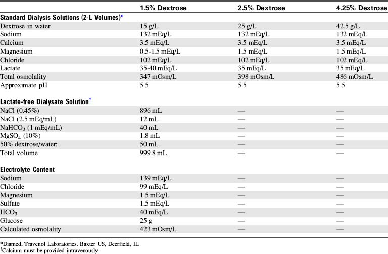
If the serum potassium concentration begins to fall after several dialysis cycles (usually 4-6 cycles are required), a small amount of potassium (typically to a maximum of 4   mEq/L) may be added to the dialysate with a physician or other on-call provider order.

Dialysis Exchange

The initial dialysate volume (inflow or exchange volume) is determined by the method of catheter placement. If the catheter is placed percutaneously at the bedside, initial exchange volumes of 20 to 30   mL/kg are used. If the catheter is placed surgically, smaller initial volumes (15-20   mL/kg) are used to reduce the likelihood of leak around the catheter.3 Small exchange volumes also may be necessary if respiratory distress is present and the child is breathing spontaneously.101 Heparin (500 units/L) usually is added to the dialysate for the first 24   hours of dialysis.

Exchange volumes are increased gradually as tolerated to 35 to 50   mL/kg/exchange. These volumes are ideal because they enable the correction of acidosis, electrolyte imbalance, and uremia.

Typically, 2.5% glucose dialysate is utilized for initial exchanges in the uremic child with acidosis and hyperkalemia. If fluid removal is not required, 1.5% glucose dialysate may be utilized. The 4.25% glucose dialysate is used for those patients with hypervolemia requiring fluid removal. The prolonged use of 4.25% glucose dialysate can be associated with hyperglycemia, hyponatremia, and hypovolemia.

For each exchange, dialysate is instilled over 5   minutes with a dwell time of 15 to 90 minutes (this may vary). The drain time varies with the size of the patient and the exchange volume; usually 5 to 10 minutes is sufficient, although drain times of 20   minutes may be required. Peritoneal dialysis is maximally effective during the first 15 to 90 minutes of dwell time. Therefore if maximum fluid removal and correction of hyperkalemia, acidosis, and uremia are required, frequent exchanges (often every 30   minutes) are performed.

Automated peritoneal dialysis cyclers are now available that provide exchange volumes as small as 50 to 100   mL. These cyclers incorporate a heater to warm the dialysate, and they automatically monitor the drain volume or outflow and can calculate the volume of fluid removed from the patient. Audible alarms indicate volume or infusion problems. Because the equipment manipulates the dialysate it can substantially reduce the nursing time needed for each exchange.

Manual peritoneal dialysis requires the use of buretrols or other graduated cylinders to monitor the precise volume of fluid infused and drained. When the exchange volume is small, buretrols can be used to measure both inflow and outflow (drain) volume. Graduated urine collection systems also can be utilized to measure outflow volume. Finally, the serial dialysate drainage bags used for continuous ambulatory peritoneal dialysis also can be utilized to measure the drain volume; clamps are used to direct the draining fluid into a separate bag following each exchange.

The large (2-L) dialysate bags should never be hung so they infuse directly into the child; if a clamp loosens or is inadvertently left open, the entire 2   L volume could be infused into the child's abdomen, causing respiratory distress and possible cardiovascular collapse. The dialysate fluid always should be warmed before use in infants and children to prevent heat loss and reduce discomfort. Room temperature dialysate may be used in adolescents unless it produces discomfort.

For dialysis in infants, exchange volumes will be small and tubing dead space should be minimized. Adult peritoneal dialysis Y tubing sets contain too much dead space for use in infants, so intravenous tubing (containing a Y or a stopcock) can be utilized to direct dialysate flow. Infant peritoneal dialysis circuits are commercially available.

Because dialysis removes fluid from the vascular space, the nurse should assess the child's volume status frequently. Signs of hypovolemia include tachycardia and signs of poor systemic perfusion (such as decreased intensity of pulses, pale mucous membranes, and cool extremities with weak pulses and sluggish capillary refill). The CVP will be low unless heart failure is present. The development of hypotension indicates critical hypovolemia and hypotensive shock.

If edema is present, peritoneal dialysis will not immediately abolish all fluid excess. However, as fluid is removed from the vascular compartment the intravascular proteins and sodium ions (osmolality) will draw water out of the edematous tissues into the vascular space, (where it can then be removed during dialysis).

Calculation of Fluid Balance

During peritoneal dialysis, two patient records of total fluid intake and output must be strictly maintained. One record documents the dialysate infused and the dialysate recovered (drain volume) at the end of each cycle (Fig. 13-9).

image image image

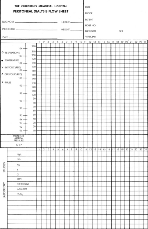
Fig. 13-9 Example of peritoneal dialysis flow chart template.

(Courtesy Children's Memorial Hospital, Chicago, IL.)

The amount of fluid recovered always should equal or exceed the amount infused; this produces a negative fluid balance. If less dialysate is recovered than was infused, the nurse should check for signs of catheter or tubing obstruction (see section, Catheter Dysfunction and Obstruction). If additional dialysate cannot be recovered, the difference between the amount infused and the amount recovered is recorded as a positive fluid balance, and a physician should be notified.

During the initial cycles of peritoneal dialysis, a larger volume may be recovered than was infused, indicating removal of intravascular fluid. When this occurs, the amount of fluid recovered in excess of the amount infused should be recorded as a negative number, because this represents fluid loss for the child. If significant fluid loss continues, a physician or on-call provider should be notified; it may be necessary to reduce the osmolality of the dialysate to prevent excessive fluid loss and dehydration.

The time and the duration of each infusion, dwell time, and drainage cycle should be recorded. Because maximum solute transfer occurs during the first 30 to 90 minutes the dialysate is in the peritoneal cavity, the dwell time is rarely longer than this. The temperature of the dialysate also should be measured and recorded; this temperature should be as close as possible to 37° C to improve the efficiency of dialysis and to minimize the child's heat loss and discomfort.

The second record of the child's fluid balance includes a total of all sources of fluid intake and output. The net dialysis balance is recorded as part of this total. It is extremely important that this record be carefully maintained, because it will aid in the evaluation of the child's progress and need for modifications in dialysis. When dialysis is begun, drug doses and administration schedules should be adjusted, because many drugs are removed by dialysis.

Potential Complications

Peritonitis

As many as one third of children who receive peritoneal dialysis develop peritonitis. The risk is directly proportional to the duration of the dialysis and is inversely proportional to the child's age. Critically ill patients are especially susceptible to the development of peritonitis. Clinical signs and symptoms of peritonitis during peritoneal dialysis include cloudy return, abdominal pain and tenderness, and leukocytosis. Fever is usually present in children, but young infants may become hypothermic. Paralytic ileus and constipation also may develop.

Because the risk of peritonitis is significant in critically ill patients, a sample of outflow often is obtained on a daily basis and should be obtained whenever peritonitis is suspected. The sample is centrifuged and a Gram stain, cell count, and culture and sensitivity tests are performed. If clinical evidence of peritonitis is present, antibiotics usually are administered as soon as the cultures from the outflow sample and blood are obtained. Cefazolin and gentamicin may be administered into the peritoneal cavity. If the patient is clinically unstable, a single dose of IV vancomycin may be given.19

The risk of fungal and bacterial infections is reduced by scrupulous attention to sterile technique during the catheter insertion process and aseptic technique during exchanges. When the outflow collection bag is examined, the bag must not be raised above the level of the bed, because elevation will produce reflux of outflow back into the peritoneal cavity.

Catheter Dysfunction and Obstruction

When dialysate will not flow either into or out of the peritoneal cavity, an external kink or an internal plug is likely to be present in the tubing. If external causes of flow obstruction are eliminated and flow does not resume, a catheter plug is presumed to be present.

A physician (or other on-call provider) can gently irrigate the catheter with normal saline or urokinase, using aseptic technique.101 Every time a break is made in the dialysis tubing system, the patient's risk of peritonitis increases. Before the dialysis tubing is separated, the connection between the tubing and the catheter must be scrubbed for 5   minutes with a povidone-iodine solution, and clamps are placed on both the catheter and the tubing. The separated ends of the tubing should be wrapped in dry sterile 4 × 4 gauze during separation. Aspiration should not be performed in an attempt to dislodge any plugs, because this may result in catheter occlusion with omentum.

A flat plate radiograph (x-ray film) of the abdomen can be made to confirm the presence of a catheter plug and rule out catheter migration. A solution consisting of three parts 1.5% dialysate and one part Renografin M60 can be infused by gravity or administered by syringe into the catheter at the time the abdominal films are made. This solution will opacify the lumen of the catheter and enable identification and location of plugs.

After catheter manipulation, the dialysis tubing can be reconnected if it is not contaminated during the manipulation. As noted above, the ends should be wrapped in sterile gauze during the manipulation. If the tubing is to be reused, ensure that no fluid leaks out of the system. If a solid column of fluid is maintained in the tubing, air does not enter the tubing and fluid does not drain out of the tubing during catheter manipulation.

With most forms of catheter obstruction, dialysate will flow freely into the peritoneal cavity, but fluid will not drain freely from the peritoneal cavity. Often the catheter floats above the level of the fluid in the peritoneal cavity or becomes wrapped in the omentum. Catheter obstruction also may be caused by constipation, which locks the catheter into a position that restricts drainage. Once the bowel is evacuated, dialysis can proceed. If the child is repositioned or turned from side to side, drainage often can be restored.

Sluggish outflow also can be caused by loops in the dialysis tubing that hang off the edge of the bed. The collection bag and tubing should be repositioned so that the tubing falls straight down from the bed to the collection bag. Any extra tubing should be coiled on the bed to facilitate drainage.

Pain

Almost all patients with new peritoneal catheters complain of pain during the initial dialysis infusions and outflows. The pain experienced upon inflow can be relieved by slowing the rate of infusion or by infusing smaller dialysate volumes.

Pain also can be caused by encasement of the catheter in a false passage; this causes the dialysate to fill only a small area of the peritoneal cavity instead of spreading throughout the peritoneal space. That small area can distend and become painful. If the catheter has been immobilized so that the dialysate flow is directed at the same point in the peritoneal cavity, it usually causes pain in the lateral or posterior peritoneal wall. It may be possible to float the catheter to another position when the abdomen is filled with dialysate. Occasionally, insertion of a new catheter may be necessary. Painful inflow also may be related to extremes of dialysate temperature.

Pain is usually present during dialysate inflow; patients rarely complain of pain only during outflow. Pain at the end of outflow will occur when the abdomen is emptied completely, and it can be abolished by stopping the outflow when a small volume of solution remains in the peritoneal cavity. The presence of this residual solution also diminishes the likelihood of catheter obstruction by omentum. Limiting the outflow time to 5-10   min also should alleviate this problem.

Miscellaneous Complications

Bloody return is a common observation during the initial 24 to 48 hours following catheter implantation. This condition is usually self limiting, and heparin still should be added to the dialysate to prevent the formation of fibrin plugs in the catheter. Heparin will not cross the peritoneal membrane, and it will not affect the patient's coagulation. If the amount of blood in the outflow seems excessive, serial hematocrits can be obtained from the outflow to quantify the amount of blood present. Transfusion may be required if excessive blood loss occurs.

Leaking around the catheter is common when the catheter is placed under urgent conditions at the bedside; it seldom occurs in surgically placed or chronic catheters. Whenever a leak occurs, the nurse should check for overfilling of the abdomen by feeling the tenseness of the abdomen at the end of inflow. The abdomen will feel full, but should not feel rigid. Reassess the catheter insertion wound and tubing to determine whether the catheter is migrating into or out of the peritoneal cavity.

If a leak occurs, weigh and then pack (using aseptic technique) sterile dressings around the catheter and change and weigh them when they become soaked, to measure the volume of the leak. Notify a physician or on-call provider of the leak volume; a smaller volume of dialysate inflow may be ordered.

Fluid leak into the abdominal subcutaneous tissues occasionally develops. The fluid is likely to accumulate in dependent perineal areas. The volume of the subcutaneous leak is usually small and is commonly reabsorbed. If a large volume leaks into closed tissue areas such as the penis or scrotum, it may be necessary to replace the catheter.

Pulmonary Complications

Because peritoneal dialysis results in abdominal fullness, it may compromise diaphragm excursion, resulting in shallow breathing and atelectasis, particularly if the patient is breathing spontaneously. Atelectasis especially in the lower lobes is more likely to develop when the child remains in the supine position.

Assess the child's breath sounds frequently, and constantly evaluate the effectiveness of ventilation. Provide chest physical therapy if areas of atelectasis are noted. If the child is alert and cooperative, encourage the child to cough and take deep breaths or perform inspiratory exercises (e.g., incentive spirometry) to prevent atelectasis. The infant may require other exercises to encourage deep breathing (see Chapter 9). The head of the child's bed should be elevated to facilitate maximal diaphragm excursion and chest expansion.

Fluid or Electrolyte Imbalance

Throughout the dialysis period, the child's electrolyte and acid-base balance should be monitored closely, and electrolyte or acid-base imbalances should be discussed immediately with an on-call provider.

Hypertonic dehydration and hemoconcentration can develop if peritoneal dialysis removes too much water too rapidly; this can result in hypernatremia and can exacerbate hyperkalemia. If dehydration is suspected the nurse should assess the child's level of hydration, heart rate, systemic perfusion, and blood pressure.

If the serum sodium concentration is elevated and the child is dehydrated or hypovolemic, free water may be administered orally, intravenously (with 5% dextrose), or intraperitoneally (using a less concentrated dialysate). The osmolality of the dialysate solution should be reduced for subsequent peritoneal dialysis.

Hypokalemia can develop if a hypokalemic dialysate is used after the serum potassium concentration normalizes. If hypokalemia develops, small doses of potassium can be added to the dialysate, or small doses of potassium chloride can be administered intravenously (0.5-1.0   mEq/kg over several hours). Hypokalemia also can develop if a 4.25% glucose dialysate is used for prolonged periods; the treatment of choice is to change the dialysate solution if the child's condition permits.

Hypoproteinemia can develop if peritoneal dialysis is required for several days, because 0.2 to 8.0   g of protein is lost per liter of outflow. Higher amounts of protein loss occur during episodes of peritonitis. As a result, the child's total protein and albumin should be monitored, and the nurse should assess the child for signs of peripheral edema. If hypoproteinemia develops, administration of amino acids may be needed.

Hyperglycemia can develop if concentrated glucose dialysate is required to eliminate large amounts of free water. The serum glucose concentration (and point of care evaluation of glucose, if available) should be monitored closely, and hyperglycemia or hypoglycemia should be reported to an on-call provider. It may be necessary to reduce the glucose concentration of the dialysate. If the patient has diabetes mellitus, exogenous insulin should be added to the dialysate.

Catheter Removal

The percutaneously placed PD catheter is removed using sterile technique. While one provider withdraws the catheter, a second provider maintains tension on the purse-string stitch that was placed during the catheter insertion. The suture is drawn tight as the catheter is withdrawn. The catheter tip can be sent to the laboratory for culture (per policy). Sterile dressings are placed over the catheterization site. If the dialysis catheter is placed surgically, it must be removed surgically.

Extended Peritoneal Dialysis: Continuous Ambulatory Peritoneal Dialysis and Continuous Cycling Peritoneal Dialysis

Some patients with AKI will require PD for a long period of time (i.e., longer than 5-10 days). In these patients a permanent cuffed peritoneal catheter can be placed surgically for ambulatory peritoneal dialysis.

Method

Continuous Ambulatory Peritoneal Dialysis

Continuous ambulatory peritoneal dialysis (CAPD) is a form of continuous dialysis that does not require bed rest or hospitalization. CAPD uses a surgically placed, cuffed PD catheter and disposable plastic bags of dialysate. Approximately four to five exchanges are performed daily, and each exchange volume totals approximately 30 to 50   mL/kg (or 0.5-2   L total). The exchange time is approximately 4 to 6 hours, which is much longer than the exchange time during acute peritoneal dialysis.101 However, during the exchanges the empty dialysate bag is clamped and strapped to the patient's abdomen, and the patient is free to be relatively active. At the end of each exchange period, the empty dialysate bag is placed at a level lower than the patient's abdomen, the drainage tubing is unclamped, and outflow from the patient's abdomen drains into the bag. The bag of outflow is then discarded, and a new disposable dialysate bag is obtained for use in the next exchange.

CAPD will not rapidly correct hypervolemia, acidosis, or hyperkalemia; therefore it is not the dialysis method of choice for acutely ill children. However, CAPD allows excellent regulation of fluid and serum electrolyte concentrations when it is used on a daily basis for the child in stable condition with chronic kidney disease. Children who receive CAPD generally require less frequent blood transfusions than children who receive chronic hemodialysis, and serum urea nitrogen and phosphorus concentrations may be better controlled than with hemodialysis. However, renal osteodystrophy and hyperphosphatemia do occur.36

Children receiving CAPD have few dietary restrictions, because their relatively continuous dialysis can remove excess fluid and allow constant regulation of electrolyte and acid-base balance. As a result, children receiving CAPD may be better nourished than those who require intermittent forms of dialysis. Control of hypertension is also excellent when children with renal failure receive CAPD.

Continuous Cycling Peritoneal Dialysis

Continuous peritoneal dialysis may be made less labor intensive with the addition of mechanical cycling to the dialysis process. Continuous cycling peritoneal dialysis (CCPD) provides mechanical (automatic) delivery of a prescribed volume of dialysate at prescribed intervals, with a set indwell time. Drainage of outflow is initiated mechanically, and audible alarms sound if flow problems occur.72 Current cycling machines are capable of infusing dialysate volumes of 50 to 100   mL.

CCPD is performed most commonly while the child sleeps. The dialysis cycling machine should not require any attention during the night unless an alarm sounds. A small dialysate volume is allowed to remain in the peritoneal cavity during the day, providing continuous dialysis. The child is able to resume school and other normal childhood activities. All exchange bags are hung at the same time when CCPD is begun at night; this can reduce the risk of contamination and peritonitis.72

Complications

The most frequent complications associated with CAPD and CCPD are mechanical problems and infection. The mechanical problems are related to cuff erosion and fluid leaks, and the infection problems are related to peritonitis.

The cuffed peritoneal catheter can erode the abdominal wall; this can cause a fluid leak and require catheter replacement. Hernias can develop from subcutaneous fluid leaks around the dialysis incision, and these hernias often require surgical repair.

The incidence of peritonitis among children receiving CAPD and CCPD varies widely in clinical reports, but averages one infection every 14.7 patient months.36 Many patients also develop local infections around the catheter site.

When selecting patients for CAPD, it is important that the child and the parents are reliable and able to follow the established protocol. Children or families must be taught the dialysis technique, and they should be instructed to contact the CAPD nurse whenever the patient experiences abdominal pain, inflow or outflow occlusion, inflammation of the catheter site, a feeling of weakness or dizziness when standing, hypotension, cloudy outflow, catheter disconnection or contamination, fever, excessive weight gain, edema, or other illness.

Because the dialysate dwells in the peritoneal cavity for a long time and the risk of peritonitis is relatively high in children receiving CAPD and CCPD, hospital protocols often try to minimize the number of personnel providing any in-hospital CAPD or CCPD that the child requires. This action minimizes the child's exposure to people and contaminants and may reduce the risk of peritonitis.

Hemodialysis

Method

Hemodialysis is one of the most efficient artificial methods of removing nitrogenous wastes from the body and of restoring fluid, electrolyte, and acid-base balance. However, pediatric hemodialysis requires the assembly of skilled personnel capable of establishing and maintaining vascular access, recognizing and responding to potential complications of dialysis, and supporting cardiorespiratory function in extremely unstable patients. If urgent dialysis is required and experienced personnel are not available, PD may be provided until the child's condition can be stabilized and the child transported to an appropriate facility.

Indications for pediatric hemodialysis are GFR <15   mL/min per 1.73   m2 or intractable complications of AKI, even at a higher GFR. GFR is roughly equivalent to the creatinine clearance estimated by the Schwartz formula (see Box 13-5).

Complications of hemodialysis include but are not limited to hypervolemia, hyperkalemia, metabolic acidosis, hyperphosphatemia, hypocalcemia, hypercalcemia, and neurologic dysfunction.52

Hemodialysis requires access in an artery, arterialized vessel, or large vein. If chronic hemodialysis is planned, an arteriovenous fistula may be created or a graft may be placed in a large artery. In the critical care unit, vascular access is often achieved with a single- or double-lumen catheter (at least size 6 French) placed through the subclavian vein into the right atrium, or from the femoral vein into the inferior vena cava.15

During hemodialysis, blood is withdrawn from the body and pumped at high flow rates to a blood compartment that makes contact with a semipermeable membrane. This blood compartment and semipermeable membrane are immersed in dialysate, which is pumped at rates that exceed blood flow rate by 50%. The dialysate flows in the direction opposite the blood flow.37

The dialysate contains a fairly standard concentration of electrolytes, but the potassium concentration usually is determined individually (based on the amount of potassium to be removed). Nitrogenous wastes pass from the blood into the dialysate as the result of a concentration gradient. Free water will move from the blood into the dialysate if the osmolality of the dialysate is greater than the serum osmolality. Free water movement is also enhanced by a positive transmembrane hydrostatic pressure from the blood compartment to the dialysate compartment. This positive pressure can be generated by the blood pump and by manipulation of resistance to blood outflow, although newer dialysis machines automatically generate the transmembrane hydrostatic pressure required to provide the volume of ultrafiltrate prescribed.37

The amount of fluid and solute removal from the blood is determined by the flow rates of the blood and dialysate, the surface area and permeability of the membrane, the concentration and osmolality gradients between the dialysate and blood, and the transmembrane pressure gradient. Electrolytes move across the semipermeable membrane as the result of concentration gradients. If the concentration of an electrolyte (e.g., potassium) or other small molecule (e.g., urea) is lower in the dialysate than in the blood, that electrolyte or molecule will move out of the blood and into the dialysate. If the concentration of an electrolyte or other molecule (e.g., glucose) is higher in the dialysate than in the blood, that electrolyte or molecule will move into the blood. Other substances can be removed from the blood as the result of ultrafiltration and solvent drag (the passive movement of solutes as the result of movement of large amounts of water).

Because hemodialysis requires that blood be drawn from and returned to the body, pumps must be present in the dialysis circuit. In addition, the filter and the tubing (the dialysis circuit) must be primed with fluid or blood before the dialysis begins. Because the circulating blood volume of the infant or child is low, extracorporeal movement of a large quantity of blood (e.g., to prime the dialysis circuit) is likely to produce hypovolemia. As a rule, the filling volume of the dialysis circuit should be no greater than the equivalent of 10% of the child's circulating blood volume. If the circuit requires a larger filling volume, the circuit should be primed with colloid (5% albumin or packed RBCs diluted with albumin to a hematocrit of 35%).37 Few centers have experience in the hemodialysis of infants, and it should not be attempted by inexperienced personnel. In adolescents, the dialysis circuit can be primed with normal saline or 5% albumin.

The dialysate contains glucose, sodium, calcium, and potassium, in concentrations that are specified by the physician. The dialysate usually contains little potassium (0-4   mEq/L) and no urea, so that high concentration gradients between the dialysate and the blood will hasten removal of these solutes from the blood. The presence of glucose in the dialysate at levels of 200 to 250   mg/dL creates a high osmolality in the dialysate, favoring the movement of water from the blood to the dialysate, provided the patient's serum glucose concentration and osmolality are lower than the glucose and osmolality of the dialysate.

The glucose concentration in patients with diabetes mellitus is often higher than 200 to 250   mg/dL. As a result, the osmotic forces will favor free water movement from the dialysate into the patient's blood, exacerbating hypervolemia. This inappropriate fluid movement can be prevented by increasing the pressure gradient across the dialysis filter (creating higher pressure within the blood compartment or generating negative pressure in the dialysate compartment) or by adding albumin to the dialysate.

The high glucose concentration in the dialysate produces a serum glucose of approximately 200   mg/dL during dialysis. This mild hyperglycemia is usually well tolerated in the patients without diabetes mellitus; however, patients with diabetes will require adjustment of insulin dose (particularly long-acting insulin) in anticipation of this period of relatively low serum glucose.

The blood and the dialysate are typically pumped through the circuit in opposite directions. This pumping maximizes the concentration and osmotic gradients between the dialysate and the blood so that dialysis can be accomplished within a short period of time (exchange time of approximately 3-4   hours).

As noted previously, the creation of either positive pressure in the blood compartment or negative pressure in the dialysate compartment or both will increase the rate of fluid removal from the blood. Positive pressure is created in the blood compartment by increasing the resistance to flow on the venous side of the blood circuit. This increase in resistance usually is accomplished by placement of an adjustable clamp on the venous blood line, and the clamp is tightened until the desired pressure in the blood compartment is reached.

Negative pressure can be applied across the filter. This negative pressure draws free water and small particles from the blood, across the semipermeable membrane, and into the ultrafiltrate.

The dialysis nurse and the bedside nurse will be responsible for continuously evaluating the effect of fluid removal on the patient's systemic perfusion. If the patient's clinical condition deteriorates, some adjustment in the rate of fluid removal is often required.

When hemodialysis is initiated, a small amount of heparin is injected into the dialysis catheters and into the dialysis circuit to prevent clot formation in the filter and tubing. Heparin will then be administered at 30-minute to 1-hour intervals or by continuous infusion. The rate of infusion is adjusted based on the activated clotting time (ACT) or the Lee-White clotting time. Heparin dose and adjustment will be determined by dialysis unit policy and procedure. Citrate may be the anticoagulant of choice in hemodialysis therapy, and it is useful in the prevention of heparin-induced thrombocytopenia. Refer to protocols published on-line17 and in print69 for additional information.

Complications

Hemodialysis is efficient, but it is extremely expensive and can produce complications that do not develop during PD or hemofiltration. These complications are related largely to hypovolemia and resultant hypotension, fluid shifts (also known as dysequilibrium), hypervolemia, bleeding, anemia, infection, or malfunction of the vascular access site. Each of these is discussed in the following sections.

Hypotension and Hypovolemia

Hypotension can develop from removal of a large amount of intravascular water, resulting in hypovolemia, or from circulatory instability. The patients most at risk for developing hypotensive crises during dialysis are patients with vasomotor instability (including patients with paraplegia or quadriplegia), low cardiac output or myocardial dysfunction, those receiving vasodilators, or those with a history of hypotensive episodes during dialysis.

If the child develops hypotension during dialysis, the dialysis nurse will reduce any transmembrane pressure created across the filter, because this pressure gradient enhances fluid removal from the blood. In addition, the bedside nurse may be required to administer albumin or other volume expanders (per unit policy or physician or on-call provider order), place the patient in modified Trendelenburg position (head flat, feet elevated), or initiate cardiopulmonary resuscitation (as needed).

To avoid hypotension, any existing hypovolemia should be corrected before dialysis is begun. In addition, the patient's blood should be drawn slowly into the circuit so the patient does not experience an acute loss of intravascular volume. If excess intravascular fluid is to be removed during dialysis, venous positive pressure or negative pressure across the filter will be applied very slowly. The dialysis nurse and the bedside nurse are both responsible for monitoring the child's systemic arterial blood pressure and systemic perfusion. Deterioration in clinical status should be reported immediately to a physician or appropriate on-call provider.

Fluid Shifts and Dysequilibrium

If many osmotically active particles such as sodium or urea are removed rapidly from the patient's blood, the patient's serum osmolality will fall quickly. As a result, free water may shift from the intravascular and interstitial spaces to the intracellular compartment, producing cerebral edema. This edema following dialysis has been called dialysis dysequilibrium syndrome. The child may complain of severe headaches or may demonstrate nausea, vomiting, confusion, irritability, or seizures.

To reduce the risk of dysequilibrium, solute removal from the blood must be gradual; peritoneal dialysis may initially be performed to gradually reduce the BUN concentration. The efficiency of the hemodialysis can be reduced: the blood flow through the filter can be slowed, the direction of dialysate flow can be changed to the same direction as the blood, or the duration of the dialysis treatment can be shortened.

Intravenous mannitol can be administered slowly to increase serum osmolality and slow the removal of water by dialysis. Mannitol should be administered if evidence of dysequilibrium develops.64

Hypervolemia

If too much fluid is administered to the patient during dialysis or if an excessive volume of fluid and blood is transfused to the patient from the circuit at the end of dialysis, the child can develop hypervolemia. This condition can produce significant cardiovascular problems, particularly if the patient has preexisting cardiac disease.

The child can rapidly develop signs of congestive heart failure, including tachycardia, peripheral vasoconstriction, hepatomegaly, periorbital edema, elevated CVP, tachypnea, and increased respiratory effort. If severe hypervolemia is present, the child can develop pulmonary edema or hypertension.

Bleeding and Anemia

Because the child's blood must be anticoagulated during dialysis, bleeding can occur. The child can bleed from wounds or puncture sites or into the brain, pericardium, or abdomen. To reduce the risk of bleeding, regional heparinization may be performed. The heparin is injected into the arterial (or inflow) tubing that carries blood from the patient to the filter. To prevent large heparin infusion to the patient, protamine sulfate is administered into the tubing that is returning blood from the filter to the patient. Protamine sulfate neutralizes heparin, but can produce a coagulopathy or hypotension if it is administered separate from or in excess of heparin, or if the patient inadvertently receives a bolus of protamine.

Bleeding also can occur in patients with renal failure, because uremia is associated with depression of platelet function. If active bleeding is present in a patient with uremia, the most common treatment is the use of desmopressin or l-deamino-8-arginine vasopressin (DDAVP). Although the exact mechanism of action is unknown, desmopressin has been shown to increase release of factor VIII from storage sites, increasing the concentration of factor VIII and minimizing the effects of uremic dysfunctional von Willebrand factor. The dose of DDAVP to treat bleeding with renal failure is approximately 10-fold higher than the dose used for treatment of diabetes insipidus; it ranges from 0.3 to 0.4   mcg/kg intravenously and will improve platelet function within 1   hour after infusion. The effects disappear within 24   hours.51 DDAVP also can be administered before surgical procedures to reduce the risk of bleeding in patients with uremia.

Patients with renal failure often have anemia. Anemia can result from loss of blood within the dialysis system (through blood leaks, loose connections, clot formation, frequent blood sampling, or dilution of blood with dialysis tubing prime), from hemorrhage, or from the effects of uremia. Red cell lysis also can occur if blood is exposed to a dialysate of significantly higher osmolality. Levels of erythropoietin are low among uremic patients; therefore RBC production and survival are both reduced.

To prevent anemia, blood sampling should be minimized. Whenever blood is drawn, the amount should be recorded in the child's record of intake and output; blood replacement should be considered whenever the blood loss totals 5% to 7% of the child's circulating blood volume (circulating blood volume is approximately 70 to 80   mL/kg in infants and children and 70   mL/kg in adolescents). Because hemodialysis often is accomplished through the use of an arterial access catheter, laboratory sampling of blood can be performed during dialysis. This procedure reduces the number of venipunctures required and allows immediate replacement of the sample amount through the dialysis circuit.

Packed RBCs usually are administered to replace lost blood. Iron therapy will not be effective in the treatment of uremia-induced anemia and can contribute to the development of iron toxicity. Anemia can complicate the care of children with associated cardiovascular disease, because cardiac output must increase to maintain oxygen delivery.

Infection and Febrile Reactions

Patients with renal disease have increased risk of infection resulting from loss of immune proteins in nephrosis, multiple invasive catheters and cannulae, compromised nutritional status, frequent handling by hospital personnel, and frequent transfusions. The risk of infection can be minimized if all healthcare personnel practice strict hand-washing technique and strict asepsis, if the child receives adequate nutrition and if hepatitis and HIV screening is performed by the blood bank (for further discussion of hepatitis see Chapter 14).

The nurse should assess all of the patient's wounds and vascular access sites daily and report any areas of inflammation to an on-call provider. All wounds should be dressed according to unit policy or physician or on-call provider order.

Patients receiving hemodialysis may experience a sudden increase in temperature, known as a febrile reaction. This fever may result from an allergic reaction to the filter, from a reaction to blood administered during dialysis, or from systemic seeding from an infected shunt or access site. A preexisting fever suddenly may become manifest when the patient's serum urea, which can act as an antipyretic, is lowered.

When fever develops, the on-call provider can request that blood cultures be obtained from two different collection points; one set of cultures is typically collected from the dialysis tubing by the dialysis nurse after a 3-min povidone-iodine (Betadine) scrub. The second culture usually is obtained from a peripheral vein, although a second culture can be obtained from the dialysis circuit if at least 30   min elapse between samples. Cultures also can be collected from the ultrafiltrate.

If a transfusion reaction is suspected, specimens are collected from the transfusion bag and patient, and they are sent to the laboratory for hemolysis and incompatibility checks (see Chapter 15). If hemolysis is present, the child's serum potassium concentration should be monitored closely, because hyperkalemia can develop. A hematocrit also should be obtained, and a serum sample should be checked for evidence of hemolysis.

Hemodialysis Access

The establishment and maintenance of vascular access in small children is one of the most challenging aspects of pediatric hemodialysis. Dependable, double lumen, cuffed venous catheters are now available and they are the acute and long-term dialysis catheters of choice. If these catheters are placed surgically in the right atrium and infused appropriately with heparin, they can be maintained for a long time. When vascular access is needed, the injection port of the catheter can be used and no skin punctures are required. The care of these catheters is identical to the care of any long-term central venous catheter; meticulous care is required to ensure catheter longevity and minimize the risk of infection. Refer to hospital policies and procedures regarding central venous catheter care (also, Box 22-6).

Arterial access can be provided by a graft. Grafts consist of tubes made of Teflon or polytetrafluoroethylene. The tube is attached to an artery and then looped subcutaneously and connected to a parallel large vein.15 After surgical placement, time is allowed for the graft to become coated with the patient's endothelial cells (i.e., the graft endothelializes) before it is used for dialysis. Vascular access is achieved by piercing the graft with standard large-bore needles.

Arteriovenous fistulae can still be created in older children to provide vascular access for hemodialysis. The fistula is surgically created in the nondominant arm by connection of the radial artery to the cephalic vein.15 Soon after the fistula is created, the vein distends and is punctured easily to obtain vascular access. The risk of infection in the fistula is low because no prosthetic material is involved in its construction. However, any arteriovenous shunt will increase venous return to the heart and may precipitate or worsen congestive heart failure.

Continued Problems of Uremia

The patient requiring hemodialysis is still susceptible to complications of uremia. Although dialysis may provide temporary relief of some fluid and electrolyte or acid-base imbalances, anemia, hypertension, infection, osteodystrophy, endocrine imbalance, pruritus, anorexia, nausea, vomiting, fatigue, ulcers, and depression can persist (see section, Chronic Kidney Disease).

Throughout the care of the child with renal failure, the nurse should consult support personnel to assist in the psychosocial care of the child and family. Frequent multidisciplinary conferences and meetings with the social worker, dietician, financial counselor, physicians, and primary nurses will help the family to be aware of the support systems available to them and ensure that communication is optimal among team members and the family. As appropriate, the bedside nurse or case manager should begin to plan for the child's discharge to home or to another unit.

Whenever the critically ill child requires dialysis, the bedside nurse remains responsible for coordinating the care of the child and family. Although a dialysis nurse may be present and responsible for the dialysis procedure and circuit, the bedside nurse still must assess and document the child's fluid balance and systemic perfusion. The dialysis nurse and the bedside nurse should coordinate efforts. It is important to time the administration of medications, blood products, and fluids based on the timing and effectiveness of the dialysis. Both the dialysis nurse and the bedside nurse must provide the child and family with skilled support and compassion.

Hemoperfusion

Definition

Hemoperfusion is a treatment which exposes blood or plasma to an adsorbent material for the removal of toxins, solutes, or other materials. Adsorbent materials used can include charcoal, resin, protein A, synthetic materials, and monoclonal antibodies.21

Although hemodialysis is highly efficient at removing water-soluble drugs with low molecular weights (e.g., salicylates, ethanol, methanol, lithium) from the blood, it does not remove protein and lipid-bound substances; therefore it is not effective in the treatment of hypercholesterolemia, hyperbilirubinemia, toxicity associated with fulminant hepatic failure, or ingested toxins. Hemoperfusion is extremely effective in the treatment of these problems.21 Large molecules such as β2-microglobulin have been successfully removed by hemoperfusion with lower cardiovascular mortality,87 and clinical trials are currently underway with new adsorbents that are effective in removing β2-microglobulin.21

Hemoperfusion uses a hemodialysis circuit; however, the blood is passed through a cartridge containing the adsorbent surface, and no dialysate is present. Life-threatening poisonings from theophylline, carbamazepine, phenobarbital, and phenytoin may be an indication for charcoal hemoperfusion.74 With the use of activated charcoal, substances normally bound by lipid or protein are quickly and effectively removed from the intravascular space. Because hemoperfusion will not remove urea, it is not the treatment of choice for uremia.

Method

In the following discussion, a charcoal filter or cartridge will be used as the example of the adsorbent surface. As noted previously, the vascular access and circuit tubing used for hemoperfusion are often identical to those required for hemodialysis.

The charcoal filter is prepared, and the filter is given a glucose-heparin rinse. Because most commercially available cartridges require a priming volume of 50 to 300   mL, the total hemoperfusion circuit is primed with the equivalent of more than 10% of the circulating volume of a small child. As a result, the circuit usually is primed with blood before initiation of hemoperfusion.24

Some anticoagulation must be provided to prevent clot formation in the cartridge. Heparin usually is infused into the inflow tubing, between the patient and the cartridge. To minimize the risk of bleeding, protamine sulfate usually is added to the blood in the circuit beyond the cartridge (i.e., between the cartridge and the patient) to bind the heparin before the blood is returned to the patient.

Approximately 3   hours are required for hemoperfusion. The activated charcoal quickly will become saturated with the drug or substance removed from the blood. Frequently, several cartridges are required to complete one treatment. To determine the amount of toxin binding provided by the cartridge, blood levels of the toxic substance are drawn from the circuit immediately proximal to and distal from the cartridge. If these levels become nearly identical or if clotting is observed in the cartridge, the cartridge should be changed.

Complications

The activated charcoal binds lipid or protein-bound toxins. In addition, it binds glucose, calcium, and platelets; therefore serum concentrations of these substances must be monitored closely during hemoperfusion. Severe thrombocytopenia is the most common complication observed during this procedure.24

Occasionally, hemoperfusion effectively removes the toxins from the blood, but rebound toxicity develops several hours later as tissue-bound toxins move into the vascular space. For this reason, serum levels of the toxin should be monitored during the hemoperfusion and at regular intervals for several hours following hemoperfusion.

Bleeding can result from thrombocytopenia or heparinization. Because the half-life of protamine sulfate is shorter than the therapeutic effect of heparin, it may be necessary to administer additional protamine sulfate several hours after hemoperfusion is performed. During and after the procedure, the child should be observed closely for any evidence of bleeding, and all body secretions should be checked for the presence of blood.

Continuous Renal Replacement Therapy

The history of pediatric continuous renal replacement therapy (pCRRT) is rich with innovation, challenges, and change. In the late 1970s and early 1980s, the care of pediatric patients with AKI requiring renal replacement therapy involved improvised arteriovenous systems. Control of ultrafiltration rates in these systems required vigilant monitoring and frequent adjustment on the effluent side of the extracorporeal system.

Today, continuous renal replacement therapy (CRRT) devices are primarily venovenous and provide compact, efficient, and controlled management of fluid and electrolytes in pediatric patients with AKI. The Prospective Pediatric Continuous Renal Replacement Therapy Registry (ppCRRT Registry) was designed to evaluate clinical and therapeutic aspects of pCRRT in a prospective, observational manner.39 The ppCRRT Registry is a voluntary, multicenter, collaborative effort designed to evaluate clinical and therapeutic aspects of pCRRT and gather information about best pCCRT practices.38

The First Acute Dialysis Quality Initiative Conference examined evidence-based research and published consensus practice guidelines for CRRT in 2002.61 Complete guidelines are available online at http://www.ccm.pitt.edu/adqi/.1

Definition

CRRT performs the work of the kidney through an extracorporeal system designed to run continuously. The patient's blood flows through a filter to remove excess fluid, waste products, and toxins and to establish electrolyte and pH balance before it is returned to the patient. CRRT is similar to hemodialysis, but treatment is continuous rather than intermittent. The continuous aspect of CRRT provides more gradual fluid and electrolyte shifts and enables more tightly calibrated fluid management. CRRT as provided by current technology enables minute-to-minute adjustments in therapy titrated to patient need.

Indications for Continuous Renal Replacement Therapy

In the setting of AKI, the indications for renal replacement therapy are fluid and solute removal. Similar to PD, CRRT provides a gradual fluid and solute removal, but CRRT allows more sensitive fluid control than PD.101 Comparable to HD, CRRT can treat hypervolemia and electrolyte imbalance but instead of correcting these imbalances over 3 to 4 hours as with HD, CRRT will correct them over 1 day or more. For these reasons, CRRT is a valuable treatment choice in critically ill pediatric patients with AKI who are hemodynamically unstable with signs of multiorgan dysfunction syndrome. In data from the ppCRRT Registry, the most common acute diagnoses in patients receiving CRRT were sepsis and cardiogenic shock, and the most common comorbid conditions were bone marrow transplant and solid organ transplants—primarily the liver.38,88

In data from the ppCRRT Registry, immediate indications for CRRT include: fluid overload and electrolyte imbalance, fluid overload only, electrolyte imbalance only, prevention of fluid overload to allow intake, or other indications such as intoxications and inborn errors of metabolism. The combination of fluid overload and electrolyte imbalance was the most frequently encountered indication and had the lowest survival.

Recent experience supports the use of CRRT as a treatment modality for sepsis, not only for the management of fluids and electrolytes but also for the removal of inflammatory mediators of sepsis.26,28,33 CRRT has been effective after stem cell transplant, for treatment of drug toxicities, in acute solute removal in tumor lysis syndrome with associated hyperkalemia and hyperuricemia and to treat inborn error of metabolism.

Continuous Renal Replacement Therapy Modalities

CRRT is a broad term that encompasses treatment modalities involving an extracorporeal system and water removal by filtration. The treatment modalities include continuous arteriovenous hemofiltration (CAVH) and a variety of modalities provided by continuous venovenous hemofiltration.

The mechanism of solute transport is the distinguishing feature of the CRRT modalities. Solutes are removed by diffusion, convection, or both.

When solute is removed by diffusion, the process is similar to that described in the hemodialysis portion of this chapter. Solutes are removed from the blood through the filter's semipermeable membrane as the result of concentration gradients. Solutes move from the area of high concentration (blood) through the semipermeable membrane into an area of low concentration (i.e., fluid on the opposite side of the membrane).

The second mechanism of solute transport is convection. Solutes in solution are carried with the water across the semipermeable membrane in response to a transmembrane pressure gradient. Convection has been described as “solvent drag”: the bulk water flow literally drags the solutes through the semipermeable membrane. In venovenous CCRT, the transmembrane pressure gradient is generated by the speed of the blood pump or blood flow rate (BFR), which transmits a positive hydrostatic pressure inside the blood tubule of the filter. The rate of the effluent pump, or effluent and dialysate pump if dialysis is used, creates a negative pressure in the space surrounding the blood tubules, therefore increasing the transmembrane pressure gradient.

Continuous Arteriovenous Hemofiltration

Definition

CAVH uses an extracorporeal circuit and a small filter that is highly permeable to water and small solutes, but is impermeable to proteins and formed elements of the blood. The filter system is joined to both an arterial and a venous catheter. Passage of arterial blood through the filter results in the formation of an ultrafiltrate of plasma that consists of water and nonprotein bound solutes. The filtered blood is then returned to the patient through the venous catheter.

Because the filter used for CAVH contains no dialysate there are no concentration gradients established across the filter. The volume and content of the ultrafiltrate is determined by the rate of blood flow through the filter (essentially determined by the patient's blood pressure), the permeability of the filter, and the transmembrane pressure. The transmembrane pressure is created largely by the patient's arterial pressure, but it also can be augmented by elevating the patient above the fluid drainage bag. The transmembrane pressure also will be enhanced by the oncotic pressure difference between the patient blood and the ultrafiltrate, which is protein-free with an oncotic pressure of zero.

The volume of ultrafiltrate can also be adjusted through the use of negative pressure generated on one side of the filter by the use of a volume-controlled infusion pump that draws ultrafiltrate at a set hourly rate. The faster the rate set for the volume-controlled pump, the more ultrafiltrate drawn from the blood through the filter every hour. The infusion pump can create resistance to flow if it is adjusted to a relatively slow hourly rate. As a result, resistance to ultrafiltrate production will be created by slow flow through the infusion pump circuit. The use of infusion pumps in this setting is not optimal for neonates and small infants, because the pumps may be inaccurate and might remove fluid at an unreliable rate. For example, the amount of ultrafiltrate could actually be greater than the hourly rate set on the infusion pump.

CAVH can provide effective therapy for the treatment of AKI complicated by hypervolemia or electrolyte or acid-base disturbances. It is particularly useful in patients with extremely limited venous access, in neonates or small infants, and in patients with unstable cardiovascular function or multisystem organ failure who are likely to be intolerant of hemodialysis. This form of renal replacement is not recommended for the rapid treatment of hyperkalemia, because the rate of ultrafiltration is low and potassium removal is slower than with hemodialysis.19

CAVH may be used to remove a small but predictable volume of fluid from the vascular space. This procedure is helpful in the management of chronically ill patients with oliguria and hypervolemia (e.g., those with severe congestive heart failure), because excess fluid can be removed and replaced with parenteral fluid that has high nutrient value.

With the appropriate equipment, CAVH systems can be assembled and implemented relatively quickly. CAVH also can provide a useful option when resources are scarce. However, in many critical care areas CAVH has been replaced by venovenous hemofiltration machines with more precise technology to provide safer and more predictable blood flow and precise ultrafiltation.12,69

Contraindications

There are few contraindications to the use of CAVH. Active bleeding is a relative contraindication; PD is preferred in such patients. A severe coagulopathy is not a contraindication to the use of CAVH, although the risk of bleeding at the access site is increased.

Method

Preparation for Continuous Arteriovenous Hemofiltration

To begin CAVH, arterial and venous access must be achieved. It is extremely important to catheterize an artery large enough to allow sufficient flow into the filter; the femoral artery is used most frequently, although the umbilical artery may be catheterized in neonates. The femoral and external jugular veins are used most frequently for venous access.

The circuit tubing should contain multiple sampling ports, and it should be primed before use. A heparin infusion system (with appropriate infusion pump) should be joined to the arterial side of the circuit, and stopcocks and tubing should be placed in the circuit to enable bypass of the filter; these stopcocks will be used when the filters are changed. The filter and circuit are prepared, and the prime fluid should be warmed to no more than 37° C.

Many CAVH filters are commercially available, and most use a hollow fiber design. It is preferable to use small filters (20-mL prime volume) designed for use in small children to keep the volume in the circuit as low as possible during treatment. The ideal filter has a short fiber length, a large surface area, and a small priming volume. If the entire extracorporeal circuit must be primed with a volume exceeding 10% of the patient's blood volume, fresh bank blood should be used to prime the circuit.

As noted previously, a volume-controlled infusion pump may be required to enhance or limit the formation of ultrafiltrate. Occasionally, an additional fluid infusion pump and infusion system are prepared to administer fluid to the patient to enable replacement of ultrafiltrate with fluid that differs in solute or nutritional content (Fig. 13-10).

image

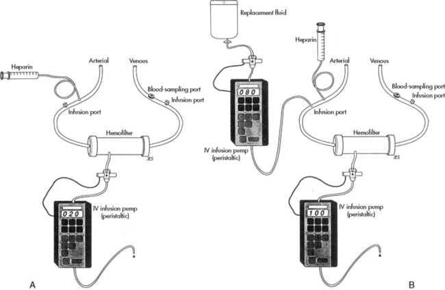
Fig. 13-10 Continuous arteriovenous hemofiltration. A, The rate of fluid removal can be controlled if a pediatric volume infusion pump is joined to the hemofilter. The variable resistance generated by the infusion pump will control the hemofilter transmembrane pressure and the filtration rate. B, Continuous hemofiltration generally requires infusion of replacement fluid to prevent volume depletion and to control the electrolytes lost through hemofiltration. Use of two pediatric infusion pumps enables controlled removal of fluid and controlled replacement of fluid and electrolytes.

(From Stark JE, Hammed J: Continuous hemofiltration and dialysis. In Blumer J, editor: A practical guide to pediatric intensive care, ed 3. St Louis, 1990, Mosby-Year Book.)

Initiation of Continuous Arteriovenous Hemofiltration

To initiate hemofiltration, the arterial and venous catheters are joined to the filter and drainage system. Clamps are placed on each limb of the circuit. The arterial and venous limbs are unclamped first, while the bypass tubing and the drainage limb remain clamped. The heparin infusion should commence when the vascular lines are unclamped. Once the area in the filter surrounding the blood tubules is filled with the patient's ultrafiltrate, the tubing of the drainage line can be unclamped.

During CAVH, the nurse is responsible for monitoring the volume of ultrafiltrate, which can be regulated with a volume-controlled infusion pump. Periodic sampling of the ultrafiltrate will be performed to monitor the solute content of the filtrate, so that appropriate replacement therapy can be provided.

Regular monitoring of the patient's activated clotting time (ACT) is required during CAVH. The heparin infusion is titrated to maintain an ACT that is approximately 1.5-fold of baseline (maximum, 200   s). The arterial ACT should be no greater than 10% above baseline to prevent bleeding.2

The filter should be examined frequently for the presence of clot formation. If clots are observed or ultrafiltrate formation decreases significantly, a physician or on-call provider should be notified and the filter probably should be changed. Filters can be expected to last approximately 12   hours, although the duration varies widely with patient condition.

Hemoglobin and hematocrit and serum chemistries should be monitored on a regular basis per unit protocol. The patient should be weighed once or twice every 24   hours.

Complications

The most common complications during CAVH are bleeding, thromboembolic events, and fluid balance problems. Bleeding occurs when heparinization is excessive, and it should be prevented with careful monitoring of arterial and venous ACTs. Anemia can result from excessive clot formation in the filters; therefore the child's hematocrit should be monitored frequently.

Air is trapped in air chambers, but thrombi entering the system or leaving the filter can embolize to the central venous system. These thrombi can be prevented by scrupulous examination of the filter and circuit and removal of any air or clots.

Hypervolemia can develop or be exacerbated by inadequate ultrafiltrate formation. Alternatively, hypovolemia can develop if an excessive volume of ultrafiltrate is removed. Electrolyte imbalances can be created by inappropriate priming fluid or excessive solute loss in the ultrafiltrate and will be detected with regular blood sampling. If electrolyte imbalances develop, adjustments are needed in the volume and solute content of the fluid replacement infusion or in the rate of ultrafiltrate removal.

Continuous Venovenous Hemofiltration Modalities

Slow continuous ultrafiltration uses an extracorporeal circuit with a hemofilter and can be set as arteriovenous or venovenous. Slow continuous ultrafiltration removes free water and small amounts of solute.

Continuous venovenous hemofiltration (CVVH) uses an extracorporeal circuit with a hemofilter. Blood is pumped from a large vein into the circuit; it flows through the filter, which removes fluid and solute, and then is pumped back into the patient through the venous line and catheter. If a double-lumen catheter is used, the blood is pulled from and returned to the same vein.

CVVH uses replacement fluid and therefore transports solutes primarily through convective transport. Replacement fluid is a physiologic solution infused into the blood component of the circuit either just before the filter (predilution) or immediately after the filter (postdilution), but before it is returned to the patient. The replacement fluid is used to replace large volumes of ultrafiltrate to help maintain the patient's fluid and electrolyte balance. The machine weighs the ultrafiltrate bag and calculates the volume of fluid removed (ultrafiltrate).

Continuous venovenous hemodialysis (CVVHD) uses an extracorporeal circuit with a filter. Blood is pumped from a large vein into the circuit, through the filter, which removes fluid and solute, and then the blood is pumped back into the patient's venous circulation. Dialysate fluid is pumped into the space surrounding the blood tubules in the filter in the direction opposite the blood flow (countercurrent flow).

Solute removal in CVVHD results from diffusion that is caused by concentration gradients between the blood and dialysate. The ultrafiltration fluid and the dialysate fluid are recovered into the same large effluent bag for disposal. The machine weighs and calculates the actual amount of fluid removed.

Continuous venovenous hemodiafiltration (CVVHDF) uses an extracorporeal system with a filter. Blood is pumped from a large vein into the circuit and through the filter, which removes fluid and solute, and then the blood is pumped back into the patient's venous circulation. CVVHDF uses dialysis fluid and replacement fluid as described previously; therefore solute transport occurs through both diffusion and convective transport mechanisms. In CVVHDF the ultrafiltrate, which includes the replacement fluid volume, and the dialysate are recovered in the same effluent bag. The machine is designed to subtract the dialysate and replacement fluid volumes from the ultrafiltrate volume to calculate the actual volume of fluid removed from the patient. Table 13-7 compares characteristics of different CRRT modalities, including the driving force of blood flow and the mechanism of solute transport.

Table 13-7 Continuous Renal Replacement Therapy Nomenclature

image

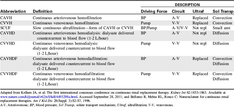
Pediatric Considerations

Access issues are extremely important in all patients who need renal replacement therapy, but these challenges become much more difficult in small, critically ill patients. Access is the most important consideration contributing to the effectiveness of therapy, and constant problems can arise when access is inadequate.100

Data from the ppCRRT Registry indicate that femoral access is used most often.88 Care must be taken with femoral catheters in neonates and young children to prevent thrombosis and bleeding at the site. Femoral catheters are subject to kinking and are positional, and high negative pressures may lodge the catheter tip against the wall of the vessel, impeding flow. Femoral catheters may also have draw and inflow problems caused by increased intraabdominal pressure. These issues can be alleviated by careful positioning and switching the outflow (arterial) port with the inflow (venous) port of the catheter.

Catheter placement and size are based on the optimal site determined for the patient based on the risk of thrombosis and infection, ease of placement, and adequacy of function. Most pediatric practitioners recommend short, large-bore catheters in the right internal jugular vein or access in the femoral vein using the shortest possible catheter that will reach the inferior vena cava.16

Circuit volume must be considered. The CRRT circuit volume can represent a large percentage of the total blood volume in neonates or infants, especially if the patient is hemodynamically unstable as the result of sepsis, shock, or cardiac failure. The guidelines17,61 recommend a blood prime if the circuit volume represents >10% of the patient's blood volume or if the patient is hemodynamically unstable. It is important to remember that packed RBCs from the blood bank have high potassium concentration and a hematocrit of approximately 80%; therefore they should be diluted by 50% using either normal saline or albumin before the circuit prime.

Blood priming can cause early clotting and in some filters may cause acute hypotension that is thought to be caused by bradykinin release.17 If a blood prime is required, several procedures may help maintain hemodynamic stability while connecting the patient to the circuit.

The first procedure prepares the bank blood for infusion into the patient. This procedure, called zero balance ultrafiltration,46 circulates the bank blood through the CRRT circuit to normalize electrolytes and improve the acid-base status of the blood before connecting the circuit to the patient.

A second procedure uses a priming mixture of 75   mL of PRBCs, 75   mL of sodium bicarbonate, and 300   mg of calcium gluconate to be transfused post hemofilter.17 In a third option, called the bypass maneuver, RBCs are administered to the patient at the same rate that the patient's blood is entering the circuit. The heparinized priming solution is discarded as it is displaced by the patient's blood. When the circuit is completely primed with the patient's blood, the venous line of the circuit is attached to the patient and CRRT treatment is begun.35

Hypothermia is a significant problem in pediatrics, especially in neonates or small infants, whenever an extracorporeal system is used. Heat loss is related to the blood flow rate (BFR) and the volume of blood in the circuit. Heat loss can be problematic even in children with higher BFR and smaller circuit volume-to-blood volume ratios. All possible measures should be instituted to maintain normothermia. Newer machines incorporate blood warmers. However, if a warmer is not included in the circuit, the nurse should institute some method of providing external warming. Blood warmers can be attached to the patient return line of the circuit to warm the blood just before it is returned to the patient. Potential external warming devices include radiant warmers, body warmers, heating mattresses, and solution heaters.

Most commercially available systems have membrane choices that will meet the patient's needs and the prescribed therapy that is based on those needs. For a list of access catheters see Table 13-8. Table 13-9 lists characteristics of CRRT hemofilters used for children, including priming volume, and Table 13-10 lists machines.

Table 13-8 Recommended Catheter Sizes and Sites for Pediatric Continuous Renal Replacement Therapy

Patient Size Catheter Size and Location Site of Insertion
Neonate <3-5   kg Two 5-French single lumen Femoral, internal jugular vein
Single 7-French double lumen, 10   cm (Medcomp) Internal jugular, femoral vein
Single 7-French double lumen, 13   cm (Cook) Femoral vein
Two 7-French umbilical venous catheters Umbilical vein
Infant or school-aged, 5-20   kg Single 7-French triple lumen, 16   cm (Arrow) Internal jugular, subclavian, or femoral vein
Single 8-French double lumen, 11-16   cm (Arrow, Kendall)  
Infant or school-aged, >20-30   kg Single 9-French double lumen, 12-15   cm (Medcomp) Internal jugular, subclavian, or femoral vein
Single 10-French double lumen, 12-19.5   cm (Mahurkar)  
Pediatric, 30-70   kg Single 11.5-French double lumen, 12-20   cm (Medcomp, Mahurkar) Internal jugular, subclavian, or femoral vein
  Single 11.5- or 12-French triple lumen, 12-20   cm (Medcomp, Mahurkar, respectively)  

Data supplied by Cook Critical Care, Bloomington, IN.; Mahurkar catheters are marketed by Kendall Company, Mansfield, MA; Medcomp, Harleysville, PA.; The Kendall Company, Mansfield, MA. Adapted from McBryde KD, Bunchman TE: Continuous renal replacement therapy. In Wheeler DS, Wong HR and Shanley TP, editors: Pediatric critical care medicine basic science and clinical evidence, London, 2007, Springer-Verlag.

Table 13-9 Hemofilters for Continuous Renal Replacement Therapy in Children

image

Table 13-10 Hemofiltration Machines

Company Machine Lines
Edwards
Lifesciences
Aquarius Adult
Peds
Gambro Prismaflex
Prisma*
Adult
Peds
Baxter BM 11
BM 11a
BM 25
Accura
Adult
Peds
Fresenius 2008 Adult
Peds
B. Braun Diapact Adult
Peds

Baxter, Baxter Healthcare Corporation, Deerfield, IL; Braun, Braun Medical Incorporated, Renal Therapies Division, Bethleham, PA; Edwards Lifesciences, Draper UT: Fresenius, Fresdius Medical Care North America, Waltham, MA; Gambro, Gambro USA, Lakewood, CO; Hospal, Meyzieu, France.

* Prisma system no longer available, but supported by Gambro.

Blood pump only; need the addition of the BM 14 to make the total of a BM 25 for the blood and ultrafiltration monitor together.

From Brophy PD, Bunchman TE: References and overview for hemofiltration in pediatrics and adolescents. Available at http://www.pcrrt.com/Protocols%20PCRRT%20Zurich.pdf, Accessed January 14, 2010.

Nutritional considerations in the pediatric patient during CRRT are focused on protein requirements. In critically ill children or in children during the postoperative period, provision of adequate dietary protein is the most important nutritional intervention. Recommended daily amounts of protein to be administered to these children range from 1.5 g/kg for patients with AKI who are not receiving dialysis to 3 to 4   g/kg for patients receiving CRRT.17 When critical illness is complicated by renal injury or failure, protein loss increases and it is imperative to consider this loss in planning the child's nutritional support. Protein loss in this setting results from increased muscle protein catabolism from insulin resistance, acidosis, and the presence of catabolic hormones and mediators.68

CRRT prescription variables include selecting the CRRT modality, the blood flow rate (BFR), the ultrafiltration rate, the dialysate rate, and the replacement fluid rate appropriate for the patient's clinical condition. In pediatric CVVH, a BFR of 4 to 6   mL/kg per minute is used, while attempting to keep the venous return pressure below 200   mm Hg. Dialysate and replacement fluid rates at 2000   mL/1.73   m2 per hour have historically been used with a minimum rate of 35   mL/kg per hour.17,100 The available data are insufficient to guide dialysate or replacement fluid rates, and wide variations exist worldwide in dialysis dosing.78,81,100 Because of the many options available, prescription decisions should be tailored to the needs of the patient.100

Contraindications and Indications

As with CAVH, there are few contraindications to venovenous CRRT. It is often the treatment of choice for critically ill pediatric patients who are hemodynamically unstable as a result of shock, sepsis, or poor cardiac function. It is also used in patients after transplant (e.g., liver, bone marrow, stem cell transplant) who are fragile and have a metabolic milieu that does not promote optimal organ function. Even bleeding is not a contraindication to venovenous CRRT.19 The patient must always be observed for bleeding at catheter sites, and vigilance must be heightened if the patient exhibits severe coagulopathy.

Method

Preparation for Venovenous Continuous Renal Replacement Therapy

When venovenous CRRT is ordered, the first step is to gather all the equipment: the machine, solutions, medications, and the circuit containing the hemofilter (cassette). With the new technology, all steps for inserting the cassette and priming the circuit are illustrated on the screen of the hemofiltration machine. It is imperative to follow each step in sequence. Do not jump to an “obvious” next step, because this can cause errors in preparation with omission of small but important details.

Initiation of Venovenous Continuous Renal Replacement Therapy

When priming of the circuit is complete, the patient is connected to the circuit. If the priming solution will be infused into the patient upon initiation of treatment, the patient's outflow catheter hub is connected to the red arterial outflow tubing of the circuit, and the venous return tubing is attached to the inflow hub of the patient's catheter.

To avoid infusing the priming solution into the patient when treatment is initiated, connect the outflow (arterial) line to the outflow hub of the patient's catheter, but do not connect the venous return tubing to the patient. As the patient's blood fills the circuit, begin the anticoagulation therapy (per physician order). Collect the priming solution in a sterile bag as it is flushed out of the circuit by the patient's blood. Once the patient's blood fills the circuit up to the venous return line and all of the prime solution has been displaced, stop the machine and attach the venous return tubing to the inflow hub of the patient's catheter, and restart the machine. This method discards the priming solution instead of infusing it into the patient, and prevents infusion of heparin and excess volume into the patient. This technique is especially suited for small patients, patients with poor cardiovascular function, and patients who are unable to handle the excess volume.

When considering priming methods, it is important to determine whether the patient can tolerate initial blood loss into the machine. To mitigate the effects of volume displacement, fluids (normal saline, albumin, packed RBCs) can be administered to the patient via the venous inflow catheter as priming takes place (similar to the bypass procedure described previously).

When the therapy is started, only the BFR and the anticoagulation therapy should be running. The other flow rates may be kept at zero to allow time to assess the patient's response to the extracorporeal treatment. Assess vital signs (heart rate, respiratory rate, and blood pressure) and systemic perfusion. If the patient is tolerating the procedure well, the flow rates can be adjusted to the prescribed settings. After a patient has received venovenous CRRT for a period of time, the critical care nurse becomes familiar with the patient's tolerance to treatment and can work with the healthcare team to address specific patient needs.

Throughout venovenous CRRT, the nurse assesses the patient's vital signs (including temperature) and systemic perfusion. The catheter insertion site is assessed frequently for bleeding and kinks or any obstruction to flow from the patient and back to the patient. Access for medications, calcium infusion (if using citrate as the anticoagulant), and maintenance fluid must be functional and frequently assessed. If possible, a triple lumen central venous catheter is ideal for performing venovenous CRRT and delivering calcium. The third lumen is used for intravenous administration of medications and fluid.69

It is essential to prevent hypothermia in pediatric patients; the patient should be kept normothermic. Cover the child with sufficient blankets, use warming pads or warming mattresses, and use a blood warmer placed as close to the patient's venous return site as possible. Many of the newer machines incorporate blood warmers in the system and they will be needed for most patients. The CRRT treatment may mask the development of fever; fever should be suspected if a patient no longer requires external warming procedures to maintain body temperature.

During the venovenous CRRT procedure the nurse is responsible for monitoring the patient, the machine and the entire circuit. This monitoring includes the catheter insertion site for bleeding, inadvertent clamping of the catheter, or kinks; the pressures of the pull (arterial) side, the filter and the venous return side; the flow rates; the character of the effluent (ultrafiltrate); the blood leak detector; the air chamber or detection site; and the circuit tubing for leaks, kinks, and to ensure proper attachment to the solution or effluent bags. The nurse must respond to each alarm and perform a thorough assessment and a step-by-step troubleshooting procedure to resolve the issue associated with the alarm.

Blood sampling and laboratory studies must be performed as needed (per protocol) to maintain appropriate anticoagulation. If heparin is used, ACT levels are drawn from a blood draw site distal to the filter. The ACT range is generally maintained in the range of 180 to 200   s or approximately 1.5-fold the baseline. If citrate anticoagulation is used, the ionized calcium for the machine should be drawn distal to the filter, and the patient's ionized calcium should be drawn from a separate site, such as the arterial line. Available protocols provide guidelines for the rate of citrate infusion into the outflow tubing and the proportional calcium infusion into the patient. The goal of the protocol is to maintain the patient's ionized calcium at 1.1 to 1.3   mmol/L and the machine's ionized calcium at 0.35 to 0.45   mmol/L.17,69

Patient management during CRRT focuses on the effectiveness of the treatment as assessed by the patient's perfusion parameters, fluid status, and serum laboratory values. The monitored laboratory values include serum electrolytes, glucose, ionized and total Ca2+, Mg2+, hemoglobin and hematocrit, pH, albumin, prothrombin time, activated partial thromboplastin time, BUN, and creatinine. Depending on the indication for CRRT, additional laboratory values may be monitored.

Complications

Complications associated with venovenous CRRT in general include bleeding, problems with fluid balance (hypervolemia or hypovolemia), hypothermia, and anemia. Bleeding is a consequence of the severity of illness and the need for optimal CRRT circuit function, which in turn relies on safe and reliable anticoagulation. Anticoagulation must be maintained within narrow parameters.

Careful patient assessment is needed. Support of optimal fluid balance requires monitoring of the patient's perfusion, CVP, meticulous records of intake and output, and close attention to the ultrafiltration rate and serial patient weights. The patient's clinical status is the best guide to treatment adjustments. Anemia can develop from clots formed in the filter or from discontinuing treatment without reinfusing the blood from the circuit back into the patient. The patient's hematocrit should be followed closely.

Anticoagulation therapy can cause specific complications in addition to those listed previously. Heparin therapy can cause systemic bleeding including epistaxis; intracranial, pulmonary, gastrointestinal, bladder and vaginal hemorrhage; and heparin-induced thrombocytopenia. These developments may require a change in anticoagulation therapy or discontinuation of all anticoagulation.18 Citrate anticoagulation can induce metabolic alkalosis (from the conversion of citrate to bicarbonate) and citrate lock, which is most common in patients with hepatic dysfunction or failure. Citrate lock occurs when citrate is not being cleared either in the filter or by the patient's liver. It is characterized by high serum total calcium and low serum ionized calcium. Treatment of this complication is to stop the citrate and calcium infusion for a designated period of time according to protocol (usually 30   min) and restarting the citrate at 70% of the prior rate.17

Brophy et al. published a multicenter evaluation of anticoagulation from ppCRRT Registry data comparing anticoagulation methods, filter life span, and patient complications. They concluded that when both heparin and citrate regional heparinization methods were commonly used, neither resulted in a longer filter life span, and citrate anticoagulation may result a in lower incidence of life-threatening complications.18

Specific diseases

Pediatric patients may require critical care for serious complications of primary renal disease such as sepsis, hypovolemia, thromboembolism, and AKI. Of the pediatric patients with failure-level AKI, primary renal diseases such as nephrotic syndrome (NS) and glomerulonephritis account for 7% to 9%.67 Some of the more common primary renal diseases are discussed in this major part of the chapter.

In general, glomerular diseases have a clinicopathologic presentation that manifests as NS or nephritic syndrome. NS is generally inflammatory in nature, with visceral epithelial cell (podocyte) injury to the glomerular capillary wall from subepithelial immune deposits. NS is addressed as a specific disease in this part.

Nephritic syndrome typically arises from mesangial or subendothelial immune deposits on the glomerular capillary wall. These deposits lead to inflammation of the glomerulus and resultant urine sediment that is said to be active, including RBCs, white blood cells, and cellular and granular casts.57,77 This active urine sediment reflects glomerular injury, inflammation, and cellular proliferation, all of which are hallmarks of this syndrome.77 Injury to the glomerulus can result from a number of disorders, but immunologic injury is the most common cause. Two major mechanisms of immunologic injury have been described: (1) glomerular deposition of circulating antigen-antibody immune complexes and (2) development of an immune complex by antibody intereaction with an antigen that is part of the glomerular wall environment. Thus, injury of the glomerular endothelium results in an active inflammatory response associated with antibody-mediated binding or immune complex formation and deposition in the subendothelial and mesangial areas.77

Pediatric diseases characterized by nephritic syndrome include acute glomerulonephritis, systemic lupus erythematosus (SLE), Henoch-Schonlein purpura nephritis (HSP), and hemolytic uremic syndrome. Information about each of these diseases is summarized separately in this part of the chapter.

Similar to any organ or localized area within an organ, the kidney and the glomerulus respond to injury in a limited manner. To this end, glomerular diseases may manifest with only nephrotic features; they may manifest with dominant nephrotic features, but with some nephritic aspects and vice versa. Table 13-11 outlines this concept of glomerular diseases manifesting features from one or both types of syndromes, but with a dominance of features in one syndrome.57

Table 13-11 Tendencies of Glomerular Diseases to Manifest Nephrotic and Nephritic Features*

Glomerular disease Nephrotic Features Nephritic Features
Minimal change glomerulopathy (MCNS or MCD) ++++
Membranous glomerulopathy ++++ +
Diabetic glomerulosclerosis ++++ +
Amyloidosis ++++ +
Focal segmental glomerulosclerosis (FSGS) +++ ++
Fibrillary glomerulonephritis +++ ++
Mesangioproliferative glomerulopathy ++ ++
Membranoproliferative glomerulonephritis ++ +++
Proliferative glomerulonephritis ++ +++
Acute diffuse proliferative glomerulonephritis + ++++
Crescentic glomerulonephritis + ++++

+ through ++++ indicates increasing tendency; –, no nephritic features.

* Most diseases can manifest both nephrotic and nephritic features, but there is usually the tendency for one to predominate.

Mesangioproliferative and proliferative glomerulonephritis (focal or diffuse) are structural manifestations of a number of glomerulonephritides, including immunoglobulin A nephropathy and lupus nephritis.

Adapted from Jennette JC and Falk RJ: Glomerular clinicopathologic syndromes. In Greenber A, editor: Primer on kidney diseases, ed 4. National Kidney Foundation, Philadelphia, 2005, Elsevier, p. 151.

Nephrotic Syndrome

NS is a clinical state characterized by the presence of massive proteinuria (the critical level for diagnosis is in excess of 40   mg/m2 per hour or 1   g/m2 body surface area per day in children and >3.5   g/day in adults), hypoalbuminemia (<2.5   g/dL), and edema (a common presenting sign). Hypercholesterolemia, hyperlipidemia, and lipiduria are associated complications.67

Etiology

NS is primarily a pediatric disease, developing in approximately 2 to 5 in 100,000 children per year.91 It is 15-fold more common in children than in adults. Although NS can occur at any age, the initial onset of the disease is most often between 2 and 7 years of age. NS is seen approximately twice as often in boys as in girls in this age group.91

NS is currently categorized into primary and secondary forms. Primary or idiopathic NS is the most common form of NS in children (90% of cases); the cause remains unclear. Secondary NS is seen less frequently (10% of cases) in children and results from infections, drugs or toxins,97 systemic diseases, or glomerular disease (e.g., membranous nephropathy, membranoproliferative glomerulonephritis).

Primary Nephrotic Syndrome

There are three distinct histologic variants of primary childhood NS based on findings from light microscopy: (1) minimal change disease (MCD), (2) focal segmental glomerulosclerosis (FSGS), and (3) mesangial proliferation.97 The histologic typing is important in the diagnosis of NS, because it predicts response to therapy and prognosis. Table 13-12 defines terms and classifications used in renal pathophysiology.

Table 13-12 Renal Pathophysiology: Classifications and Definitions

Term Definition
Focal Involves <50% of glomeruli on light microscopy*
Diffuse Involves >50% of glomeruli on light microscopy
Segmental Involves part of the glomerular tuft, usually in a focal manner
Global Involves the entire glomerular tuft; can be seen in either focal or diffuse disease
Membranous Thickening of the glomerular capillary wall, usually with distinctive basement membrane “spikes”
Proliferative Increased number of cells in the glomerulus; these cells can be either proliferating glomerular cells or infiltrating circulating inflammatory cells
Exudative Prominent infiltration of neutrophils; a specific proliferative type
Membranoproliferative Presence of thickening of the glomerular capillary wall with distinctive double contours or “tram tracks” and proliferative changes in the glomeruli
Crescent Accumulation of cells (mostly mononuclear cells derived from the circulation and proliferated parietal epithelial cells) within Bowman's space; crescents often compress the capillary tuft and are associated with more severe disease
Glomerulosclerosis Segmental or global capillary collapse or obsolescence with closure of the capillary lumens; it is presumed that there is little or no filtration across sclerotic areas
Glomerulonephritis Any condition associated with inflammation in the glomerular tuft
Mesangium Important component of the glomerulus; glomerular capillary network is attached and organized around the mesangium; consists of mesangial cells and mesangial matrix
Mesangial Cells Possess properties of smooth muscle cells, surround glomerular capillaries, secrete the extracellular matrix, exhibit phagocytic activity that removes macromolecules from the mesangium, secrete prostaglandins and proinflammatory cytokines
Mesangial Matrix Network of fibrous and fluid-filled material that is excreted by the mesangial cells; surrounds them and supports them
Podocytes Tubular epithelial cells of Bowman's capsule that compose the visceral epithelial layer; they have long fingerlike processes called foot processes that surround the glomerular capillaries and interdigitate to cover the basement membrane that separates them from the endothelial cells; foot processes are separated by gaps called filtration slits
Filtration barrier/membrane Composed of three layers: fenestrated glomerular endothelial cells, basement membrane, and foot processes of the podocytes
Slit diaphragm Thin porous diaphragms that bridge the filtration slit between the foot processes of the podocytes; appear as continuous structure viewed on electron microscopy (EM); composed of several proteins: nephrin, podocin, α-actinin 4, and CD2-AP; create the size-selective filter, which impedes filtration of proteins and macromolecules

* This limitation to light microscopy is important, because most glomerular diseases involve almost all the glomeruli if the latter are examined by electron or immunofluorescence microscopy.

Based on data from Rennke HG and Denker BM: Pathogenesis of major glomerular and vascular diseases. In Rennke HG, Denker BM, editors: Renal pathophysiology: the essentials, ed 2. Philadelphia, 2007, Lippincott Williams and Wilkins; Koeppen BM, Stanton BA: Structure and function of the kidneys. In Koeppen BM, Stanton BA, editors: Renal physiology, ed 4. 2007, Mosby-Elsevier.

Minimal change nephrotic syndrome (MCNS), also called MCD, represents approximately 85% of primary NS in children. The onset is after 1 year of age and peaks between 2 and 3 years of age; it is unlikely to develop after approximately 7 of age.97 In MCD, glomeruli are normal or nearly normal when examined by light microscopy, with only a minimal increase in mesangial cells and matrix. The mesangial cells and matrix support the glomerular capillaries. Separation of MCD from other types of primary NS is important because children with MCD are most likely to respond to treatment and have the best long-term prognosis.91

FSGS is the second most common histologic subtype of primary NS, seen in approximately 15% of children. The incidence of FSGS has increased in recent years, doubling and tripling in its occurrence, especially in African American populations.13,67 In FSGS, the glomeruli show mesangial proliferation and “segmental areas of capillary collapse with obliteration of the capillary lumens, entrapment of hyaline material in some capillaries and adhesion of the tuft to Bowman's capsule.”77 Progression to AKI is common. Of note, FSGS may recur in 20% to 30% of patients after initial renal transplantation and in higher percentages after retransplantation.67

Approximately 5% of children who develop primary NS have mesangial proliferation characterized by diffuse increase in mesangial cells and matrix on light microscopy. These children have signs and symptoms identical to children with MCNS (MCD); however, only 50% respond to the initial course of steroid treatment.

Secondary Nephrotic Syndrome

NS can also result from secondary renal involvement associated with systemic diseases. The most common causes are infections, drugs or toxins, sickle cell disease, renal vein thrombosis, Hodgkin's disease, systemic lupus erythematosus (SLE), or Henoch-Schonlein purpura.67

Congenital Nephrotic Syndrome

Congenital NS is an inherited, autosomal recessive form of NS originally described in Finnish newborns. The diagnosis is generally made within the first 2 months of life. The gene responsible for congenital NS is located on chromosome 19 and encodes nephrin, an important protein in the slit diaphragm of glomerular podocytes. Nephrin is essential for normal glomerular function because it prevents filtration of protein macromolecules.102

Congenital NS can cause a large placenta and prematurity. The clinical presentation includes massive proteinuria, edema, abdominal distension, ascites, umbilical hernia, and separation of cranial sutures. These patients do not respond to steroid or immunosuppressive therapy.67

Pathophysiology

Primary NS is believed to have immune pathogenesis, although the precise mechanism remains to be elucidated.91 Primary NS may be related to lymphocyte dysfunction and suppressor cytokines, or lymphocytes may initiate the syndrome. The permeability of the glomerular filtering membrane is increased, with electron microscopy revealing effacement or fusion of foot processes of podocytes (cells forming the outer layer of the glomerular capillary). The foot processes of the podocytes normally interconnect, leaving small filtration slits that normally do not allow filtration of protein.

The increase in glomerular capillary permeability may be related to a change in the electrostatic charge of the glomerular capillary wall by a highly cationic plasma protein. Under normal conditions, the glomerular endothelial cells and basement membrane express negatively charged glycoproteins, and they repel negatively charged proteins. If the charge becomes positive, then large, negatively charged proteins are no longer electrically repelled, and they begin to pass through the glomerular membrane, resulting in severe proteinuria.102

Common Elements

The presentation of NS in general reflects noninflammatory injury to the glomerular capillary wall. The common structural finding in all nephrotic conditions is prominent and extensive injury to the glomerular visceral epithelial cells (see Fig. 13-4), resulting in effacement of the foot processes. This damage increases glomerular permeability resulting in massive proteinuria.77

Regardless of the type of NS, the proteinuria causes a fall in serum albumin levels and a decrease in intravascular colloid osmotic (oncotic) pressure. This change enhances movement of fluid from the intravascular to the extravascular spaces, producing edema and decreased intravascular volume. The fall in intravascular volume produces a fall in GFR and stimulates the release of aldosterone and antidiuretic hormone. These hormones produce an increase in sodium and water reabsorption that may temporarily increase intravascular volume, but further reduces intravascular osmolality and encourages further movement of fluid into the tissues. Thus the patient with NS may demonstrate a low or high intravascular volume, determined in part by renin, ADH and aldosterone secretion, and subsequent sodium and water retention.

All patients will demonstrate edema. Generalized edema is likely to develop once the serum albumin concentration falls below 2   g/dL. It is of interest that most studies have failed to document elevated levels of renin, angiotensin, or aldosterone even during times of significant sodium retention. As a result, the precise cause of edema is uncertain and may be associated with all of the preceding responses in addition to diminished atrial natriuretic hormone, activities of inflammatory cytokines, and physical factors within the vasa recti.91

Clinical Signs and Symptoms

Periorbital edema is often the first sign noted by parents of children with NS. Initially the eyes are puffy only in the morning and appear normal later in the day. Soon, however, the child develops dependent edema. If the child is ambulatory, the edema will first be apparent in the ankles and feet. It is common for children to be diagnosed with allergies during this time. As the edema becomes more generalized, it will be noted in the abdomen. The development of abdominal distension may accentuate inguinal and umbilical hernias, and labial and scrotal edema may be excessive. Moderate hepatomegaly often is noted. If ascites becomes severe, it may compromise diaphragm excursion and cause respiratory distress.

Generalized edema may also produce diarrhea, vomiting, and anorexia. These gastrointestinal symptoms and the presence of a generalized catabolic state further deplete body protein stores. Malnutrition and loss of muscle mass may become severe, but may not become apparent until the edema resolves. During the edematous phase, fever may be absent despite the presence of infection.

The patient with NS is oliguric; the urine has an acid pH and contains large quantities of protein, so it foams. Urine may be tinted pink or red from microscopic hematuria; it may contain granular and cellular casts and lipid bodies. Severe hypoalbuminemia is generally present among children with NS, and most have albumin levels of less than 2.5   g/dL. Serum complement levels are low in some patients with nephritic syndrome (e.g., membranoproliferative glomerulonephritis or systemic lupus erythematosus nephritis), but are normal in children with MCNS.

Total serum calcium concentration may be falsely low. Because hypoalbuminemia is present, the fraction of the total calcium bound to protein is reduced; therefore serum ionized calcium concentration may be normal. The patient with chronic NS, however, may demonstrate genuine hypocalcemia resulting from loss of vitamin products, including vitamin D. Water retention may contribute to the development of hyponatremia; hyperaldosteronism and poor dietary intake may produce hypokalemia.10 Approximately one fourth of the children with MCNS demonstrate an increase in serum creatinine and BUN. Children who have focal glomerulosclerosis may demonstrate glycosuria and bicarbonaturia, with only moderate reduction of creatinine clearance.

A secondary anemia may be present and may be severe when significant glomerular disease is associated with renal failure. Because the patient's skin is edematous, the pallor of anemia may be more pronounced. If the child's intravascular volume is reduced, hemoconcentration may maintain the measured hematocrit within normal limits.

The risk of thromboembolism is high among patients with NS. These children demonstrate a hypercoagulability that may be related to increased platelet aggregation and increased levels of beta-thromboglobulin, as well as decreased fibrinolytic activity. As a result, the nurse should monitor for signs of thromboembolic events and minimize venipunctures and use of longdwelling central venous or arterial catheters.

During NS relapse, edema can accumulate rapidly in tissues, depleting the intravascular volume. If this fluid shift from the intravascular space occurs during sodium restriction and diuretic therapy, hypovolemia may result in circulatory collapse.

Children with NS usually develop anorexia and demonstrate growth failure. The administration of steroids and immunosuppressive agents can further compromise the child's growth. In addition, the steroids and immunosuppressive agents increase the child's risk of infection, as does the loss of immune proteins in the urine. Sepsis, especially gram-positive sepsis, frequently occurs among children with NS in relapse.

A renal biopsy is necessary to firmly establish the degree of glomerular damage associated with NS, but it may not be performed as part of the initial workup. Because MCNS is the most common form of NS occurring in children, a 4-week course of corticosteroid therapy abolishes symptoms in approximately 93% of children with NS who are between 1 and 7 years of age.67

If the child with NS does not respond to the initial steroid therapy, a renal biopsy is often indicated.67 The biopsy specimen is examined using both electron and light microscopy techniques. Identification of the degree of glomerular involvement will enable better evaluation of therapy and establishment of prognosis.

Management

Treatment of the child with NS is aimed at restoration or maintenance of adequate circulating blood volume and systemic perfusion, minimization of glomerular damage and maximization of renal function, maintenance of fluid and electrolyte balance, maximization of patient comfort, and prevention of infection. Respiratory function also must be monitored and supported.

If the child with NS has signs of poor systemic perfusion (e.g., tachycardia, cool, clammy extremities, and decreased intensity of peripheral pulses) and intravascular volume depletion (no hepatomegaly, low CVP, small heart silhouette on chest radiograph), then a large-bore central venous catheter should be inserted if one is not already in place. This catheter will enable measurement of CVP and infusion of intravenous fluids. Usually bolus administration of 10 to 20   mL/kg of saline, Ringer's lactate, albumin, or a mixture of saline and albumin (80   mL of saline plus 20   mL of 25% albumin) will reestablish adequate intravascular volume (see “Management of Shock” in Chapter 6). Fluid resuscitation should proceed with care to avoid pulmonary edema, and the healthcare team should be prepared to support ventilation.

The use of albumin may cause rapid fluid shift from the third space into the vascular space. If the child is oliguric, fluid administration probably should be curtailed as soon as intravascular volume is adequate as indicated by a CVP >5   mm Hg, with good systemic perfusion and a heart rate that is appropriate for age and clinical condition.

Once systemic perfusion is acceptable, laboratory studies should be performed, including complete blood count, serum electrolytes, calcium, phosphorus, BUN, creatinine, total protein, albumin, globulin, cholesterol, triglycerides, complement (C3), and urinalysis. Each time the child voids, the urine should be measured and tested for proteins. The urine specific gravity will be falsely elevated in the presence of proteinuria or with administration of osmotic diuretics. The urine osmolality is the best indicator of renal function, because it reflects renal concentrating ability and is not affected by the presence of large molecules in the urine.

Collection of urine samples from the infant or child with NS may be difficult. Catheterization should be avoided if possible, because it introduces risk of infection. Adhesive urine collection bags can irritate the edematous perineal skin; therefore they should be avoided. Small children may be allowed to void on nonabsorbant surfaces, such as the outside of a disposable diaper, so that some urine can be collected for analysis. Cotton balls may be placed in the diaper to catch the urine; the urine is then squeezed out of the cotton ball into a container to be sent for analysis. The specimen should be labeled as that collected into a cotton ball, because it will not yield reliable cell counts.

The child with NS requires careful measurement of all sources of fluid intake and output. The child's daily weight should be measured using the same scale and technique at the same time of day. Frequent, but rough, estimates of the degree of edema should be made. If ascites is present, the child's abdominal girth should be measured at least once daily.

Usually, children with NS are asymptomatic except for the discomfort caused by edema. Bed rest is necessary only during acute infections or when severe incapacitating edema is present. Because bed rest is associated with problems of large vessel venous stasis, possible decubitus, and possible development of contractures, mobility is encouraged as soon as it is feasible.

Salt restriction, albumin infusion, or diuretics may be necessary to reduce edema. All of these methods of reducing edema, except albumin infusion, can result in intravascular volume depletion, so they should be used with caution. In addition, the hemoconcentration produced by edema and vigorous diuresis can aggravate the hypercoagulability, resulting in increased risk of thromboembolic events.

If a diuretic is prescribed, the nurse should assess the child's systemic perfusion carefully before administering the drug, and then monitor perfusion during and after diuresis. Furosemide is the most commonly used diuretic for children with NS, whether administered by scheduled doses or by infusion67 (see Table 13-2).

Furosemide (1-2   mg/kg per intravenous dose) may be prescribed; this dose can be increased each time the drug is given until a maximum of 4 to 5   mg/kg every 12   hours is reached. If the patient develops resistance to furosemide, other diuretics such as metolazone, hydrochlorothiazide, and spironolactone (an aldosterone antagonist) can be used.

For maximal diuretic effect, the infusion of salt-poor albumin may be ordered to be followed by intravenous administration of furosemide (1-2   mg/kg) within 30   minutes. The administration of 25% albumin before diuresis will diminish the risk of hypovolemia, and a target albumin level of approximately 2.8   g/dL is considered sufficient to restore intravascular volume and oncotic pressure.67 The nurse should monitor for signs of hypervolemia after albumin administration, although risks can be reduced by infusing albumin at a slow continuous rate. Signs of hypervolemia include tachycardia, hypertension, and congestive heart failure. The increase in intravascular volume is usually only transient while the albumin remains in the vascular space.

If the child is between 1 and 7 years of age with a normal complement (C3) concentration and minimal hematuria, MCNS is likely to be present, and steroid therapy is the treatment of choice. Prednisone is given in a dose of 2   mg/kg per 24 hours, for a maximum total daily dose of 80   mg in divided doses.65 Proteinuria should disappear within the first 2 weeks of therapy in most children. Once the child responds to the prednisone, the dose can be tapered over a period of several months while the child or parents continue to test the child's urine for proteinuria.

If the child continues to demonstrate proteinuria after 28 days of continuous prednisone therapy, a renal biopsy usually is planned to determine the etiology of the NS. NS that is unresponsive to prednisone may include MCNS or mesangial proliferation or focal glomerulosclerosis.

Treatment of steroid-resistant or relapsed NS may require the use of alkylating agents such as cyclophosphamide or chlorambucil. These drugs can produce alopecia, leukopenia, and increased susceptibility to infection, so careful evaluation and preparation of the child and family is required before such drugs are prescribed. Cyclosporine has become one of the most commonly used drugs for children with steroid-resistant NS (SRNS). More recently, tacrolimus has been used with prednisone to treat SRNS, with a high rate of complete remission reported.67

The child will be extremely uncomfortable if severe edema is present. Measures should be taken to avoid friction between adjacent skin surfaces, such as between the inner leg and the scrotum or between the chest and under arm areas. Rolls of cotton can be placed in these areas, or nonperfumed talc or cornstarch can be placed over friction points. Because the skin is extremely fragile, tape or adhesive dressings should be avoided. The bedridden child should be turned frequently to avoid pressure sores over bony prominences.

Resistance to infection is compromised by loss of immune proteins in the urine, poor systemic perfusion, steroid and/or immunosuppressive therapy, poor nutrition, and appetite loss. The nurse and dietician should make every effort to provide the child with small, frequent, nutritious, and appetizing meals.

The development of significant abdominal effusion can increase the risk of peritonitis, causing unexplained fever and ascites. Administration of prophylactic antibiotics usually is not indicated, because it can foster the growth of resistant organisms. The pneumococcal vaccine should be administered to any patient with new onset NS and every 5 years thereafter to reduce the risk of infection.

The mortality rate of NS is low (3% to 7%), and the prognosis for children with MCNS is best if the child has only signs of proteinuria and responds immediately to prednisone therapy. If the child has nonresponsive (i.e., steroid-resistant) or steroid-dependent NS, recovery is less complete, and relapses may occur frequently. If the child is unresponsive to steroids (i.e., has SRNS), immunosuppressive drugs, and alkylating agents, the prognosis is poor because many of these patients (estimated risk >40% within 5 years of diagnosis) develop end-stage renal disease (ESRD). SRNS causes greater than 10% of pediatric ESRD.67 Severe glomerular sclerosis that is resistant to treatment often is associated with a fulminant and fatal course.

Because the child's illness is often sudden and the prognosis is usually uncertain for several weeks, it is imperative that the child and family receive adequate support and consistent information from all members of the healthcare team.

Acute Glomerulonephritis

Etiology

Acute glomerulonephritis is a significant cause of AKI in pediatric patients and among children who require acute dialysis.67 Glomerulonephritis can be a primary or secondary disease resulting in glomerular injury leading to hematuria, mild proteinuria, edema, hypertension, and oliguria. The injury to the glomerulus is characterized by glomerular inflammation and cellular proliferation. Nephritogenic forms of streptococcus have been associated with the most common form of glomerulonephritis in children, though other bacterial, viral, parasitic, pharmacologic, and toxic agents have also been implicated.25

Pathophysiology

When antibody reacts with circulating antigen that is unrelated to the kidney and forms immune complexes, these complexes are deposited in the glomerulus. The deposits contain immunoglobulins and complement that can be visualized with immunofluorescence microscopy. This deposition stimulates glomerular endothelial proliferation, mesangial proliferation, and invasion of white blood cells. When antibody reacts with antigen located on the glomerular basement membrane, the deposition of immunoglobulin and complement forms crescents.59,77 These deposits also activate the inflammatory response.

Clinical Signs and Symptoms

The onset of glomerulonephritis is usually abrupt with manifestations of nephritis: azotemia (excess of urea or other nitrogenous wastes), oliguria, edema, hypertension, proteinuria, and active urine sediment.57 If glomerulonephritis is related to a nephritogenic streptococcal infection, it generally develops in children between the ages of 2 and 12 years, and symptoms develop approximately 7 to 14 days after a group A beta-hemolytic streptococcal pharyngitis or 21 to 40 days after streptococcal skin infection (impetigo).59 The symptoms are usually self limiting, although prolonged hematuria and proteinuria occasionally occur.

Macroscopic hematuria (excretion of rusty colored urine) is a frequent presenting sign, although microscopic hematuria is occasionally present. The child usually develops systemic edema; periorbital edema may be noted initially, although generalized edema is often present. Proteinuria is present, but is not as severe as that seen with NS.

Hypertension is present in 50% to 60% of children with acute streptococcal glomerulonephritis,25,67 and it can be severe. Occasionally, hypertensive encephalopathy develops and will be associated with signs of altered level of consciousness, irritability, and increased intracranial pressure.

Signs of hypervolemia may be associated with signs of congestive heart failure, including tachycardia, hepatomegaly, high CVP, tachypnea, and increased respiratory effort. Radiologic evidence of cardiomegaly and pulmonary edema are often apparent.

The child with glomerulonephritis is often oliguric but rarely anuric. The GFR is reduced, but renal concentrating ability may be normal. The fractional excretion of sodium often is reduced, and sodium and water retention develop (see “Acute Kidney Injury” earlier in this chapter for information regarding fractional excretion of sodium). Hematuria with RBC casts are the characteristic urinalysis findings associated with acute glomerulonephritis and are documented in nearly half of involved patients.57 A renal biopsy rarely is indicated to confirm the diagnosis of glomerulonephritis.

Changes in the child's serum and urinary electrolyte concentrations often resemble those associated with prerenal failure (see Table 13-4). The creatinine is often normal, but the BUN typically is elevated. The child may demonstrate dilutional hyponatremia, and the serum albumin concentration will be low. Serum hyperkalemia also may develop and produce associated changes in cardiac rhythm (see “Hyperkalemia” earlier in this chapter, and in Chapter 12). A dilutional anemia may also be observed.

Antibodies to streptococcal products (such as antistreptolysin-O) usually can be documented in patients with poststreptococcal glomerulonephritis from pharyngeal infection, but may not be present after a skin infection. The most consistent indicators of a recent streptococcal infection are serum titers of antihyaluronidase and antideoxyribonuclease B (antiDNAase B). High serum immunoglobulin (Ig) G levels and low C3 levels will be present.59

Management

Most children with acute glomerulonephritis will recover completely if complications of renal disease can be prevented.25 If AKI failure develops, fluid restriction will be required, and treatment of electrolyte and acid-base imbalances will be necessary (see section, Acute Renal Failure and Acute Kidney Injury). In general, treatment is symptomatic and supportive.

Sixty percent of children with acute glomerulonephritis will develop hypertension.25 The nurse should notify the on-call provider if hypertension develops and perform frequent neurologic examinations to detect any deterioration in the child's level of consciousness. The child with significant hypertension may develop headaches and signs of encephalopathy, including nausea, vomiting, irritability, lethargy, seizures, coma, and increased intracranial pressure. Standard therapies to treat hypertension include sodium restriction and drug therapy including diuretic therapy with furosemide, calcium channel blockers, vasodilators and ACE inhibitors.25

Significant hyperkalemia must be treated on an urgent basis to prevent the development of malignant arrhythmias. Administration of calcium, sodium bicarbonate, glucose and insulin, or a sodium polystyrene sulfonate (Kayexalate) enema may be needed (see “Hyperkalemia” in this chapter and in Chapter 12).

If signs of congestive heart failure develop, the child will require treatment with fluid restriction and diuretics. Intravenous inotropic agents and/or vasodilators may be necessary. (See “Management of Shock” in Chapter 6, and “Congestive Heart Failure” in Chapter 8).

Because antibiotic administration does not influence the recovery of children with glomerulonephritis, antibiotic administration is indicated only if the child has positive bacterial cultures.

Systemic Lupus Erythematosus: Renal Involvement

SLE is a chronic inflammatory autoimmune disease affecting multiple systems. The etiology and pathogenesis are incompletely understood, although clinicopathologic studies have enabled classification of the lupus nephritis that results from renal injury (Table 13-13).

Table 13-13 World Health Organization Classification of Lupus Nephritis and General Clinical Manifestations

Classification Histologic Findings* Clinical Manifestations
Class I No histologic abnormalities SLE signs and symptoms
Class II

   II-A

   II-B
Mesangial lupus nephritis: mesangial deposits of immunoglobulin and complement
Includes mild mesangial hypercellularity and increased matrix
Includes moderate mesangial hypercellularity and increased matrix
Hematuria, normal renal function, and proteinuria of <1   g/24   hours
Class III Focal segmental lupus glomerulonephritis: mesangial deposits in almost all glomeruli and subendothelial deposits; occasional glomeruli show necrosis, crescent formation, and sclerosis Hematuria, normal renal function and proteinuria of <1   g/24   hours; some have NS, reduced renal function, or AKI failure
Class IV Diffuse proliferative lupus nephritis: all glomeruli contain significant mesangial and subendothelial deposits of immunoglobulin and complement; capillary walls thickened and demonstrate necrosis, crescent formation and scarring Most common and severe form; hematuria, NS, reduced renal function or AKI failure; highest risk for progression to end-stage renal disease
Class V Membranous lupus nephritis: immune complexes deposited on epithelial side of glomerular membrane cause diffuse thickening (spikes on glomerular basement membrane) and granular deposits of IgG and C3, and linear staining of IgG, IgA, and C3 Least common form of NS

AKI, Acute kidney injury; C3, complement; Ig, immunoglobulin; NS, nephrotic syndrome; SLE, systemic lupus erythematosus.

* Based on light microscopy, immunofluorescence, and electron microscopic findings.

Data from Davis ID, Avner ED: Membranous glomerulopathy (glomerulonephritis). In Kliegman RM, et al, editors: Nelson textbook of pediatrics, ed 18. Philadelphia, 2007, WB Saunders; Davis ID, Avner ED: Membranoproliferative (mesangiocapillary) glomerulonephritis. In Kliegman RM, et al, editors: Nelson textbook of pediatrics, ed 18. Philadelphia, 2007, WB Saunders; Davis ID, Avner ED: Glomerulonephritis associated with systemic lupus erythematosus. In Kliegman RM, et al, editors: Nelson textbook of pediatrics, ed 18. Philadelphia, 2007, WB Saunders.

Renal disease occurs in 30% to 70% of children with SLE, primarily mediated by deposition of immune complexes in the kidney. Patients with SLE who have clinical manifestations of proteinuria or hematuria, a positive serologic test result for lupus, and other clinical signs typical of SLE are diagnosed with lupus nephritis after a renal biopsy. Children typically present with signs of SLE after 5 years of age, with a peak incidence in late childhood. SLE is highly prevalent in adolescent females, and these adolescents develop nephritis far more frequently than do adults.29 African American females have a 10-fold higher incidence of SLE than Caucasians.67

The clinical findings in class II and some patients with class III lupus nephritis show hematuria and proteinuria of less than 1 g/24   hours (see Table 13-13). These findings are considered the milder forms. The more severe forms of lupus nephritis, represented by most patients in class III and those in class IV, are associated with decreased renal function including AKI and NS. NS is present in 45% to 80% of patients with SLE and renal disease.67 Class V, the least common form of lupus nephritis, is also associated with NS.

Immunosuppressive treatment involves an induction of prednisone followed by a low steroid taper. For severe forms of the disease, intravenous cyclophosphamide is administered (500-1000   mg/m2) every month for 6 months then every 3 months for 18 months.25 Combination therapy of prednisone and cyclophosphamide has improved renal survival by 40%.67

Henoch-Schonlein Purpura

Henoch-Schonlein purpura (also known as anaphylactoid purpura) is a disease of childhood with the greatest incidence between 2 and 8 years of age. It is a disease of unknown etiology manifested by nonthrombocytopenic purpura in the lower extremities and buttocks in addition to pain, joint swelling, and signs of glomerular disease. HSP occurs most frequently in the winter months and often follows an upper respiratory infection. It affects boys more commonly than girls and recurrent episodes may occur.

HSP includes vasculitis with IgA dominant immune deposits affecting small vessels such as capillaries, venules, and arterioles. The areas most commonly affected are the skin, gut, and glomeruli. Active disease is characterized by elevated serum concentrations of the cytokines tumor necrosis factor-α and interleukin-6.25

Microscopic hematuria and nephritis may be present. The renal biopsy demonstrates antibody-antigen complexes with glomerular fibrin deposition. Patients with the worst prognosis include those who have NS or an AKI associated with oliguria, uremia, and hypertension (see section, Acute Renal Failure and Acute Kidney Injury). Most children will recover with supportive care; there is no specific treatment for this disorder.77

Hemolytic-Uremic Syndrome

Etiology

Hemolytic uremic syndrome (HUS) is a thrombotic microangiopathic disease involving endothelial damage, which leads to platelet and fibrin deposition in small vessels. It has several etiologies.

HUS is characterized by a triad of symptoms: acute hemolytic anemia, thrombocytopenia, and AKI causing uremia. HUS is classified as postdiarrheal (D+HUS, the typical form) or nondiarrheal (D-HUS, the atypical form). HUS is the most common cause of AKI in children25,59; it affects both sexes equally and most cases occur in children younger than 4 years of age. Advances in treatment of the D+HUS have greatly improved prognosis with most children regaining normal renal function.75

Pathophysiology

The inciting event most commonly associated with D+HUS is the absorption of verotoxin (also called a Shiga toxin) from a form of Escherichia coli, known collectively as Shiga toxin-producing E. coli (STEC). In North America, the most commonly identified STEC is E. coli O157:H7 (also called E. coli O157).41 STEC is transmitted through ingestion of contaminated and undercooked or uncooked beef, or contaminated milk or cheese. Contaminated water, fruits, vegetables, and juices have been implicated less frequently.75

The primary site of injury with hemolytic uremic syndrome is the endothelial lining of the small arteries and arterioles, particularly in the kidney. This microangiopathic process results in the intravascular deposition of platelets and fibrin, resulting in partial or complete occlusion of small arterioles and capillaries in the kidney. As erythrocytes and platelets traverse these partially occluded vessels, they are fragmented by mechanical injury from the narrowed vessels and the fibrin strands. RBC fragmentation also results from oxidative damage.75 The life span of the erythrocytes is reduced, and the damaged erythrocytes are removed from the circulation by the spleen; this results in a severe and often rapidly progressing anemia.

Most patients with HUS also demonstrate thrombocytopenia for 1 to 2 weeks. It is not clear whether this thrombocytopenia results from destruction of platelets, consumption of the platelets, or aggregation of the platelets within the kidney. Platelet survival time is drastically reduced, from a normal survival of 7 to 10 days to approximately 1.5 to 5 days. Platelet antigen has been found in the kidneys of affected patients and HUS patients often demonstrate a thrombocytopathia. In addition, there is evidence of peripheral platelet destruction.

As noted previously, HUS is associated with damage to the glomerular endothelial cells. The cells tend to swell and detach from the glomerular basement membrane, and the space between the cells and the membrane becomes filled with lipid, fibrin strands, platelets, and cell fragments. The glomerular capillary lumen often is occluded by fibrin, thrombi, and platelets. As a result, renal blood flow and GFR can be reduced in a degree proportional to the glomerular injury.

Renal ischemia may produce cortical necrosis, and renal tubular injury also may be seen. Although much of this damage is reversible, recurrences can occur or progressive renal failure can develop.

HUS may involve the gastrointestinal or central nervous systems. Young children often have a mild gastroenteritis that can progress to bloody diarrhea. The development of neurologic symptoms, particularly coma, is associated with a poor prognosis. The patient may develop irritability, seizures, abnormal posturing, hemiparesis, or hypertensive encephalopathy.

In contrast to D+HUS, the presentation of D-HUS (the atypical form) does not include a prodrome of enterocolitis and diarrhea. D-HUS can be genetic, acquired, or idiopathic in origin. Atypical HUS also predominantly affects children, and the glomerular endothelial pathology is similar to that of D+HUS. However, D-HUS has a worse prognosis and a tendency to recur, and a large percentage of the patient's progress to end-stage renal disease.25 D-HUS is likely to recur even after renal transplant.

Clinical Signs and Symptoms

The appearance of D+HUS closely follows or is coincident with an episode of mild gastroenteritis that may include bloody stools. HUS also may follow upper respiratory infections, urinary tract infections, measles, or varicella.41 Within 1 to 2 days, the child demonstrates a notable pallor, with purpura, rectal bleeding, or other signs of hemorrhage (e.g., petechiae or ecchymoses).

The child's peripheral blood smear shows fragmented RBCs (a microangiopathic hemolytic anemia), fibrin split products, and a decreased platelet count (thrombocytopenia) seen in 90% of patients. The hemoglobin concentration is typically 5 to 9   g/dL. Within a few days of the onset of anemia, the reticulocyte count will be high. The serum bilirubin level usually is not elevated, although hepatosplenomegaly is often present.

Renal manifestations range from mild insufficiency to AKI. If the child exhibits oliguria or anuria, the serum creatinine, BUN, phosphate, and potassium will be elevated. Hyperkalemia can develop and progress rapidly if gastrointestinal bleeding and gastrointestinal reabsorption of blood products occurs. Congestive heart failure, pulmonary edema, and hypertension can result from decreased renal function, hypervolemia, and increased plasma renin activity.

Examination of the child's urine reveals the presence of fibrin, proteinuria, microscopic or macroscopic hematuria, and urinary cell casts. Evidence of consumptive coagulopathy (decreased levels of fibrinogen, factor V, and factor VIII) is not present.

Most children younger than 2 years demonstrate mild hemolytic anemia and renal involvement, and the course of the disease is short. Mortality has declined to less than 10% as a result of aggressive treatment focused on the support of fluid and electrolyte balance and nutrition, with early dialysis. A small number of patients recover renal function slowly, whereas some will develop chronic renal failure. Recurrence of HUS has been reported after renal transplantation, particularly in patients with D-HUS.25

Management

The management of the child with hemolytic uremic syndrome requires assessment and support of fluid and electrolyte balance, administration of RBCs as needed, management of hypertension, and recognition and treatment of neurologic complications. Insensible fluid loss (300-400   mL/m2 per day) is replaced with 5% dextrose and 0.45% sodium chloride without potassium supplement. Urine output is typically replaced milliliter for milliliter with 0.45% sodium chloride. Furosemide is given intravenously in a dose of 1 to 5   mg/kg every 6   hours to convert oliguric renal failure to the polyuric form, which is ostensibly easier to treat.75

Approximately one third to half of children with D+HUS75 and the majority of those with D-HUS become anuric or develop oliguric renal failure, and fluid and electrolyte therapy must be adjusted accordingly. Renal replacement therapy, usually in the form of PD, may be necessary. If the illness is severe and is complicated by intestinal perforation or severe neurologic symptoms, CVVH is used (see Care of the Child During Dialysis, earlier in this chapter). The healthcare team should review doses and dosing intervals of all drugs that the child is receiving and adjust as needed.

Anemia is treated through careful administration of packed RBCs whenever the child becomes symptomatic or the hematocrit falls below 20%. Frequent transfusions may be required, because the life span of transfused erythrocytes is shortened in the presence of HUS. Only small amounts (3-5   mL/kg) of blood should be administered at any one time if hypervolemia, hypertension, or hyperkalemia are complicating the renal failure. Often transfusions will be planned around the institution of CRRT to manage fluid balance. Fresh packed RBCs are preferable to older bank blood, because the potassium content of fresh blood is low. Administration of leukocyte-poor packed RBCs may be ordered if irreversible renal failure and ultimate renal transplantation are anticipated. Platelet transfusions should be avoided, because they can create further consumptive coagulopathy and microthrombosis.75

Hypertension related to hypervolemia should be managed with hemofiltration or dialysis. Pharmacologic agents may be needed (see Box 13-7).104 Doses and dosing intervals should be adjusted as needed in the presence of renal dysfunction.

The nurse should perform careful neurologic assessments at least every hour. Signs of irritability, lethargy, seizures, posturing, or hemiparesis should be reported immediately. If the child develops signs of neurologic deterioration, refer to Increased Intracranial Pressure in Chapter 11.

If bloody diarrhea persists or the child develops abdominal distension with decreased intestinal motility, provide nothing by mouth and plan to administer caloric requirements through parenteral alimentation. Calories should be provided chiefly with glucose, using less protein than would be provided for patients without renal failure. Maximize caloric intake within the fluid restrictions necessary to prevent hypervolemia.

The parents of the child with hemolytic uremic syndrome will require a great deal of support. They will require reassurance that there was no way they could have known that the child's prodromal illness would lead to serious illness. It is imperative that all members of the healthcare team use consistent terminology and provide a consistent prognosis to minimize confusion.

Chronic Kidney Disease

Etiology

Chronic kidney disease (CKD) is an irreversible, progressive decrease in renal function that leads to ESRD. CKD is defined by the Kidney Disease Outcome Quality Initiative (KDOQI) as either kidney damage or a decreased level of kidney function as measured by the GFR. The criteria for kidney damage (with or without a decrease in GFR) specify damage for 3 months or more as defined by pathologic abnormalities or markers of kidney damage, such as abnormalities in the blood or urine or abnormal imaging tests. The second defining criterion of CKD is a GFR of <60   mL/minute per 1.73   m2 body surface area (BSA) for 3 or more months with or without kidney damage. Regardless of the etiology of the renal injury, the continued decline in GFR reflects the process of sclerosis and nephron loss, which is irreversible.22

The KDOQI has published a classification system for the stages of CKD, with emphasis on early detection and treatment to prevent or delay the progression of the disease. This staging of CKD22 is as follows:

Stage I: Kidney damage with normal or increased GFR (≥90   mL/minute per 1.73   m2 BSA)

Stage II: Kidney damage with mild reduction in GFR (60-89   mL/minute per 1.73   m2 BSA)

Stage III: Moderate reduction in GFR (30-59   mL/minute per 1.73   m2 BSA)

Stage IV: Severe reduction in GFR (15-29   mL/minute per 1.73   m2)

Stage V: Kidney failure; GFR < 15   L/minute per 1.73   m2 BSA

The kidney reserve function is such that more than 50% of renal capacity must be lost before imbalances occur. Although manifestations of renal insufficiency vary, continued dialysis will be necessary for the patient with severe renal insufficiency, whereas the patient with moderate renal impairment typically responds to careful medical and dietary management.

Functional disturbances associated with CKD involve impaired removal of metabolic byproducts as well as fluid excess and electrolyte and acid-base imbalances. Renal dysfunction will affect the growth and formation of bones, RBC formation, and general body growth and will greatly alter the child's daily life.

Pathophysiology

Chronic kidney disease may result from malformation of the renal system, infections, inherited renal disorders, severe trauma, or glomerular disease. The leading causes of CKD in young children are congenital defects, and cystic and hereditary diseases. ESRD from glomerulonephritis, hereditary diseases, and acquired diseases such as HUS are seen in older children.10

At stage III, chronic renal disease is associated with moderate reduction in the GFR with or without kidney damage. At this level the serum urea is greater than 20   mg/dL, and serum creatinine is greater than 1.5   mg/dL. (Normal creatinine concentration in infants and small children is approximately 0.3-0.8   mg/dL; the creatinine concentration in larger children and adults is approximately 0.7-1.5   mg/dL.)40

Uremia

Uremia refers to the cluster of symptoms, clinical signs, and biochemical changes associated with the accumulation of waste products and the fluid and electrolyte imbalances that occur in patients with CRD. These changes can include hypervolemia, electrolyte and acid-base imbalances, anemia, hypertension, renal osteodystrophy, metastatic calcification, and accumulation of uremic toxins.41

Sodium and Water Balance

The characteristic feature of early renal insufficiency is a defect in the renal ability to concentrate urine. This defect leads to the production of urine with a fixed osmolality. The patient may maintain a relatively normal serum sodium concentration despite a marked reduction in GFR, because the remaining functioning nephrons handle more sodium. Most patients with chronic renal disease are able to excrete reasonable quantities of sodium and maintain normal serum sodium concentration, provided that acute increases in sodium intake are avoided.

Sodium and water restriction may result in hyponatremia because the diseased kidneys are unable to conserve sodium. The resultant urinary loss of sodium and water can produce volume depletion, further reductions in the GFR, and a greater increase in BUN. Prolonged administration of diuretics also can lead to sodium depletion.

A change in sodium balance is seen in severe ESRD. In these patients, the low GFR is inadequate to excrete sufficient amounts of sodium and water in light of sodium and water intake. Retention of sodium and water produce edema and vascular congestion, often with resultant hypertension, pulmonary edema, and heart failure. These complications often must be treated with dialysis.

Potassium Balance

Because the entire quantity of potassium filtered by the glomerulus is reabsorbed by the proximal tubule, the maintenance of a stable serum potassium concentration requires potassium secretion by the distal tubules. When renal damage is present, undamaged nephrons have the ability to increase potassium secretion. Patients with chronic renal failure and chronically low GFR may generate urine containing a secreted potassium concentration in excess of the amount present in the filtrate. For this reason, it usually is not necessary to restrict dietary potassium until the GFR is at extremely low levels.

The patient with chronic renal insufficiency requires a longer period of time to rid the body of excess potassium. As a result, acute hyperkalemia can result from the ingestion of a large potassium load, hemolysis, acidosis, or a catabolic state associated with fever. Hypokalemia occasionally develops as a result of a decreased potassium intake or diuretic therapy.

Acidosis

One of the primary functions of the kidney is the excretion of metabolic acids. This function involves three aspects of tubular function: reabsorption of bicarbonate, secretion of ammonium ions, and secretion of titratable acids (acidification of urinary buffers). Patients with chronic renal disease generally develop metabolic acidosis caused by bicarbonate wasting and decreased distal tubule ability to produce ammonia once the GFR decreases by 30% to 40%.41 Exogenous bicarbonate administration might only increase urinary bicarbonate loss. The rate of ammonia production decreases in proportion to the fall in GFR. The ability of the kidney to form phosphate buffers remains effective until late stages of CKD.41

Calcium, Phosphate, and Bone

Patients with CKD have reduced intestinal absorption of calcium. This reduced absorption may result from deficiency in the active form of vitamin D that is produced by the kidney.

When the GFR falls below 25% of normal, the plasma phosphate concentration begins to rise. Under normal conditions, a reciprocal fall in the serum level of ionized calcium follows phosphate retention, because the ionized calcium and phosphate form a precipitate. This lowering of the ionized calcium stimulates release of parathyroid hormone (PTH). PTH normally increases renal excretion of phosphate and promotes bone reabsorption, liberating calcium and phosphate ions. It simultaneously reduces renal phosphate reabsorption. PTH also assists the kidney in the formation of active vitamin D, which increases intestinal calcium absorption and bone reabsorption. The net result of PTH secretion is normally a fall in serum phosphate and a rise in serum ionized calcium, removing the stimulus for PTH secretion.

When renal disease is present, the rise in phosphate concentration results in a fall in serum ionized calcium. The low serum calcium level stimulates PTH release. Because the kidneys are impaired, they are unable to excrete more phosphate and cannot synthesize vitamin D to increase intestinal calcium absorption. The serum calcium remains low, triggering an increase in PTH synthesis and secretion, causing chronic bone reabsorption. This excessive secretion of PTH by the parathyroid is referred to as secondary hyperparathyroidism, and it causes renal osteodystrophy (Fig. 13-11).

image

Fig. 13-11 Advanced concepts: pathophysiology of hyperphosphatemia in chronic kidney disease.

Anemia

Chronic renal failure affects both RBCs and platelets. RBC production is impaired by a decrease in production of erythropoietin, and the life span of the RBC is shortened by uremia. Although the platelet count is normal, platelet function is reduced (a thrombocytopathia is present).

Uremic Encephalopathy and Neuropathy

The cause of uremic encephalopathy in patients with CKD is unknown. It seems to be related to changes in the fluid and electrolyte balance, serum osmolality, and accumulation of uremic toxins. Ultimately, these abnormalities can affect the brain cell membrane permeability, the sodium-potassium pump, and the cerebral uptake of glucose.64

CKD also can be associated with the development of a peripheral neuropathy. With this neuropathy, demyelination of distal portions of the nerves can occur, resulting in decreased nerve conduction.31

Clinical Signs and Symptoms

Patients with CKD often have vague complaints of fatigue, weakness, anorexia, nausea, abdominal pain, and headaches. Growth failure is usually present. Specific signs and symptoms include an initial polyuria and polydipsia, mild edema (especially around the eyes), and oliguria. The child's complexion may be sallow or pale with a faint uremic tint. Skin rashes or arthritis also may be present. The child usually has a history of previous kidney or urologic disease or of an episode of renal injury.

Initial evaluation includes analysis of serum electrolytes, phosphate, pH, bicarbonate, BUN, creatinine, PCO2 and base deficit or excess, hematocrit, hemoglobin, white cell count, and blood culture. Urine is collected for culture, sediment, pH, osmolality, and sodium. A 24-hour or timed urine collection may be performed to quantify urine volume and creatinine and protein excretion.

The child with CKD usually demonstrates a normal serum sodium and potassium concentration (unless chronic diuretic therapy is provided, and then hypokalemia may be present), a high serum phosphate and low serum calcium concentration, a high BUN, high uric acid, and an elevated serum creatinine concentration. Metabolic acidosis may be present, and serum bicarbonate ion concentration is low. If an infection is present, the child's white blood cell count may be elevated. In addition, the child is usually anemic, with a prolonged bleeding time resulting from thrombocytopathia.

If uremic encephalopathy develops, the child may demonstrate signs of increased intracranial pressure, such as irritability, lethargy, or seizures. If a uremic neuropathy is present, the child may develop muscle cramps, tetany, weakness, or muscle wasting.64

Management

The hospitalized child with CKD requires careful fluid and electrolyte therapy. Parents or primary caregivers of children with CKD are often valuable resources regarding the child's food preferences and feeding techniques. The dietician will assist by helping plan menus on an individual basis.

Children with CKD are more susceptible to infections and need careful skin and wound attention. The staff must be extremely careful to use good hand-washing technique before and after examining the child.

Once the child develops any form of renal failure, doses of medications must be adjusted (see section, Adjustment of Medication Dosages in the Management of Acute Renal Injury earlier in this chapter).

Treatment of uremia in children with CKD can be accomplished through dietary restrictions, dialysis, or both. Because protein is essential for normal growth and development in children, dietary protein restriction is generally not recommended.98

Indications for dialysis in children with chronic kidney disease include: hypervolemia or congestive heart failure, deterioration in neurologic status, severe bleeding, metastatic calcification as a result of calcium phosphate precipitation, severe hyperkalemia, acidosis, BUN greater than 125 to 150   mg/dL, serum sodium concentration above 160   mEq/L, or serum calcium concentration above 12   mg/dL.

Generally, once the child with CKD is in stable condition, dialysis can be scheduled at relatively regular intervals. Next, hemodialysis or a form of PD can be used to maintain fluid and electrolyte balance (see “Dialysis,” earlier in this chapter). Whenever possible, children with CKD are prepared for renal transplantation.

Nutritional support for children with chronic renal failure is extremely challenging. Anorexia is common among patients with CKD, and inadequate ingestion of protein and carbohydrates compounds the severe growth failure associated with the disease. Tube feedings can provide 100% of the recommended dietary allowances for infants (100   Kcal/kg per day) and older children (40-70   Kcal/kg per day) for normal growth (see Chapter 14). However, no catch-up growth can be achieved by providing calories exceeding recommended daily allowances.

Daily protein requirement for children is approximately 2.5   g/kg. The aim should be to provide proteins that are metabolized into useable amino acids—that is, amino acids that do not produce as much nitrogenous wastes. These proteins include: eggs, milk, meat, fish, and foul (listed from lowest to highest in nitrogenous waste production).98 Patients receiving PD should receive the higher ranges of protein. Meals must be appetizing, and the caloric content of foods should be maximized. For example, if the child enjoys drinking milkshakes, then they should contain protein and caloric supplements so that the child ingests more than milk and ice cream. Regular consultation with a dietician is usually necessary.

Fluid, sodium, and potassium restrictions usually are not required when the patient is receiving PD. If hemodialysis is provided, the need for fluid restrictions is determined by the amount of remaining renal function and urine output present, by the child's volume status, and by the success of fluid removal during dialysis.

If the patient develops mild hyperkalemia, treatment should focus on elimination of the cause of the elevation. If the hyperkalemia is severe, however, and ECG changes develop, urgent treatment with calcium, sodium bicarbonate, glucose plus insulin, sodium polystyrene sulfonate, or dialysis are indicated (see “Hyperkalemia” and “Acute Kidney Injury,” this chapter).

It is important to regulate the child's phosphate intake and intestinal absorption. Phosphate binders, especially calcium-based phosphate binders, should be prescribed when phosphate cannot be controlled by dietary restriction.22 Control of the serum phosphate concentration helps prevent hypocalcemia and hyperparathyroidism and the bone mineral disease that results.

Anemia is often present in children with CKD. The 2007 clinical practice guidelines recommend maintaining the hemoglobin concentration in children with CKD between 11 and 12   g/dL using erythropoietin stimulating agents.22

Oral hygiene and skin care are extremely important because urea tends to accumulate in the mouth and on the skin, which can cause odor, irritation, and discomfort. Uremia is usually especially high just before dialysis treatments.

Renal (Kidney) Transplantation

Indications

The major indication for renal transplantation is deterioration in renal function leading to ESRD, requiring chronic dialysis therapy. Children in ESRD have the greatest chance of surviving 5 years with transplantation (93% compared with 79% for hemodialysis and 82% for peritoneal dialysis).92 Although transplantation is virtually always preferable to dependence on dialysis, the availability of continuous ambulatory peritoneal dialysis and continuous cycled peritoneal dialysis offers tolerable options for support of the child while awaiting transplantation, and they probably offer improved transplantation selection criteria and survival.

Preemptive transplantation (transplantation before dialysis is required) has the benefit of preventing potential complications of dialysis as well as limiting the adverse effects created by complete kidney failure. This strategy is challenging because it requires predicting when kidney failure will become significant, transplanting only when necessary and avoiding dialysis. As stated in the KDOQI Guidelines, “the initiation of dialysis therapy remains a decision informed by clinical art, as well as by science and the constraints of regulation and reimbursement.”22

If transplantation is considered, the nephrologist, the transplant surgeon, and the nurse transplant coordinator will each discuss with the child (as age-appropriate) and family the type of transplant recommended (cadaver versus living related), and the expected posttransplant care regimen. The child's role in postoperative activities and the immunosuppressive regimen should be discussed before the procedure takes place.

Preparation for Transplantation

Multiple diagnostic studies will be necessary to evaluate the renal transplant recipient (Table 13-14). Because many children with renal disease demonstrate associated anomalies of the urinary tract, a voiding cystourethrogram (VCUG) is performed to ensure that drainage of the urinary system is normal. If abnormalities, such as reflux are detected, they usually are repaired surgically before the child is listed for transplantation.

Table 13-14 Preoperative Evaluation of the Renal Transplant Patient

Tissue Typing and General Evaluation Metabolic Evaluation Infectious and Immune Evaluation
Recipient
ABO blood type, tissue type Bili (total and direct) Measles, mumps, rubella and Varicella titer
  Total protein, albumin Hepatitis B surface antigen
Chest radiograph Lipid profile, fasting blood sugar Hepatitis B antibody
Echocardiogram Magnesium Hepatitis profile
Stool guaiac × 2 CBC with differential, platelet count HIV antibody, VDRL
Dental evaluation PT, aPTT, thrombosis evaluation  
Ophthalmology evaluation PTH-N terminal  
Urinalysis (UA), urine culture SMA 12, alkaline phosphatase CMV titer; EBV titer
Voiding cystourethrogram (VCUG) and/or ultrasound   Herpes titer
Urology evaluation if indicated   PPD
    Immunization records
    Communicable disease history
Donor
ABO tissue type, initial crossmatch CBC, SMA 12 CMV titer; EBV titer
History, physical Renal and liver panel HIV antibody
ECG, chest radiograph Lipid panel Hepatitis B surface antigen
Urine culture, UA   VDRL
Renal CT scan    
Psychiatric evaluation    
24-hour urine-volume, protein, creatinine Amylase, lipase  
Final pretransplant testing immediately before transplant surgery
Cross-match recipient with donor—if positive, transplant canceled PT, aPTT, SGOT, SGPT, alkaline phosphatase Nasopharyngeal culture
Chest radiograph   Peritoneal fluid culture (if peritoneal dialysis patient)
Urine analysis, urine culture    
  BUN, creatinine, SMA-12, CBC with differential  

The child must be free of infection at the time of transplantation. In addition, a social worker interviews the family and determines the support they will require and their ability to comply with the child's posttransplant care requirements. A psychosocial evaluation of the child and family is required by federal law. There should also be a discussion with the family regarding alternative therapies and the option to decline escalation of care. Families will need support for their decisions.

Blood type identification and tissue typing is performed to identify the category of tissue that is most likely to result in successful transplantation. Use of a living donor is preferable to a cadaver donor for long-term outcome. The most important factors in finding a suitable donor have been shown to be ABO blood type compatibility and youth.

Typing of human leukocyte antigens (HLAs) is performed on all potential donors and recipients. These antigens are located on the sixth chromosome, and three pairs of antigens—including A, B, and DR antigens—are present in each patient. Long-term graft survival is best if the transplant recipient and the kidney are HLA-compatible.

Each child receives a haploid or haplotype from each parent; the haploid contains the genetic material from either the sperm or egg (half of the genetic material required). Therefore a biologic parent always shares one haplotype with the child. Siblings may demonstrate identical haplotypes (25% probability), share one haplotype (50% probability), or share no haplotype (25% probability). Transplants from living related donors with identical haplotypes are more successful than all other transplants, because the donor will share identical chromosomal material with the recipient.94

Parents and family members of the patient will be tissue typed. Identical twins will, of course, match all six antigens. If a compatible family donor is identified, baseline laboratory studies must be performed, including complete blood count with white cell differential, serum electrolytes, liver and renal function studies, and cytomegalovirus, HIV, and hepatitis B antigen screening. An ECG and chest radiograph also are performed. If the results of all serum studies are acceptable, a renal computed axial tomography scan, renal arteriograms, and psychiatric evaluation are performed. These studies are more extensive than those required of a cadaver donor, because the family member must be assured of adequate renal function even after the donation of one kidney; two functioning structurally normal kidneys must be present in order for one kidney to be donated. The donor must be prepared for surgery and a potentially painful recovery. Laparoscopic kidney donation has minimized the risk to the donor with far quicker recovery time. In addition, the donor and recipient must be prepared for the psychological stress of possible rejection of the transplanted kidney.

A cadaver kidney is obtained when a kidney to be donated is matched with a computerized listing of potential recipients available from the United Network of Organ Sharing and regional organ procurement organizations. This network lists the age; ABO blood type, presence or absence of Rh (rhesus) D-antigen (i.e., Rh+ or Rh-), and tissue type; antigen sensitivity; time on list; and urgency status of recipients on the network roster. When a kidney is donated, it becomes available to the patient with the highest priority based on time waiting, urgency of condition, tissue matching, percentage of reactive antibody, and age.

The United Network of Organ Sharing agrees that children younger than 18 years have priority on the list. This priority of pediatric over adult transplant recipients was approved because significant deleterious effects of transplant delay on growth and development have been documented in pediatric patients with renal failure. Current policies give pediatric transplant candidates priority for kidneys from donors younger than 35 years.93 The impetus for this prioritization was an attempt to decrease the need for a second transplant in pediatric patients and to attempt to provide more effective use of organs. In the past, older transplant patients were dying with functioning grafts from younger donors, and children were requiring second transplants after they received initial grafts from older donors.

Kidneys are preferentially allocated locally, then regionally, and then cross-regionally. A cadaver kidney should ideally be transplanted within 24   hours of harvest, because longer preservation times are associated with a higher incidence of acute tubular necrosis and primary nonfunction.

The final step in matching the donated kidney and the recipient is the panel reactive antibody (PRA) which is used to detect antibodies in the recipient to the HLA antigens in the potential donor. The PRA test is reported as a percentage; a high percentage means that the recipient possesses preformed antibodies to the donor cells and will reject the kidney acutely.

If all crossmatches, the PRA test, and assessment of the donor, the donated kidney, and the recipient indicate that the donor is healthy and infection free and the donor and recipient are compatible, the transplant is performed. The procedure lasts approximately 4   hours. In children larger than approximately 20   kg, the new kidney is placed in the retroperitoneal space in the right or left iliac fossa. The renal artery of the donated kidney is sewn to the recipient iliac artery, the donor vein is sewn to the recipient's external iliac vein, and the ureter is implanted into the posterior wall of the recipient bladder. In small children (less than 20   kg), the kidney can be placed in the intraperitoneal space. It is routinely attached to the aorta and vena cava. The dialysis catheter remains in place if kidney function is not immediately observed during surgery, so the dialysis catheter will be available if needed for dialysis postoperatively.

Posttransplant Care

The child usually returns from renal transplantation with a urinary catheter, large-bore peripheral intravenous catheters, and one central venous monitoring catheter. A multilumen central venous catheter will enable simultaneous CVP measurement and intravenous access. The first dose of immunosuppressive agent can be administered preoperatively or intraoperatively.

The goals of posttransplant care include maintenance of effective circulating blood volume, maintenance of systemic and kidney perfusion, and prevention of infection. Fluid balance and daily weight must be monitored closely and reported to the on-call provider. Routine postoperative cardiorespiratory support and psychosocial and family support also will be required.

The transplant surgery and flank incision will create a significant amount of pain postoperatively. Adequate analgesia must be provided (see Chapter 5 for further information).

Fluid Therapy

The child's intravascular volume must be carefully assessed (Box 13-11) and supported throughout posttransplant care, because hypovolemia will result in further compromise of renal function and may contribute to thrombosis of the transplanted graft. Transplanted kidneys are exquisitely sensitive to volume changes and initially lack the ability to protect renal perfusion and GFR if hypovolemia develops. Systemic perfusion, heart rate, CVP, and arterial pressure should be monitored continuously.

Box 13-11 Assessment of Intravascular Volume

Clinical Signs of Adequate Intravascular Volume

Adequate systemic perfusion (severe hypovolemia will compromise systemic perfusion)

Central venous pressure 0-5   mm Hg; no systemic edema

Pulmonary artery wedge pressure 4-8   mm Hg; no pulmonary edema

Good skin turgor, round (not tense) fontanelle (in infants)

Body weight appropriate for age

Heart rate appropriate for age and clinical condition

Normal heart size on chest radiograph

Urine volume approximately 1-2   mL/kg per hour

Laboratory Results Consistent with Adequate Intravascular Volume

BUN 5-22; serum sodium 135-145   mEq/L, serum osmolality 275-295   mOsm/L

Hematocrit appropriate for age and condition

Urine specific gravity less than 1.020

Clinical Signs and Laboratory Results Consistent with Significant Hypovolemia

Poor systemic perfusion

Tachycardia

Oliguria with increased urine specific gravity and osmolality

Dry mucous membranes, sunken fontanelle

CVP and pulmonary artery wedge pressure (PAWP) less than 5   mm Hg

Rise in serum sodium concentration and hematocrit

Clinical Signs and Laboratory Results Consistent with Significant Hypervolemia

Tachycardia

Hepatomegaly and systemic edema

Moist mucous membranes, full and tense fontanelle

Hypertension

Clinical and radiographic evidence of pulmonary edema, possible pericardial effusion

Increased heart size on chest radiograph

Decrease in serum sodium concentration and hematocrit

BUN, Blood urea nitrogen; CVP, central venous pressure.

The CVP usually is maintained between 5 and 10   mm Hg to ensure the presence of adequate circulating blood volume. The fluid administration rate will total insensible water losses (approximately 300-400   mL/m2 body surface area per day) plus urinary losses. One intravenous infusion will consist of 0.45% sodium chloride (occasionally normal saline is alternated with 5% dextrose and 0.45% sodium chloride) and may contain sodium bicarbonate. This fluid replaces urine output (milliliter for milliliter for urine output up to 200   mL/hour) exclusively; the replacement rate is calculated on an hourly or 30-minute basis. The second intravenous infusion is used to administer a glucose-containing solution at a rate designed to replace insensible water losses. Glucose is monitored (including point-of-care testing if available) and is frequently elevated as the result of steroid use; fluid intake is adjusted accordingly.

The CVP will be maintained with intravenous infusion of normal saline and occasional 5% albumin infusion. Care should be taken to maintain the CVP at 5 to 10   mm Hg, but overhydration should be avoided. If the transplanted kidney is healthy and functioning well, an osmotic diuresis usually is observed immediately after surgery; urine output of 100 to 200   mL/hour or more is often observed during the first 24 to 72 hours after transplantation.

Approximately 25% of transplanted kidneys develop acute tubular necrosis,49 which can result in oliguria and may produce complications of renal failure, including hyperkalemia and acidosis. If ATN develops, fluid restriction will be necessary, and dialysis ultimately may be required. Indications for dialysis and treatment of AKI are presented earlier in the chapter (see section, Acute Renal Failure and Acute Kidney Injury).

Accurate documentation of fluid intake and output must be maintained scrupulously, and a physician (or the transplant coordinator or other on-call provider) should be notified of any decrease in urine output. If urine output falls, the urine collection system should be checked for the presence of kinks or clots. With physician (or other on-call provider) order, the urinary drainage system can be entered using aseptic technique, and the urinary catheter can be irrigated gently with sterile solution. If the irrigant does not return through the catheter, the catheter may be obstructed and catheter replacement is required.

Serum electrolytes and urine and serum osmolality should be monitored closely. The child's BUN and creatinine concentrations should approach normal by the third day. Additional information about postoperative care, and particularly about surgical complications following kidney transplantation, can be found in Chapter 17.

Infection

Strict hand-washing technique and careful surveillance for signs of infection are mandatory in the treatment of the immunocompromised renal transplant patient. All hospital personnel involved in the care of the child, and family members must protect the child from infection, and visitors must be screened carefully for evidence of transmittable disease.

A urinary catheter is required during the postoperative period to avoid tension on the incision site in the bladder; the catheter serves as a potential site of infection. The catheter should be immobilized with secure tape in a position that avoids tension on the catheter. The collection tubing should be coiled on the bed to facilitate gravity drainage and should never be elevated to allow reflux of urine into the bladder. The final portion of the tubing should drain straight down into the collection chamber, without the development of dependent loops.

The child's temperature and white blood cell count should be monitored closely. Any signs of infection should be reported to a physician or other on-call provider immediately, and appropriate cultures of blood and urine should be obtained.

Potential Causes of Renal Failure

After renal transplantation, renal failure can develop as the result of prerenal (e.g., hypovolemia, acute tubular necrosis) or postrenal causes. Prerenal causes of renal failure should be prevented with adequate intravascular fluid therapy and careful monitoring of blood volume and systemic perfusion.

ATN is most likely to occur in a cadaveric kidney that had prolonged cold ischemic time before transplantation. Preprocurement ischemia will also contribute to the development of posttransplant ATN.

If ATN develops, the child will be oliguric or anuric and will be susceptible to all the complications associated with AKI failure. When urine output falls, a fluid challenge of 10   mL/kg of normal saline may initially be provided. In addition, furosemide can be administered to encourage a diuresis.

Hypervolemia, acidosis, and hyperkalemia all require aggressive treatment. Hypervolemia usually produces hypertension and can result in cardiovascular dysfunction and congestive heart failure. Dialysis is indicated for the treatment of congestive heart failure associated with hypervolemia and for a serum potassium concentration exceeding 6.0 to 6.5   mEq/L. Renal function often returns within several days after surgery.

Postrenal causes of post transplant renal failure include obstruction of the newly anastomosed ureter. In addition, urine may leak from the anastomosis into the abdomen. Such a leak will be apparent if a renal scan or ultrasound examination is performed. If such obstruction or leak occurs, placement of a urinary stent, creation of a nephrostomy, or reimplantation of the ureters may be required.48

Renal Vascular Complications

Partial or complete obstruction of the renal artery may occur from torsion or kinking of the vessel. Renal artery obstruction usually produces escalating hypertension with oliguria. A renal scan, ultrasound examination with Doppler, or arteriogram enables distinction of arterial obstruction from ATN. Reoperation or balloon angioplasty will be required to relieve the obstruction.

In pediatric transplantation, vascular thrombosis is a potential cause of graft failure. It generally develops in the first 24 to 48   hours, but may develop as late as 7 to 15 days after transplant.48 If a computed tomography scan or magnetic resonance imaging confirms the presence of the thrombosis, reoperation will be required. For additional information about renal artery stenosis and thrombosis, see “Renal Transplantation, Surgical Complications” in Chapter 17).

Rejection

Despite effective tissue cross-matching, most transplanted kidneys (except those received from identical twins) will be rejected to some degree. Hyperacute rejection begins almost immediately when blood flow to the kidney is established in surgery. Within minutes to hours, circulating preformed antibodies begin destroying the kidney, which must be removed immediately. However, hyperacute renal rejection mediated by preformed or T cell-mediated antibodies is rare.

Acute renal rejection may occur within 2 to 10 days of transplantation, although it may develop within the first 2 months after transplantation. Acute rejection can result in an acute fall in urine output, fever, and tenderness over the graft. This rejection may be identified early, before signs or symptoms are apparent. Leukopenia can also be noted, although it may be associated with excessive immunosuppressive therapy. Additional signs of rejection include hypertension, rising serum creatinine, weight gain, proteinuria, decreased fractional excretion of sodium, evidence of renal tubular acidosis, and abnormal imaging studies.94

A renal biopsy will be performed to document the presence of rejection and differentiate it from other causes of renal failure. If rejection continues, removal of the kidney may be necessary, although it is rare to have a rejection episode refractory to treatment.

The immunosuppressive drugs used to prevent rejection vary among transplant centers. New protocols are being devised to limit the use of prednisone to reduce or avoid growth failure and other steroid side effects such as edema, hypertension, central nervous system effects and glucose intolerance.32 The immunosuppressive agent cyclosporin A was most widely used in the past, but new protocols are being designed to limit its use to prevent nephrotoxity resulting in renal damage, hypertension, and increasing serum creatinine.

Tacrolimus is a drug in the same category as cyclosporine A (both are calcineurin inhibitors), but it is less nephrotoxic and is being used more widely in recent years.48 Initiation of therapy using either drug is delayed until good function is established in the new kidney (Table 13-15). Additional information about immunosuppressive agents can be found in Chapter 17.

Table 13-15 Immunosuppressive Drugs: Category, Mechanism of Action, and Major Side Effects

Category Mechanism of Action Major Side Effects
Antibodies:
Polyclonal: Atgam and antithymocyte globulin
Target T-lymphocyte surface antigens to inactivate peripheral T-lymphocytes; prevents initiation of the immune response to the allograft Increased risk of infection, serum sickness
Monoclonal:
Muromonab CD-3 (OKT3)
Binds with CD-3 receptor complex on T-cells and renders T-cell receptor incapable of activating the cell Cytokine release syndrome: fever, capillary leak, and neurologic symptoms
Basiliximab and daclizumab Target interleukin 2 (IL-2) receptor on activated T-lymphocytes and prevent T-lymphocytes from responding to IL-2 Few if any
Calcineurin Inhibitors:
Cyclosporin A and tacrolimus
Inhibits calcineurin phosphatase and reduces IL-2 expression. Disrupts the signal from T-cell receptor to nucleus and prevents T-lymphocyte activation Increased risk of malignancy, nephrotoxicity (cyclosporine greater than tacrolimus), neurotoxicity, diabetes, hypertension, cardiotoxicity, and hyperlipidemia, hirsutism (cyclosporine), alopecia (Tacrolimus)
Target of Rapamycin Inhibitors:
Sirolimus and everolimus
Targets T-lymphocytes and to lesser extent B-lymphocytes by binding with a protein that prevents their proliferation in response to activation Increased risk of infection (boxed warning issued by FDA), delayed wound healing, lymphoceles, hyperlipidemia, and thrombocytopenia, nephrotoxicity (when combined with calcineurin inhibitors)
Antiproliferative Agent:
Mycophenolate mofetil = replacing azathioprine in kidney transplantation*
Blocks T-lymphocyte proliferation by inhibiting its ability to synthesize DNA (selectively inhibits inosine monophosphate dehydrogenase, which interferes with DNA purine synthesis) Gastrointestinal: diarrhea
Hematologic: increased risk of infection, leukopenia
Azathioprine (Imuran) Prevents proliferation of leukocytes by inhibiting purine synthesis Hematologic: increased risk of infection and malignancy; leukopenia, thrombocytopenia, anemia
Corticosteroids:
Glucocorticoids (prednisone and derivatives)
Anti-inflammatory effects target antigen-presenting cells and inhibit cytokine production; block the production of chemokines that signal immune cells to migrate to area of inflammation and block the migration itself Growth suppression, glucose intolerance, hypokalemia, alkalosis, edema, hypertension, headache, seizures

* Formica RN Jr, Lakkis FG: Kidney transplantation: management and outcome. In Greenberg A, editor: Primer on kidney disease, ed 4. National Kidney Foundation, Philadelphia, 2005, Elsevier.

Data from Formica RN, Lakkis FG: Kidney transplantation: Management and outcome. In Greenberg A, editor: Primer on kidney diseases, ed 4. National Kidney Foundation, Philadelphia, 2005, Elsevier, pp. 547-548. Chapter 69; and Hanevold CD, Wynn JJ, Ortiz LA: Renal Transplantation. In Wheeler DS, Wong HR, Shanley TP, editors: Pediatric critical care medicine basic science and clinical evidence, London, 2007, Springer-Verlag London Limited, pp. 1253-1254. Chapter 107.

Late Complications

The most common cause of death during the first year after renal transplantation is infection, especially from Epstein-Barr virus (EBV) and cytomegalovirus (CMV). Antibody titers for both of these viruses are screened in the donor and recipient before transplantation. Cytomegalovirus can lead to direct graft tissue injury and loss.94

Malignancies are another serious complication that can lead to death. Most (up to 73%) result from posttransplantation lymphoproliferative disease caused by Epstein-Barr virus (EBV) infection during aggressive immunosuppressive therapy.94 Other complications include, but are not limited to, recurrence of glomerular disease in the new kidney, ATN, bleeding, and drug toxicity.32

Diagnostic studies

Most of the tests used to evaluate renal function—including creatinine clearance, fractional excretion of sodium, renal failure index, and urine osmolality—have been discussed in previous sections of this chapter. The formulas for these calculations are included in Box 13-12.

Box 13-12 Common Formulas Used in Evaluation of Renal Function

eCrCl, Estimated creatinine clearance; FENa, fractional excretion of filtered sodium; GFR, glomerular filtration rate; SCr, serum creatinine.

Fractional Excretion of Filtered Sodium (FENa)


image


Prerenal azotemia is associated with a FENa less than 1% (less than <2.5% in neonates), and intrarenal renal failure is associated with a FENa greater than 2% (greater than 3.5% in neonates). This equation is not valid if recent diuretic therapy has been provided.

Glomerular Filtration Rate (GFR)


image


This relationship is not valid in the presence of severe renal dysfunction.

Estimated Creatinine Clearance (eCrCl) using the Schwartz Formula


image


Estimated creatinine clearance in mL/min/m2 body surface area is equal to K (a constant value that varies with age) times the height in centimeters divided by serum creatinine in mg per deciliter.

Age K value
<2 years 0.45
2 to <13 years 0.55
13 to <20 years 0.70 for males; 0.55 for females

Estimation of Serum Osmolality


image


image


image


image


image


Because many techniques for the clinical evaluation of renal function require an accurately timed collection of a urine specimen, the technique for collection of a 24-hour urine specimen is reviewed briefly here. Whenever timed collection of a urine specimen is planned, the child, family, and nursing staff should have specific instructions about the collection, and these should be documented in the patient's care plan.

A timed urine collection begins when the first urine specimen is collected and discarded. Next, all urine is saved in appropriate containers until the end of the collection period. At the time that the collection is to end, the patient is encouraged to void (or the Foley catheter tubing and urometer are emptied) and the collection container is labeled with the patient's name, hospital number, the time the collection was begun, and the time the collection ended.

It is important that the nurses discuss contingency plans with a physician or other appropriate provider in the event that a portion of the collection is lost or discarded inadvertently. If the continuous collection is interrupted, it may be possible to perform studies on the volume collected over the duration of the study up to the time of the specimen loss (e.g., instead of a 24-hour creatinine clearance study, a 12- or 16-hour study may be performed). If estimates of the quantity of lost urine are available, the collection may continue, with special note made of the quantity and timing of specimen loss. Contingency plans should be discussed with the nursing staff and the specimen laboratory before the collection is begun. The plans should be documented carefully in the nursing care plan so there is no confusion about appropriate response to specimen loss.

Finally, some timed urine collections require the use of specially prepared containers and refrigeration of the urine sample during the collection period. These specifications should be followed strictly, or the study results may be inaccurate.

If the child has acute renal failure and oliguria, it is important to know that urine collections over long periods of time are not necessary to evaluate renal function. Fractional excretion of sodium, creatinine clearance, and urine osmolality usually can be calculated from small quantities of urine. Many of these calculations will, however, require simultaneous collection of blood samples for measurement of serum sodium or creatinine concentrations or serum osmolality.

References

1 . ADQI workshop results, http://www.ccm.pitt.edu/adqi/, 2012 Accessed February 29

2 . ADQI II: ARF Research. 1. Defining Acute Renal Failure. Available at, http://www.ccm.pitt.edu/adqi/adqi02.html, 2012 Accessed February 29

3 . ADQI Documents IV: Primary Prevention of Acute Renal Failure. Available at, http://www.ccm.pitt.edu/adqi/adqi04.html, 2012 Accessed February 29

4 Akcan-Arikan A., et al. Modified RIFLE criteria in critically ill children with acute kidney injury. Kidney Int. 2007;71:1028.

5 Allon M. Disorders of potassium metabolism. In Greenberg A., editor: Primer on kidney disease, ed 5, National Kidney Foundation, Philadelphia: Elsevier, 2009.

6 Andreoli S.P. Clinical Evaluation and Management. In Avner E.D., et al, editors: Pediatric nephrology, ed 5, Philadelphia: Lippincott Williams and Wilkins, 2004.

7 Aronoff G.R., et al. Drug prescribing in renal failure dosing guidelines for adults and children, ed 5. Philadelphia: American College of Physicians; 2007.

8 Askenazi O.J., Bunchman T.E. Pediatric acute kidney injury: the use of the RIFLE criteria. Kidney Int. 2007;71:963-964.

9 Bailey D., et al. Risk factors of acute renal failure in critically ill children: a prospective descriptive epidemiological study. Pediatr Crit Care Med. 2007;8:29-35.

10 Ball J.W., Bindler R.C. Child health nursing partnering with children and families. Upper Saddle River, NJ: Pearson Education; 2006.

11 Bellomo R., et al. Acute renal failure—definition, outcome measures, animal models, fluid therapy and information technology needs: the Second International Consensus Conference of the Acute Dialysis Quality Initiative (ADQI) Group. Crit Care. 2004;8:R204-R212.

12 Benfield M.R., Bunchman T.E. Management of acute renal failure. In Avner E.D., et al, editors: Pediatric nephrology, ed 5, Philadelphia: Lippincott Williams and Wilkins, 2004.

13 Birk P.E. The not-so-minimal lesions of the idiopathic nephrotic syndrome of childhood. Kidney Int. 2007;71:284-285.

14 Breault D.T., Majzoub J.A. Other abnormalities of arginine vasopressin metabolism and action. In Kliegman R.M., et al, editors: Nelson textbook of pediatrics, ed 18, Philadelphia: WB Saunders, 2007.

15 Breen C., Windt K. Hemodialysis: assist. In: Verger J.T., Lebet R.M., editors. AACN procedure manual for pediatric acute and critical care. St Louis: Saunders, 2008.

16 Brophy P.D. Access in Pediatric CRRT. Available at http://www.pcrrt.com/pcrrtA2004.html, 2010. Accessed September 21

17 Brophy P.D., Bunchman T.E. References and overview for hemofiltration in pediatrics and adolescents. Available at http://www.pcrrt.com/Protocols%20PCRRT%20Zurich.pdf, 2009. Accessed September 4

18 Brophy P.D., Somers M.J.G., Baum M.A. Multi-centre evaluation of anticoagulation in patients receiving continuous renal replacement therapy (CRRT). Nephrol Dial Transplant. 2005;20:1416-1421.

19 Butt W.W., Skippen P.W., Jouvet P. Renal replacement therapies. In Nichols D.G., editor: Roges’ textbook of pediatric intensive care, ed 4, Philadelphia: Lippincott Williams and Wilkins, 2008.

20 Carcillo J.A., et al. Shock: an overview. In: Wheeler D.S., Wong H.R., Shanley T.P., editors. Pediatric critical care medicine basic science and clinical evidence. London: Springer-Verlag London Limited, 2007.

21 Cheung A. Hemodialysis and hemofiltration. In Greenberg A., editor: Primer on kidney disease, ed 5, National Kidney Foundation, Philadelphia: Elsevier, 2009.

22 Chronic kidney disease: evaluation, classification, and stratification. Available at http://www.kidney.org/professionals/kdoqi/guidelines.cfm, 2008. Accessed July 3

23 Coffman T.M. Kidney function impairment caused by therapeutic agents. In Greenberg A., editor: Primer on kidney disease, ed 5, National Kidney Foundation, Philadelphia: Elsevier, 2009.

24 Dabbagh S., Atiyeh B. Hemoldialysis and hemoperfusion. In Levin D.L., Morriss F.C., editors: Essentials of pediatric intensive care, ed 2, New York: Churchill Livingston, 1997.

25 Davis I.D., Avner E.D. Glomerular disease. In Kliegman R.M., et al, editors: Nelson textbook of pediatrics, ed 18, Philadelphia: WB Saunders, 2007.

26 Dellinger R.P., et al. Surviving sepsis campaign: international guidelines for management of severe sepsis and septic shock: 2008. Intensive Care Med. 2008;34:17-60.

27 Devarajan P., Woroniecki R.P. Acute tubular necrosis. Available at http://www.emedicine.com/ped/topic28.htm, 2008. Accessed March 15

28 DiCarlo J.V., Alexander S.R. Hemofiltration for cytokine-driven illnesses: the mediator delivery hypothesis. Int J Artif Organs. 2005;28:777-786.

29 Dooley M.A., Nachman P.H. Kidney manifestations of systemic lupus erythematosus and rheumatoid arthritis. In Greenberg A., editor: Primer on kidney disease, ed 4, Philadelphia: Elsevier, 2005. National Kidney Foundation

30 Elder J.S. Vesicoureteral reflex. In Kliegman R.M., et al, editors: Nelson textbook of pediatrics, ed 18, Philadelphia: WB Saunders, 2007.

31 Ellison D.H. Edema and the clinical use of diuretics. In Greenberg A., editor: Primer on Kidney Disease, ed 5, National Kidney Foundation, Philadelphia: Elsevier, 2009.

32 Formica R.N.Jr, Lakkis F.G. Kidney transplantation: management and outcome. In Greenberg A., editor: Primer on kidney disease, ed 4, Philadelphia: Elsevier, 2005. National Kidney Foundation

33 Fortenberry J.D., Paden M.L. Extracorporeal therapies in the treatment of sepsis: experience and promise. Semin Pediatr Infect Dis. 2006;17:72-79.

34 Ganong W.F. Review of medical physiology, ed 22. New York: Lange Medical Books-McGraw-Hill; 2005.

35 Gardner J.: Nursing Issue 13, Available at, http://www.pcrrt.com/talks/pcrrtT2006.html. 2011. (select Gardner Nursing Issue 13 file on page) Accessed September 28

36 Gokal R., Hutchinson A.J. Peritoneal dialysis. In Greenberg A., editor: Primer on kidney disease, ed 4, Philadelphia: Elsevier, 2005. National Kidney Foundation

37 Goldstein S.L., Jabs K. Hemodialysis. In Avner E.D., Harmon W.E., Niaudet P., editors: Pediatric nephrology, ed 5, Philadelphia: Lippincott Williams and Wilkins, 2004.

38 Goldstein S.L., et al. Pediatric patients with multi-organ dysfunction syndrome receiving continuous renal replacement therapy. Kidney Int. 2005;67:653-658.

39 Goldstein S.L., et al. The prospective pediatric continuous renal replacement therapy (PPCRRT) registry: design, development and data assessed. Int J Artif Organs. 2004;27:9-14.

40 Gomez R.A., Norwood V.F. The kidney in infants and children. In Greenberg A., editor: Primer on kidney disease, ed 4, Philadelphia: Elsevier, 2005. National Kidney Foundation

41 Gray M., Huether S., Forshee B. Alterations of renal and urinary tract function in children. In McCance K.L., Huether S.E., editors: Pathophysiology: the biologic basis of disease in adults and children, ed 5, St Louis: Elsevier, 2006.

42 Greenbaum L.A. Electrolyte and acid-base disorders. In Kliegman R.M., et al, editors: Nelson textbook of pediatrics, ed 18, Philadelphia: WB Saunders, 2007.

43 Greenbaum L. Maintenance and replacement fluid. In Kliegman R.M., et al, editors: Nelson textbook of pediatrics, ed 18, Philadelphia: WB Saunders, 2007.

44 Greenberg A. Urinalysis. In Greenberg A., editor: Primer on kidney disease, ed 5, Philadelphia: Elsevier, 2009. National Kidney Foundation

45 Guyton A.C. Micturition, diuretics, and kidney disease. In Guyton A.C., Hall J.E., editors: Textbook of medical physiology, ed 9, Philadelphia: WB Saunders Company, 1996.

46 Hackbarth R.M., et al. Zero balance ultrafiltration (Z-BUF) in blood primed CRRT circuits achieves electrolyte and acid-base homeostasis prior to patient connection. Pediatr Nephrol. 2005;20:1328-1333.

47 Hackbarth R.M., Maxvold N.J., Bunchman T.E. Acute Renal failure and end-stage renal disease. In Nichols D.G., editor: Rogers’ textbook of pediatric intensive care, ed 4, Philadelphia: Lippincott Williams and Wilkins, 2008.

48 Hanevold C.D., Wynn J.J., Ortiz L.A. Renal transplantation. In: Wheeler D.S., Wong H.R., Shanley T.P., editors. Pediatric critical care medicine basic science and clinical evidence. London: Springer-Verlag London Limited, 2007.

49 Harmon W.E. Pediatric renal transplantation. In Avner E.D., Harmon W.E., Niaudet P., editors: Pediatric nephrology, ed 5, Philadelphia: Lippincott Williams and Wilkins, 2004.

50 Hauser G.J., Kulick A.F. Electrolyte disorders in the pediatric intensive care unit. In: Wheeler D.S., Wong H.R., Shanley T.P., editors. Pediatric critical care medicine basic science and clinical evidence. London: Springer-Verlag, 2007.

51 Hedges S.J., et al. Evidence-based treatment recommendations for uremic bleeding. Nat Clin Pract Nephrol. 2007;3:138-153.

52 Hemodialysis guidelines from the kidney disease outcome quality initiative. Available at http://www.kidney.org/professionals/KDOQI/guideline_upHD_PD_VA/index.htm, 2011. Accessed September 22

53 Hoste E.A., Schurgers M. Epidemiology of acute kidney injury: how big is the problem? Crit Care Med. 2008;36:S146-S151.

54 Huether S.E. Mechanisms of hormonal regulation. In McCance K.L., Huether S.E., editors: Pathophysiology: the biologic basis of disease in adults and children, ed 5, St Louis: Elsevier, 2006.

55 Huether S.E. Structure and function of the renal and urologic systems. In McCance K.L., Huether S.E., editors: Pathophysiology: the biologic basis of disease in adults and children, ed 5, St Louis: Elsevier, 2006.

56 Hus C. Clinical evaluation of kidney function. In Greenberg A., editor: Primer on kidney disease, ed 5, Philadelphia: Elsevier, 2009. National Kidney Foundation

57 Jennette J.C., Falk R.J. Glomerular clinicopathologic syndromes. In Greenberg A., editor: Primer on kidney disease, ed 5, Philadelphia: Elsevier, 2009. National Kidney Foundation

58 Jennette C.J., Falk R.J. Kidney involvement in systemic vasculitis. In Greenberg A., editor: Primer on kidney disease, ed 5, Philadelphia: Elsevier, 2009. National Kidney Foundation

59 Jones D.P., Chesney R.W., Friedman A.L. Glomerulotubular dysfunction and acute renal failure. In Fuhrman B.P., Zimmerman J.J., editors: Pediatric critical care, ed 3, Philadelphia: Mosby, 2006.

60 Kellum J.A. Acute kidney injury. Crit Care Med. 2008;36:S142.

61 Kellum J.A., et al. The first international consensus conference on continuous renal replacement therapy. Kidney Int. 2002;62:1853-1863.

62 Koeppen B.M., Stanton B.A. Glomerular filtration and renal blood flow. In Koeppen B.M., Stanton B.A., editors: Renal physiology, ed 4, Philadelphia: Mosby Elsevier, 2007.

63 Koeppen B.M., Stanton B.A. Regulation of calcium and phosphate homeostasis. In Koeppen B.M., Stanton B.A., editors: Renal physiology, ed 4, Philadelphia: Mosby Elsevier, 2007.

64 Kovalik E.C. Endocrine and neurologic manifestations of kidney failure. In Greenberg A., editor: Primer on kidney disease, ed 5, Philadelphia: Elsevier, 2009. National Kidney Foundation

65 Lechner B.L., Siegel N.J. Minimal change disease. In Greenberg A., editor: Primer on kidney disease, ed 4, Philadelphia: Elsevier, 2005. National Kidney Foundation

66 Lee C., Robertson J., Shilkofski N. Drug doses. In Robertson J., Shilkofski N., editors: The Harriet Lane handbook, ed 17, Philadelphia: Mosby, 2005.

67 Lin J., Smoyer W.E. Renal disorders in the pediatric intensive care unit: glomerulonephritis and nephrotic syndrome. In: Wheeler D.S., Wong H.R., Shanley T.P., editors. Pediatric critical care medicine basic science and clinical evidence. London: Springer-Verlag, 2007.

68 Maxvold N.J. Nutrient Support in Critically Ill Children with ARF. http://www.pcrrt.com/talks/pcrrtT2006.html, 2011. Must select Maxvold Norma Nutri from list on page. Accessed September 28

69 McBryde K.D., Bunchman T.E. Continuous renal replacement therapy. In: Wheeler D.S., Wong H.R., Shanley T.P., editors. Pediatric critical care medicine basic science and clinical evidence. London: Springer-Verlag, 2007.

70 Mehta R.L., et al. Acute kidney injury network: report of an initiative to improve outcomes in acute kidney injury. Crit Care. 2007;11:R31.

71 Nguyen M.T., Bissler J.J. Acute renal failure. In: Wheeler D.S., Wong H.R., Shanley T.P., editors. Pediatric critical care medicine basic science and clinical evidence. London: Springer-Verlag, 2007.

72 O'Connor P. Peritoneal dialysis: pass management. In: Verger J.T., Lebet R.M., editors. AACN procedure manual for pediatric acute and critical care. St Louis: Saunders, 2008.

73 Pinsk M.N., Norwood V.F. Renal structure and function. In Fuhrman B.P., Zimmerman J.J., editors: Pediatric critical care, ed 3, Philadelphia: Mosby-Elsevier, 2006.

74 Powers K.S., Cholette J.M., Abboud P. Toxic exposures: diagnosis and management. In: Wheeler D.S., Wong H.R., Shanley T.P., editors. Pediatric critical care medicine basic science and clinical evidence. London: Springer-Verlag, 2007.

75 Proulx F., Tesh V.L. Renal diseases in the pediatric intensive care unit: thrombotic microangiopathy, hemolytic uremic syndrome, and thrombotic thrombocytopenic purpura. In: Wheeler D.S., Wong H.R., Shanley T.P., editors. Pediatric critical care medicine basic science and clinical evidence. London: Springer-Verlag, 2007.

76 Quigley R., Alexander S.R. Acute renal failure. In: Levin D.L., Morris F.C., editors. The essentials of pediatric intensive care. St Louis: Churchill Livingston, 1997.

77 Rennke H.G., Denker B.M. Renal pathophysiology: the essentials, ed 2. Philadelphia: Lippincott Williams and Wilkins; 2007.

78 Ricci Z., Bellomo R., Ronco C. Dose of dialysis in acute renal failure. Clin J Am Soc Nephrol. 2006;1:380-388.

79 Roberts K.E. Fluid and electrolyte balance: critical care case studies. Critical Care Nursing Clinics of North America. Philadelphia: Elsevier; 2005.

80 Robertson J. Blood chemistries and body fluids. In Robertson J., Shilkofski N., editors: The Harriet Lane handbook, ed 17, Philadelphia: Mosby, 2005.

81 Ronco C., Zaccari R., Bellomo R. Current worldwide practice of dialysis dose prescription in acute renal failure. Curr Opin Crit Care. 2006;12:551-556.

82 Safirstein R.L. Pathophysiology of acute renal failure. In Greenberg A., editor: Primer on kidney disease, ed 4, National Kidney Foundation, Philadelphia: Elsevier, 2005.

83 Schwartz G.J. Potassium. In Avner E.D., Harmon W.E., Niaudet P., editors: Pediatric nephrology, ed 5, Philadelphia: Lippincott Williams and Wilkins, 2004.

84 Sperling M.A. Hypoglycemia. In Kliegman R.M., et al, editors: Nelson textbook of pediatrics, ed 18, Philadelphia: WB Saunders, 2007.

85 Stoll B.J. The newborn infant. In Kliegman R.M., et al, editors: Nelson textbook of pediatrics, ed 18, Philadelphia: WB Saunders, 2007.

86 Stone B. Fluids and electrolytes. In Robertson J., Shilkofski N., editors: The Harriet Lane handbook, ed 17, Philadelphia: Mosby, 2005.

87 Suzuki Y., et al. Efficacy of the CTR-001 direct hemoperfusion adsorption column in sepsis. Crit Care. 2008;12:459.

88 Symons J.M., et al. Demographic characteristics of pediatric continuous renal replacement therapy: a report of the prospective pediatric continuous renal replacement therapy registry. Clin J Am Soc Nephrol. 2007;2:732-738.

89 Taketomo C., editor. Children's Hospital Los Angeles pediatric dosing handbook and formulary. Hudson OH: Lexi-Comp, 2008-2010.

90 Trachtman H. Sodium and water. In Avner E.D., Harmon W.E., Niaudet P., editors: Pediatric nephrology, ed 5, Philadelphia: Lippincott Williams and Wilkins, 2004.

91 Travis L. Nephrotic syndrome. Available at http://www.emedicine.com/ped/topic1564.htm, 2008. Accessed July

92 . US Renal Data System, USRDS 2007 Annual Data Report: Atlas of Chronic Kidney Disease and End-Stage Renal Disease in the United States: Transplantation. Bethesda, MD, 2007, National Institutes of Health, National Institute of Diabetes and Digestive and Kidney Diseases. Available at. http://www.usrds.org/2007/pdf/07_tx_07.pdf, 2009 Accessed September

93 . United Network for Organ Sharing. Policies 3.5. Organ Distribution: Allocation of Deceased Kidneys. Available at, www.unos.org/PoliciesandBylaws2/policies/pdfs/policy_7.pdf, 2011 Revised June 29, 2011. Accessed September 30

94 Urizar R.E. Renal transplantation. In Kliegman R.M., et al, editors: Nelson textbook of pediatrics, ed 18, Philadelphia: WB Saunders, 2007.

95 Venkaturaman R. Can we prevent acute kidney injury? Crit Care Med. 2008;36:S166-S171.

96 Verbalis J. Hyponatremia and hypoosmolar disorders. In Greenberg A., editor: Primer on kidney disease, ed 5, Philadelphia: Elsevier, 2009. National Kidney Foundation

97 Vogt B.A., Avner E.D. Conditions particularly associated with proteinuria. In Kliegman R.M., et al, editors: Nelson textbook of pediatrics, ed 18, Philadelphia: WB Saunders, 2007.

98 Vogt B.A., Avner E.D. Renal failure. In Kliegman R.M., et al, editors: Nelson textbook of pediatrics, ed 18, Philadelphia: WB Saunders, 2007.

99 Vogt B.A., Avner E.D. Toxic nephropathy. In Kliegman R.M., et al, editors: Nelson textbook of pediatrics, ed 18, Philadelphia: WB Saunders, 2007.

100 Walters S., Porter C., Brophy P.D. Dialysis and pediatric acute kidney injury: choice of renal support modality. Pediatr Nephrol. 2009;24:37-48.

101 Warady B.A., Morgenstern B.Z., Alexander S.R. Peritoneal dialysis. In Avner E.D., Harmon W.E., Niaudet P., editors: Pediatric nephrology, ed 5, Philadelphia: Lippincott Williams and Wilkins, 2004.

102 Wartiovaara J., et al. Nephrin strands contribute to a porous slit diaphragm scaffold as revealed by electron tomography. J Clin Invest. 2004;114:1475-1483.

103 Wood E.G., Lynch F. Electrolyte management in pediatric critical illness. In Fuhrman B.P., Zimmerman J.J., editors: Pediatric critical care, ed 3, Philadelphia: Mosby, 2006.

104 Zaritsky A., Whitby D. Hypertension in the pediatric intensive care unit. In Fuhrman B.P., Zimmerman J.J., editors: Pediatric critical care, ed 3, Philadelphia: Mosby, 2006.