Chapter 9 Digestive system
The maintenance of life depends on a constant supply of energy. Food provides energy and the digestive system has evolved to extract it from the nutrients taken into the body and then to excrete the indigestible remains. The digestive process occurs in several stages:
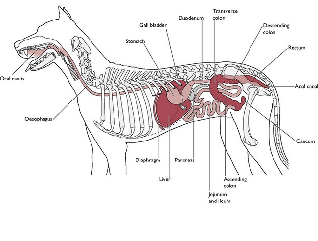
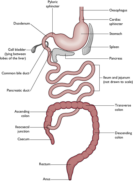
The digestive system (Figs 9.1, 9.2) consists of the following parts:
There are also several accessory glands, without which the digestive process cannot be completed:
The oral cavity
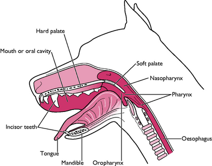
The oral cavity (Fig. 9.3) is also known as the mouth or buccal cavity and contains the tongue, teeth and salivary glands. The function of the oral cavity is as follows:
The oral cavity is formed by the following bones of the skull:
The mandibles articulate with the temporal bones of the skull forming the temporomandibular joint. In carnivores the action of the joint is scissor-like to shear flesh off the bones of their prey.
The upper and lower jaws are linked by skin, forming the cheeks, under which lie the muscles of mastication (see Ch. 4). These muscles lie over the temporomandibular joint and give strength to the biting action. The entrance to the mouth is closed by the lips, composed of muscle covered in skin. The upper lip is split vertically by a division known as the philtrum (see Ch. 8, Fig. 8.1).
The entire oral cavity is lined by a layer of mucous membrane. It is reflected on to the jawbones, forming the gums. The mucous membrane covers the hard palate and extends over a flap of soft tissue at the back of the oral cavity – this is the soft palate, which extends caudally between the oral and nasal cavities and divides the pharynx into the oropharynx and nasopharynx (Fig. 9.3) (see also Ch. 8).
The tongue
The functions of the tongue are:
The tongue lies on the floor of the oral cavity and is made of striated muscle fibres running in all directions. This enables the tongue to make delicate movements. The muscles are attached at the root of the tongue to the hyoid bone (see Ch. 8) and to the sides of the mandibles. The tip of the tongue is unattached and very mobile.
In an anaesthetised animal, the sublingual vein, running under the tongue, may be used for intravenous injections or for collecting blood samples, while the sublingual artery may be used for monitoring the pulse rate.
The tongue is covered in mucous membrane. The dorsal surface is thicker and arranged in rough papillae, which assist in control of the food bolus and in grooming. Some papillae are adapted to form taste buds, mainly found at the back of the tongue. Taste buds are well supplied with nerve fibres, which carry information about taste to the forebrain (see Ch. 5). Running along the underside of the tongue are the paired sublingual veins and arteries.
The teeth
The teeth are hard structures embedded in the upper and lower jaw. Each jaw forms a dental arch or dentary – there are four dental arches in total. The teeth pierce the gums to sit in sockets or alveoli. The membrane covering the gums is known as the gingival membrane or periodontal membrane.
Structure
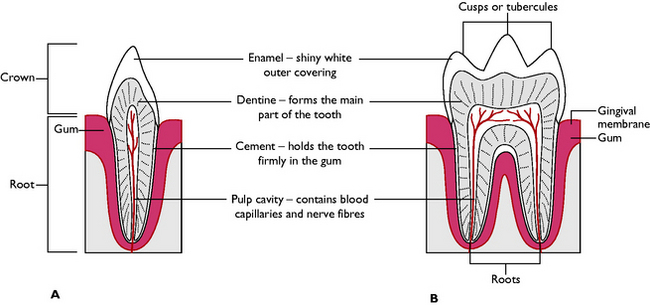
All teeth have a basic structure (Fig. 9.4). In the centre of each tooth is a pulp cavity. This contains blood capillaries and nerves, which supply the growing tooth. In young animals the cavity is relatively large but, once the tooth is fully developed, it shrivels and contains only a small blood and nerve supply. After a tooth has stopped growing the only changes occurring will be due to wear.
Function
The teeth of a carnivore are adapted to shearing and tearing the flesh off the bones of their prey. There are four types of tooth, which are classified by their shape and position in the jaw (Fig. 9.5). This is summarised in Table 9.1.
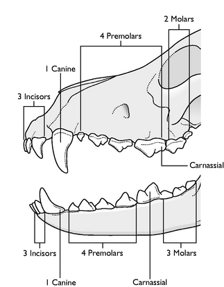
Table 9.1 Tooth types and functions
| Type | Position and shape | Function |
|---|---|---|
| Incisor (I) | Lie in the incisive bone of the upper jaw and in the mandible of the lower jaw; small, pointed with a single root | Fine nibbling and cutting meat; often used for delicate grooming |
| Canines (C) – ‘eye teeth’ | One on each corner of the upper and lower jaws; pointed, with a simple curved shape; single root deeply embedded in the bone | Holding prey firmly in the mouth |
| Premolars (PM) – ‘cheek teeth’ | Flatter surface with several points known as cusps or tubercles; usually have two or three roots arranged in a triangular position to give stability in the jawbone | Shearing flesh off the bone using a scissor-like action; flattened surface helps to grind up the flesh to facilitate swallowing and digestion |
| Molars (M) – ‘cheek teeth’ | Similar shape to premolars; usually larger with at least three roots | Shearing and grinding meat – NB There are no molars in the deciduous dentition |
| Carnassials | Largest teeth in the jaw; similar shape to other cheek teeth; these are the first lower molar and the last upper premolar on each side | Very powerful teeth sited close to the angle of the lips; this type is only found in carnivores |
Dentition
Dogs and cats have two sets of teeth in their lifetime:
Eruption times vary with the species, the type of tooth and the species of animal and are summarised in Table 9.2.
| Tooth type | Dog | Cat |
|---|---|---|
| Deciduous dentition | ||
| Incisors | 3–4 weeks | Entire dentition starts to erupt at 2 weeks and is complete by 4 weeks |
| Canines | 5 weeks | |
| Premolars | 4–8 weeks | |
| Molars | Absent | |
| Permanent dentition | ||
| Incisors | 3.5–4 months | 12 weeks |
| Canines | 5–6 months | |
| Premolars | 1st premolars 4–5 months; remainder 5–7 months | Variable; full dentition present by 6 months |
| Molars | 5–7 months | |
Dental formulae
Each species has a characteristic dental formula and this enables the veterinary surgeon to monitor the numbers of teeth lost as a result of disease or ageing. Each type of tooth is referred to by its initial letter: I is for incisor, etc. The following numbers show how many teeth of each type there are in the upper and lower jaws on one side of the head. The numbers are then multiplied by two to give the total number of teeth in the mouth.
Salivary glands
These are paired glands lying around the area of the oral cavity. Their secretions, known collectively as saliva, pour into the cavity via ducts. Saliva contains 99% water and 1% mucus – there are no enzymes in the saliva of the dog and the cat (Fig. 9.6).
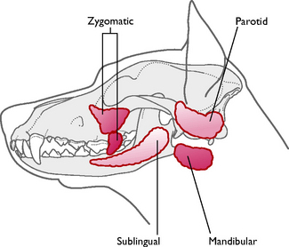
The positions of the glands are:
Production of saliva is continuous but may be increased by such factors as the sight and smell of food (particularly in dogs), fear, pain and irritant gases or other chemicals such as organophosphates. Salivation also often occurs just prior to vomiting and may be a warning sign to the owner!
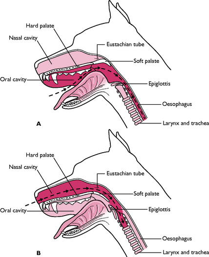
Pharynx
The pharynx forms a crossover point between the respiratory and digestive systems. It is a muscular tube lined with mucous membrane, connecting the back of the nasal and oral cavities with the oesophagus and the larynx and trachea. The soft palate extends caudally towards the epiglottis of the larynx and divides the pharynx into the nasopharynx and oropharynx (Fig. 9.3; see also Ch.8). The walls of the pharynx contain diffuse areas of lymphoid tissue known as the tonsils. Their function is to protect the animal against disease (see Ch. 7). The most obvious are the palatine tonsils lying one on each side of the pharynx in a shallow recess. The Eustachian or auditory tube connects the pharynx to the middle ear. It enables the air pressure on either side of the tympanic membrane to equalise and thus maintains the flexibility and function of the ear drum (see Ch. 5).
The pharynx conveys food from the mouth into the oesophagus by means of a process known as deglutition or swallowing (Fig. 9.7). This occurs in the following stages:
Oesophagus
This is a simple tube that carries food from the pharynx to the stomach (Figs 9.1, 9.2). In the neck, the oesophagus lies dorsal to the trachea and slightly to the left of it. It passes through the thoracic cavity, running within the mediastinum, dorsal to the heart base and between the two lungs. The oesophagus enters the abdominal cavity via the oesophageal hiatus of the diaphragm, which separates the thorax and abdomen.
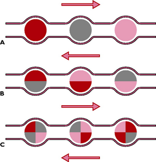
The walls of the oesophagus are lined with stratified squamous epithelium arranged in longitudinal folds. This protects against damage by food and allows for widthways expansion as the boluses pass down. Within the walls are circular and longitudinal bands of smooth muscle fibres. Contraction of these muscles brings about a series of peristaltic waves that force the food along the tube (Fig. 9.8). Food can pass in the reverse direction – antiperistalsis – seen during vomiting. The average time taken for food to pass down the oesophagus is 15–30 seconds but this depends on the type of food: liquids take a shorter time than dry foods.
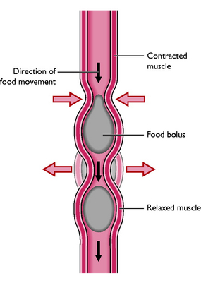
Fig. 9.8 Representation of peristalsis. Muscular contraction within the tube forces the food bolus along.
Diagnosis of conditions of the digestive tract may be helped by the use of a ‘barium meal’ or barium series. Liquid barium sulphate is administered orally and a series of radiographs are taken over a period of several hours. Figure 9.9 shows the result of the use of barium as a contrast medium and illustrates the anatomy of the canine digestive tract.
The abdominal part of the digestive system
The majority of the digestive tract lies within the abdominal cavity and can be divided into three parts:
Stomach
The stomach of the dog and cat is described as being simple and digestion is said to be monogastric. The functions of the stomach are:
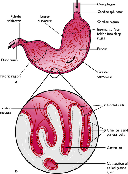
The stomach is a C-shaped, sac-like organ lying on the left side of the cranial abdomen. Food enters from the oesophagus via the cardiac sphincter and leaves from the pyloric sphincter (Fig. 9.2). The inner curve of the sac is called the lesser curvature and the outer curve the greater curvature. The entire organ is covered in a layer of visceral peritoneum or mesentery, as are all the organs in the abdominal cavity. The mesentery attached to the inner curvature is called the lesser omentum, while that attached to the greater curvature is called the greater omentum. The spleen lies within the layers of the greater omentum. The walls of the stomach are thick and easily distended by food. When empty, the stomach lies under the ribs but when full the stomach may occupy a third of the abdomen.
Gastric dilatation and volvulus (GDV) is a condition of large breeds of dog, such as the German Shepherd. It occurs when the stomach distends with gas and half-fermented food, becomes unstable and twists around on itself, preventing the escape of the gas. The dog suffers extreme discomfort and may go into shock and die if not treated to relieve the distension. In such cases, the stomach feels tight and drum-like and may fill half the abdomen.
Structure of the stomach wall
The stomach can be divided into three regions: the cardia, fundus and pylorus. Most of the gastric glands lie within the fundic region of the stomach. The walls of the stomach are lined with mucous membrane, the gastric mucosa. This is thrown into deep longitudinal folds known as rugae which flatten out when the stomach fills with food. Within the mucosa are the gastric pits (Fig. 9.10), consisting of three types of cell responsible for the secretion of gastric juices:
Food enters the stomach via the cardiac sphincter. Distension of the stomach stimulates the secretion of the hormone gastrin from the stomach walls, which initiates the production of the gastric juices. Two types of muscular movement of the stomach break up and mix the food with the juices:
Gastric emptying
Food in the stomach is broken up and partially digested, resulting in a soup-like liquid with an acid pH known as chyme. Chyme is released in spurts through the pyloric sphincter into the duodenum, where digestion continues. The time taken for food to pass through the stomach depends on the type of food – liquids may take about half an hour, while more fatty or solid foods take about 3 hours.
Vomiting is the ‘return of ingesta and fluids against the normal direction of swallowing and peristalsis’. This is a very common presenting sign and there are three types:
Small intestine
The small intestine is the major site of enzymic digestion and absorption. It is a long, relatively narrow tube and may be up to 3.5 times the body length. Food passes along the small intestine and is mixed with digestive juices by peristalsis and by rhythmic segmentation. It is divided into three parts, each of which has a similar structure but shows functional adaptations (Figs 9.2, 9.9):
Structure of the intestinal wall
Each part of the small intestine has a similar structure (Fig. 9.12). The epithelial layer is folded into millions of tiny, leaf-shaped folds called villi (sing. villus). Their function is to increase the surface area of the epithelium to maximise the efficiency of the digestive and absorptive processes. Extending from each epithelial cell is a border of tiny microvilli, which form a ‘brush border’ to further increase the surface area. (see Ch. 2, Fig. 2.2)
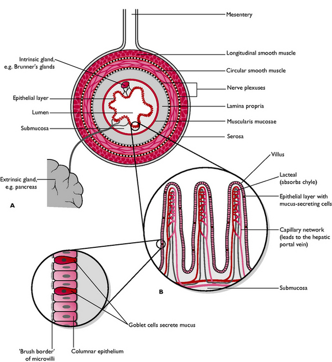
Fig. 9.12 A Cross-section through an area of intestine to show structure. B Enlargement to show the villi and absorptive epithelium.
The pancreas
The pancreas is a pale pink, lobular gland lying in the ‘U’ of the duodenum, whose secretions are essential to the digestive process. It lies outside the digestive tract and is described as an extrinsic gland. The pancreas has an endocrine part (see Ch. 6) and an exocrine part and is classed as a mixed gland. The exocrine part secretes digestive enzymes and bicarbonate into the duodenum via the pancreatic duct (Fig. 9.2).
The gall bladder
The gall bladder lies within the lobes of the liver and is a reservoir for bile, produced by the liver. Bile contains bile salts needed for fat digestion and is stained yellow-green by bile pigment or bilirubin produced from the breakdown of old erythrocytes. It is secreted into the duodenum via the common bile duct.
Digestion
Food is ingested in the form of proteins (polypeptides), carbohydrates (polysaccharides) and fats (lipids). For food to be a useable source of energy, it must be broken down into soluble molecules that are small enough to be absorbed through the wall of the small intestine and into the bloodstream – this is digestion. In mammals this process occurs by the use of chemicals called enzymes. Each enzyme is designed to act on a specific material or substrate, and not on any other. They are usually named after the substrate on which they act, e.g. lipase acts on lipids (fats); maltase acts on maltose.
An enzyme is a protein that acts as a catalyst, increasing the speed of a reaction. Some enzymes may be produced in an inactive form, known as a precursor, which must be converted to the active form by another enzyme before they can work.
There are four sources of digestive juices containing the enzymes: gastric juices, pancreatic juices, bile salts and intestinal juices. Digestion in the carnivore begins in the stomach.
Gastric juice
This is secreted by the gastric pits in response to production of the hormone gastrin from the stomach wall (see Ch. 6). It contains:
The liquid food resulting from this process of gastric juice digestion is known as chyme.
Pancreatic juice
This secretion is produced by the exocrine part of the pancreas and occurs in response to the hormones cholecystokinin and secretin (from the wall of the duodenum) and gastrin (from the stomach wall), and stimuli from the autonomic nervous system (see Ch. 6). It contains:
Bile salts
The presence of chyme in the duodenum causes the gall bladder to contract and produce bile. Bile emulsifies fat globules so that they have a larger surface area on which enzymes can act, and also activates lipases.
Intestinal juice
Secretion of intestinal juice is stimulated by the hormone secretin produced in response to the passage of chyme through the pyloric sphincter (see Ch. 6). The intestinal juices are produced by:
A number of enzymes are present, many of which are also produced by the pancreas:
The result of the digestive process is that the basic constituents of food are converted into small molecules that can now be absorbed.
Absorption
The main site for absorption is the villi of the small intestine. The efficiency of the absorptive process is increased by:
During absorption amino acids and simple sugars are absorbed by the blood capillaries and are carried by the hepatic portal vein to the liver. Fatty acids and glycerol are absorbed by the lacteals. They form a fatty milky liquid known as chyle, which is carried to the cisterna chyli, lying in the dorsal abdomen (see Ch. 7, Fig. 7.7). Here it is mixed with lymph and carried to the heart by the thoracic duct, where it joins the blood circulation.
Large intestine
This is a short tube of a wider diameter than the small intestine. Each part has a similar structure to that of the small intestine. However, in the lumen there are no villi and no digestive glands, but there are more goblet cells. These secrete mucus, which lubricates the faeces as it passes through. The large intestine is divided into:
 External anal sphincter: outer ring of striated muscle; control is voluntary
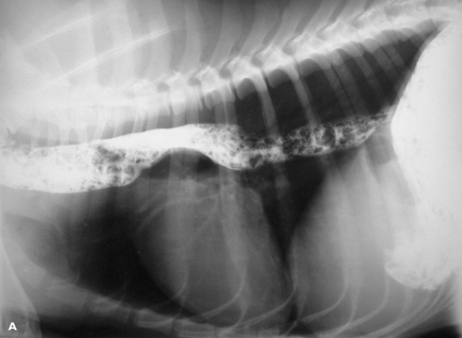
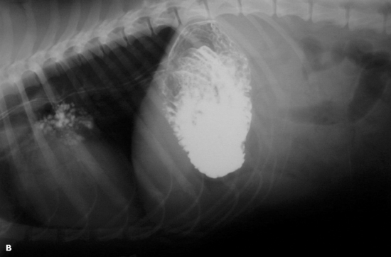
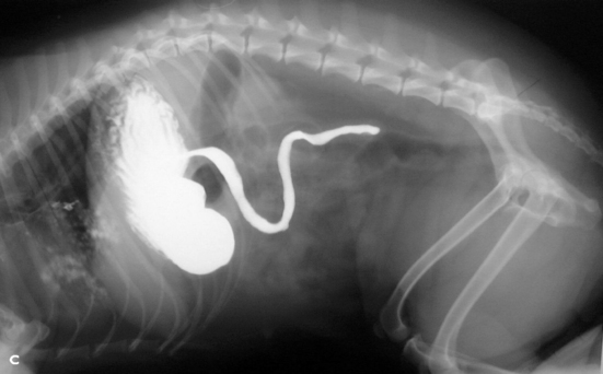
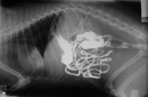
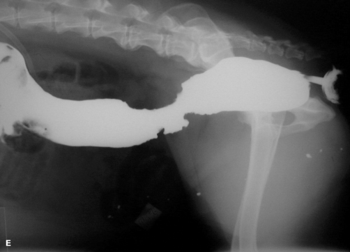
Fig. 9.9 Barium X-ray series illustrating the digestive tract. A The barium (radio-opaque white material) is in the oesophagus after being swallowed. B Barium enters the stomach. C Barium starts to leave the stomach and enter the duodenum. D Most of the barium is now in the small intestine (a small amount remains in the stomach and some has entered the large intestine). E Barium in the colon and rectum applied by enema.
The lumen of the sphincter is constricted and lined with deep longitudinal folds of mucous membrane that stretch to allow the passage of bulky faeces.
Defaecation
The faecal mass passes through the large intestine by means of peristalsis, antiperistalsis, rhythmic segmentation and infrequent but strong contractions known as mass movements. These movements are involuntary but, as the faecal mass enters the pelvic cavity, stretching of the rectal wall stimulates voluntary straining brought about by abdominal contractions. The anal sphincter, which is normally held tightly closed, relaxes, the abdominal muscles contract and the mass is forced out.
Composition of faeces
Normal faeces have a colour and smell that is characteristic of the species. They are described as being ‘formed’ – the water content is such that the faeces keep their shape (watery faeces do not). Normal faeces contain:
Lying between the two anal rings, in the ‘20 to 4’ position, are the anal sacs. These are modified sebaceous glands, which vary from pea to marble size depending on the species and breed of animal. Their secretions coat the faeces as it passes out and the characteristic smell is used by the animal for territorial scent marking, particularly in wild species. Impacted anal sacs may be a problem in dogs fed on softer diets.
The sequence of muscle contraction and nervous control involved in defaecation requires coordination by the nervous system. Any damage to the spinal cord, e.g. disc prolapse or severe trauma, may result in faecal incontinence or faecal retention.
Diarrhoea is ‘frequent evacuation of watery faeces’. This is a common presenting sign. It may be acute or chronic and is an indication of some form of intestinal disturbance, e.g. maldigestion, malabsorption, increased peristalsis or gastrointestinal irritation. However, the most common cause of diarrhoea, in dogs especially, is dietary mismanagement. This can be easily corrected by starvation for 24 hours followed by a light diet of fish or chicken and rice for a further 24 hours.
The liver
The liver is the largest gland in the body and lies in the cranial abdomen. The cranial aspect is convex and is in contact with the diaphragm. The caudal aspect is concave and is in contact with the stomach, duodenum and right kidney (Fig. 9.1). The liver is deep red in colour as it has a large volume of blood flowing through it. It is divided into several large lobes, in the centre of which is the falciform ligament. This is the remains of fetal blood vessels from the umbilicus and is of little significance in the adult animal (see Ch. 7). The gall bladder lies between the lobes in the centre of the caudal aspect. It stores bile, which pours into the duodenum via the common bile duct.
Histologically, the liver consists of thousands of cells known as hepatocytes (Fig. 9.13). These are responsible for all the many functions of the liver. The hepatocytes are arranged in hexagonal liver lobules surrounded by connective tissue. Running through the lobules between the hepatocytes are minute blood sinusoids carrying blood from the hepatic portal vein. Each hepatocyte is bathed in plasma, which percolates across the lobule to drain into the central vein. The central veins flow into the hepatic vein and so to the caudal vena cava. Thus the products of digestion are carried from the small intestine by the blood to every hepatocyte where they are used for metabolism. The liver tissue receives arterial blood in the hepatic artery.
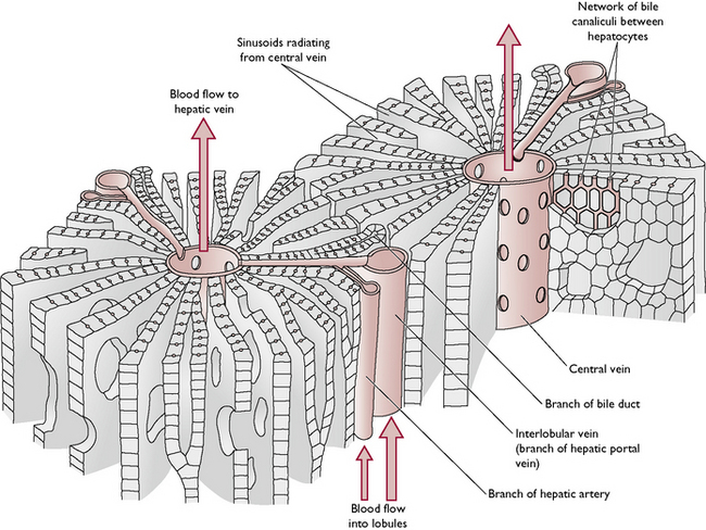
Between the hepatocytes are tiny channels known as bile canaliculi, into which bile is secreted. The canaliculi form an interconnecting network that eventually drains into the gall bladder. There is no direct connection between the sinusoids and the canaliculi.
The liver has many functions and is essential to normal health.
Formation of plasma proteins: Albumin maintains the balance of fluids in the body; fibrinogen and prothrombin are involved in the blood clotting mechanism; globulins form part of the immune mechanism Regulation of amino acids: there are 20 amino acids that are essential to form body protein. The liver cells use these amino acids to make new proteins and as the ‘building blocks’ of other organic compounds. Amino acids that are not essential may be converted into more useful ones by a process known as transamination