Introduction to Head and Neck Anatomy
1 Define and pronounce the key terms and anatomic terms in this chapter.
2 Discuss the clinical applications of head and neck anatomy by dental professionals.
3 Discuss normal anatomic variation and how it applies to head and neck structures.
4 Correctly complete the review questions and activities for this chapter.
5 Apply the correct anatomic nomenclature during dental clinical procedures.
Position in which the body is erect, with arms at the sides, palms and toes directed forward, and eyes looking forward.
Plane created by an imaginary line that divides the body at any level into anterior and posterior parts.
Plane created by an imaginary line that divides the body at any level into superior and inferior parts.
Clinical Applications
The dental professional must have a thorough understanding of head and neck anatomy when performing patient examination procedures, both extraoral and intraoral (Figure 1-1). This will help determine whether any abnormalities or lesions exist and possibly indicate their cause and amount of involvement. This will also provide a basis for the description of the lesion and its location for record-keeping purposes.
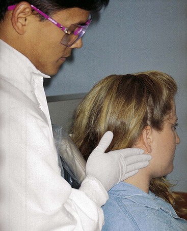
FIGURE 1-1 Extraoral examination of the patient (as well as intraoral examination) is based on an understanding of head and neck anatomy.
When taking radiographs, the dental professional uses surface landmarks for easy film placement and consistency. In addition to these landmarks, an understanding of anatomy is important in the mounting and analysis of the films.
A patient may also present with features of a temporomandibular joint disorder. A dental professional must understand the normal anatomy of the joint to understand the various disorders associated with it.
The administration of local anesthesia is also based on landmarks of the head and neck. Knowledge of anatomy helps the dental professional plan for use of a local anesthetic to reduce pain during various dental procedures. This knowledge also allows for correct placement of the syringe and its anesthetic agent, potentially avoiding complications.
During examination of the patient, the dental professional may note the presence of dental infection. It is important to know the source of the infection as well as the areas to which it could spread by way of certain anatomic features of the head and neck. This background in anatomy will help the dental professional understand the spread of dental infection.
To initially consider patient care through anatomic study, this text takes mainly a systemic approach to the study of head and neck anatomy after its two initial background chapters, Chapters 1 and 2, and through most of its chapters, Chapters 3-10, an approach that takes a look at each system separately (e.g., skeletal, muscular). Another way to study anatomy for patient care integration is the regional approach, which is taken up later within Chapter 11, since it focuses on the faciae and fascial spaces of the head and neck. Both approaches, when used in the order presented in this text, are complementary and effective ways to study head and neck anatomy and prepare for patient care considerations.
To reinforce the material already presented and make it readily useful for clinicians, the final chapter, Chapter 12, emphasizes this important clinical approach to head and neck anatomy during the consideration of the spread of infection. Chapter 9 also has an expanded clinical emphasis, covering the anatomy of local anesthesia. In addition, all the other chapters include important clinical ramifications when appropriate, such as related pathology.
Anatomic Nomenclature
Before beginning the study of head and neck anatomy, the dental professional may need to review anatomic nomenclature, which is the system of names for anatomic structures. This review will allow for easy application of these terms to the head and neck area when examining a patient, for use in the patient’s record, or during other clinical procedures.
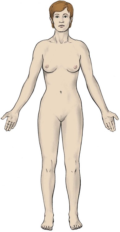
The nomenclature of anatomy is based on the body being in anatomic position (Figure 1-2). In anatomic position, the body is standing erect. The arms are at the sides with the palms and toes directed forward and the eyes looking forward. This position is assumed even when the body may be supine (on the back) or prone (on the front) or even with respect to the patient’s head and neck when sitting in a dental chair.
When studying the body in anatomic position, certain terms are used to refer to areas in relationship to other areas (Figure 1-3). The front of an area in relationship to the entire body is its anterior part. The back of an area is its posterior part. The ventral part is directed toward the anterior and is the opposite of the dorsal part (the posterior) when considering the entire body.
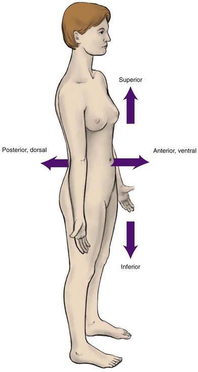
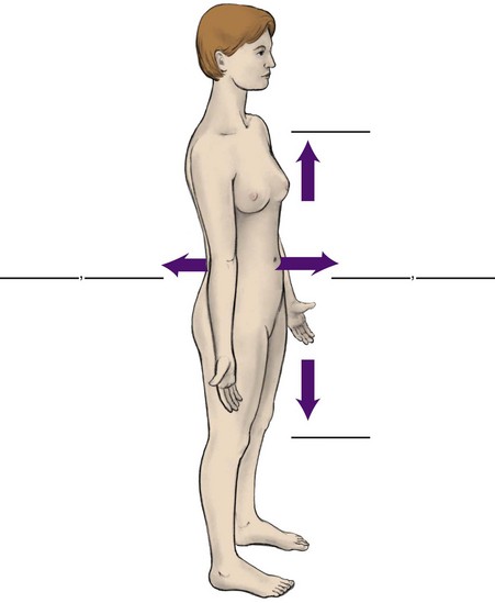
FIGURE 1-3 Body in anatomic position with the anterior (or ventral), posterior (or dorsal), superior, and inferior areas noted.
Other terms can be used to refer to areas in relationship to other areas of the body. An area that faces toward the head and away from the feet is its superior part. An area that faces away from the head and toward the feet is its inferior part. As an example, the face is on the anterior side of the head, and the hair is superior and posterior to the face. The apex or tip is the pointed end of a conical structure such as the tongue apex or tip.
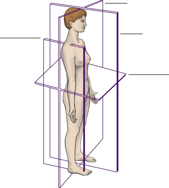
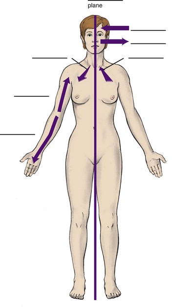
The body in anatomic position can be divided by planes or flat surfaces (Figure 1-4). The median plane or midsagittal plane is created by an imaginary line dividing the body into equal right and left halves. On the surface of the body, these halves are generally symmetric, yet the same symmetry does not apply to all internal structures.
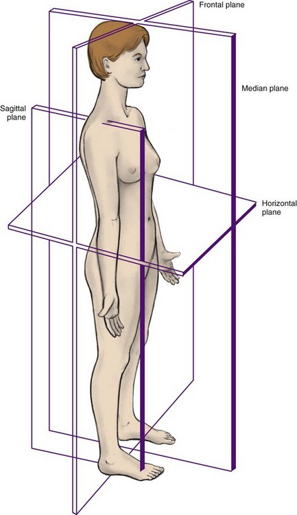
FIGURE 1-4 Body in anatomic position with the median, sagittal, horizontal, and frontal planes noted.
Other planes can be created by different imaginary lines. A sagittal plane is any plane created by an imaginary plane parallel to the median plane. A frontal plane or coronal plane is created by an imaginary line dividing the body at any level into anterior and posterior parts. A horizontal plane is created by an imaginary line dividing the body at any level into superior and inferior parts and is always perpendicular to the median plane.
Parts of the body in anatomic position can also be described in relationship to these planes (Figure 1-5). A structure located at the median plane (e.g., the nose) is considered median. An area closer to the median plane of the body or structure is considered medial. An area farther from the median plane of the body or structure is considered lateral. For example, the eyes are medial to the ears, and the ears are lateral to the eyes.
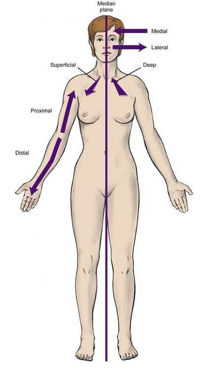
FIGURE 1-5 Body in anatomic position with the medial (or proximal), lateral (or distal), and superficial (or deep) areas noted.
Terms can be used to describe the relationship of parts of the body in anatomic position. An area closer to the median plane is considered by anatomists to be proximal, and an area farther from the median plane is distal. For example, in the upper limb the shoulder is proximal and the fingers are distal.
Additional terms can be used to describe relationships between structures. A structure on the same side of the body is considered ipsilateral. A structure on the opposite side of the body is considered contralateral. For example, the right leg is ipsilateral to the right arm but contralateral to the left arm.
Certain terms can be used to give information about the depth of a structure in relationship to the surface of the body. A structure located toward the surface of the body is superficial. A structure located inward, away from the body surface, is deep. For example, the skin is superficial, and the bones are deep.
Terms also can be used to give information about location in hollow structures such as the braincase of the skull. The inner side of the wall of a hollow structure is referred to as internal. The outer side of the wall of a hollow structure is external.
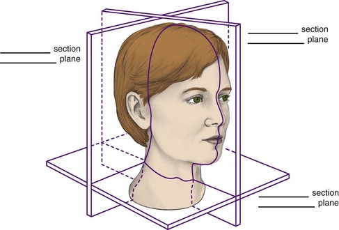
The body or parts of it in anatomic position can also be divided into sections along various planes in order to study the specific anatomy of a region (Figure 1-6). The midsagittal section or median section is a division through the median plane. The frontal section or coronal section is a division through any frontal plane. The transverse section or horizontal section is a division through a horizontal plane.
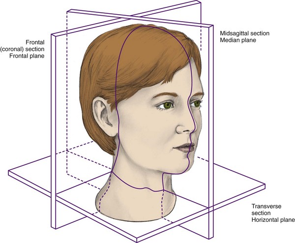
FIGURE 1-6 Head and neck in anatomic position with the midsagittal, transverse, and frontal sections and related planes noted.
It is important to keep in mind when looking at diagrams or even clinical photographs, especially those of dissections, to first note any directional aids (e.g., view, section) and then pick out a familiar structure (e.g., apex of tongue or nose, mandible) to allow for orientation. This process will help in the study of the head and neck.
Normal Anatomic Variation
When studying anatomy, the dental professional must understand that there can be anatomic variations of head and neck structures that are still within normal limits. The number of bones and muscles in the head and neck is usually constant, but specific details of these structures can vary from patient to patient. Bones may have different sizes of processes. Muscles may differ in size and details of their attachments. Joints, vessels, nerves, glands, lymph nodes, fasciae, and spaces of an individual can vary in size, location, and even presence. The most common variations of the head and neck that affect dental treatment are discussed in this text.
Identification Exercises
Identify the structures on the following diagrams by filling in each blank with the correct anatomic term. You can check your answers by looking back at the figure indicated in parentheses for each identification diagram.
1. Which of the following planes divides the body in anatomic position into right and left halves?
2. Which of the following terms is used to describe an area of the body that is farther from the median plane?
3. Structures on the same side of the body are considered
4. An area of the body in anatomic position that faces toward the head is considered
5. Through which plane of the body in anatomic position is a midsagittal section taken?
6. Which of the following statements concerning anatomic position is CORRECT?
A Body is erect with eyes looking forward.
B Arms are at sides with palms directed backward.
7. Which of the following sections is considered also a horizontal section?
8. Structures that are located inward, away from the body surface, are considered
9. Which of the following planes divides any part of the body further into anterior and posterior parts?
10. Which of the following is a CORRECT statement concerning human anatomy?
A Apex of a conical structure is the flat base.
B Two halves of the body are completely symmetric.
11. Which of the following is a CORRECT statement when considering facial features?
12. Proximal refers to a body part that is
A closer to the medial plane of the body than another part.
B farther from the medial plane of the body than another part.
C farther from the point of attachment to the body than another part.
D closer to the point of attachment to the body than another part.
13. The median plane placed through the body will divide the right arm and the
14. A frontal plane placed through the body will ALWAYS bisect the
15. If a transverse plane occurs through the navel, which of the following statements is CORRECT?