Glomerular Diseases

Overview

Glomerular diseases are a group of conditions that damage the kidney’s filtering units (glomeruli). Glomerulonephritis is also a group of diseases that affect the glomeruli but specifically manifest with hematuria. Glomerulonephritis is a glomerular disease, but not all glomerular diseases are termed glomerulonephritis. Glomerular diseases are the most common cause of ESRD worldwide, while glomerulonephritis is the third leading cause of end-stage kidney disease in the United States.

The glomeruli are tufts of capillaries connecting the afferent and efferent arterioles of the nephron (see Fig. 18-6). The capillaries are supported by a stalk made up of mesangial cells and a basement membrane and are arranged in lobules. The circulating blood is filtered in the glomeruli, with the urine filtrate being an end-product. Glomerular damage produces two types of syndromes: the nephrotic syndrome and the nephritic syndrome.

Nephrotic syndrome is not a specific kidney disease but rather occurs as a result of any disease that causes damage to the kidney-filtering units. Nephrotic syndrome is principally associated with proteinuria, which occurs with such diseases as diabetes, amyloidosis, and membranous glomerulopathy. Nephritic syndrome is correlated with hematuria. Glomerular diseases that result in a nephritic syndrome include lupus nephritis, immunoglobulin A (IgA) nephropathy, and acute diffuse proliferative glomerulonephritis. Overlap of the two syndromes is common, and a precise diagnosis often requires a kidney biopsy.

Etiologic Factors

Most cases of nephritic syndrome and some cases of nephrotic syndrome have an immune origin and are part of a systemic process, such as lupus nephritis or membranoproliferative glomerulonephritis. Two different mechanisms have been proposed to account for the pathologic changes seen in glomerular diseases. The first is due to the deposition of a circulating antigen/antibody complex into some portion of the glomerulus (the glomerular basement membrane or GBM, mesangium), followed by an inflammatory response and damage. Injury is caused via the second mechanism when an antigen is deposited into the glomerulus with subsequent antibody interaction with the antigen, followed by an inflammatory response. Antigens may be exogenous, as seen in poststreptococcal glomerulonephritis, or endogenous, as noted with lupus nephritis.

Risk Factors

The presence of a variety of disorders can increase the risk of glomerular damage. Diabetes, principally type 2 diabetes, is a significant risk factor for CKD and the development of nephrotic syndrome. Age is another factor in the development of some diseases associated with nephrotic syndrome. For example, minimal change glomerulopathy is seen in children under the age of 10 and accounts for more than 80% of cases of nephrotic syndrome in children. Race is also a factor in the development of glomerular disease. Focal segmental glomerulosclerosis (FSGS) is the most common cause of nephrotic syndrome in African Americans, while membranous nephropathy is seen more commonly in Caucasians. FSGS is also seen more often in persons who are obese.

Pathogenesis

Damage to the glomerular epithelial cells or the GBM allows larger molecules, such as protein, to escape out of the circulation and into the urine, causing nephrotic syndrome. Rupture of a capillary wall or proliferation of mesangial cells leads to hematuria and nephritic syndrome. The processes that cause this damage vary depending on the underlying glomerular disease.

Nephritic syndromes are caused by antibody/antigen complexes. Damage and clinical manifestations depend on where these complexes are deposited in the glomerulus. IgA nephropathy results when circulating antibodies (IgA antibodies) complex with an antigen (currently not defined) but are not able to be filtered through the glomerulus. These complexes stimulate an inflammatory response, accompanied by the release of cytokines and growth factors. Mesangial cell proliferation and glomerular scarring result.

Poststreptococcal glomerulonephritis occurs when antigen (streptococcal) is deposited between the glomerular epithelial cells and the GBM, resulting in an antibody response and damage to the GBM. This disruption of the GBM results in proteinuria as well as nephritis. Lupus nephritis can result from either antigen deposition followed by antibody reaction or the deposition of antibody/antigen complexes. These depositions can also occur in several locations. Those found in the mesangium result in proliferation of mesangial cells. Complexes placed in the GBM cause proteinuria, while deposition in the subepithelial space leads to nephrosis-range proteinuria. If the antigen is chronically produced, the recurrent inflammatory reactions lead to chronic glomerulonephritis. These changes adversely affect the glomerular filtration mechanism and alter capillary permeability.

While the nephritic syndrome results most often from depositions of immune complexes, causes of nephrotic syndrome vary, with many not well understood. For example, minimal change disease shows very few abnormalities on microscopic or electron microscopic inspection. This disease may result from damage created by a lymphocyte product. Causes of other diseases, such as membranous nephropathy, are better understood. Antigen is initially deposited in the GBM with subsequent antibody interaction and inflammatory response. These immune complexes also trigger the complement system, causing further damage to the GBM and allowing large amounts of protein to escape from the plasma.

Clinical Manifestations

Glomerular disease causing a nephrotic syndrome produces proteinuria (greater than 3 g in 24 hours), hypoalbuminemia, hyperlipidemia, lipiduria, and edema. The significant loss of protein from the kidney accounts for the hypoalbuminemia, which in turn reduces the plasma oncotic pressure in the vessels. Fluid flows to areas of greater protein concentration, which in this instance is outside the blood vessel, causing edema. The kidney perceives a loss in volume and retains both fluid and sodium, thus increasing the edema. Edema is the principal symptom that brings affected people to the physician’s office. The edema can be severe enough to be disabling.

The loss of protein also stimulates the liver to produce cholesterol, leading to hypercholesterolemia (cholesterol can be as high as 300 to 400 mg/dl). High cholesterol not only increases atherosclerosis of the coronary arteries but also worsens existing kidney disease. Other clinical manifestations include coagulation abnormalities from the loss of coagulation proteins, resulting in a venous thrombotic event (i.e., pulmonary embolism, deep venous thrombosis, or renal vein thrombosis). Hypothyroidism may occur secondary to the loss of thyroxine, and anemia may develop because of the loss of transferrin and erythropoietin.

The nephritic syndrome is characterized by hematuria, but oliguria, hypertension, and renal insufficiency often accompany this syndrome. The urine typically contains abnormally shaped erythrocytes (sometimes called “Mickey Mouse cells”), which distinguishes the hematuria of nephritic syndromes from that of urinary or bladder sources (normally shaped erythrocytes).

Proteinuria may be present, depending on which part of the glomerulus is affected. Hematuria may be asymptomatic, as with IgA nephropathy, or clinically apparent, as with anti–GBM antibody disease. Proliferation of mesangial cells may occur. If less than 50% of the glomeruli are affected, the disease may be asymptomatic. If greater than 50% of glomeruli are involved, hematuria and proteinuria are more profound. Renal insufficiency may be mild or may lead to a rapid loss in function. This depends on the percentage of kidney involved and the severity of the disease. Epithelial crescents form when the disease involves Bowman’s space. Crescent formation or extracapillary proliferation is caused by the accumulations of macrophages, fibroblasts, proliferating epithelial cells, and fibrin within Bowman’s space. Crescentic glomerulonephritis defines a disease process that results when more than 50% of the glomeruli have crescents. Progressive disease can result in ESRD.

MEDICAL MANAGEMENT

DIAGNOSIS.

The diagnosis of glomerular disease requires analysis of urine, looking for protein, casts (protein, erythrocyte, or lymphocyte “castings” of the tubules), and erythrocytes. The erythrocytes are often misshaped, demonstrating the damage acquired while going through the glomerulus. A 24-hour urine collection may be required to assess the amount of proteinuria. Blood pressure is often elevated, and anemia may develop. A kidney biopsy is frequently needed to make the precise diagnosis.

TREATMENT.

Treatment depends on the specific cause of the glomerular disease, but there are several features that may be shared and the treatment may be similar. Diseases causing nephrotic syndrome are treated with an ACE inhibitor or ARB to control blood pressure and to reduce proteinuria.

Hypercholesterolemia is usually treated with a hydroxymethylglutaryl–coenzyme A reductase inhibitor (statin). Erythropoietin can be employed if anemia is symptomatic, and vitamin D sterols may be needed to prevent a deficiency in vitamin D. Fluid is restricted, and diuretics are often used to reduce edema. Diseases with immune-associated injury often require treatment with prednisone, cyclosporine, or cytotoxic agents. Others, such as membranoproliferative glomerulonephritis, improve with treatment of the underlying disorder (i.e., treating hepatitis C–induced disease with interferon-alfa-2 with ribavirin). Some diseases progress and require dialysis or transplantation.

18-6   SPECIAL IMPLICATIONS FOR THE THERAPIST

Glomerular Diseases

Therapists working with clients with a diagnosis of diabetes, systemic lupus erythematosus, vasculitis, and hypertension need to be aware of the association of glomerulonephritis with these disorders. Being vigilant for the clinical manifestations of glomerulonephritis (e.g., edema, hypertension, hematuria, oliguria) is important, and their presence warrants referral of the client to a physician.

An awareness of the side effects associated with diuretics is also important. Potential side effects include muscle weakness, fatigue, muscle cramps, headaches, and depression, all of which can interfere with the rehabilitation program. (See Chapter 5 for additional information regarding diuretics; see also Table 12-5.) The onset of any of these complaints also warrants communication with a physician. Finally, many clients with glomerulonephritis progress to chronic renal failure.

DISORDERS OF THE BLADDER AND URETHRA

Bladder Cancer

Overview and Incidence

The bladder is lined with transitional cells and in some places, such as at the trigone, epithelial cells. Transitional cell carcinoma is the most common type of bladder cancer, accounting for 90% of all cases. Transitional cell bladder cancer is a heterogenous group of cancers with a wide spectrum of aggressiveness and clinical manifestations. Squamous cell carcinoma of the bladder is unusual, accounting for 8% of all cases, typically resulting from chronic inflammation. A more rare form, adenocarcinoma, accounts for the remaining 2% of cases and is thought to arise from remnants of the embryologic urachus (ligaments).

There are approximately 50,000 cases (21 per 100,000 people) of bladder cancer each year in the United States. As the fourth leading cause of cancer in men and eighth leading cause of cancer death in the United States, bladder cancer is more common than is generally appreciated.96 Overall, the incidence of bladder cancer is increasing in industrialized countries, although the survival rate is improving.

Etiologic and Risk Factors

The specific cause of bladder cancer is unknown, but multiple risk factors are linked with the development of bladder cancer (Box 18-3). The strongest and most significant risk factor is smoking. Sixty-five percent to seventy-five percent of all individuals with bladder cancer have a strong smoking history. Cigarette smokers are twice as likely as nonsmokers to develop bladder cancer.

Box 18-3   RISK FACTORS FOR BLADDER CANCER

• Cigarette smoking

• Occupational exposures

• Truck driving (diesel exhaust)

• Painting

• Leatherworking

• Metalworking

• Male gender

• Age 55 and older

• Previous treatment with cyclophosphamide or iphosphamide (chemotherapy)

• Previous pelvic radiation (e.g., ovarian cancer treatment)

• European descent

• History of chronic bladder infections (such as with spinal cord injury, stroke), kidney or bladder stones, infection with parasite causing schistosomiasis

• Long-term catheterization (e.g., dementia, Alzheimer’s disease, neurologic impairment)

• History of previous bladder cancer

• Family history of bladder cancer or retinoblastoma gene inheritance

• Under investigation:

• Gene-environment interaction

• Coffee

• Fluid intake

• Bacon consumption

• Gonorrhea infection

Occupational exposures are also related to bladder cancer, particularly exposure to β-naphthylamine, 4aminobiphenyl (ABP), and benzidine, used in the dye and rubber tire industries. Although these chemicals have been banned, others are currently being investigated for possible association, and those in many occupations appear to be at risk for the development of bladder cancer, such as painters, metalworkers, and truck drivers.12,65,184 It is estimated that up to 20% of bladder cancer is caused by an occupational exposure.106

More than 90% of cases occur in people over 55 years old, making age another risk factor. Caucasians are twice as likely as African Americans to develop the disease, and men develop bladder cancer four times more often than women. A previous history of bladder cancer; previous treatment with high doses of the chemotherapy drugs cyclophosphamide or iphosphamide; and radiation to the pelvis also place people at higher risk.

Chronic inflammation, such as from recurrent UTIs, kidney or bladder stones, or the parasite causing schistosomiasis, increase the risk for the uncommon development of squamous cell carcinoma of the bladder. It is believed that chronic irritation and inflammation cause transitional cells to undergo metaplasia and transform into malignant cells. This may also occur in areas already epithelialized with squamous cells, such as at the trigone.

Rare risk factors include uncommon birth defects such as exstrophy (where there is a defect in the abdominal wall), inheritance of the retinoblastoma gene, or a family history of bladder cancer.39,57 Increased fluid intake may decrease the risk of bladder cancer, leading to speculation that a more frequent urine flow decreases the time of contact between carcinogens and the bladder epithelium.129 Long-term catheterization (e.g., in spinal cord injury) is a risk factor for squamous cell bladder cancer due to chronic inflammation.

The relationship between coffee and bladder cancer remains controversial despite decades of research. Studies have published varying results, but the findings to date indicate that there may be a weak association between coffee drinking and bladder cancer.166,186,209 Genes most likely play a role in the development of bladder cancer, and specific genes may put some persons at risk once exposed to a carcinogen.172

Pathogenesis

Tumors of the urinary collecting system can arise from epithelial, mesenchymal, or hematopoietic tissues, but the majority of bladder cancers arise from the epithelium. Approximately 90% of these cancers are transitional cell carcinomas, with squamous cell carcinomas and adenocarcinomas making up the remainder. Bladder cancer is believed to develop through reversible premalignant stages followed by irreversible steps, ending in invasive cancer that can give rise to distant metastases. Variations in the clinical course suggest that different forms of bladder cancer develop along different molecular pathways, leading to tumor presentations of various malignant potential.202

The systemic absorption of environmental carcinogens, including cigarette smoke, followed by storage in the bladder exposes the urinary epithelium to concentrated levels of the agents for prolonged periods. This interaction of urine-soluble carcinogens with the epithelium is known as contact chemical carcinogenesis. The precise molecular events leading to the formation of bladder cancer are not known, but several genes appear to be involved. One includes the 9p21 locus (chromosome 9), which contains the CDKN2A/ARF tumor suppressor gene.9 Another factor may be the inability to repair deoxyribonucleic acid (DNA) following damage from carcinogens.164 Further research is needed to determine which genes are involved and how risk factors interact in the development of bladder cancer.

Clinical Manifestations

Painless hematuria is the most common sign of bladder cancer. Gross hematuria is present in up to 85% of people with this condition, and microscopic hematuria is present in a majority of the remainder. The onset of hematuria is often sudden, and the hematuria is frequently intermittent; the degree of hematuria is not related to the volume of tumor or its stage.43 Clots may form and cause urethral blockage with resultant bladder enlargement and painful spasms. The intermittent pattern of bleeding can result in a delay in diagnosis. Other signs of voiding dysfunction may also be present, including frequency, urgency, and dysuria.

Lymphedema of the lower extremities may occur secondary to locally advanced masses or pelvic lymph node involvement. Obstruction of the ureter can lead to hydroureter or hydronephrosis. In the presence of advanced disease, back pain secondary to metastases to the vertebral bones may occur. Metastatic disease may also lead to liver or pulmonary symptoms.

MEDICAL MANAGEMENT

PREVENTION.

Although there is no specific way to prevent bladder cancer, modification of risk factors may help. Smoking cessation is the number one prevention strategy for bladder cancer. Reducing exposure to industrial or occupational carcinogens would also lower the incidence of this type of malignancy. Large total fluid intake may reduce the risk of bladder cancer by reducing the time of contact between carcinogens and the bladder epithelium.21,129

Vitamins and increasing consumption of fruits and vegetables initially showed benefit in reducing the risk for bladder cancer, but larger studies have not supported this relationship.85,130

Currently, screening of the general population for bladder cancer is not recommended, principally due to a lack of evidence for its effectiveness (large studies have not been continued for more than 10 years). But there is evidence to suggest that screening people at high risk (i.e., smokers, people with an occupational exposure) may be beneficial.127 Early detection of bladder cancer when still superficial can reduce mortality.

The best specific tests to use to screen for bladder cancer have not been determined, but in high-risk individuals, a urine dipstick test, evaluating for the presence of hematuria, and urine cytology are economical and noninvasive. Even with the presence of intermittent symptoms, these screening tests may miss many tumors. Some experts suggest cystoscopy, which visualizes the bladder and has an increased ability to detect tumors that are intermittently symptomatic. Cystoscopy, however, is more invasive and expensive. Further research is needed to determine the most appropriate candidates and tests to screen for bladder cancer.

The majority of bladder cancers are low-grade, superficial carcinomas that do not tend to metastasize. However, 50% to 90% of bladder cancers will recur, depending on the grade and stage. With recurrence, 10% to 50% will progress in stage or grade. Regular follow-up for early detection of cancer recurrence is important for anyone with a previous history of bladder cancer.

DIAGNOSIS.

Bladder cancer is seldom recognized in its preclinical stage but rather is detected once symptoms present, usually hematuria. Younger people with hematuria most often have a benign cause, such as a UTI or kidney or bladder stones.

Evaluation usually consists of a history, physical examination, urinalysis, and urine culture. If the cause is not determined with these measures, further evaluation should be done. For people over the age of 50 with hematuria (gross or microscopic), a history, physical examination, urinalysis, urine cytology, and cytoscopy should be performed. It is important to note risk factors the client may have for bladder cancer.

Cystoscopy allows the urologist to view the bladder for tumor and take a biopsy and cytology washings for evaluation. The cytology samples improve the ability to detect smaller tumors (especially if they are flat), since they are difficult to distinguish from normal bladder tissue.

If tumor is noted in the biopsy specimen, depth of involvement can be determined, which aids in determining staging and treatment. Staging of the tumor may involve ultrasound, CT scan, bone scan, or other tests. A number of urine-based markers, including telomerase and nuclear matrix protein 22 (NMP22), are under investigation for their potential usefulness in diagnosing transitional cell cancer and in monitoring for recurrence.77,107,169

STAGING.

The TNM staging system is a staging scheme based on the progressive depth of invasion of tumor into the bladder wall that has been used to assign treatment, assess outcomes, and predict prognosis. Cancer cells that are present along the surface of the bladder mucosa but have not yet invaded, also called in situ carcinomas, do not yet have the potential for metastasis and are classified as Tis.

Tumors that have penetrated the basement membrane and invaded into the submucosa/lamina propria but not the muscularis propria are categorized as T1. T2 tumors invade the muscularis propria, while T3 tumors invade through the wall into perivesical tissue. T4 tumors invade other organs and structures, such as the prostate, uterus, vagina, pelvic wall, or abdominal wall (Fig. 18-8).

image

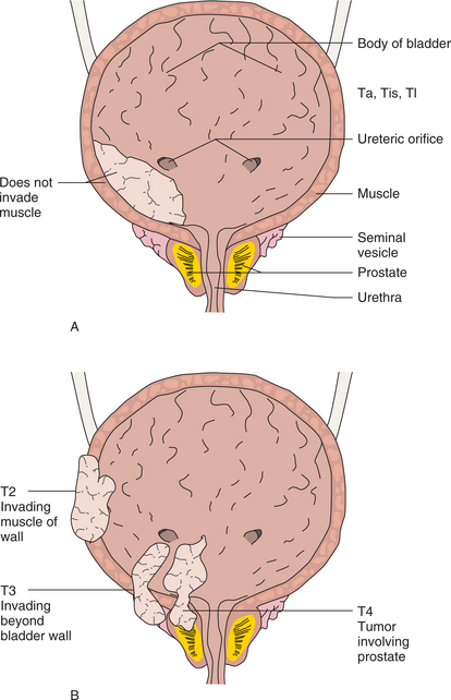
Figure 18-8 Bladder cancer staging using the TNM method. A, In Ta, Tis, and T1 tumors, cells do not invade muscle. B, If the muscle is involved, the tumor is staged as T2. T3 tumors invade beyond the bladder wall but do not involve other organs. T4 tumors are locally invasive to outside structures such as the prostate as shown or systemically with distant metastases (not shown).

Once tumor is invasive, it has the capacity to metastasize and commonly first reaches lymph nodes near the bladder. The presence of lymph node involvement, number of lymph nodes affected, and distance of involved lymph nodes from the bladder determine the N categorization (N0 for no involvement, N1 for nodes near the bladder, and N2 for nodes further away). Distant sites of metastasis include the liver, lung, and bone. The stage is determined by combining the T, N, and M status (e.g., stage 1 is T1, N0, M0). About 74% of bladder cancer is diagnosed as T1 or T2 tumors; 19% is T3; and 3% is T4.

TREATMENT.

Treatment of bladder cancer is determined on the basis of the stage of the tumor and the person’s general health. Surgery is the principal treatment for bladder cancer. Transurethral resection (TUR), surgery completed through the urethra using a rigid cystoscope called a resectoscope, is performed for early and superficial tumors. After the removal of the lesion, the tumor bed is treated either with a high-beam laser or fulguration (electric current used to destroy tumor tissue). Most clients can return home the same day or the next day. Complications include bleeding and discomfort. Long-term effects of repeated TUR include fibrosis of the bladder and loss of continence.

Cystectomy is performed for invasive bladder cancer. If the tumor is small, a partial cystectomy may be performed in order to salvage functioning bladder tissue.103 This approach is controversial, with some urologists preferring cystectomy even for selected clients with small tumor mass. A radical cystectomy is the surgery of choice for larger, invasive tumors or multiple tumors. This procedure removes the bladder and adjacent lymph nodes. The prostate is removed in men, and the uterus, ovaries, and a portion of the vagina are removed in women.

Following cystectomy, reconstructive surgery is performed to create a urine drainage system to compensate for the loss of the bladder. A urostomy procedure allows drainage of urine into a bag outside the abdomen. This is the least preferred long-term method of draining urine. The surgery that uses a short piece of small or large intestine to create a pouch or conduit from the ureters to the outside of the body is called an ileal conduit procedure.

Another surgical option is the creation of a continent diversion. In this procedure, the reserve pouch (intestine) has a valve. This valve allows urine to be stored until a catheter is placed to drain the urine. Newer reconstructive methods create a “neobladder” from intestine, which is then attached to the urethra. This surgery allows clients the ability to urinate normally, using a Valsalva maneuver (increasing intraabdominal pressure). The surgical complications from these types of procedures include infection, urine leakage, and obstruction. Sexual side effects are common, particularly for men. Impotence has been an issue with radical cystectomies in the past, but newer techniques have reduced the risk of nerve damage. If impotence does occur, function can improve with time. Generally, younger men (under the age of 60) are more likely to regain function than older men.

Chemotherapy is administered in two ways: intravesically or systemically. Intravesical chemotherapy is given through a catheter into the bladder, directly affecting the lining of the bladder. Chemotherapy agents are not absorbed into the deep layers of the bladder; therefore, intravesical chemotherapy is effective only for superficial cancers. Systemic chemotherapy is usually given in combination for invasive cancer (such as methotrexate, vinblastine, doxorubicin, and cisplatin). Radiotherapy in conjunction with concurrent chemotherapy is recommended less frequently because of the long-term consequences, although it is an option for treating clients unable to tolerate surgery because of health issues.

Another therapy option for treating low-stage bladder cancer is intravesical immunotherapy with bacillus Calmette-Guérin (BCG; a bacterium sometimes used to immunize people against tuberculosis). BCG is administered through a catheter into the bladder. The immune system responds to the BCG and becomes activated. These activated cells are then believed to recognize the cancer cells as foreign and destroy them. BCG is typically administered on a weekly basis for 6 weeks. BCG may reduce the risk of recurrence by as much as 50%. Side effects include flulike symptoms and a burning sensation in the bladder. Rarely, the bacterium can spread into the bloodstream, causing sepsis.

Stage 0 and 1 tumors are treated with TUR followed by intravesical BCG. If the tumor does not respond to the BCG, intravesical chemotherapy is administered, although either BCG or intravesical chemotherapy can be used first.68 However, over half of the people with stage 1 tumors will have a recurrence, and 20% to 30% will have a cancer that is invasive.

Stage 2 is treated with a radical cystectomy, with or without lymph node removal. Selected people may undergo a partial cystectomy with fulguration. Systemic chemotherapy may be given prior to or after surgery to treat any small micrometastases not seen during the staging process. A radical cystectomy is also the treatment for stage 3.

Chemotherapy may be added prior to surgery (neoadjuvant) to improve survival, but further investigations are needed to determine the appropriate timing for chemotherapy.94 Stage 4 therapy focuses on quality of life and slowing tumor growth. A radical cystectomy may be performed with chemotherapy if there are no distant metastases. Various combinations of therapy may be employed when distant metastases are present.

PROGNOSIS.

Despite the continued increase in the number of new cases occurring each year, the mortality attributed to bladder cancer has remained fairly stable. Stage and grade of the tumor are prognostic indicators for local failure. The 5-year survival rates with treatment are 84% for white males, 71% for black males, 76% for white females, and 51% for black females (most likely reflecting delay in diagnosis and inadequate care due to socioeconomic issues). Individuals with T1 tumors have a 90% 5-year survival, while for those with muscleinvasive tumors survival at 5 years is 50%; those with deep muscle invasion will go on to have metastatic disease within 2 years.5

Bladder cancer is sensitive to chemotherapy and immunotherapy but also has a high incidence of local recurrence, usually within the first 2 years. In approximately 30% of cases metastasis develops during the course of the disease, and 50% of individuals with muscle-invasive disease at the time of diagnosis already have distant metastases. Although rare, long-term survival with recurrent cancer can be achieved in some individuals. Continued improvements in the management of bladder cancer will improve the prognosis in the future.

18-7   SPECIAL IMPLICATIONS FOR THE THERAPIST

Bladder Cancer

The risk of severe late radiation sequelae is low (less than 5%), and about 75% of long-term survivors maintain a normally functioning bladder. The therapist may likely treat those individuals who have residual bladder control problems. The high rate of cancer recurrence requires therapists to be vigilant in observing for the onset or return of symptoms and signs suggestive of urogenital system disease or metastatic spread. Anyone reporting visible blood in the urine must be evaluated further by a physician.

Neurogenic Bladder Disorders

Overview

Voiding dysfunction associated with neurologic pathology is termed a neurogenic bladder disorder. There are many types of voiding dysfunction that can interfere with normal urine storage and coordinated, voluntary release.

Voiding dysfunction associated with neurologic pathology can be classified using one of many descriptive systems available. Each categorization scheme has advantages and disadvantages. For example, the Bors-Comarr Classification is well suited for clients with voiding dysfunction secondary to spinal cord injury but not as useful for clients with other problems.

Urodynamic classification correlates urodynamic findings and symptoms, while the International Continence Society Classification separates storage and voiding abnormalities and expands many of the urodynamic classification categories. The Lapides Classification is well known and helpful to nonurologists, so will be used as the classification system for this section. It correlates cystometry findings with clinical symptoms.

This classification system separates voiding dysfunction into five categories: (1) sensory neurogenic bladder, (2) motor paralytic bladder or motor neurogenic bladder, (3) uninhibited neurogenic bladder, (4) reflex neurogenic bladder, and (5) autonomous neurogenic bladder. This system provides a framework for understanding neurogenic bladder disorders, particularly for the nonurologist, but applies only to those disorders with a neurologic basis for pathology. Many clients may demonstrate a mixture of sensory and motor abnormalities, and symptoms may overlap between categories.

Incidence

Voiding dysfunction is a common problem associated with many types of neurologic diseases. In the United States, over 91,000 people are released from the hospital each year with a neurologic disease or spinal cord injury.30 Voiding dysfunction is costly and leads to significantly decreased quality of life, particularly in the older adult when long-term care is often considered for this problem in an otherwise healthy adult.

Etiologic Factors

The common disorders that can result in neurogenic bladder dysfunction include cerebrovascular accident, dementia, Parkinson’s disease, multiple sclerosis, and brain tumors. Neurogenic bladder dysfunction can also occur secondary to spinal cord lesions such as spinal cord injury, herniated intervertebral disc, vascular lesions, spinal cord tumors, and myelitis.

Local pelvic irritation can result in spasm of the external bladder sphincter, impairing urinary function. The local irritation can occur in the presence of vaginitis, perineal inflammation, urethral inflammation, and chronic prostatitis. Hypotonic (flaccid) bladder dysfunction can be secondary to meningomyelocele, spina bifida, and diabetes mellitus.

Pathogenesis

The process of micturition involves a complex interplay of nerves. Proper functioning of these nerves is needed for voiding to occur. The normal structures involved in micturition include the brain, the spinal cord, and nerves to the bladder. The type of voiding dysfunction that occurs is dependent on the underlying cause and which nerves are affected (Box 18-4).

Box 18-4   TYPES OF MICTURITION PATHOLOGY

• Sensory neurogenic bladder occurs when there is a disruption of the nerves between the bladder and the spinal cord or the afferent nerves to the brain. Diabetes, tabes dorsalis (from syphilis), and pernicious anemia (vitamin B12 deficiency) are the most common causes. Initial changes include an abnormal sensation in response to bladder distention. Affected people will not recognize the need to void, and unless frequent voiding is instigated, the bladder becomes chronically distended. This eventually leads to bladder hypotonicity with urine retention.

• Motor paralytic bladder results from the destruction of the parasympathetic motor nerves that innervate the bladder. This may occur with extensive pelvic surgery or trauma. Clinical symptoms can initially vary, ranging from a mild inability to initiate or maintain a urine stream to painful urine retention. Like sensory neurogenic bladder, with chronic bladder overdistension, motor paralytic bladder results in a distended bladder with large-volume urine retention.

• Uninhibited neurogenic bladder refers to damage of the corticoregulatory tract. This nomenclature is somewhat outdated; the term is no longer used to classify bladder dysfunction but still may be found in the literature. It was presumed that the regulatory site for reflex bladder control was located in the sacral spinal cord or the micturition reflex center. If there was damage to the corticoregulatory tracts, there was, in turn, a disinhibition of the micturition center, leading to incontinence. Conditions classified as leading to this type of neurogenic bladder included cerebrovascular accidents, brain or spinal cord tumors, Parkinson’s disease, demyelinating diseases, and brain tumors. Clinical symptoms included frequency, urgency, and urge incontinence. Bladder sensation is normal but involuntary contractions occur at urine low volumes. Voluntary contractions can be initiated by the affected person but the capacity to store urine is decreased.

• Reflex neurogenic bladder describes the condition of the bladder following a spinal cord injury or “postspinal shock.” This occurs when there is a complete disruption between the sacral spinal cord and the brainstem, as seen with traumatic spinal cord injury or transverse myelitis.

    Other disorders or disease processes that cause significant demyelination of the spinal cord can also lead to reflex neurogenic bladder. The bladder lacks sensation and the person is unable to determine when the bladder is distended. Affected people also are unable to initiate micturition (voiding) and develop sphincter dyssynergia incontinence (the external sphincter tightens during micturition as the detrusor muscle is contracting, resulting in increased intravesicular pressure and vesicoureteral reflux).

• Autonomous neurogenic bladder refers to the complete separation of sensory and motor nerves of the bladder from the spinal sacral cord. Damage to the sacral roots or cord or the pelvic nerves can lead to this type of voiding dysfunction. Clients with this problem are unable to initiate voiding and there is no bladder sensation, leading to large-volume bladder capacity and distension.

    This type of voiding dysfunction can be seen in clients with spinal shock. Initial cystometric findings can be similar to those in the late stages of motor or sensory paralytic bladder (large capacity with low bladder pressure), but with continued inflammation and nerve damage, the bladder can lose capacity and compliance.

The micturition reflex center is located in the brainstem. The efferent (exiting) neurons travel down the spinal cord in the reticulospinal tract to the detrusor muscle of the bladder. Parasympathetic nerves originate in the spinal cord at the level of S2, S3, and S4 and innervate the bladder wall via the pelvic nerve.

Preganglionic sympathetic nerves have their origin in the spinal cord at the levels of T10 through L2 and travel through the sympathetic chain ganglion to the bladder neck and fundus. The bladder neck contains the internal urethral sphincter (involuntary muscle). The external urethral sphincter (voluntary muscle) is innervated by the pudendal nerves, which originate in the spinal cord at the level of S2 through S4.

Damage to nerves involved in micturition can result in different types of voiding dysfunction. Cerebral injury (above the micturition reflex center) leads to loss of voluntary inhibition of voiding and a hyperreflexic bladder, but coordinated sphincter function is retained. This results in the ability to completely void, but because of the hyperreflexic bladder, incontinence occurs. This can be seen in brain tumors, cerebral palsy, cerebrovascular accidents, dementia, Parkinson’s disease, pernicious anemia, and Shy-Drager syndrome.

Lesions in the region of the micturition center to S2 result in loss of voluntary inhibition and coordinated sphincter activity. Since the sphincters are unable to coordinate their activity (the sphincter remains closed although bladder contractions occur), high pressure along with ureteral reflux result, termed dyssynergic sphincter function (detrusor-sphincter dyssynergia). This leads to urinary retention and incomplete voiding. Diseases that can result in this type of voiding problem include anterior spinal cord lesions, ischemia, multiple sclerosis, myelodysplasia, and trauma.

Complete spinal cord injury above T5 or T6 leads to autonomic dysreflexia (loss of sensation of bladder distension results in overdistension) and detrusor-sphincter dyssynergia. This type of injury also involves the sympathetic nerves and loss of sympathetic inhibition, leading to systemic sympathetic symptoms such as hypertension, facial flushing, perspiration, and headache. Since the vagal nerve is intact, bradycardia accompanies this syndrome.

Spinal cord lesions at the level of S2 and below lead to bladder areflexia and dysfunction of the external sphincter. Since parasympathetic nerves are not affected (ganglia are in or near the bladder wall), bladder tone is preserved, but bladder compliance decreases with time (secondary to repeated infection, fibrosis, and changes in innervation).

The external sphincter maintains some tone but the bladder neck does not relax, leading to obstructive problems when voiding (overflow incontinence). Acute transverse myelitis, diabetes, Guillain-Barré syndrome, herniated intervertebral disc, myelodysplasia, pelvic surgery, tabes dorsalis (syphilis), and trauma can cause this type of neurogenic bladder.

Diabetic bladder neuropathy occurs in 43% to 87% of people with type 1 diabetes mellitus and 60% to 75% of people with type 2 diabetes.63,104 The actual neurologic damage and symptoms vary among clients with diabetes and include diabetic cystopathy (impaired bladder sensation, increased postvoid residuals, increased bladder capacity, and decreased bladder contractility), detrusor overactivity, bladder outlet obstruction (seen in men), and urge and stress incontinence. Diabetic bladder neuropathy occurs when other diabetic complications are apparent (e.g., diabetic retinopathy, microalbuminuria).

Clinical Manifestations

Neurogenic bladder dysfunction is manifested by partial or complete urinary retention, incontinence, urgency, suprapubic pain, or frequent urination. Common complications include UTIs, kidney stones, and deterioration in renal function.

MEDICAL MANAGEMENT

DIAGNOSIS.

Numerous tests can be used to assess the anatomic and physiologic status of the bladder, associated structures, and nervous system. Urodynamic testing includes many types of urologic studies and is frequently performed to categorize abnormalities and determine cause and most appropriate treatment.

Urodynamic studies include uroflowmetry, cystometry, urethral pressure studies, pressure-flow micturition studies, electrophysiologic studies, and video urodynamic studies. All or a portion of these tests can be performed as appropriate. If spinal cord or brain abnormalities are suspected, an MRI is beneficial.

Many diseases lead to voiding dysfunction, and a complete history and physical can often reveal the cause. Other tests can be performed as needed to diagnose these disease processes.

TREATMENT.

The primary goals of treatment include preventing incontinence, bladder overdistention, UTIs, and renal damage. Treatment modalities include catheterization, pharmacologic agents, bladder training, and surgery.

Clean intermittent catheterization is a commonly employed intervention to avoid bladder overdistention. It is usually performed at 4-hour intervals and aids in reducing the risk for vesicourethral reflux and kidney damage. Permanent indwelling catheters are used only in specific medical situations, and alternatives should be used as possible.

Indications for short-term indwelling urinary catheter include the following: (1) for accurate monitoring of urine output, (2) for relief of urinary obstruction, (3) for prevention of obstruction from large clots when hematuria is present, (4) for surgical procedures involving general or spinal anesthesia, and (5) for incontinence when pressure ulcers are present.

Although a medical necessity in certain situations, permanent indwelling catheters carry a risk. UTIs, urethral irritation, epididymo-orchitis, pyelonephritis, renal calculi, and cancer have all been associated with the use of permanent indwelling catheters in people with spinal cord injury.41,201 Although associated with fewer adverse effects, intermittent catheterization can lead to reduced quality of life.139

Catheterization is frequently used in conjunction with medications. Anticholinergic agents are used to treat voiding dysfunctions that include detrusor hyperactivity. These agents relax the bladder, reduce high pressures, and increase bladder capacity. Side effects include dry mouth, gastrointestinal disturbances, drowsiness, cognitive impairment, hallucinations, and delirium.

While a relatively new treatment, botulinum toxin A injections into the detrusor muscle appear to reduce involuntary bladder contractions in people with neurogenic detrusor overactivity.4,101 Long-term studies are needed to determine appropriate duration and efficacy of treatment.

Bladder training methods are designed to enhance bladder function and prevent complications. Adequate fluid intake is important for the prevention of infection and overconcentrated urine. With a hyperreflexive bladder or detrusor-sphincter dyssynergia the abnormally concentrated urine can stimulate afferent nerve endings, exacerbating the bladder disorder. This could increase vesicular pressures, vesicoureteral reflux, and overflow incontinence.

Fluid intake must be controlled and monitored to prevent bladder distention. Biofeedback techniques using electromyography or cystometry help the person control external sphincter function or increase intravesicular pressure sufficiently to overcome outflow resistance.

A variety of surgical interventions for neurogenic bladder exist, although due to the invasive nature of the surgery, it is often utilized after more conservative methods have failed. Procedures include bladder augmentation cystoplasty (colon, ileus, stomach, or ureter can be used); cystectomy with or without continent diversion; ureteral and bladder neck suspension; artificial urinary sphincter implantation115; ileovesicostomy, ileal conduit, or placement of suprapubic catheters; denervation procedures and electrostimulation (for complete lesions); and sacral nerve neuromodulation (for incomplete lesions).

18-8   SPECIAL IMPLICATIONS FOR THE THERAPIST

Neurogenic Bladder Disorders

PREFERRED PRACTICE PATTERNS

See also Special Implications for the Therapist: Urinary Incontinence later in the chapter.

5C:

Impaired Motor Function and Sensory Integrity Associated with Nonprogressive Disorders of the Central Nervous System—Congenital Origin or Acquired in Infancy or Childhood

5D:

Impaired Motor Function and Sensory Integrity Associated with Nonprogressive Disorders of the Central Nervous System—Acquired in Adolescence or Adulthood

5F:

Impaired Peripheral Nerve Integrity and Muscle Performance Associated with Peripheral Nerve Injury (flaccid bladder)

5H:

Impaired Motor Function, Peripheral Nerve Integrity, and Sensory Integrity Associated with Nonprogressive Disorders of the Spinal Cord

Therapists provide care for many people who have sustained spinal cord injuries and cerebrovascular accidents or who have myelomeningoceles, multiple sclerosis, or brain tumors. Neurogenic bladder disorders are usually only one of the complications associated with these conditions, but familiarity with this complication is important. The potential for UTIs, renal calculi, and renal damage is high in those with neurogenic bladder disorders.

The development of any of these comorbidities can interfere with the rehabilitation process. Several medical conditions such as UTI, diabetes, congestive heart failure, bladder cancer, and enlarged prostate can be mistaken for a bladder control problem. Familiarity with the signs and symptoms associated with these potential diseases is a necessity. Detection of any of these symptoms warrants communication with a physician.

Incontinence associated with any of the bladder conditions discussed here can be greatly improved and even eliminated in many people through a program of exercise and behavioral intervention. Specific guidelines are available.91,92,185,197

Urinary Incontinence

See the section on Pelvic Floor Disorders in Chapter 20.

Definition and Overview

UI may be defined as an involuntary loss of urine that is sufficient to be a problem and occurs most often when bladder pressure exceeds sphincter resistance.50 The following four categories can be used to classify UI:

1. Functional incontinence occurs in people who have normal urine control but who have difficulty reaching a toilet in time because of muscle or joint dysfunction.

2. Stress incontinence is the loss of urine during activities that increase intraabdominal pressure such as coughing, lifting, or laughing.

3. Urge incontinence is the sudden unexpected urge to urinate and the uncontrolled loss of urine. Urge incontinence is often related to reduced bladder capacity or detrusor instability (the latter is also referred to as overactive bladder, hyperreflexive bladder, detrusor-sphincter dyssynergia, or detrusor hyperreflexia).

4. Overflow incontinence is the constant leaking of urine from a bladder that is full but unable to empty.

Some people can have more than one type of incontinence (most often stress and urge incontinence together), known as mixed incontinence. Postsurgical incontinence refers to the incontinence a person can develop following urologic surgery.

UI is common, particularly in older adults. Approximately 10% to 35% of community-dwelling and 50% to 60% of nursing home adults have incontinence. Yet the condition is poorly understood, underdiagnosed, and often inadequately treated. Many people are embarrassed to acknowledge that they are incontinent. Only 20% to 50% of incontinent adults seek medical care.199 Others regard incontinence as part of the normal aging process. The economic price tag associated with UI is estimated to be greater than $26 billion per year in the United States.90 Incontinence can be a significant contributory factor related to falls in older adults,19 pressure sores, UTIs, institutionalization, depression, and isolation.

Prevalence

UI is more prevalent in women than men and in the aging over the young. The prevalence of UI was first reported in 1988 at the National Institutes of Health Consensus Conference on Adult Urinary Incontinence. At that time, an estimated 10 million adults in the United States experienced incontinence, including 15% to 30% of community-dwelling older adults and more than 50% of nursing home residents.33

Others cite a range of 13 to 25 million individuals in the United States.50 In fact, it is estimated that 50% of all admissions to skilled nursing facilities today are a direct result of UI, and another large portion of affected individuals are homebound.

Since the first consensus conference, an increased awareness of and focus on the problem of UI has come about. Until recently, the impact of UI on working women employed full time, a population generally characterized as healthy, has not been the focus of research. One survey at a large university center reported that 21% of the women surveyed (age 18 and older) reported UI at least monthly.55

Risk Factors

A wide range of factors can contribute to the increased risk of developing UI (Box 18-5). Some risk factors are more likely to lead to one type or another or several types of incontinence. Since women are more likely than men to develop UI, there are several factors associated with childbirth and gynecologic surgery. Women who have had multiple pregnancies and deliveries (whether cesarean section or vaginal birth) have a higher incidence of UI. Hysterectomy, the presence of a cystocele, and uterine prolapse may also increase a woman’s risk.

Box 18-5   RISK FACTORS FOR URINARY INCONTINENCE

• Altered local anatomy and physiology

• Pelvic floor muscle weakness (e.g., pregnancy/multiple pregnancies; childbirth/delivery [vaginal or cesarean section]; episiotomy; high fetal gestational weight; any pelvic surgery, including hysterectomy for women, prostatectomy for men)

• Cystocele or uterine prolapse

• Congenital sphincter muscle weakness or damaged sphincter muscle

• Pudendal nerve damage (e.g., childbirth trauma, pelvic surgery, radiation)

• Neurologic disorder (e.g., myelomeningocele, multiple sclerosis, brain injury, Parkinson’s disease, cerebral palsy, spinal cord injury, stroke)

• Psychogenic (e.g., childhood and/or adult sexual trauma for both males and females, negative sexual experiences, emotional stress)

• History of benign prostatic hyperplasia (BPH)

• Constipation, fecal impaction

• Tobacco use

• History of recurrent urinary tract infections

• Medications

• α-Adrenergic blockers (antihistamines, decongestants)

• Antibiotics

• Antiparkinson agents

• Diuretics

• Hormone replacement therapy (estrogen plus progestin in postmenopausal women)

• Hypertensives

• Tranquilizers, sedatives

• Tricyclic antidepressants

• Decreased estrogen (deficiency)

• Bladder irritation

• Caffeine

• Alcohol (?)

• Restrictive clothing, restraints

• Loss of activities of daily living skills for toileting

• Decreased or impaired mobility

• Impaired cognitive function

• Radiation therapy

• Obesity

• Race (Caucasian)

• Higher socioeconomic status

Consistent evidence shows that frequency of UI (particularly urge incontinence) increases with age. One large study showed that the prevalence of urge and mixed incontinence increased with age, while stress incontinence was not related to advancing age. This may be because the prevalence of stress incontinence remained unchanged.16

Medications commonly prescribed for other illnesses can also increase the risk of incontinence. These pharmacologic agents can interfere with conscious inhibition of voiding, induce a quick diuresis (diuretics),131 produce urinary retention, or reduce urethral resistance to the point of stress incontinence. Tranquilizers and sedatives may impair awareness of the usual cues related to urinary urgency and may also depress the cerebral corticoregulatory tract, affecting detrusor muscle activity.117 Drugs producing anticholinergic side effects (e.g., major tranquilizers and antiparkinsonian agents) may cause urinary retention because of failure of bladder contraction. Laxatives, estrogen, and antibiotics are also associated with an increased risk for UI.54,86,168

Although estrogen deficiency is a risk factor for UI, hormone replacement, including estrogen plus progestin, in healthy postmenopausal has been linked with incontinence.75,162 Topical estrogen can be used if incontinence is due to atrophic vaginitis or severe vaginal atrophy.

High caffeine intake (more than 400 mg/day) can also contribute to the development of urge incontinence, while studies involving alcohol consumption have reported mixed results.64,171 Constipation and other bowel problems increase the risk of UI, which may be related to pelvic organ prolapse.26 Recurrent UTI is also an independent risk factor for developing UI, as is increased body mass index (BMI). Obesity is a common condition among women in developed countries and is believed to have a major impact on stress incontinence. The proposed mechanisms for this association include the increased intraabdominal pressures that adversely stress the pelvic floor and the effect of obesity on the neuromuscular function of the genitourinary tract.35

Race and socioeconomic class may also play roles in the development of UI. It appears that Caucasian women are more likely to have UI than African American women, while women from a higher socioeconomic class have an increased risk of UI. The risk associated with smoking is unclear, but smoking may be an independent risk factor for UI.

Overflow incontinence can occur as a result of conditions that cause urethral or bladder neck obstruction, such as benign prostatic hyperplasia (see Chapter 19), fecal impaction, or organ prolapse (see Figs. 20-9 and 20-10).

Lastly, as older adults lose their mobility and manual dexterity secondary to a multitude of ailments, getting to the bathroom or commode in a timely fashion and manipulating clothing become increasingly difficult. The presence of two or more diseases significantly increases the likelihood of developing UI. Common illnesses associated with UI include diabetes, stroke, hypertension, cognitive impairment, parkinsonism, arthritis, and hearing and visual impairments.

Pathogenesis and Clinical Manifestations

Incontinence, as discussed earlier, is categorized depending on the cause and pathophysiology. Each type of UI has associated clinical manifestations. Mixed incontinence, particularly urge and stress incontinence, is common, with overlapping symptoms from both types of incontinence.

Functional incontinence lacks a pathologic origin and is the consequence of chronic impairments of physical or cognitive function that make toileting in a timely fashion difficult.

Urge incontinence is often caused by involuntary bladder (detrusor) spasms and is associated with both increased frequency and urgency. Normal bladder control is a minimum of 2 hours and usually gauged as 3 to 5 hours without toileting. Urinating more than eight times a day and/or two or more times a night is a symptom of urge incontinence.

Normal urination involves signals from the cortex of the brain, through the pons, spinal cord, peripheral autonomic system, sensory afferent innervation of the lower urinary tract, and finally to the bladder itself. Normally, the bladder is designed to hold a pint of urine for several hours. As it fills, the detrusor muscle relaxes to allow the bladder to stretch and accommodate more urine; the sphincter contracts to prevent urine from escaping through the urethra.

Pelvic floor muscles also support the base of the bladder and close off the bladder end of the urethra, further blocking the flow of urine. When the bladder is full, a message to get to the bathroom is received, at which time the sphincter and pelvic muscles relax, allowing urine to flow into the urethra, while the detrusor muscle contracts to squeeze the urine out of the bladder.

Because much of this system involves the nervous system, overactive bladder is often associated with neurologic conditions such as stroke, multiple sclerosis, advanced diabetes, spinal cord injury, and dementia. Urge incontinence is characterized by leakage (sometimes large-volume accidents) after a sudden precipitant urge to urinate or by events such as trying to insert a key in the door, running hands under water (or hearing running water), or passing by a bathroom.

Stress incontinence results from weakness or loss of tone in the pelvic floor muscles, urethral sphincter failure, hypermobility of the ureterovesical junction, or damage to the pudendal nerve (e.g., tumor, childbirth) (see Figs. 20-7 and 20-8). It is accompanied by leakage that is coincident with increases in intraabdominal pressure (e.g., coughing, sneezing, laughing, bending, high-impact physical activity or exercise). Someone with mixed urge and stress incontinence may exhibit signs of both types.

Other clinical manifestations, depending on the underlying pathology, may include constant dribbling, leakage without warning, frequency, urgency, nocturia, hesitancy, weak stream, or straining to void. Prolapsed bladder, uterus, and/or bowel may accompany or contribute to leakage, especially when caused by multiple pregnancies and deliveries.

Overflow incontinence is usually the result of a neurologic problem such as hypotonic or underactive detrusor secondary to drugs, fecal impaction, diabetes, lower spinal cord injury, or multiple sclerosis. It can also occur as the result of obstruction (e.g., prostatic hyperplasia, genital prolapse, or surgical overcorrection of urethral detachment in women). In this situation, as the bladder fills and becomes overdistended, the pressure inside the bladder finally exceeds the maximal urethral pressure and urine “overflows.”

Overflow incontinence often manifests with symptoms of stress and urge incontinence and is characterized by frequent or constant dribbling, inability to completely empty the bladder, hesitancy, weak stream, need to strain to void, bladder distension, and urinary urgency and frequent urination. Anticholinergics, narcotics and α-adrenergic agonists can worsen these symptoms.

MEDICAL MANAGEMENT

PREVENTION.

Preventive education is the key to eliminating a large majority of incontinence. Many health care professionals advocate early education for adolescent girls and young women before they become pregnant. Prepartum and postpartum pelvic floor muscle training has been shown to have immediate and long-term effects in preventing incontinence, improving quality of life, and improving sexual dysfunction.11,133,134

Proper instruction in pelvic floor exercise emphasizing both fast-twitch and slow-twitch muscle fibers is an important part of prevention. Brief verbal or written instruction in performing a Kegel pelvic muscle contraction is not adequate preparation as measured in more than 50% of cases investigated.7 A properly performed Kegel exercise should result in a significant increase in the force of the urethral closure without an appreciable Valsalva effort. Improperly done, the Kegel technique can potentially promote incontinence.

The best time to explain a Kegel exercise is during a pelvic examination. The medical practitioner can describe the exercise and verify that it is done correctly during the examination. A trained practitioner, such as a physical therapist, can also provide follow-up assessment and training with necessary biofeedback to ensure the success of a properly performed exercise program. This may be particularly helpful in older adults, who often have a difficult time localizing pelvic muscles.

Avoiding constipation through proper nutrition, adequate hydration, and responding to the need to toilet should be part of the health curriculum for children. The therapist can be very instrumental in establishing screening programs for all ages to assess for risk factors for UI or for the presence of incontinence and teaching proper lifting techniques, lifestyle management, behavioral techniques, and pelvic floor protection during increased abdominal pressure (e.g., lifting, laughing, coughing, sneezing, vomiting). Anyone experiencing leaking during exercise needs a prescriptive exercise program both for the leaking and modifying the exercise that precipitates the leaking.

DIAGNOSIS.

Since UI is related to a wide variety of disorders and factors, a detailed investigation may be necessary to determine the cause(s). An important part of this investigation is a voiding diary to determine the frequency, timing, and amount of voiding and to assess the numerous other risk factors potentially associated with incontinence.50

A careful history will investigate medication usage, prescribed and OTC; past and current illnesses; and surgical and birth histories. Physical examination should include a pelvic, genitourinary, and rectal examination to evaluate for the presence of prolapse, fecal impaction, atrophic vaginitis, cystocele, masses, prostate hypertrophy, and prostate nodule.

Urinalysis may reveal hematuria or infection. In order to differentiate between urge and overflow incontinence, a postvoid residual should be obtained. If obstruction is not present, the bladder should contain less than 100 ml of urine after voiding. Many of the cases of incontinence can be diagnosed without significant invasive techniques and treated by modifying reversible risk factors. The mnemonic DIAPPERS summarizes the reversible causes of incontinence: D for delirium, I for infection, A for atrophic urethritis/vaginitis, P for pharmaceutical, P also for psychologic disorders, E for excessive urine output (associated with congestive heart failure or hyperglycemia), R for restricted mobility, and S for stool impaction.194

Clients who have abnormal physical or laboratory findings may require further workup, particularly if surgery is planned or the diagnosis is in doubt. Cystoscopy is beneficial in the presence of hematuria to assess for bladder cancer. Urodynamic evaluation is used in select clients who may have nonspecific symptoms or for whom a more precise diagnosis of obstruction is needed. Cystometry is used to assess bladder capacity, sensation, voluntary control, and contractility. Cystometry consists of filling the bladder with water or carbon dioxide and recording changes in intravesicular pressure. When stress incontinence is suspected, provocative stress testing is carried out. The client is asked to cough vigorously while the examiner observes for urine loss. The test is initially done in the lithotomy position but if the result is negative, is repeated in a standing position.50 Dynamic MRI is being investigated to study specific aspects of voiding associated with stress incontinence.10,83

TREATMENT.

Management of UI depends on the type of incontinence and the person’s age and general health but usually falls into one of three categories: behavioral, pharmacologic, and surgical. Behavioral intervention is considered the first line of treatment for pelvic muscle rehabilitation and includes a combination of lifestyle and dietary changes, prescriptive exercises including Kegel exercises and Beyond Kegel exercises,92 pelvic floor electrical stimulation, biofeedback therapy, support devices such as pessaries, and vaginal weight training.

Pessaries are devices inserted into the vagina that are designed to support the bladder and bladder neck. These devices come in a variety of shapes and sizes and are made of flexible or rigid silicone, latex, or acrylic. Some pessaries can stay in the vagina for up to 3 months before being removed, cleaned, and replaced; others are used just during exercise or sexual intercourse. A properly fitted pessary should not interfere with bowel or bladder function and should not be uncomfortable.

A new technology using noninvasive pulsed magnetic fields (extracorporeal magnetic innervation) has recently been used in the treatment of stress and urge incontinence, with mixed results.66,196,208

Functional Incontinence.

Functional incontinence occurs from mobility and access deficits, such as being confined to a wheelchair or needing a walker to ambulate. Deficits in dexterity, such as weakness from a stroke or neuropathy and loss of motion from arthritis, may keep the individual from getting pants unfastened or underpants pulled down in time to avoid an accident. Altered mentation from dementia or Alzheimer’s disease can also contribute to untimely urination without a urologic structural problem.

Because this type of incontinence has no urologic pathology but is due to other factors, treatment is aimed at correcting or improving the underlying problems. For example, if physical impairments make mobility difficult, thus affecting the client’s ability to reach a bathroom, appropriate devices (i.e., bedside commode) and therapy can be provided. Clothing should be easy to remove.

Physical and occupational therapists can often help improve mobility skills. Behavioral strategies for scheduled voiding and assistance may help avoid accidents. If cognitive impairment is a factor, prompted voiding can decrease the number of incontinence episodes. In extended care facilities, a toileting assistance program can be established.

Stress Incontinence.

Stress incontinence can be treated by behavioral and surgical methods. Behavior modifications include pelvic floor muscle training (Kegels, Beyond Kegel exercises),92 with or without biofeedback and electrical stimulation. A recent Cochrane review reported pelvic floor muscle training to be effective for the treatment of stress or mixed incontinence.81

Studies evaluating electrical stimulation show only slight improvement over placebo, while the use of vaginal cones was found to be inferior to pelvic floor training.80 Self-positioning pessaries are available as an alternative to pads and surgery.51

Surgeries that may be used to correct pelvic floor laxity include an open retropubic colposuspension, anterior vaginal repair, bladder neck needle suspension, and suburethral sling procedure. Surgical correction of a cystocele or uterine prolapse may improve symptoms. Periurethral injections with a bulking agent (such as collagen) may be administered to increase sphincter resistance in women and in men who have stress incontinence due to postprostatectomy changes. Artificial urinary sphincters can be considered but have a cure rate of about 50% with high morbidity.69

Urge Incontinence.

Various interventions are used to treat urge incontinence, with the primary focus being behavioral modification. Biofeedback and pelvic floor training along with bladder retraining can significantly improve urge incontinence.206 Bladder training consists of scheduled voiding (trying to increase time between each void) and urge-suppression techniques. For clients with cognitive impairment, prompted voiding can be beneficial and decrease incontinent episodes (remind every 2 hours to toilet). Electrical stimulation has also been used with some success. Clients should also be encouraged to manage constipation and avoid caffeine.

Pharmacologic therapy is frequently used in conjunction with behavioral modifications. First-line drug therapy for urge incontinence is the use of anticholinergics or muscarinic antagonists, which inhibit involuntary detrusor contractions.82 The most common side effect is dry mouth. Other adverse effects include drowsiness, cognitive impairment, delirium, and hallucinations. These side effects are more likely to occur in older adults than younger clients. Tolterodine is associated with fewer adverse effects than oxybutynin.124

Other medications have shown some benefit but are not as effective as the anticholinergics/muscarinic antagonists or have off-label usage. Estrogens have been used to treat UI, although there are few data to support their use, and oral hormone therapy has been associated with increased incidence of incontinence. In men with symptoms of urge incontinence and prostatic hypertrophy, alfuzosin and tamsulosin (α-adrenergic antagonists) can be used initially. If these agents do not alleviate symptoms, obstruction should be ruled out before starting an anticholinergic agent.140

Overflow Incontinence.

Once the diagnosis of overflow incontinence is made, treatment then depends on the presence of obstruction or weak detrusor muscles. Surgery can be performed to alleviate obstruction (e.g., stricture dilation or transurethral prostatectomy). If a weak detrusor muscle is the cause, a few measures can be utilized to improve symptoms. One behavior modification is to have the client double-void. Medications such as cholinergic agonists or α-adrenergic antagonists are not typically efficacious in long-term use. If these measures are not appropriate or not effective, clean intermittent catheterization may be required.

18-9   SPECIAL IMPLICATIONS FOR THE THERAPIST

Urinary Incontinence

See the section on Pelvic Floor Disorders in Chapter 20.

PREFERRED PRACTICE PATTERNS

Selection of the most appropriate practice patterns depends in part on the underlying etiologic factors and pathogenesis of the UI.

4C:

Impaired Muscle Performance

4D:

Impaired Joint Mobility, Motor Function, Muscle Performance, and Range of Motion Associated with Connective Tissue Dysfunction

4I:

Impaired Joint Mobility, Motor Function, Muscle Performance, and Range of Motion Associated with Bony or Soft Tissue Surgery

5B:

Impaired Neuromotor Development (spina bifida, myelomeningocele)

5C:

Impaired Motor Function and Sensory Integrity Associated with Nonprogressive Disorders of the Central Nervous System—Congenital Origin or Acquired in Infancy or Childhood

5F:

Impaired Peripheral Nerve Integrity and Muscle Performance Associated with Peripheral Nerve Injury (pudendal nerve injury)

5H:

Impaired Motor Function, Peripheral Nerve Integrity, and Sensory Integrity Associated with Nonprogressive Disorders of the Spinal Cord

7A:

Primary Prevention/Risk Reduction for Integumentary Disorders

Therapists have an important direct role in the assessment and treatment intervention of UI. Physical therapy guides the rehabilitation of muscle imbalance and pelvic alignment and promotes pelvic muscle awareness and function through biofeedback, electrostimulation, therapeutic exercise, and a behavioral management approach.23,111

The pelvic rehabilitation program is designed to prevent recurrence of the impairment and to restore bowel, bladder, sexual, and supportive muscle functioning.155 Quality-of-life issues can be assessed using the Urge Impact Scale (URIS) for older people with urge incontinence.44

For some clients seen by therapists who do not specialize in pelvic floor rehabilitation, UI will be a comorbidity or a condition that has yet to be evaluated. Many adults think that incontinence is an inevitable part of aging and do not report the problem. Any perimenopausal or postmenopausal woman, any woman who has been pregnant, anyone (male or female) over the age of 60 (earlier if prostate or bladder infection or cancer is evident), and any person with multiple risk factors should be screened for UI.

Anyone with onset of incontinence with concomitant cervical spine pain (even without a history of trauma or known cause) may be experiencing cervical disc protrusion, requiring additional screening and evaluation. Additionally, the possibility of genitourinary disorders as a result of sexual abuse or assault exists and requires careful assessment.

The generally positive rapport that develops between client and therapist may facilitate the acknowledgment that UI exists and uncover the potential underlying risk factors. Specific questions should be included in the history to help bring this information to light, such as the following:

• Do you leak urine when you lift, cough, sneeze, or stand up?

• Do you get up at night to urinate (how often)?

• Do you go to the bathroom more often than every 2 to 3 hours?

• How much water do you drink in a waking day?

• Are you constipated?

The therapist may be in a position to direct the person to the appropriate physician for evaluation. Successful intervention in UI may enhance rehabilitation efforts geared to improve the client’s physical and social activity level. A variety of examinational, evaluative, and diagnostic tools and management options are available. The National Association for Continence (http://www.nafc.org) also offers a wide variety of information, and the Women’s Health Section of the American Physical Therapy Association (APTA) offers a broad range of information and courses on incontinence specific to the physical therapist.

For an internal pelvic floor assessment, including a manual muscle test and assessment of trigger points, a consent-to-treat form is recommended. This form should include an explanation of the internal examination and specifically addresses the client’s right to refuse or stop the evaluation at any time.

Some clinicians offer to have a third person in the room during the assessment, either a friend or relative of the client’s or another woman from the health care facility. Any unspoken behaviors or indication of discomfort on the part of the client should prompt the physical therapist to stop and communicate with the client before continuing or discontinuing. Issues of childhood incest or sexual assault or adult sexual issues may be a significant contributing factor, requiring combined pelvic muscle rehabilitation and psychotherapy or sexual abuse counseling.

Understanding the use of medications and potential side effects for these conditions is essential; many of the same medications used to treat incontinence can also cause incontinence if used inappropriately. Medications to treat other conditions can have side effects that cause incontinence. A concise listing of this information is available.92

Exercise and Incontinence

Studies to verify the effectiveness of exercise programs to strengthen pelvic muscles administered by nurses have provided evidence-based protocols.167 Several references for physical therapy–based intervention approaches are also available,11,133,134 but further research documentation of outcomes is needed.

Comparison of biofeedback and exercise programs administered by physical therapists showed equal effectiveness in reducing nocturnal urinary frequency and improved subjective outcome; only the group receiving physical therapy experienced reduced daytime urinary frequency.141

Although pelvic floor muscle exercises have proven effective in the short term, long-term noncompliance may hamper continued success. Predictors of compliance may include amount of urinary loss per wet episode and individual perception of the ability to do the exercises as recommended under various circumstances. This information should be taken into consideration during the education portion of any home program. Further studies to determine components of adherence behavior are needed.1

STRESS INCONTINENCE

Exercises can be performed to retrain and strengthen the pelvic floor musculature. This type of exercise has been advocated for decades.105 Exercise management, including adductor and obturator assist and “quick flick” exercises, can help develop adequate sphincteric function and improve bladder and bladder outlet position in the pelvis.

A quick flick exercise is a quick contraction followed by release with full relaxation of the pelvic muscles while the gluteals, abdominals, adductors, and obturator internus muscles remain relaxed. Quick flicks are designed to improve the strength and function of the fast-acting fibers, primarily of the urogenital diaphragm and external sphincter muscles. These fibers are important for prevention of leaking during coughing, sneezing, lifting, and pulling, because these fibers act with speed and intensity to maintain urinary control.92

Restoring normal pelvic floor strength and bladder control is essential before resuming vigorous physical activity or exercise. The therapist is very instrumental in teaching contraction of the appropriate muscles (e.g., the pelvic diaphragm, urogenital diaphragm, and external sphincter muscles) without muscle contraction in the anal area or of the gluteal muscles and with complete relaxation of the pelvic muscles between contractions.

Physiologic quieting including hand warming, diaphragmatic breathing, and body/mind quieting can be used to normalize bladder function and autonomic nervous system innervation of bladder and bowel.92 With verbal and manual cues, biofeedback (auditory, visual, or electronic), physiologic quieting, and electromyography (surface or intravaginal), the client can be taught to disassociate pelvic floor muscle activity from other hip and pelvic muscle activity and to maintain pelvic floor muscle tone while avoiding a Valsalva maneuver. Weighted vaginal cones and electrical stimulation can also be used to rehabilitate the pelvic floor.

URGE INCONTINENCE

Continence requires the ability to inhibit automatic detrusor contractions, and pelvic floor exercises facilitate the use of urge inhibition techniques. Pelvic floor rehabilitation specifically using pelvic floor muscle exercises for an overactive bladder may be able to suppress the desire to void by reducing detrusor and increasing urethral pressure, resulting in suppression of the micturition reflex.

Pelvic floor muscle contraction appears to prevent internal sphincter relaxation produced by the micturition reflex. Failure of the internal sphincter to relax seems to cause reflex detrusor relaxation, an action mediated through the voluntary urinary inhibition reflex.178

The bladder needs to be trained to respond to a specific voiding schedule. Clients void at scheduled intervals to suppress the micturition reflux, increase bladder capacity, and decrease urinary frequency. The initial retraining interval is usually 60 minutes. The voiding intervals are increased by 15-to 60-minute extensions using urge inhibition if it occurs too soon.

Success is marked by voiding at 3-to 4-hour intervals, continence (perhaps measured by number of pads used), minimal sensory symptoms, and functional capacity (minimum of 300 ml of urine or 8 or more seconds of a steady urine stream). Biofeedback (visual, auditory, or electronic) and physiologic quieting (e.g., diaphragmatic breathing) can also be used to train the client to inhibit abdominal muscle activity and bladder contractions, thereby reducing detrusor pressure.199

An episode of urge incontinence at least once a week increases the risk of fractures by 34% because of falls at night. Early diagnosis and appropriate treatment of urge incontinence may decrease the risk of fracture.18 The therapist can be very instrumental in performing a home assessment for the older adult with urge incontinence. Placing strategically located night lights near the bed, hallway to the bathroom, and bathroom and removing objects along the path (e.g., throw rugs) are essential preventive steps to take.

For the individual with incontinence and postural orthostatic hypotension, low blood pressure (or even use of hypertensives with the potential for hypotension as a side effect) and rising from bed quickly at night can also result in falls. Assessing for this complication and teaching prevention measures are recommended (see the section on Orthostatic [Postural] Hypotension in Chapter 12).

*References 50, 91, 92, 150, 185, 197, 199.

References

1. Alewijnse, D., et al. Predictors of intention to adhere to physiotherapy among women with urinary incontinence. Health Educ Res. 2001;16(2):173–186.

2. American Physical Therapy Association (APTA). Guide to physical therapist practice, rev ed 3. Alexandria, VA: The Association, 2003.

3. Anemia and chronic kidney disease. Am J Kidney Dis. 2007;49(3 suppl 3):S79–S86.

4. Apostolidis, A., Popat, R., Harper, M., et al. Successful treatment with botulinum toxin A after failed augmentation ileocystoplasty. Nat Clin Pract Urol. 2007;4(5):280–284.

5. Araki, M., Nieder, A.M., Manoharan, M., et al. Lack of progress in early diagnosis of bladder cancer. Urology. 2007;69(2):270–274.

6. Bailey, D.T., Dalton, C., Joseph Daugherty, F., et al. Can a concentrated cranberry extract prevent recurrent urinary tract infections in women? A pilot study. (5):2007.

7. Baldassare, J.S., Kaye, D. Special problems of urinary tract infection in the elderly. Med Clin North Am. 1991;75:375–390.

8. Bayliss, D. Starting and managing an intradialytic exercise program. Nephrol News Issues. 2006;20(9):47–52.

9. Berggren de Verdier, P.J., Kumar, R., Adolfsson, J., et al. Prognostic significance of homozygous deletions and multiple duplications at the CDKN2A (p16INK4a)/ARF (p14ARF) locus in urinary bladder cancer. Scand J Urol Nephrol. 2006;40(5):363–369.

10. Bo, K. Dynamic MRI of the pelvic floor muscles in an upright sitting position. Neurourol Urodyn. 2001;20(2):167–174.

11. Bo, K., Talseth, T., Vinsnes, A. Randomized controlled trial on the effect of pelvic floor muscle training on quality of life and sexual problems in genuine stress incontinent women. Acta Obstet Gynecol Scand. 2000;79(7):598–603.

12. Boffetta, P., Silverman, D.T. A meta-analysis of bladder cancer and diesel exhaust exposure. Epidemiology. 2001;12:125–130.

13. Borghi, L., Schianchi, T., Meschi, T., et al. Comparison of two diets for the prevention of recurrent stones in idiopathic hypercalciuria. N Engl J Med. 2002;346:77–84.

14. Bove, P., Kaplan, D., Dalrymple, N., et al. Reexamining the value of hematuria testing in patients with acute flank pain. J Urol. 1999;162:685–687.

15. Boyko, E.J., Fihn, S.D., Scholes, D., et al. Diabetes and the risk of acute urinary tract infection among postmenopausal women. Diabetes Care. 2002;25(10):1778–1783.

16. Brown, J.S., Grady, D., Ouslander, J.G., et al. Prevalence of urinary incontinence and associated risk factors in postmenopausal women, Heart and Estrogen/Progestin Replacement Study (HERS) Research Group. Obstet Gynecol. 1999;94:66–70.

17. Brown, J.S., Vittinghoff, E., Kanaya, A.M., et al. Urinary tract infections in postmenopausal women: effect of hormone therapy and risk factors. Obstet Gynecol. 2001;98(6):1045–1052.

18. Brown, J.S., Vittinghoff, E., Wyman, J.F., et al. Urinary incontinence: does it increase risk for falls and fractures? Study of Osteoporotic Fractures Research Group. J Am Geriatr Soc. 2000;48(7):847–848.

19. Brown, J.S., Vittinghoff, E., Wyman, J.F., et al. Urinary incontinence: does it increase risk for falls and fractures? Study of Osteoporotic Fractures Research Group. J Am Geriatr Soc. 2000;48:721–725.

20. Brown, P., Ki, M., Foxman, B. Acute pyelonephritis among adults: cost of illness and considerations for the economic evaluation of therapy. Pharmacoeconomics. 2005;23(11):1123–1142.

21. Bruemmer, B., et al. Fluid intake and incidence of bladder cancer among middle-aged men and women in a three-county area of western Washington. Nutr Cancer. 1997;29(2):163–168.

22. Bump, R.C., et al. Assessment of Kegel pelvic muscle exercise performance after brief verbal instruction. Am J Obstet Gynecol. 1991;165(2):322–327.

23. Burgio, K.L., et al. Behavioral vs. drug treatment for urge urinary incontinence in older women. JAMA. 1998;280(23):1995–2000.

24. Cashion, A.K., et al. Heart rate variability, mortality, and exercise in patients with end-stage renal disease. Prog Transplant. 2000;10(1):10–16.

25. Centers for Disease Control and Prevention (CDC), National Center for Infectious Diseases: Viral hepatitis, 2001. Available on-line at http://www.cdc.gov/ncidod/diseases/hepatitis Accessed May 12, 2008.

26. Chiarelli, P., Brown, W., McElduff, P. Leaking urine: prevalence and associated factors in Australian women. Neurourol Urodyn. 1999;18:567–577.

27. Childs, R., et al. Regression of metastatic renal-cell carcinoma after nonmyeloablative allogeneic peripheral-blood stem-cell transplantation. N Engl J Med. 2000;343(11):750–758.

28. Chow, W.H., Gridley, G., Fraumeni, JR, Jr., et al. Obesity, hypertension, and the risk of kidney cancer in men. N Engl J Med. 2000;343:1305–1311.

29. Cibulka, R., Racek, J. Metabolic disorders in patients with chronic kidney failure. Physiol Res. 2007. [[Epub ahead of print]].

30. Clinical classifications for health policy research: hospital inpatient statistics, 1995. Table 1: Statistics for 1995 HCUP-3 nationwide inpatient sample, by expanded CCHPR diagnosis (principal diagnosis only), section 6: Diseases of the nervous system and sense organs. Available on-line at http://www.ahcpr.gov Accessed May 12, 2008.

31. Cochrane Database Syst Rev. 2004(2):0–CD001321.

32. Cohen, H.T., McGovern, F.J. Renal-cell carcinoma. N Eng J Med. 2005;353(23):2477–2490.

33. Consensus Conference. Urinary incontinence in adults. JAMA. 1989;261:2685–2690.

34. Coppes, M.J., Pritchard-Jones, K. Principles of Wilms’ tumor biology. Urol Clin North Am. 2000;27(3):423–433.

35. Cummings, J.M., Rodning, C.B. Urinary stress incontinence among obese women: review of pathophysiology therapy. Int Urogynecol J Pelvic Flood Dysfunct. 2000;11(1):41–44.

36. Curhan, G.C. A 44-year-old woman with kidney stones. JAMA. 2005;293:1107–1114.

37. Curhan, G.C., Knight, E.L., Rosner, B., et al. Lifetime nonnarcotic analgesic use and decline in renal function in women. Arch Intern Med. 2004;164:1519–1524.

38. Curhan, G.C., Willett, W., Speizer, F., et al. Comparison of dietary calcium with supplemental calcium and other nutrients as factors affecting the risk for kidney stones in women. Ann Intern Med. 1997;126:497–504.

39. Czene, K., Lichtenstein, P., Hemminki, K. Environmental and heritable causes of cancer among 9.6 million individuals in the Swedish Family-Cancer Database. Int J Cancer. 2002;99:260–266.

40. Darnley, M.J., DiMarco, N.M., Aukema, H.M. Safety of chronic exercise in a rat model of kidney disease. Med Sci Sports Exer. 2000;32(3):576–580.

41. Delnay, K.M., et al. Bladder histological changes associated with chronic indwelling urinary catheter. J Urol. 1999;161(4):1106–1109.

42. Dome, J.S., Cotton, C.A., Perlman, E.J., et al. Treatment of anaplastic histology Wilms’ tumor: results from the fifth National Wilms’ Tumor Study. J Clin Oncol. 2006;24(15):2352–2358.

43. Droller, M.J. Bladder cancer: state-of-the-art care. CA Cancer J Clin. 1998;48(5):269–284.

44. DuBeau, C.E., Kiely, D.K., Resnick, N.M. Quality of life impact of urge incontinence in older persons: a new measure and conceptual structure. J Am Geriatr Soc. 1999;47(8):989–994.

45. Durstine, J.L., et al. Physical activity for the chronically ill and disabled. Sports Med. 2000;30(3):207–219.

46. Eknoyan, G., Levin, N.W. K/DOQI clinical practice guidelines for chronic kidney disease: evaluation, classification, and stratification. Am J Kidney Dis. 2002;39(2):S1–266.

47. Ensrud, K.E., Lui, L.Y., Taylor, B.C., et al. Renal function and risk of hip and vertebral fractures in older women. Arch Intern Med. 2007;167(2):115–116.

48. Evans, N., Forsyth, E. End-stage renal disease in people with type 2 diabetes: systemic manifestations and exercise implications. Phys Ther. 2004;84(5):454–463.

49. Falagas, M.E., Betsi, G.I., Tokas, T., et al. Probiotics for prevention of recurrent urinary tract infections in women: a review of the evidence from microbiological and clinical studies. Drugs. 2006;66(9):1253–1261.

50. Fantl JA, et al.: Urinary incontinence in adults: acute and chronic management. Clinical practice guideline no. 2, 1996 update. AHCPR publication no. 96-0682, Rockville, MD, March 1996, U.S. Department of Health and Human Services, Public Health Service, Agency for Health Care Policy and Research.

51. Farrell, S.A., Baydock, S., Amir, B., et al. Effectiveness of a new self-positioning pessary for the management of urinary incontinence in women. Am J Obstet Gynecol. 2007;196(5):474.e1–474.e8.

52. Fihn, S.D. Acute uncomplicated urinary tract infection in women. N Eng J Med. 2003;349(3):259–266.

53. Fihn, S.D., Johnson, C., Pinkstaff, C., et al. Diaphragm use and urinary tract infections: analysis of urodynamic and microbiological factors. J Urol. 1986;136:853–856.

54. Finkelstein, M.M. Medical conditions, medications, and urinary incontinence: analysis of a population-based survey. Can Fam Physician. 2002;48:96–101.

55. Fitzgerald, S.T., et al. Urinary incontinence: impact on working women. AAOHN J. 2000;48(3):112–118.

56. Flanigan, R.C., Salmon, S.E., Blumenstein, B.A., et al. Nephrectomy followed by interferon alfa-2b compared with interferon alfa-2b alone for metastatic renal-cell cancer. N Engl J Med. 2001;345:1655–1659.

57. Fletcher, O., Easton, D., Anderson, K., et al. Lifetime risks of common cancers among retinoblastoma survivors. J Natl Cancer Inst. 2004;96:357–363.

58. Fored, C.M., Ejerblad, E., Lindblad, P., et al. Acetaminophen, aspirin, and chronic renal failure. N Eng J Med. 2001;345:1801–1808.

59. Foxman, B. Epidemiology of urinary tract infections: incidence, morbidity, and economic costs. Am J Med. 2002;113:5S–11S.

60. Foxman, B., et al. Urinary tract infections: self-reported incidence and associated costs. Ann Epidemiol. 2000;10(8):509–515.

61. Foxman, B., Geiger, A.M., Palin, K., et al. First-time urinary tract infection and sexual behavior. Epidemiology. 1995;6:162–168.

62. Foxman, B., Klemstine, K.L., Brown, P.D. Acute pyelonephritis in US hospitals in 1997: hospitalization and in-hospital mortality. Ann Epidemiol. 2003;13:144–150.

63. Frimodt-Miller, C. Diabetic cystopathy: epidemiology and related disorders. Ann Intern Med. 1980;92:318.

64. Fultz, N.H., Herzog, A.R., Raghunathan, T.E., et al. Prevalence and severity of urinary incontinence in older African American and Caucasian women. J Gerontol A Biol Sci Med Sci. 1999;54:M299–M303.

65. Gaertner, R.R., Theriault, G.P. Risk of bladder cancer in foundry workers: a meta-analysis. Occup Environ Med. 2002;59:655–663.

66. Galloway, N.T., et al. Update on extracorporeal magnetic innervation (ExMI) therapy for stress urinary incontinence. Urology. 2000;56(6 suppl 1):82–86.

67. Garcia, J.A., Rini, B.I. Recent progress in the management of advanced renal cell carcinoma. CA Cancer J Clin. 2007;57(2):112–125.

68. Gardmark, T., Jahnson, S., Wahlquist, R., et al. Analysis of progression and survival after 10 years of a randomized prospective study comparing mitomycin-C and bacillus Calmette-Guerin in patients with high-risk bladder cancer. BJU Int. 2007;99(4):817–820.

69. Gibbs, C.F. Office management of geriatric urinary incontinence. Am J Med. 2007;120(3):211–220.

70. Gilbertson, D. Racial differences in trends of end-stage renal disease, by primary diagnosis-United States, 1994-2004. Racial differences in trends of end-stage renal disease, by primary diagnosis-United States, 1994-2004. 2007;56(11):253–256.

71. Go, A.S., Chertow, G.M., Fan, D., et al. Chronic kidney disease and the risks of death, cardiovascular events, and hospitalization. N Engl J Med. 2004;351:1296–1305.

72. Godley, P.A., Stinchcombe, T.E. Renal cell carcinoma. Curr Opin Oncol. 1999;11:213–217.

73. Goldstraw, M.A., Kirby, M.G., Bhardwa, J., et al. Diabetes and the urologist: a growing problem. BJU Int. 2006.

74. Gooch, K., Culleton, B.F., Manna, B.J., et al. NSAID use and progression of chronic kidney disease. Am J Med. 120(3), 2007. [280.e1-7].

75. Grady, D. Postmenopausal hormones and incontinence: the Heart and Estrogen/Progestin Replacement Study. Obstet Gynecol. 2001:116–120.

76. Grimlund, K. Phenacetin and renal damage at a Swedish factory. Acta Med Scand Suppl. 1963;405:1–26.

77. Grossman, H.B., Soloway, M., Messing, E., et al. Surveillance for recurrent bladder cancer using a point-of-care proteomic assay. JAMA. 2006;295:299–305.

78. Guilleminault, C., Cetel, M., Philip, P. Dopaminergic treatment of restless legs and rebound phenomenon. Neurology. 1993;43:445.

79. Gupta, K., Hooton, T.M., Roberts, P.L., et al. Patient-initiated treatment of uncomplicated recurrent urinary tract infections in young women. Ann Intern Med. 2001;135:9–16.

80. Hay-Smith, E.J., Bo, K., Berghmans, L.C., et al, Pelvic floor muscle training for urinary incontinence in women. Cochrane Database Syst Rev. 2003(1):CD001407.

81. Hay-Smith, E.J., Dumoulin, C. Pelvic floor muscle training versus no treatment, or inactive control treatments, for urinary incontinence in women. Cochrane Database Syst Rev. 2006;25(1):CD005654.

82. Hay-Smith, J., Herbison, P., Ellis, G., et al. Which anticholinergic drug for overactive bladder symptoms in adults. Cochrane Database Syst Rev. 2005;20(3):CD005429.

83. Hedlund, H., et al. The clinical value of dynamic magnetic resonance imaging in normal and incontinent women: a preliminary study on micturition. Scand J Urol Nephrol Suppl. 2001;207:87–91.

84. Henrich, W.L., Agodoa, L.E., Barrett, B., et al. Analgesics and the kidney: summary and recommendations to the Scientific Advisory Board of the National Kidney Foundation from an ad hoc committee of the National Kidney Foundation. Am J Kidney Dis. 1996;27:162–165.

85. Holick, C.N., De Vivo, I., Feskanich, D., et al. Intake of fruits and vegetables, carotenoids, folate, and vitamins A, C, E and risk of bladder cancer among women (United States). Cancer Causes Control. 2005;16(10):1135–1145.

86. Holroyd-Leduc, J.M., Straus, S.E. Management of urinary incontinence in women: scientific review. JAMA. 2004;291:986–995.

87. Hooton, T.M. Recurrent urinary tract infection in women. Int J Antimicrob Agents. 2001;17(4):259–268.

88. Hooton, T.M., Stamm, W.E. Diagnosis and treatment of uncomplicated urinary tract infection. Infect Dis Clin North Am. 1997;11:551–581.

89. Hu, K.K., Boyko, E.J., Scholes, D., et al. Risk factors for urinary tract infections in postmenopausal women. Arch Intern Med. 2004;164(9):989–993.

90. Hu, T-W., Moore, K., Subak, L., et al. Economics of incontinence. In: Abrams P., Cardozo L., Khoury S., et al, eds. Incontinence. ed 2. Plymouth, England: Health Publications; 2002:3–20.

91. Hulme, J. Beyond Kegels book II: a clinician’s guide. Missoula, MT: Phoenix Publishing, 1999.

92. Hulme, J. Geriatric incontinence: a behavioral and exercise approach to treatment. Missoula, MT: Phoenix Publishing, 1999.

93. Ifudu, O. Care of patients undergoing hemodialysis. N Engl J Med. 1998;339(15):1054–1062.

94. Izawa, J.I., Chin, J.L., Winquist, E., et al. Timing cystectomy and perioperative chemotherapy in the treatment of muscle invasive bladder cancer. Can J Urol. 2006;113(suppl 3):48–53.

95. Jackson, S.L., Boyko, E.J., Scholes, D., et al. Predictors of urinary tract infection after menopause: a prospective study. Am J Med. 2004;117(12):903–911.

96. Jemal, A., Siegel, R., Ward, E., et al. Cancer statistics, 2007. CA Cancer J Clin. 2007;57(1):43–66.

97. Jensen, P.B., et al. Changes in cardiac muscle mass and function in hemodialysis patients during growth hormone treatment. Clin Nephrol. 2000;53(1):25–32.

98. Johnson, J.R., Kuskowski, M.A., Wilt, T.J. Systematic review: antimicrobial urinary catheters to prevent catheter-associated urinary tract infection in hospitalized patients. Ann Intern Med. 2006;144(2):116–126.

99. Kalapurakal, J.A., Dome, J.S., Perlman, et al. Management of Wilms’ tumour: current practice and future goals. Lancet Oncol. 2004;5(1):37–46.

100. Kalpakian, M.A., Mehrotra, R. Vascular calcification and disordered mineral metabolism in dialysis patients. Semin Dial. 2007;20(2):139–143.

101. Karsenty, G., Reitz, A., Lindemann, G., et al. Persistence of therapeutic effect after repeated injections of botulinum toxin type A to treat incontinence due to neurogenic detrusor overactivity. Urology. 2006;68(6):1193–1197.

102. Kasinskas, C., Piazza, D. Chronic renal failure-a clinical pathway. Acute Care Perspect. 2004;13(3):1–4.

103. Kassouf, W., Swanson, D., Kamat, A.M., et al. Partial cystectomy for muscle invasive urothelial carcinoma of the bladder: a contemporary review of the M.D. Anderson Cancer Center experience. J Urol. 2006;175(6):2058–2062.

104. Kebapci, N., Yenilmez, A., Efe, B., et al. Bladder dysfunction in type 2 diabetic patients. Neurourol Urodyn. 2007.

105. Kegel, A.H. Progressive resistance exercises in the functional restoration of the perineal muscles. Am J Obstet Gynecol. 1948;56:238.

106. Kirkali, Z., Chan, T., Manoharan, M., et al. Bladder cancer: epidemiology, staging and grading, and diagnosis. Urology. 2005;66(6 suppl 1):4–34.

107. Konety, B.R., Getzenberg, R.H. Urine-based markers of urological malignancy. J Urol. 2001;165(2):600–611.

108. Konstantinidou, E. Exercise training in patients with end-stage renal disease on hemodialysis: comparison of three rehabilitation programs. J Rehabil Med. 2002;34:40–45.

109. Kontiokari, T., Laitinen, J., Jarvi, L., et al. Dietary factors protecting women from urinary tract infection. Am J Clin Nutr. 2003;77(3):600–604.

110. Kotarinos, R.K. Interstitial cystitis. J OB/GYN PT. 1994;18(4):5–7.

111. Kotarinos, R.K. Pelvic floor physical therapy in urogynecologic disorders. Curr Womens Health Rep. 2003;3(4):334–339.

112. Krambeck, A.E., Murat, F.J., Gettman, M.T., et al. The evolution of ureteroscopy: a modern single-institution series. Mayo Clin Proc. 2006;81(4):468–473.

113. Kuo, R.L., Aslan, P., Abrahamse, P.H., et al. Incorporation of patient preferences in the treatment of upper urinary tract calculi: a decision analytical view. J Urol. 1999;162:1913–1919.

114. Kutner, N.G., Zhang, R., McClellan, W.M. Patient-reported quality of life early in dialysis treatment: effects associated with usual exercise activity. Nephrol Nurs J. 2000;27(4):357–367.

115. Lai, H.H., Hsu, E.I., The, B.S., et al. 13 years of experience with artificial urinary sphincter implantation at Baylor college of Medicine. J Urol. 2007;177(3):1021–1025.

116. Lam, J.S., Leppert, J.T., Belldegrun, A.S., et al. Novel approaches in the therapy of metastatic renal cell carcinoma. World J Urol. 2005;23:202–212.

117. Landi, F., Cesari, M., Russo, A., et al. Benzodiazepines and the risk of urinary incontinence in frail older persons living in the community. Clin Pharmacol Ther. 2002;72:729–734.

118. Lawson, C.R., Doulton, T.W., MacGregor, G.A. Autosomal dominant polycystic kidney disease: role of the renin-angiotensin system in raised blood pressure in progression of renal and cardiovascular disease. J Renin Angiotensin Aldosterone Syst. 2006;7(3):139–145.

119. Leutholtz, B.C., Ripoll, I. Exercise and disease management. Boca Raton, FL: CRC Press, 1999.

120. Lewis, E.J. Treating hypertension in the patient with overt diabetic nephropathy. Semin Nephrol. 2007;27(2):182–194.

121. Li, C.M., Kim, C.E., Margolin, A.A., et al. CTNNB1 mutations and overexpression of Wnt/beta-catenin target genes in WT1-mutant Wilms’ tumors. Am J Pathol. 2004;165(6):1943–1953.

122. Lotan, Y., Gettman, M.T., Roehrborn, C.G., et al. Management of ureteral calculi: a cost comparison and decision making analysis. J Urol. 2002;167:1621–1629.

123. Mailloux, L.U. Hypertension in chronic renal failure and ESRD: prevalence, pathophysiology, and outcomes. Semin Nephrol. 2001;21(2):146–156.

124. Malone-Lee, J., Shaffu, B., Anand, C., et al. Tolterodine: superior tolerability than and comparable efficacy to oxybutynin in individuals 50 years old or older with overactive bladder: a randomized controlled trial. J Urol. 2001;165:1452–1456.

125. Marild, S., Jodal, U. Incidence rate of first-time symptomatic urinary tract infection in children under 6 years of age. Acta Paediatr. 1998;87:549–552.

126. Maschio, G., Alberti, D., Janin, G., et al. Effect of the angiotensin-converting-enzyme inhibitor benazepril on the progression of chronic renal insufficiency, The Angiotensin-Converting-Enzyme Inhibition in Progressive Renal Insufficiency Study Group. N Engl J Med. 1996;334:939–945.

127. Mason, T.J., Vogler, W.J. Bladder cancer screening at the DuPont Chambers Works: a new initiative. J Occup Med. 1990;32:874–877.

128. Mercer, T. Low-volume exercise rehabilitation improves functional capacity and self-reported functional status of dialysis patients. Am J Phys Med Rehab. 2002;81(3):162–167.

129. Michaud, D.S., et al. Fluid intake and the risk of bladder cancer in men. N Engl J Med. 1999;340(18):1390–1397.

130. Michaud, D.S., Pietinen, P., Taylor, P.R., et al. Intakes of fruits and vegetables, carotenoids and vitamins A, E, C in relation to the risk of bladder cancer in the ATBC cohort study. Br J Cancer. 2002;87(9):960–965.

131. Moller, L.M., Lose, G., Jorgensen, T. Risk factors for lower urinary tract symptoms in women 40 to 60 years of age. Obstet Gynecol. 2000;96:446–451.

132. Moore, G.E. Exercise prescription in renal failure. In: Shankar K., ed. Exercise prescription. Philadelphia: Hanley and Belfus, 1999.

133. Morkved, S., Bo, K. Effect of postpartum pelvic floor muscle training in prevention and treatment of urinary incontinence: a one-year follow up. BJOG. 2000;107(8):1022–1028.

134. Morkved, S., Bo, K. The effect of postpartum pelvic floor muscle exercise in the prevention and treatment of urinary incontinence. Int Urogynecol J Pelvic Floor Dysfunct. 1997;8(4):217–222.

135. Motzer, R.J., Hutson, T.E., Tomczak, P., et al. Sunitinib versus interferon alfa in metastatic renal-cell carcinoma. N Eng J Med. 2007;356(2):115–124.

136. National Institute of Diabetes and Digestive and Kidney Diseases (NIDDK): Research updates. Diabetes in Hispanic Americans, 2005. Available on-line at www.niddk.nih.gov/ Accessed May 1, 2007.

137. National Institute of Diabetes and Digestive and Kidney Diseases (NIDDK): Research updates. Survival Rates, 2002. Available on-line at www.niddk.nih.gov/ Accessed May 1, 2007.

138. Novick, A.C. Nephron-sparing surgery for renal cell carcinoma. Annu Rev Med. 2002;53:393–407.

139. Oh, S.J. Health-related quality of life of patients using clean intermittent catheterization for neurogenic bladder secondary to spinal cord injury. Urology. 2005;65(2):306–310.

140. Ouslander, J.G. Drug therapy: management of overactive bladder. J Engl J Med. 2004;350:786–799.

141. Pages, I.H., et al. Comparative analysis of biofeedback and physical therapy for treatment of urinary stress incontinence in women. Am J Phys Med Rehabil. 2001;80(7):494–502.

142. Painter, P. Low-functioning hemodialysis patients improve wit exercise training. Am J Kidney Dis. 2000;36:600–608.

143. Painter, P. Why exercise can make a difference. Nephrol News Issues. 2006;20(9):50, 52.

144. Painter, P.L. Exercise training during hemodialysis: rates of participation. Dial Transplant. 1988;17:165–168.

145. Painter, P.L., et al. Effects of exercise training during hemodialysis. Nephron. 1986;43:87–92.

146. Painter, P.L., et al. Low-functioning hemodialysis patients improve with exercise training. Am J Kidney Dis. 2000;36(3):600–608.

147. Painter, P.L., et al. Physical functioning and health related quality of life changes with exercise training in hemodialysis patients. Am J Kidney Dis. 2000;35(3):482–492.

148. Parks, J.H., Asplin, J.R., Coe, F.L. Patient adherence to long-term medical treatment of kidney stones. J Urol. 2001;166:2057–2060.

149. Pastan, S., Bailey, J. Dialysis therapy. N Engl J Med. 1998;338(20):1428–1437.

150. Pauls, J. Urinary incontinence and impairment of the pelvic floor in the older adult. In Guccione AA, ed.: Geriatric physical therapy, ed 2, St Louis: Mosby, 2000.

151. Pearle, M.S., Calhoun, E.A., Curhan, G.C. Urolithiasis. In: Litwin MS, Saigal CS, eds. Urologic diseases in America. U.S. Department of Health and Human Services, Public Health Service, National Institutes of Health, National Institute of Diabetes and Digestive and Kidney Diseases. NIH publication no. 04-5512. Washington, DC: U.S. Government Printing Office; 2004:3–39.

152. Pechter, U. Beneficial effects of water-based exercise in patients with chronic kidney disease. Int J Rehabil Res. 2003;26(2):153–156.

153. Pianta, T. Improving physical functioning in the elderly dialysis patient: relevance of physical therapy. ANNA J. 1999;26(1):11–14.

154. Powell, C.R., et al. Impact of body weight on urinary electrolytes in urinary stone formers. Urology. 2000;55(6):825–830.

155. Press, J., Lawler, M., Smith, J.C. Physical medicine and rehabilitation. JAMA. 1999;282(10):925–926.

156. Qunibi, W.Y. Cardiovascular calcification in nondialyzed patients with chronic kidney disease. Semin Dial. 2007;20(2):134–138.

157. Ramunni, A., Saracino, A., Esposito, T., et al. Renal vascular resistance and renin-angiotensin system in the pathogenesis of early hypertension in autosomal dominant polycystic kidney disease. Hypertens Res. 2004;27(4):221–225.

158. Raz, R., Colodner, R., Rohana, Y., et al. Effectiveness of estriol-containing vaginal pessaries and nitrofurantoin macrocrystal therapy in the prevention of recurrent urinary tract infection in postmenopausal women. Clin Infect Dis. 2003;36(11):1362–1368.

159. Renal Data System: USRDS 2003 annual data report: atlas of end-stage renal disease in the United States Bethesda, MD, 2003, National Institutes of Health, National Institute of Diabetes and Digestive and Kidney Diseases.

160. Rexrode, K.M., Buring, J.E., Glynn, R.J., et al. Analgesic use and renal function in men. JAMA. 2001;286:315–321.

161. Ries, L.A.G. SEER cancer statistics review, 1975-2003. Bethesda, MD: National Cancer Institute; 2006. Available on-line at http://seer.cancer.gov/csr/1975_2003/ Accessed May 15, 2007.

162. Rossouw, J.E. Risks and benefits of estrogen plus progestin in healthy postmenopausal women: principal results from the Women’s Health Initiative randomized controlled trial. JAMA. 2002;288:321–333.

163. Saint, S., Kaufman, S.R., Rogers, M.A., et al. Condom versus indwelling urinary catheters: a randomized trial. J Am Geriatr Soc. 2006;54(7):1055–1061.

164. Sakano, S., Matsumoto, H., Yamamoto, Y., et al. Association between DNA repair gene polymorphisms and p53 alterations in Japanese patients with muscle-invasive bladder cancer. Pathobiology. 2006;73(6):295–303.

165. Sala, E., et al. Impaired muscle oxygen transfer in patients with chronic renal failure. Am J Physiol Regul Integr Comp Physiol. 2001;280(4):R1240–1248.

166. Sala, M., Cordier, S., Chang-Claude, J., et al. Coffee consumption and bladder cancer in nonsmokers: a pooled analysis of case-control studies in European countries. Cancer Causes Control. 2000;11(10):925–931.

167. Sampselle, C.M., et al. Continence for women: a test of AWHONN’s evidence-based protocol in clinical practice. J Wound Ostomy Continence Nurs. 2000;27(2):109–117.

168. Samuelsson, E., Victor, A., Svardsudd, K. Determinants of urinary incontinence in a population of young and middle-aged women. Acta Obstet Gynecol Scand. 2000;79:208–215.

169. Sanchini, M.A., Gunelli, R., Nanni, O., et al. Relevance of urine telomerase in the diagnosis of bladder cancer. JAMA. 2005;294:2052–2056.

170. Schmeider, R.E., Delles, C., Messerli, F.H. Diuretic therapy and the risk for renal cell carcinoma. J Nephrol. 2000;13(5):343–346.

171. Schmidbauer, J., Temml, C., Schatzl, G., et al. Risk factors for urinary incontinence in both sexes: analysis of a health screening project. Eur Urol. 2001;39:565–570.

172. Schnakenberg, E., et al. Susceptibility genes: GSTM1 and GSTM3 as genetic risk factors in bladder cancer. Cytogenet Cell Genet. 2000;91(1-4):234–238.

173. Scholes, D., Hooton, T.M., Roberts, P.L., et al. Risk factors associated with acute pyelonephritis in healthy women. Ann Intern Med. 2005;142(1):20–27.

174. Schrier, R.W., McFann, K.K., Johnson, A.M. Epidemiological study of kidney survival in autosomal dominant polycystic kidney disease. Kidney Int. 2003;63(2):678–685.

175. Segasothy, M., Samad, S.A., Zulfigar, A., et al. Chronic renal disease and papillary necrosis associated with the long-term use of nonsteroidal anti-inflammatory drugs as the sole or predominant analgesic. Am J Kidney Dis. 1994;24:17–24.

176. Segura, J.W., Preminger, G.M., Assimos, D.G., et al. Ureteral Stones Clinical Guidelines Panel summary report on the management of ureteral calculi. J Urol. 1997;158:1915–1921.

177. Seltzer, M.A., Low, R.K., McDonald, M., et al. Dietary manipulation with lemonade to treat hypocitraturic calcium nephrolithiasis. J Urol. 1996;156:907–909.

178. Shafik, A. Overactive bladder inhibition in response to pelvic floor muscle exercises. World J Urol. 2003;20(6):374–377.

179. Shekarriz, B., Lu, H.F., Stoller, M.L. Correlation of unilateral urolithiasis with sleep posture. J Urol. 2001;165(4):1085–1087.

180. Shekarriz, B., Stoller, M.L. Uric acid nephrolithiasis: current concepts and controversies. J Urol. 2002;168:1307–1314.

181. Snyder, T.E. An exercise program for dialysis patients. Am J Nurs. 1989:362–364.

182. Stamatelou, K.K., Francis, M.E., Jones, C.A., et al. Time trends in reported prevalence of kidney stones in the United States: 1976-1994. Kidney Int. 2003;63:1817–1823.

183. Stamm, W.E. Scientific and clinical challenges in the management of urinary tract infections. Am J Med. 2002;113:1S–4S.

184. Steenland, K., Palu, S. Cohort mortality study of 57,000 painters and other union members: a 15 year update. Occup Environ Med. 1999;56:315–321.

185. Stephenson, R., O’Connor, L.J. Obstetric and gynecologic care in physical therapy, ed 2. Thorofare, NJ: Slack, 2000.

186. Tavani, A., La Vecchia, C. Coffee and cancer: a review of epidemiological studies, 1990-1999. Eur J Cancer Prev. 2000;9(4):241–256.

187. Teichman, J.M.H. Acute renal colic. N Eng J Med. 2004;350:684–693.

188. Thomas-Hawkins, C. Symptom distress and day-to-day changes in functional status in chronic hemodialysis patients. Nephrol Nurs J. 2000;27(4):369–379.

189. Thomas-Hawkins, C., Zazworsky, D. Self-management of chronic kidney disease. Am J Nurs. 2005;105(10):40–48.

190. Tsakiris, D. Morbidity and mortality reduction associated with the use of erythropoietin. Nephron. 2000;85(suppl 1):2–8.

191. Ungar, A., et al. Changes in renal autacoids and hemodynamics associated with aging and isolated systolic hypertension. Prostaglandins Other Lipid Mediat. 2000;62(2):117–133.

192. United States Renal Data System (USRDS). Annual data report (2004). Am J Kidney Dis. 2005;45:8–280.

193. Van Drongelen, J., Kiemeney, LALM, Debruyne, F.M.J., et al. Impact of urometabolic evaluation on prevention of urolithiasis: a retrospective study. Urology. 1998;52:384–391.

194. Vapnek, J.M. Urinary incontinence: screening and treatment of urinary dysfunction. Geriatrics. 2001;56:25–29.

195. Vasser, G.J., Chan, K.F., Teichman, J.M.H., et al. Holmium: YAG lithotripsy: photothermal mechanism. J Endourol. 1999;13:181–190.

196. Voorham-van der Zalm, P.J., Pelger, R.C., Stiggelbout, A.M., et al. Effects of magnetic stimulation in the treatment of pelvic floor dysfunction. BJU Int. 2006;97(5):1035–1038.

197. Wallace K, Sandalcidi D: Urinary incontinence step by step, 2001. Available on-line at http://www.progressivetherapeutics.com or at (888) 340-1500. Accessed May 12, 2008.

198. Warren, J.W., Abrutyn, E., Hebel, J.R., et al. Guidelines for antimicrobial treatment of uncomplicated acute bacterial cystitis and acute pyelonephritis in women, Infectious Diseases Society of America (IDSA). Clin Infect Dis. 1999;29:745–758.

199. Weinberger, M.W. Conservative treatment of urinary incontinence. Clin Obstet Gynecol. 1995;38:175–188.

200. Weiner, D.E. Causes and consequences of chronic kidney disease: implications for managed health care. J Manag Care Pharm. 2007;13(3):1–9.

201. West, D.A., et al. Role of chronic catheterization in the development of bladder cancer in patients with spinal cord injury. Urology. 1999;53(2):292–297.

202. Wijkstrom, H., et al. Prevention and treatment of urologic premalignant and malignant tumors. Scand J Urol Nephrol Suppl. 2000;205:116–135.

203. Wilder, E., eds. Gynecologic physical therapy manual. Alexandria, VA: Women’s Health Section of the American Physical Therapy Association; 1997. Available on-line at http://www.apta.org Accessed May 12, 2008.

204. Wilson, A. Effect of exercise during dialysis on fatigue in individuals with end-stage renal disease. Acute Care Perspect. 2006;15(1):20–24.

205. Wilson, A.M. Treatment of individuals with end-stage renal disease. Acute Care Perspect. 2003;12(3-4):16–27.

206. Wyman, J.F., Fantl, J.A., McClish, D.K., et al. Comparative efficacy of behavioral interventions in the management of female urinary incontinence. Am J Obstet Gynecol. 1998;179:999–1007. [for the Continence Program for Women Research Group].

207. Yagoda, A., abi-Rached, B., Petrylak, D. Chemotherapy for advanced renal-cell carcinoma: 1983-1993. Semin Oncol. 1995;22:42–60.

208. Yokoyama, T., Fujita, O., Nishiguchi, J., et al. Extracorporeal magnetic innervation treatment for urinary incontinence. Int J Urol. 2004;11(8):602–606.

209. Zeegers, M.P., Dorant, E., Goldbohm, R.A. Are coffee, tea, and total fluid consumption associated with bladder cancer risk? Results from the Netherlands Cohort Study. Cancer Causes Control. 2001;12(3):231–238.

210. Ziari, M., Shen, S., Amato, R.J. Metastatic renal cell carcinoma to the nose and ethmoid sinus. Urology. 2006;67(1):199.