Treatment of Osteoarthritis

Physiology and Pathophysiology

Osteoarthritis Defined

Degenerative joint disease, or osteoarthritis, is defined as a loss of articular cartilage and chondrocyte death. In its primary form, it is considered a disease of “wear and tear,” and inflammation does not play a significant role in the onset of the disease process.287 Although the role of inflammation in the pathogenesis of the disease is also limited compared with that of other joint disorders (i.e., sepsis or immune-mediated diseases), the role of inflammation is sufficiently important that drugs modifying inflammation have become important in the management of degenerative joint disease. However, the concept of chondroprotection in the damaged joint has become a focus for therapy, as have agents that modify the disease process.

Degenerative joint disease is a progressive disease characterized by degeneration and destruction of articular cartilage.288,289 Certain conditions are predisposed to cause secondary degenerative joint disease, although it can occur as the primary disorder. Secondary degenerative joint disease can develop as a result of abnormal joint mechanics (e.g., instability) or direct trauma.

Cartilage Physiology and Pathophysiology

In order to understand the potential role of disease-modifying agents in the treatment of osteoarthritis, a discussion of normal and diseased cartilage is warranted. Normal cartilage is avascular and tightly adheres to cortical bone.290,291 A load-bearing and gliding surface of the joint is formed such that a frictionless surface occurs throughout the range of motion of the joint. The fibrous capsule of the joint contains a layer of synovial cells that are very vascular and serve as a selective membrane, precluding passage of molecules greater than 12,000 molecular weight. Synovial fluid produced by the cells lubricates and nourishes cartilage. Hyaline cartilage contains a small number of chondrocytes that synthesize the matrix in which they are embedded. The matrix is composed of collagen fibers interspersed in a well-structured manner with proteoglycan aggregates of varying molecular weights (Figures 29-12 and 29-13). Proteoglycans comprise glycosaminoglycans encircling a core protein. The proteoglycan complex in turn is bound (by a link protein) to hyaluronic acid. Chondroitin sulfate is the principal proteoglycan of mature cartilage, with other sulfates (e.g., keratin, dermatan) making up the remainder. Chondroitin sulfates are glycosaminoglycans composed of alternating sulfated residues of a glucuronic acid and a galactosamine. Sources of chondroitin sulfate for commercial purposes include bovine trachea, nasal septum, and shark cartilage. Proteoglycans are large, hydrophilic (containing oxygen, nitrogen, and sulfur) molecules that trap water, thus maintaining the gel-like consistency of cartilage, and act as an elastic shock absorber. Chondrocytes are very metabolically active, constantly breaking down and resynthesizing proteoglycan and collagen. The substrates and energy for these activities are transported to, and waste material from, the cartilage by a synovial “pump” mechanism.

image

Figure 29-12 Hyaline articular cartilage is characterized by highly metabolic chondrocytes surrounded by an extracellular matrix that they secrete (inset, top left). The matrix is composed of collagen, which provides strength to the joint, and polysulfated glycosaminoglycans (PGAGs). The PGAGs cause retention of water, thus providing a cushion to joint stresses. The PGAGs are composed of a central protein core (inset, top right) to which is attached keratin sulfates (base) and chondroitin sulfates. The PGAG attaches to hyaluronic acid.

image

Figure 29-13 Chondroitin sulfates structurally are very similar to heparin and hyaluronic acid (not shown). The negative charges of the repeating units cause retention of water. Inappropriate chemical configuration of the chondroitin sulfates contributes to abnormal stresses on the joint and ultimately continued articular damage.

The initial insult leading to cartilage degeneration may vary (i.e., injury, congenital malformation, chronic overload, age), but the sequence of events is similar. The changes occur well before clinical (including radiographic) signs are evident. The initial lesion in osteoarthritis occurs in cartilage. Chondromalacia (softening of the cartilage) occurs early in the course of disease. Collagen turnover is markedly increased by the chondrocytes; reparation may not yield the appropriate (type II) collagen. Ultimately, collagen loss may predominate. Species differences in the repair of collagen are likely to exist. Proteoglycans are also lost as degenerative joint disease progresses. Initially, proteoglycan synthesis is markedly increased, but the normal ratios of high-molecular-weight versus low-molecular-weight proteoglycans may not be maintained. Eventually, proteoglycan synthesis markedly decreases. Hyaluronic acid concentrations also decrease.

The loss of cartilage matrix is mediated, in part, by proteolytic enzymes such as metalloproteinases, including collagenases, stromelysin, and aggrecanases, and lysosomal enzymes released (stimulated by IL-1 or TNF) by synovial cells or chondrocytes.287 IL-1 and IL-6, TNF, and nitric oxide also act as cellular or molecular mediators.287,292-295 Mediators (eicosanoids, IL-1, and TNF) act to upregulate catabolic enzymes of destruction while downregulating mediators that inhibit catabolic actions.293 The catabolic process of cartilage degradation worsens as these enzymes are released. Chondrocyte death may occur early in the process of degenerative joint disease. Synovial cells phagocytize the products of degradation and initiate a (chemical) inflammatory process. Collagen is exposed; fissures develop in the cartilage. Local tissue degradation increases, and leukocytes are activated, eventually leading to a viscous cycle of degradation and inflammation. Synovial fluid amount is increased, which reduces the content of hyaluron.287

As cartilage continues to bear weight, mechanical destruction and physiologic changes continue. The damaged cartilage cannot bear weight appropriately, and subchondral bone is exposed to forces that normally would be dampened. Subchondral sclerosis occurs, and apposing articular surfaces become eburnated. Cartilage homeostasis is interrupted, limiting access to fluid-containing nutrients. Fluid released into the synovial joint may not be efficiently absorbed. In addition, mediators of inflammation are released by both chondrocytes and synovial cells. The joint becomes painful as a result. Microfractures and fissures allow synovial fluid to penetrate into the bone, with resulting subchondral cyst formation. The damaged cartilage attempts to repair the damage as it occurs by synthesizing new proteoglycan and collagen. Osteoarthritis probably occurs when the catabolic process overwhelms the repair process.

Therapy with Drugs and Other Agents

The goals of drug therapy for degenerative joint disease should be (1) to control pain, (2) to increase mobility, (3) to prevent continued degradation of the joint, and (4) to provide support to reparative processes. In addition to drug therapy, dietary management (i.e., weight control) and exercise control should be implemented, and surgical options should be considered when appropriate. Mechanisms of therapeutic drugs designed to retard the deterioration of degenerative joint disease include inhibition of synovial cell–derived cytokines and chondrocyte-derived degradative enzymes, inactivation of superoxide radicals, stimulation of matrix synthesis, and enhancement of synovial fluid lubrication.296,297

Advances in the pathophysiology of degenerative joint disease (osteoarthritis) have provided new therapeutic foci. The progressive degeneration of articular cartilage that characterizes this disease reflects an imbalance between cartilage matrix synthesis and breakdown. The role of inflammation in the pathophysiology of degenerative joint disease is controversial. The impact of NSAID therapy can be a double-edged sword: The effects may be either harmful or beneficial, depending on the drug. The primary effect of NSAIDs in the disease may be analgesic rather than antiinflammatory.296 A number of other antiinflammatory drugs have been studied for their efficacy in the treatment of degenerative joint disease, and clinical trials addressing their use for treatment of osteoarthritis has been previously discussed with each drug. The dose of NSAID needed to control pain associated with osteoarthritis may vary greatly among animals. Drugs can control pain not associated with inflammation at doses lower than those necessary to control pain associated with inflammation. On the other hand, for some animals pain may be controlled only if inflammation is successfully controlled. The choice of the most appropriate NSAID should be based on both efficacy and safety.

Until relatively recently, little justification existed for the selection of one NSAID over another for treatment of osteoarthritis in dogs. Approval of the newer class of NSAIDs, which are more selective for COX-2 in their inhibition compared with COX-1, has, however, provided a realistic first-choice drug. Of the conventional NSAIDs, aspirin has been the second drug of choice simply because it is a known entity; phenylbutazone may also be used because it is approved for use in dogs in the US. In countries in which ketoprofen is approved, it may be the second choice. Chondroprotection by the NSAID also should be considered. Differential effects on healthy or damaged cartilage among the newer NSAIDs are likely to exist. For example, etodolac appears to contribute to damage, whereas carprofen appears to facilitate its repair at therapeutic concentrations.211 Caution is recommended with use of all drugs (or supplements) whose action targets COX, with relative potential toxicity likely being greatest for conventional NSAIDs, less for COX-1–protective (often referred to as COX-2–selective) and least for dual-acting (COX- and lipoxygenase- inhibiting) drugs. Ideally, NSAID use for treatment of osteoarthritis is limited to “rescue” situations in animals with mild to moderate disease, with maintenance therapy focusing on disease-modifying agents (discussed later). NSAIDs might be reserved in such patients for short term treatment of acute conditions (or “flare-ups”). If NSAIDs are required for maintenance therapy, simultaneous use of disease-modifying agents should be considered so that the animal may benefit from any synergistic effect the combination of therapies might have,298 as well as to decrease the amount of time that an NSAID is necessary, to protect the cartilage from any NSAID damage that might occur, and to potentially protect the gastrointestinal tract.91 Selection of an NSAID for treatment of osteoarthritis in cats is confounded by the lack of data identifying COX-1 protection. Meloxicam and ketoprofen each are characterized by shorter half-lives in the cat (compared with the dog), which may or may not contribute to the relative tolerance the cat has to these drugs compared with other NSAIDs. Further, meloxicam is approved in the U.S. for use in cats. As such, meloxicam might be a first choice for treatment of pain or inflammation associated with osteoarthritis in the cat. However, concerns regarding renal disease indicate the need for pre- and during treatment evaluation of renal function. The FDA has led the manufacturer of meloxicam to state that meloxicam should not be used beyond a single dose in cats, increasing the risk of liability for veterinarians that use the drug off label. Signed informed consent is recommended if meloxicam is used off-lable. Care must be taken that the daily dose does not exceed 0.03 mg/kg (or 0.1 to 0.15 mg/cat). Time may reveal dual inhibitors to be well tolerated by cats. Although used safely as a platelet inhibitor, the use off aspirin at doses necessary to control pain in cats is likely to be unsafe. Disease-modifying agents also should be used in cats.

The use of glucocorticoids alone for the treatment of osteoarthritis is discouraged. In general, these drugs should not be used for prolonged periods because of their chondrodestructive effects, which are likely to occur at clinically used doses. Rather, use, including intraarticular injections, should focus on short-term management or in patients for which other therapies have failed. The combination of NSAIDs with drugs that control inflammation through mechanisms other than COX inhibition should be strongly considered. These include disease-modifying agents (discussed later). For acute flare ups in animals already receiving the maximum dose of a chosen (safe) NSAID, the use of opioids, tramadol, or NMDA receptor inhibitors (e.g., amantadine) should be considered. Although scientific evidence is needed, gabapentin increasingly is being used based on anecdotal recommendations for control of chronic pain.

Disease-Modifying Agents: Chondroprotectants

A number of compounds are able to modify the progression of osteoarthritis, most commonly by supporting the cartilage. These products help achieve the goals of preventing further degradation and providing support for the reparative cartilage.Although variable, evidence exists that supports the use of these products as sole or adjuvant therapy in the treatment of the damaged joint (see the discussion of recommendations regarding the use of disease-modifying agents in the treatment of joint disease). Care should be taken to not overinterpret studies that fail to demonstrate a statistical difference between treatment groups (including placebo); such studies do not demonstrate a lack of treatment effect, but rather, are unable to demonstrate a treatment effect. Commonly, treatment design fails to effectively address the complex nature of the conditions being treated, particularly in terms of sample size.

Injectable Products

Polysulfated Glycosaminoglycans

Efforts to treat osteoarthritis have focused on drugs that favorably shift the balance from degradation to synthesis of cartilage matrix. Two compounds composed of PGAGs are available in the United States and Canada: Adequan and pentosan polysulfate. Hyaluronic acid (e.g., Legend) is also a PGAG but differs in structure sufficiently to be addressed separately. Because of similarities in structure, however, the disposition of these drugs (at least as much as is understood) and their assumed mechanism of action are similar. On the one hand, subtle differences in chemical structure may impact physiologic function (e.g., water retention in cartilage), and thus change efficacy.On the other hand, for products with demonstrated efficacy but presumed different mechanisms of action, combination therapy in the damaged joint. Their use should not be limited to osteoarthritis, but should include any conditions associated with of joint damage—trauma, immune-mediated diseases, septic or drug-induced damage, surgery (including prophylaxis) and others. Further, prophylactic use should be considered in animals predisposed to joint damage (e.g., conformation predisposing to joint damage or intensive sports training). Because PGAGs are responsible for normal functions in a variety of body tissues, their potential applications include disorders other than osteoarthritis (e.g., interstitial cystitis and glomerulonephritis).

Chemistry

A PGAG s a polymeric chain of repeating units of hexosamine and hexuronic acid. Considered a hypersulfated compound, approximately 14% of the drug is sulfated. It is extracted and purified from bovine tracheal tissues.299 Normal cartilage matrix is composed of proteoglycan complexes, collagen, and water. Side chains of glycosaminoglycans (keratin and chondroitin) are attached to the core protein of the proteoglycan molecule by a strand of hyaluronate (see Figures 29-12 and 29-13). Water trapped between these complexes accounts for the resiliency of cartilage. PGAGs closely mimic the proteoglycan complexes found in normal articular cartilage.

Pharmacologic effects

PGAGs appear to be chondroprotective in both in vitro and in vivo models. In vivo models have included chemically and traumatically induced cartilage damage.300,301 Cartilage degradation is retarded in the presence of PGAG. Although the mechanisms of these protective actions are not known, chondrocyte proliferation and matrix biosynthesis appear to be important.301 Collagen, proteoglycan, and hyaluronic acid syntheses increase.302 In addition, proteolytic enzymes such as collagenase,302,303 leukocyte elastase,304 proteases,299,305 and lysosomes are inhibited,305 although these actions are likely to be complex.302 Complement activity is also inhibited; the degree of inhibition appears to be related to the sulfate load of the chondroitin sulfate matrix.306 PGAGs appear to have no effect on the ability of IL-1 to stimulate metalloproteinase activity in cartilage.307

Disposition and safety

Deposition of PGAG in normal and damaged cartilage has been demonstrated after parenteral administration. Drug that is not retained in cartilage is excreted primarily by the kidneys with minimal degradation of the parent compound. Toxicity is limited in all species studied. In dogs the LD50 is 1000 mg/kg.299 Heparin and PGAG are chemically similar. Adverse effects related to the anticoagulant activity of PGAG have been suggested. One study found coagulation times to be prolonged after administration (see below). This suggests that PGAGs should not be administered at the time surrounding a surgical procedure. Heparin-associated thrombocytopenia, a presumed immunologically mediated decrease in circulating platelet numbers, has been reported in human patients receiving PGAGs.308

At the time of this publication, several injectable PGAG products are being marketed as “generic” Adequan. These products (e.g., Chondroprotec for horses) are not “generic” in that they have undergone no FDA approval process. That such products contain the same ingredients and will provide the same level of response as approved products cannot be assumed without inbiased scientific evaluation. Although a peer review paper disputing the efficacy of such products could not be identified at the time of this publication, a review of the internet reveals testimonial based evidence of the lack of efficacy of these products and the lack of understanding regarding their approval status. Although veterinarians might be legally empowered to prescribe or recommend such products, liability and standard of care concerns should lead to caution regarding their use. Differences in chemistry may be very subtle but may impact efficacy both safety and efficacy. Among the biggest concerns might be the impact of adulteration, as has occurred for heparin, also a PGAG (see Chapter 17). That these products can become unsafe if composed of improper PGAGs has been recently demonstrated.

Clinical use

Adequan has been approved for use in dogs for the treatment of osteoarthritis. The drug might, however, be considered in any situation in which the joint has been or will be injured. This includes trauma, elective surgical procedures, and arthritis associated with immune-mediated or infectious conditions. Additionally, PGAGs should be considered in conjunction with NSAIDs for their chondroprotective effects, as well as with the intent to potentially discontinue the NSAID. Disease-modifying agents including PGAG or its precursors (see later discussion of nutraceuticals) might also be considered as preventive therapy in animals that are likely to develop osteoarthritis for whatever reason, including conformation problems. Use of these drugs before clinical signs of osteoarthritis develop may prolong the time until NSAIDs are necessary. In patients with osteoarthritis, the time to clinical response is likely to be directly related to the severity of disease. Treatment that is begun before the joint is markedly damaged is more likely to be successful. Adequan may negatively impact hemostasis. Dogs treated at 10 times the recommended dose develop hemotomas and prolonged protime (package insert). Surgical candidates probably should not be treated with Adequan on the day of or prior to surgery unless coagulation studies indicated no effect. Care should be taken to not use Adequan in patients with bleeding disorders (see package insert), which might include patients receiving aspirin. The impact on patients receiving COX-2 preferential drugs is not clear.

KEY POINT 29-23

Injectable polysulfated glycosaminoglycans should be anticipated to act more rapidly than orally administered products.

Pentosan Polysulfate

Pentosan polysulfate (PPS) is isolated from beechwood hemicellulose and synthetically modified by adding sulfates to its repeating units of xylan pyranoses. Thus, unlike Adequan, it is not derived from animal sources. It is available as an injectable product, and an oral product has been approved in the United States for people with interstitial cystitis, a syndrome that may reflect a quantitative and qualitative defect in bladder mucosal glycosaminoglycans.309,310 It is approved for use under the market name Cartrophen for treatment of osteoarthritis in dogs in Canada and Europe, where it has been marked for at least 15 years.

PPS has a number of pharmacologic effects. Response of interstitial cystitis (based on resolution of pain in humans) ranges from 6% to 20%. In Europe PPS is used to treat thrombosis and hyperlipidemia, and an application for its use in treatment of osteoarthritis is pending. For cartilage PPS may improve subchondral and synovial membrane blood flow. In addition, it modulates cytokine actions, stimulates hyaluronic acid synthesis, and maintains PGAG content in joints.302,311 When PPS was administered intramuscularly (2 mg/kg once weekly) in a model of osteoarthritis in dogs, cartilage damage was significantly decreased. In a double-blind clinical trial in 40 dogs, after 3 mg/kg intramuscularly per week, lameness, body condition, pain on joint manipulation, and willingness to exercise were improved at 4 weeks.312 The work of Budsberg and coworkers313 supports the efficacy of PPS in selected surgeries. The investigators prospectively studied the efficacy of PPS in dogs (n = 40; 10 per group) with spontaneous cranial cruciate rupture using a parallel, randomized, blinded, placebo-controlled design. Dogs were divided into four groups on the basis of radiographic score (low versus high) and a partial meniscectomy was performed. Within each group animals were randomly assigned to receive either placebo or 3 mg/kg PPS weekly for 4 weeks after surgery. Animals were evaluated for 48 weeks, with biomarkers of collagen cleavage, aggrecan activity, and osteocalcin activity measured in serum. Radiographs and gait analysis were included in the postoperative evaluation. A total of 10 response variables were compared among the groups. Although lameness scores did not differ, PPS-treated dogs generally improved faster than placebo-treated dogs, and in those dogs with partial meniscectomies, biochemical markers were reduced compared with placebo-treated dogs. An oral dose has not been established for dogs.

PPS appears to be safe, but like other PGAG-like compounds, it appears to prolong clotting times and may cause thrombocytopenia. Safety information from the manufacturer available online314 indicated an adverse event reporting incident of 0.01% with no signalment predisoposition. Clinical signs considered to be “probably related” to the drug, based on their appearance within 10 to 15 minutes of administration, included vomiting (78% of those reports, or 0.0047% of animals dosed) and a change in demeanor (i.e., quiet, etc; 0.0062% of animals). Experimentally, at 3 mg/kg, PPS increased partial thromboplastin time (PTT) and thrombin time (TT) but not prothrombin time (PT) above baseline in dogs, with the peak effect at 2 hours and resolution by 8 hours. Although hemorrhage has been reported in clinical cases using PPS, few of those reports have been designated as probable cause. Local reactions at the injection site did not occur in experimental studies, even at 30 mg/kg. The posibility that PPS and similar products worsens gastrointestinal hemorrhage induced by NSAIDs has not been addressed; indeed, evidence suggests a protective nature is imparted by related products in the gastrointestinal tract. It should be noted that information provided by the manufacturer of Cartrophen indicates that generic products are not therapeutically equivalent because they differ both chemically and thus in their interation with target proteins. As such, differences in therapeutic response among these products in general might be anticipated.

The use of this product for syndromes other than osteoarthritis in humans (e.g., interstitial cystitis, thrombosis) potentially might lead to similar uses in animals. For example, a study of mouse mesangial cells found that PPS decreased proliferation and net extracellular matrix production, mechanisms that may explain its apparent ability to slow the progression of glomerular sclerosis.315,316 PPS inhibits calcium oxalate crystallization in vitro317 and is being studied for possible use in vivo. The compound is being studied for its apparently clinically beneficial effects for the treatment of acquired immune deficiency syndrome–related Kaposi’s sarcoma in human patients.318

Hyaluronic Acid

Hyaluronic acid is a linear polydisaccharide (glucuronic acid combined with glucosamine) that is an essential component of synovial fluid, where it is chemically linked to proteoglycans in articular cartilage. As such, it helps to form large, aggregating proteoglycans in articular cartilage (Figure 16-12).296 Its mode of action is not certain, but it is assumed to function as a lubricant by increasing viscosity of synovial fluid. It may also act as an antiinflammatory. Studies in horses support its efficacy in the treatment of osteoarthritis. After intraarticular injection, the drug persists in joints for several days. The drug also has been given intravenously; the half-life in horses is 96 hours, but no studies could be found regarding dogs. Hyaluronic acid exists in variable molecular weights. High-molecular-weight hyaluronic acid inhibits phagocytosis, lymphocyte migration, and synovial permeability and stimulates hyaluronic acid synthesis.319 Prior treatment with glucocorticosteroids or bony changes limits response. Hyaluronic acid appears to be very safe; side effects tend to be associated with administration of the drug. The drug has been used with variable success after intraarticular injection in horses320 and dogs.321

The role of viscosupplementation in the treatment of human osteoarthritis has been reviewed.322 A meta-analysis of 22 studies of hyaluron use in humans found it to have a small effect compared with placebo, but the study also found publication bias.322 Accordingly, as with glucocorticoids, intraarticular administration of hyaluron tends to be limited in humans to patients who have not responded to other therapies.

Oral Disease-Modifying Agents

Veterinary Nutraceuticals

The use of oral disease-modifying agents (slow-acting disease-modifying agents)323,324 for the treatment of damaged joints remains controversial despite increasing evidence regarding their potential contributions. Currently, with the exception of oral PPS (for which an approved drug version exists), these products can be classified as veterinary nutraceuticals.323-327 The North American Veterinary Nutraceutical Council (no longer active) defined a veterinary nutraceutical as “a [non-drug] substance that is produced in a purified or extracted form and administered orally to a patient to provide agents required for normal body structure and function and administered with the intent of improving the health and well-being of animals.”325,326

Regulatory considerations

Neither human nor veterinary nutraceuticals, botanicals, herbs or other novel ingredients (including botanicals or biological) undergo any mandated federal approval process, and therefore neither quality, safety nor efficacy necessarily have been documented before they are marketed. Chapter 4 addressed some of the safety issues associated with these products, and particularly those of plant origin. Unfortunately, clients may not realize the lack of regulation of these products, and counseling by veterinarians regarding the implications may be important.

The issues relating to quality, safety, and efficacy of disease-modifying agents have been reviewed,327 but the complexity of their nature and the impact the lack of regulation has on products warrant a short review. The FDA historically has held dietary supplements to the same standards applied to foods, requiring evidence of safety as well as evidence that labeling was truthful and not misleading. In 1994 the dietary supplement industry was successful in lobbying Congress to pass the Dietary Supplement Health and Education Act of 1994 (DSHEA), which amends the Food, Drug and Cosmetic Act such that it addresses these products. The amendment, which legally defines a dietary supplement, effectively restricts the FDA’s ability to regulate dietary supplements. Various label claims that refer to effects on structure or function of the body are now allowed on these products. Premarket safety evaluation is no longer required; rather, the manufacturer is responsible for ensuring safety, However, the FDA is responsible for monitoring the safety, despite the lack of a mandated mechanism for adverse event reporting. Rather, all adverse event reporting is voluntary, which may preclude effective safety assessment of products by the FDA. Consequently, the burden of proof that a product is unsafe rests with the FDA. Since DSHEA passage, the FDA has been able to require that manufacturers of hebal products include proper herbal names, and when relevant, the generic drug name for the product (e.g., ma guang for caffeine), and to indicate the part of the plant (e.g, stem, leaf, flower), from which the ingredient is derived.

The Center for Veterinary Medicine (CVM) of the FDA has determined that DSHEA does not apply to animals or animal feeds, including veterinary nutraceuticals. Therefore the term dietary supplements does not apply to veterinary products and theoretically should not be used when referring to products marketed for veterinary use. The term animal dietary supplements will nonetheless be used in this document. The CVM believes that public health is better served if the special concessions given to human dietary health supplements by the DSHEA do not apply to those given to food-producing animals. This reflects their goal of ensuring that harmful residues from either the compound or its metabolite do not reach food intended for human consumption and their concern that extrapolation of information among species receiving novel ingredients is complex and difficult. Most notably, the CVM is concerned that current ‘‘production drugs’’ (i.e., those that increase the production of food) might be considered by manufacturers or users of these products to fall under the lax guidelines of DSHEA, thus increasing the risk of human exposure to unapproved products. Thus, unlike human products, the FDA-CVM has retained its ability to regulate animal dietary supplements, although the lack of resources necessary to adequately regulate their sale will limit regulation. However, in contrast to dietary supplements, states can and do restrict the sale of products for animals through an alternative mechanism. Federal and state feed officials have organized the American Association of Feed Control Officials (AAFCO),328 whose stated goal is to ‘‘provide a mechanism for developing and implementing uniform and equitable laws, regulation, standards, and enforcement policies for regulating the manufacture, distribution, and sale of animal feeds’’ such that the use of these products is ‘‘safe, effective and useful.’’ Although nonregulatory, the AAFCO nonetheless influences state regulation and thus the marketing of oral products administered to animals in many (although not all) states. For those states that follow the AAFCO’s guidelines, a manufacturer seeking the sale of an unapproved oral product in a state must provide information necessary for the product to be recognized as an ‘‘ingredient’’ that has been ‘‘defined’’ by the AAFCO. Sale of a product that is not a drug, food, or feed additive is more likely to be denied by a state feed official if the product is not listed as a defined ingredient in the AAFCO publication.

The dramatic increase in veterinary nutraceutical use in the last 15 years led the AAFCO to focus more aggressively on the regulation of these products. The National Animal Supplement Council (NASC),329 based in the United States, has taken an active approach in working with the AAFCO to implement voluntary actions among nutraceutical manufacturers that will cause the AAFCO to respond to their products positively, thus allowing their sale. Veterinarians should be reminded that because DSHEA specifically applies only to human beings and neither dietary supplements (humans) nor novel ingredients (other animals) are not approved drugs, the use or prescription of these products is not protected by the Animal Medical Drug Use Clarification Act of 1994, which otherwise legalizes veterinary extralabel drug use.

Quality assurance

Several sources of scientific information indicate the need to focus on those products whose quality assurance can be verified. Chondroitin sulfate products (CDS) offer an example of consistent mislabeling. A University of Maryland study, funded in part by Nutramax Laboratories, found deviations from label claims for CDS in 84% (9 of 11) of the products studied; the amount by which products were mislabeled ranged from 0% to 115%.329a Further, the study found that products costing less than or equal to $1 per 1200 mg of CDS were seriously deficient (less than 10% of the label claim), which suggests that cheaper products should be avoided. Costliness did not guarantee accuracy, however. Several of the most expensive products also were found in this study to be mislabeled. In contrast to CDS, the glucosamine of only 1 of 14 products was mislabeled in this study. However, another study found glucosamine sulfate to vary 60% to 140% of the label claim.329b ConsumerLab330 (www.Consumerlabs.com) is a for-profit laboratory (with income based largely on subscription to their site) that offers a seal of ‘‘validation’’ for dietary supplements that are appropriately labeled. The efforts of the laboratory are two fold in origin: dependent, based on requests from a sponsoring manufacturer or independent, that is, an unsponsored investigation. In either case, the products to be tested are obtained from commercial sites (grocery or health food stores) rather than from the manufacturer, thus avoiding manufacturer-induced bias through product selection. The samples are subjected to ingredient analysis by independent laboratories. The ‘‘pass’’ criteria vary for each ingredient but are based on comparing content as determined from ingredient analysis to the labeled ingredients. Criteria include labeling of ingredients by proper name (for herbs, this includes the part of the plant and herbal names); a misatch between the listed and measured ingredients; and lack of contaminants, including toxicants (heavy metals, cleaning agents, and [particularly for herbs] pesticides or insecticides), metabolites, and other degradative products of the active ingredient. Products that pass analysis are allowed to place the ConsumerLab seal on the product label. Members of the public can read selected information on the ConsumerLab website for a fee ($23 annual fee at the time of this printing) for ingredients that have been tested. Criteria for passing and failing and reasons for failure for specific ingredients can be viewed, as can a list of proprietary products that have passed. Animal products have just recently been included in the review. A review of the website reveals that an important proportion of both human and animal products fail quality assessment; in general, animal products have fared more poorly than products marketed to humans. Among the passing products are those manufactured by Nutramax Laboratories, although a number of other human products also have passed. The USP127 has also recently implemented the Dietary Supplement Verification Program (DSVP), a voluntary standards assessment program for human dietary supplements; veterinary products have not yet been included in their review. Although its activities are nonregulatory, the USP criteria are recognized in the Food, Drug and Cosmetic Act and its amendments, including the DSHEA. The NASC Compliance Plus program offers a potential mechanism for ensuring the accuracy in labeling of veterinary products; however, it is not clear if their program is is based on independent product analysis or if their criteria include the the use of ingredients that meet USP standards.

KEY POINT 29-24

Because they undergo no premarket approval process, dietary supplements for animals or humans often lack in quality.

Assessing Quality

Veterinarians and consumers should fully evaluate a product under consideration.327 First, the user should establish whether the product has been manufactured according to good manufacturing practices. The label should include the exact amount of each active ingredient; labels that combine metric and apothecary systems may be intentionally misleading because conclusions regarding product content are more difficult to determine. Each product listed on the label should contain a specific dosage. The source of each compound should be noted on the label. Note that products based on whole body tissues (e.g., mussel-containing glycosaminoglycans) will lack milligram contents of specific compounds (e.g., chondroitin sulfate). When individual products are listed, the purity of the compounds is likely to vary among and within products. The stated purity may not be the actual purity, particularly if good manufacturing procedures are not followed. Anderson324 noted that 70% of products analyzed for glucosamine and chondroitin sulfate did not meet the labeled claims.

Safety and Efficacy Considerations

The lack of efficacy data should not lead to the assumption that the benefits of these products are negligible. Establishing efficacy for many novel ingredients, including disease-modifying agents targeting cartilage, may be very difficult because of the complex nature of their actions and the dependence of their actions on other endogenous molecules.326 For example, the disease-modifying agents accumulate in tissues and have a carryover effect that requires a minimum of 6 to 8 weeks of therapy, necessitating long studies.331 A placebo effect of more than 40% mandates the need for controls. Studies that do not demonstrate a significant difference between treatment groups should not be considered evidence of “no effect” unless the power of the study to demonstrate an effect is sufficient. Establishing safety may be easier simply because many adverse events are dose and duration dependent. It should be noted, however, that with poorly manufactured products, harm may occur not only because of the compounds themselves but also because of possible contaminants. Additional harm may occur if the client neglects traditional therapies in the belief that the nutraceutical agent will be sufficiently effective.

Nutraceutical Disease-Modifying Agents

Nutraceutical products that contain various forms of glycosaminoglycans or their component parts (aggregates form proteoglycans, the major constituent of cartilage matrix), such as glucosamines or chondroitin sulfates, appear most promising for treatment of osteoarthritis according to studies supporting their efficacy332-340 and safety.341 Presumably, as precursor nutrients, chondroitin sulfates, glucosamines, and other ingredients that comprise these will be extracted from the serum by chondrocytes and used to synthesize proteoglycans. During periods in which cartilage degradation exceeds cartilage formation, the need for precursor molecules may exceed availability, inhibiting the repair process. The availability of orally administered compounds not only increases the efficiency of the ability of the chondrocytes to repair damaged cartilage, as is demonstrated by increased synthesis, but also leaves less opportunity for formation of inappropriate molecules. The role of glucosamine and chondroitin sulfate, the major component of oral disease-modifying agents, in veterinary medicine has recently been reviewed.342,342a

Glucosamine

Glucosamine is an amino sugar that is among the aminosugars necessary for synthesis of mucopolysaccharides such as chondroitin, heparin and hyaluronic acid. As such, it is important in chondrocyte synthesis of PGAG. A deficiency of the compound has been implicated as a cause of decreased PGAG synthesis in early osteoarthritis. Alternatively, glucosamine also may stimulate synovial production of hyaluronic acid.339

Glucosamine is not available in foods. It is derived from either bovine cartilage or chitin, the hard outer shells of shrimp, lobsters, and crabs. Several glucosamine salts are available, including sulfate, hydrochloride, N-acetyl, and hydroiodide; although it is glucosamine that is active, the oral absorption of the salts varies.

After intravenous injection of radiolabeled (at a carbon atom) glucosamine sulfate, about 10% appears in plasma, with the rest rapidly disappearing from plasma as it is incorporated into plasma globulins. After oral administration, at least 88% of the compound is absorbed, with oral bioavailability being only 44%, due to first-pass extraction. Radioactivity remains in the body, with a half-time of approximately 95 hours. Radioactivity rapidly appears in multiple tissues, including articular cartilage. After intravenous administration, 49% of the dose is excreted in the lungs as radiolabled CO2, presumably after hepatic or other peripheral tissue metabolism. However, another 29% is excreted in the urine.344 A meta-analysis of studies on glucosamine in humans concluded that the compound is well absorbed after oral administration; first-pass metabolism due to incorporation into proteins reduced oral bioavailability to 26%.340,343 Glucosamine sulfate kinetics have been studied in dogs as well and appear to resemble that in humans.344,345 After oral administration, absorption was rapid and nearly complete in dogs (87%). Glucosamine hydrochloride also is well absorbed in dogs, with peak concentrations occurring in 1.5 to 2 hours; bioavailability after a single dose is 12% although bioavailability is likely to increase, as it does with chondroitin sulfates, after multiple dosing.331 Glucosamine hydrochloride exhibits a dose-dependent effect on plasma glucosamine concentrations, although, unlike chondroitin sulfates, glucosamine does not appear to accumulate in dogs with multiple dosing.331

KEY POINT 29-25

If only a single disease-modifying agent is chosen, glucosamine is a reasonable first choice because it is the rate-limiting step in polysulfated glycosaminoglycans synthesis; its oral absorption and safety have been demonstrated; and among the supplements, it tends to be one that is labeled appropriately.

The safety and efficacy of glucosamine have been well reviewed in human medicine.287,342a, 343 Glucosamine appears to be safe; an LD50 value cannot be established in mice or rats, even at doses of 5000 mg/kg orally or 3000 mg/kg intramuscularly, or 1500 mg/kg IV. Safety has been established both in the dog and cat after medium to high doses for 30 days.341,346 Oral doses ranging from 160 to 2000 mg/kg for up to 180 days were associated with no adverse effects in dogs.343 Safety in animals with comorbidity may be a concern. The impact of glucosamine on glucose metabolism, particularly through the hexosamine pathway,343 has led to concern regarding diabetic control. However, glucosamine metabolism follows a different path than radiolabeled glucose,344 and glucosamine does not appear to interact with glucose disposition.347 Diabetes could not be induced in rats genetically predisposed to sugar-induced diabetes mellitus after treatment with glucosamine, chondroitin sulfate, or the combination of the two at 3 to 7 times the recommended dose.348 In a separate series of studies in humans, glucose concentrations did not change and histologic lesions could not be identified as a result of glucosamine administration.343 Using a placebo-controlled, double-blinded, randomized clinical trial, human patients requiring medical management of type 2 diabetes mellitus and receiving Cosequin (1200 mg CDS, 1500 mg glucosamine) for 90 days were monitored in an outpatient clinic. Diabetic control did not change in either group during the study period, and concentrations of glycosylated hemoglobin did not differ between treatment or placebo groups.349 With rare to no exceptions, reviews of clinical trials in humans reveal that the use of glucosamine is not associated with adverse effects in humans.343 Indeed, some trials find fewer adverse effects associated with glucosamine than with placebo.343

The use of glucosamine for treatment of osteoarthritis remains controversial despite in vitro and in vivo studies supporting its efficacy in improving lameness scores and mobility in human and animal models of degenerative joint disease. Some of the controversy probably reflects poor design of selected clinical trials.340 However, several meta-anlayses and reviews of clinical trials generally find glucosamine (hydrochloride or sulfate) moderately effective for treatment of osteoarthritis in humans.342a,343 A prospective study found glucosamine to be highly cost effective in the treatment of human osteoarthritis.343a

All glucosamine salts appear to be equally effective,336 although one in vitro study suggests that the N-acetyl salt may be less efficacious, perhaps owing to less absorption. This is in contrast to galactosamine salts (also found in PGAGs) and glucuronide salts, which do not appear to be effective in damaged joints.336 Dosing differences should be expected among the glucosamine products. The differences reflect, in part, the different salts, with the molecular weight of the salts being hydrochloride (36.5), N-acetyl (58), and sulfate (96). This compares to a molecular weight of 192 for glucosamine. Thus approximately 16% of the molecular weight of glucosamine hydrochloride is represented by the (hydrochloride) salt, compared with 24% of the N-acetyl salt and 33% of the sulfate salt. Consequently, on the basis of weight alone (of the total salt), the dose of glucosamine sulfate should be 15% higher. An additional adjustment must be made for differences in bioavailability. Some glucosamine salts, including sulfate, are accompanied by sodium or potassium, whose molecular weight also may be included with the active moiety. Sulfate has been proposed as the active moiety responsible for efficacy of glucosamine sulfate insofar as it is required for glycosaminoglycan synthesis. Because sulfate, but not glucosamine, increases in plasma and sulfate concentrations appear in the joint, some investigators believe that it is the sulfate moiety of glucosamine sulfate that is responsible for its effects.350 Therefore the sulfate salt might be the preferred form, but clinical trials comparing the salts are warranted. An injectable glucosamine preparation is available, and the acetyl salt is available as an enema preparation.

A number of studies support the use of glucosamine for treatment of osteoarthritis. In 2001 Reginster and coworkers351 reported on the success of glucosamine sulfate in the treatment of knee osteoarthritis in humans in a non–manufacturer-sponsored study. The placebo-controlled, blinded study encompassed 3 years, a sample size that exceeded 200, and included outcome measures that were less subjective to bias (radiographic measurements) compared with clinical assessment. The investigators have since published additional studies352 in different sample populations, with similar success. Glucosamine has been compared to NSAIDs as well. A meta-analysis353 comparing ibuprofen and glucosamine for treatment of osteoarthritis pain found glucosamine to be a reasonable alternative to or adjuvant with ibuprofen or other NSAIDs.

Chondroitin Sulfate

Chondroitin sulfates are glycosaminoglycans (repeating units of galactosamine sulfate and glucuronic acid) that can be found in many tissues (see Figure 29-13). In cartilage matrix they bind to and support collagen. Differences in molecular weight result in variable oral bioavailability with lower weight molecules being more bioavailable. Chondroitin 4-sulfate is mammalian in origin, and it is the most abundant chondroitin in growing mammalian cartilage. Chondroitin sulfate generally is derived from bovine trachea. Processing is costly and variable, with differences in fractionation, particle size, molecular weight, location, purity (presence of other PGAGs, such as keratan or dermatan sulfate), and degree of sulfation evident among products. As such, products containing CDS are more likely to be mislabeled with regard to CDS content compared with glucosamine, which is less expensive to manufacture.

With age, chondrocyte secretion of chondroitin 4-sulfate may decline, contributing to the initiation of degenerative joint disease. Chondroitin 6-sulfate is derived from shark cartilage and conceptually may be less ideal than chondroitin 4-sulfate. Chondroitin sulfates appear not only to increase synthesis of PGAGs but also to competitively inhibit the actions of metalloproteases in cartilage matrix. They have a variety of other in vitro and in vivo effects on cartilage. In humans (dosed at 1 to 1.5 g/day) they decrease the need for NSAIDs.

Despite its large molecular size, 70% of CDS is absorbed in various sizes ranging from intact chains to monomer subunits after oral administration. In humans more than 70% of radioactivity was absorbed and distributed to urine and tissues after oral administration of radiolabled low molecular weight (14,000) CDS.347,354 The presence of intestinal chondroitinases in carnivorous and omnivorous animals has been postulated as the reason for absorption.336 In contrast to glucosamine, chondrotin sulfate accumulates after multiple dosing (for 7 days); bioavailability of 200% was reported, indicating that a carryover or residual effect might be expected after dosing is discontinued.331

In dogs oral chondroitin increases serum glycosaminoglycans. Because of its ubiquitous location in the body, indications other than joint disease should be considered for chondroitin sulfates, including cardiovascular diseases associated with thrombogenesis and indications previously noted for PPS. Attention should be paid to the source of chondroitin sulfates in nutraceuticals. Syndromes such as interveterbral disc disease (discs are comparised of CDS), tracheal collapse (a hylaline cartilage related problem), and chronic urinary (cystitis) infection may be rationale indications. Purified preparations are expensive, but the amount of chondroitin in whole animal tissues (mussel, shark cartilage, sea cucumber, or sea algae) cannot be determined from the label. Bioavailability of the chondroitin sulfates in such products is not known.

Efficacy of Glucosamine–Chondrotin Sulfate Combination

Glucosamine by itself might be expected to be the most effective of the oral nutraceuticals for the following reasons: (1) It is the rate-limiting step in PGAG synthesis; (2) it is generally labeled more accurately than chondroitin sulfate; and (3) clinical trials have demonstrated a clinical effect using glucosamine alone. However, the combination product might offer some advantages. A number of studies support the efficacy of the combination of chondroitin sulfate and glucosamine.355,356 Many are sponsored by the manufactuer, most commonly Nutramax Laboratories. This should not be surprising insofar as this company holds the patent for their glucosamine–chondrotin sulfate product (Cosamin for humans and Cosequin for animals) and strives to ensure product quality and safety. In a single-center study sponsored by the manufacturer, humans with mild to moderate radiographic damage in the knee improved more when receiving Cosamine compared with placebo after 6 months of therapy.357 Some evidence supports the possible efficacy of combination products in lower-back degenerative diseases. Leffler and coworkers358 reported improvement in Navy personnel (n = 34) afflicted with knee or lower-back cartilage degeneration osteoarthritis after 16 weeks of Cosamin therapy compared with control. A European meta-analysis of studies found a large treatment effect in persons receiving chondroitin sulfate (of similar molecular weight to the Nutramax products Cosamin or Cosequin) and a moderate treatment effect for glucoasmine.359 Fajardo and Di Cesare360 reviewed meta-analyses examining the effect of glucosamine and chondroitin on treatment of human osteoarthritis. In general, the studies revealed that glucosamine provides significant effects on most outcome measures, including structural efficacy (based on radiography of joint spaces), whereas effects of chondroitin tend to be significant for symptomatic outcome measures.

Clegg and coworkers361 reported the results of a large multicenter double-blinded clinical trial that compared placebo (negative control), celecoxib (positive control), glucosamine (1500 mg), chondroitin sulfate (1200 mg), and the combination of glucosamine and chondroitin sulfate in humans (n = 1583), with osteoarthritis graded as mild (n = 1229) and moderate and severe (n = 354). The primary outcome measure was a 20% decrease in knee pain by week 24 of treatment. The only treatment that significantly reduced pain compared with placebo was celecoxib; however, the placebo effect was 60%, underscoring the importance of negative control groups. More important, the group receiving the combination therapy tended to respond better than the placebo group (p = 0.09). Further, within the subgroup of patients with moderate to severe pain, the combination product, but no other treatment, was significantly more effective than placebo.

Efficacy also has been demonstrated in dogs using a variety of models, including in vitro studies and experimental and spontaneous disease. Most of these studies also have been manufacturer sponsored. With use of in vitro methods, biosynthetic activity was greater in canine cartilage cores incubated in serum collected from dogs receiving Cosequin for 1 month compared to incubation in serum collected from the same dogs before Cosequin administration.362 Under experimental conditions, in surgically induced instability in rabbits, the combination product was found to be superior to placebo, glucosamine hydrochloride, chondroitin sulfate, or manganese alone (Figure 29-14). The results of this study led investigators to suggest that the combination product Cosequin was more effective (and perhaps acted synergistically) than individual ingredients. However, the dose that animals received was higher than that recommended. Using an experimental model in dogs, Canapp and coworkers363 demonstrated a protective effect of Cosequin in dogs when administered 21 days before induction of chemical synovitis. These studies do not reflect a comprehensive review of the literature but nonetheless provide evidence of efficacy.

image

Figure 29-14 Histologic evidence of the clinical efficacy of disease-modifying agents. A surgically induced instability model of osteoarthritis was induced in rabbits. Treated rabbits received 0.38 g glucosamine hydrogen chloride and 0.304 g sodium chondroitin sulfate (Cosequin, Nutramax Laboratories, Inc., Edgewood, Md.) per kilogram body weight per day. Histologic samples were collected at 16 weeks from the center of the medial condyle. Differences in lesions between the groups were significant (p <0.02). (Photographs courtesy Nutramax Laboratories.)

Other Oral Supplements

Nutritional products for treatment of osteoarthritis were scientifically reviewed in human medicine.364 These studies were randomized (human or animal) clinical trials that focused on osteoarthritis, published in peer-reviewed journals, and based on nonsynthesized (i.e., natural) orally administered products. Because it is considered synthetic, SAMe was excluded (it is not stable in its endogenous form), whereas chondroitin sulfates and glucosamine were excluded because they were already well reviewed. Surprisingly, only of 52 of 2026 potential studies met all criteria. Each was then scored on a scale of −2 (evidence of no efficacy) to 2 (very good evidence of efficacy). Funding sponsorship for the trial as a possible source of bias was not addressed. No positive effects were found in 11 of 52 trials. An example of some of the major nutrients that emerged from the review and their respective scores are as follows: avocado soybean unsaponifiables ([ASU] score 1.58); methylsulfonylmethane (MSM) (1.21), vitamins B3 or C (0.75); lipids from green-lipped mussels (0.58); boron (a nonmetallic trivalent chemical whose concentration is less in femoral osteoarthritis than in normal osteoarthritis; score of 0.50); cetyl myristoleate (a lipid of sperm whale and beaver gland origin; score of 0.58); ginger [0.42], vitamin E (0.17); hyperimmune milk (−0.9); and collagen hydrosylate (−0.17).

Avocado Soybean Unsaponifiable Lipids

The term ASU lipids refers to the residue left behind after the lipids associated with avocados and soybeans are saponified (ie “soapafied”). Saponification occurs through the addition of an alkali. For ASU the primary ingrediants associated with the beneficial effects are phytosterols. They appear to inhibit cholesterol absorption and endogenous cholesterol biosynthesis.365 Among those present in ASU are beta-sitoseterol, which is particularly potent as an antiinflammatory. Among the anabolic, anticatabolic, and antiinflammatory effects cited for ASU are increased collagen and aggrecan synthesis, decreased collagenase, aggrecanase and matrix metalloproteinase activity, and decreased production of IL-6 and IL-8 and PGE2 activity. ASU increases transforming growth factor in normal canine synovial fluid. The most common avocado to soybean ratio studied was 1:2; it was the most effective, followed by products with a ratio of 1:1; products with a ratio of 2:1 were less effective. In general, ASU appears to improve symptoms of osteoarthritis after several months and may slow down joint space loss. However, further studies are needed to confirm structure-modifying effects and long-term effects.

KEY POINT 29-26

A variety of nutraceutical ingredients target inflammation. The ideal supplement or supplement combination, however, should always include orally bioavailable glucosamine and chondroitin sulfates.

S-Adenosylmethionine

S-Adenosylmethionine (also addressed in Chapter 19) also is a nutraceutical product, but its mechanism for treatment of osteoarthritis is less clear and probably reflects antiinflammatory effects. It is synthesized in the body from methionine and is responsible for a number of biological reactions, serving as a methyl donor. In the joint it may act to transulfate glycosaminoglycans. Its precursor (methionine) cannot be administered during states of deficiency without toxicity. The product must be prepared as a salt because it is unstable; it is extremely hygroscopic, and the tablet cannot be broken without loss of efficacy. In human clinical trials (controlled and uncontrolled), SAMe has improved lameness scores and mobility. In vitro studies suggest that SAMe increases proteoglycan synthesis and protects the cartilage.323 This is being studied in dogs. The human dose is 600 mg daily for the first 2 weeks, followed by 400 mg daily. Clinical response may not be evident for 1 or 2 months.

The efficacy of SAMe for treatment of osteoarthritis in humans compared with placebo or an NSAID was studied through meta-analysis.366 Eleven studies (controlled clinical trials) met the critiera of the study. The authors concluded that SAMe was as effective as NSAIDS in reducing pain and improving function, without the adverse effects traditionally associated with NSAIDs. Limitations of the studies included higher than recommended doses for some studies; a short intervention for most studies (28 to 30 days), which may have underestimated NSAID efficacy; and, in general, study of only osteoarthritis of the knee or hip, which limited extrapolation of data to osteoarthritis of other joints. The mechanisms and other aspects of SAMe are addressed in greater depth in Chapter 19.

Phycocyanin

Phycocyanin (PC) is a phycobiliprotein chromoprotein that is a major pigment of the microalgae Spirulina. Among its function in algae is to harvest light, which also serves to protect the organism from undue damage associated with oxygen radical formation produced by exposure to ultraviolet light. Like many other light-harvesting pigments, it is an effective antioxidant compound. Structurally, it is similar to bilirubin, which also has antioxidant activities.367 Romay and coworkers367 reviewed the evidence supporting PC as a selective COX-2 inhibitor. First, antioxidant properties in general have been demonstrated in vitro, including scavenging of hydroxyl, alkoyl, peroxyl, and peroxynitrite radicals. Lipid peroxidation has been demonstrated, also in vitro. Neutrophil activation is decreased. Antiinflammatory effects have been demonstrated using inflammatory rodent models in a dose-dependent model. Because the models are also used to demonstrate inhibition of COX or lipoxygenase activities, the antiinflammatory effects of PC were considered to potentially reflect inhibition of AA metabolism, as well as prevention of lipid peroxidation. Separating out the direct inhibitionof COX or lipoxygenase from inhibition of fatty acid (i.e., AA) release from cell membranes is difficult. Studies in human whole blood have supported COX-2 inhibition, although selectivity was not demonstrated. However, a subsequent in vitro study using cloned human COX-1 and COX-2 does demonstrate selectivity.367 The appropriateness of using COX-1 to COX-2 ratios as indicators of efficacy or safety has already been discussed. By virtue of its antioxidant effects, largely through animal models, PC has demonstrated potential efficacy as a hepatoprotectant and a treatment for arthritis and inflammatory bowel disease. Because efficacy has been demonstrated after oral absorption, it is possible that it is not active as the intact compound. Although its combined use as an antiinflammatory with disease-modifying agents containing glucosamine and chondroitin sulfates is reasonable, issues associated with dietary supplments in general should be addressed, including species differences, safety, and quality assurance of the product. This is particularly true if PC is an inhibitor of COX; its safety should be confirmed in target species, particularly when combined with NSAIDs.

Other Antiinflammatory Drugs

Orgotein

Orgotein, or superoxide dismutase, is a copper- and zinc-containing metolloprotein that can be an effective antiinflammatory. As an endogenous intracellular enzyme, it occurs at very low concentrations in many tissues, but particularly the liver, where it scavenges tissue-damaging oxygen radicals. Phagocytic cells (neutrophils and macrophages) generate large amounts of cytotoxic superoxides during the inflammatory process. Among the radicals apparently scavenged by orgotein is peroxynitrite, a long-lasting radical that can contribute to chondrocyte death.323 The half-life of phagocytic cells is prolonged in the presence of superoxide dismutase.369,370 Approximately 2 to 6 weeks of therapy may be required before therapeutic benefits are realized.

Orgotein is characterized by a wide margin of safety, with the lethal dose being over 40,000 times the therapeutic dose. As a large molecule, efficacy by any route other than intraarticular is questionable owing to poor absorption. The drug has, however, also been administered clinically both intramuscularly and orally.371 Absorption of the oral preparation has not been documented. Molecular size limits renal elimination of the drug. After intraarticular administration, orgotein was 94% effective in horses lame for less than 2 months, compared with only 49% efficacy in horses lame for longer than 2 months before treatment.372 The use of orgotein in combination with disease-modifying agents is a rational approach for control of inflammation; however, other antiinflammatories may be necessary for effective control of inflammation.

Dimethylsulfoxide

Dimethylsulfoxide (DMSO) is a hygroscopic solvent derived from wood pulp. It is used as a drug vehicle because of its ability to dissolve drugs that are not soluble in water.373,374

Pharmacologic Effects

As an antiinflammatory, DMSO is a scavenger of free oxygen radicals. Antiinflammatory effects have been reported in acute musculoskeletal injuries and CNS inflammatory processes and after trauma.375,376 Chronic diseases are less responsive to the antiinflammatory effects of DMSO. Immunomodulation may be responsible for some of the antiinflammatory effects of DMSO. The drug inhibits white blood cell migration and antibody production. Fibroblast proliferation is also inhibited. The analgesic effects of DMSO have been compared with those of narcotic analgesics. Analgesia has been reported in a variety of situations, including acute and chronic musculoskeletal disorders and postoperative pain. Although nerve blockade has been reported in vitro, it is unlikely that concentrations occur in vivo sufficient to affect this response. Opiate receptors also do not seem to be involved. Other pharmacologic effects include inhibition or stimulation of enzymes, vasodilation (due to either histamine release or anticholinesterase effects), inhibition of platelet aggregation, radioprotection, cryopreservation, and antimicrobial (antifungal, bacterial, and viral) activity.373,375 Diuresis occurs after topical, oral, or parenteral administration, probably because of its hygroscopic nature and ability to pull water into the tubules. DMSO (3 mg/kg in 20% solution) has been reported to protect the kidneys against ischemic insults. A sedative effect has also been reported in several species.373

Disposition

After oral administration of 1 g/kg, peak plasma drug concentrations occur within 4 to 6 hours, and detectable levels persist in the plasma for 400 hours.375 Within 20 minutes of topical application, DMSO penetrates the skin and can be detected in all organs of the body.373 Peak plasma drug concentrations occur 2 hours after topical administration.375 Its ability to penetrate the skin is believed to reflect exchange and interchange with water in biologic membranes. Mucous membranes, lipid membranes of cells and organelles, and the blood–brain barrier are similarly penetrated without irreversible membrane damage.373 Tooth enamel and keratin appear to be the only tissues that DMSO does not penetrate.375 DMSO facilitates penetration of other substances across membranes; cutaneous penetration of steroids, sulfadiazine, phenylbutazone, and other drugs has been documented.373,374 Enhanced absorption of therapeutic drugs can lead to toxicity, particularly for anesthetic, cardioactive, and anticholinesterase drugs.

DMSO is partially metabolized by hepatic microsomal enzymes,373 but the primary route of elimination appears to be in the urine as the parent compound.375 Although a significant amount of DMSO may be eliminated in the bile, most undergoes enterohepatic circulation.375 Hepatic metabolism of a small amount of DMSO (3% to 6%) to dimethylsulfide and subsequent pulmonary excretion of this metabolite accounts for the halitosis that occurs regardless of the route of administration.375

Adverse Effects

DMSO is characterized by a large safety margin. Signs associated with near lethal intravenous doses include sedation, diuresis, intravascular hemolysis, and hematuria. Death is preceded by hypotension; prostration; convulsions; and respiratory distress characterized by dyspnea, tachypnea, and pulmonary edema. Phlebitis and venous obstruction may occur with intravenous dosing. Intravascular hemolysis is concentration and rate dependent, and concentrations less than 10% are recommended for intravenous administration. Susceptibility to hemolysis will vary with species on account of differences in erythrocyte fragility. Nephrotoxicity has been reported in some species. Necropsy lesions include hematuria, hemoglobinuria, and mild tubular nephrosis. Chronic toxicity studies in laboratory animals have documented hepatotoxicity, which may be due to its metabolism by the liver to toxic metabolites. DMSO may also enhance hepatotoxicity of other drugs, as well as hepatic binding and metabolism of selected carcinogens.

Teratogenicity has also been reported in some animals. Ocular toxicity occurs with daily, long-term administration and develops more rapidly in young animals. Lesions occur in the lens and appear as altered lucency, making animals myopic. Histologic abnormalities are not apparent. Such a response was reported in one horse that received 0.6 g/kg daily cutaneously for 2 months. Skin reactions are common, particularly at higher concentrations, and are manifested as erythema, warmth, and local vasodilation. A wheal and flare response and pruritus may also occur. Repeated application may result in drying and desquamation of the epithelium.373

Clinical Use

DMSO is approved for topical application in horses suffering from acute swelling caused by trauma and in the treatment of acute or chronic otitis. In humans DMSO is approved for interstitial cystitis. Although not approved, DMSO has been recommended for therapy in male cats suffering from urinary tract obstruction.373 Other reported applications of DMSO include facilitation of healing of skin wounds (including habronemiasis of horses), acral lick dermatitis in dogs, postoperative fibrous adhesions, acute CNS trauma, inflammation, edema or ischemia, intervertebral disc disease, fibrocartilaginous embolization, ischemic insults, postoperative myositis, rheumatic diseases, myasthenia gravis, and chronic musculoskeletal conditions. DMSO also inhibits alcohol dehydrogenase and thus has been recommended for the treatment of ethylene glycol toxicity.373

Methylsulfonylmethane

Methylsulfonylmethane is a naturally occurring metabolite of DMSO that has also received attention as a food additive for control of musculoskeletal inflammation. Limited data364 are available to support the use of this compound for therapy of osteoarthritis.

Use of Modulators of Inflammation in the Treatment of Shock and Central Nervous System Trauma

A number of drugs that modulate the inflammatory response have been studied for their effect in the patient suffering from shock, particularly that associated with the release of bacterial toxins such as endotoxin (septic shock). Because of the oxygen radical scavenging ability of some of these drugs, studies have also focused on their use in treatment of damage to the CNS.

Pathophysiology of Septic Shock

The pathophysiology of septic shock is addressed in Chapter 8. This discussion focuses on the inflammatory mediators associated with the syndrome. Increasingly, sepsis is recognized to reflect an exaggerated systemic inflammatory response to infectious organisms. Mediators of the response include lipopolysaccharide of gram-negative bacteria, lipoteichoic acid of gram-positive bacteria, and peptidoglycan from both. Cytokines released by host cells (macrophages and circulating monocytes) play a pivotal role in the pathyophysiology.

Gram-positive bacteria appear to secrete superantigens, which bind to both major histocompatibility complex molecules and T-cell receptors, initiating massive cytokine production. Lipoteichoic acid and endotoxin both stimulate cytokine production. The lipid A component of lipopolysaccharide, endotoxin, found on the surface of gram-negative organisms, is a highly conserved molecule responsible for the sequelae of endotoxic shock. The manifestation begins as endotoxin released from dying gram-negative organisms interacts with receptors on cells of the host defense system: macrophages, neutrophils, platelets, and lymphocytes. Endotoxin also directly interacts with vascular endothelial cells. In response, cells either release the mediators of endotoxic shock or render other cells more reactive to cellular signals and subsequent mediator release. Mediators of endotoxic shock are grouped as cytokines, lipid mediators, or secondary mediators.377,378 Cytokines are small polypeptides released from inflammatory cells, especially macrophages. TNF and IL-1 are the two cytokines that appear to be primarily responsible for the cascade of endotoxemia. Their effects in turn are often mediated by nitric oxide. Lipid mediators are derived from AA, located in the phospholipids of cell membranes, particularly those of neutrophils, platelets, vascular endothelium, and vascular smooth muscle. Examples include PGs (including thromboxane), LTs, and platelet-activating factor.

Both cytokines and the lipid mediators act as signaling mechanisms among inflammatory cells, platelets, and the vascular endothelium through negative and positive feedback mechanisms. When the positive feedback loops overwhelm the negative feedback loops, the pathophysiology of endotoxic shock becomes a clinical reality. The pathophysiology reflects, in part, the direct effects of endotoxin (e.g., it directs activation of Hageman factor and complement components) and the combined or individual effects of the mediators. Secondary mediators (e.g., histamine, serotonin, vasopressin, angiotensin II, catecholamines, and opioids) are released in response to cytokines and lipid mediators, resulting in the general signs of endotoxic shock. Adhesion molecules, selectins, and leukocyte integrins are among the humoral mediators associated with the pathophysiology. Disruption in hemostasis balance also is affected by inflammatory cytokines. Interleukins 1α, 1β, and TNF-α activate tissue factor and subsequent coagulation; endotoxin also increased the activity of fibrinolytic inhibitors, contributing to an imbalance in the coagulation cascade. Changes in peripheral vasculature (i.e., constriction and dilation) coupled with activation of clotting factors and inhibition of fibrinolysis, results in widespread coagulopathy, microbembolization, and vascular endothelial damage.The clinical signs associated with each stage of shock depend on the mediators released during that stage and vary among species. The complex interactions of these mediators, however, if allowed to progress, can result in multiple organ failure in any species simply because of the cumulative effects of hypoxia: oxygen radical, lysosomal enzymes, thrombosis, and metabolic derangements.

Drug therapy is most likely to be successful when initiated early during the course of endotoxic shock. The role of steroidal compounds in the treatment of shock in animals was reviewed by Howe.379 A number of investigators have recently reviewed the role of mediator-specific antiinflammatory agents in the prevention and treatment of septic shock.380,381 Examples of these drugs include antibiodies to TNF, soluble TNF receptors, agonists of IL-1or platelet activating receptors, and drugs that target bradykinin or PGs. Thus far, these drugs appear to have demonstrated efficacy in clinical trials. Meta-analyses indicate that their use should be associated with clinical benefits in the individual patient.381 However, because the targets of these drugs play complex roles in the pathophysiology of septic shock, their general use should be discouraged; rather their use should be matched to patient conditions that are more likely to benefit from treatment.

Role of Lipid Peroxidation in Tissue Injury

Lipid peroxide formation is the result of free radical–mediated cell and tissue injury caused by lipid peroxides within cell membranes and organelles. Both structure and function of the membranes and organelles are disrupted by lipid peroxide formation. Lipid peroxidation is a potentially geometrically progressing reaction that spreads over the surface of cell membranes, impairing phospholipid-dependent enzymes; ionic gradients across the cell membrane,;and, if sufficiently severe, membrane lysis (Figure 29-15). Its importance, along with the generation of oxygen radicals, is evident early in the pathophysiology of CNS trauma. Lipid peroxidation is only one of several sources of oxygen radial formation. Other sources include the AA cascade; catecholamine oxidation; mitochondrial “leak”; oxidation of extravasated hemoglobin; and, as the inflammatoryprocess proceeds, infiltrating neutrophils.

image

Figure 29-15 Lipid peroxidation of cell membranes reflects a cascade of events that begins with neurologic damage. Oxygen radical formation is potentiated in the presence of iron and is inhibited by endogenous protectants such as superoxide dismutase (SOD) (which converts the superoxide radical to hydrogen peroxide) and catalase. Catecholamine release can contribute to damage. The ability of the radicals to move down the cell membrane is facilitated by the fluidity of the membrane. Calcium influx accompanying cell membrane damage contributes to the formation of arachidonic acid metabolites and subsequent inflammation. AA, Arachidonic acid; CNS, central nervous system; PAF, platelet-activating factor.

Lipid peroxidation is initiated by a reactive oxygen molecule. After CNS trauma, the generation of oxygen radicals during the normal reduction of oxygen overwhelms normal control mechanisms. Xanthine/xanthine oxidase, PG synthetase, and other mechanisms result in superoxide anion formation. Although superoxide anion is not in itself very reactive, it becomes more so by accepting a proton, thereby becoming more able to penetrate cell membranes. Other sources of superoxide anion include catecholamines; ascorbic acid and glutathione act to inhibit superoxide anion. The superoxide anion can also become more dangerous by conversion by way of superoxide dismutase to hydrogen peroxide. Inflammatory cells are an important source of hydrogen peroxide, as is degradation of monoamines mediated by monoamine oxidase. Mitochondria contain high concentrations of superoxide dismutase. Although hydrogen peroxide is not very damaging to intact tissues, it can easily penetrate cell membranes unless destroyed first by catalase. In the cell membrane, hydrogen peroxide interacts with iron to yield the highly reactive hydroxyl radical.

Normally, the conversion of oxygen to water is well controlled by the presence of superoxide dismutase, catalase, and endogenous antioxidants, with vitamin E being one of the most important membrane-bound antioxidants. After CNS trauma, normal control mechanisms are lost, and lipid peroxidase formation begins. Iron plays a crucial role in this process. Free iron released from hemoglobin, transferrin, or ferritin in the presence of a lowered tissue pH or oxygen radicals catalyzes radical-initiated peroxidation (see Figure 29-15). Iron–oxygen complexes probably initiate lipid peroxidase formation; damage during ischemic injury from radicals will be affected by the amount and location of iron (and copper) ions. Unfortunately, these ions become more available during injury. Acidosis, which often accompanies ischemia (anaerobic environment and lactic acidosis), increases the solubility of iron. Free calcium released during the injury stimulates phosopholipase A2 and the AA cascade. Metabolites of AA are important sources of reactive oxygen species. Inflammatory cells become an important source of continued AA metabolism. Decreased concentrations of vitamin E, ascorbic acid, and glutathione (induced by scavenging of oxygen radicals) predicate the occurrence of lipid peroxidation.

Drugs That Impair Mediators of Sepsis

Nonsteroidal Antiinflammatory Drugs

A number of NSAIDs have been studied for their ability to block response to mediators of endotoxic shock. Indomethacin and ibuprofen have shown efficacy in human patients.380,382 Flunixin meglumine has been studied in horses and dogs.141,383 As with glucocorticoids, however, the effects of NSAIDs must be realized within the first 2 hours of the onset of endotoxic shock—that is, before mediators have been able to stimulate response. The use of NSAIDs may shunt AA substrate to the lipoxygenase pathway, which may cause detrimental effects. Thus drugs that impair both arms of the AA cascade may prove more useful. The efficacy of ketoprofen, an NSAID that appears to inhibit both PGs and LTs, has been shown to ameliorate many of the effects of endotoxin infusion.384 The combined use of NSAIDs with LT antagonists apparently has not been reported in endotoxic shock, but the advent of dual inhibitors warrants further studies. Prolonged therapy with NSAIDs is not advisable because of toxic effects. Although gastrointestinal toxicity is the major concern in most animals, the patient suffering from endotoxic shock may be more predisposed.

Glucocorticoids

Glucocorticoids are discussed in Chapter 17. Glucocorticoids inhibit the enzyme phospholipase A2 and the release of TNF and IL-2 from activated macrophages.385 Glucocorticoids also alter synthesis of and biologic response to collagenase, lipase, and plasminogen activator. The immunosuppressive actions of glucocorticoids are more pronounced on the cellular arm than the humoral arm of the immune system. Glucocorticoids have minimal effects on plasma immunoglobulin concentrations but can modulate immunoglobulin function. Immunosuppressive actions of glucocorticoids, like their antiinflammatory actions, involve disruption of intercellular communication of leukocytes through interference with lymphokine production and biological action; however, these effects are largely transrepressive (see Chapter 17). Glucocorticoids block the effects of macrophage-inhibiting factor and interferon-γ (IFN-γ) on macrophages. IFN-γ, which is released from activated T cells, plays an important role in facilitating antigen processing by macrophages. Glucocorticoids inhibit the synthesis and release of IL-1 by macrophages, thereby suppressing the activation of T cells. Glucocorticoids also inhibit IL-2 synthesis by activated T cells. Interleukin-2 plays a critical role in amplification of cell-mediated immunity. Additionally, glucocorticoids suppress the bactericidal and fungicidal actions of macrophages.

Septic shock

Earlier experimental models of septic shock in animals indicated that glucocorticoids can be of benefit but only if administered before or concurrently with endotoxin administration—that is, within the first 2 hours. In canine models severe mesenteric vasoconstriction within the first 15 minutes can lead to irreversible shock. Thus glucocorticoids provided no beneficial effects when administered 30 to 60 minutes after administration of the endotoxin. Rapid-acting, water-soluble agents such as dexamethasone sodium phosphate (4 to 8 mg/kg intravenously), prednisolone sodium succinate or sodium phosphate (30 mg/kg intravenously), or methylprednisolone sodium succinate (30 mg/kg intravenously) have been recommended, at shock doses, which are 5 to 10 times the immunosuppressive dose. However, more recent data suggest that the use of glucocorticoids in patients with sepsis is controversial. Multiple meta-analyses of randomized human studies have demonstrated that high doses of glucocorticoids (e.g., methylprednisolone at 30 mg/kg) invariably do not prevent septic shock, reverse the shock state, or improve the 14-day mortality rate, despite theoretical and experimental animal evidence to the contrary. Indeed, mortality rates were greater in one study, presumably because of immunosuppression and secondary infection.381 In contrast, stress-dose (or physiologic dose) glucocorticoids (e.g., hydrocortisone at 200 mg/day; approximately 3 mg/kg) intended to replace deficient corticosteroids (due to adrenal suppression) in patients with severe and refractory shock may be of benefit (see Chapter 17). Patients with persistent vasopressor-dependent shock should be targeted,381 although controversies continue regarding this indication as well.380 In human patients use of glucocorticoids is controversial, with no improvement in survival in some studies.386

Hemorrhagic shock

The use of glucocorticoids for treatment of hemorrhagic shock is controversial. Some studies in dogs suggest that dexamethasone sodium phosphate (5 mg/kg intravenously) may improve blood flow to the kidneys, lungs, and gastrointestinal tract. Other supportive measures, particularly aggressive fluid therapy, must also be instituted. Appropriate fluid replacement therapy will ensure adequate drug distribution to target tissues.

Oxygen radical scavengers

A neuroprotective role has been recognized for certain glucocorticoids and, most notably, methylprednisolone. Interest stemmed from the observation that the ability to inhibit CNS lipid peroxidation and influence other pathophysiologic processes strongly correlated with neurologic recovery. The neuroprotective effects have been separated from the glucocorticoid activity by the discovery of nonglucocorticoid steroids that are able to equal or surpass the antioxidant effects of methylprednisolone (see later discussion of lazaroids).

In a feline model of spinal injury, methylprednisolone (30 mg/kg) attenuates posttraumatic lipid peroxidation. In addition, perhaps because of the inhibitory effect on lipid peroxidation, methylprednisolone supports energy metabolism, reduces or prevents posttraumatic ischemia and neurofilament degradation, reduces intracellular calcium accumulation (resulting in the AA cascade), and inhibits vasoactive PGs (PGF and thromboxane). In addition, like other steroids, methylprednisolone may increase spinal neuronal excitability, which may also be important to neurophysiologic recovery. Several pertinent points must be appreciated regarding these effects of methylprednisolone on spinal cord injury. First, these effects occur only at a high concentration (i.e., that achieved with an intravenous dose of 30 mg/kg). Second, the effects are biphasic, with loss at 60 mg/kg. As with many protective mechanisms, the drug must be administered early in the pathophysiologic process because spinal uptake of methylprednisolone decreases rapidly with time after injury. Loss of effect may reflect a decrease in blood flow to damaged tissues or the irreversible nature of lipid peroxidase. Finally, the time course of neuroprotection of methylprednisolone follows the disappearance of the drug from plasma or tissue—that is, it lasts only 2 to 6 hours (the half-life of the drug in feline spinal tissue). Thus the drug must be administered frequently to preserve tissues and maximize the potential for recovery. The role of glucocorticoids in treatment of intervertebral disc disease is addressed in Chapter 27).

Lazaroids

After the unique protective effect of methylprednisolone among the glucocorticoids in damaged nerve tissue was recognized, attempts were made to refine the structure of the steroidal molecule such that the neuroprotective (anti lipid peroxidase) effects would be maintained but the glucocorticoid effects minimized.393 The result of these efforts was the synthesis of the 21-aminosteroids or lazaroids. Tirilazad mesylate is the prototypic drug (Figure 29-16). Lazaroids have been specifically designed to localize in cell membranes and inhibit (iron-mediated) lipid peroxidase. Although initial investigations were oriented toward acute trauma, because inhibition of lipid peroxidase may decrease neuronal degeneration, these drugs may also be useful for chronic neurodegenerative processes.

image

Figure 29-16 Tirilazad mesylate is an example of a lazaroid (a 21-aminosteroid). Compared with methylprednisolone (left), a glucocortiocoid capable of scavenging oxygen radicals, the 21-aminosteroids are devoid of steroidal activity. Their ability to inhibit lipid peroxidation and provide a neuroprotective effect occur in part by insertion into the lipid bilayer of the cell membrane. Cell membrane fluidity is decreased, thus impairing the ability of oxygen radical formation to cascade across the lipid layer. Lipid peroxidation is thus minimized.

Tirilazad mesylate is a very lipophilic drug that preferentially distributes to the lipid components of cell membranes. Because neuronal tissue is composed of a greater proportion of lipid components, these drugs preferentially accumulate in neuronal tissue. Its pharmacologic actions are complex and include a radical scavenging–antioxidant effect and a physiochemical interaction with the cell membrane such that the fluidity of the membrane is decreased (see Figure 29-16). Although action against iron-mediated lipid peroxidase was sought for these drugs, they will in fact inhibit lipid peroxidase in iron-free systems as well. Tirilazad has proved to be an effective inhibitor of lipid peroxidase in all in vitro models studied. It also acts to reduce hydroxyl radicals by either direct scavenging abilities or decreased lipid peroxidase. Stabilization of cell membranes is considered an important part of its protective action. The nitrogen component of the steroid is thought to interact with the phosphate of the “head” groups (hydrophilic portion) of the bilipid layer by way of ionic interactions. The steroidal component localizes in the lipophilic portion of the membrane, compressing the phospholipid groups. Restriction of the movement of the cell membrane reduces the potential for lipid peroxidase by restricting the movement of lipid peroxyl and alkoxyl radicals in the membrane.

Tirilazad also has a high affinity for vascular endotheliulm. It appears to be able to protect vascular endothelium from damage by reactive oxygen species, possibly by preserving endothelium-derived relaxing function. It also appears to protect the blood–brain barrier against traumatically or chemically induced permeability. Tirilazad may protect other endothelial cells during trauma or hypoxic damage, such as the hepatic endothelium during hemorrhagic shock.

Animal models used to study the effects (not safety) of tirilazad have been primarily rats and monkeys. A few studies have focused on dogs or cats. Models of CNS damage have included that induced by cardiac arrest, altered cerebral blood flow, and subarachnoid hemorrhage. The clinical pharmacology of tirilazad has been studied in humans. It appears to be a flow-limited hepatically cleared drug. In humans there is a discrepancy in the elimination half-life of approximately 4 hours in one report and, after steady state, 35 hours. The difference appears to be a longer terminal phase after multiple dosing compared with single dosing; it is likely that volume of distribution cannot be accurately assessed after single dosing. The disposition of tirilazad is linear and does not appear to change with plasma drug concentration. Safety studies in humans have revealed the drug to be safe. Pain occurs at the site of injection, but this has been overcome by dilution of the drug, a change in the site of injection, and frequent catheter changes. Tirilazad does not appear to adversely affect the heart, blood pressure, or hepatic or renal function. Tirilazad has no glucocorticoid activity and will not alter parameters indicative of glucocorticoids (i.e., glucose, hematologic indices, adrenocorticotropic hormone, or cortisol). Apparent indications for tirilazad in humans include acute head or spinal injury, subarachnoid hemorrhage, and ischemic stroke.

The lazaroids have also demonstrated efficacy in traumatic shock,387 hemorrhagic shock,388 and endotoxemia.389,390 Lazaroids decrease neutrophil accumulation, maintain arterial pressure, decrease myocardial injury, and increase survival. Lazaroids decrease formation of the eicosanoid mediators and production of TNF. When administered to dogs within 30 minutes of endotoxin infusion, lazaroids attenuated the effects of endotoxin.391 Further studies are indicated before the use of lazaroids is confirmed. Currently, no lazaroid is approved for use in the United States.

Recombinant Human Activated Protein C

Activated protein C (APC) is an endogenous protein that simultaneously promotes fibrinolysis and inhibits thrombosis and inflammation, thus potentially modulating some of the negative sequelae of sepsis. In septic patients concentrations of protein C are decreased and conversion of protein C to activated protein C is inhibited. The role of protein C in septic shock led to the development and subsequent approval of a recombinant product, drotrecogin-activated, used in the treatment of human septic shock. In humans clinical trials largely support the safety and efficacy of the drotrecogin-alfa, activated, as indicated by decreased mortality rates.380 Its efficacy has been attributed to effects beyond those on the coagulation cascade, with antiapoptotic effects on endothelial cells suggested. The use of APC may be of most benefit in patients with a high risk of death but may be harmful in patients with a low risk of death.381,392 Consequently, the use of this product in small animals should be based on scientific studies that establish the optimal pharmacokinetic–pharmacodynamic relationship.

Inhibitors of Soluble Epoxide Hydrolase

The role of cytochrome P450 in the production of biologically active compounds, including the patient with septic shock, is largely overlooked. However, EETs or HETEs and EpOMEs are among the chemicals derived from the vascular endothelium that mediate vascular relaxation responses, as well as antiinflammatory effects in septic shock. Proposed mechanisms by which EETs modify the response of septic shock include inhibition of transcription factor nuclear factor-κB and IB kinase, thus preventing amplification resulting from macrophage production of proinflammatory proteins, such as TNF-α, IL-6, iNOS, and COX-2. Once formed, EETs and EpOMEs are further metabolized by soluble (in the cytoplasm) epoxide hydrolase (SEH). Persistence of these mediators by inhibition of SHE is being investigated as a therapeutic target in the patient in septic shock.261 Drugs that target SHE not only facilitate EETs but also may facilitate the antiendotoxic effects of lipoxin.

References

1. Lees P., Landoni M.F., Giraudel J. Pharmacodynamics and pharmacokinetics of nonsteroidal anti-inflammatory drugs in species of veterinary interest. J Vet Pharmacol Ther. 2004;27(6):479-490.

2. Vane J., Botting R. Inflammation and the mechanism of action of antiinflammatory drugs. FASEB J. 1987;1:89-96.

3. Botting R. Inhibitors of cyclooxygenase: Mechanisms, selectivity and uses. J Phys Pharm. 2006;57(5):113-124.

4. D’Acquisto F., May M.J., Ghosh S. Inhibition of nuclear factor kappa B (NFκB): an emerging theme in anti-inflammatory therapies. Mol Interv. 2002;2:22-35.

5. Mohammed F.F., Smookler D.S., Khokha R. Metalloproteinases, inflammation, and rheumatoid arthritis. Ann Rheum Dis. 2003;62:43-47.

6. Boynton C.S., Dick C.F., Mayor G.H. NSAIDs: an overview. J Clin Pharmacol. 1988;28:512-517.

7. Hochberg M.C. NSAIDs: mechanisms and pathways of action. Hosp Pract. 1989;15:185-198.

8. Robinson D.R. Eicosanoids, inflammation, and antiinflammatory drugs. Clin Exp Rheumatol. 1989;7:155-161.

9. Warner T.D. Cyclooxygenases: new forms, new inhibitors, and lessons from the clinic. FASEB J. 2004;18(7):790-804.

10. Weissmann G. The actions of NSAIDs. Hosp Pract. 1991;15:60-76.

11. FitzGerald G.A., Burke A., Smyth E. Recent developments with traditional NSAIDs and COX-2 inhibitors” (Update). In Brunton L.L., Lazo J.S., Parker K.L., editors: Goodman & Gilman’s the pharmacological basis of therapeutics, ed 11, New York: McGraw-Hill, 2006.

12. Griswold D.E., Adams J.L. Constitutive cyclooxygenase (COX-1) and inducible cyclooxygenase (COX-2): rationale for selective inhibition and progress to date. Med Res Rev. 1996;16(2):181-206.

13. Williams C.S., DuBois R.N. Prostaglandin endoperoxide synthase: why two isoforms? Am J Physiol. 1996;270(3 Pt 1):G393-G400.

14. Crawford L.J. COX-1 and COX-2 tissue expression: implications and predictions. J Rheumatol. 1997;49(Suppl 24):15-19.

15. Cashman J.N. The mechanisms of action of NSAIDs in analgesia. Drugs. 1996;52(Suppl 5):13-23.

16. Donnelly M.T., Hawkey C.J. Review article: COX-II inhibitors: a new generation of safer NSAIDs? Alimentary Pharmacol Ther. 1997;11(2):227-236.

17. Kiefer W., Dannhardt G. Novel insights and therapeutical applications in the field of inhibitors of COX-2. Curr Med Chem. 2004;11:3147-3161.

18. Hinz B., Brune K. Pain and osteoarthritis: new drugs and mechanisms. Curr Opin Rheumatol. 2004;16(5):628-633.

19. Krause D.L., Müller N. Neuroinflammation, microglia and implications for anti- inflammatory treatment in Alzheimer’s disease. Int J Alzheimers Dis. 2010. Jun 14;2010. pii: 732806. PubMed PMID: 20798769; PubMed Central PMCID: PMC2925207

19a. Izarraga I., Chambers J.P., Johnson C.B. Synergistic depression of NMDA receptor-mediated transmission by ketamine, ketoprofen and L-NAME combinations in neonatal rat spinal cords in vitro. Br J Pharmacol. 2008;153(5):1030-1042.

20. Warner T.D., Giulano F., Vojnovic I., et al. Nonsteroid drug selectivities for cyclo-oxygenase-1 rather than cyclo-oxygenase-2 are associated with human gastrointestinal toxicity: a full in vitro analysis. Proc Natl Acad Sci U S A. 1999;96(13):7563-7568.

20a. Jones P., Lamdin R. Oral cyclo-oxygenase 2 inhibitors versus other oral analgesics for acute soft tissue injury: systematic review and meta-analysis. Clin Drug Investig. 2010;30(7):419-437.

20b. Vuolteenaho K., Moilanen T., Moilanen E. Non-steroidal anti-inflammatory drugs, cyclooxygenase-2 and the bone healing process. Basic Clin Pharmacol Toxicol. 2008;102(1):10-14.

20c. Souyri C., Olivier P., Grolleau S., et al. Severe necrotizing soft-tissue infections and nonsteroidal anti-inflammatory drugs. Clin Exp Dermatol. 2008;33(3):249-255.

20d. Roumie C.L., Choma N.N., Kaltenbach L., et al. Non-aspirin NSAIDs, cyclooxygenase-2 inhibitors and risk for cardiovascular events-stroke, acute myocardial infarction, and death from coronary heart disease. Pharmacoepidemiol Drug Saf. 2009;18(11):1053-1063.

21. Goodman L., Torres B., Punke J., et al. Effects of firocoxib and tepoxalin on healing in a canine gastric mucosal injury model. J Vet Intern Med. 2009;23(1):56-62.

22. Bertolini A., Ottan A., Sandrini M. Dual-acting antinflammatory drugs: a reappraisal. Pharm Res. 2001;44(6):437-450.

23. Knapp D.W. Expression of cyclooxygenase-1 and -2 in naturally occurring canine cancer. Prostaglandins Leukot Essent Fatty Acids. 2004;70(5):479-483.

24. Hennig R., Grippo P., Ding X.Z., et al. 5-Lipoxygenase, a marker for early pancreatic intraepithelial neoplastic lesions. Cancer Res. 2005;65(14):6011-6016.

25. Higgins A.J. The biology, pathophysiology and control of eicosanoids in inflammation. J Vet Pharmacol Ther. 1985;8:1-18.

26. Lees P., Taylor J.B.O., Higgins A.J., et al. Phenylbutazone and oxyphenbutazone distribution into tissue fluids in the horse. J Vet Pharmacol Ther. 1986;9:204-212.

27. Laudanno O.M., Cesolari J.A., Esnarriaga J., et al. In vivo selectivity of nonsteroidal anti-inflammatory drugs on COX-1–COX-2 and gastrointestinal ulcers, in rats [in Spanish]. Acta Gastroenterol Latinoam. 1998;28(3):249-255.

28. Little D., Jones S.L., Blikslager A.T. Cyclooxygenase (COX) inhibitors and the intestine. J Vet Intern Med. 2007;21:367-377.

29. Streppa H.K., Jones C.H., Busberg S.C. Cyclooxygenase selectivity of nonsteroidal anti-inflammatory drugs in canine blood. Am J Vet Res. 2002;63:91-94.

30. Kawai S., Nishida S., Kato M., et al. Comparison of cyclooxygenase-1 and -2 inhibitory activities of various nonsteroidal anti-inflammatory drugs using human platelets and synovial cells. Eur J Pharmacol. 1998;347(1):87-94.

31. Wilson J.E., Chandrasekharan V., Westover K.D., et al. Determination of expression of cyclooxygeans 1 and 2 isoenzyme in canine tissues and their differential sensitivity to nonsteroidal anti-inflammatory drugs. Am J Vet Res. 2004;65:910-918.

32. Ricketts A.P., Lundy K.M., Seibel S.B. Evaluation of selective inhibition of canine cyclooxygenase 1 and 2 by carprofen and other nonsteroidal anti-inflammatory drugs. Am J Vet Res. 1998;59(11):1441-1446.

33. Brideau C., Van Staden C., Chan C.C. In vitro effects of cyclooxygenase inhibitors in whole blood of horses, dogs, and cats. Am J Vet Res. 2001;62(11):1755-1760.

34. Giraudel J.M., Toutain P.L., Lees P. Development of in vitro assays for the evaluation of cyclooxygenase inhibitors and predicting selectivity of nonsteroidal anti-inflammatory drugs in cats. Am J Vet Res. 2005;66(4):700-709.

35. Bryant C.E., Farnfield B.A., Janicke H.J. Evaluation of the ability of carprofen and flunixin meglumine to inhibit activation of nuclear factor kappa B. Am J Vet Res. 2005;64(2):211-215.

36. Braten D.C. Clinical pharmacology of NSAIDs. J Clin Pharmacol. 1988;28:518-523.

37. Kenyon C.J., Hooper G., Tierney D., et al. The effect of food on the gastrointestinal transit and systemic absorption of naproxen from a novel sustained release formulation,. J Contro Release. 1995;34(1):31-36.

38. Munsiff I.J., Koritz G.D., McKiernan B.C., et al. Plasma protein binding of theophylline in dogs. J Vet Pharmacol Ther. 1988;11:112-114.

39. Galbraith E.A., McKellar Q.A. Protein binding and in vitro serum thromboxane B2 inhibition by flunixin meglumine and meclofenamic acid in dog, goat and horse blood. Res Vet Sci. 1996;61(1):78-81.

40. Toutain P.L., Bousquet-Mélou A. Free drug fraction vs. free drug concentration: a matter of frequent confusion. J Vet Pharmacol Ther. 2002;25:460-463.

41. Paulson S.K., Engel L., Reitz B., et al. Evidence for polymorphisn in the canine metabolism of the cyclooxygenase 2 inhibitor, celecoxib. Drug Metab Dispos. 1999;27:1133-1142.

42. Aitken M.M., Sanford J. Plasma levels following administration of sodium meclofenamate by various routes. Res Vet Sci. 1975;19:241-244.

43. Lepist E.-I., Jusko W.J. Modeling and allometric scaling of s(+)-ketoprofen pharmacokinetics and pharmacodynamics: a retrospective analysis. J Vet Pharmacol Ther. 2004;27:211-218.

43a. Morris T., Stables M., Hobbs A., et al. Effects of low-dose aspirin on acute inflammatory responses in humans. J Immunol. 2009;183(3):2089-2096.

44. Lees P., Higgins A.J. Clinical pharmacology and therapeutic uses of non-steroidal anti-inflammatory drugs in the horse. Equine Vet J. 1985;17:83-96.

44a. Lees P., Giraudel J., Landoni M.F., et al. PK-PD integration and PK-PD modelling of nonsteroidal anti-inflammatory drugs: principles and applications in veterinary pharmacology. J Vet Pharmacol Ther. 2004;27(6):491-502.

45. Giraudel J.M., King J.N., Jeunesse E.C., et al. Use of a pharmacokinetic/pharmacodynamic approach in the cat to determine a dosage regimen for the COX-2 selective drug rebenacoxib. J Vet Pharmacol Ther. 2008;32:18-30.

46. Laird J.M., Herrero J.F., Garcia de la Rubia P., et al. Analgesic activity of the novel COX-2 preferring NSAID, meloxicam in mono-arthritic rats: central and peripheral components. Inflamm Res. 1997;46(6):203-210.

47. Greisman L.A., Mackowia P.A. Fever: beneficial and detrimental effects of antipyretics. Curr Opin Infect Dis. 2002;15:241-245.

48. Brandt K.D. The mechanism of action of nonsteroidal antiinflammatory drugs. J Rheumatol. 1991;18:120-121.

48a. Martel-Pelletier J., Lajeunesse D., Reboul P., et al. Therapeutic role of dual inhibitors of 5-LOX and COX, selective and non-selective non-steroidal anti-inflammatory drugs,. Ann Rheum Dis. 2003;62(6):501-509.

49. Knapp D.W., Richardson R.C., Bottoms G.D., et al. Phase I. Trial of piroxicam in 62 dogs bearing naturally occurring tumors. Cancer Chemother Pharmacol. 1992;29:214-218.

50. Knapp D.W., Richardson R.C., Chan T.C., et al. Piroxicam therapy in 34 dogs with transitional cell carcinoma of the urinary bladder. J Vet Intern Med. 1994;8:273-278.

51. Knapp D.W., Chan T.C., Kuczek T., et al. Evaluation of in vitro cytotoxicity of nonsteroidal anti-inflammatory drugs against canine tumor cells. Am J Vet Res. 1995;56(6):801-805.

51a. Martinez J.M., Sali T., Okazaki R., et al. Drug-induced expression of nonsteroidal anti-inflammatory drug-activated gene/macrophage inhibitory cytokine-1/prostate-derived factor, a putative tumor suppressor, inhibits tumor growth. J Pharmacol Exp Ther. 2006;318(2):899-906.

52. Braun D.P., Bonomi P.D., Taylor S.G., et al. Modification of the effects of cytotoxic chemotherapy on the immune responses of cancer patients with a nonsteroidal, antiinflammatory drug, piroxicam. A pilot study of the Eastern Cooperative Oncology Group. J Biol Response Mod. 1987;6:331-345.

53. Trepanier L.A. Potential interactions between non-steroidal anti-inflammatory drugs and other drugs. J Vet Emerg Crit Care. 2005;15(4):248-253.

54. Hulisz D., Lagzdins M. Drug-induced hypertension. US Pharamcist. 2008;33(9):HS11-HS20.

55. Fillastre J., Leroy A., Borsa-Lebas F., et al. Effects of ketoprofen (NSAID) on the pharmacokinetics of pefloxacin and ofloxacin in healthy volunteers. Drugs Exp Clin Res. 1992;18:487-492.

56. Jayaraman L., Sood J. An unusual cause of convulsions following general anesthesia. J Anaesth Clin Pharmcol. 2005;21(3):333-334.

57. Loke Y.K., Trivedi A.N., Singh S. Meta-analysis; gastrointestinal bleeding due to interaction between selective serotonin uptake inhibitors and non-steroidal anti-inflammatory drugs. Aliment Pharmacol Ther. 2008;27(1):31-40.

58. Woodward K.N. Veterinary pharmacovigilance. Part 6. Predictability of adverse reactions in animals from laboratory toxicology studies. J Vet Pharmacol Ther. 2005;28:213-231.

59. Jonnes R.D., Baynes R.E., Nimitz C.T. Nonsteroidal anti-inflammatory drug toxicosis in dogs and cats: 240 cases. J Am Vet Med Assoc. 1992;201:475-477.

59a.http://www.fda.gov/AnimalVeterinary/SafetyHealth/ProductSafetyInformation/ucm055394.htm (accessed November 22, 2010).

60. Horii Y., Ikenaga M., Shimoda M., et al. Pharmacokinetics of flunixin in the cat: enterohepatic circulation and active transport mechanism in the liver. J Vet Pharmacol Ther. 2004;27(2):65-69.

61. Ellison G.W. NSAIDs: How ulcerogenic are they? Proc Am Coll Vet Int Med. 1995:445-446.

62. Shaw N., Burrows C.F., King R.R. Massive gastric hemorrhage induced by buffered aspirin in a greyhound. J Am Anim Hosp Assoc. 1997;33(3):215-219.

63. Dye T.L. Naproxen toxicosis in a puppy. Vet Hum Toxicol. 1997;39(3):157-159.

64. Giannoukas A.D., Baltoyiannis G., Milonakis M., et al. Protection of the gastroduodenal mucosa from the effects of diclofenac sodium: role of highly selective vagotomy and misoprostol. World J Surg. 1996;20(4):501-506.

65. Vonderhaar M.A., Salisbury S.K. Gastroduodenal ulceration associated with flunixin meglumine administration in three dogs. J Am Vet Med Assoc. 1993;203:92-95. Published erratum appears in J Am Vet Med Assoc 203:869, 1993

66. Mathews K.A., Paley D.M., Foster R.A., et al. A comparison of ketorolac with flunixin, butorphanol, and oxymorphone in controlling postoperative pain in dogs,. Can Vet J. 1996;37(9):557-567.

67. McCormack K., Brune K. Classical absorption theory and the development of gastric mucosal damage associated with the non-steroidal anti-inflammatory drugs. Arch Toxicol. 1987;60:261-269.

68. Peura D.A., Goldkind L. Balancing the gastrointestinal benefits and risks of nonselective NSAIDs. Arthritis Res Ther. 2005;7(Suppl 4):S7-S13.

69. Wallace J.L. The 21st Gaddum Memorial Lecture, Building a better aspirin: gaseous solutions to a century-old problem. Br J Pharmacol. 2007;152:421-428.

70. Chastain C.B. Aspirin: new indications for an old drug. Compend Small Anim. 1987;9:165-170.

71. Silverstein F.D. Improving the gastrointestinal safety of NSAIDs. The development of misoprostol—from hypothesis to clinical practice. Dig Dis Sci. 1998;43:447-458.

72. Mazué G., Richez P., Berthe J. Pharmacology and comparative toxicology of non-steroidal anti-inflammatory agents. In: Ruckebusch Y., Toutain P.L., Koritz G.D., editors. Veterinary pharmacology and toxicology. Boston: MTP Press Limited; 1983:321-331.

73. Dickman A., Ellershaw J. NSAIDs: gastroprotection or selective COX-2 inhibitor? Palliative Medicine. 2004;18(4):275-286.

74. Boston S.E., Moens N.M., Kruth S.A., et al. Endoscopic evaluation of the gastroduodenal mucosa to determine the safety of short-term concurrent administration of meloxicam and dexamethasone in healthy dogs. Am J Vet Res. 2003;64(11):1369-1375.

75. Meddings J.B., Kirk D., Olson M.E. Noninvasive detection of nonsteroidal anti-inflammatory drug-induced gastropathy in dogs. Am J Vet Res. 1995;56(8):977-981.

76. Neiger R. NSAID-induced gastrointestinal adverse effects in dogs—can we avoid them? J Vet Intern Med. 2003;17(3):259-261.

77. Palmer R.H., DeLapp R. Gastrointestinal toxicity in elderly osteoarthritis patients treated with NSAIDs. Inflammopharmacology. 2000;8(1):19-30.

78. Dow S.W., Rosychuk R.A., McChesney A.E., et al. Effects of flunixin and flunixine plus prednisone on the gastrointestinal tract of dogs. Am J Vet Res. 1990;51:1131-1137.

79. Kietzmann M., Meyer-Lindenberg A., Engelke A., et al. Pharmacokinetics and tolerance of an orally administered combination preparation containing phenylbutazone and prednisolone in the dog. Deutsche Tierarztl Wochenschr. 1996;103(1):14-16.

80. Lascelles B.D., Blikslager A.T., Fox S.M., et al. Gastrointestinal tract perforation in dogs treated with a selective cyclooxygenase-2 inhibitor: 29 cases (2002-2003). J Am Vet Med Assoc. 2005;227:1112-1117.

81. Brune K. Safetyof anti-inflammatory treatment—new ways of thinking. Rheumatology. 2004;43(Suppl 1):i16-i20.

82. Peterson K.D., Keefe T.J. Effects of meloxciam on severity of lameness and other clinical signs of osteoarthritis. J Am Vet Med Assoc. 2004;225:1056-1060.

83. Reimer M.E., Johnston S.A., Leib M.S., et al. The gastroduodenal effects of buffered aspirin, carprofen, and etodolac in healthy dogs,. J Vet Intern Med. 1999;13(5):472-477.

84. Mason L., Moore R.A., Edwards J.E., et al. Topical NSAIDs for acute pain: a meta-analysis. BMC Family Practice. 2004;5:10.

85. Collins L.G., Tyler D.E. Experimentally induced phenylbutazone toxicosis in ponies: description of the syndrome and its prevention with synthetic prostaglandin E2. Am J Vet Res. 1985;46:1605-1615.

86. Boulay J.P., Lipowitz A.J., Klausner J.S. Effect of cimetidine on aspirin-induced gastric hemorrhage in dogs. Am J Vet Res. 1986;47(8):1744-1746.

86a. Watkins .P. Omeprazole induction of cytochrome P45O1A2: the importance of selecting an appropriate human model. Hepatology. 1993;17:748-750.

86b. Li X.Q., Andersson T.B., Ahlström M. Comparison of inhibitory effects of the proton pump inhibiting drugs omeprazole, esomeprazole, lansoprazole, pantoprazole and rabeprazole on human cytochrome P45O activities. Drug Met Rev. 2004;32:821-837.

87. Bowersox T.S., Lipowitz A.J., Hardy R.M., et al. The use of a synthetic prostaglandin E1 analog as a gastric protectant against aspirin-induced hemorrhage in the dog,. J Am Anim Hosp Assoc. 1996;32(5):401-407.

88. Murtaugh R.J., Matz M.E., Labato M.A., et al. Use of synthetic prostaglandin E1 (misoprostol) for prevention of aspirin-induced gastroduodenal ulceration in arthritic dogs. J Am Vet Med Assoc. 1993;202:251-256.

89. Johnston S.A., Leib M.S., Forrester S.D., et al. The effect of misoprostol on aspirin-induced gastroduodenal lesions in dogs. J Vet Int Med. 1995;19:32-38.

90. Shield M.J. Diclofenac/misoprostol: novel findings and their clinical potential. J Rheumatol. 1998;25(Suppl) 51):31-41.

90a. Cook D., Guyatt G., Marshall J., et al. A comparison of sucralfate and ranitidine for the prevention of upper gastrointestinal bleeding in patients requiring mechanical ventilation,. N Engl J Med. 338, 1998. 791–791

91. Cho C.-H., Liu E.S., Shin V.Y. Polysaccharides: A new role in gastrointestinal protection. Cho C.-H., Wang J.-Y., editors. Gastrointestinal mucosal repair and experimental therapeutics, Front Gastrointest Res. 25:180-189. 2002.

92. Bjarnason I., Hayliar J., Smethurst P., et al. Metronidazole reduces intestinal inflammation and blood loss in non-steroidal anti-inflammatory drug induced enteropathy. Gut. 1992;33:1204-1208.

93. Canadian Agency for Drugs and Technologies in Health (CADTH): Preventing NSAID induced GI complications: an economic evaluation of alternative strategies in Canada, 2007. Accessed November 9, 2009, at cadth.ca/media/compus/pdf/compus_economic_evaluation_pud_model_e.pdf.

94. Lewis J.H. Hepatic toxicity of nonsteroidal anti-inflammatory drugs. Clin Pharmacol. 1984;3:128-138.

95. Martin K., Andersson L., Stridsberg M., et al. Plasma concentration, mammary excretion and side-effects of phenylbutazone after repeated oral administration in healthy cows. J Vet Pharmacol Ther. 1984;7:131-138.

96. Carlisle C.H., Penny R.H.C., Prescott C.W., et al. Toxic effects of phenylbutazone on the cat. Br Vet J. 1968;124:560-566.

97. Watson A.D.J., Wilson J.T., Turner D.M., et al. Phenylbutazone-induced blood dyscrasias suspected in three dogs. Vet Rec. 1980;107:239-241.

98. Markel M.D. What is your diagnosis? J Am Vet Med Assoc. 1986;188:307-308.

99. Wallau J.L. Distribution and expression of cyclooxygenase (COX) isoenzymes, their physiologic role, and the categorization of nonsteroidal antiinflammatory drugs (NSAIDs). Am J Med. 1999;107:11S-16S.

100. Dunn J.M., Simonson J., Davidson E.W., et al. Nonsteroidal anti-inflammatory drugs and renal function. J Clin Pharmacol. 1988;28:524-529.

101. Angio R.G. Nonsteroidal antiinflammatory drug-induced renal dysfunction related to inhibition of renal prostaglandins. Drug Intell Clin Pharmacol. 1987;21:954-960.

102. Efrati S., Berman S., Siman-Tov Y., et al. N-acetylcysteine attenuates NSAID-induced rat renal failure by restoring intrarenal prostaglandin synthesis. Nephrol Dial Transplant. 2007;22:1873-1881.

103. Selig C.B., Maloley P.A., Campbell J.R. Nephrotoxicity associated with concomitant ACE inhibitor and NSAID therapy. South Med J. 1990;83:1144-1148.

104. Swan S.K., Rudy D.W., Lasseter K.C., et al. Effect of cyclooxygenase-2 inhibtion on renal function in persons receiving a low salt diet. Ann Intern Med. 2000;133:1-9.

105. Crandell D.E., Mathews K.A., Dyson D.H. Effect of meloxicam and carprofen on renal function when administered to healthy dogs prior to anesthesia and painful stimulation. Am J Vet Res. 2004;65(10):1384-1390.

106. Lobetti R.G., Joubert K.E. Effect of administration of nonsteroidal anti-inflammatory drugs before surgery on renal function in clinically normal dogs. Am J Vet Res. 2000;61(12):1501-1506.

107. Steagall P.V.M., Moutinho F.Q., Mantovani F.B., et al. Evaluation of the adverse effects of subcutaneous carprofen over six days in healthy cats. Res Vet Sci. 2009;86:115-120.

108. Gunew M.N., Menrath V.H., Marshall R.D. Long-term safety, efficacy and palatability of oral meloxicam at 0.01-0.03 mg/kg for treatment of osteoarthritic pain in cats. J Fel Med Surg. 2008;10:235-241.

109. Fullerton T., Sica D.A., Blum R.A. Evaluation of the renal protective effect of misoprostol in elderly, osteoarthritic patients at risk for nonsteroidal anti-inflammatory drug-induced renal dysfunction. J Clin Pharmacol. 1993;33:1225-1232.

110. Pouteil-Noble C., Chapuis F., Berra N., et al. Misoprostol in renal transplant recipients: a prospective, randomized, controlled study on the prevention of acute rejection episodes and cyclosporin Anephrotoxicity. Dial Transplant. 1994;9(5):552-555.

111. Boers M., Bensen W.G., Ludwin D., et al. Cyclosporine nephrotoxicity in rheumatoid arthritis: no effect of short term misoprostol treatment. J Rheumatol. 1992;19(4):534-537.

112. Aitio M- L. N-acetylcysteine—passe-partout or much ado about nothing? Br J Clin Pharmacol. 2006;61(1):1-15.

113. Levin A., Pate G.E., Shalansky S., et al. N-acetylcysteine reduces urinary albumin excretion following contrast administration: evidence of biological effect. Nephrol Dial Transplant. 2007.

114. Jackson M.L. Platelet physiology and platelet function: inhibition by aspirin. Compend Contin Educ Pract Vet. 1987;9:627-638.

115. Konstantinopoulos P.A., Lehmann D.F. The cardiovascular toxicity of selective and nonselective cyclooxygenase inhibitors: comparisons, contrasts, and aspirin confounding. J Clin Pharmacol. 2005;45(7):742-750.

116. Hennan J.K., Huang J., Barrett T.D., et al. Effects of selective cyclooxygenase-2 inhibition on vascular responses and thrombosis in canine coronary arteries. Circulation. 2001;104(7):820-825.

116a. Rimon G., Sidhu R.S., Lauver D.A., et al. Coxibs interfere with the action of aspirin by binding tightly to one monomer of cyclooxygenase-1. Proc Natl Acad Sci USA 5. 2010;107(1):28-33.

117. Daminet S., Croubels S., Duchateau L., et al. Influence of acetylsalicylic acid and ketoprofen on canine thyroid function tests. Vet J. 2003;166(3):224-232.

118. Panciera D.L., Johnston S.A. Results of thyroid function tests and concentrations of plasma proteins in dogs administered etodolac. Am J Vet Res. 2002;63(11):1492-1495.

119. Sauve F., Paradis M., Refsal K.R., et al. Effects of oral administration of meloxicam, carprofen, and a nutraceutical on thyroid function in dogs with osteoarthritis. Can Vet J. 2003;44:474-479.

120. Clemmons R.M., Meyers K.M. Acquisition and aggregation of canine blood platelets: basic mechanisms of function and differences because of breed origin. Am J Vet Res. 1984;45:137-144.

121. Berliner S., Weinberger A., Shoenfeld Y., et al. Ibuprofen may induce meningitis in (NZB X NZW) mice. Arthritis Rheum. 1985;28:104-107.

122. Syvlia L.M., Forlenza S.W., Brocavich J.M. Aseptic meningitis associated with naproxen. DICP Ann Pharmacother. 1988;22:399-401.

122a. Botting R. Vane’s discovery of the mechanism of aspirin changed our understanding of its clinical pharmacology. Pharmacol Rep. 2010;62:518-525.

123. Romano M. Lipid mediators: lipoxin and aspirin-riggered 15-epi-lipoxins. Inflamm Allergy Drug Targets. 2006;5:81-90.

123a. Morris T., Stables M., Hobbs A., et al. Effects of low-dose aspirin on acute inflammatory responses in humans. J Immunol. 2009;183(3):2089-2096.

124. Conlon P.D. Nonsteroidal drugs used in the treatment of inflammation. Vet Clin North Am Small Anim Pract. 1988;18:1115-1131.

125. Davis L.E., Westfall B.A. Species differences in biotransformation and excretion of salicylate. Am J Vet Res. 1972;33:1253-1262.

126. Short C.R., Hsieh L.C., Malbrough M.S., et al. Elimination of salicylic acid in goats and cattle. Am J Vet Res. 1990;51:1267-1270.

127. USP. Veterinary pharmaceutical information monographs, anti-inflammatories anonymous. J Vet Pharmacol Therap. 27(Suppl 1), 2004.

128. Parton K., Balmer T.V., Boyle J. The pharmacokinetics and effects of intravenously administered carprofen and salicylate on gastrointestinal mucosa and selected biochemical measurements in healthy cats,. J Vet Pharmacol Ther. 2000;23(2):73-79.

129. Robinson M.G. New oral salicylates in therapy of chronic idiopathic inflammatory bowel disease. Gastroenterol Clin North Am. 1989;18:43-50.

130. Takaya T., Sawada K., Suzuki H., et al. Application of a colon delivery capsule to 5-aminosalicylic acid and evaluation of the pharmacokinetic profile after oral administration to beagle dogs. J Drug Targeting. 1997;4(5):271-276.

131. Beasley V.R., Buck W.B. Acute ethylene glycol toxicosis: a review. Vet Hum Toxicol. 1980;22:255.

132. Larson E.J. Toxicity of low doses of aspirin in the cat. J Am Vet Med Assoc. 1963;143:837-840.

133. Palmoski M.J., Colyer R.A., Brandt K.D. Marked suppression by salicylate of the augmented proteoglycan synthesis in osteoarthritic cartilage. Arthritis Rheum. 1980;23(1):83-91.

134. Morton D.L., Knottenbelt D.C. Pharmacokinetics of aspirin and its application in canine veterinary medicine. J S Afr Vet Assoc. 1989;60:191-194.

135. Lipowitz A.J., Boulay J.P., Klausner J.S. Serum salicylate concentrations and endoscopic evaluation of the gastric mucosa in dogs after oral administration of aspirin-containing products. Am J Vet Res. 1986;47:1586-1589.

136. Konturek S.J. Physiology and pharmacology of prostaglandin. Dig Dis Sci. 1986;31:6S-19S.

137. Jezyk P.F. Metabolic diseases: an emerging area of veterinary pediatrics. Compend Contin Educ Pract Vet. 1983;5:1026-1031.

138. Yeary R.A., Swanson W. Aspirin dosages for the cat. J Am Vet Med Assoc. 1973;163:1117-1178.

139. Davis L.E., Westfall B.A., Short C.R. Biotransformation and pharmacokinetics of salicylate in newborn animals. Am J Vet Res. 1973;34:1105-1108.

140. Oehme F.W. Aspirin and acetaminophen. In: Kirk R., editor. Current veterinary therapy (small animal practice) X. Philadelphia: Saunders; 1986:188-190.

141. Hardie E.M., Kolata R.J., Rawlings C.A. Canine septic peritonitis: treatment with flunixin meglumine. Circ Shock. 1983;11:159-173.

142. Moore J.N., Hardee M.M., Hardee G.E. Modulation of arachidonic acid metabolism in endotoxic horses: comparison of flunixin meglumine, phenylbutazone, and a selective thromboxane synthetase inhibitor. Am J Vet Res. 1986;47:110-113.

143. Templeton C.B., Bottoms G.D., Fessler J.F., et al. Endotoxin-induced hemodynamic and prostaglandin changes in ponies: effects of flunixin meglumine, dexamethasone, and prednisolone. Circ Shock. 1987;23:231-240.

144. Jarlov N., Andersen P.H., Haubro P., et al. Pathophysiology of experimental bovine endotoxicosis: endotoxin induced synthesis of prostaglandins and thromboxane and the modulatory effect of some non-steroidal anti-inflammatory drugs. Acta Vet Scand. 1992;33:1-8.

145. Davidson J.R., Lantz G.C., Salisbury S.K., et al. Effects of flunixin meglumine on dogs with experimental gastric dilatation-volvulus. Vet Surg. 1992;21:113-120.

146. Hardie E.M., Hardee G.E., Rawlings C.A. Pharmacokinetics of flunixin meglumine in dogs. Am J Vet Res. 1985;46:235-237.

147. McKellar Q.A., Galbraith E.A., Bogan J.A., et al. Flunixin pharmacokinetics and serum thromboxane inhibition in the dog. Vet Rec. 1989;24:651-654.

148. Scherkl R., Frey H.H. Pharmacokinetics of ibuprofen in the dog. J Vet Pharmacol Ther. 1987;10:261-265.

149. Dunayer E. Ibuprofen toxicosis in dogs, cats, and ferrets. Vet Med. 2004;7:580-586.

150. Williams R.L., Upton R.A. The clinical pharmacology of ketoprofen,. J Clin Pharmacol. 1988;28:S13-S22.

151. Schmitt M., Guentert T.W. Biopharmaceutical evaluation of ketoprofen following intravenous, oral, and rectal administration in dogs. J Pharm Sci. 1990;79:614-616.

152. Castro E., Soraci A., Fogel F., et al. Chiral inversion of R(-) fenoprofen and ketoprofen enantiomers in cats. J Vet Pharmacol Therap. 2000;23:265-271.

153. Cailleteau J.G. Ketoprofen in dentistry: a pharmacologic review. Oral Surg Oral Med Oral Pathol. 1988;66:620-624.

154. Beaver W.T. Ketoprofen: a new nonsteroidal anti-inflammatory analgesic. J Clin Pharmacol. 1988;28:S1.

155. Stambough J., Drew J. A double-blind parallel evaluation of the efficacy and safety of a single dose of ketoprofen in cancer pain. J Clin Pharmacol. 1988;28:S34-S39.

156. Avouac B., Teule M. Ketoprofen: the European experience. J Clin Pharmacol. 1988;28:S2-S7.

157. Turek M.D., Baird W.M. Double-blind parallel comparison of ketoprofen, acetaminophen plus codeine, and placebo in postoperative pain. J Clin Pharmacol. 1988;28:S23-S28.

158. Glew A., Aviad A.D., Keister D.M., et al. Use of ketoprofen as an antipyretic in cats. Can Vet J. 1996;37:222-225.

159. Slingsby L.S., Waterman-Pearson A.E. Comparison of pethidine, buprenorphine and ketoprofen for postoperative analgesia after ovariohysterectomy in the cat. Vet Rec. 1998;143:185-189.

160. Insel P.A. Analgesic, antipyretic and anti-inflammatory agents and drugs employed in treatment of gout. In: Hardman J.G., Limbird L.D., editors. Goodman and Gilman’s the pharmacological basis of therapeutics. ed 9. New York: McGraw-Hill; 1996:617-659.

161. Paddleford R.R. Analgesia and pain management. In Paddleford R.R., editor: Manual of small animal anesthesia, ed 2, Philadelphia: Saunders, pp 227–246, 1999.

162. Pasloske K., Renaud R., Burger J., et al. Pharmacokinetics of ketorolac after intravenous and oral single dose administration in dogs. J Vet Pharmacol Ther. 1999;22:314-319.

163. Frey H.H., Rieh B. Pharmacokinetics of naproxen in the dog. Am J Vet Res. 1981;42:1615-1617.

164. Zech R., Scherkl R., Hashem A., et al. Plasma and tissue kinetics of phenylbutazone and naproxen in dogs. Arch Int Pharmacodyn Ther. 1993;325:113-128.

165. Gfeller R.W., Sandors A.D. Naproxen-associated duodenal ulcer complicated by perforation and bacteria- and barium sulfate-induced peritonitis in a dog. J Am Vet Med Assoc. 1991;198:644-646.

166. Roudebush P., Morse G.E. Naproxen toxicosis in a dog. J Am Vet Med Assoc. 1981;179:805-806.

167. Ratliffe A., Rosenwasser M.P., Mahmud F., et al. The in vivo effects of naproxen on canine experimental osteoarthritic articular cartilage: composition, metalloproteinase activity and metabolism. Agents Actions. 1993;39(Suppl):207-211.

168. De Backer P., Braeckman R., Belpaire F., et al. Bioavailability and pharmacokinetics of phenylbutazone in the cow. J Vet Pharmacol Ther. 1980;3:29-33.

169. Mills P.C., Ng J.C., Hrdlicka J., et al. Disposition and urinary excretion of phenylbutazone in normal and febrile greyhounds. Res Vet Sci. 1995;59(3):261-266.

170. Mills P.C., Ng J.C., Skelton K.V., et al. Phenylbutazone in racing greyhounds: plasma and urinary residues 24 and 48 hours after a single intravenous administration. Aust Vet J. 1995;72(8):304-308.

171. Murray M.J. Phenylbutazone toxicity in a horse. Compend Contin Educ Pract Vet. 1985;7:S389-S394.

172. Tandy J., Thorpe E. A fatal syndrome in the dog following administration of phenylbutazone,. Vet Rec. 1967;81:398-399.

173. Kalbhen D.A. The influence of NSAIDs on morphology of articular cartilage. Scand J Rheumatol Suppl. 1988;77:13-22.

174. Jolly W.T., Whittem T., Jolly A.C., et al. The dose-related effects of phenylbutazone and a methylprednisolone acetate formulation (Depo-Medrol) on cultured explants of equine carpal articular cartilage. J Vet Pharmacol Ther. 1995;18(6):429-437.

175. Thomas A.D., Bowater I.C., Vine J.H., et al. Uptake of drugs from topically applied anti-inflammatory preparations applied to racing animals. Aust Vet J. 1997;75:897-901.

176. Weiss D.J., Klausner J.S. Drug associated aplastic anemia in dogs: eight cases (1984-1988). J Am Vet Med Assoc. 1990;196:472.

177. Galbraith E.A., McKellar Q.A. Pharmacokinetics and pharmacodynamics of piroxicam in dogs. Vet Rec. 1991;128:561.

178. Heeb H.L., Chun R., Koch D.E., et al. Single dose pharmacokinetics of piroxicam in cats. J Vet Pharmacol Ther. 2003;26(4):259-263.

179. Bergh M.S., Budsberg S.C. The coxib NSAIDs: potential clinical and pharmacologic importance in veterinary medicine. J Vet Intern Med. 2005;19(5):633-643.

180. Fox S.M., Johnston S.A. Use of carprofen for the treatment of pain and inflammation in dogs. J Am Vet Med Assoc. 1997;210(10):1493-1498.

181. McKellar Q.A., Delatour P., Lees P. Stereospecific pharmacodynamics and pharmacokinetics of carprofen in the dog. J Vet Pharmacol Ther. 1994;17(6):447-454.

182. Clark T.P., Chieffo C., Huhn J.C., et al. The steady-state pharmacokinetics and bioequivalence of carprofen administered orally and subcutaneously in dogs. J Vet Pharmacol Therap. 2003;26:187-192.

183. Priymenko N., Garnier F., Ferre J.P., et al. Enantioselectivity of the enterohepatic recycling of carprofen in the dog. Drug Metab Dispos. 1998;26(2):170-176.

184. Vasseur P.B., Johnson A.L., Budsberg S.C., et al. Randomized, controlled trial of the efficacy of carprofen, a nonsteroidal antiinflammatory drug, in the treatment of osteoarthritis in dogs. J Am Vet Assoc. 1995;206:807-811.

185. Forsyth S.F., Guilford W.G., Haslett S.J., et al. Endoscopy of the gastroduodenal mucosa after carprofen, meloxicam and ketoprofen administration in dogs. J Small Anim Pract. 1998;39(9):421-424.

186. Craven M., Chandler M.L., Steiner J.M., et al. Acute effects of carprofen and meloxicam on canine gastrointestinal permeability and mucosal absorptive capacity. J Vet Intern Med. 2007;21:917-923.

187. MacPhail C.M., Lappin M.R., Meyer D.J., et al. Hepatocellular toxicosis associated with administration of carprofen in 21 dogs. J Am Vet Med Assoc. 1998;212(12):1895-1901.

188. Pfizer Carprofen Technical Report: 1998; Pfizer Animal Health.

189. Forsyth S.F., Guilford W.G., Pfeiffer D.U. Effect of NSAID administration on creatinine clearance in healthy dogs undergoing anaesthesia and surgery. J Small Anim Pract. 2000;41(12):547-550.

190. Bostrom I.M., Nyman G.C., Lord P.E., et al. Effects of carprofen on renal function and results of serum biochemical and hematologic analyses in anesthetized dogs that had low blood pressure during anesthesia. Am J Vet Res. 2002;63(5):712-721.

191. Hickford F.H., Barr S.C., Erb H. Effect of carprofen on hemostatic variables. Am J Vet Res. 2001;62:1642-1646.

192. Benton H.P., Vasseur P.G., Broderick-Villa A., et al. Effect of carprofen on sulfated glycosaminoglycan metabolism, protein synthesis and prostaglandin release by cultured osteoarthritic canine chondrocytes. Am J Vet Res. 1997;58:286-291.

193. Lascelles B.D., Butterworth S.J., Waterman A.E. Postoperative analgesic and sedative effects of carprofen and pethidine in dogs. Vet Rec. 1994;134(8):187-191.

194. Lascelles B.D., Cripps P.J., Jones A., et al. Efficacy and kinetics of carprofen, administered preoperatively or postoperatively, for the prevention of pain in dogs undergoing ovariohysterectomy. Vet Surg. 1998;27(6):568-582.

195. Welsh E.M., Nolan A.M., Reid J. Beneficial effects of administering carprofen before surgery in dogs. Vet Rec. 1997;141:251-253.

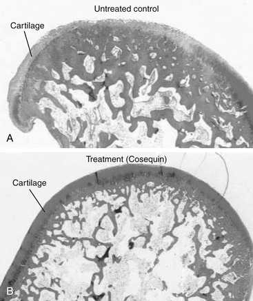
196. Borer L.R., Peel J.E., Seewald W., et al. Effect of carprofen, etodolac, meloxicam, or butorphanol in dogs with induced acute synovitis. Am J Vet Res. 2003;64(11):1429-1437.

197. Moreau M., Dupuis J., Bonneau N.H., et al. Clinical evaluation of a nutraceutical, carprofen and meloxicam for the treatment of dogs with osteoarthritis. Vet Rec. 2003;152(11):323-329.

198. Aragon C.L., Hofmeister E.H., Budsberg S.C. Systematic review of clinical trials of treatments for osteoarthritis in dogs. J Am Vet Med Assoc. 2007;230(4):514-521.

199. Taylor P.M., Delatour P., Landoni F.M., et al. Pharmacodynamics and enantioselective pharmacokinetics of carprofen in the cat. Res Vet Sci. 1996;60(2):144-151.

200. Lascelles B.D., Cripps P., Mirchandarin S., et al. Carprofen as an analgesic for postoperative pain in cats: dose titration and assessment of efficacy in comparison to pethidine hydrochloride. Small Anim Prac. 1995;36:535-541.

201. Balmer T.V., Irvine D., Jones R.S., et al. Comparison of carprofen and pethidine as postoperative analgesics in the cat. J Small Anim Pract. 1998;39(4):158-164.

202. Runk A., Kyles A.E., Downs M.O. Duodenal perforation in a cat following the administration of nonsteroidal antiinflammatory medication. J Am Anim Hosp Assoc. 1999;35:52-55.

203. Slingsby L.S., Waterman-Pearson A.E. Postoperative analgesia in the cat after ovariohysterectomy by use of carprofen, ketoprofen, meloxicam or tolfenamic acid. J Small Anim Pract. 2000;41(10):447-450.

204. Taylor P.M., Steagall P.V.M., Dixon M.J., et al. Carprofen and buprenorphine prevent hyperalgesia in a model of inflammatory pain in cats. Res Vet Sci. 2007;83:369-375.

205. Simmons K: Personal communication, Novartis Animal Health, October, 2004.

206. Gierse J.K., Staten N.R., Casperson G.F., et al. Cloning, expression, and selective inhibition of canine cyclooxygenase-1 and cyclooxygenase-2. Vet Ther. 2002;3(3):270-280.

207. Millis D.L., Weigel J.P., Moyers T., et al. The effect of deracoxib, a new COX-2 inhibitor, on the prevention of lameness induced by chemical synovitis in dogs. Vet Ther. 2002;3(4):7-18.

208. Sennello K.A., Leib M.S. Effects of deracoxib or buffered aspirin on the gastric mucosa of healthy dogs. J Vet Intern Med. 2006;20:1291-1296.

209. Glaser K.B. Cyclooxygenase selectivity and NSAIDS: Cyclooxygenase-2 selectivity of etodolac (Lodine). Inflammopharmacology. 1995;3:335-345.

210. Cayan M.N., Kraml M., Gerdinandi E.S., et al. The metabolic disposition of etodolac in rats, dogs, and man. Drug Metab Rev. 1981;12:339.

211. Lippiello L., Han M.S., Henderson T. Protective effect of the chondroprotective agent CosequinDS on bovine articular cartilage exposed in vitro to nonsteroidal antiinflammatory agents. Vet Ther. 2002;2(3):128-135.

211a. Klauss G., Giuliano E.A., Moore C.P., et al. Keratoconjunctivitis sicca associated with administration of etodolac in dogs: 211 cases (1992-2002). J Am Vet Med Assoc. 2007;230(4):541-547.

212. Budsberg S., Johnston S., Schwarz P., et al. Evaluation of etodolac for the treatment of osteoarthritis of the hip in dogs: a prospective multicenter study (abstract). Vet Surg. 1996;25:420.

213. McCann M.E., Andersen D.R., Zhang D., et al. In vitro effects and in vivo efficacy of a novel cyclooxygenase-2 inhibitor in dogs with experimentally induced synovitis. Am J Vet Res. 2004;65(4):503-512.

214. Pollmeier M., Toulemonde C., Fleishman C., et al. Clinical evaluation of firocoxib and carprofen for the treatment of dogs with osteoarthritis. Vet Rec. 2006;159:547-551.

215. Engelhardt G., Bogel R., Schnitzer C., et al. Meloxicam: influence on arachidonic acid metabolism, Part 1. In vitro findings. Biochem Pharmacol. 1996;51:21.

216. Engelhardt G., Bogel R., Schnitzer C., et al. Meloxicam: influence on arachidonic acid metabolism, Part II. In vivo findings. Biochem Pharmacol. 1996;51:29.

217. Engelhardt G., Homma D., Schlegel K., et al. General pharmacology of meloxicam, Part II. Effects on blood pressure, blood flow, heart rate, ECG, respiratory minute volume and interactions with paracetamol, pirenzepine, chlorthalidone, phenprocoumon and tolbutamide. Gen Pharmacol. 1996;27(4):679-688.

218. Hare J.E., Darling H., Doig P.A. Metacam oral suspension: current safety data, Boehringer Ingelheim Vetmedica. Toronto, Ontario: c/o Jonathan Hare, Janssen Animal Health, 19 Green Belt Drive; 1998.

219. Kay-Mugford P.A., Grimm K.A., Weingarten A.J., et al. Effect of preoperative administration of tepoxalin on hemostasis and hepatic and renal function dogs. Vet Ther. 2004;5:120-127.

220. Busch U., Schmid J., Heinzel G., et al. Pharmacokinetics of meloxicam in animals and the relevance to humans. Drug Metab Dispos. 1998;26(6):576-584.

221. Staerkel P., Horsmans Y. Meloxicam-induced liver toxicity. Acta Gastroenterol Belg. 1999;62(2):255-256.

222. Enberg T.B., Braun L.D., Kuzman A.B. Gastrointestinal perofration in five dogs associated with the administration of meloxicam. J Vet Emerg Crit Care. 2006;16(1):34-43.

223. Niza N.M., Felix N., Vilela C.L. Cutaneous and ocular adverse reactions in a dog following meloxicam administration. Vet Dermatol. 2007;18:45-49.

224. Doig P.A., Purbrick K.A., Hare J.E., et al. Clinical efficacy oand tolerance of meloxicam in dogs with chronic osteoarthritis. Can Vet J. 2000;41:296-300.

225. Van Bree H., Justus C., Quirke J.F. Preliminary observations on the effects of meloxicam in a new model for acute intra-articular inflammation in dogs. Vet Res Commun. 1994;18(3):217-224.

226. Jones C.J., Streppa H.K., Harmon B.G., et al. In vivo effects of meloxicam and aspirin on blood, gastric mucosal, and synovial fluid prostanoid synthesis in dogs. Am J Vet Res. 2002;63(11):1527-1531.

227. Lascelles B.D., Henderson A.J., Hackett I.J. Evaluation of the clinical efficacy of meloxicam in cats with painful locomotor disorders. J Small Anim Pract. 2001;42(12):587-593.

228. Slingsby L.S., Waterman-Pearson A.E. Comparison between meloxicam and carprofen for postoperative analgesia after feline ovariohysterectomy. J Small Anim Pract. 2002;43(7):286-289.

229. Mathews K.A., Pettifer G., Foster R., et al. Safety and efficacy of preoperative administration of meloxicam, compared with that of ketoprofen and butorphanol in dogs undergoing abdominal surgery. Am J Vet Res. 2001;62(6):882-888.

230. Budsberg S.C., Cross A.R., Quandt J.E., et al. Evaluation of intravenous administration of meloxicam for perioperative pain management following stifle joint surger in dogs. Am J Vet Res. 2002;63:1557-1563.

231. Rainsford K.D., Skerry T.M., Chindemi P., et al. Effects of the NSAIDs meloxicam and indomethacin on cartilage proteoglycan synthesis and joint responses to calcium pyrophosphate crystals in dogs. Vet Res Commun. 1999;23(2):101-113.

232. Justus C., Quirke J.F. Dose-response relationship for the antipyretic effect of meloxicam in an endotoxin model in cats. Vet Res Commun. 1995;19(4):321-330.

233. Metacam Symposium on Arthritic Disease in Cats, Seville, Spain, June 1-3, 2007.

234. Giraudel J.M., Toutain P.L., King J.N., et al. Differential inhibition of cyclooxygenase isoenzymes in the cat by the NSAID robenacoxib. J Vet Pharmacol Ther. 2009;32(1):31-40.

235. Jung M., Lees P., Seewald W., et al. Analytical determination and pharmacokinetics of robenacoxib in the dog. J Vet Pharmacol Therap. 2008;32:41-48.

236. Hoeijmakers M., Coert A., van Helden H., et al. The pharmacokinetics of vedaprofen and its enantiomers in dogs after single and multiple dosing,. J Vet Pharmacol Ther. 2005;28:305-312.

237. Nell T., Bergman J., Hoeijmakers M., et al. Comparison of vedaprofen and meloxicam in dogs with musculoskeletal pain and inflammation. J Small Anim Pract. 2002;43(5):208-212.

238. Paulson S.K., Zhang J.Y., Jessen S., et al. Comparison of celecoxib metabolism and excretion in mouse, rabbit, dog, cynomolgus monkey and rhesus monkey. Xenobiotica. 2000;30(7):731-744.

239. Hunter R.P., Radlinsky M., Koch D.E., et al. Single and multiple dose pharmacokinetics and synovial fluid concentrations of celecoxib in greyhound dogs. Proc ACVIM. #286, 2003.

240. Moreau M., Daminet S., Martel-Pelletier J., et al. Superiority of the gastroduodenal safety profile of licofelone over rofecoxib, a COX-2 selective inhibitor, in dogs. J Vet Pharmacol Therap. 2005;28:81-86.

241. Halpin R.A., Geer L.A., Zhang K.E., et al. The absorption, distribution, metabolism and excretion of rofecoxib, a potent and selective cyclooxygenase-2 inhibitor, in rats and dogs. Drug Metab Dispos. 2000;28(10):1244-1254.

242. Toutain P.L., Cester C.C., Haak T., et al. Pharmacokinetic profile and in vitro selective cyclooxygenase-2 inhibition by nimesulide in the dog. J Vet Pharmacol Ther. 2001;24(1):35-42.

243. Toutain P.L., Cester C.C., Haak T., et al. A pharmacokinetic/pharmacodynamic approach vs. a dose titration for the determination of a dosage regimen: the case of nimesulide, a Cox-2 selective nonsteroidal anti-inflammatory drug in the dog,. J Vet Pharmacol Ther. 2001;24(1):43-55.

244. Ewing G.O. Indomethacin-associated gastrointestinal hemorrhage in a dog. J Am Vet Med Assoc. 1972;161:1665-1668.

245. Mburu D.N., Mbugua S.W., Skoglund L.A., et al. Effects of paracetamol and acetylsalicylic acid on the post-operative course after experimental orthopaedic surgery in dogs. J Vet Pharmacol Ther. 1988;11:163-171.

246. St. Omer V.V., McKnight E.D. Acetylcysteine for treatment of acetaminophen toxicosis in the cat. J Am Vet Med Assoc. 1980;176:911-913.

247. Cullison R.F. Acetaminophen toxicosis in small animals: clinical signs, mode of action, and treatment. Compend Contin Educ. 1984;6:315-321.

248. Savides M.C., Oehme F.W., Leipold H.W. Effects of various antidotal treatment on acetaminophen toxicosis and biotransformation in cats. Am J Vet Res. 1985;46:1485-1489.

249. Jackson J.E. Cimetidine protects against acetaminophen toxicity. Life Sci. 1982;31:31-35.

250. Ruffalo R.L., Thompson J.F. Cimetidine and acetylcysteine as antidote for acetaminophen overdose. South Med J. 1982;75:954-958.

251. Wallace K.P., Center S.A., Hickford F.H., et al. S-Adenosyl-L-Methionine (SAMe) for the trestment of acetaminophen toxicity in a dog. J Am Anim Hosp Assoc. 2002;38:246-254.

252. Aronson L.R., Drobatz K. Acetaminophen toxicosis in 17 cats. J Vet Emerg Crit Care. 1996;6(2):65-69.

253. Cartier L.-J., Leclerc P., Pouliot M., et al. Toxic levels of acetaminophen produce a major positive interference on glucometer elite and accu-check advantage glucose meters. Clin Chem. 1998;44(4):893-894.

253a. Flood A.R. The role of acetaminophen in the treatment of osteoarthritis,. Am J Manag Care. 2010;16:S48-S54.

254. Johnston S.A., Budsberg S.C. Nonsteroidal anti-inflammatory drugs and corticosteroids for the management of canine osteoarthritis. Vet Clin North Am Small Anim Pract. 1997;27:841-862.

255. Hjelle J.J., Grauer G.F. Acetaminophen induced toxicosis in dogs and cats. J Am Vet Med Assoc. 1986;188:742-746.

256. Savides M.C., Oehme F.W. Acetaminophen and its toxicity. J Appl Toxicol. 1983;3:96-111.

257. Francavilla A., Makowka L., Polimeno L., et al. A dog model for acetaminophen-induced fulminant hepatic failure. Gastroenterology. 1989;96:470-478.

258. Goodman L., Coles T.B., Budsberg S. Leukotriene inhibition in small animal medicine. J Vet Pharmacol Ther. 2008;31:387-398.

259. Newcombe D.S. Leukotrienes: regulation of biosynthesis, metabolism, and bioactivity. J Clin Pharmacol. 1988;28:530-549.

260. Parkinson J.F. Lipoxin and synthetic Lipoxin analogs: an overview of anti-inflammatory functions and new concepts in immunomodulation. Inflamm Allergy Drug Targets. 2006;5:91-106.

261. Schmelzer R., Kubala L., Newman J.W., et al. Soluble epoxide hydrolase is a therapeutic target for acute inflammation. Proc Nat Acad Sci. 2005;102(28):9772-9777.

262. Ding X.Z., Hennig R., Adrian T.E. Lipoxygenase and cyclooxygenase metabolism: new insights in treatment and chemoprevention of pancreatic cancer. Mol Cancer. 2003;2:10-22.

263. Daffonchio L., Rossoni G., Clavenna G., et al. Protective activity of ketoprofen lysine salt against the pulmonary effects induced by bradykinin in guinea-pigs. Inflammation Res. 1996;45(5):259-264.

264. Martel-Pelletier J., Lajeunesse D., Reboul P., et al. Therapeutic role of dual inhibitors of 5-LOX and COX, selective and non-selective non-steroidal anti-inflammatory drugs. Ann Rheum Dis. 2003;62:501-509.

265. Jovanovic D.V., Fernandes J.C., Martel-Pelletier J., et al. In vivo dual inhibition of cyclooxygenase and lipoxygenase by ML-3000 reduces the progression of experimental osteoarthritis: suppression of collagenase 1 and interleukin-1 b synthesis. Arthritis Rheum. 2000;44:2320-2330.

266. Agnello K.A., Reynolds L.R., Budsberg S.C. In vivo effects of tepoxalin, an inhibitor of cyclooxygenase and lipoxygenase, on prostanoid and leukotriene production in dogs with chronic osteoarthritis. Am J Vet Res. 2005;66(6):966-972.

267. Punke J.P., Speas A.L., Reynolds L.R., et al. Effects of firocoxib, meloxicam, and tepoxalin on prostanoid and leukotriene production by duodenal mucosa and other tissues of osteoarthritic dogs. Am J Vet Res. 2008;69(9):1203-1209.

268. Hall G. Personal communication, Technical Service Veterinarian. Schering-Plough. April, 2009.

269. Bosmans T., Gasthuys F., Duchateau L., et al. A comparison of tepoxalin-buprenorphine combination and buprenorphine for postoperative analgesia in dogs: a clinical study,. J Vet Med A Physiol Pathol Clin Med. 2007;54(7):364-369.

269a. Gupat A., Kumar A., Kulkarni S. Licofelone attenuates MPTP-induced neuronal toxicity: behavioral, biochemical and cellular evidence. Inflammopharmacology. 2010;18:223-232.

270. Undem B.J. Pharmacotherapy of asthma. In Brunton L.L., Lazo J.S., Parker K.L., editors: Goodman & Gilman’s the pharmacological basis of therapeutics, ed 11, New York: McGraw-Hill, pp 717–736, 2006.

271. Serafin W.E. Drugs used in the treatment of asthma. In: Hardman J.G., Limbird L.D., editors. Goodman and Gilman’s the pharmacological basis of therapeutics. ed 9. New York: McGraw-Hill; 1996:659-682.

272. Ambrus J.L., Anain J.M., Anain S.M., et al. Dose-response effects of pentoxifylline on erythrocyte filterability: clinical and animal model studies. Clin Pharmacol Ther. 1990;48:50-56.

273. Ambrus J.L., Stadler S., Kulaylat M. Hemorrheologic effects of metabolites of pentoxifylline (Trental). J Med. 1995;26:65-75.

274. Zargari O. Pentoxifylline: a drug with wide spectrum applications in dermatology. Dermatology Online Journal. 14. 11. 2008. 2. http://dermatology.cdlib.org/1411/reviews/pentoxy/zargari.html. Accessed February 12, 2009, at(1 of 10)

275. Rees C., Boothe D.M., Boeckh A., et al. Dosing regimen and hemotologic effects of pentoxifylline and its active metabolites in normal dogs. Vet Ther. 2003;4(2):188-196.

276. Quezado Z.M.N., Hoffman W.D., Banks S.M. Increasing doses of pentoxifylline as a continuous infusion in canine septic shock. J Pharmacol Experiment Therapeut. 1999;288:107-113.

277. Apaydin B.B., Paksoy M., Arb T., et al. Influence of pentoxifylline and interferon-alpha on prevention of stricture due to corrosive esphagitis. Eur Surg Res. 2001;33:225-231.

278. Skidgel R.A., Erdös E.G. Histamine, bradykinin, and their antagonists. In Brunton L.L., Lazo J.S., Parker K.L., editors: Goodman & Gilman’s the pharmacological basis of therapeutics, ed 11, New York: McGraw-Hill, pp 629–651, 2006.

278a. Moy A.B., Winter M., Kamath A., et al. Histamine alters endothelial barrier function at cell-cell and cell-matrix sites. Am J Physiol Lung Cell Mol Physiol. 2000;278(5):L888-L898.

279. Obradovic T., Dobson G.G., Shingaki T., et al. Assessment of the first and second generation antihistamines brain penetration and role of P-glycoprotein. Pharm Res. 2007;24(2):318-327.

280. Hansson H., Bergvall K., Bondesson U., et al. Clinical pharmacology of clemastine in healthy dogs. Vet Dermatol. 2004;15(3):152-158.

281. Bizikova P., Papich M.G., Olivry T. Hydroxyzine and cetirizine pharmacokinetics and pharmacodynamics after oral and intravenous administration of hydroxyzine to healthy dogs. Vet Dermatol. 2008;19(6):348-357.

282. Papich M.G., Schooley E.K., Reinero C.R. Pharmacokinetics of cetirizine in healthy cats. Am J Vet Res. 2008;69(5):670-674.

283. Devillier P. Comparing the new antihistamines: the role of pharmacological parameters. Clin Exp Allergy. 2006;36:5-7.

284. Cook C.P., Scott D.W., Miller W.H.Jr., et al. Treatment of canine atopic dermatitis with cetirizine, a second generation antihistamine: a single-blinded, placebo-controlled study. Can Vet J. 2004;45(5):414-417.

285. DeBoer D.J., Griffin C.E. The ACVD task force on canine atopic dermatitis XXI: antihistamine pharmacotherapy,. Vet Immunol Immunopathol. 2001;81:323-329.

286. Scott D.W., Miller W.H.Jr. Antihistamines in the management of allergic pruritus in dogs and cats. J Small Anim Pract. 1999;40:359-364.

287. Fajardo M., Di Cesare P.E. Disease-modifying therapies for osteoarthritis: current status. Drugs Aging. 2005;22(2):141-161.

288. Gardner D.L. The nature and causes of osteoarthrosis,. Br Med J. 1983;286:418-424.

289. Jones A.C., Doherty M. The treatment of osteoarthritis. Br J Clin Pharmacol. 1992;33:357-363.

290. Vaughan-Scott T., Taylor J.H. The pathophysiology and medical management of canine osteoarthritis. J S Afr Vet Assoc. 1997;68(1):21-25.

291. Kelly G.S. The role of glucosamine sulfate and chondroitin sulfates in the treatment of degenerative joint disease,. Altern Med Rev. 1998;3(1):27-39.

292. Pelletier J.P., DiBattista J.A., Roughley P., et al. Cytokines and inflammation in cartilage degradation. Rheum Dis Clin North Am. 1993;19:545-568.

293. Pelletier J.P., Mineau F., Ranger P., et al. The increased synthesis of nitric oxide induced by IL-1 in human chondrocytes markedly reduced the synthesis of IL-1 receptor antagonists: a possible role in osteoarthritic cartilage degradation,. Osteoarthritis Cartilage. 1996;4(1):77-84.

294. Mitchell P.G., Yocum S.A., Lopresti L.L., et al. Collagenase 1 and collagenase 3 expression in IL 1 treated cartilage explants: regulation of net activity by endogenous produced inhibitors. Trans ORS. 459, 1997.

295. Singer I.I., Scott S., Kawka D.W., et al. Aggrecanase and metalloproteinase specific aggrecan neo-epitopes are induced in the articular cartilage of mice with collagen 11 arthritis. Osteoarthritis Cartilage. 1997;5(6):407-418.

296. Pinals R.S. Pharmacologic treatment of osteoarthritis. Clin Ther. 1992;14:336-346.

297. Altman R.D., Kapila P., Dean D.D., et al. Future therapeutic trends in osteoarthritis. Scand J Rheumatol Suppl. 1989;77:37-42.

298. Kelly G.S. The role of glucosamine sulfate and chondroitin sulfates in the treatment of degenerative joint disease,. Altern Med Rev. 1998;3(1):27-39.

299. White G.W. Adequan: a review for the practicing veterinarian. Vet Rev. 1988;8:463-467.

300. Francis D.J., Forrest M.J., Brooks P.M., et al. Retardation of articular cartilage degradation by glycosaminoglycan polysulfate, pentosan polysulfate, and DH-40J in the rat air pouch model. Arthritis Rheum. 1989;32:608-616.

301. Hannan N., Ghosh P., Bellenger C., et al. Systemic administration of glycosaminoglycan polysulphate (arteparon) provides partial protection of articular cartilage from damage produced by meniscectomy in the canine. J Orthop Res. 1987;5:47-59.

302. Nethery A., Giles I., Jenkins K., et al. The chondroprotective drugs, arteparon and sodium pentosan polysulphate, increase collagenase activity and inhibit stromelysin activity in vitro,. Biochem Pharmacol. 1992;44:1549-1553.

303. Halverson P.B., Cheung H.S., Struve J., et al. Suppression of active collagenase from calcified lapine synovium by arteparon. J Rheumatol. 1987;14:1013-1017.

304. Rao N.V., Kennedy T.P., Rao G., et al. Sulfated polysaccharides prevent human leukocyte elastase-induced acute lung injury and emphysema in hamsters. Am Rev Respir Dis. 1990;142:407-412.

305. Montefiori D.C., Robinson W.E., Modliszewski A., et al. Differential inhibition of HIV-1 cell binding and HIV-1-induced syncytium formation by low molecular weight sulphated polysaccharides. J Antimicrob Chemother. 1990;25:313-318.

306. Biffoni M., Paroli E. Complement in vitro inhibition by a low sulfate chondroitin sulfate (matrix). Drugs Exp Clin Res. 1991;27:35-39.

307. Arsenis C., McDonnell J. Effects of antirheumatic drugs on the interleukin-1a induced synthesis and activation of proteinases in articular cartilage explants in culture. Agents Actions. 1989;27:261-264.

308. Greinacher A., Michels I., Schäfer M., et al. Heparin associated thrombocytopenia in a patient treated with polysulphated chondroitin sulphate: evidence for immunological crossreactivity between heparin andpolysulphated glycosaminoglycan. Br J Haematol. 1992;81:252-254.

309. Jepsen J.V., Sall M., Rhodes P.R., et al. Long-term experience with pentosan polysulfate in interstitial cystitis. Urology. 1998;51(3):381-387.

310. Barrington J.W., Stephenson T.P. Pentosan polysulphate for interstitial cystitis. Int Urogynecol J Pelvic Floor Dysfunct. 1997;8(5):293-295.

311. Ghosh P., Hutadilok N. Interactions of pentosan polysulfate with cartilage matrix proteins and synovial fibroblasts derived from patients with osteoarthritis. Osteoarthritis Cartilage. 1996;4(1):43-53.

312. Read R.A., Cullis-Hill D., Jones M.P. Systemic use of pentosan polysulphate in the treatment of osteoarthritis. J Small Anim Pract. 1996;37(3):108-114.

313. Budsberg S.C., Bergh M.S., Reynolds L.R., et al. Evaluation of pentosan polysulfate sodium in the postoperative recovery from cranial cruciate injury in dogs: a randomized, placebo-controlled clinical trial. Vet Surg. 2007;36(3):234-244.

314. Hannon R.L., Smith J.G., Cullis-Hill D., et al. Safety of Cartrophen Vet in the dog: review of adverse reaction reports in the UK2003. J Small Anim Pract. 2003:202-208.

315. Elliot S.J., Striker L.J., Stetler W.G., et al. Pentosan polysulfate decreases proliferation and net extracellular matrix production in mouse mesangial cells. J Am Soc Nephrol. 1999;10:62-68.

316. Striker G.E., Lupia E., Elliot S., et al. Glomerulosclerosis, arteriosclerosis, and vascular graft stenosis: treatment with oral heparinoids. Kidney Int Suppl. 1997;63:S120-S123.

317. Senthil D., Malini M.M., Varalakshmi P. Sodium pentosan polysulphate—a novel inhibitor of urinary risk factors and enzymes in experimental urolithiatic rats. Ren Fail. 1998;20(4):573-580.

318. Schwartsmann G., Sprinz E., Kalakun L., et al. Phase II study of pentosan polysulfate (PPS) in patients with AIDS-related Kaposi’s sarcoma. Tumori. 1996;82(4):360-363.

319. Suzuki Y., Yamaguchi T. Effects of hyaluronic acid on macrophage phagocytosis and active oxygen release. Agents Actions. 1993;38(1-2):32-37.

320. Asheim A., Lindblad G. Intra-articular treatment of arthritis in race-horses with sodium hyaluronate. Acta Vet Scand. 1976;17:379-394.

321. Campos J.F.A. Efficacy of sodium hyaluronate in the treatment of hip dysplasia in dogs. Ahora Vet. 1998;17:59-61.

322. Fajardo M., Di Cesare P.E. Disease-modifying therapies for osteoarthritis; current status. Drugs Aging. 2006;22(2):141-161.

323. Anderson M. Oral chondroprotective agents, part I: common drugs used today. Compend Contin Educ Small Anim Pract. 1999;21(7):601-609.

324. Anderson M. Oral chondroprotective agents, Part II: evaluation of products and future compounds. Compend Contin Educ Small Anim Pract. 1999;21(9):861-865.

325. Boothe D.M. Nutraceuticals in veterinary medicine: Part I. Definitions and regulatory considerations. Compend Contin Educ Pract Vet. 1997;19:1248-1255.

326. Boothe D.M. Nutracueticals in veterinary medicine: Part II. Evaluating safety and efficacy. Compend Contin Educ Pract Vet. 1997;20:15-21.

327. Deal C.L., Moskowitz R.W. Nutraceuticals as therapeutic agents in osteoarthritis. The role of glucosamine, chondroitin sulfate, and collagen hydrolysate. Rheum Dis Clin North Am. 1999;25(2):379-395.

328. American Association of Feed Control Officials (AAFCO). Accessed November 9, 2009, at www.aafco.org.

329. National Animal Supplement Council (NASC). Accessed November 9, 2009, at www.nasc.cc.

329a. Adebowale A.O., Cox D.S., Liang Z., et al. Analysis of glucosamine and chondroitin sulfate content in marketed products and the Caco-2 permeability of chondroitin sulfate raw materials. J Am Nutr Assoc. 2000;3:37-44.

329b. Russell A.S., Aghazadeh-Habashi A., Jamali F. Active ingredient consistency of commercially available glucosamine sulfate products. J Rheumatol. 2002;29(11):2407-2409.

330. ConsumerLab. Accessed November 9, 2009, at www.Consumerlabs.com.

331. Adebowale A., Du J., Liang Z., et al. The bioavailability and pharmacokinetics of glucosamine hydrochloride and low molecular weight chondroitin sulfate after single and multiple doses to beagle dogs. Biopharm Drug Dispos. 2002;23(6):217-225.

332. Hanson R.R., Smalley L.R., Huff G.K., et al. Treatment with an oral glucosamine-chondroitin sulfate compound for degenerative joint disease in horses: 25 cases. Proc Vet Orthoped Soc. 1996;24:5.

333. Hanson R.R. Oral glycosaminoglycans in treatment of degenerative joint diseases in horses. Equine Pract. 1997;18:18-22.

334. White G.W., Sanders T., Jones E.W., et al. Efficacy of an orally administered sulfated glycosaminoglycan supplement in an induced equine carpitis model. Proc Am Assoc Equine Practitioners. 1996;42:139-141.

335. Anderson M.A., Slater M.R., Hammad T.A. Results of a survey of small-animal practitioners on the perceived clinical efficacy and safety of an oral nutraceutical. Prev Vet Med. 1999;38:65-73.

336. Bucci L.R. Chondroprotective agents: glucosamine salts and chondroitin sulfates. Townsend Letters for Doctors. 1994:52-55. January

337. McNamara P.S., Barr S.C., Idouaraine A., et al. Effects of an oral chondroprotective agent (Cosequin) on cartilage metabolism and canine serum. Proc Vet Orthoped Soc. 1997;24:35.

338. Phillipi A.F. Glucosamine, chondroitin and manganese ascorbate for degenerative joint disease of the knee or low back: a randomized, double-blind, placebo-controlled pilot study. Mil Med. 1999;164:85-91.

339. McCarty M.F. Enhanced synovial production of hyaluronic acid may explain rapid clinical response to high-dose glucosamine in osteoarthritis. Med Hypoth. 1998;50(6):507-510.

340. Barclay T.S., Tsourounis C., McCart G.M. Glucosamine. Ann Pharmacother. 1998;32(5):574-579.

341. McNamara P.S., Barr S.C., Hollis N.E. Hematologic, hemostatic and biochemical effects in dogs receiving an oral chondroprotective agent for thirty days. Am J Vet Res. 1996;57:1390-1394.

342. Neil K.M., Caron J.P., Orth M.W. The role of glucosamine and chondroitin sulfate in treatment for prevention of osteoarthritis in animals. J Am Vet Med Asoc. 2005;226:1079-1089.

342a. Reginster J.Y., Bruyere 0, Neuprez A. Current role of glucosamine in the treatment of osteoarthritis. Rheumatology. 2007;46(5):730-735.

343. Anderson J.W., Nicolosi R.J., Borzelleca J.F. Glucosamine effects in humans: a review of effects on glucose metabolism, side effects, safety considerations and efficacy. Food Chem Toxicol. 2005;43(2):187-201.

344. Setnikar I., Rovati L.C. Absorption, distribution, metabolism and excretion of glucosamine sulfate. A review. Arzneimittelforschung. 2001;51(9):699-725.

345. Setnikar I., Giacchetti C., Zanolo G. Pharmacokinetics of glucosamine in the dog and man. Pharmatherapeutica. 1984;3:538-549.

346. McNamara P.S., Barr S.C., Erb H.N., et al. Hematological, hemostatic, and biochemical effects in cats receiving an oral chondroprotective agent for thirty days. Vet Ther. 2000;1(2):108-117.

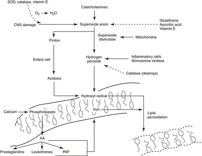
347. Volpi N. Oral bioavailability of chondroitin sulfate (Condrosulf) and its constituents in healthy male volunteers. Osteoarthritis Cartilage. 2002;10(10):768.

348. Echard B.W., Talpur N.A., Funk K.A., et al. Effects of oral glucosamine and chondroitin sulfate alone and in combination on the metabolism of SHR and SD rats. Mol Cell Biochem. 2001;225(1-):85-91.

348a. Scholtissen S., Bruyère O., Neuprez A., et al. Glucosamine sulphate in the treatment of knee osteoarthritis: cost-effectiveness comparison with paracetamol. Int J Clin Pract. 2010;64(6):756-762.

349. Scroggie D.A., Albright A., Harris M.D. The effect of glucosamine-chondroitin supplementation on glycosylated hemoglobin levels in patients with type 2 diabetes mellitus: a placebo-controlled, double-blinded, randomized clinical trial,. Arch Intern Med. 2003;163(13):1587-1590.

350. Hoffer L.J., Kaplan L.N., Hamadeh M.J., et al. Sulfate could mediate the therapeutic effect of glucosamine sulfate. Metabolism. 2001;50(7):767-770.

351. Reginster J.Y., Deroisy R., Rovati L.C., et al. Long-term effects of glucosamine sulphate on osteoarthritis progression: a randomised, placebo-controlled clinical trial. Lancet. 2001;357(9252):251-256.

352. Bruyere O., Pavelka K., Rovati L.C., et al. Glucosamine sulfate reduces osteoarthritis progression in postmenopausal women with knee osteoarthritis: evidence from two 3-year studies. Menopause. 2004;11(2):138-143.

353. Ruane R., Griffiths P. Glucosamine therapy compared to ibuprofen for joint pain. Br J Community Nurs. 2002;7(3):148-152.

354. Conte A., Volpi N., Palmieri L. Biochemical and pharmacokinetic aspects of oral treatment with chondroitin sulfate. Arzneimittelforschung. 1995;45(8):918-925.

355. Gregory S., Kelly N.D. The role of glucosamine sulfate and chondroitin sulfates in the treatment of degenerative joint disease. Altern Med Rev. 1998;3:27-39.

356. Leffler C.T., Philippi A.F., Leffler S.G., et al. Glucosamine, chondroitin and manganese ascorbate for degenerative joint disease of the knee or low back: a randomized, double-blind, placebo-controlled pilot study. Mil Med. 1999;164(2):85-91.

357. Das A.K., Hammad T.A. Efficacy of a combination of FCHG49 glucosamine hydrochloride, TRHI22 low molecular weight sodium chondroitin sulfate and manganese ascorbate in the management of knee osteoarthritis. Osteoarthritis Cartilage. 2000;8(5):343-350.

358. Leffler C.T., Philippi A.F., Leffler S.G., et al. Glucosamine, chondroitin, and manganese ascorbate for degenerative joint disease of the knee or low back: a randomized, double-blind, placebo-controlled pilot study. Mil Med. 1999;164(2):85-91.

359. McAlindon T., LaValley M., Gulin J., et al. Glucosamine and chondroitin for treatment of osteoarthritis - a systematic quality assessment and meta-analysis. J Am Med Assoc. 2000;283(11):1469-1475.

360. Fajardo M., Di Cesare P.E. Disease-modifying therapies for osteoarthritis: current status. Drugs Aging. 2005;22(2):141-161.

361. Clegg D.O., Reda D.J., Harris C.L., et al. Glucosamine, chondroitin sulfate, and the two in combination for painful knee osteoarthritis. N Engl J Med. 2006;354(8):795-808.

362. Lippiello L., Idouraine A., McNamara P.S., et al. Cartilage stimulatory and antiproteolytic activity is present in sera of dogs treated with a chondroprotective agent. Canine Pract. 1999;24(1):18-19.

363. Canapp S.O., McLaughlin R.M., Hoskinson J.J., et al. Scintigraphic evaluation of glucosamine HCl and chondroitin sulfate as treatment for acute synovitis in dogs. Am J Vet Res. 1999;60(12):1552-1557.

364. Ameye L.G., Chee W.S.S. Osteoarthritis and nutrition. From nutraceuticals to functional foods: a systematic review of the scientific evidence. Arthritis Res Ther. 2006;8:R127.

365. Lippiello L., Nardo J.V., Harlan R., et al. Metabolic effects of avocado/soy unsaponifiables on articular chondrocytes. Evid Based Complement Alternat Med. 2008;5(2):191-197.

366. Soeken K.L., Lee W.-L., Bausell R.B., et al. Safety and efficacy of S-adenosylmethionine (SAMe) for osteoarthritis. J Family Pract. 2002;51:425-430.

367. Romay Ch, Gonzalez R., Ledon N., et al. C-Phycocyanin: a biliprotein with antioxidant, anti-inflammatory and neuroprotective effects. Curr Protein Pept Sci. 2003;4:207-216.

368. Reddy C.M., Bhat V.B., Kiranmai G., et al. Selective inhibition of cyclooxygenase-2 by C-Phycocyanin, a biliprotein from Spirulina platensis. Biochem Biophys Res Commun. 2000;277:599-603.

369. Salin M., McCord J.M. Free radicals and inflammation: protection of phagocytosing leukocytes by superoxide dismutase. J Clin Invest. 1975;56:1319-1323.

370. Tobin T. Pharmacology review: the nonsteroidal anti-inflammatory drugs. II. Equiproxen, meclofenamic acid, flunixin and others. J Equine Med Surg. 1979;6:298-302.

371. Breshears D.E., Brown C.D., Riffel D.M., et al. Evaluation of orgotein in treatment of locomotor dysfunction in dogs. Mod Vet Pract. 1974;55:85-93.

372. Ahlengard S., Tufvesson G., Pettersson H., et al. Treatment of traumatic arthritis in the horse with intra-articular orgotein (Palosein). Equine Vet J. 1978;10:122-124.

373. Brayton C.F. Dimethyl sulfoxide (DMSO). Cornell Vet. 1986;76:61-90.

374. Alsup E.M. Dimethyl sulfoxide. J Am Vet Med Assoc. 1984;185:1011-1014.

375. Wong L.K., Reinertson E.L. Clinical considerations of dimethyl sulfoxide. Iowa State Univ Vet. 1984;46:89-95.

376. Spitzer W.O. Drugs as determinants of health and disease in the population. J Clin Epidemiol. 1991;44:823-830.

377. Olson N.C., Hellyer P.W., Dodam J.R. Mediators and vascular effects in response to endotoxin. Br Vet J. 1995;151:489-522.

378. Whittle B.J.R. Nitric oxide in physiology and pathology. Histochem J. 1995;27:727-737.

379. Howe L.B. Treatment of endotoxic shock: glucocorticoids, lazaroids, nonsteroidals and others. Vet Clin North Am. 1998;28:249-267.

380. Vincent J.L. Evidence-based medicine in the ICU: important advances and limitations. Chest. 2004;126:592-600.

381. Minneci P., Deans K., Natanson C., et al. Increasing the efficacy of anti-inflammatory agents used in the treatment of sepsis. Eur J Clin Microbiol Infect Dis. 2003;22:1-9.

382. Kettelhut I.C., Fiers W., Goldgerg A.L. The toxic effects of tumor necrosis factor in vivo and their prevention by cyclooxygenase inhibitors,. Proc Natl Acad Sci USA. 1987;84:4273.

383. Shuster R., Traub-Dargatz J., Baxter G. Survey of diplomates of the American College of Veterinary Internal Medicine and the American College of Veterinary Surgeons regarding clinical aspects and treatment of endotoxemia in horses. J Am Vet Med Assoc. 1997;210:87-92.

384. Sigurdsson G.H., Youssef H. Amelioration of respiratory and circulatory changes in established endotoxic shock by ketoprofen. Acta Anesthesiol Scand. 1994;38:33-39.

385. Boumpas D.T., Chrousos G.P., Wilder R.L., et al. Glucocorticoid therapy for immune-mediated diseases. Basic and clinical correlates. Ann Intern Med. 1993;119:1198-1208.

386. Bone R.C., Fisher C.J., Clemmer T.P., et al. A controlled clinical trial of high dose methylprednisolone in the treatment of severe sepsis and septic shock. N Engl J Med. 1987;317:653-658.

387. Aoki N., Lefer A.M. Protective effects of a novel non-glucocorticoid 21 aminosteroid (U74006F) during traumatic shock in rats. J Cardiovasc Pharmacol. 1990;15:205-210.

388. Hall E.D., Yonkers P.A., McCall J.M. Attenuation of hemorrhagic shock by the non-glucocorticoid 21-aminosteroid U74006F. Eur J Pharmacol. 1988;147:299-303.

389. Semrad S.D., Rose M., Putnam M.L. Efficacy and toxicity of lazaroid (U74006F) in neonatal endotoxemia. Circ Shock. 1989;27:358-359.

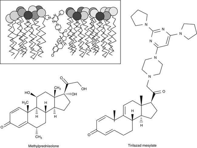
390. Semrad S.D., Rose M.L., Adams J.L. Effects of tirilazad mesylate (U74006F) on eicosanoid and tumor necrosis factor generation in healthy and endotoxic neonatal calves. Circ Shock. 1993;40:235-242.

391. Zhang H., Spapen H., Minikis P., et al. Tirilazad mesylate (U-74006F) inhibits effects of endotoxin in dogs. Am J Physiol. 1995;286:H1847-H1855.

392. Morris P.E., Light R.B., Garber G.E. Identifying patients with severe sepsis who should not be treated with drotrecogin alfa (activated). Am J Surg. 2002;184:19S-24S.

393. Kavanagh R.J., Kam P.C. Lazaroids: efficacy and mechanism of action of the 21-aminosteroids in neuroprotection. Br J Anaesth. 2001;86(1):110-119.

Data on package insert provided among the NSAID may differ depending, in part, on requirements of the FDA. As such, bioavailability data may not be available for all drugs. This may preclude accurate comparisons among drugs.