Chapter 40 Oxygenation

Margaret Wheeler

KEY TERMS

LEARNING OUTCOMES

Mastery of content will enable you to:

Define the key terms listed.

Describe the structure and function of the cardiovascular and pulmonary systems.

Identify physiological processes of cardiac output, myocardial blood flow and coronary artery circulation.

Describe the relationship of cardiac output, stroke volume (preload, afterload, contractility) and heart rate.

Identify physiological processes involved in ventilation, perfusion and exchange of oxygen and carbon dioxide.

Describe neural and chemical regulation of ventilation.

Describe the impact of a patient’s level of health, age, lifestyle and environment on tissue oxygenation.

Identify and describe clinical outcomes as a result of disturbances in conduction, altered cardiac output, impaired valvular function, myocardial ischaemia and impaired tissue perfusion.

Identify and describe clinical outcomes of ventilation/perfusion mismatch, diffusion impairment, alveolar hypoventilation and alveolar hyperventilation.

Identify nursing care interventions in the primary care, acute care and restorative and extended-care settings that promote oxygenation.

Scientific knowledge base

Oxygen is required to sustain life. The function of the cardiac and respiratory systems is to supply the body’s oxygen demands. Cardiopulmonary physiology involves delivery of deoxygenated blood to the right side of the heart and then to the pulmonary circulation where oxygen uptake occurs. Oxygenated blood then moves from the lungs to the left side of the heart and is subsequently delivered to the tissues. Blood is oxygenated and carbon dioxide eliminated through the mechanisms of ventilation, the mechanical movement of gas into and out of the lungs, and respiration, the exchange of oxygen and carbon dioxide at tissue level. Neural and chemical regulators control respiratory rate and depth in response to changing tissue oxygen demands (Brashers 2010a, 2010b).

Cardiovascular physiology

The function of the cardiovascular system is to deliver oxygen, nutrients and other substances to the tissues and to remove the waste products of cellular metabolism through the cardiac pump, the circulatory vascular system and the integration of other systems, for example respiratory, gastrointestinal and renal systems (Brashers, 2010b).

Structure and function

The right ventricle pumps blood through the pulmonary circulation while the left ventricle pumps blood to the systemic circulation, supplying oxygen and nutrients to the tissues and removing carbon dioxide and wastes (Figures 40-1 and 40-2). The circulatory system exchanges oxygen, carbon dioxide, nutrients and waste products between the blood and the tissues.

image

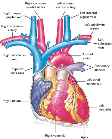
FIGURE 40-1 Diagram showing serially connected pulmonary and systemic circulation. Right heart chambers propel unoxygenated blood through the pulmonary circulation; left heart chambers propel oxygenated blood through the systemic circulation.

From Canobbio MM 1990 Cardiovascular disorders. St Louis, Mosby.

image

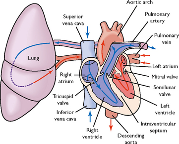
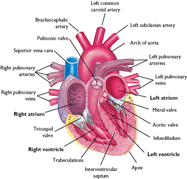
FIGURE 40-2 Schematic representation of blood flow through the heart. Arrows indicate direction of flow.

From Lewis SM and others 2004 Medical–surgical nursing: assessment and management of clinical problems, ed 6. St Louis, Mosby.

MYOCARDIAL PUMP

The pumping action of the heart is essential for the maintenance of oxygen delivery. Decreased pump effectiveness, as occurs in heart failure, results in a diminished stroke volume—the volume of blood ejected from the ventricles during each contraction. Shock, haemorrhage and dehydration decrease pump effectiveness by reducing the volume of circulating blood, thereby decreasing stroke volume.

The chambers of the heart, the right and left atria and ventricles, fill during diastole and partially empty during systole. The effectiveness of the diastolic and systolic events of the cardiac cycle can be assessed by monitoring the patient’s blood pressure (see Chapter 27). The myocardial fibres have contractile properties that enable them to stretch during filling. In a healthy heart this stretch is proportionally related to the strength of contraction. As the myocardium stretches, the strength of the subsequent contraction increases; this is known as Starling’s law of the heart (or Frank–Starling relationship). In the diseased heart, Starling’s law does not apply because the stretch of the myocardium is beyond the heart’s physiological limits. The subsequent contractile response results in insufficient ventricular ejection (volume), and blood begins to ‘back up’ in the pulmonary (left heart failure) or systemic (right heart failure) circulation.

CARDIAC BLOOD FLOW

Blood flow through the heart is unidirectional. There are four heart valves that ensure this forward blood flow (Figure 40-3). During ventricular diastole, the atrioventricular (mitral and tricuspid) valves open and blood flows from the higher-pressure atria into the relaxed ventricles. The closure of these valves represents S1, or the first heart sound of auscultation. After ventricular filling, the systolic phase begins. As the systolic intraventricular pressure rises, the atrioventricular valves close, preventing the backflow of blood into the atria, and ventricular contraction begins (isovolumetric contraction). During the systolic phase, ventricular pressure rises, causing the semilunar (aortic and pulmonic) valves to open. As the ventricles eject blood, the intraventricular pressure falls and the semilunar valves close, thus preventing the backflow of blood from the aorta and pulmonary artery into the ventricles. Closure of the aortic and pulmonic valves represents S2, or the second heart sound. Patients with valvular disease may have backflow or regurgitation of blood through an incompetent valve, causing a murmur or click that is heard on auscultation (see Chapter 27).

image

FIGURE 40-3 Cross-sectional view of the heart showing atrium, ventricles and valves.

Modified from Canobbio MM 1990 Cardiovascular disorders. St Louis, Mosby.

CORONARY ARTERY CIRCULATION

To maintain adequate blood flow to the pulmonary and systemic circulation, myocardial blood flow must supply sufficient oxygen and nutrients to the myocardium itself. Blood in the atria and ventricles does not supply oxygen and nutrients to the myocardium. The coronary circulation is the branch of the systemic circulation that supplies the myocardium with oxygen and nutrients and removes waste. The coronary arteries fill during ventricular diastole (Brashers, 2010b). The right and left coronary arteries arise from the aorta just above and behind the aortic valve through openings called the coronary ostia (coronary openings). The left coronary artery generally feeds the left atrium and ventricle, while the right coronary artery feeds the right atrium and ventricle. However, there is variation in branching of coronary arteries among individuals (Figure 40-4) (Box 40-1).

image

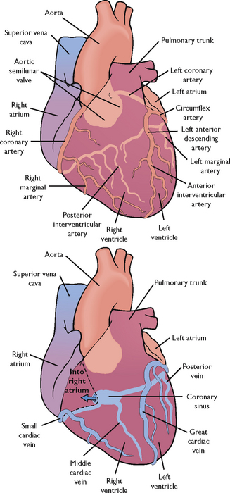
FIGURE 40-4 Coronary arteries and veins.

From Lewis SM and others 2004 Medical–surgical nursing: assessment and management of clinical problems, ed 6. St Louis, Mosby.

BOX 40-1 CORONARY ARTERIES

RIGHT CORONARY ARTERY

Supplies:

Right atrium

Right ventricle

Part of posterior and inferior surface of left ventricle

AV node, Bundle of His (90% of population)

Sinus node (55% of population)

LEFT CORONARY ARTERIES

LEFT ANTERIOR DESCENDING (LAD) ARTERY

Supplies:

Lateral left ventricular wall

Majority of septum

Anterior and diaphragmatic aspects of left ventricle

CIRCUMFLEX ARTERY

Supplies:

Sinus node (45% of population)

Left atrium

Posterior surfaces of left ventricle

Posterior aspects of septum

NB: further anatomical variations exist in the population.

Adapted from Bond EF 2010 Cardiac anatomy and physiology. In Woods SL, Froelicher ESS, Motzer SU and others, editors, Cardiac Nursing, ed 6. Philadelphia, Lippincott Williams & Wilkins.

SYSTEMIC CIRCULATION

The arteries and veins of the systemic circulation deliver nutrients and oxygen to the tissues and remove waste from them. Oxygenated blood flows from the left ventricle by way of the aorta and into large systemic arteries. These arteries branch into smaller arteries, then into arterioles and finally into the smallest vessels, the capillaries. At capillary level the exchange of oxygen and carbon dioxide, nutrients and wastes occurs, resulting in tissue oxygenation. The waste products exit the capillary network by way of the venules that join to form veins. These veins form larger veins, which carry deoxygenated blood to the right side of the heart, where it is returned to the pulmonary circulation.

BLOOD FLOW REGULATION

The amount of blood ejected from the left ventricle each minute is the cardiac output. The normal cardiac output is 4–6 L/min in the healthy 70 kg adult at rest. The circulating volume of blood changes according to the oxygen and metabolic needs of the body. For example, during exercise, pregnancy and fever the cardiac output increases, but during sleep it decreases. Cardiac output is represented by the following formula:


Cardiac output (CO) = Stroke volume (SV) × Heart rate (HR)

Cardiac output in the older adult may be affected by increased arterial wall tension and moderate myocardial hypertrophy due to an increased systolic blood pressure.

Stroke volume is the amount of blood ejected from the left ventricle with each contraction. It can be affected by the amount of blood in the left ventricle at the end of diastole (preload), the resistance to left ventricular ejection (afterload) and myocardial contractility (see section below).

Preload is essentially the end-diastolic volume. As the ventricles fill, they stretch. The greater the stretch on the ventricle, the greater the contraction and the greater the stroke volume (Starling’s law). In clinical situations, the preload and subsequent stroke volume can be manipulated by changing the amount of circulating blood volume. For example, in the patient with haemorrhagic shock, fluid therapy and replacement of blood increases volume, thus increasing the preload and cardiac output. If volume is not replaced, preload decreases, the cardiac output decreases and ultimately the venous return to the right atrium decreases, further decreasing preload and cardiac output.

Afterload is the resistance to left ventricular ejection—the work the heart must overcome to fully eject blood from the left ventricle. The diastolic aortic pressure is a good clinical measure of afterload. In a patient with an acute hypertensive crisis, the afterload is increased, increasing the cardiac workload. Afterload in this situation can be manipulated by decreasing systemic blood pressure.

The continuous monitoring of cardiovascular haemodynamics is usually performed in critical-care units. Some step-down or special-care units may also have the capability to continuously monitor haemodynamics.

MYOCARDIAL CONTRACTILITY

Myocardial contractility also affects stroke volume and cardiac output. Poor contraction of the myocardium decreases the amount of blood ejected by the ventricles during each contraction. Myocardial contractility can be increased by drugs that increase the force of contraction, such as digitalis preparations and sympathomimetic drugs (drugs that mimic the effects of the sympathetic nervous system), e.g. adrenaline. Injury to the myocardial muscle, such as an acute myocardial infarction, can cause a decrease in myocardial contractility. The myocardium of the older adult is more rigid and slower in recovering its contractility (Steven, 2011).

Heart rate affects blood flow because of the interaction between rate and diastolic filling time. With a sustained heart rate greater than 160 beats per minute, diastolic filling time decreases, decreasing stroke volume and cardiac output. The heart rate of the older adult is slow to increase under stress. With the normal ageing process there are significant changes to cardiac structure and function, and the stroke volume may increase to increase the cardiac output and blood pressure (Jet, 2008).

CONDUCTION SYSTEM

The rhythmic relaxation and contraction of the atria and ventricles depend on continuous, organised transmission of electrical impulses. These impulses are generated and transmitted by way of the cardiac conduction system (Figure 40-5).

image

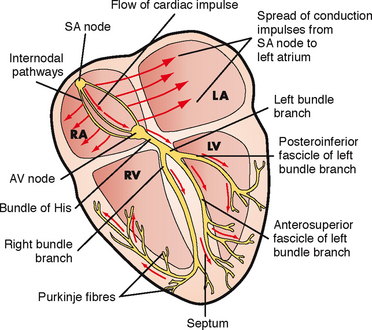
FIGURE 40-5 Conduction system of the heart. LA, left atrium; LV, left ventricle; RA, right atrium; RV, right ventricle; SA, sinoatrial; AV, atrioventricular.

From Lewis SM and others 2007 Medical–surgical nursing: assessment and management of clinical problems, ed 7. St Louis, Mosby.

The heart’s conduction system generates the necessary action potentials that conduct the impulses required to initiate the electrical chain of events resulting in the heartbeat. The autonomic nervous system, consisting of sympathetic and parasympathetic branches, influences the rate of impulse generation as well as the speed of transmission through the conductive pathway and the strength of atrial and ventricular contractions. Sympathetic nerve fibres, which increase the rate of impulse generation and the speed of impulse transmission, innervate all parts of the atria and ventricles. Parasympathetic fibres from the vagus nerve, which decrease this rate, also innervate these parts, as well as the sinoatrial and atrioventricular nodes (Brashers, 2010b).

The conduction system originates with the sinoatrial (SA) node, the ‘pacemaker’ of the heart. The SA node is in the right atrium next to the entrance of the superior vena cava (Brashers, 2010b). Impulses are initiated at the SA node at an intrinsic rate of 60–100 beats per minute. The resting adult rate is approximately 75 beats per minute. Resting heart rate for infants and children varies with age: 80–150 beats per minute for age 3 months to 2 years, 70–110 beats per minute for 2–10 years and 55–90 beats per minute for over 10 years (Hockenberry and others, 2009).

The electrical impulses are then transmitted through the atria along intra-atrial pathways to the atrioventricular (AV) node. The AV node mediates impulses between the atria and the ventricles. The intrinsic rate of the normal AV node is 40–60 beats per minute. The AV node assists atrial emptying by delaying the impulse before transmitting it through the bundle of His and the ventricular Purkinje network. The intrinsic rate of the bundle of His and the ventricular Purkinje network is 20–40 beats per minute.

An electrocardiogram (ECG) reflects the electrical activity of the conduction system. It monitors the regularity and path of the electrical impulse through the conduction system; however, it does not reflect muscular work of the heart. This electrical impulse is required to stimulate the myocardium to then generate the mechanical event of ventricular contraction. The normal sequence on the ECG is called normal sinus rhythm (NSR) (Figure 40-6).

image

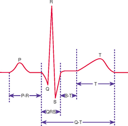
FIGURE 40-6 Normal ECG waveform.

From Potter PA, Perry AG 2013 Fundamentals of Nursing, ed 8. St Louis, Mosby.

NSR implies that the impulse originates at the SA node and follows the normal sequence through the conduction system. The P wave represents the electrical conduction through both atria and is normally small and rounded. Atrial contraction follows the P wave. The P–R interval represents the impulse travel time through the AV node and the bundle of His to the Purkinje fibres. The normal length of the P–R interval is 3–5 small squares on the ECG paper (0.12–0.20 second). An increase in the P–R of > 5 small squares (0.20 second) indicates that there is a block in the impulse transmission through the AV node, whereas a P-R interval of < 2 small squares (0.08 second) indicates the initiation of the electrical impulse from a source other than the SA node. A P–R interval of > 5 small squares (0.20 second) should be reported.

The QRS complex indicates that the electrical impulse has travelled through the ventricles. Normal QRS duration is 1.5–3 small squares on the ECG paper (0.06–0.12 second). An increase in QRS duration indicates a delay in conduction time through the ventricles. Ventricular contraction usually follows the QRS complex. A QRS complex wider than 3 small squares (0.12 second) should be reported.

The T wave represents ventricular repolarisation. The T wave is normally rounded, rising more slowly from the baseline than it returns. T waves can be flattened or inverted in hypokalaemia and peaked in hyperkalaemia.

The S–T segment extends from the end of the QRS (J point) to the start of the T wave and should be at the baseline. Significant displacement from the baseline may be indicative of disease processes such as acute coronary syndrome (Jacobson, 2010a).

Respiratory physiology

Most cells in the body obtain their energy from chemical reactions involving oxygen and the elimination of carbon dioxide. The exchange of oxygen and carbon dioxide occurs between environmental air and the blood (Figure 40-7). There are three steps in the process of oxygenation: ventilation, diffusion and perfusion of lungs (Brashers, 2010a). For the exchange of oxygen and carbon dioxide to occur, the organs, nerves and muscles of respiration must be intact and the central nervous system able to regulate the respiratory cycle. The respiratory centre lies in the brainstem, which contains a group of neurons (dorsal respiratory group) that regulates the automatic rhythm of respiration. The pneumotaxic and apneustic centres of the respiratory centre adjust inspiratory rate and depth. Central chemoreceptors detect slight changes in pH in spinal fluid, reflecting arterial carbon dioxide (see Chapter 39). Changes in carbon dioxide and therefore pH are detected and inspiratory rate and depth adjusted accordingly. Peripheral chemoreceptors detect arterial carbon dioxide and oxygen, but predominantly oxygen, and are therefore principally responsible for adjusting respiration in response to hypoxaemia. Central chemoreceptors are more sensitive than peripheral chemoreceptors (Brashers, 2010a).

image

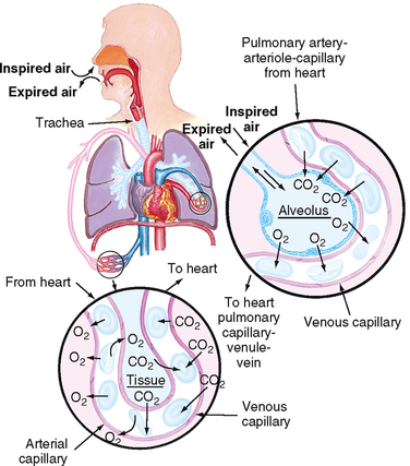
FIGURE 40-7 Structures of the pulmonary system.

From Thompson J and others 1993 Mosby’s manual of clinical nursing, ed 3. St Louis, Mosby.

Structure and function

Normal adult respiratory rate ranges between 12 and 20 breaths per minute. Respiratory rate in infants and children varies: 25–30 breaths per minute for age 3 months to 2 years, 19–25 breaths per minute for 2–10 years, and 16–19 breaths per minute for over 10 years (Hockenberry and others, 2009). Respiration can be altered by conditions or diseases that change the structure and function of the lung. The respiratory muscles, pleural space, lungs and alveoli (Figure 40-8) are essential for ventilation, perfusion and exchange of oxygen and carbon dioxide (Box 40-2). The pleura is a membrane which adheres to the lungs and then folds back to connect to the chest wall. The visceral pleura adheres to the lungs, while the parietal pleura adheres to the chest wall. The area between these two is the pleural space, normally a potential space containing a small amount of fluid and negative in pressure relative to atmosphere (Brashers, 2010a). Problems arise if the negative pressure of this space is not maintained, as for example when air (pneumothorax), blood (haemothorax) or fluid (pleural effusion) enters the space (Brashers, 2010a).

image

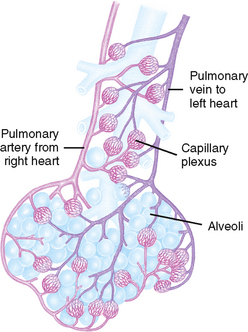
FIGURE 40-8 Alveoli at the terminal end of the lower airway.

From Thompson J and others 1993 Mosby’s manual of clinical nursing, ed 3. St Louis, Mosby.

BOX 40-2 MAJOR ANATOMICAL STRUCTURES OF THE THORAX AND THEIR FUNCTIONS

INSPIRATORY MUSCLES

DIAPHRAGM

Contraction causes the diaphragm to descend, creating a negative pleural pressure and increasing the vertical dimension of the lungs, which contributes to inflation of the lungs. The increase in vertical dimension and the decrease in intrapulmonary pressure (negative with respect to atmospheric pressure) cause air to enter the lungs.

EXTERNAL INTERCOSTAL MUSCLES

Contraction elevates the anterior ends of the ribs, causing them to move upwards and outwards. This increases the anteroposterior dimension of the thorax.

ACCESSORY MUSCLES

Accessory muscles include the scalene, sternocleidomastoid and trapezius muscles. Contraction elevates the first two ribs and the sternum.

EXPIRATORY MUSCLES

INTERNAL INTERCOSTAL MUSCLES

Contraction pulls the ribs down and in, thereby decreasing the anteroposterior diameter of the thorax.

ABDOMINAL RESPIRATORY MUSCLES

Abdominal respiratory muscles include the rectus, transversus abdominis, internal oblique and external oblique muscles. Contraction depresses the lower ribs, forces the diaphragm up and decreases the vertical dimension of the thoracic cavity.

PLEURAL SPACE

The pleural space is a potential space that is only a thin film of liquid lying between the outer layer of the lung (visceral pleura) and the inner layer of the chest cavity (parietal pleura). It permits a smooth, gliding movement of the lungs along the chest wall. Normally, air is not present in the pleural space.

LUNGS

LEFT (TWO LOBES) AND RIGHT (THREE LOBES)

The lungs transfer oxygen from the atmosphere into the alveoli and carbon dioxide from the alveoli to the lungs to be excreted as a waste product. They also filter toxic material from circulation and metabolise compounds such as angiotensin I, bradykinin and prostaglandins.

ALVEOLI

Alveoli transfer oxygen and carbon dioxide to and from the blood through the alveolar membrane. These tiny air sacs expand during inspiration, greatly increasing the surface area over which exchange of oxygen and carbon dioxide occurs.

Ventilation is the process of moving gases into and out of the lungs. Normal ventilation occurs at a rate of approximately 4 L/min. Ventilation requires coordination of the muscular and elastic properties of the lung and thorax, as well as intact innervation. The major inspiratory muscle of respiration is the diaphragm. It is innervated by the phrenic nerve derived from spinal nerves C3, C4 and C5. A mnemonic to aid memory of the innervation of the diaphragm is ‘C3, 4, 5 keeps the diaphragm alive’.

WORK OF BREATHING

Breathing is the effort required to expand and contract the lungs. The work of breathing is determined by the degree of compliance of the lungs, airway resistance, presence of active expiration, and use of accessory muscles of ventilation.

Inspiration is an active process, stimulated by chemical receptors in the aorta. Expiration is a passive process that depends on the elastic recoil properties of the lungs, requiring little or no muscle work. Elastic recoil is produced by elastic fibres in lung tissue and by surface tension in the fluid film lining the alveoli. Surfactant is the chemical produced in the lungs by alveolar type 2 cells that reduces the surface tension of the alveoli and keeps them from ‘sticking shut’ when collapsed (Brashers, 2010a). Patients with advanced chronic obstructive pulmonary disease (COPD) lose the elastic recoil of the lungs and thorax. As a result, the patient’s work of breathing is increased.

Accessory muscles of respiration can increase lung volume during inspiration, though not as effectively as the diaphragm. Accessory muscles include the sternocleidomastoid and scalene muscles on the side of the neck and the intercostal muscles (Brashers, 2010a). Patients with COPD, especially emphysema, frequently use these muscles to increase lung volume. During assessment the nurse may see obvious movement of these neck muscles and retraction of the muscles between the ribs. Prolonged use of the accessory muscles of respiration does not promote effective ventilation and causes fatigue.

Compliance is the ability of the lungs to distend or to expand in response to increased intra-alveolar pressure. Compliance is decreased in diseases such as pulmonary oedema, interstitial and pleural fibrosis, and congenital or traumatic structural abnormalities such as kyphosis (curving of the spine causing a roundness of the back) or fractured ribs.

Airway resistance is the pressure difference between the mouth and the alveoli in relation to the rate of flow of inspired gas. Airway resistance can be increased by an airway obstruction, small-airway disease (such as asthma) and tracheal oedema. When resistance is increased, the amount of air travelling through the anatomical airways is decreased.

Decreased lung compliance, increased airway resistance, active expiration or the use of accessory muscles increases the work of breathing, resulting in increased energy expenditure. To meet this expenditure, the body increases its metabolic rate, and the need for oxygen, as well as for the elimination of carbon dioxide, increases. This sequence is a vicious cycle for a patient with impaired ventilation, causing further deterioration of respiratory status and the ability to oxygenate adequately.

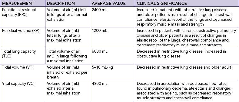
LUNG VOLUMES AND CAPACITIES

Spirometry is used to measure the volume of air entering or leaving the lungs. Variations in lung volumes may be associated with health states such as pregnancy, exercise, obesity, or obstructive and restrictive conditions of the lungs. The amount of surfactant, degree of compliance and strength of respiratory muscles can affect pressures and volumes within the lungs.

The volume of air in the lungs is measured in various ways and combined to describe lung capacities (see pulmonary function tests later in the chapter).

Gases are moved into and out of the lungs through pressure changes (Figure 40-9). Intrapleural pressure is negative relative to atmospheric pressure, which is 760 mmHg at sea level. For air to flow into the lungs, intrapleural pressure must become more negative, setting up a pressure gradient between the atmosphere and the alveoli.

image

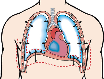
FIGURE 40-9 Frontal section of chest showing movement of the lung and chest wall during inspiration and expiration. During inspiration the inspiratory muscles contract and the chest expands. Alveolar pressure becomes subatmospheric with respect to pressure at the airway opening, and air flows into the lungs. During expiration the inspiratory muscles relax. Recoil of the lung causes alveolar pressure to exceed pressure at the airway opening and air to flow out of the lungs. Single arrows show excursion of the lungs and chest wall. Double arrows show movement of the lung bases.

From Lewis SM and others 2004 Medical–surgical nursing: assessment and management of clinical problems, ed 6. St Louis, Mosby.

PULMONARY CIRCULATION

The main function of the pulmonary circulation is to move blood to and from the alveolocapillary membrane for gas exchange to occur. The pulmonary circulation is a reservoir for blood so that the lung can increase its blood volume without large increases in pulmonary artery or venous pressures. The pulmonary circulation also acts as a filter, removing small thrombi before they can reach vital organs.

The pulmonary circulation begins at the pulmonary artery, which receives poorly oxygenated mixed venous blood from the right ventricle. Blood flow through this system depends on the pumping ability of the right ventricle, which has an output of approximately 5 L/min. The flow continues from the pulmonary artery through the pulmonary arterioles to the pulmonary capillaries, where blood comes in contact with the alveolar–capillary membrane and the exchange of oxygen and carbon dioxide occurs. The now oxygen-rich blood then circulates through the pulmonary venules and pulmonary veins, returning to the left atrium.

Pressure and resistance within the pulmonary circulatory system is lower than that within the systemic circulatory system. The walls of the pulmonary vessels are thinner and contain less smooth muscle. The lung accepts the total cardiac output from the right ventricle and, except in some specific pathophysiological circumstances, does not direct blood flow from one region to another.

EXCHANGE OF OXYGEN AND CARBON DIOXIDE

Oxygen and carbon dioxide are exchanged in the alveoli and the capillaries of the body tissues. Oxygen is transferred from the alveoli of the lungs to the pulmonary circulation, and carbon dioxide is transferred from the pulmonary circulation to the alveoli to be exhaled as a waste product. At the tissue level, oxygen is transferred from the arterial blood to tissues, and carbon dioxide is transferred from tissues to the venous blood to return to the pulmonary circulation and alveoli and be exhaled. This transfer depends on the process of diffusion.

Diffusion is the movement of molecules from an area of higher concentration to an area of lower concentration (see Chapter 39). Diffusion of oxygen and carbon dioxide occurs at the alveolar–capillary membrane. Oxygen diffuses into the pulmonary circulation from the alveoli and carbon dioxide diffuses from the pulmonary circulation into the alveoli to be expired.

OXYGEN TRANSPORT

The oxygen transport system consists of the respiratory and cardiovascular systems. Delivery depends on the amount of oxygen entering the lungs (ventilation), blood flow to the lungs and tissues (perfusion), rate of diffusion, and oxygen-carrying capacity. The capacity of the blood to carry oxygen is influenced by the amount of dissolved oxygen in the plasma, amount of haemoglobin and tendency of haemoglobin to bind with oxygen. Only a relatively small amount of required oxygen, less than 1%, is dissolved in the plasma (Brashers, 2010a). Most oxygen is transported by haemoglobin, which serves as a carrier for oxygen and carbon dioxide. The haemoglobin molecule combines with oxygen to form oxyhaemoglobin. The formation of oxyhaemoglobin is easily reversible, allowing haemoglobin and oxygen to dissociate, which frees oxygen to enter tissues.

CARBON DIOXIDE TRANSPORT

Carbon dioxide diffuses into red blood cells and is rapidly hydrated into carbonic acid (H2CO3) because of the presence of carbonic anhydrase. The carbonic acid then dissociates into hydrogen (H+) ions and bicarbonate (HCO3) ions. The hydrogen ion is buffered by haemoglobin, and the HCO3diffuses into the plasma (see Chapter 39). In addition, some of the carbon dioxide in red blood cells reacts with amino acid groups, forming carbamino compounds. This reaction can occur rapidly without the presence of an enzyme. Reduced haemoglobin (deoxyhaemoglobin) can combine with carbon dioxide more easily than oxyhaemoglobin, and therefore venous blood transports the majority of carbon dioxide.

REGULATION OF RESPIRATION

Regulation of respiration is necessary to ensure sufficient oxygen intake and carbon dioxide elimination to meet the body’s demands (e.g. during exercise, infection or pregnancy). Neural and chemical regulators control the process of respiration. Neural regulation includes the central nervous system control of respiratory rate, depth and rhythm. Chemical regulation involves the influence of chemicals such as carbon dioxide and hydrogen ions on the rate and depth of respiration (Box 40-3).

BOX 40-3 NEURAL AND CHEMICAL REGULATION OF RESPIRATION

NEURAL REGULATION

Maintains rhythm and depth of respiration and balance between inspiration and expiration.

CEREBRAL CORTEX

Voluntary control of respiration delivers impulses to the respiratory motor neurons by way of the spinal cord; accommodates speaking, eating and swimming.

MEDULLA OBLONGATA

Automatic control of respiration occurs continuously.

CHEMICAL REGULATION

Maintains appropriate rate and depth of respirations based on changes in the blood’s carbon dioxide, oxygen and hydrogen ion (H+) concentration. Chemical regulation can occur during physical exercise and in some illnesses. It is a short-term adaptive mechanism.

CHEMORECEPTORS

Located in the medulla, aortic body and carotid body. Changes in chemical content of oxygen, carbon dioxide and hydrogen ions stimulate chemoreceptors, which in turn stimulate neural regulators to adjust the rate and depth of ventilation to maintain normal arterial blood gas levels.

Factors affecting oxygenation

Adequacy of ventilation, perfusion and transport of oxygen to the tissues is influenced by four types of factors: (1) physiological, (2) developmental, (3) behavioural and (4) environmental. Physiological factors are outlined below; developmental, behavioural (lifestyle) and environmental factors are discussed in the following section (nursing knowledge base).

Physiological factors

Any condition that affects cardiopulmonary functioning directly affects the body’s ability to meet oxygen demands. The general classifications of cardiac disorders include disturbances in conduction, impaired valvular function, myocardial hypoxia, cardiomyopathic conditions and peripheral tissue hypoxia. Respiratory disorders are caused by one or more of the following factors: ventilation/perfusion (V/Q) mismatch, diffusion impairment, alveolar hypoventilation and alveolar hyperventilation.

Other physiological processes affecting oxygenation are presented in Table 40-1.

TABLE 40-1 PHYSIOLOGICAL PROCESSES AFFECTING OXYGENATION

PROCESS EFFECT ON OXYGENATION
Airway obstruction Limits delivery of inspired oxygen to alveoli
Anaemia Decreases oxygen-carrying capacity of blood
Decreased chest wall motion (e.g. from musculoskeletal impairments) Prevents lowering of diaphragm and reduces anteroposterior diameter of thorax on inspiration, reducing volume of air inspired
Fever, pregnancy, infection Increases metabolic rate and tissue oxygen demand
High altitude Atmospheric oxygen concentration is lower and inspiratory oxygen concentration decreases
Toxic inhalant Decreases oxygen-carrying capacity of blood

DECREASED OXYGEN-CARRYING CAPACITY

Haemoglobin carries 99% of the diffused oxygen to tissues (Brashers, 2010a). Anaemia and inhalation of toxic substances decrease the oxygen-carrying capacity of blood by reducing the amount of available haemoglobin to transport oxygen. Anaemia, a lower than normal haemoglobin level, is a result of decreased haemoglobin production, increased red blood cell destruction and/or blood loss. Patients complain of fatigue, decreased activity tolerance and increased breathlessness, as well as pallor (especially seen in the conjunctiva of the eye) and an increased heart rate.

Carbon monoxide is the most common toxic inhalant that decreases the oxygen-carrying capacity of blood. The affinity for haemoglobin to bind with carbon monoxide is greater than 200 times its affinity to bind with oxygen, creating a functional anaemia. Because of the bond’s strength, carbon monoxide is not easily dissociated from haemoglobin, making the haemoglobin unavailable for oxygen transport.

DECREASED INSPIRED OXYGEN CONCENTRATION

When the concentration of inspired oxygen declines, the oxygen carried by the blood is decreased. Decreases in the fraction of inspired oxygen concentration (FiO2) can be caused by an upper-airway obstruction (e.g. croup in the child) or lower-airway obstruction (e.g. COPD in the adult) limiting delivery of inspired oxygen to alveoli; decreased environmental oxygen, such as at high altitudes; or decreased inspiration as a result of an incorrect oxygen concentration setting on respiratory therapy equipment.

HYPOVOLAEMIA

Conditions such as shock and severe dehydration resulting from extracellular fluid loss and reduced circulating blood volume cause hypovolaemia. With a significant fluid loss, the body tries to adapt by increasing the heart rate and peripheral vasoconstriction through sympathetic nervous system stimulation to increase the volume of blood returned to the heart and, in turn, increase the cardiac output. Consider the child with vomiting and diarrhoea from gastroenteritis. The nursing assessment will likely reveal a child who speaks minimally and is tachypnoeic (Airway and Breathing); pale, tachycardic with capillary refill greater than 3 seconds (Circulation); and is listless (Disability). The loss in circulating volume caused by the extreme fluid loss from the vomiting and diarrhoea means that preload and therefore stroke volume (SV) fall, dropping cardiac output (CO). Remember that CO = SV × HR. The sympathetic nervous system responds by increasing HR (tachycardic) and causing vasoconstriction which increases afterload (pale and capillary refill greater than 3 seconds), all to try to improve CO. At the same time, the respiratory centre is stimulated to increase respiratory rate (tachypnoeic) in an effort to deliver more oxygen. As CO has fallen, perfusion to the brain is diminished and so the child is listless.

INCREASED METABOLIC RATE

Increases in metabolic activity result in an increased oxygen demand. When body systems are unable to meet this increased demand, the level of oxygenation declines. An increased metabolic rate is a normal physiological response to pregnancy, wound healing and exercise because the body is building tissue. Most people can meet the increased oxygen demand and do not display signs of oxygen deprivation. Fever increases the tissues’ need for oxygen, and as a result carbon dioxide production also increases. If the febrile state persists, the metabolic rate remains high and the body begins to break down protein stores, resulting in muscle wasting and decreased muscle mass. Respiratory muscles such as the diaphragm and intercostal muscles are also wasted. The body attempts to adapt to the increased carbon dioxide levels by increasing the rate and depth of respiration. The patient’s work of breathing increases and the patient eventually displays signs and symptoms of hypoxaemia. Those patients with pulmonary diseases are at greater risk of hypoxaemia and hypercapnoea due to V/Q mismatch and alveolar hypoventilation (see later in the chapter). Assessment findings include an increased rate and depth of respiration, use of the accessory muscles of respiration, pursed-lip breathing and decreased activity tolerance.

CONDITIONS AFFECTING CHEST WALL MOVEMENT

Any condition that reduces chest wall movement can result in decreased ventilation. If the diaphragm cannot fully descend with breathing, the volume of inspired air decreases and less oxygen is delivered to the alveoli and subsequently to tissues. For example, thoracic or abdominal injury or surgery will cause considerable pain, which inhibits diaphragmatic and chest wall movement, reducing inspiration and therefore delivery of oxygen.

PREGNANCY

As the fetus grows during pregnancy, the greater size of the uterus pushes abdominal contents upwards against the diaphragm. In the last trimester of pregnancy, the inspiratory capacity declines, resulting in dyspnoea on exertion and increased fatigue.

OBESITY

Obese patients have reduced lung volumes from the heavy lower thorax and abdomen, particularly when in the recumbent and supine positions. Obese patients have a reduction in compliance as a result of encroachment of the abdomen into the chest, increased work of breathing and decreased lung volumes, and they may have fatigue and carbon dioxide retention. In some patients an obesity–hypoventilation syndrome develops in which oxygenation is decreased and carbon dioxide is retained, resulting in daytime sleepiness. The obese patient is also susceptible to pneumonia after an upper respiratory tract infection because the lungs cannot fully expand and pulmonary secretions are not mobilised in the lower lobes.

MUSCULOSKELETAL ABNORMALITIES

Musculoskeletal impairments in the thoracic region reduce oxygenation. Such impairments may result from abnormal structural configurations, trauma, muscular diseases and diseases of the central nervous system. Abnormal structural configurations impairing oxygenation include those that affect the rib cage, such as pectus excavatum (abnormal development of ribs causing caved in appearance), and those that affect the vertebral column, such as kyphosis.

TRAUMA

Two or more ribs fractured in two or more places results in a flail chest, a condition in which fractures cause instability in part of the chest wall. The unstable chest wall allows the lung underlying the injured area to contract on inspiration and bulge on expiration, resulting in hypoxia. Chest wall or upper abdominal incisions may also decrease chest wall movement as the patient uses shallow respirations to minimise chest wall movement to avoid pain. Narcotic analgesia, e.g. morphine, is often prescribed to relieve the pain, thereby facilitating improved chest wall expansion. Should excessive or high doses of narcotic analgesics be administered, this may depress the respiratory centre in the brain, further decreasing respiratory rate and chest-wall expansion and therefore decreasing oxygenation.

NEUROMUSCULAR DISEASES

Diseases such as muscular dystrophy affect oxygenation of tissues by decreasing the patient’s ability to expand and contract the chest wall. Ventilation is impaired, and atelectasis, hypercapnoea and hypoxaemia can occur. Myasthenia gravis and Guillain-Barré syndrome affect respiratory functioning and result in alveolar hypoventilation (see later in the chapter). Myasthenia gravis interferes with the normal transmission of impulses from nerves to muscles, involving the whole body, including muscles of respiration. Guillain-Barré syndrome causes inflammation and paralysis of muscle groups, which usually results in an ascending pattern of paralysis. Respiratory muscles become paralysed as paralysis ascends to the thoracic region.

CENTRAL NERVOUS SYSTEM ALTERATIONS

Diseases or trauma involving the medulla oblongata and spinal cord may result in impaired respiration. When the medulla oblongata (contains the respiratory centre) is affected, neural regulation of respiration is damaged and abnormal breathing patterns may develop. If the phrenic nerve is damaged, the diaphragm may not descend, thus reducing inspiratory lung volumes and causing hypoxaemia. Cervical trauma at vertebrae C3 to C5 can result in paralysis of the phrenic nerve. Spinal cord trauma below the fifth cervical vertebra usually leaves the phrenic nerve intact but damages nerves that innervate the intercostal muscles, preventing anteroposterior chest expansion.

INFLUENCES OF CHRONIC DISEASE

Oxygenation can be decreased as a direct consequence of chronic disease. It can also be decreased as a secondary effect, as with anaemia. The physiological response to chronic hypoxaemia is the development of a secondary polycythaemia. This adaptive response is the body’s attempt to increase the amount of circulating haemoglobin to increase the available oxygen-binding sites.

Alterations in cardiac functioning

Illnesses and conditions that affect cardiac rhythm, strength of contraction, blood flow through the atria and ventricles, myocardial blood flow and peripheral circulation cause alterations in cardiac functioning.

Disturbances in conduction

Some disturbances in conduction are a result of electrical impulses that do not originate from the SA node. These rhythm disturbances are called arrhythmias, meaning a deviation from the normal sinus rhythm (Table 40-2). Arrhythmias may occur as a primary conduction disturbance as a response to ischaemia, valvular abnormality, anxiety or drug toxicity; caffeine, alcohol or tobacco use; or as a complication of acid–base or electrolyte imbalance (see Chapter 39).

TABLE 40-2 COMMON CARDIAC ARRHYTHMIAS

image

Aehlert B 2011 ECGs made easy, ed 4. St Louis, Mosby; Australian Resuscitation Council (ARC) 2010 Guideline 8, cardiopulmonary resuscitation. Melbourne, ARC. Online. Available at www.resus.org.au 30 May 2011; Moser DB, Riegel B 2008 Cardiac nursing: a companion to Braunwald’s Heart disease. Philadelphia, Saunders.

Images from Aehlert (2011).

Arrhythmias are classified by site of impulse origin and mechanism of the cardiac response. Cardiac response can be one of tachycardia (> 100 beats per minute), bradycardia (< 60 beats per minute), a premature (early) beat or a blocked (delayed or absent) beat. Tachyarrhythmia can lower cardiac output and blood pressure. Tachyarrhythmia reduces cardiac output by decreasing diastolic filling time. Bradyarrhythmia lowers cardiac output because of the decreased heart rate.

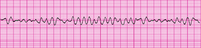
Abnormal impulses originating above the ventricles are referred to as supraventricular arrhythmias. The abnormality of the wave form is the configuration and placement of the P wave. Ventricular conduction usually remains normal and a normal QRS complex is observed. Junctional arrhythmias represent an abnormal site of impulse conduction above or below the AV node. The P wave can occur before, during or after the QRS complexes and is often inverted if visible. Because the beat originates above the ventricle, ventricular conduction and the QRS complex are usually normal. One of the most common supraventricular arrhythmias is atrial fibrillation and occurs most frequently with ageing and in heart failure. There is disorganised chaotic depolarisation of the atria at a very rapid rate (400–600 beats/min) resulting in fibrillatory waves. Conduction of many of these beats is blocked at the AV node and conduction through the ventricle is usually normal. The ventricular rate varies but is usually quite rapid. Due to the lack of synchrony between atria and ventricles, thrombi can form in the atria, potentially causing a stroke when sinus rhythm is restored (Jacobson, 2010b). Patients often require anticoagulation, usually with warfarin. A new anticoagulant, dabigatran, appears superior with fewer side effects (Talati and White, 2011). Management focuses on either rate control or rhythm control, with neither approach holding an outcome advantage over the other (Van Gelder and others, 2002; Wyse and others, 2002).

Ventricular arrhythmias represent an ectopic site of impulse formation within the ventricles. The configuration of the QRS complex is usually widened and bizarre. P waves may or may not be present; often they are buried in the QRS complex. Ventricular tachycardia and ventricular fibrillation are life-threatening arrhythmias that require immediate intervention. Ventricular tachycardia is considered a life-threatening arrhythmia because of the decreased cardiac output and the potential to deteriorate into ventricular fibrillation (Jacobson, 2010b).

Altered cardiac output

Failure of the myocardium to eject sufficient volume to the systemic and pulmonary circulations can result in heart failure. Failure of the myocardial pump results from primary coronary artery disease, cardiomyopathic conditions, valvular disorders and pulmonary disease.

LEFT-SIDED HEART FAILURE

Left-sided heart failure is an abnormal condition characterised by impaired functioning of the left ventricle due to elevated pressures. If left ventricular failure is significant, the amount of blood ejected from the left ventricle drops greatly, resulting in decreased cardiac output. Consider the patient experiencing an acute episode of heart failure. The nursing assessment will likely reveal a person who speaks in phrases, is tachypnoeic, is using accessory muscles of respiration, has crackles on auscultation of lung fields, has low oxygen saturations (Airway and Breathing) and is hypotensive and tachycardic with capillary refill greater than 3 seconds (Circulation). As the left ventricle is unable to pump effectively and stroke volume and therefore CO is reduced (hypotensive), sympathetic nervous system stimulation increases HR (tachycardic) and causes vasoconstriction (capillary refill greater than 3 seconds) to compensate. The increased volume and therefore pressure in the left ventricle due to its ineffective pumping action causes the pressure to be reflected backwards into the pulmonary circulation. The increased pressure forces fluid out of the pulmonary circulation into the interstitium and alveoli (crackles on auscultation), impairing oxygenation (low oxygen saturations). The respiratory centre is stimulated, resulting in an increased respiratory rate (tachypnoeic) to compensate and try to deliver more oxygen. Other assessment findings may include decreased activity tolerance, dizziness and confusion as a result of tissue hypoxia from the diminished cardiac output. Also, cough and paroxysmal nocturnal dyspnoea may be evident due to the pulmonary congestion.

RIGHT-SIDED HEART FAILURE

Right-sided heart failure results from impaired functioning of the right ventricle characterised by venous congestion in the systemic circulation. Right-sided heart failure more commonly results from pulmonary disease or as a result of long-term left-sided failure. The primary pathological factor in right-sided failure is elevated pulmonary vascular resistance (PVR). As the PVR continues to rise, the right ventricle must generate more work, and the oxygen demand of the heart increases. As the failure continues, the amount of blood ejected from the right ventricle declines, and blood begins to ‘back up’ in the systemic circulation. Clinically, the patient has weight gain due to fluid retention, distended neck veins, hepatomegaly and splenomegaly and dependent peripheral oedema.

Impaired valvular function

Valvular heart disease is an acquired or congenital disorder of a cardiac valve characterised by stenosis and obstructed blood flow or valvular degeneration and regurgitation of blood. When stenosis occurs in the semilunar valves (aortic and pulmonic valves), the adjacent ventricles must work harder to move the ventricular volume beyond the stenotic valve. Over time, the stenosis can cause the ventricle to hypertrophy (enlarge), and if the condition is untreated, left- or right-sided heart failure can occur. If stenosis occurs in the atrioventricular valves (mitral and tricuspid valves), the atrial pressure rises, causing the atria to hypertrophy. When regurgitation occurs, there is a backflow of blood into an adjacent chamber. For example, in mitral regurgitation the mitral leaflets do not close completely. When the ventricle contracts, blood escapes back into the atria, causing a murmur or ‘whooshing’ sound (see Chapter 27).

Myocardial ischaemia

Myocardial ischaemia results when the supply of blood to the myocardium from the coronary arteries is insufficient to meet the oxygen demands of the organ. Atherosclerotic plaque develops, narrowing the lumen of the coronary artery and leading to thrombus formation and eventually occlusion. Two common manifestations of this ischaemia are angina pectoris and myocardial infarction. These collectively are termed acute coronary syndrome. When a patient first presents with ischaemic type chest pain, the diagnosis of acute coronary syndrome is made until it is determined whether the presentation is angina pectoris or a myocardial infarction. This prevents delay in management (Aroney and others, 2006).

ANGINA

Angina pectoris is usually a transient imbalance between myocardial oxygen supply and demand due to reversible myocardial ischaemia. The condition results in chest pain that is aching, sharp, tingling or burning, or that feels like pressure. The chest pain may be left-sided or substernal and may radiate to the left or both arms, and to the jaw, neck and back. In some patients, anginal pain may not radiate. The pain can last from 1 to 15 minutes. Patients report that pain is often precipitated by activities that increase myocardial oxygen demand (e.g. exercise, anxiety or stress). The pain is usually relieved with rest and coronary vasodilators, the most common being a glyceryl trinitrate preparation, e.g. Anginine (Brashers, 2010c).

MYOCARDIAL INFARCTION

Myocardial infarction results from sudden decreases in coronary artery blood flow or an increase in myocardial oxygen demand without adequate coronary artery perfusion. Infarction occurs when ischaemia (which is reversible) remains untreated or inadequately treated and becomes necrosis (which is not reversible) of myocardial tissue (Brashers, 2010c).

Chest pain associated with myocardial infarction is usually described as crushing, squeezing or stabbing. The pain may be retrosternal and left praecordial, and it may radiate down the left arm to the neck, jaws, teeth, epigastric area and back. The pain occurs at rest or exertion, lasts more than 30 minutes and is unrelieved by rest, position change or sublingual glyceryl trinitrate administration.

Research indicates that there is a significant difference between men and women in relation to coronary artery disease. It is known that women do not always present the same type of symptoms as men (Miracle, 2010). Symptoms are likely to be more subtle in women, with atypical presentation such as a sensation of indigestion, heaviness or general discomfort. Dyspnoea, fainting, nausea, fatigue and palpitations are also common symptoms in women. A first presentation of coronary artery disease in women is likely to be angina, while in men a first presentation is more likely to be a myocardial infarction (Miracle, 2010). Oestrogen replacement in healthy postmenopausal women may reduce and prevent coronary artery disease. Additional risk factors for coronary artery disease in women include menopause and hormonal contraceptives, such as birth-control pills.

• CRITICAL THINKING

Wanda Johnson is a 56-year-old postmenopausal woman with a history of hypertension. What would you include in the teaching portion of her plan of care?

Alterations in respiratory functioning

Illnesses and conditions that affect ventilation or oxygen transport cause alterations in respiratory functioning. The main alterations are ventilation/perfusion (V/Q) mismatch, diffusion impairment, alveolar hypoventilation, alveolar hyperventilation and hypoxia.

Alveolar hyperventilation

The goal of ventilation is to produce a normal arterial carbon dioxide tension (PaCO2) between 35 and 45 mmHg and maintain a normal arterial oxygen tension (PaO2) between 95 and 100 mmHg. Arterial oxygen levels can be monitored using a non-invasive oxygen saturation monitor. The normal range is 95–100%.

Alveolar hyperventilation is a state of ventilation in excess of that required to eliminate the normal venous carbon dioxide produced by cellular metabolism. Anxiety, infections, drugs or an acid–base imbalance can induce alveolar hyperventilation, as well as hypoxia associated with pulmonary embolus or shock. Acute anxiety can lead to alveolar hyperventilation and may cause loss of consciousness from excess carbon dioxide exhalation. Fever can cause alveolar hyperventilation. For each increase of 1°C there is a 12% increase in metabolic rate, thereby increasing carbon dioxide production. The clinical response is an increased rate and depth of respiration.

Alveolar hyperventilation may also be chemically induced. Salicylate (aspirin) poisoning causes excessive stimulation of the respiratory centre as the body attempts to compensate for excess carbon dioxide. Amphetamines also increase ventilation by raising carbon dioxide production. Alveolar hyperventilation can also occur as the body tries to compensate for metabolic acidosis by producing a respiratory alkalosis. For example, the patient with diabetes mellitus who has gone into diabetic ketoacidosis is producing large amounts of metabolic acids. The respiratory system tries to correct the acid–base imbalance by increasing the respiratory rate. Ventilation increases to reduce the amount of carbon dioxide available to form carbonic acid (see Chapter 39).

Alveolar hyperventilation produces many signs and symptoms that can be assessed (Table 40-3). Haemoglobin does not release oxygen to tissues as readily, and tissue hypoxia results. As symptoms worsen, the patient may become more agitated, which further increases the respiratory rate and can result in respiratory alkalosis.

TABLE 40-3 DIFFERENTIATING ALVEOLAR HYPERVENTILATION FROM HYPOVENTILATION

HYPERVENTILATION HYPOVENTILATION
FEATURES

Mental status

Disorientation

Light-headedness

Dizziness

Disorientation

Lethargy

Dizziness

Headache

Decreased ability to follow instructions

Convulsions

Coma

CARDIOVASCULAR SIGNS AND SYMPTOMS

Tachycardia

Chest pain

Shortness of breath

Arrhythmias

Cardiac arrest

NEUROLOGICAL SIGNS AND SYMPTOMS

Blurred vision

Paraesthesias

Extremity and circumoral numbness

Carpopedal spasm (tetany)

Tinnitus

 
OTHER
Acid–base and electrolyte imbalances Acid–base and electrolyte imbalances

Alveolar hypoventilation

Alveolar hypoventilation occurs when alveolar ventilation is inadequate to meet the body’s oxygen demand or to eliminate sufficient carbon dioxide. As alveolar ventilation decreases, PaCO2 is elevated (hypercapnoea). Severe atelectasis can produce alveolar hypoventilation. Atelectasis is a collapse of the alveoli that prevents normal diffusion of oxygen and carbon dioxide. As alveoli collapse, less of the lung can be ventilated and alveolar hypoventilation occurs.

Patients with COPD have a progressive deterioration of the airways, leading to a loss of lung function resulting in alveolar hypoventilation. Some have a chronically elevated PaCO2 and low PaO2. In this instance, the central chemoreceptors primarily responsible for detecting changes in pH (and PaCO2) are less sensitive, and peripheral chemoreceptors are now the primary stimulus to ventilation. Remember that peripheral chemoreceptors are more sensitive to oxygen than carbon dioxide (Brashers, 2010a). A small percentage of these patients in response to supplemental oxygen will experience a significant elevation in PaCO2, which causes drowsiness and lethargy. This is known as hypercapnoeic encephalopathy or CO2 narcosis (Simmons & Simmons, 2004). To avoid this complication, supplemental oxygen therapy should be titrated to achieve oxygen saturations of 88–92% (Beasley and others, 2011).

Signs and symptoms of alveolar hypoventilation are presented in Table 40-3. If untreated, the patient’s status can rapidly decline. Convulsions, unconsciousness and death can result. Treatment for alveolar hyperventilation and alveolar hypoventilation requires improving tissue oxygenation, restoring ventilatory function, treating the underlying cause and achieving acid–base balance.

Ventilation/perfusion mismatch

For oxygenation to occur, both adequate ventilation and adequate perfusion is needed. This is termed the ventilation/perfusion (V/Q) ratio and is normally approximately 4 : 5 (ventilation 4 L/min : perfusion 5 L/min). If there is impairment with either mechanism, a V/Q mismatch occurs. The patient with pneumonia will have V/Q mismatch due to impaired ventilation, while a patient with a pulmonary embolism will have a V/Q mismatch due to impaired perfusion. The overall result is a lowering of oxygen (Pierce, 2007).

Diffusion impairment

The rate of diffusion of oxygen can be affected by the thickness of the alveolar–capillary membrane and the available surface area for diffusion. Increased thickness of the membrane impedes diffusion because oxygen takes longer to diffuse across. Patients with pulmonary oedema or pneumonia have an increased thickness of the alveolar–capillary membrane, resulting in slowed diffusion, slowed exchange of oxygen and impaired delivery of oxygen to tissues. The surface area of the membrane can be altered as a result of a chronic disease (e.g. emphysema), an acute disease (e.g. pneumothorax) or a surgical process (e.g. lobectomy). The alveolar–capillary membrane can be destroyed or may thicken, changing the rate of diffusion. When fewer alveoli are functioning, the surface area is decreased. This alteration in membrane thickness and/or surface area which results in impaired gas exchange is termed diffusion impairment (Pierce, 2007).

Hypoxia

Hypoxia is inadequate tissue oxygenation at the cellular level. This can result from a deficiency in oxygen delivery or oxygen utilisation at the cellular level. Hypoxia can be caused by:

1. a decreased haemoglobin level and lowered oxygen-carrying capacity of the blood

2. a diminished concentration of inspired oxygen, which may occur at high altitudes

3. the inability of the tissues to extract oxygen from the blood, as with cyanide poisoning

4. diffusion impairment as in pneumonia

5. poor tissue perfusion with oxygenated blood, as with shock

6. V/Q mismatch as with multiple rib fractures or chest trauma.

The clinical signs and symptoms of hypoxia include apprehension, restlessness, inability to concentrate, a declining level of consciousness, dizziness and behavioural changes (Box 40-4). The patient with a narcotic overdose, such as a heroin overdose, which depresses the respiratory centre leading to a decreased ventilatory drive, may display signs of alveolar hypoventilation. During early stages of hypoxia, the blood pressure is elevated unless the condition is caused by shock. As the hypoxia worsens, the respiratory rate may decline as a result of respiratory muscle fatigue.

BOX 40-4 HYPOXIA—SIGNS AND SYMPTOMS

Restlessness

Apprehension, anxiety

Disorientation

Decreased ability to concentrate

Decreased level of consciousness

Increased fatigue

Dizziness

Behavioural changes

Increased pulse rate

Increased rate and depth of respiration

Elevated blood pressure

Cardiac arrhythmias

Pallor

Cyanosis

Clubbing

Dyspnoea

Hypoventilation

PAEDIATRIC CARDINAL SIGNS

Restlessness

Irritability

Increased rate of respiration

Increased pulse rate

Profuse sweating

Nasal flaring

Intercostal, subcostal recession

Tracheal tug

Grunt on expiration

Head bobbing

Cyanosis, blue discolouration of the skin and mucous membranes caused by the presence of desaturated haemoglobin in capillaries, is a late sign of hypoxia. The presence or absence of cyanosis is not a reliable measure of oxygenation status. Central cyanosis, observed in the tongue, soft palate and conjunctiva of the eye, where blood flow is high, indicates hypoxaemia. Peripheral cyanosis, seen in the extremities, nail beds and earlobes, is often a result of vasoconstriction and stagnant blood flow.

Hypoxia is a life-threatening condition. Untreated, it can produce cardiac arrhythmias that result in death. Hypoxia is managed by administration of oxygen and treatment of the underlying cause, such as airway obstruction.

Nursing knowledge base

Developmental factors

The developmental stage of the patient and the normal ageing process can affect tissue oxygenation.

Premature infants

Premature infants are at risk of respiratory distress syndrome (previously known as hyaline membrane disease), which is caused by a surfactant deficiency. The surfactant-synthesising ability of the lungs develops late in pregnancy, about the seventh month, and may therefore be lacking in preterm infants (Wheeler, 2009).

Infants and toddlers

Maternal antibodies provide protection from infection in the infant under 3 months of age. Infants and toddlers are at risk of upper respiratory tract infections as a result of frequent exposure to other children and exposure to secondhand smoke. In addition, during the teething process some infants develop nasal congestion, which encourages bacterial growth and increases the potential for respiratory tract infection. Upper respiratory tract infections are usually not dangerous, and infants or toddlers recover with little difficulty. Common airway infections are nasopharyngitis (e.g. rhinoviruses, respiratory syncytial virus and adenovirus), pharyngitis (e.g. viral and beta-haemolytic streptococci), Haemophilus influenzae infection and tonsillitis. Airway obstruction can also occur with aspirated foreign objects, such as food, buttons and lollies. Airways are smaller in younger children and can narrow considerably from oedema of mucous membranes accompanying an infection. The chest wall is thin and therefore very compliant, and accessory muscles of respiration are poorly developed. Both of these characteristics limit the child’s ability to increase depth of breathing or tidal volume, the volume of air in a breath. Consequently, tachypnoea is a characteristic response to illness (Kendrick and Morrison, 2007; Wilson, 2009).

School-age children and adolescents

School-age children and adolescents are exposed to respiratory infections and respiratory risk factors such as second-hand smoke and cigarette smoking. A healthy child usually does not have adverse pulmonary effects from respiratory infections. A person who starts smoking in adolescence and continues to smoke into middle age, however, has an increased risk of cardiopulmonary disease and lung cancer.

Young and middle-aged adults

Young and middle-aged adults are exposed to multiple cardiopulmonary risk factors: an unhealthy diet, lack of exercise, stress, occupational hazards, drugs and smoking. Reducing these modifiable factors may decrease the patient’s risk of cardiac or pulmonary diseases. This is also the time when lifelong habits and lifestyles are established. It is important to help these patients make good choices and informed decisions about the rest of their lives and their healthcare practices.

Older adults

WORKING WITH DIVERSITY FOCUS ON OLDER ADULTS

Older adults generally have a respiratory rate between 16 and 25 breaths per minute. Because they depend on changes in intra-abdominal pressure, positioning can greatly affect their breathing pattern. Position the patient to maximise ventilation. Positions such as semi-Fowler’s and high-Fowler’s provide the best ventilation. The older patient should be encouraged to sit up in the chair to promote good lung expansion.

Frequent, smaller meals and fewer bloating and gas-producing foods will help prevent an over-distended abdomen and reduce pressure on the diaphragm.

Obese patients may need to sleep in semi-Fowler’s position.

The cardiac and respiratory systems undergo changes throughout the ageing process. In the arterial system, atherosclerotic plaques develop and the systemic blood pressure may rise. Chest wall compliance is decreased in the older adult as a result of osteoporosis and calcification of the costal cartilages. The respiratory muscles weaken, and the pulmonary vascular circulation becomes less distensible (see Working with diversity). The trachea and large bronchi become enlarged from calcification of the airways, and alveoli enlarge, decreasing the surface area available for gas exchange. In addition, the number of functional cilia is reduced. Decreased ciliary action and effectiveness of cough mechanisms put the older adult at increased risk of respiratory infections (Ebersole and others, 2008). Ventilation and diffusion of oxygen and carbon dioxide decline with age. Osteoporotic changes of the thoracic cage and kyphosis of the vertebrae occur normally with ageing (Jet, 2008). With these changes the lungs are unable to expand fully, leading to lower oxygenation levels (Table 40-4).

TABLE 40-4 CHANGES IN THE AGEING LUNG

FUNCTION PATHOPHYSIOLOGICAL CHANGE KEY CLINICAL FINDINGS
Breathing mechanics

Decreased chest-wall compliance

Decreased pulmonary compliance

Loss of elastic recoil

Decreased respiratory muscle mass and strength

Uneven alveolar ventilation

Decreased thoracic wall excursion

Decreased vital capacity

Increased residual volume

Decreased forced expiratory volume

Decreased airway clearance (coughing)

Oxygenation

Increased physiological dead space

Ventilation/perfusion mismatch

Decreased alveolar surface area

Decreased carbon dioxide diffusion capacity

Decreased arterial partial pressure of oxygen (PaO2)

Increased alveolar–arterial oxygen gradient

Ventilation control and breathing pattern Decreased responsiveness of central and peripheral chemoreceptors to hypoxaemia and hypercapnia

Decreased tidal volume

Increased respiratory rate

Increased minute ventilation

Decreased airway clearance

Lung defence mechanisms

Decreased number of cilia and effectiveness of the mucociliary clearance

Decrease in mucous production

Diminished cough reflex

Decreased T-cell function

Decreased immunoglobulin A (IgA) production

Increased risk of infection

Thickened secretions

Increased risk of aspiration

Sleep and breathing

Decreased ventilatory drive

Decreased tone of upper airway muscles

Decreased arousal

Increased risk of apnoea, hypopnoea and arterial oxygen desaturation during sleep

Increased risk of aspiration

Snoring

Obstructive sleep apnoea

Exercise capacity

Muscle deconditioning and efficiency

Decreased muscle mass

Decreased reserves

Decreased maximum oxygen consumption

Breathlessness at low exercise levels

Modified from Pierson DJ 1992 Effects of aging on the respiratory system. In Pierson DJ, Kacmarek RM, editors, Foundations of respiratory care. New York, Churchill Livingstone; Criddle LM 2009 Caring for the critically ill elderly patient. In Carlson KK, editor 2009 Advanced critical care nursing. St Louis, Saunders.

Lifestyle factors

Lifestyle factors that influence cardiopulmonary functioning include nutrition, exercise, cigarette smoking, substance abuse and stress (Box 40-5).

BOX 40-5 CARDIOPULMONARY HEALTH PROMOTION*

Maintain ideal bodyweight.

Eat a low-fat, low-salt, kilojoule-appropriate diet.

Monitor cholesterol and triglyceride levels.

Engage in regular aerobic exercise.

Use stress-reduction techniques.

Be smoke-free.

Avoid secondhand smoke and other pollutants.

Use a filter mask when exposed to occupational hazards.

Monitor blood pressure.

Get an annual influenza vaccine if at risk of developing influenza.

Get a pneumococcal vaccine if appropriate.

Reduce exposure to secondary infections.

*Target population: young to older adults.

Nutrition

Nutrition affects cardiopulmonary function in several ways. Severe obesity decreases lung expansion, and the increased bodyweight increases oxygen demands to meet metabolic needs. The malnourished patient may experience respiratory muscle wasting, resulting in decreased muscle strength and respiratory excursion. Cough efficiency is reduced secondary to respiratory muscle weakness, putting the patient at risk of retention of pulmonary secretions. Diets high in fat increase cholesterol and atherogenesis in the coronary arteries.

Patients who are obese and/or malnourished are at risk of anaemia. Diets high in carbohydrates may play a role in increasing the carbon dioxide load for patients with carbon dioxide retention. As carbohydrates are metabolised, an increased load of carbon dioxide is created and excreted via the lungs.

Restricting dietary sodium levels to below 75 mmol/day can reduce blood pressure by 4–5 mmHg in hypertensive patients. The effect is more marked in systolic blood pressure and on standing, and is greater in the elderly and in severely hypertensive patients (National Heart Foundation of Australia, 2006a).

Potassium supplements can also lower blood pressure in people with moderate to severe hypertension. Potassium supplements should not normally be used with potassium-sparing diuretics or angiotensin-converting enzyme (ACE) inhibitors or in patients with renal insufficiency (National Heart Foundation of Australia, 2006a).

Exercise

Exercise increases the body’s metabolic activity and oxygen demand. The rate and depth of respiration increase, enabling the person to inhale more oxygen and exhale excess carbon dioxide. A physical exercise program has many benefits (see Chapter 33). People who exercise 3–4 times per week for 20–40 minutes have a lower pulse rate and blood pressure, decreased cholesterol level, increased blood flow and greater oxygen extraction by working muscles. In addition to the effects on blood pressure, exercise training has the potential to make people feel better, will facilitate weight reduction in the obese and has metabolic benefits (National Heart Foundation of Australia, 2006b).

Cigarette smoking

Cigarette smoking is associated with a number of diseases, including heart disease, COPD and lung cancer. Cigarette smoking can worsen peripheral vascular and coronary artery diseases (National Heart Foundation of Australia, 2009). Inhaled nicotine causes vasoconstriction of peripheral and coronary blood vessels, increasing blood pressure and decreasing blood flow to peripheral vessels. The risk of lung cancer is 10 times greater for a person who smokes than for a non-smoker. Exposure to secondhand smoke increases the risk of lung cancer in the non-smoker. While lung cancer rates among men are falling, the rates among women continue to rise. Women who take oral contraceptive pills and smoke cigarettes are at increased risk of cardiovascular problems such as thrombophlebitis and pulmonary emboli (Cancer Council New South Wales, 2005).

Substance abuse

Excessive use of alcohol and other drugs can impair tissue oxygenation in two ways. First, the person who chronically abuses substances often has a poor nutritional intake. With the resultant decrease in intake of iron-rich foods, haemoglobin production declines. Second, excessive use of alcohol and certain other drugs can depress the respiratory centre, reducing the rate and depth of respiration and the amount of inhaled oxygen. Substance abuse, by either smoking or inhaling substances such as crack cocaine or inhaling fumes from paint or glue cans, causes direct injury to lung tissue that can lead to permanent lung damage and impaired oxygenation.

Stress/anxiety

A continuous state of stress or severe anxiety increases the body’s metabolic rate and the oxygen demand. The body responds to anxiety and other stresses with an increased rate and depth of respiration. Most people can adapt, but some, particularly those with chronic illnesses or acute life-threatening illnesses such as a myocardial infarction, cannot tolerate the oxygen demands associated with anxiety.

Environmental factors

The environment can also influence oxygenation. The incidence of pulmonary disease is higher in smoggy, urban areas than in rural areas. In addition, a person’s workplace may increase the risk of pulmonary disease. Occupational pollutants include asbestos, talcum powder, dust and airborne fibres. For example, agricultural workers are exposed to airborne grain dust, pesticides and microorganisms. Asbestosis is an occupational lung disease that develops after exposure to asbestos. The lung in asbestosis is characterised by diffuse interstitial fibrosis, creating a restrictive lung disease. It can also cause pleural mesotheliomas and pleural plaques. People at risk of developing asbestosis include those working with textiles, fireproofing or milling, or in the production of paints, plastics or some prefabricated construction. People exposed to asbestos who also smoke are at increased risk of developing lung cancer.

Critical thinking synthesis

Successful critical thinking requires a synthesis of knowledge, experience, information gathered from patients, critical-thinking attitudes, and intellectual and professional standards. Clinical judgments are dynamic and require the nurse to anticipate the information necessary, analyse the data and make decisions regarding the patient. During assessment the nurse must consider all elements that build towards identifying appropriate nursing problems (Figure 40-10).

image

FIGURE 40-10 Critical thinking model for oxygenation assessment phase.

To understand the oxygen demands of a patient and the ability of the patient’s body to meet those demands, the nurse integrates knowledge from nursing and other disciplines, previous experiences, and information gathered from patients. The nurse must consider current and future oxygenation demands, plan for change in patient status and develop a plan that can change with the changing demands of the patient. The use of resources such as those supplied by the Cancer Council, the National Heart Foundation of Australia, the Australian Safety and Compensation Council and Asthma Australia, along with the guidelines set by professional bodies, are valuable in the care, education and management of patients with altered oxygenation.

NURSING PROCESS

ASSESSMENT

The nursing assessment of a patient’s cardiopulmonary functioning should include data collected from the following areas:

nursing history of the patient’s normal and present cardiopulmonary function, past impairments in circulatory or respiratory functioning, and measures that the patient may use to optimise oxygenation

physical examination of the patient’s cardiopulmonary status, including inspection, palpation, percussion and auscultation

review of laboratory and diagnostic test results, including a full blood count, ECG, pulmonary function test, sputum, and oxygenation such as arterial blood gas tests or pulse oximetry.

Nursing history

The nursing history should focus on the patient’s ability to meet oxygen needs. The nursing history for cardiac function includes pain and characteristics of pain, dyspnoea, fatigue, peripheral circulation, cardiac risk factors and the presence of past or concurrent cardiac conditions. The nursing history for respiratory function includes the presence of a cough, sputum production, shortness of breath, changes in exercise tolerance, wheezing, pain, environmental exposure, frequency of respiratory tract infections, pulmonary risk factors, past respiratory problems, current medication use and smoking history or secondhand smoke exposure.

FATIGUE

Fatigue is a subjective sensation in which the patient reports a loss of endurance. Fatigue in the patient with cardiopulmonary alterations is often an early sign of a worsening of the chronic underlying process. There are numerous fatigue scoring tools available, most of which require further research for validation. A visual analogue fatigue scale is a simple and quick tool to use. This rates fatigue on a scale of 0–10, with 10 being the worst level of fatigue and 0 representing no fatigue. Other short tools which have good psychometric properties are the Fatigue Severity Scale, the Fatigue Impact Scale and the Brief Fatigue Inventory (Whitehead, 2009).

DYSPNOEA

Dyspnoea is a clinical sign of hypoxia and manifests as breathlessness. It is the subjective sensation of difficult or uncomfortable breathing. Physiological dyspnoea is shortness of breath associated with exercise or excitement. Pathological dyspnoea is the inability to catch a breath without relation to activity or exercise.

Dyspnoea can be associated with clinical signs such as exaggerated respiratory effort, use of the accessory muscles of respiration, nasal flaring and marked increases in the rate and depth of respirations. There are a number of dyspnoea scoring tools available; two validated tools include the Modified Medical Research Council (MMRC) Dyspnoea Scale and the Modified Borg Scale (Australian Lung Foundation, 2009). The Modified Borg Scale is an easy-to-use 10-point scale with descriptors to quantify the severity of breathlessness, and can be used to determine the effect of treatment (Australian Lung Foundation, 2009) (see Box 40-6). The Modified Borg Scale is an effective tool to assess the dyspnoea associated with heart failure and COPD.

BOX 40-6Borg Scale—breathlessness

Borg CR10 Scale®. © Gunnar Borg. To obtain the scale with correct instructions see www.borgperception.ce

0 Nothing at all
0.5 Very, very slight
1 Very slight
2 Light breathlessness
3 Moderate
4 Somewhat severe
5 Severe
6  
7 Very severe
8  
9 Very, very severe
10 Maximal

The nursing history of dyspnoea includes the circumstances under which it occurred, such as with exertion, stress or respiratory tract infection. The extent to which dyspnoea affects the patient’s activities of daily living should also be assessed. The nurse also determines whether the patient’s perception of dyspnoea affects the ability to lie flat. Orthopnoea is an abnormal condition in which the person must use multiple pillows when preparing for sleep and rest or must sit with the arms elevated and leaning forward to breathe. The number of pillows required for sleeping, such as two or three, usually quantifies the presence of orthopnoea.

COUGH

A cough is a sudden, audible expulsion of air from the lungs. The person breathes in, the glottis is partially closed and the accessory muscles of expiration contract to expel the air forcibly. Coughing is a protective reflex to clear the trachea, bronchi and lungs of irritants and secretions. The carina, the point of bifurcation of the right and left mainstem bronchi, is the most sensitive area for cough production. A cough is difficult to evaluate, and almost everyone has periods of coughing. Patients with a chronic cough tend to deny, underestimate or minimise their coughing, often because they are so accustomed to it that they are unaware of how often it occurs.

Coughing is classified according to the time when the patient most frequently coughs. Patients with chronic sinusitis may cough only in the early morning or immediately after rising from sleep. This clears the airway of mucus resulting from sinus drainage occurring overnight in sleep. Patients with chronic bronchitis generally produce sputum all day, although greater amounts are produced after rising from a semi-recumbent or supine position. This is as a result of the dependent accumulation of sputum in the airways and is associated with reduced mobility (see Chapter 33).

Once the nurse determines that the patient has a cough, it must be identified as productive or non-productive and its frequency must be assessed. A productive cough results in the patient producing sputum, material coughed up from the lungs that may be swallowed or expectorated. Sputum contains mucus, cellular debris and microorganisms, and it may contain pus or blood. The nurse must collect data about the type and quantity of sputum (Box 40-7). The patient is instructed to try to produce some sputum, being careful not to simply clear the throat to produce a sample of saliva. Sputum should not be collected after meals, as the specimen may be contaminated by food. The nurse then inspects it for colour, consistency, odour and amount. The patient is asked about the amount of sputum produced in a day, using household measures to estimate amounts (teaspoon, tablespoon, cupful).

BOX 40-7 SPUTUM CHARACTERISTICS

COLOUR

Clear

White

Yellow

Green

Brown

Red

Streaked with blood

CHANGES IN COLOUR

Same colour throughout the day

Clearing with coughing

Progressively darker

ODOUR

None

Foul

QUALITY

Same as usual

Increased

Decreased

CONSISTENCY

Frothy

Watery

Tenacious, thick

PRESENCE OF BLOOD

Occasional

Early morning

Bright or dark red

Blood-tinged

If haemoptysis (bloodstained sputum) is reported, the nurse determines if it is associated with coughing and bleeding from the upper respiratory tract, from sinus drainage or from the gastrointestinal tract (haematemesis). In addition, the haemoptysis should be described according to amount, colour and duration and whether it is mixed with sputum. When a patient reports bloody or blood-tinged sputum, diagnostic tests, such as examination of sputum specimens, chest X-ray examinations, bronchoscopy and other X-ray studies, should be performed.

WHEEZING

Wheezing is characterised by a high-pitched musical sound caused by high-velocity movement of air through a narrowed airway. Wheezing may be associated with asthma, acute bronchitis or pneumonia. Wheezing can occur on inspiration, expiration or both. The nurse should determine any precipitating factors, such as respiratory infection, allergens, exercise or stress.

PAIN

The presence of chest pain needs to be thoroughly evaluated with regard to location, duration, radiation and frequency. Cardiac pain does not occur with respiratory variations and is most often on the left side of the chest and radiates to the left arm in men. Chest pain in women is much less definitive and may be a sensation of choking, breathlessness or pain that radiates through to the back. Pericardial pain resulting from an inflammation of the pericardial sac is usually non-radiating and may occur with inspiration.

Pleuritic chest pain is peripheral and may radiate to the scapular regions. It is worsened by inspiratory manoeuvres, such as coughing, yawning and sighing. Pleuritic pain is often caused by an inflammation or infection in the pleural space and is described as knife-like, lasting from a minute to hours and always in association with inspiration.

Musculoskeletal pain may be present following exercise, rib trauma and prolonged coughing episodes. This pain is also aggravated by inspiratory movements and may easily be confused with pleuritic chest pain.

ENVIRONMENTAL OR GEOGRAPHICAL EXPOSURES

Environmental exposure to many inhaled substances is closely linked to respiratory disease. The nurse should investigate exposures in the patient’s home and workplace. The most common environmental exposures in the home are cigarette smoke and carbon monoxide. The nurse should determine whether a patient who is a non-smoker is passively exposed to smoke. Carbon monoxide poisoning can result from a blocked flue or chimney or from a poorly maintained heater. The patient may have vague complaints of general malaise, flu-like symptoms and excessive sleepiness. People are particularly at risk in the late autumn when central heating or other heating devices are used or they begin to use a fireplace again.

An employment history is obtained to assess exposure to substances such as asbestos, coal, cotton fibres, fumes or chemical inhalants. This is particularly important with middle-aged and older adults, who may have worked in places without regulations to protect workers from carcinogens, such as asbestos.

RESEARCH HIGHLIGHT
Research focus

For many years, clinicians (both nursing and medical) believed that patients with chronic obstructive pulmonary disease (COPD) would stop breathing if given oxygen or too much oxygen. This was related to the hypoxic drive theory which has been disproven. Despite the theory being disproven, some clinicians continued to follow this belief (Simmons and Simmons, 2004). On the other hand, other clinicians administered high concentrations of oxygen, desiring to avoid the significant risks associated with hypoxia (Beasley and others, 2011). However, research has shown that high concentrations of oxygen in some patients with COPD can significantly increase carbon dioxide levels resulting in drowsiness and lethargy, termed hypercapnoeic encephalopathy or CO2 narcosis. The British Thoracic Society therefore developed guidelines for administration of oxygen to patients with COPD, recommending titration of oxygen to achieve oxygen saturation levels of 88–92% (O’Driscoll and others, 2008). These guidelines have not been widely adopted. Researchers hypothesised that the poor uptake of the guidelines may be related to the lack of clear evidence of benefit for titrated oxygen (Austin and others, 2010).

Research abstract

A randomised controlled trial was conducted to compare high-flow oxygen therapy with titrated oxygen therapy for patients with an acute exacerbation of COPD (Austin and others, 2010). The setting was Hobart, Tasmania. 405 patients with a presumed diagnosis of COPD participated in the trial. 226 patients received high-flow oxygen, while 179 received oxygen titrated to an oxygen saturation level of 88–92%. Titrated oxygen is any inspired oxygen concentration that achieves a patient oxygen saturation level of 88–92%. Mortality was 9% for those patients receiving high-flow oxygen and 4% for those patients receiving titrated oxygen. Of those patients who were proven to have COPD confirmed by lung function testing, mortality was 9% in the high-flow oxygen group and 2% in the titrated oxygen group.

Also, the COPD patients who received titrated oxygen were less likely to have a respiratory acidosis or hypercapnoea. Overall, the use of titrated oxygen significantly reduced mortality for COPD patients. Mortality was also significantly reduced for those patients with breathlessness but who did not have COPD (Austin and others, 2010).

Evidence-based practice

Titrated oxygen therapy to achieve an oxygen saturation level of 88–92% should be administered to all patients with COPD.

• Nurses should closely monitor oxygen saturations, increasing or decreasing inspired oxygen to achieve the target oxygen saturation.

Education plans should be developed to effect this change in practice. This should include identification of facilitators and barriers to implementation. Education strategies should optimise facilitators and overcome barriers.

References

Austin MA, Wills KE, Blizzard L, et al. Effect of high flow oxygen on mortality in chronic obstructive pulmonary disease patients in prehospital setting: randomized controlled trial, Br Med J. 2010;341:c5462. doi: 10.1136/bmj.c5462.

Beasley R, Patel M, Perrin K, et al. High-concentration oxygen therapy in COPD. Lancet. 2011;378(9795):969–970.

O’Driscoll BR, Howard LS, Davison AG. Guideline for emergency oxygen use in adults patients. Thorax. 2008;63(Suppl vi):1–80.

Simmons P, Simmons M. Informed nursing practice: the administration of oxygen to patients with COPD. Medsurg Nurs. 2004;13(2):82–86.

Exposure to substances may occur during travel. Severe acute respiratory syndrome (SARS) is an infection of the respiratory system that causes about 10% fatality. From its inception in China, it subsequently spread to many South-East Asian countries and threatened Australia and New Zealand.

RESPIRATORY INFECTIONS

A nursing history should contain information about the patient’s frequency and duration of respiratory tract infections. Although everyone occasionally experiences a cold, for some people it can result in bronchitis or pneumonia. On average, people will have four colds per year. The nurse should determine if the patient has had a pneumococcal or flu vaccine in the past and should also ask about any known exposure to tuberculosis and the results of the tuberculin skin test.

The patient’s risk of human immunodeficiency virus (HIV) infection is also determined. Patients with a history of intravenous (IV) drug use and multiple unprotected sexual partners are at risk of developing HIV infection. Patients may not display any symptoms of HIV infection until they present with Pneumocystis carinii (PCP) or Mycoplasma pneumoniae. Presentation with PCP or Mycoplasma pneumoniae indicates a significant depression of the patient’s immune system and progression to acquired immune deficiency syndrome (AIDS).

RISK FACTORS

The nurse must also investigate familial and environmental risk factors, such as a family history of lung cancer or cardiovascular disease. Documentation should include which blood relatives have had the disease and their present level of health or age at time of death. Other family risk factors include the presence of infectious diseases, particularly tuberculosis. The nurse should determine who in the patient’s household has been infected and the status of treatment.

MEDICATIONS

The last component of the nursing history should describe medications the patient is using. These include prescribed and over-the-counter medicines, traditional medicine, herbal medicines, alternative therapies and illicit drugs and substances. Such medications may have adverse effects by themselves or because of interactions with other drugs. A person using a prescribed bronchodilator drug, for example, may decide that using an over-the-counter inhalant as well will be beneficial. Many of these contain ephedrine or ma huag, a natural ephedrine, which acts like adrenaline. This product may react with the prescribed medication by increasing or decreasing the effect of the prescribed medication.

As with all medication, the nurse assesses the patient’s knowledge and ability to use the ‘five rights’ (or ‘seven rights’) of medication administration (see Chapter 31). Of particular importance is the nurse’s assessment of the patient’s understanding of potential side effects of the medications. Patients should be able to recognise adverse reactions and be aware of the dangers in combining prescribed medications with over-the-counter drugs.

When patients are prescribed drugs for which toxic levels can be monitored by blood analysis, the nurse needs to review these laboratory values. Common drugs that can be monitored include theophylline preparations, digoxin preparations and anticoagulants such as warfarin. Toxic effects of these medications can impair cardiopulmonary functioning. Illicit drugs, particularly parenterally administered narcotics, which are often diluted with talcum powder, can cause pulmonary disorders resulting from the irritant effect of the powder on lung tissues.

Physical examination

The physical examination performed to assess the patient’s level of tissue oxygenation includes evaluation of the entire cardiopulmonary system. Inspection, palpation, auscultation and percussion techniques are used (see Chapter 27). Also refer to Boxes 27-1 and 27-2 for examples of focused cardiovascular and respiratory examinations using a head-to-toe approach.

EXAMINATION TECHNIQUES
INSPECTION

Using inspection techniques, the nurse performs a head-to-toe observation of the patient for skin and mucous membrane colour, general appearance, level of consciousness, adequacy of systemic circulation, breathing patterns and chest wall movements (Tables 40-5 to 40-7). Any abnormalities should be investigated during palpation, percussion and auscultation.

TABLE 40-5 INSPECTION OF CARDIOPULMONARY STATUS

LOCATION ABNORMALITY CAUSE
Eyes Xanthelasma (yellow lipid lesions on eyelids) Hyperlipidaemia
Corneal arcus (whitish opaque ring around junction of cornea and sclera) Hyperlipidaemia in young to middle-aged adults, normal finding in older adults with arcus senilis
Pale conjunctivae Anaemia
Cyanotic conjunctivae Hypoxaemia
Petechiae on conjunctivae Fat embolus or bacterial endocarditis
Mouth and lips Cyanotic mucous membranes Decreased oxygenation (hypoxia)
Pursed-lip breathing Associated with chronic lung disease
Neck veins Distension Associated with right-sided heart failure
Nose Flaring nares Air hunger, dyspnoea
Chest Retractions Increased work of breathing, dyspnoea
Asymmetry Chest wall injury
Skin Peripheral cyanosis Vasoconstriction and diminished blood flow
Central cyanosis Hypoxaemia
Decreased skin turgor Dehydration (normal finding in older adults as a result of decreased skin elasticity)
Dependent oedema Associated with right- and left-sided heart failure
Periorbital oedema Associated with kidney disease
Fingertips and nail beds Cyanosis Decreased cardiac output or hypoxia
Splinter haemorrhages Bacterial endocarditis
Clubbing Chronic hypoxaemia

TABLE 40-6 RESPIRATORY PATTERN

From Weilitz PB 1991 Pocket guide to respiratory care. St Louis, Mosby.

TYPE AND PAT TERN RATE (BREATHS PER MINUTE) CLINICAL SIGNIFICANCE
Eupnoea 16–20 Normal
image
Tachypnoea >35

Respiratory failure

Response to fever

Anxiety

Shortness of breath Respiratory infection

image
Bradypnoea <10

Sleep

Respiratory depression

Drug overdose

Central nervous system (CNS) lesion

image
Apnoea Periods of no respiration lasting >15 seconds

May be intermittent, such as in sleep apnoea

Respiratory arrest

image
Hyperpnoea 16–20

Can result from anxiety or response to pain

Can cause marked respiratory alkalosis, paraesthesia, tetany, confusion

image
Kussmaul’s Usually >35, may be slow or normal Tachypnoea pattern associated with diabetic ketoacidosis, metabolic acidosis or renal failure
image
Cheyne-Stokes Variable Increasing and decreasing pattern caused by alterations in acid–base status; underlying metabolic problem or neurocerebral insult
image
Biot’s Variable Periods of apnoea and shallow breathing caused by CNS disorder; found in some healthy people
image
Apneustic Increased Increased inspiratory time with short grunting expiratory time; seen in CNS lesions of the respiratory centre
image

TABLE 40-7 ASSESSMENT OF ABNORMAL CHEST-WALL MOVEMENT

ABNORMALITY CAUSE
Increased anteroposterior diameter Emphysema, chronic obstructive pulmonary disease, advancing age
Paradoxical breathing—asynchronous breathing; chest contraction during inspiration and expansion during expiration Flail chest resulting from rib fractures due to chest trauma or cardiopulmonary resuscitation
Retraction or recession—sinking in of soft tissues of chest between and around cartilaginous and bony ribs, such as intercostal space, intraclavicular space and trachea, and substernally* worsening with need for increased inspiratory effort Any condition that causes increased inspiratory effort (e.g. airway obstruction, asthma, tracheobronchitis)

*Infants can experience sternal and substernal retractions with only slight inspiratory effort because the thin chest wall is highly compliant.

Inspection includes observations of the nails for clubbing. Clubbed nails, obliteration of the normal angle between the base of the nail and the skin, are seen in patients with prolonged oxygen deficiency, endocarditis and congenital heart defects.

PALPATION

Palpation of the chest provides assessment data in several areas. It documents the type and amount of thoracic excursion, elicits any areas of tenderness and can identify tactile fremitus, thrills, heaves and the apical impulse. Palpation also allows the nurse to feel for abnormal masses or lumps in the axilla and breast tissue.

Palpation of the extremities provides data about the peripheral circulation, the presence and quality of peripheral pulses, skin temperature, colour and capillary refill (see Chapter 27). Palpation should assess for the presence or absence of peripheral oedema in the feet and legs. Patients with alterations in their cardiac function, such as those with chronic heart failure or hypertension, often have pedal or lower extremity oedema. Oedema is graded from 1+ to 4+, depending on the depth of visible indentation after firm application of a finger.

Palpation of the pulses in the neck and extremities is performed to assess arterial blood flow. A scale of 0 (absent pulse) to 3+ (full, bounding pulse) is used to describe what is palpated. The normal pulse is graded as 2+, and a weak, thready pulse is graded as 1+.

PERCUSSION

Percussion allows the nurse to detect the presence of abnormal fluid or air in the lungs. It is also used to determine diaphragmatic excursion (see Chapter 27).

AUSCULTATION

Auscultation enables the nurse to identify normal and abnormal heart and lung sounds (see Chapter 27). Auscultation of the cardiovascular system should include assessment for normal S1 and S2 sounds, the presence of abnormal S3 and S4 sounds (gallops) and murmurs or rubs. Auscultation of murmurs and rubs is an advanced skill. Auscultation is also used to identify a bruit (pronounced ‘brew-ee’) over the carotid arteries, abdominal aorta and femoral arteries.

Auscultation of lung sounds involves listening for movement of air throughout all lung fields: anterior, posterior and lateral. Adventitious breath sounds such as crackles and wheeze occur with collapse of a lung segment, fluid in a lung segment or narrowing or obstruction of an airway. Auscultation also evaluates the patient’s response to interventions for improving the respiratory status.

EXAMINATION OF INFANTS AND CHILDREN

Due to the physiological characteristics whereby the ability to increase tidal volume is limited, infants and young children with respiratory distress will exhibit tachypnoea without an increase in depth of breathing. Also evident may be tracheal tug (recession of trachea), nasal flaring, tachycardia and listlessness. Poor feeding may be apparent due to fatigue and the energy required for feeding and breathing (Kendrick and Morrison, 2007; Wilson, 2009). The older child in respiratory distress will show increased depth of breathing as well as tachypnoea. Substernal, suprasternal and intercostal retractions may also be seen, along with restlessness and anorexia (Wilson, 2009). A previously present stridor or wheeze which suddenly disappears may indicate deterioration, as the child is unable to produce enough tidal volume to cause the added sounds. This is a sign of imminent collapse. Also, a decreasing respiratory rate needs to be closely monitored as this may also indicate imminent collapse due to fatigue rather than improvement. Signs of early cardiovascular compromise are often subtle, and include unexplained tachycardia, pale, cool peripheries, tachypnoea and irritability. Hypotension is a late sign of cardiovascular compromise (Kendrick and Morrison, 2007; Wilson, 2009).

Diagnostic tests

CARDIAC CONDUCTION TESTS

Tests used to determine the cardiac conduction of the heart include ECG, Holter monitor, the exercise stress test and electrophysiological studies.

ELECTROCARDIOGRAM

The ECG produces a graphic recording of the heart’s electrical activity from twelve separate views, detecting transmission of impulses and the electrical position of the heart (the axis).

HOLTER MONITOR

The Holter monitor is a portable device that records the heart’s electrical activity and produces a continuous ECG tracing over a specified period, generally for 24 hours. The Holter monitor allows patients to continue with their normal activities while recording the heart’s electrical activity. Patients keep a diary of activity, noting when they experience rapid heartbeats or periods of dizziness. Correlation between activities and abnormal electrical activity can then be determined.

EXERCISE STRESS TESTS

are used to evaluate the cardiac response to physical stress. These provide information on myocardial response to increased oxygen requirements and determine the adequacy of coronary blood flow. Heart rate, electrical activity and cardiac recovery time are reflected in the ECG tracing. In addition, data about the patient’s blood pressure, presence of chest pain, changes in respiration, colour and rate of muscular fatigue are monitored. There are more false-positive findings in women; it is therefore a less valuable tool for evaluation of cardiac response in women.

THALLIUM STRESS TEST

Thallium imaging involves IV injection of thallium-201, a potassium analogue that accumulates in the heart in proportion to blood flow. Stress testing determines whether coronary blood flow changes with increased activity (treadmill exercises).

ELECTROPHYSIOLOGICAL STUDIES

An electrophysiological study (EPS) is an invasive measure of electrical activity. An electrode catheter is inserted into the right atrium, usually via the femoral vein. Electrical stimulation is then delivered through the catheter while the ECG monitors and computers record the heart’s electrical response to the stimulus. Specific arrhythmias can also be induced to determine the pathways through the heart, provide more specific information about difficult-to-treat arrhythmias and assess the adequacy of antiarrhythmic medication.

MYOCARDIAL CONTRACTION AND BLOOD FLOW STUDIES

Echocardiography, scintigraphy, cardiac catheterisation and angiography are used to determine myocardial contraction and blood flow.

Echocardiography is a non-invasive measure to evaluate the internal structures of the heart and heart wall motion. Sonar (radar) technology is used to measure ultrasonic waves and translate them into formed images. The echocardiogram graphically demonstrates overall cardiac performance. This can be performed using a transthoracic or transoesophageal approach.

Scintigraphy, or radionuclide angiography, is a non-invasive imaging technique. Radioisotopes are used to evaluate cardiac structures, myocardial perfusion and contractility.

Cardiac catheterisation and angiography are invasive procedures used for viewing cardiac chambers, valves, the great vessels and coronary arteries and to measure pressure and volumes within the four chambers. The procedures require insertion of a catheter into the heart via a percutaneous venous puncture. Contrast material is injected through the catheter, and fluoroscopic pictures are obtained. Often a device called a stent is inserted to open the occluded coronary artery. Both right- and left-sided catheterisation can be performed.

Diagnostic cardiac catheterisation is usually done as a day-surgery procedure. If there are no complications from the procedure, the patient may go home in 4–8 hours. Some patients may need to stay overnight for observation. Other patients may be taken directly to the operating room if the catherisation reveals significant, life-threatening coronary artery disease and/or blockage. Complications associated with the cardiac catheterisation procedure include arrhythmias, bleeding at the puncture site, haematoma and stroke.

VENTILATION AND OXYGENATION STUDIES

Pulmonary function tests, peak expiratory flow rates, arterial blood gas tests, oximetry and full blood counts (see section on blood studies) are used to assess the adequacy of ventilation and oxygenation.

PULMONARY FUNCTION TESTS

determine the ability of the lungs to efficiently exchange oxygen and carbon dioxide. Basic ventilation studies are performed with a spirometer and recording device as the patient breathes through a mouthpiece into a connecting tube. For example, measurements can include tidal volume (Vt), inspiratory reserve volume (IRV) residual volume (RV) and functional residual capacity (FRC) (Table 40-8). Pulmonary functions are variable by ethnic group (see Working with diversity).

TABLE 40-8 PULMONARY FUNCTION MEASUREMENTS

image

Pulmonary function tests are usually performed in a pulmonary function laboratory. A nose clip prevents air from being inhaled or exhaled through the nose. The patient breathes through a mouthpiece attached to a spirometer for measuring lung volume, and is asked at certain times in the test to inhale or exhale as much air as possible. The patient’s cooperation is critical to ensure accurate results.

WORKING WITH DIVERSITY FOCUS ON CULTURAL CARE

Pulmonary functions vary between cultures as a result of the variation in chest size. Caucasians have the largest chest volumes, followed by Africans then Asians. The variations in the chest size affect the forced expiratory volume (FEV1), forced vital capacity (FVC) and the FEV1/FVC ratio.

image
PEAK EXPIRATORY FLOW RATE

The peak expiratory flow rate (PEFR) is the point of highest flow during maximal expiration. The PEFR reflects changes in large-airway sizes and correlates well with the FEV1 (forced expiratory volume in 1 second). The peak expiratory flow meter is a handheld instrument that allows patients with asthma to monitor their disease. All patients with asthma should monitor their PEFR, just as patients with diabetes monitor their blood glucose level.

ARTERIAL BLOOD GAS TESTS

Arterial blood gas (ABG) measurement is performed in conjunction with pulmonary function tests to determine the hydrogen ion concentration, partial pressure of carbon dioxide and oxygen concentration and oxyhaemoglobin saturation. ABG tests provide information about diffusion of oxygen and carbon dioxide across the alveolar–capillary membrane and adequacy of tissue oxygenation (Chapter 39).

OXIMETRY

Continuous measurements of capillary oxygen saturation are available with cutaneous oximetry (see Skill 28-4). Oxygen saturation is the percentage of haemoglobin saturated with oxygen. Transcutaneous oximeter measurements have the advantages of being easy to use, non-invasive and readily available. Patients with ventilation/perfusion (V/Q) abnormalities such as pneumonia, emphysema, chronic bronchitis, asthma, pulmonary embolism or chronic heart failure are ideal candidates for pulse oximetry.

SKILL 40-4 Using home liquid oxygen equipment

DELEGATION CONSIDERATIONS

This task can be delegated to appropriately trained nurse assistants. The nurse is responsible for assessing and checking the device set-up and the patient.

Instruct care provider in the proper way to set up and use home oxygen equipment.

Instruct care provider in unexpected outcomes associated with use of home oxygen and the need to inform the nurse if any occur.

EQUIPMENT

Nasal cannula equipment (see Skill 40-3)

Primary and portable liquid oxygen source for mobility (see Figure 40-23)

STEPS RATIONALE

1. Assess:


a. Patient for need for home oxygen therapy.

Candidates for home oxygen have an arterial partial pressure of oxygen (PaO2) of ≤55 mmHg or an oxygen saturation of 88% on room air, or a PaO2 of 55–59 mmHg or an oxygen saturation of 86–89% with evidence of right heart failure, cor pulmonale or polycythaemia.
 

b. Patient’s or family’s ability to use oxygen equipment properly, or for appropriate use of oxygen equipment in home setting.

Physical or cognitive impairments may require instructing family members or significant others on how to operate home oxygen equipment.
 

c. Patient’s and family’s ability to observe for signs and symptoms of hypoxia: apprehension, anxiety, decreased ability to concentrate, decreased level of consciousness, increased fatigue, dizziness, behavioural changes, increased pulse, increased respiratory rate, pallor or cyanosis of the mucous membranes.

Hypoxia can occur at home despite use of oxygen therapy. It can be caused by worsening of patient’s physical condition or another underlying condition, such as a change in the respiratory status.

2. Explain procedure to patient and family.

Reinforces information given to patient and family; allows opportunity to ask questions.

3. Perform hand hygiene.

Reduces transmission of infection.

4. Demonstrate steps for preparation and completion of oxygen therapy.

Teaches psychomotor skill and enables patient to ask questions.

5. Prepare primary and portable oxygen.


a. Place primary oxygen source in clutter-free environment.

Primary oxygen source replaces compressed oxygen cylinders.
 

b. Check oxygen levels of both sources by reading gauge on top (see illustrations).

Ensures adequate amount of oxygen available for use and timely refills of primary source.
 

c. Refill portable source by placing on top of primary source and pressing down firmly. Check oxygen gauge to determine fullness of portable source (see illustration).

Provides secure connection and prevents leakage of oxygen into room. If not seated securely, the cold liquid oxygen will leak out, creating a snowlike precipitate.
 

d. Select prescribed rate.

Ensures delivery of prescribed amount of oxygen.
 

e. Connect nasal cannula and oxygen tubing to oxygen source.

Connects oxygen source to delivery method.

6. Have patient and family perform each step with guidance from the nurse.

Allows nurse to correct for errors in technique and discuss their implications.
Critical decision point: Discuss signs and symptoms of respiratory tract infection: fever; increased sputum production; change in colour, consistency or smell of sputum; difficulty clearing secretions; or shortness of breath.
image

Step 5 Check oxygen levels of both primary (left) and portable (right) oxygen sources.

© BOC Gases Australia. Reproduced with permission

RECORDING AND REPORTING  

Record patient’s and family’s ability to safely use the home oxygen equipment.

If multiple care providers are in the home, report the type of equipment, patient’s and family’s understanding and any concerns to the other care providers.


The most common oximetry is done with the pulse oximeter, which displays the amplitude of the pulse with the oxygen saturation reading. The nurse usually attaches a non-invasive sensor to the patient’s finger, toe, earlobe or bridge of the nose to monitor capillary blood oxygen saturation. If using the finger or toe, dark-coloured or frosted nail polish should be removed as this may interfere with sensing and result in falsely low readings. Similarly, false fingernails may also interfere with readings. The nasal probe is recommended in low perfusion states because the blood flow in the nasal septum anterior ethmoid artery remains greater than peripheral flow in compromised flow states. Continuous monitoring of oxygen saturation is useful in assessing sleep disorders, exercise tolerance, weaning from mechanical ventilation, and transient decreases in oxygen saturation. Use during cardiac resuscitation is not reliable (Pierce, 2007).

The accuracy of the pulse oximetry value is directly related to the perfusion of the probe area. Patients with poor tissue perfusion caused by shock, hypothermia or peripheral vascular diseases may not have reliable oximetry measures. The accuracy of the pulse oximetry is decreased when the systolic blood pressure is less than 90 mmHg. Spot-check oximetry readings have little clinical value; trends over time provide the best information about the patient’s oxygenation.

BLOOD STUDIES
FULL BLOOD EXAMINATION

A full blood examination (FBE) determines the number and type of red and white blood cells per cubic millimetre of blood. The nurse obtains a venous blood sample by performing a venepuncture. Normal values for a FBE vary with age and gender.

The FBE measures the haemoglobin level in the red blood cells (erythrocytes). A deficiency in red blood cells decreases the blood’s oxygen-carrying capacity because there are fewer haemoglobin molecules available to carry oxygen to tissues. When the number of red blood cells is increased, such as with polycythaemia in chronic lung conditions and cyanotic heart conditions, the oxygen-carrying capacity of the blood is increased. However, increased red blood cells increase blood viscosity and the patient’s risk of thrombus formation.

CARDIAC ENZYMES

are used to diagnose acute myocardial infarction. The primary markers of myocardial damage are the plasma cardiac troponins I (TnI) and T (TnT) (Chew and others, 2011). Blood is taken for troponin levels when the patient first presents with symptoms, and serial serum levels are repeated over several days. Elevated tropionin levels are indicative of a myocardial infarction (Chew and others, 2011).

Other, less specific markers include creatine phosphokinase (CK) and CKMB (isoenzyme portion of CK specific for myocardial damage).

SERUM ELECTROLYTES

It is important to monitor the serum electrolytes of patients with cardiac disease, and especially if receiving diuretic therapy for hypertension and suffering from chronic heart failure. The potassium (K+) level should be between 3.5 and 5 mmol/L. Patients receiving diuretic therapy are at risk of hypokalaemia. Patients taking diuretics are usually monitored within 4 weeks of initiation of therapy, then every 6–12 months.

The nurse should also be alert for hyperkalaemia (elevated potassium) in patients receiving ACE inhibitors. ACE inhibitors are used for patients with chronic heart failure or left ventricular dysfunction following acute myocardial infarction, and to treat systemic hypertension. Hypokalaemia and hyperkalaemia can result in cardiac arrhythmias.

CHOLESTEROL

Patients with risk factors for coronary artery disease need to have their cholesterol, low-density lipoprotein (LDL) cholesterol, high-density lipoprotein (HDL) cholesterol and triglyceride levels measured and monitored. A fasting total triglyceride level should be less than 1.5 mmol/L, and total cholesterol should be less than 5.0 mmol/L. LDL should be less than 2.0 mmol/L and HDL should be greater than 1.0 mmol/L (National Heart Foundation of Australia and the Cardiac Society of Australia and New Zealand, 2005).

Factors such as cigarette smoking, obesity, lack of regular exercise, beta-adrenergic blocking agents, genetic disorders of HDL metabolism, hypertriglyceridaemia and type 2 diabetes contribute to low levels of HDL cholesterol. High levels of LDL cholesterol (hypercholesterolaemia) are caused by excessive intake of saturated fatty acids, dietary cholesterol intake and obesity. Familial hypercholesterolaemia and hyperlipidaemia are also contributing factors, as well as hypothyroidism, nephrotic syndrome and type 1 diabetes. Obesity, excessive alcohol intake, type 1 diabetes, beta-adrenergic blocking agents and familial hypertriglyceridaemia cause hypertriglyceridaemia.

• CRITICAL THINKING

Chen Lee, age 72 years, has been having chest pain, shortness of breath and pain down his left arm for about 2 hours. He comes to the emergency department for care. What problems can you identify for this patient?

VIEWING STRUCTURES OF THE RESPIRATORY SYSTEM

Chest X-ray examination, bronchoscopy and lung scans are used to show structures of the respiratory system.

CHEST X-RAY EXAMINATION

A chest X-ray consists of a radiograph of the thorax that allows the doctor and nurse to observe the lung fields for fluid (e.g. occurs with pneumonia), masses (e.g. lung cancer), fractures (e.g. rib and clavicular fractures) and other abnormal processes (e.g. tuberculosis). Usually anterior/posterior (AP) and lateral films are taken to adequately show all the lung fields.

BRONCHOSCOPY

is a visual examination of the tracheobronchial tree through a narrow, flexible fibre-optic bronchoscope. Bronchoscopy is performed to obtain a biopsy and fluid or sputum samples and to remove mucous plugs or foreign bodies that have become lodged in the airways.

The patient is usually kept NBM (nil by mouth) for 4 hours or more before bronchoscopy. The nurse may prepare the patient for the procedure by administering medications such as a sedative or atropine to reduce oral secretions. The nurse continues to observe and monitor the patient after the procedure for signs and symptoms of respiratory distress, haemoptysis or hypoxia. Before beginning oral fluids, the nurse assesses that the patient’s gag and swallow reflex is intact, as often a topical local anaesthetic agent is sprayed on the posterior pharyngeal wall.

LUNG SCAN

The most common lung scan is the computed tomography (CT) scan. CT scanning combines radiographical and computer technology. X-ray beams pass through a section or plane of the thorax from different angles, and the computer calculates tissue absorption and displays a printout and scan picture of the tissues, showing densities of various intrathoracic structures. A CT scan can identify abnormal masses by size and location but cannot identify tissue types; this requires a biopsy.

DETERMINING ABNORMAL CELLS OR INFECTION IN THE RESPIRATORY TRACT

Tests to determine whether there are abnormal cells or infection in the respiratory tract include throat cultures, sputum specimens, skin testing and thoracentesis. Any specimens collected for culture and sensitivity (C&S) to detect the presence of pathogenic microorganisms should be collected prior to the commencement of antibiotics.

THROAT CULTURES

A throat culture sample is obtained by swabbing the oropharynx and tonsillar regions with a sterile swab. The throat culture determines the presence of pathogenic microorganisms. If a C&S is ordered, the antibiotics to which the microorganisms are resistant and those to which they are sensitive can also be determined.

When obtaining a throat culture, the nurse inserts the swab into the pharyngeal region and passes it along reddened areas and areas with exudate. Some patients have an active gag reflex, making it difficult to obtain the specimen. The reflex may be less active if the patient is sitting straight and leaning forward slightly. The patient may be able to control gagging if told that the procedure will take only a few seconds.

SPUTUM SPECIMENS

are obtained to identify the type of organism growing in the sputum. Early-morning specimens yield the most reliable results. A C&S identifies a specific microorganism and its drug resistance and sensitivities. A sputum specimen may also be obtained to screen for the presence of acid-fast bacilli (AFB). The AFB specimen is obtained on three consecutive days in the early morning before eating. Sputum for cytology is a sputum specimen obtained to identify abnormal lung cancer by cell type. It also involves a serial collection of three early-morning specimens before eating.

The nurse must teach the patient to cough effectively so that the sputum specimen consists of mucus from deep in the bronchus, and not saliva. The colour, consistency, amount and odour of the sputum are recorded, and the date and time the specimen was sent to a specific laboratory for analysis is documented.

SKIN TESTING

enables the clinician to determine the presence of bacterial, fungal or viral pulmonary diseases. The antigen is injected intradermally (see Chapter 31); the injection site may be circled, and the patient is instructed not to wash it off. This procedure enables the clinician to evaluate the response. Tuberculin skin tests are read at 48 hours. Positive results are based on the size of the induration, which is a palpable, elevated, hardened area around the patient’s injection site. It is caused by oedema and inflammation from the antigen–antibody reaction. The induration is measured in millimetres. Reddened flat areas are not positive reactions and should not be measured. Tuberculin testing in older adults is less reliable (see Working with diversity).

THORACENTESIS

WORKING WITH DIVERSITY FOCUS ON OLDER ADULTS

The tuberculin skin test is an unreliable indicator of tuberculosis in older adults. They frequently display false-positive or false-negative skin test reactions. Older adults are at an increased risk of reactivation of dormant organisms that have been present for decades as a result of age-related changes in the immune system.

The standard 5-TU Mantoux test is given and repeated or repeated with the 250-TU strength test to create a booster effect. If the older adult has a positive reaction, a complete history is necessary to determine any risk factors.

Weight loss, night sweats, haemoptysis and fatigue are signs and symptoms that should alert the nurse to possible tuberculosis. Early morning sputum for acid-fast bacilli (AFB) and a chest X-ray are usually indicated.

is perforation of the chest wall and pleural space with a needle to aspirate fluid for diagnostic or therapeutic purposes or to remove a specimen for biopsy. The procedure is performed with aseptic technique using a local anaesthetic. The patient usually sits upright with the anterior thorax supported by pillows or an overbed table (Figure 40-11).

image

FIGURE 40-11 Position for thoracentesis.

From Wilson SF, Thompson JM 1990 Respiratory disorders. St Louis, Mosby.

Whether this procedure is painful depends on the patient’s tolerance for pain (see Chapter 41). The nurse can reduce the patient’s anxiety by explaining the procedure and telling the patient what to expect. The patient must understand the importance of holding the breath as requested and of not coughing during the procedure. Sudden movements may result in lung puncture by the thoracentesis needle. The patient is instructed to notify the doctor before coughing or sneezing, so that the needle can be withdrawn.

After the procedure the nurse monitors the patient for signs of pneumothorax (air in the pleural space): sudden shortness of breath (see Box 40-6), tracheal deviation, oxygen desaturation and anxiety. The development of a pneumothorax following thoracentesis is an emergency. This type of pneumothorax is known as a tension pneumothorax and can result in cardiopulmonary arrest if it is not treated promptly.

Patient expectations

The nurse should ask patients what they expect with regard to their care, including what they expect from the encounter and what their priority is for management of their health. It also includes involving patients in the decision-making process about what will happen to them. For example, planning a quit-smoking or weight-reduction program for a patient who is not ready for the change will be frustrating for both the patient and the nurse. Short-term realistic goals should be established that build to a larger goal. For example, reducing the fat in the patient’s diet may start out with replacing food such as whole milk with 2% milk and gradually introducing skimmed milk. A plan for adding exercise to the patient’s lifestyle may start with a commitment to exercise once a week for 20 minutes, or the patient may commit to a weight-reduction plan of 2 kg per month.

It is important to remember that the goals and expectations of the nurse may not always coincide with those of the patient. By considering the patient’s concerns and expectations, the nurse will establish a relationship that can encompass other healthcare goals and expected outcomes.

NURSING DIAGNOSIS

Patients with an altered level of oxygenation can have problems that are primarily of a cardiovascular or pulmonary origin (Box 40-8). Each diagnosis is based on specific defining characteristics and the related aetiology. The nurse uses the information gathered in the nursing assessment to identify and cluster the defining characteristics. The clustered defining characteristics support the diagnostic process (Box 40-9).

BOX 40-8 NURSING DIAGNOSES

CARDIOPULMONARY DYSFUNCTION

Activity intolerance

Airway clearance, ineffective

Anxiety

Breathing pattern, ineffective

Cardiac output, decreased

Coping, ineffective individual

Fear

Gas exchange, impaired

Health maintenance, altered

Infection, risk of

Knowledge deficit (specify)

Tissue perfusion, altered (cardiopulmonary)

Ventilation, inability to sustain spontaneous

BOX 40-9 SAMPLE NURSING DIAGNOSTIC PROCESS

CARDIOPULMONARY DYSFUNCTION
ASSESSMENT ACTIVITIES DEFINING CHARACTERISTICS NURSING DIAGNOSIS
Observe patient while breathing.

Dyspnoea

Tachypnoea

Use of accessory muscles

Nasal flaring

Diaphoresis

Ineffective airway clearance related to thickened pulmonary secretions.
Inspect patient’s skin and mucous membranes.

Cyanotic nail beds

Circumoral cyanosis

Pale mucous membranes

 
Auscultate lung fields.

Lower lobe crackles

Inspiratory wheezes throughout fields

 
Observe cough and inspect sputum.

Poor cough

Patient tires trying to produce sputum

Thick, yellow sputum


• CRITICAL THINKING

Mr Majid has recently migrated to Australia to join his family. He is at the medical centre to see a doctor because he has become increasingly tired, has had a persistent cough and has been losing weight. What questions would be important to ask when completing the health history interview?

PLANNING

Goals and outcomes

During planning, the nurse again synthesises information from multiple resources (Figure 40-12). Critical thinking ensures that the patient’s plan of care integrates all that the nurse knows about the individual, as well as key critical-thinking elements. Professional standards are especially important, and often establish scientifically proven guidelines for selecting effective nursing interventions.

image

FIGURE 40-12 Critical thinking model of oxygenation planning phase.

Depending on the patient’s clinical condition, the care may be directed by an appropriate clinical pathway. A clinical pathway is a standardised management plan based on current evidence. Where the patient’s clinical condition is complex, the nurse develops an individualised plan of care to manage identified problems. The nurse and patient set realistic expectations of care. Goals are to be individualised and realistic with measurable outcomes.

Patients with impaired oxygenation require a nursing care plan directed towards meeting the actual or potential oxygenation needs of the patient (see Sample nursing care plan). Individual outcomes are derived from patient-centred needs. The nurse identifies specific outcomes of nursing care and identifies the appropriate interventions necessary to achieve the desired outcome. The plan includes one or more of the following patient-centred outcomes (Ackley and Ladwig, 2011):

The patient maintains a patent airway.

The patient achieves and maintains adequate gas exchange and ventilation demonstrated by respiratory rate and pattern and pulse oximetry.

The patient achieves and maintains adequate cardiac output demonstrated by blood pressure, heart rate and rhythm.

The patient achieves and maintains adequate electrolyte and acid–base balance.

The patient achieves maintenance and promotion of lung expansion.

The patient demonstrates effective coughing and clear breath sounds.

Tissue oxygenation is maintained or improved.

The patient will be able to increase endurance for activities of daily living.

Setting priorities

Alterations in oxygenation are always among the most important problems and require immediate and continual work until the problem is overcome. The patient’s level of health, age, lifestyle and environmental risks affect the level of tissue oxygenation. Patients with severe impairments in oxygenation often require nursing interventions in multiple areas. A clinical pathway or clinical practice guideline can provide a multidisciplinary template for care.

Continuity of care

When the patient is discharged, care is assumed by the patient, a family member or a community healthcare professional. Patients who have chronic oxygenation problems can live comfortably in their own homes for many years with appropriate equipment and the support of their healthcare professionals.

SAMPLE NURSING CARE PLAN

American Association of Respiratory Care (AARC) 2003 Bronchopulmonary hygiene protocol. Online. Available at www.aarc.org/resources/protocol_resources/documents/broncho_hygiene_algorithm.pdf 6 Oct 2011; Kaufman JS 2008 Nursing management: obstructive pulmonary disease. In Brown D, Edwards H, editors, Lewis’s Medical-surgical nursing, ed 2. Sydney, Mosby, pp. 665–720; Tang CY, Taylor, NF, Blackstock FC 2010 Chest physiotherapy for patients admitted to hospital with an acute exacerbation of chronic obstructive pulmonary disease (COPD): a systematic review. Physiother 96(1):1–13.

RESPIRATORY ALTERATIONS
ASSESSMENT*

Mr Edwards, an older adult with a history of COPD, comes to the medical centre with complaints of coughing. He states that he has been coughing for about a week, and his ribs are getting sore. He denies sputum production and states that there is nothing to cough up. He notes that his mouth is dry, however, and he has had increased fatigue over the past week. He continues to smoke 2–3 cigarettes a day, an improvement from his previous 10–15 per day. His skin and mucous membranes are dry. Lung sounds reveal crackles in the upper lobes. The lower lobes are clear. He is unable to produce a sputum sample for evaluation.

NURSING DIAGNOSIS: Ineffective airway clearance related to retained secretions.

PLANNING

GOALS EXPECTED OUTCOMES

Patient will be able to effectively clear secretions.

Patient’s oxygenation will be optimised.

Lung sounds will be normal in 48 hours.

Sputum will be thin, white and watery.

Respiratory rate will be within 20–24 breaths per minute in 48 hours. Respiratory pattern will be normalised with no use of accessory muscles. SpO2 will be > 92%.

Patient will be able to clear airway by coughing.

INTERVENTIONS RATIONALE
Airway management  

Adjust fluid intake to ensure sputum remains liquid (Kauffman, 2008).

Have patient deep-breathe and cough every 2 hours 4–5 times (Kauffman, 2008).

Consider chest physiotherapy (CPT) if there is evidence of infiltrates on chest X-ray and sputum production (Tang and others, 2010).

Humidification: humidified oxygen if oxygen delivery > 6 L/min.

Fluids help to liquefy secretions and promote ease of removal.

Retained secretions predispose patient to atelectasis and pneumonia.

Standards for CPT include sputum production greater than 25 mL/day or infiltrates on chest X-ray (American Association of Respiratory Care, 2003).

Decreases insensible fluid loss and helps liquefy secretions.

Intervention classification labels from Kaufman JS 2008 Nursing management: obstructive pulmonary disease. In Brown D, Edwards H, editors, Lewis’s Medical-surgical nursing, ed 2. Sydney, Mosby, pp. 665–720.

EVALUATION

Observe patient’s ability to deep-breathe and cough effectively.

Auscultate for adventitious lung sounds.

Assess patient’s respiratory rate, pattern and use of accessory muscles.

SpO2 > 92%.

Assess patient’s level of hydration.

Observe appearance of sputum.

*Defining characteristics are shown in bold type.

IMPLEMENTATION

Nursing interventions for promoting and maintaining adequate oxygenation are included in the domains of provision and coordination of care and collaborative and therapeutic practice (Australian Nursing and Midwifery Council, 2005). These include independent nursing actions such as health-promotion and prevention behaviours, positioning and coughing techniques and interdependent or dependent interventions such as oxygen therapy, lung inflation techniques, hydration, medications and chest physiotherapy.

Health promotion

Maintaining the patient’s optimal level of health is important in reducing the number and/or severity of respiratory symptoms. Prevention of respiratory infections is foremost in maintaining optimal health. The nurse provides cardiopulmonary-related health information to patients and their families (Boxes 40-10 and 40-11; see also Working with diversity).

BOX 40-10 PATIENT TEACHING FOR CARDIOVASCULAR DISEASE

OBJECTIVES

Patient will be able to demonstrate knowledge of relevant modifiable cardiovascular risk factors and their management.

Patient will have an awareness of behaviour modification required to minimise the impact of relevant risk factors.