Chapter 33 Promoting mobility
Mastery of content will enable you to:
• Describe the role of musculoskeletal and nervous systems in the regulation of movement.
• Describe physiological and pathological influences on body alignment and joint mobility.
• Identify key changes in physiological and psychosocial function associated with immobility.
• Perform a focused nursing assessment of mobility and activity tolerance.
• Perform a body systems assessment for the complications of immobility.
• Describe and plan interventions for maintaining mobility and activity tolerance during the acute, restorative and continuing care of clients.
• Perform safe positioning and transfer techniques when helping a client to move up in bed, repositioning a client, helping a client into a sitting position and transferring a client from a bed to a chair or from a bed to a stretcher.
• Perform active and passive range-of-motion exercises.
• Describe essential techniques when helping a client to safely mobilise and use aids for walking.
• Describe important considerations when planning an exercise program for clients across the life span and for those with specific chronic diseases.
• Evaluate the nursing care plan for maintaining mobility and activity tolerance.
Movement is often taken for granted until it is lost. Individuals experiencing health breakdown often develop problems with mobility and movement which make it difficult to get into and out of beds and chairs, stand up, sit down and walk. Losing the ability to move purposefully and easily impinges on the individual’s ability to remain physically independent in daily activities and invariably reduces quality of life.
Nurses use a range of interventions aimed at promoting mobility and movement. These typically include methods of helping patients walk, positioning of limbs, physical exercises, supportive equipment and techniques to help patients transfer from one place to another (Kneafsey, 2007). When working in acute care settings, encouraging patients to mobilise may be a low priority compared with meeting patients’ other needs. Yet, for the patient with impaired physical mobility, the resultant inactivity may lead to life-threatening complications such as a pressure injury, deep-vein thrombosis or pulmonary infection, as well as longer-term decreased functional ability.
This chapter will briefly review anatomy and physiology relating to the client’s activity–exercise pattern, outline the nursing assessment of mobility and discuss some key nursing interventions to promote mobility and movement and prevent the complications of immobility.
The coordinated efforts of the musculoskeletal and nervous systems to maintain balance, posture and body alignment provide the foundation for safe and effective movement of the body during normal physical activity. Use of proper body mechanics is important for the safety and wellbeing of nurses and clients (see Table 33-1).
TABLE 33-1 BODY MECHANICS FOR HEALTHCARE WORKERS
ACTION | RATIONALE |
---|---|
When planning to move a client, arrange for adequate help. Use mechanical aids | Two workers lifting together divide the workload by 50%. Mechanical aids reduce worker and client injury |
Encourage client to help as much as possible | Promotes client’s independence and strength while minimising workload |
Keep back, neck, pelvis and feet aligned. Avoid twisting | Reduces risk of injury to lumbar vertebrae and muscle groups. Twisting increases risk of injury |
Flex knees; keep feet wide apart | A broad base of support increases stability |
Position self close to client (or object being lifted) | The force is minimised. 5 kg at waist height close to body is equal to 50 kg at arms’ length |
Use arms and legs (not back) | The leg muscles are stronger, larger muscles capable of greater work without injury |
Slide client towards yourself using a slide sheet | Sliding requires less effort than lifting. Slide sheet minimises shearing forces, which can damage client’s skin |
Set (tighten) abdominal and gluteal muscles in preparation for move | Preparing muscles for the load minimises strain and stabilises the trunk |
Body alignment refers to the relationship of one body part to another along a horizontal or vertical line. Correct alignment reduces strain on musculoskeletal structures, maintains adequate muscle tone and contributes to balance.
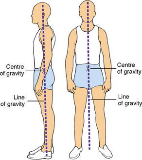
This is achieved when a relatively low centre of gravity is balanced over a wide, stable base of support and a vertical line falls from the centre of gravity through the base of support. The base of support is the foundation. When the vertical line from the centre of gravity does not fall through the base of support, the body loses balance. Body balance is also enhanced by proper posture—the body position that most favours function, requires the least muscular work to maintain and places the least strain on muscles, ligaments and bones.
The maintenance of proper body alignment and posture is achieved by using two simple techniques. First, the base of support can easily be widened by separating the feet to a comfortable distance. Second, balance is increased by bringing the centre of gravity closer to the base of support. This is achieved by bending the knees and flexing the hips until the person is squatting and still maintaining proper back alignment by keeping the trunk erect.
The nervous system is responsible for muscle tone and regulates and coordinates the amount of pull exerted by the individual muscles.
Coordinated body movement is a result of weight, centre of gravity and balance. Weight is the force exerted on the body mass by gravity. When an object is lifted, the lifter must overcome the object’s weight and be aware of its centre of gravity; in symmetrical objects, the centre of gravity is located at the exact centre of the object. The force of weight is always directed downwards. An object that is unbalanced has its centre of gravity away from the midline and falls without support. Because people are not geometrically perfect, their centres of gravity are usually at 55–57% of standing height and located in the midline. Like unbalanced objects, clients who fail to maintain a balance with their centre of gravity are unsteady, which places them at risk of falling.
Friction is a force that occurs in a direction opposing that of movement. As the nurse turns, transfers or moves a client up in bed, friction must be overcome. The greater the surface area of the object to be moved, the greater the friction. Friction can be reduced by following some basic principles, including using client-movement devices such as a slide sheet.
A passive or immobilised client has the potential to produce greater friction to movement. Thus, when possible, the nurse should use the client’s strength and mobility in association with the appropriate lifting aid when transferring or moving the client up in bed. This can be done by explaining the procedure and telling clients when to move. The use of a slide sheet reduces friction because the client is more easily moved along the bed’s surface.
Exercise is physical activity for the purpose of conditioning the body, improving health and maintaining fitness. It may also be used as a therapeutic measure. The exercise program chosen and developed for a client depends heavily on the individual’s activity tolerance, the kind and amount of exercise or activity that the person is able to perform. Physiological, emotional and developmental factors influence the client’s activity tolerance.
A program of regular physical activity and exercise promotes physical and psychological health (Department of Health and Ageing, 2010). An active lifestyle is important for maintaining and promoting health; it is also an essential treatment modality for many chronic diseases. A program of regular physical activity and exercise enhances functioning of all body systems, including cardiopulmonary functioning (endurance), musculoskeletal fitness (flexibility and bone integrity), weight control and maintenance (body image) and psychological wellbeing. For example, Dinas and colleagues’ (2011) review of the evidence suggests that exercise and physical activity have beneficial effects on depression symptoms that are comparable to those of antidepressant treatments.
The best program of physical activity includes a combination of exercises that produce different physiological and psychological benefits. Isotonic, isometric and resistive isometric are three categories of exercise classified according to the type of muscle contraction involved. Isotonic exercises cause muscle contraction and change in muscle length (isotonic contraction)—examples are walking, swimming, dance aerobics, jogging, bicycling, and moving arms and legs with light resistance. The benefits of isotonic exercises are increased circulation and respiratory functioning; increased osteoblastic activity (activity by bone-forming cells), thus combating osteoporosis; and increased muscle tone, mass and strength (Patton and Thibodeau, 2010).
Isometric exercises involve tightening or tensing of muscles without moving body parts (isometric contraction)—examples are quadriceps set exercises and contraction of the gluteal muscles. This form of exercise is ideal for clients who are unable to tolerate the increase in activity that is expected during isotonic exercises. Isometric exercises are easily accomplished by an immobilised client in bed. The benefits are increased muscle mass, tone and strength (thus decreasing the potential for muscle wasting); increased circulation to the involved body part; and increased osteoblastic activity (Patton and Thibodeau, 2010).
Isometric exercises may also be resistive. Resistive isometric exercises are those in which the person contracts the muscle while pushing against a stationary object or resisting the movement of an object. A gradual increase in the amount of resistance and length of time that the muscle contraction is held will increase muscle strength and endurance. Examples of resistive isometric exercises are push-ups, pushing against a footboard to move up in bed, and hip lifting. In hip lifting, the client, who is in a sitting position, pushes with the hands against a surface such as the seat of a chair and raises the hips. Resistive isometric exercises help to promote muscle strength and provide sufficient stress against bone to promote osteoblastic activity.
Coordinated body movement involves integrated functioning of the skeletal, muscular and nervous body systems. Because of the integrative nature of these three systems in mechanical support of the body, they are often considered as a single functional unit.
Bones perform five major functions in the body: support, protection, movement, mineral and fat storage and haematopoiesis (blood-cell formation) (Marieb and Hoehn, 2010). In the discussion of body mechanics, two of these functions—support and movement—are most important. In support, bones serve as the framework and contribute to the shape, alignment and positioning of the body parts. In movement, bones together with their joints constitute levers for muscle attachment. As muscles contract and shorten, they pull on bones, producing joint movement.
An articulation, or joint, is the connection between bones. Each joint is classified according to its structure and degree of mobility. On the basis of connective structures, joints are classified as fibrous, cartilaginous or synovial. Fibrous joints fit closely together and are fixed, permitting little, if any, movement. Cartilaginous joints have little movement but are elastic and use cartilage to unite separate body surfaces. Synovial joints, or true joints, are freely movable and are the most mobile, numerous and anatomically complex of the body’s joints.
These are structures that support the skeletal system. Ligaments are white, shiny, flexible bands of fibrous tissue that bind joints and connect bones and cartilage. Ligaments are elastic and aid joint flexibility and support. In some areas of the body, ligaments also have a protective function. Tendons are white, glistening, fibrous bands of tissue that connect muscle to bone. Cartilage is avascular, supporting connective tissue with the flexibility of a firm, plastic material. The characteristics of cartilage permit it to sustain weight and serve as a shock-absorber between articulating bones.
When we walk, talk, run, breathe or participate in physical activity, we do so by the contraction of skeletal muscles. There are over 600 skeletal muscles in the body. In addition to facilitating movement, these muscles determine the form and contour of our bodies. Most of our muscles span at least one joint and attach to both articulating bones. When contraction occurs, one bone is fixed while the other moves. The origin is the point of attachment that remains still; the insertion is the point that moves when the muscle contracts (Marieb and Hoehn, 2010).
The muscles of movement are located near the skeletal region, where movement is caused by a lever system. The lever system makes the work of moving a weight or load easier. It occurs when specific bones, such as the humerus, ulna and radius, and the associated joints, such as the elbow, act as a lever. Thus, the force applied to one end of the bone to lift a weight at another point tends to rotate the bone in the direction opposite to that of the applied force. Muscles that attach to bones of leverage provide the necessary strength to move the object.
Gravity pulls on parts of the body all the time. The only way the body can be held in position is for muscles to exert a force on bones in the opposite direction. Muscles accomplish this counterforce by maintaining a low level of sustained contraction. Poor posture places more work on muscles to counteract the force of gravity. This leads to fatigue and can eventually interfere with bodily functions and cause deformities.
The antagonistic, synergistic and antigravity muscle groups are coordinated by the nervous system and maintain posture and initiate movement.
• Antagonistic muscles bring about movement at the joint. During movement, the active mover muscle contracts while its antagonist relaxes. For example, during flexion of the arm the active mover, the biceps brachii, contracts and its antagonist, the triceps brachii, relaxes. During extension of the arm the active mover, now the triceps brachii, contracts and the new antagonist, the biceps brachii, relaxes.
• Synergistic muscles contract to accomplish the same movement. When the arm is flexed, the strength of the contraction of the biceps brachii is increased by contraction of the synergistic muscle, the brachialis. Thus with synergistic muscle activity there are now two active movers—the biceps brachii and the brachialis—which contract while the antagonistic muscle, the triceps brachii, relaxes.
• Antigravity muscles are involved with joint stabilisation. These muscles continuously oppose the effect of gravity on the body and permit a person to maintain an upright or sitting posture. In an adult, the antigravity muscles are the extensors of the leg, the gluteus maximus, the quadriceps femoris, the soleus muscles and the muscles of the back.
Skeletal muscles support posture and carry out voluntary movement. The muscles are attached to the skeleton by tendons, which provide strength and permit motion. The movement of the extremities is voluntary and requires coordination from the nervous system.
Movement and posture are regulated by the nervous system. The major voluntary motor area, located in the cerebral cortex, is the precentral gyrus. A majority of motor fibres descend (via pyramidal pathways) from the motor cortex and cross at the level of the medulla. Thus the motor fibres from the right motor cortex initiate voluntary movement for the left side of the body, and vice versa. The cerebellum and basal ganglia integrate information from the motor cortex as well as sensory input to plan and coordinate complex motor activities (Marieb and Hoehn, 2010).
Transmission of impulses from the nervous system to the musculoskeletal system is an electrochemical event. The neurotransmitter acetylcholine transfers the electric impulse from the nerve across the neuromuscular junction to stimulate the muscle, causing contraction.
Proprioception is the awareness of the position of the body and its parts. Proprioceptors are specialised receptors located in skeletal muscles, tendons, joints, ligaments and connective-tissue coverings of bones and muscles (Marieb and Hoehn, 2010). During daily activities, proprioceptors constantly advise the brain of our body movements and position by monitoring how much the tissues containing the receptors are stretched.
Balance centres in the central nervous system (i.e. vestibular nuclei in the brain stem and cerebellum) respond to information from three main sources about the body’s position in space. The vestibular apparatus in the inner ear (receptors in the semicircular canals and vestibule) respond to changes in body position and send signals to the brain. Balance centres also receive input through vision as well as somatic receptors, such as proprioceptors (Marieb and Hoehn, 2010). These centres initiate reflexes that fix the eyes on objects and activate muscles to maintain balance.
Many pathological conditions affect mobility. Although a complete description of each is beyond the scope of this chapter, an overview of four pathological influences is presented here: postural abnormalities, impaired muscle development, damage to the central nervous system and direct trauma to the musculoskeletal system. Common abnormal assessment findings of the musculoskeletal system are presented in Table 33-2.
TABLE 33-2 COMMON ASSESSMENT ABNORMALITIES OF THE MUSCULOSKELETAL SYSTEM
FINDING | DESCRIPTION | POSSIBLE AETIOLOGY AND SIGNIFICANCE |
---|---|---|
Ankylosis | Scarring within a joint leading to stiffness or fixation | Chronic joint inflammation |
Atrophy | Wasting of muscle, characterised by decreased circumference and flabby appearance leading to decreased function and tone | Muscle denervation, contracture, prolonged disuse as a result of immobilisation |
Contracture | Resistance of movement of muscle or joint as a result of fibrosis of supporting soft tissues | Shortening of muscle or ligaments, tightness of soft tissue, incorrect positioning of immobilised extremity |
Crepitation (crepitus) | Crackling sound or grating sensation as a result of friction or broken bone or cartilage bits in joint | Fracture, dislocation, chronic inflammation, osteoarthritis |
Effusion | Fluid in joint possibly with swelling and pain | Trauma, especially to knees; inflammation |
Ganglion | Small fluid-filled synovial cyst usually on dorsal surface of wrist or foot | Degeneration of connective tissue close to tendons and joints leading to formation of small cysts |
Hypertrophy | Increase in size of muscle as a result of enlargement of existing cells | Exercise or other increased stimulation, increased androgens |
Kyphosis (dowager’s hump) | Anteroposterior or forward bending of thoracic spine with convexity of curve in posterior direction | Poor posture, tuberculosis, arthritis, osteoporosis, growth disturbance of vertebral epiphyses |
Lordosis | Lumbar spinal deformity resulting in anteroposterior curvature with concavity in posterior direction | Secondary to other spinal deformities, muscular dystrophy, obesity, flexion contracture of hip, congenital dislocation of hip |
Pes planus | Flatfoot | Congenital condition, muscle paralysis, mild cerebral palsy, early muscular dystrophy |
Scoliosis | Deformity resulting in lateral curvature of thoracic spine | Idiopathic or congenital condition, fracture or dislocation, osteomalacia |
Subluxation | Partial dislocation of joint | Instability of joint capsule and supporting ligaments (e.g. from trauma, arthritis) |
Valgus (bow legs) | Angulation of bone away from midline | Alteration in gait, pain, arthritis |
Varus (knock-knees) | Angulation of bone towards midline | Alteration in gait, pain, arthritis |
From Brown D, Edwards H, editors, Lewis’s Medical–surgical nursing: assessment and management of clinical problems. ed 3. Sydney, Mosby.
Congenital or acquired postural abnormalities affect the efficiency of the musculoskeletal system, as well as body alignment, balance and appearance. During assessment, the nurse observes body alignment and range of motion (ROM). Postural abnormalities can cause pain and impair alignment and mobility. Some postural abnormalities may limit ROM. Nurses intervene to maintain maximum ROM in unaffected joints and then may design interventions to strengthen affected muscles and joints, improve the patient’s posture and adequately use affected and unaffected muscle groups. Referral to and/or collaboration with a physiotherapist may enhance the nurse’s interventions for a patient with a postural abnormality.
Injury and disease can lead to numerous alterations in musculoskeletal function. The muscular dystrophies are a group of familial disorders that cause degeneration of skeletal muscle fibres. The most prevalent of the muscle diseases in childhood, the muscular dystrophies are characterised by progressive, symmetrical weakness and wasting of skeletal muscle groups, with increasing disability and deformity (Nash, 2011).
Damage to any component of the central nervous system that regulates voluntary movement results in impaired body alignment and mobility. Neurological diseases such as multiple sclerosis and Parkinson’s disease can cause progressive mobility problems. The motor cortex can be damaged by trauma such as a head injury, stroke or meningitis. Motor impairment is directly related to the location and amount of destruction of the motor cortex. For example, a person with a right-sided cerebral haemorrhage with complete necrosis is likely to have destruction of the right motor cortex and left-sided hemiplegia.
Trauma to the descending pathways in the spinal cord involved in motor control, such as the pyramidal (corticospinal) tracts, also impairs mobility. The most common trauma is transection of the spinal cord in which motor fibres are damaged. If the injury is complete, it is likely to cause a complete bilateral loss of voluntary motor control below the level of the trauma. Spinal cord trauma frequently results from motor vehicle accidents, gunshot or knife wounds to the neck and back.
Direct trauma to the musculoskeletal system can result in contusions, sprains and fractures. A fracture is a disruption of bone tissue continuity. Fractures most commonly result from direct external trauma, but they can also occur as a consequence of some deformity of the bone (e.g. pathological fractures of osteoporosis, Paget’s disease or osteogenesis imperfecta). As the fracture heals, bone begins to repair. The fractured bone initiates a cellular process that results in bone formation. Young children are able to form new bone more easily than adults and, as a result, have few complications after a bone fracture. Treatment includes positioning the fractured bone in proper alignment and immobilising it to promote healing and to restore function. Immobilisation results in some muscle atrophy, loss of tone and joint stiffness.
Acquired or congenital conditions that affect the structure of the musculoskeletal or nervous system impair body alignment or joint mobility. Impairment can be temporary or permanent. Regardless of the duration of the impairment, the nursing care plan includes interventions that maintain the present level of alignment and joint mobility and/or increase the patient’s level of motor function.
Fully understanding mobility requires more than an overview of normal body mechanics and the regulation of movement by the musculoskeletal and nervous systems. Having knowledge of how immobility affects physical and psychosocial functioning can help you prevent, assess for and manage complications caused by impaired mobility. Putting this nursing knowledge and skill into practice can mean the difference between patient recovery and independence, and costly and life-threatening complications, functional decline and disability.
Nurses have always been concerned with the clinical problems caused by immobility and have developed a body of knowledge over time. It is instructive to read a collection of classic articles published almost half a century ago in the American Journal of Nursing, entitled ‘The hazards of immobility’, which summarised the physiological changes that occur in healthy, ill and injured people who undergo bed rest (Olson, 1967). The articles described the detrimental effects of immobility on cardiovascular, respiratory, gastrointestinal, musculoskeletal, urinary, metabolic and psychosocial health, with implications for nursing practice. Today there is a large and growing body of nursing research examining the effectiveness of interventions to prevent and manage the complications of immobility.
Mobility refers to a person’s ability to move about freely, whereas immobility refers to the inability to move about freely. Mobility and immobility are best understood as the end points on a continuum. Some patients move back and forth on this continuum, but for other patients immobility is absolute and continues indefinitely. The terms ‘bed rest’ and ‘impaired physical mobility’ are frequently used when discussing patients on the mobility–immobility continuum.
Bed rest is an intervention that restricts patients to bed for therapeutic reasons. For example, patients with acute myocardial infarction are initially encouraged to remain on bed rest to reduce oxygen demand and cardiac workload. Bed rest is most often prescribed by nursing and medical staff and may have different interpretations among healthcare professionals. For example, in some settings bed rest may mean strict confinement to the bed or ‘complete bed rest’, while in others patients on bed rest may be assisted to use a bedside commode or to the bathroom. Patients with a wide variety of conditions are placed on bed rest, and the duration of bed rest depends on the illness or injury and the patient’s prior state of health.
Impaired physical mobility is defined as a ‘state in which the individual experiences or is at risk of experiencing limitation of physical movement but is not immobile’ (Carpenito-Moyet, 2010:279). Alterations in the level of physical mobility can result from prescribed restriction of movement in the form of bed rest, physical restriction of movement because of external devices (e.g. a cast or skeletal traction), voluntary restriction of movement or impairment of motor or skeletal function.
The deleterious effects of muscular deconditioning associated with lack of physical activity may be apparent within days. The normal individual on complete bed rest loses muscle strength from baseline levels at a rate of 3% a day (Nash, 2011). Bed rest is also associated with other systemic complications. The term ‘disuse atrophy’ has been used to describe the pathological reduction in the normal size of muscle fibres after prolonged inactivity from bed rest, trauma, casting, or local nerve damage (Nash, 2011).
In a classic study, Deitrick and others (1948) found that even young healthy men put on bed rest had physiological problems. Periods of immobility or prolonged bed rest can cause major physiological and psychological effects. These effects can be gradual or immediate, and vary from patient to patient. The greater the extent and the longer the duration of immobility, the more pronounced the consequences. The patient with complete mobility restrictions is continually at risk of complications.
All body systems work more efficiently with some form of movement. Exercise has been shown to have positive outcomes for all major systems of the body. Therefore, when there is an alteration in mobility, each body system is at risk of impairment. The severity of the impairment depends on the patient’s overall health, degree and length of immobility, and age. For example, older adults with chronic illnesses develop pronounced effects of immobility more quickly than do younger patients with the same degree of immobility.
Immobility disrupts normal metabolic functioning, including decreasing the metabolic rate; altering the metabolism of carbohydrates, fats and proteins; causing fluid, electrolyte and calcium imbalances; and causing gastrointestinal disturbances such as decreased appetite and slowing of peristalsis. However, in the presence of an infectious process, immobilised patients may have an increased basal metabolic rate as a result of fever or wound healing because these increase cellular oxygen requirements.
A deficiency in kilojoules and protein is characteristic of patients with a decreased appetite secondary to immobility. Proteins are constantly being synthesised and broken down into amino acids in the body to be reformed into other proteins. Amino acids that are not used are excreted. The body can synthesise certain non-essential amino acids, but depends on ingested proteins to supply the eight essential amino acids. When more nitrogen (the end-product of amino-acid breakdown) is excreted than is ingested in proteins, the body is said to have a negative nitrogen balance, and weight loss, decreased muscle mass and weakness result from tissue catabolism (tissue breakdown). Protein loss leads to muscle loss.
Another metabolic change is increased calcium loss from bones, resulting in hypercalcaemia. As a result, urinary excretion of calcium increases because immobility causes the release of calcium into the circulation.
Decreased gastrointestinal motility that develops from immobility can lead to various impairments of gastrointestinal functioning. Constipation is a common problem, although diarrhoea may result from a faecal impaction (accumulation of hardened faeces). This finding is not normal diarrhoea but rather liquid stool passing around the area of impaction (see Chapter 37). Left untreated, faecal impaction can result in a mechanical bowel obstruction that may partially or completely occlude the intestinal lumen, blocking normal propulsion of liquid and gas. The resulting fluid in the intestine produces distension and increases intraluminal pressure. Over time, intestinal function becomes depressed, dehydration occurs, absorption ceases and fluid and electrolyte disturbances worsen.
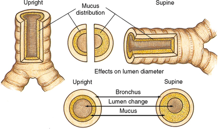
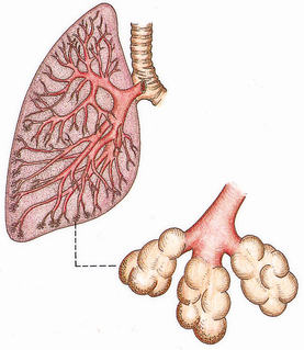
Regular aerobic exercise is known to enhance respiratory functioning. Lack of movement and exercise places patients at higher risk of respiratory complications. Postoperative and immobile patients are at high risk of developing pulmonary complications. The most common respiratory complications are atelectasis (collapse of alveoli) and hypostatic pneumonia (inflammation of the lung from stasis or pooling of secretions). In atelectasis a bronchiole or a bronchus becomes blocked by secretions and the distal lung tissue (alveoli) collapses as the existing gas is absorbed, producing hypoventilation. The extent of atelectasis is determined by the site of the blockage. A lung lobe or even a whole lung may be collapsed. Pooling secretions and decreased ability to cough productively increase the collection of mucus in the bronchi, particularly when the patient is in the supine, prone or lateral position (Figure 33-1). Secretions accumulate in the dependent regions of the airways (Figure 33-2). Because mucus is an excellent medium for bacterial growth, hypostatic pneumonia may result.
Orthostatic (or postural) hypotension is defined as a reduction in systolic blood pressure of at least 20 mmHg or diastolic blood pressure of at least 10 mmHg within 3 minutes of standing from a lying position (Consensus Committee of the American Autonomic Society and the American Academy of Neurology, 1996). Patients with orthostatic hypotension may present with lightheadedness, blurred vision, dizziness, weakness and fatigue, or with syncope. In the immobilised patient, decreased circulating fluid volume, pooling of blood in the lower extremities and decreased autonomic response occur. These factors result in decreased venous return, followed by a decrease in cardiac output, which is reflected by a decline in blood pressure.
As the workload of the heart increases, so does oxygen consumption. The heart therefore works harder and less efficiently during periods of prolonged rest. As immobilisation increases, cardiac output falls, further decreasing cardiac efficiency and increasing workload.
Patients are also at risk of thrombus formation, such as a deep-vein thrombosis (DVT) (Figure 33-3). A thrombus is an accumulation of platelets, fibrin, clotting factors and the cellular elements of the blood attached to the interior wall of a vein or artery, sometimes occluding the lumen of the vessel. There are three factors that contribute to venous thrombus formation (known as Virchow’s triad):
1. venous stasis (e.g. poor venous return associated with bed rest and immobility)
2. damage to the endothelium (inner lining of the vein), and
3. hypercoagulability of the blood (Soars, 2012).
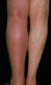
A dislodged thrombus, called an embolus, may travel through the circulatory system to the lungs (called a pulmonary embolism or PE) or brain, where they are life-threatening.
The effects of immobility on the musculoskeletal system can include permanent impairment of mobility. Restricted mobility may result in loss of endurance, strength and muscle mass. Other effects of restricted mobility affecting the skeletal system are impaired calcium metabolism and impaired joint mobility.
Decreased mobility and muscle atrophy—loss of muscle tissue—may occur with immobility. Atrophy is a phenomenon widely observed in response to illness, decreased activities of daily living (ADLs), and immobilisation (Boonyarom and Inui, 2006). Because of protein breakdown, the patient loses lean body mass, which is composed partially of muscle. Therefore, the reduced muscle mass is unable to sustain activity without increased fatigue. The muscle mass is decreased from metabolic causes and disuse. As immobility continues and the muscles are not exercised, there is continued decrease in mass. Loss of endurance, decreased muscle mass and strength, and joint instability subsequent to immobility put patients at risk of falls (see Chapter 15).
Immobilisation causes two skeletal changes: impaired calcium metabolism and joint abnormalities. Because immobilisation results in bone resorption, the bone tissue is less dense, or is atrophied and disuse osteoporosis results. When osteoporosis occurs, the patient is at risk of pathological fractures. Immobilisation and non-weightbearing activities increase the rate of bone resorption. Bone resorption also causes calcium to be released in the blood, and hypercalcaemia results.
One in two females and one in three males over the age of 60 years are likely to sustain a fracture due to osteoporosis (Department of Medicine, University of Melbourne, 2007). More women are affected because of the postmenopausal rapid decline of oestrogen, a hormone that plays a central role in the maintenance of bone mass balance. About 25% of Australians over 50 years of age with hip fractures die within the first 12 months, and 50% require long-term nursing care (Department of Medicine, University of Melbourne, 2007). Although primary osteoporosis is different in origin from the osteoporosis that results from immobility, it is imperative for nurses to recognise that immobilised patients may be at high risk of accelerated bone loss if they have primary osteoporosis. Early patient evaluation and consultation and referral to nutritionists and physiotherapists are important interventions for preventing disability in patients with primary osteoporosis who become immobilised.
Immobility can lead to joint contractures. A joint contracture is an abnormal and possibly permanent condition characterised by fixation of the joint (Figure 33-4). It is caused by disuse, atrophy and shortening of the muscle fibres. When a contracture occurs, the joint cannot obtain full range of motion. Contractures may leave a joint in a non-functional position, as seen in some patients who are permanently curled into a fetal position (Fergusson and others, 2007).
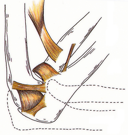
FIGURE 33-4 Contracture of the elbow: inability to extend to a 90-degree angle (dotted line) and to a 180-degree angle (not illustrated).
One common and debilitating contracture is footdrop (Figure 33-5). When footdrop occurs, the foot is permanently fixed in plantar flexion. Walking is difficult with the foot in this position, since the person cannot dorsiflex the foot. The patient with footdrop is therefore unable to lift the toes off the ground. Patients who have suffered strokes with resulting left- or right-sided paralysis (hemiplegia) are susceptible to footdrop.
The patient’s urinary elimination is altered by immobility. In the upright position, urine flows out of the renal pelvis and into the ureters and bladder because of gravitational forces. When the patient is recumbent or flat, the kidneys and the ureters move towards a more level plane. Urine formed by the kidney must enter the bladder unaided by gravity. Because the peristaltic contractions of the ureters are insufficient to overcome gravity, the renal pelvis may fill before urine enters the ureters (Figure 33-6). This condition is called urinary stasis and increases the risk of urinary tract infection (UTI) and renal calculi (see Chapter 38).
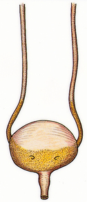
FIGURE 33-6 Stasis of urine with reflux to ureters.
From Potter PA, Perry AG 2013 Fundamentals of nursing, ed 8. St Louis, Mosby.
Renal calculi are calcium stones that lodge in the renal pelvis and pass through the ureters (Figure 33-7). Immobilised patients are at risk of calculi because of altered calcium metabolism and the resulting hypercalcaemia.
As the period of immobility continues, fluid intake can diminish; other causes, such as fever, increase the risk of dehydration. As a result, urinary output declines and urine is usually highly concentrated. This concentrated urine increases the risk of calculi formation and infection. Poor perineal care after bowel movements, particularly in women, increases the risk of urinary tract contamination by Escherichia coli bacteria. Another cause of UTIs in immobilised patients is the use of an indwelling urinary catheter.
The direct effect of pressure on the skin is compounded by the changes in metabolism that accompany immobility. Older adults and patients with paralysis also have a greater risk of developing pressure ulcers. Any break in the skin’s integrity is difficult to heal in the immobilised patient.
A pressure ulcer is an impairment of the skin as a result of prolonged ischaemia to tissues (see Chapter 30). The ulcer is characterised initially by inflammation and usually forms over a bony prominence. Ischaemia develops when the pressure on the skin is greater than the pressure inside the small peripheral blood vessels supplying blood to the skin.
Tissue metabolism depends on the body’s receipt of oxygen and nutrients from the blood supply and the elimination of metabolic wastes. Any factor that interferes with perfusion affects cellular metabolism and, as a result, the function of the cell. Pressure affects cellular metabolism by decreasing or obliterating tissue circulation. When a patient lies in bed or sits in a chair, the weight of the body is on bony prominences. The longer the pressure is applied, the longer the period of ischaemia and therefore the greater the risk of skin breakdown.
Immobilisation may lead to emotional and behavioural responses, sensory alterations and changes in coping. These changes are individualised to each patient. In addition, immobilised patients may also have social and family difficulties.
The most common emotional changes are depression, behavioural changes, sleep–wake disturbances and impaired coping. The immobilised patient can become depressed because of changes in role, self-concept and other factors. Depression is an affective disorder characterised by extreme feelings of sadness, melancholy, dejection, worthlessness, emptiness and hopelessness out of proportion to reality. Depression can result from worrying about present and future levels of health, finances and family needs. Because immobilisation removes patients from their daily routine, they have more time to worry about disability. Worrying can quickly increase depression, causing withdrawal. Assessing behavioural changes throughout restricted mobility helps the nurse to identify changes in self-concept, recognise early signs of depression and develop nursing interventions (see Chapter 23).
Behavioural changes resulting from immobilisation vary widely, depending on the patient. Common behavioural changes include hostility, fear and anxiety. Early in the nursing process the nurse should interview the patient and the patient’s family and friends about normal behavioural patterns to gain baseline data. If unexpected behaviours are observed later, the nurse can intervene to reduce the effects of immobilisation on the patient’s behavioural patterns.
The immobilised patient requires frequent and regular nursing care. Because of physiological hazards, the patient cannot be allowed to sleep for 8 hours without a change of position or other nursing care. Disruption of normal sleeping patterns can further cause behavioural changes. Nursing interventions should be used to ensure that the patient receives sufficient sleep (see Chapter 35). The patient who is on bed rest and able to change position during sleep does not require continuous physical nursing care directed at reducing the hazards of immobility. Unless other treatment activities are required during the night, the care plan for the physiologically stable patient on bed rest should provide for uninterrupted sleep.
Long-term immobility or bed rest can affect usual coping patterns. Such a patient may withdraw and become passive. The passive patient allows nurses to provide care but is not interested in increasing independence or being involved in care. Early in the care of an immobilised patient, the nurse should assess the patient’s normal coping mechanisms. The nurse then designs a nursing care plan that will enable the patient to continue to use these coping abilities or help the patient develop new ones.
Developmental changes tend to be associated more with immobility in the very young and in older adults. The immobilised young or middle-aged adult who has been healthy may experience few, if any, developmental changes. However, there are exceptions and patients must be fully assessed for developmental implications. One exception might be a mother who has complications at childbirth and as a result cannot interact with the newborn as expected.
The newborn infant’s spine is flexed and lacks the anteroposterior curves of the adult (see Chapter 20). As the baby grows, musculoskeletal development permits support of weight for standing and walking. Posture is awkward because the head and upper trunk are carried forward. Because bodyweight is not evenly distributed along a line of gravity, posture is off-balance, and falls occur often. When the infant, toddler or preschooler is immobilised, it is usually because of trauma or the need to correct a congenital skeletal abnormality. Prolonged immobilisation can delay the child’s gross motor skills and intellectual development. Nurses caring for immobilised children should plan activities that provide physical and psychosocial stimulation.
The adolescence stage is usually initiated by a tremendous growth spurt (see Chapter 20) where growth is frequently uneven. Prolonged immobilisation may alter adolescent growth patterns, causing the adolescent to lag behind peers in gaining independence. When immobilisation occurs, social isolation is often a major concern for this age group.
An adult who has correct posture and body alignment feels good and generally appears self-confident. The healthy adult also has the necessary musculoskeletal development and coordination to carry out ADLs (see Chapter 21). When periods of prolonged immobility occur, all physiological systems are at risk. In addition, the role of the adult may change with regard to the family or social structure, such as loss of identity associated with a job.
A progressive loss of total bone mass occurs with the older adult. Some of the possible causes of this loss include decreased physical activity, hormonal changes and actual bone resorption. The effect of bone loss is weaker bones and, as a result, older adults may walk more slowly, walk bent over, take smaller steps and appear less coordinated. When balance is impaired, they are at greater risk of falls and injuries (see Chapter 22). Falls in older adults are associated with immediate serious consequences such as fractures and head injuries, as well as longer-term problems such as disability, fear of falling, and loss of independence (Rawsky, 1998).
Older adults are also at risk of decreased functional status and impaired mobility secondary to hospitalisation (see Box 33-1). Immobilisation of older adults may increase their physical dependence on others, which accelerates functional losses. Immobilisation of some older adults can result from degenerative diseases, trauma or chronic illness. For some older adults, immobilisation occurs gradually and progressively; whereas for others, especially those who have had a stroke, immobilisation is sudden. When providing nursing care for an older adult, the nurse should develop a care plan that encourages the patient to perform as many self-care activities as possible, thereby maintaining the highest level of mobility (see Research highlight). Murphy and Reid (2003) suggest that nurses may inadvertently contribute to a patient’s immobility by providing unnecessary help with activities such as bathing and transferring.
BOX 33-1 HAZARDS OF HOSPITALISATION OF THE OLDER ADULT
• For many older people, hospitalisation results in functional decline despite cure or repair of the condition for which they were admitted. Hospitalisation can result in complications unrelated to the problem that caused admission or to its specific treatment, and for reasons that are explainable and avoidable.
• Usual ageing is often associated with functional change, such as a decline in muscle strength and aerobic capacity; vasomotor instability; reduced bone density; diminished pulmonary ventilation; altered sensory continence, appetite and thirst; and a tendency towards urinary incontinence. Hospitalisation and bed rest superimpose factors such as enforced immobilisation, reduction of plasma volume, accelerated bone loss, increased closing volume and sensory deprivation. Any of these factors may thrust vulnerable older people into a state of irreversible functional decline.
• The factors that contribute to a cascade to dependency are identifiable and can be avoided by modification of the usual acute hospital environment by de-emphasising bed rest, removing the hazard of the high hospital bed with rails and actively facilitating ambulation and socialisation.
From Creditor MC 1993 Hazards of hospitalization of the elderly. Ann Intern Med 118(3):219.
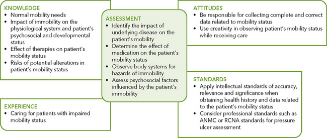
Critical thinking and sound clinical judgment when caring for patients with impaired mobility requires nurses to draw on their knowledge of mobility and immobility and on previous experience, and apply critical-thinking attitudes and professional standards (Figure 33-8). Patients with impaired mobility often have multiple needs which must be prioritised. To understand the impact of immobility on the patient and family, the nurse integrates knowledge from nursing and other disciplines, previous experiences and information gathered from patients.
In older patients, acute medical illness that requires hospitalisation is a sentinel event that often precipitates disability. This results in the subsequent inability to live independently and complete basic activities of daily living (ADLs). This nursing research examined the association between mobility levels of older adults during hospitalisation and functional outcomes at discharge and 1-month follow-up.
Design: Prospective cohort study.
Setting: A 900-bed teaching hospital in Israel.
Participants: 525 older (≥ 70 years) acute medical patients hospitalised for a non-disabling condition.
Measurements: In-hospital mobility was assessed using a previously validated scale. The main outcomes were decline from premorbid baseline functional status at discharge (ADLs) and at 1-month follow-up (ADLs and instrumental ADLs). Hospital mobility levels and functional outcomes were assessed according to prehospitalisation functional trajectories. Logistic regressions were modelled for each outcome, controlling for functional status, morbidity and demographic characteristics.
Results: 46% of participants had declined in ADLs at discharge and 49% at follow-up; 57% had declined in IADLs at follow-up. Mobility during hospitalisation was twice as high in participants with no pre-admission functional decline.
Low versus high in-hospital mobility was associated with worse basic functional status at discharge (adjusted odds ratio (AOR) = 18.03; 95% confidence interval (CI) = 7.68–42.28) and at follow-up (AOR = 4.72; 95% CI = 1.98–11.28) and worse IADLs at follow-up (AOR = 2.00; 95% CI = 1.05–3.78). The association with poorer discharge functional outcomes was present in participants with pre-admission functional decline (AOR for low vs high mobility = 15.26; 95% CI = 4.80–48.42) and in those who were functionally stable (AOR for low vs high mobility = 10.12; 95% CI = 2.28–44.92).
• The results of this study show that low levels of in-hospital mobility were associated with functional decline of older adults at discharge and, even more so, at follow-up.
• In-hospital mobility, unlike personal and disease-related risk factors, is a potentially modifiable care aspect that has important implications for in-hospital care regimens.
• Level of mobility was significantly related to functional outcomes of patients whether or not they had functional decline before their hospitalisation, indicating the value of developing and evaluating a policy for the early and effective mobilisation of all older hospitalised patients in general medical units.
You are a student nurse caring for Albert Devereaux, an alert and oriented 81-year-old man, admitted to hospital after a fall with complaints of dizziness and syncope. His blood pressure (BP) on admission was 80/40 mmHg. At the aged-care nursing facility where he lives, he ambulated with a walker independently but, since a recent episode of syncope, he has complained of weakness and needs another person to assist him while walking as a fall precaution.
Nursing staff found Mr Devereaux lying on the floor on his right hip, and he explained ‘I had to go to the bathroom. I know I should have called for help, but the nurses are so busy. I figured I could go myself. Only two more steps and I could have reached my walker. I just slipped, is all.’ He reports pain in his right hip that is a 5 on a 0–10 numerical pain rating scale, which he describes as a ‘ache’ that is worse with movement of his right leg.
An X-ray of his right hip is taken, which is negative for a fracture. You take a set of orthostatic vital signs: lying BP = 110/80 mmHg, heart rate = 73 beats per minute; standing BP = 90/50 mmHg, heart rate = 92 beats per minute. His other vital signs are temperature 36.8°C, respiratory rate 16 breaths per minute, oxygen saturation (SpO2) 99% on room air. No injuries are noted on inspection and palpation of his head and neck. Pupils are equal and react briskly to light. His chest is clear to auscultation, heart sounds are irregular. There is full range of motion in the upper extremities and handgrip strength is bilaterally equal. His abdomen is soft and non-tender. There is no physical deformity of the right hip or other injuries apparent, but a moderate amount of ecchymosis is visible over his right hip that extends around to his lower back and right upper buttock. He demonstrates normal movement and sensation in both feet. He has requested the urinal once for 200 mL of clear yellow urine. An electrocardiogram confirms his known history of atrial fibrillation.
Mr Devereaux is admitted, and his plan of care includes assessment of orthostatic vital signs every shift and fall precautions. You explain to him how to use the call light and he remains on bed rest with side rails up. Later in the shift Mr Devereaux tells you he is worried about the consequences of this fall for his mobility and independence.
Adapted from Douglas C 2001 Pulling it all together. In Lewis P, Foley D, editors, Weber & Kelley’s Health assessment in nursing. Philadelphia, Lippincott Williams & Wilkins.
• CRITICAL THINKING
1. Based on the clinical example above, construct a concept map which demonstrates all the significant assessment data from the case study and the relationships or links among this data.
2. What priority nursing interventions would you implement for Mr Devereaux to maintain his safety and promote normal movement and mobility?
3. How will you assess for and prevent the complications of immobility while Mr Devereaux remains on bed rest?
4. How would you further explore the psychosocial impact of this fall on Mr Devereaux and his fears about loss of mobility and independence?
The use of the nursing process, critical application of anatomy and physiology knowledge, and experience with patients enables the nurse to develop individualised care plans for patients with mobility impairments and for those who are at risk. A care plan is designed to improve the patient’s functional status, promote self-care, maintain psychological wellbeing and prevent the complications of immobility.
Assessment of patient mobility focuses on range of motion, gait, exercise and activity tolerance and body alignment. When unsure of the patient’s abilities, the nurse should begin assessment of mobility with the patient in the most supportive position and move to higher levels of mobility according to the patient’s tolerance. Generally, the nurse starts assessing movement while the patient is lying down, and then proceeds to assessing sitting positions in bed, transfers to chair and finally gait by watching the patient walk. This helps to protect the patient’s safety.
Range of motion (ROM) is the maximum amount of movement available at a joint in one of the three planes of the body: sagittal, frontal or transverse (Figure 33-9). Joint mobility in each of the planes is limited by ligaments, muscles and the nature of the joint. However, some joint movements are specific to each plane. In the sagittal plane, movements are flexion and extension (fingers and elbows), dorsiflexion and plantar flexion (feet) and extension (hip). In the frontal plane, movements are abduction and adduction (arms and legs) and eversion and inversion (feet). In the transverse plane, movements are pronation and supination (hands) and internal and external rotation (hips).
FIGURE 33-9 Planes of the body.
From Potter PA, Perry AG 2013 Fundamentals of nursing, ed 8. St Louis, Mosby.
To assess ROM, the nurse asks questions about and physically examines the patient for stiffness, swelling, pain, limited movement and unequal movement. Table 33-3 presents the normal ROM ranges for each joint.
Patients whose mobility is restricted because of illness, disability or trauma require ROM exercises to reduce the hazards of immobility. ROM exercises may be active (patients are able to move all joints through their ROM unassisted) or passive (patients are unable to move independently, and the nurse moves each joint through its ROM), or somewhere in between. With a weak patient, for example, the nurse may merely provide support while the patient performs most of the movement, or the patient may be able to move some joints actively while the nurse passively moves others. The nurse first assesses the patient’s ability to engage in active ROM exercises and the need for assistance, teaching or reinforcement. In general, exercises should be as active as health and mobility allow. Contractures may develop in joints not moved periodically through their full ROM.
Gait is the manner or style of walking, including rhythm, measure of beat, cadence and speed. Assessing gait allows the nurse to draw conclusions about balance, posture and the ability to walk without assistance.
Gait can easily be assessed as the client walks into the room, by asking the client to walk a short distance or while assisting the client to mobilise. A normal gait should demonstrate:
Exercise and activity tolerance is the kind and amount of exercise or work that a person is able to perform. Assessment of activity tolerance is necessary when planning activity such as walking, ROM exercises or ADLs such as bathing for patients with acute or chronic illness. Activity-tolerance assessment includes data from physiological, emotional and developmental domains. This assessment is applicable in all clinical settings and is quickly completed by the nurse.
As activity is begun, patients should be monitored for symptoms such as dyspnoea, fatigue or chest pain and/or a change in vital signs from baseline. The weak or deconditioned patient is unable to sustain activity because the greater energy needed to complete the activity creates fatigue and generalised weakness. Even seemingly simple tasks such as eating and moving in bed may need to be monitored. When decreased activity tolerance is noted, the nurse should assess the time needed by the patient to recover. Decreasing recovery time may indicate improving activity tolerance.
People who are depressed, worried or anxious are frequently unable to tolerate exercise. Depressed patients are usually not motivated to participate. Patients who are worried or anxious tire easily because they expend a great deal of energy in worry and anxiety. Thus they may experience physical and emotional exhaustion.
Developmental changes also affect activity tolerance. As the infant enters the toddler stage, the activity level increases and the need for sleep declines. The school-aged child expends mental energy in learning and may require more rest after school or before strenuous play. The adolescent going through puberty may require more rest because much of the body’s energy is expended on growth and hormone changes.
Changes may still occur through the adult years, but many of these changes are related to work and lifestyle choices. Pregnancy may cause fluctuations in a woman’s energy tolerance, especially during the first and third trimesters when she may have increased fatigue. Hormonal changes and fetal development use body energy, and the woman may be unable or unmotivated to carry out physical activities. During the last trimester, fetal development consumes a great deal of the mother’s energy and the size and location of the fetus may limit the ability to take a deep breath, resulting in less oxygen being available for physical activities.
As a person grows older, activity tolerance changes. Muscle mass is reduced, posture changes and the composition of bones is altered. There are often changes in the cardiorespiratory system, such as decreased maximum heart rate and decreased lung compliance, which affect the intensity of exercise. As age progresses, the older individual may still exercise but will do so at a reduced intensity.
There is an overall improvement of physiological functioning as a result of exercise. All systems become stronger and function more efficiently. Nurses may therefore plan interventions directed at increasing exercise. However, nurses in rehabilitation and acute care settings often care for patients with impaired mobility, and therefore develop nursing therapies designed to minimise the hazards of immobility.
Assessment of body alignment can be carried out with the patient standing, sitting or lying down. The first step in assessing body alignment is to put patients at ease so that unnatural or rigid positions are not assumed. When the body alignment of an immobilised or unconscious patient is assessed, pillows and positioning supports should be removed from the bed and the patient placed in the supine position.
The nurse should focus assessment of body alignment for the standing patient on the following points:
• The head is erect and midline.
• When observed posteriorly, the shoulders and hips are straight and parallel.
• When observed posteriorly, the vertebral column is straight.
• When the patient is observed laterally, the head is erect and the spinal curves are aligned in a reversed S pattern. The cervical vertebrae are anteriorly convex, the thoracic vertebrae are posteriorly convex and the lumbar vertebrae are anteriorly convex.
• When observed laterally, the abdomen is comfortably tucked in and the knees and ankles are slightly flexed. The person appears comfortable and does not seem conscious of the flexion of knees or ankles.
• The arms hang comfortably at the sides.
• The feet are placed slightly apart to achieve a base of support, and the toes are pointed forwards.
• When the patient is viewed anteriorly, the centre of gravity is in the midline, and the line of gravity is from the middle of the forehead to a midpoint between the feet. Laterally the line of gravity runs vertically from the middle of the skull to the posterior third of the foot (Figure 33-10).
The nurse assesses alignment of the sitting patient (Figure 33-11) by the following observations:
• The head is erect, and the neck and vertebral column are in straight alignment.
• The bodyweight is evenly distributed on the buttocks and thighs.
• The thighs are parallel and in a horizontal plane.
• Both feet are supported on the floor. With patients of short stature, a footstool is used and the ankles are comfortably flexed.
• A 2–4 cm space is maintained between the edge of the seat and the popliteal space on the posterior surface of the knee. This space ensures that there is no pressure on the popliteal artery or nerve to decrease circulation or impair nerve function.
• The patient’s forearms are supported on the armrest, in the lap or on a table in front of the chair.
FIGURE 33-11 Correct body alignment when sitting.
From Potter PA, Perry AG 2013 Fundamentals of nursing, ed 8. St Louis, Mosby.
It is particularly important to assess alignment when sitting if the patient has muscle weakness, muscle paralysis or nerve damage. Because of these alterations, the patient has diminished sensation in the affected area and is unable to perceive pressure or decreased circulation. Proper alignment while sitting reduces the risk of musculoskeletal system damage in such a patient. The patient with severe respiratory disease may assume a posture of leaning on the table in front of the chair in an attempt to breathe more easily.
People who are conscious and healthy have voluntary muscle control and normal perception of pressure. As a result, they usually assume a position of comfort when lying down. Because their ROM, sensation and circulation are within normal limits, they change positions when they perceive muscle strain and decreased circulation.
Assessment of body alignment with the patient in the lateral position (Figure 33-12) is best done when the patient is restricted to bed and not able to move well. All positioning supports should be removed from the bed except for the pillow under the head, and the body should be supported by an adequate mattress. This position allows for full view of the spine and back and will help provide other baseline body alignment data, such as whether the patient can remain positioned without aid. The vertebrae should be in straight alignment without observable curves. The position should not cause discomfort. Conditions that create a risk of damage to the musculoskeletal system when lying down include patients with impaired mobility (e.g. in traction or with arthritis), decreased sensation (e.g. sensory deficits after stroke), impaired circulation (e.g. diabetes) or lack of voluntary muscle control (e.g. spinal cord injuries).
The nurse assesses the patient with impaired mobility or activity tolerance for complications of immobility by performing a head-to-toe body systems physical assessment (refer to Chapter 27 for normal findings). In addition, nursing assessment should focus on the psychosocial and developmental impact of immobility. Common abnormal assessment findings associated with immobility for each body system are presented in Table 33-4.
TABLE 33-4 BODY SYSTEMS ASSESSMENT FOR COMPLICATIONS OF IMMOBILITY
SYSTEM | ASSESSMENT TECHNIQUES | ABNORMAL FINDINGS |
---|---|---|
Metabolic | Inspection | Slowed wound healing, abnormal laboratory data Muscle atrophy, decreased amount of subcutaneous fat |
Respiratory | Inspection | Increased respiratory rate, asymmetrical chest wall movement, dyspnoea |
Auscultation | Crackles, wheezes | |
Cardiovascular | Palpation | Increased heart rate, weak peripheral pulses, peripheral oedema |
Auscultation | Orthostatic hypotension, third heart sound | |
Musculoskeletal | Inspection, palpation | Erythema, increased diameter in calf or thigh, muscle atrophy |
Palpation | Decreased ROM, joint contracture | |
Skin | Inspection | Break in skin integrity |
Elimination | Inspection | Decreased urine output, cloudy or concentrated urine, decreased frequency of bowel movements |
Palpation | Distended bladder and abdomen | |
Auscultation | Decreased bowel sounds |
When assessing metabolic functioning, the nurse uses measures of height, weight and muscle size to evaluate muscle atrophy. In addition, assessment of volume status (e.g. mucous membranes, skin turgor) and intake and output records for fluid balance are useful. Dehydration and oedema can increase the rate of skin breakdown in an immobilised patient. Monitoring laboratory data such as electrolytes, serum protein (albumin and total protein) levels and blood urea nitrogen (BUN) help the nurse determine metabolic functioning.
Assessing wound healing to evaluate alterations in the exchange of nutrients and monitoring food intake and elimination patterns will help to determine altered gastrointestinal functioning and potential metabolic problems. If an immobilised patient has a wound, the rate of healing indicates how well nutrients are being delivered to tissues. Normal progression of healing indicates that metabolic needs of injured tissues are being met. Anorexia occurs commonly in immobilised patients. The patient’s food intake should be assessed before the tray is removed to determine the amount eaten. Nutritional imbalances can be avoided if the nurse assesses the patient’s dietary patterns and food preferences early in immobilisation (see Chapter 36) (see Working with diversity).
In addition to respiratory rate, inspect chest-wall movements during the full inspiratory–expiratory cycle. If a patient has an atelectatic area, chest movement may be asymmetrical. Auscultate lung fields to identify diminished breath sounds, crackles or wheezes. Auscultation should begin posteriorly at the bases of the lungs and dependent lung fields because pulmonary secretions tend to collect in these lower regions. A complete respiratory assessment identifies the presence of secretions and can be used to determine nursing interventions necessary for optimal respiratory function.
WORKING WITH DIVERSITYFOCUS ON CULTURAL CARE
Dairy products are key sources of the calcium necessary to prevent osteoporosis. However, people’s ability to tolerate the lactose in dairy products is thought to be related to their ethnic background. People with an Asian background often have lactose intolerance, but research has shown that they may be able to tolerate small amounts of dairy products. Australian Aboriginal peoples and those of Greek and Mediterranean origin are also reported to experience lactose intolerance more than Caucasians. To ensure bone health, education is needed about sources of calcium, both dairy and non-dairy. Although generally in lower amounts per serving than dairy foods, the non-dairy sources of calcium include green leafy vegetables, legumes and cereals. Calcium-fortified foods and calcium supplements are another option for people who cannot meet their calcium needs from foods naturally containing this mineral.
• For all patients, be aware of their normal dietary habits.
• When assessing a person’s nutritional intake, note the sources of calcium.
• Offer alternatives to dairy products for people with suspected or actual lactose intolerance.
• Provide education to patients about sources of calcium.
• Consider lactose intolerance as a cause of diarrhoea, gaseous distension and abdominal cramping.
Adapted from Greenfield H 2003 Can Chinese children drink milk? Nutrition Today 38(3):77; McBean L and others 2001 Healthy eating in later years. Nutrition Today 36(4):192; Weaver CM 2003 Does good nutrition conflict with cultural sensitivities? Nutrition Today 38(3):76.
Cardiovascular-system assessment of the immobilised patient includes blood-pressure monitoring, evaluation of apical and peripheral pulses and observation for signs of venous stasis (e.g. oedema and poor wound healing). Although not all patients will experience orthostatic hypotension, patients should have their vital signs monitored during the first few attempts at sitting or standing.
When getting the patient from a supine position into a chair, move the patient gradually. When performing this procedure, document orthostatic changes. The nurse first obtains baseline blood pressure (BP) and pulse measurements with the patient in the supine position. The nurse then raises the patient to a high-Fowler’s position and waits at least 2 minutes before measuring BP and heart rate again to detect significant postural changes. The nurse remains with the patient in the high-Fowler’s position to allow their body to adapt while continually monitoring the patient for dizziness or light-headedness. Then the nurse has the patient sit at the side of the bed with the feet on the floor. If there is no dizziness or drop in BP (>10 mmHg), the nurse assists the patient to a chair and retakes the BP for comparison with the baseline.
The nurse also assesses the apical and peripheral pulses. Recumbency increases cardiac workload and results in an increased heart rate. In some patients, particularly older adults, the heart may not tolerate the increased workload. A third heart sound (S3) heard at the apex can be an early indication of heart failure. Monitoring peripheral pulses allows the nurse to evaluate perfusion. The absence of a peripheral pulse in the lower extremities, particularly one that was previously present, should be documented and reported to the patient’s medical officer.
Oedema may be caused by venous stasis and reduced cardiac output because of increased workload. Because oedema moves to dependent body regions, assessment of the immobilised patient should include the sacrum, legs and feet. With decreased perfusion, peripheral body regions, such as the hands, feet, nose and earlobes, will be colder than central body regions.
Finally, the nurse assesses the venous system, because a venous thromboembolism is a hazard of restricted mobility. To assess for a DVT, the nurse removes the patient’s elastic compression stockings and/or sequential compression devices (SCDs) every 8 hours and inspects and palpates the calves for redness, warmth and tenderness. Calf pain elicited on dorsiflexion of the foot (a positive Homans’ sign) is no longer considered a reliable sign of DVT. In addition, calf circumference should be measured daily. To do this, the nurse marks a point on each calf 10 cm from the mid-patella. The circumference is measured each day using the mark for placement of the tape measure. Unilateral increase in calf diameter can be an early indication of thrombosis. Because DVT can also occur in the thigh, thigh measurements should be taken daily if the patient is prone to thrombosis. DVTs can also occur in the upper limbs. In many patients, DVTs can be prevented by active exercise and compression devices in conjunction with prescribed anticoagulant treatment.
Musculoskeletal system abnormalities that may be identified during nursing assessment include decreased muscle tone and strength, loss of muscle mass and contractures. Assessment may indicate losses in muscle tone and mass. Muscle atrophy is a common complication arising as a result of bed rest and lack of weightbearing (Boonyarom and Inui, 2006).
Assessment of passive and active ROM is important as a baseline against which later measurements can be compared to evaluate whether a loss in joint mobility has occurred. Objective data about the degree of joint ROM can be measured with a goniometer, although this is rarely done in the acute care setting (see Figure 33-13).
FIGURE 33-13 Goniometer, used here to verify correct degree of elbow flexion for crutch use.
From Potter PA, Perry AG 2013 Fundamentals of nursing, ed 8. St Louis, Mosby.
Disuse osteoporosis cannot be identified by physical assessment. However, postmenopausal women, patients taking steroids, and people with increased serum and urine calcium levels probably have a greater risk of bone demineralisation. The risk of disuse osteoporosis should be considered when planning nursing interventions. Not only may falls result in injury, but falls may occur because of pathological fractures secondary to osteoporosis. Patients who are at risk of osteoporosis should have their diet assessed for calcium intake. Some patients have a lactose intolerance and need dietary teaching about alternative sources of calcium.
The nurse must continually assess the patient’s skin integrity and signs of pressure areas by assessing for colour changes such as non-blanchable redness, localised heat, oedema and induration. Skin assessment can be conducted when the patient is turned, hygiene measures are performed or elimination needs are provided for. At a minimum, skin assessment should occur every 2 hours (see Chapter 30 for skin integrity and pressure-area assessment).
The patient’s elimination status should be evaluated on each shift, and total intake and output should be evaluated every 24 hours. The nurse should determine that the patient is receiving the correct amount and type of fluids orally or parenterally. Inadequate intake and output or fluid and electrolyte imbalances can increase the risk of renal system impairment, ranging from recurrent infections to acute kidney injury. Dehydration can also increase the risk of skin breakdown, thrombus formation, respiratory infections and constipation.
Assessment of elimination status should also include the adequacy of dietary choices and the frequency and consistency of bowel movements. Accurate assessment enables the nurse to intervene before constipation and faecal impaction occur.
Often the focus of immobility is on the easily visible physical problems such as skin impairment, yet the psychosocial and developmental aspects of immobility should not be overlooked. Changes in a patient’s psychosocial status often occur slowly and may be overlooked by healthcare providers. Abrupt changes in personality may have a physiological cause, such as adverse reactions to medications or an acute infection. For example, compromised older patients may have confusion as their main presenting symptom when experiencing a pulmonary embolus or an acute UTI. Confusion in older adults is not normal and should be thoroughly examined (Poole and Mott, 2003).
Common reactions to immobilisation include boredom, feelings of isolation, depression and anger. The nurse should observe for changes in emotional status. Examples of change that may indicate psychosocial concerns are a cooperative patient who becomes less cooperative or an independent patient who asks for more help than is usual. The nurse should try to determine the reasons for such alterations. Identifying how the patient usually copes with loss is vital (see Chapter 25). A change in mobility status, whether permanent or not, may cause a grief reaction. Families are a key resource for information about behaviour changes.
Unexplained changes in the sleep–wake cycle must be identified and corrected. Most can be prevented or minimised, such as those occurring because of nursing activities, a noisy environment or discomfort. They may also occur because of medications such as analgesics or sleep medications (see Chapter 35).
Because psychosocial changes usually occur gradually, the nurse should observe the patient’s behaviour on a daily basis. If behavioural changes occur, the nurse should determine the causes and evaluate the changes as short-term or long-term. Identifying the cause helps the nurse design appropriate nursing interventions.
Assessment of the immobilised patient should include developmental considerations to ensure that the patient’s needs are identified. The nurse determines whether the young child can meet developmental tasks and is progressing normally. The child’s development may regress or be slowed because of immobilisation. By identifying a child’s overall developmental needs, the nurse can design nursing therapies to maintain normal development. The nurse may also need to assure the parents that developmental delays are usually temporary.
Immobilisation of a family member affects normal role-relationship patterns and the family’s functioning. The family’s response to this change may lead to problems, stress and anxieties. Children seeing parents who are immobile may have difficulty understanding what is happening.
As discussed above, hospitalisation of the older adult commonly leads to reduced mobility and functional status even when the acute illness that necessitated hospitalisation is successfully treated. Nursing assessment at admission and over the course of hospitalisation is important to identify changes in functional status. Covinsky and others (2011) suggest that the key domains that should be documented include baseline ADL function, mobility and cognition. These are simple and brief assessments that the nurse can make during daily patient care. A decline in function needs prompt investigation to determine why the change occurred and what can be done to return the patient to an optimal level of functioning as soon as possible.
The two nursing diagnoses most directly related to mobility problems are impaired physical mobility and disuse syndrome. The diagnosis of impaired physical mobility is used for the patient who has some limitation but is not completely immobile. The diagnosis of disuse syndrome should be considered for the patient who is experiencing or at risk for the adverse effects of immobility (Carpenito-Moyet, 2010). Beyond these diagnoses, the list of potential diagnoses is extensive, since immobility affects multiple body systems (Box 33-2).
Assessment reveals clusters of data that indicate whether a patient is at risk or whether a problem exists. Assessment also identifies pertinent defining characteristics that support the diagnostic label and probable cause of the diagnosis. Locating the probable cause of the diagnosis (based on assessment data) is important to planning patient-centred goals and subsequent nursing interventions that will best help the patient.
Impaired physical mobility related to bed rest requires different interventions from impaired physical mobility related to acute low back pain. Thus, it is critical that nursing assessment activities cluster defining characteristics which ultimately support the nursing diagnosis selected (Box 33-3). The diagnosis related to bed rest requires interventions aimed at keeping the patient as mobile as possible and encouraging the patient to do self-care and ROM in bed. The diagnosis related to pain requires the nurse to help the patient with comfort measures so that the patient is then more able to move. In both situations the nurse would explain the importance of activity to healthy body functioning.
BOX 33-3 SAMPLE NURSING DIAGNOSTIC PROCESS
IMPAIRED PHYSICAL MOBILITY | ||
---|---|---|
ASSESSMENT ACTIVITIES | DEFINING CHARACTERISTICS | NURSING DIAGNOSIS |
Measure ROM during exercises of extremities. | Impaired physical mobility related to left shoulder pain. | |
Ask patient about perception of pain. | Patient complains of sharp pain in shoulder. | |
Ask patient about endurance and activity tolerance. | Patient reports decreased muscle strength in left shoulder. |
ACTIVITY INTOLERANCE | ||
---|---|---|
ASSESSMENT ACTIVITIES | DEFINING CHARACTERISTICS | NURSING DIAGNOSIS |
Ask client about perception of effects of exercise. | Client’s report of fatigue or weakness during or after exercise. | Activity intolerance related to sedentary lifestyle. |
Measure pulse and blood pressure before and after exercise. | Abnormal heart rate and blood pressure in response to exercise. | |
Observe client’s respirations. |
Often the physiological dimension is the major focus of nursing care for patients with impaired mobility. Thus the psychosocial and developmental dimensions are neglected. Yet they are important to health. For example, during immobilisation, social interaction and stimuli are decreased. Ultimately the patient may become isolated, withdrawn and bored. Such patients may frequently use the nurse’s call bell to request minor physical attention when their real need is greater socialisation. Nursing diagnoses for health needs in developmental areas reflect changes from the patient’s normal activities. Immobility can lead to a developmental crisis if the patient is unable to resolve problems.
Immobility may also directly lead to complications such as pulmonary emboli or pneumonia. If these conditions develop, the nurse will collaborate with the medical or nurse practitioner for prescribed therapy to intervene. The nurse is alert for these potential complications and works to prevent them.
• CRITICAL THINKING
1. Identify the significant or abnormal assessment data in the clinical example opposite. Explain how and why this data is abnormal given the client’s presenting problem.
2. Construct a concept map which demonstrates all the significant assessment data from the clinical scenario and the relationships or links among this data.
3. Identify three nursing problems from the clinical scenario and list all the supporting assessment data for each problem identified.
4. How would you develop a nursing care plan to manage each of the identified problems?
You are a student nurse caring for Abby O’Brien, a 31-year-old woman on an orthopaedic ward. During your initial shift assessment you elicit the following data.
Abby states, ‘I fell off my horse while riding. The horse stepped on my leg and crushed the bone in my upper leg.’ She reports pain in her right leg of severity 6 (on a 0–10 numerical pain rating scale) and says the prescribed analgesia only helps a little. She wants to move but cannot because of the traction. Abby states, ‘My bottom hurts because I can’t move around.’ She tells you ‘I haven’t had a bowel movement for 3 days now; the last time I did it was hard to pass. Normally at home I go every day.’ Abby hasn’t been hungry either. She says, ‘The food is horrible.’ She states that she is so bored she cannot stand it. ‘I am used to being active—being stuck in bed is driving me crazy. TV shows aren’t worth watching.’
• Vital signs: temperature 36.7°C, heart rate 88 beats per minute, respiratory rate 16 breaths per minute, blood pressure 108/72 mmHg; height 165 cm, weight 61 kg.
• Medication: oxycodone (an opioid analgesic) 5 mg by mouth every 6 hours for pain relief. She has taken these regularly for the last several days.
• Diet: regular diet. Has eaten, on average, 30% of meals. Fluid intake has averaged 1000 mL/day.
• Activity: client is on complete bed rest.
• Respiratory: breathing even and unlaboured. Lungs are clear to auscultation.
• Cardiovascular: all distal pulses palpable. Heart rate and rhythm regular. No peripheral oedema.
• Abdomen: slightly distended. Hypoactive bowel sounds throughout abdomen.
• Musculoskeletal: right leg in skeletal traction. Normal sensation to foot/toes, capillary refill < 2 seconds. Other extremities: full range of motion. No pain over joints and muscles.
• Integument: skin warm and dry. Pin sites for traction without redness or drainage. 5 cm diameter redness over sacrum. Skin intact.
Adapted from Wilson SF, Giddens JF 2009 Health assessment for nursing practice, ed 4. St Louis, Mosby.
During planning, the nurse synthesises information from resources such as knowledge of the role of respiratory and physiotherapy standards, e.g. skin care guidelines, protocols for patients at risk of falls, attitudes such as creativity and perseverance, and past experiences with immobilised patients (Figure 33-14). Critical thinking ensures that the patient’s plan of care integrates all that the nurse knows about the individual, as well as key critical-thinking elements. Evidence-based clinical guidelines are especially important to consider when the nurse develops a plan of care.
The nurse develops an individualised plan of care for each nursing diagnosis. The nurse and patient set realistic expectations for care, and goals are set that are individualised, realistic and measurable. The nurse plans therapeutic interventions for patients with actual problems with or risks to body alignment and mobility. The nurse plans therapies according to severity of risks to the patient, and the plan is individualised according to the patient’s developmental stage, level of health and lifestyle. Care planning must take priority-setting into consideration, so that immediate needs are attended to first. The immediacy of any problem is determined by the effect the problem has on the patient’s mental and physical health.
The interventions planned for the patient may be done directly by the registered nurse or delegated to nurse assistants. Assistants can reinforce leg exercises, use of the incentive spirometer and coughing and deep-breathing (see Chapter 40). They may turn and position patients, apply compression stockings and assess leg circumferences and height and weight.
It can be easy to overlook the potential complications of immobility until they occur. The nurse must therefore be vigilant in monitoring the patient, reinforcing prevention techniques and supervising assistants in carrying out activities aimed at preventing immobility complications. Maintaining body alignment is especially important for patients with actual or potential limitations in mobility. Although turning and positioning of a comatose patient may be delegated, the nurse must ensure that it is done correctly and that the position is changed frequently to reduce the risk of poor alignment and future injury to the skin and musculoskeletal system. The frequency of turning is based on patient assessment of the risk of pressure injury.
The nurse frequently collaborates with another healthcare team member such as a physiotherapist or occupational therapist when considering mobility needs. For example, physiotherapists are a resource for planning ROM or strengthening exercises and occupational therapists are a resource for planning ADLs that patients need to modify or relearn. Discharge planning is begun when a patient enters the healthcare system. In anticipation of the patient’s discharge from an institution, a referral may be made to help the patient remain mobile or regain mobility at home. Therefore, consideration must be given to the patient’s home environment when planning therapies to maintain or improve body alignment and mobility.
Health promotion activities include various interventions that can be divided into education, prevention and early detection. Some examples of these health-promotion activities are how to lift correctly and prevent falls (see Chapter 15) and early detection of scoliosis (see Chapter 27). Most health-promotion interventions related to mobility are educative and preventive. In this section, using proper body mechanics and exercise are emphasised.
Evelyn Brown is an 84-year-old woman admitted for a right-sided total hip replacement (THR) for osteoarthritis. It is day 4 postoperatively. Mrs Brown is afebrile and her wound is clean, dry and intact. Observations are within normal limits. During the shift you find that she is not able to transfer from chair to bed without assistance. She states that she is ‘afraid of falling’ and frequently refuses to get out of bed. She rates her pain as a 1 on a scale of 0–10. She states that she needs analgesia to help her sleep during the night but does not need any during the day.
NURSING DIAGNOSIS: Impaired mobility related to musculoskeletal impairment secondary to THR and fear of falling.
GOALS | EXPECTED OUTCOMES |
---|---|
Patient will remain free of complications of immobility. |
INTERVENTIONS† | RATIONALE |
---|---|
Circulatory care | |
Patients are at high risk of venous thromboembolism (VTE) following total hip replacement. Evidence-based clinical guidelines recommend LMWH as an effective therapy to prevent VTE after hip replacement (National Health and Medical Research Council, 2009). | |
Application increases venous tone, improving venous return and reducing venous stasis. Evidence-based clinical guidelines recommend these interventions after hip replacement whether or not pharmacological thromboprophylaxis is used (National Health and Medical Research Council, 2009). | |
Moving slowly will decrease the likelihood of orthostatic hypotension. Moving the patient slowly will also avoid the perception by the patient of being rushed, which may cause the patient to become more fearful. | |
Skin surveillance | |
To monitor for pressure areas and plan appropriate intervention. | |
Reducing the pressure on the heels is so that tissue blood flow is maintained to prevent pressure areas. | |
Positioning | |
Reduces anxiety. | |
Helps to strengthen muscles used in transfer. | |
†Intervention classification labels from McCloskey JC, Bulechek GM 2000 Nursing interventions classification (NIC), ed 3. St Louis, Mosby.
*Defining characteristics are shown in bold type.
The number of injuries in occupational settings has increased over the years, and back pain is the most common and expensive injury (White, 2007). Back injuries are often the direct result of improper lifting and bending. The most common back injury is strain on the lumbar muscle group, which includes the muscles around the lumbar vertebrae.
Nurses are especially at risk of injury to lumbar muscles when lifting, transferring or positioning immobilised patients. Because of this, most facilities in Australia have adopted a manual handling policy where the concept of no lifting is central, as well as providing training to staff on the use of devices and mechanical aids that are alternatives to lifting when transferring, moving and repositioning patients. It is important to assess the weight and obtain assistance when moving objects or clients that would be hazardous to your or the client’s health.
Nurses need to adopt good manual handling techniques to protect themselves, those they supervise and the patients for whom they care. When assisting to mobilise patients, the nurse should assess the patient’s mobility care plan, weight to be mobilised and what assistance (if any) is needed. Another consideration is the condition of the patient and whether they can provide help while being moved. If help is needed, the nurse should determine the number of persons required and equipment needed to safely transfer the patient. The manual handling protocols of the healthcare agency should be checked before transferring or mobilising patients (Commonwealth of Australia, 2007).
While nurses should adhere to a no-lift approach in their workplace, you may encounter situations where it is necessary to transfer patients under less than ideal circumstances. In these extreme circumstances you should adopt proper body mechanics to minimise the risk of injury:
• Keep the weight to be lifted as close to the body as possible; this action places the object in the same plane as the lifter and close to the centre of gravity for balance.
• Bend at the knees; this helps to maintain the centre of gravity and uses the stronger leg muscles to do the lifting (Figure 33-15).
• Avoid twisting. Twisting can overload the spine and lead to serious injury.
• Tighten abdominal muscles and tuck the pelvis; this provides balance and helps protect the back.
• Maintain the trunk erect and knees bent so that muscle groups work together in a coordinated manner.
Although many health problems can cause or contribute to immobility, it is important to remember that exercise programs can enhance wellness. Exercise is known to reduce the risk of many health problems such as cardiovascular disease, diabetes and osteoporosis. Thus, exercise should be a key prescription given by nurses. Functional decline from disuse is a major concern as ageing occurs. Nurses can contribute to promoting health for many patients by encouraging or starting managed exercise programs. Older adults can enjoy and benefit from exercises other than traditional Western exercises such as walking or swimming. Hospitalised patients should be encouraged to do stretching, ROM exercises and ambulate within the limits of their condition.
A sedentary lifestyle contributes to the development of health-related problems. Nurses promote health by encouraging clients to engage in a regular exercise program (Box 33-4). Low to moderate levels of exercise are associated with a range of health benefits. A holistic approach is taken to develop and implement a plan to enhance the client’s overall physical fitness. The recommendations for physical activity and fitness should be discussed with the client, and a program of exercise designed in collaboration with the client. The national physical activity guidelines for Australians (Department of Health and Ageing, 2010) outline the minimum levels of physical activity required to gain a health benefit and ways to incorporate incidental physical activity into everyday life (see Box 33-5).
BOX 33-4 PROCEDURAL GUIDELINES FOR HELPING CLIENTS TO EXERCISE
1. Be aware of any medical limitations (e.g. weightbearing status, untreated fracture, cardiovascular disease).
2. Teach clients breathing skills to help reduce anxiety and to fully oxygenate tissues and expand lungs.
3. Always know the client’s limitations.
4. Do not force a muscle or a joint during exercise.
5. Let each client move at their own pace.
6. Posture, body alignment and good body mechanics should be maintained during exercise.
7. Monitor vital signs before, during and after exercise.
8. Stop exercising if the client has pain, shortness of breath or a change in vital signs.
9. Clients should wear shoes and comfortable clothing.
10. Know what the client’s mobility skills were before hospitalisation.
11. Keep a record of the client’s progress and provide feedback as the client exercises.
BOX 33-5 AUSTRALIAN NATIONAL PHYSICAL ACTIVITY GUIDELINES
• Being physically active every day is important for the healthy growth and development of infants, toddlers and pre-schoolers.
• For infants (birth to 1 year), physical activity—particularly supervised floor-based play in safe environments—should be encouraged from birth.
• Toddlers (1–3 years) and preschoolers (3–5 years) should be physically active every day for at least 3 hours, spread throughout the day.
• Children younger than 2 years of age should not spend any time watching television or using other electronic media (DVDs, computer and other electronic games), and for children 2–5 years of age these activities should be limited to less than 1 hour per day.
• Infants, toddlers and preschoolers should not be sedentary, restrained or kept inactive for more than 1 hour at a time, with the exception of sleeping.
• A combination of moderate and vigorous activities for at least 60 minutes a day is recommended.
• Children typically accumulate activity in intermittent bursts ranging from a few seconds to several minutes, so any sort of active play will usually include some vigorous activity.
• Most importantly, kids need the opportunity to participate in a variety of activities that are fun and suit their interests, skills and abilities. Variety will also offer children a range of health benefits, experiences and challenges.
• Children shouldn’t spend more than 2 hours a day using electronic media for entertainment (e.g. computer games, TV, internet), particularly during daylight hours.
• At least 60 minutes of physical activity every day is recommended. This can built up throughout the day with a variety of activities.
• Physical activity should be done at moderate to vigorous intensity. There are heaps of fun ways to do it:
• Vigorous activities are those that make you ‘huff and puff’. For additional health benefits, try to include 20 minutes or more of vigorous activity at least 3–4 days a week.
• Try to be active in as many ways as possible. Variety is important in providing a range of fun experiences and challenges and provides an opportunity to learn new skills.
• Make the most of each activity in your day. For example, you can walk the dog and replace short car trips with a walk or bike ride.
There are four steps for better health for Australian adults. Together, steps 1–3 recommend the minimum amount of physical activity you need to do to enhance your health. They are not intended for high-level fitness, sports training or weight loss. To achieve best results, try to carry out all three steps and combine an active lifestyle with healthy eating. Step 4 is for those who are able, and wish, to achieve greater health and fitness benefits:
• Step 1—think of movement as an opportunity, not an inconvenience; where any form of movement of the body is seen as an opportunity for improving health, not as a time-wasting inconvenience.
• Step 2—be active every day in as many ways as you can. Make a habit of walking or cycling instead of using the car, or do things yourself instead of using labour-saving machines.
• Step 3—put together at least 30 minutes of moderate-intensity physical activity on most, preferably all, days. You can accumulate your 30 minutes (or more) throughout the day by combining a few shorter sessions of activity of around 10–15 minutes each.
• Step 4—if you can, also enjoy some regular, vigorous activity for extra health and fitness. This step does not replace Steps 1–3. Rather, it adds an extra level for those who are able, and wish, to achieve greater health and fitness benefits.
It’s never too late to start becoming physically active, and to feel the associated benefits. ‘Too old’ or ‘too frail’ are not of themselves reasons for an older person not to undertake physical activity. Most physical activities can be adjusted to accommodate older people with a range of abilities and health problems, including those living in residential-care facilities. Many improved health and wellbeing outcomes have been shown to occur with regular physical activity. These include helping to:
• maintain or improve physical function and independent living
• improve social interactions and quality of life and reduce depression
• build and maintain healthy bones, muscles and joints, reducing the risk of injuries from falls
• reduce the risk of heart disease, stroke, high blood pressure, type II diabetes and some cancers.
There are five physical activity recommendations for older Australians.
1. Older people should do some form of physical activity, no matter what their age, weight, health problems or abilities.
2. Older people should be active every day in as many ways as possible, doing a range of physical activities that incorporate fitness, strength, balance and flexibility.
3. Older people should accumulate at least 30 minutes of moderate-intensity physical activity on most, preferably all, days.
4. Older people who have stopped physical activity, or who are starting a new physical activity, should start at a level that is easily manageable and gradually build up to the recommended amount, type and frequency of activity.
5. Older people who continue to enjoy a lifetime of vigorous physical activity should carry on doing so in a manner suited to their capability into later life, provided recommended safety procedures and guidelines are adhered to.
From Department of Health and Ageing (DoHA) 2010 Physical activity guidelines. Canberra, DoHA. Online. Available at www.health.gov.au/internet/main/publishing.nsf/Content/health-pubhlth-strateg-phys-act-guidelines 24 Mar 2012. Used by permission of the Australian Government.
No matter what exercise prescription is implemented for the client, a warm-up and a cool-down period should be included in the program. The warm-up period usually lasts about 5–10 minutes and is task-specific to increase the temperature of the muscles to be used. The warm-up activity prepares the muscles and decreases the potential for injury. Stretching alone does not prevent injury. The cool-down period follows the exercise routine and usually lasts about 5–10 minutes. This allows the body to readjust gradually to baseline functioning and provides an opportunity to combine movement such as stretching with relaxation-enhancing mind–body awareness. Many clients find it difficult to incorporate an exercise program into their daily lives because of time constraints. For these clients it is beneficial to reinforce that many ADLs can be used to accumulate the recommended 30 minutes or more per day of moderate-intensity physical activity.
Other clients, including older adults, may benefit from a prescribed exercise and physical fitness program carefully designed to meet their needs and expectations. An exercise prescription may incorporate a combination of aerobic exercise, stretching and flexibility exercises and resistance training and can include all age groups, with special attention being paid to retired people. Aerobic exercise includes such activities as walking, running, cycling, aerobic dance, skipping and squash. The recommended frequency of aerobic exercise is 3–5 times per week or every other day. Cross-training is recommended for the client who prefers to exercise every day. For example, the client may run one day and do yoga the next day.
Stretching and flexibility exercises include active ROM that allows for stretching of all muscle groups and joints. This form of exercise is ideal for warm-up and cool-down periods. Benefits include increased flexibility, improved circulation and posture and an opportunity for relaxation.
Resistance training increases muscle strength and endurance, and is associated with increased muscle strength and bone density and reduction in some types of disease development. Formal resistance training includes weight training, but the same benefits can be obtained by performing ADLs such as pushing a vacuum cleaner, raking leaves or mowing the lawn. Some clients may use weight training to bulk up their muscles. However, the purpose of weight training from a health perspective is to develop tone and strength and to stimulate and maintain healthy bone.
• CRITICAL THINKING
Your clinical rotation is in extended care and you are working in assisted living. The registered nurse working in the assisted-living wing asks you to help her with a program entitled ‘Lifestyle choices: living life to its fullest’. She asks you to participate and discuss how regular exercise can improve overall health and to show how exercise can be incorporated into activities of daily living. Develop a content outline and justify the teaching strategies you intend to use.
In the acute care setting, specific interventions are designed to reduce the impact of immobility on the patient and to position and transfer patients correctly.
The immobilised patient requires a high-protein, high-kilojoule diet with vitamin B and C supplements. Protein is needed to repair injured tissue and rebuild depleted protein stores. A high-kilojoule intake provides sufficient fuel to meet metabolic needs and to replace subcutaneous tissue. Supplementation with vitamin C is necessary to replace protein stores. Vitamin B complex is needed for skin integrity and wound healing.
If the patient is unable to eat, nutrition must be provided parenterally or enterally. Enteral feedings include delivery through a nasogastric or gastrostomy tube of highprotein, high-kilojoule solutions with complete requirements of vitamins, minerals and electrolytes (see Chapter 36). Total parenteral nutrition refers to intravenous delivery of nutritional supplements through a central venous catheter.
Nursing interventions for the respiratory system are aimed at maintaining a patent airway, promoting expansion of the chest and lungs, preventing stasis of pulmonary secretions and promoting adequate exchange of respiratory gases.
Immobilised patients and those on bed rest are generally weakened. If weakness progresses, the cough reflex gradually becomes inefficient. The stasis of secretions in the lungs may be life-threatening for an immobilised patient because hypostatic pneumonia can easily develop. Dislodging and mobilising the stagnant secretions reduces the risk of pneumonia. Assessment findings that indicate this condition include productive cough with greenish-yellow sputum, fever, pain on breathing, crackles and/or wheezes and dyspnoea. The nurse should actively work with the patient to deep-breathe and cough every 1–2 hours as described in promoting chest expansion, below.
In the immobilised patient, an obstructed airway is usually the result of a mucous plug. The nurse can implement several therapies, such as chest physiotherapy, to reduce the risk of mucous plugs and to maintain a patent airway. Nasotracheal or orotracheal suction techniques may be used to remove secretions in the upper airways of a patient who is unable to cough productively. This procedure must be performed aseptically. The nurse places a suction catheter in the patient’s nose or through the mouth and applies suction. The nurse can also suction secretions from an artificial airway such as an endotracheal or tracheal tube. The nurse inserts a catheter into the artificial airway in a sterile procedure. This removes pulmonary secretions from the upper and lower airways (see Chapter 40 for suctioning techniques).
The nurse promotes chest expansion with several interventions. Changing the position of the patient at least every 2 hours allows the dependent lung regions to re-expand. Re-expansion maintains the elastic recoil property of the lungs and clears the dependent lung regions of pulmonary secretions.
The nurse should encourage the patient to deep-breathe and cough every 1–2 hours. Alert patients can be taught to deep-breathe or yawn every hour or to use an incentive spirometer (see Chapter 40). The nurse teaches the patient to take in three deep breaths and cough with the third exhalation. This technique produces a more forceful, productive cough without excessive fatigue. These respiratory interventions will aid alveolar expansion and prevent atelectasis. Coughing reduces the stasis of pulmonary secretions. For unconscious patients with an artificial airway, the nurse can expand the chest and lungs using an Ambu-bag.
If abdominal binders or rib supports are required, they should be removed every 2 hours to allow the patient to breathe deeply. Binders must be assessed for correct positioning and adjusted as necessary to prevent interference with respirations. Often patients will wear the binder only when moving about. Specific medical prescriptions for binders will vary.
Stagnant secretions accumulating in the bronchi and lungs may lead to growth of bacteria and subsequent development of pneumonia. Stagnation of secretions can be reduced by changing the patient’s position every 2 hours. This change helps mobilise secretions.
The immobile patient should take in a minimum of 2000 mL of fluid a day, if not contraindicated, to help keep mucociliary clearance normal. In patients free from infection and with adequate hydration, pulmonary secretions will appear thin, watery and clear. The patient can easily remove the secretions with coughing. Without adequate hydration the secretions are thick and tenacious and difficult to remove. Encouraging fluids also benefits in helping with bowel and urine elimination and aids in maintaining circulation and skin integrity.
Chest physiotherapy including percussion and positioning is an effective method of preventing pulmonary secretion stasis. These techniques help the patient to drain secretions from specific segments of the bronchi and lungs into the trachea so that the patient can cough and expel the secretions. Respiratory assessment findings identify areas of the lungs requiring chest physiotherapy (see Chapter 40).
The effects of bed rest or immobilisation on the cardiovascular system include orthostatic hypotension, increased cardiac workload and thrombus formation. Nursing therapies are desiged to minimise or prevent these alterations.
After bed rest, patients usually have an increased pulse rate, a decrease in pulse pressure and an increase in fainting in response to a tilting or an erect posture. Interventions should be directed towards reducing or eliminating the effects of orthostatic hypotension. The nurse attempts to get the patient moving as soon as the physical condition allows, even if this only involves dangling legs from the bed or moving to a chair. This activity maintains muscle tone and increases venous return. Isometric exercises, those activities that involve muscle tension without muscle shortening, do not have any beneficial effect on preventing orthostatic hypotension but may improve activity tolerance. When getting an immobile patient up for the first time, the nurse should usually be assisted by at least one other person. This is a precautionary step. The patient will still be expected to do as much of the transfer as the condition allows.
The nurse designs interventions to reduce cardiac workload, which is increased by immobility. A primary intervention is to discourage the patient from using the Valsalva manoeuvre. When using this manoeuvre the patient holds their breath, which increases intrathoracic pressure. This decreases venous return and cardiac output. When the strain is released, venous return and cardiac output immediately increase and systolic blood pressure and pulse pressure rise. These pressure changes produce a reflex bradycardia and a possible decrease in blood pressure that may cause sudden cardiac death in patients with heart disease. The nurse teaches the patient to breathe out while moving or being lifted up in bed.
Prevention of venous thromboembolism is the goal of care through an aggressive program of prophylaxis. The National Health and Medical Research Council’s National Institute of Clinical Studies (NICS) has developed an evidence-based clinical practice guideline for the prevention of venous thromboembolism in patients admitted to Australian hospitals (NICS, 2009; available online).
Prevention begins with identification of patients at risk and continues throughout the time patients are immobile or otherwise at risk. Many interventions reduce the risk of thrombus formation in the immobilised patient. Leg exercises, position changes, fluids and teaching should begin when the patient becomes immobile. Preoperative patients should be given this information before surgery (see Chapter 44). Other interventions such as medications and intermittent pneumatic compression (IPC) devices require a medical prescription. Maintenance and administration of prophylaxis is a nursing role, and nurses can determine when the patient is fully mobile postoperatively, decreasing the continued risk of DVT.
Unfractionated heparin (heparin sodium, commonly known as heparin) and low-molecular-weight heparin (e.g. enoxaparin) are the most commonly used anticoagulant drugs in the prophylaxis of DVT. Common dosage for low-dose unfractionated heparin (LDH) therapy is 5000 units given subcutaneously every 12 hours for low- to moderate-risk patients. Because of the action of these medications, the patient is assessed for signs of bleeding, such as increased bruising, melaena or blood-positive stools and bleeding gums. Although the majority of patients receiving LDH do not experience side effects, the risk of bleeding remains present.
IPC devices—also referred to as sequential compression devices (SCDs)—consist of sleeves or stockings made of fabric or plastic that are wrapped around the leg and secured with Velcro (Box 33-6). Foot-pump SCDs are also available. The sleeves are then connected to a pump that alternately inflates and deflates the stocking around the leg. A typical cycle is inflation for 10–15 seconds and deflation for 45–60 seconds. Inflation pressures average 40 mmHg. Use of SCDs on the legs decreases venous stasis by increasing venous return through the deep veins of the legs. For optimal results, use of SCDs is begun as soon as possible and maintained until the patient is fully mobile. Graded compression stockings can help prevent DVT, but patients must receive the right size, and the SCDs must be used correctly.
BOX 33-6 PROCEDURAL GUIDELINES FOR APPLICATION OF SEQUENTIAL COMPRESSION DEVICES (SCDS)
1. Measure patient for proper-size stocking by measuring around the largest part of the patient’s thigh.
2. Place a protective stocking over the patient’s leg.
3. Wrap the stocking around the leg, starting at the ankle, with the opening over the patella (see illustration).
4. Attach the stockings to the insufflator and verify that the intermittent pressure is between 35 and 45 mmHg.
Elastic compression stockings also help maintain external pressure on the muscles of the lower extremities and thus may promote venous return (Skill 33-1). When considering applying graded compression stockings, the nurse first needs to assess the patient’s suitability for wearing them. The stockings should not be applied if there is any local condition affecting the leg (e.g. any skin lesion, gangrenous condition or recent vein ligation), as application may compromise circulation. The stockings must be applied properly, and they must be removed and reapplied (see Skill 33-1) at least twice a day. In addition, the stockings should always be clean and dry. It may be useful for the patient to have two pairs.
SKILL 33-1 Applying elastic stockings
Applying elastic stockings can be delegated to nurse assistants. The following information should be given when delegating this task:
• Avoid activities that promote venous stasis (e.g. crossing legs, wearing garters or placing pillows under the knees).
• When possible, elevate legs to improve venous return.
• Elevate legs before applying stockings.
• Avoid wrinkles in the stockings.
• Observe for allergic reactions, skin irritation and thrombophlebitis.