Chapter 67Other Joint Conditions
Several types of joint diseases are rare and have been presented only in case reports or small retrospective studies. This makes generalizations about these diseases difficult and searching for information a tedious process.
Diseases of Soft Tissues of the Joint
Soft tissue structures serve to support the joint, and disease can result in abnormal stresses to the articular cartilage and hence chronic progression of articular cartilage degeneration and osteoarthritis (OA) (see Chapter 61). Preoperative diagnosis of some soft tissue injuries is difficult, often leaving the surgeon with little to do other than evaluate the injured area and debride fibrillated tissue during arthroscopic surgery. With the advent of newer diagnostic techniques such as magnetic resonance imaging (see Chapter 21) and increased use of ultrasonography (see Chapter 17), soft tissue injuries can be characterized better preoperatively.
Most soft tissues of joints, such as ligaments, menisci, and joint capsules, function to support joints. Joint ligaments maintain alignment of opposing and adjacent bones in joints. They form a connection between opposing bones (such as the tibia and femur in the stifle) and between adjacent bones in more complex joints (such as between the carpal bones). They function to maintain alignment and allow for movement of the joint. The meniscal cartilages function as cushions between the tibia and femur of the femorotibial joints. Proper material characteristics are necessary for maintenance of the joint environment. The joint capsule is also responsible for maintaining joint support and provides a barrier for synovial fluid. Because the capsule surrounds the entire joint, appropriate elasticity is needed to maintain joint flexibility (see Chapter 66).
Ligament Injuries
Joint ligament injury can be an incidental finding during surgery or a devastating cause of lameness. For instance, medial palmar intercarpal ligament damage can be an incidental finding during arthroscopic surgery and has been seen incidentally in necropsy specimens (see Figure 23-4 and Chapter 38).1
Injury to cruciate ligaments and menisci of the stifle usually causes substantial pain (see Chapter 46), but cutting the cranial cruciate ligament in an attempt to create a model of OA did not lead to OA in horses. Therefore further destabilization of the joint beyond cruciate injury must usually occur, with possible damage to other soft tissue structures, including the joint capsule.
Collateral ligament injury caused by a bad step or laceration can result in subtle or severe lameness. I have seen low-grade lameness in a horse that had complete tearing of the medial collateral ligament of the metatarsophalangeal joint (Figure 67-1). Laxity of the joint was detected by manipulation and stress radiographs. The joint was immobilized in a cast for 6 weeks, followed by use of a splint for an additional 6 weeks. Reinjury occurred after splint removal, and the joint was recast. After immobilization for 16 weeks the injury healed well enough for trail riding. Others have described collateral ligament abnormalities in the carpus and its close association with OA.2
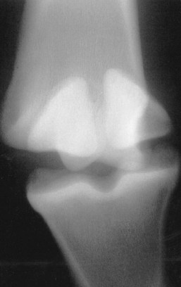
Fig. 67-1 A 3-year-old Paint gelding had acute swelling on the medial aspect of the metatarsophalangeal joint. A stress dorsoplantar radiographic image shows subluxation of the metatarsophalangeal joint caused by complete rupture of the medial collateral ligament (medial is to the left).
Ligament injuries usually heal slowly, and gradual return to function is needed to strengthen the tissues. The consequences of collateral ligament injury depend on the severity of damage to the ligament, contamination in the joint, and damage to other structures in the joint and may be difficult to predict at the time of injury. Long-term instability may place abnormal stresses on the joint and lead to progressive OA. Cutting the lateral collateral and lateral collateral sesamoidean ligaments of the metacarpophalangeal joint resulted in lameness, increased joint circumference, decreased range of motion, and osteophyte formation in 8 weeks, resulting in OA.1
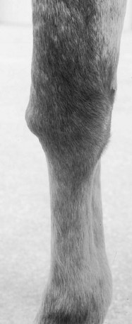
Hygroma
A hygroma is an adventitious or acquired bursa on the dorsum of the carpus caused by trauma from falling, getting up and down, or hitting a fence or by chronically pawing and hitting the dorsum of the carpus.3-5 Nonpainful, fluctuant, uniform soft tissue swelling occurs on the dorsal aspect of the carpus. Pressure does not induce swelling in any associated joints or tendon sheaths. Range of motion of the carpus may be reduced, but lameness is unusual. Injection of radiodense contrast agent into the hygroma or ultrasonographic examination confirms its extraarticular position.
Spontaneous resolution of hygromas may occur, but treatment is often necessary. Drainage and injection of antiinflammatory agents has been used with varying success; in many horses repeated injection is necessary.4,5 I have seen spontaneous resolution after injection of a radiodense contrast agent. Injection of atropine (7 mg) may resolve the swelling. Owners should be warned that bandaging is an essential component of treatment and that long-term chronic thickening may occur.
Other treatments include incisional drainage; injection of irritants, such as iodine or Lugol’s solution; and blistering,4,5 but contrast radiography should be performed to ensure that the hygroma is an isolated structure. Although preoperative contrast radiographs may show no communication between a hygroma and a joint or tendon sheath, one might exist in the form of a one-way valve from the joint into the mass.6 Treatment involving drainage of the mass with a Penrose drain and bandaging has been used successfully for recurrent hygromas. Drains usually are removed once drainage stops, 2 to 7 days after placement.7 Surgical excision can be performed in horses with chronic hygroma and is best accomplished if the fluid sac is left intact and dissected from the other tissues (see Chapter 38).8 Soft tissue and skin closure are routine, and a splint can be used to prevent flexion for better healing. Prognosis for resolution of hygroma is often good, although some degree of thickening usually persists.
Synovial Hernia
A synovial hernia is a defect in a joint capsule or tendon sheath through which the synovial membrane can protrude. The condition rarely causes lameness but is a cosmetic blemish (see Chapter 74). A well-defined, round, soft tissue mass can be palpated over a joint, and with palpation fluid often can be moved between the hernia and the underlying joint or tendon sheath (Figure 67-2). The hernia may disappear with joint flexion. Contrast agent injected into the hernial sac is detected in the underlying joint or tendon sheath, although a one-way valve may be present, limiting movement of contrast material. If the synovial hernia is of cosmetic concern, surgical excision can be performed, with a good prognosis for soundness provided no other joint diseases are present.9
Ganglion
A ganglion is a fluid-filled structure that connects to a joint or tendon sheath through a one-way tract from the joint into the mass.3 Unlike a synovial hernia, the mass lacks a synovial lining and often is filled with mucin. Ganglions are rare in the horse but common in people, and they have been reported around the stifle and the carpus.3 A ganglion adjacent to the fetlock was associated with lameness that was alleviated by regional analgesia and after surgical excision of the mass.10 However, communication with the digital flexor tendon sheath and joint was not demonstrable by ultrasonography. Demonstrating connection between a ganglion and an adjacent joint by injection of radiographic contrast agent into the mass may or may not be possible (see Chapter 74).7
Synovial Fistula
Synovial fistulae are communications between two synovial structures, usually a joint and tendon sheath. They have occurred between the antebrachiocarpal joint and the common digital extensor tendon; the middle carpal joint and the extensor carpi radialis tendon sheath or the common digital extensor tendon sheath; the proximal interphalangeal joint and long digital extensor tendon sheath; and the extensor carpi radialis tendon sheath and a carpal hygroma.6,11-13 Additional joint damage is often present in association with the fistula, causing lameness referable to the area.
Swelling in the joint and nearby tendon sheath occurs, and fluid is often movable between the structures (Figure 67-3). Radiology may reveal additional joint or tendon sheath damage, and contrast agent injected into one of the structures is visible in the other.11
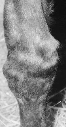
Fig. 67-3 Dorsal view of a carpus with effusion of the antebrachiocarpal joint and common digital extensor tendon sheath. A synovial fistula was seen during arthroscopic surgery.
Occasionally a fistula can be seen during arthroscopic surgery, but closure of the fistula requires an arthrotomy. However, arthroscopic surgery for treatment of a primary problem, without repair of the fistula, has resulted in resolution of lameness, without resolution of the swelling. I do not close these fistulae unless a cosmetic effect is important, or if the swelling itself is impeding performance, or if medical therapy fails to alleviate lameness.
Neoplasia
Joint-associated tumors in horses are rare and consequently behave unpredictably, and relying on treatment information from other species is difficult. Soft tissue tumors in horses are vascular, fibrous, or synovial in origin. Benign vascular masses such as hemangiomas have been seen in carpal and digital flexor tendon sheaths of horses.3 The digital flexor tendon sheaths were distended with bloodstained fluid, but no associated lameness occurred, and surgical excision of the masses was curative.
Fibromas may occur as slow-growing masses near the tarsus, stifle, and distal radius. These masses are rarely erosive to associated bone. Complete surgical excision may be curative, but incomplete excision can result in recurrence.12,14 A fibroma on the proximal lateral aspect of the tarsus was incompletely resected, and over a 4-month period the mass regrew to larger than its original size.15
Villonodular (proliferative) synovitis is a common traumatic injury on the proximal dorsal aspect of the metacarpophalangeal joints of racehorses and is not a tumor (see Chapters 36 and 66). Keratinization of a villonodular synovitis was associated with severe lameness.16 It was suggested that this was a form of epidermal inclusion cyst, the result of inadvertent introduction of epidermal tissue into the joint after repeated arthrocentesis for previous infectious arthritis, resulting in a foreign body reaction. Surgical resection resulted in resolution of lameness. Pigmented villonodular synovitis has occurred in the metatarsophalangeal and femoropatellar joints, resulting in chronic lameness in a mule.17
Synovial cell sarcomas have been identified in the antebrachium,15 a digital flexor tendon sheath,18 and the proximal interphalangeal joint associated with soft tissue swelling and variable lameness. The masses had infiltrated the soft tissues and caused localized inflammatory bone loss because of pressure. There may be recurrence after surgical excision.19 Chondrosarcomas are rare, but they have been described in a metacarpophalangeal joint20 and the carpal region,21 associated with expansible radiolucent lesions. Osteosarcoma is also quite rare but has been described in the proximal aspect of the tibia and periarticular to the shoulder joint.22 The horse with the periarticular mass was successfully managed with excision.22 Osteosarcoma was described in the tarsus of a horse and involved the calcaneus and the proximal intertarsal joint.23 Mast cell tumors have also been identified around joints as mineralized masses that are responsive to excision.24 They do not appear to invade joint capsules in general. A secondary melanosarcoma in a shoulder joint that caused severe lameness has been described.25
A hemangiosarcoma occurred in the tarsal sheath, and surgical excision resulted in relief of lameness.26 However, in a retrospective study of hemangiosarcoma in young horses, the investigators found masses in numerous joints that not only caused radiological evidence of pressure atrophy on surrounding bones but also were unresponsive to surgical excision.27 A neonatal foal with unusual congenital hemangiosarcomas in numerous sites, including the fetlock and stifle joints, has been described.28 The synovial fluid from those joints was hemorrhagic to orange in appearance.
Osteochondromatosis
Synovial chondromatosis and osteochondromatosis are conditions involving pieces of uncalcified and calcified hyaline cartilage, respectively. Synovial chondromatosis is a disease in which hyaline cartilage can occur in the joint in pedunculated form, within the synovial membrane, or free within the joint. Osteochondromatosis results when endochondral ossification of the mass occurs, often making it difficult to differentiate from osteochondral fragmentation. Osteochondromatosis has occurred in the femorotibial joint.3,29 Secondary osteochondromatosis also occurs within joints with OA. The condition is often painful in people, and surgical removal is indicated. Osteochondromatosis has occurred incidentally in horses without lameness.