Chapter 89Cryotherapy
Cryotherapy has been used widely in veterinary medicine since the 1970s, primarily for tumor ablation. Percutaneous cryotherapy, called freeze firing or freezing, is a useful palliative technique for various musculoskeletal disorders in the horse, but little research has been published on specific techniques or results.1-4 Most of the information in this chapter comes from our clinical experience using cryotherapy to manage selected lameness conditions. Cryotherapy generally is used for pain management, and our recommendations are made assuming the horse will continue or resume athletic performance. The term cryotherapy (cold therapy) is sometimes used to refer to intentional cooling of a body part to reduce inflammation or the effects of inflammatory mediators. For instance, cryotherapy is used to prevent laminitis by inducing digital vasoconstriction (see Chapter 34).
Mechanisms of Cryonecrosis
Freezing mammalian tissue results in direct and indirect cell destruction. Direct cell injury occurs by formation of intracellular and extracellular ice crystals, which destroy cell walls and cause intracellular dehydration, respectively. Intracellular dehydration causes severe electrolyte concentration and pH shifts, which damage lipoprotein membranes and organelles. Loss of cellular homeostasis results in cell death. Indirect cell injury occurs by damage to the endothelium of arterioles and venules, causing increased vascular permeability, edema, and hemoconcentration. Local tissue damage occurs from thrombosis and infarction of small vessels. Two rapid-freeze, slow-thaw cycles are used. Rapid freezing maximizes intracellular crystal formation and crystal size. Slow thawing causes additional cell damage by a process of recrystallization, during which time crystals increase in size before melting. Precooled tissue freezes faster than normal tissue; therefore a second freeze-thaw cycle optimizes the processes of tissue destruction.
Because cryotherapy causes tissue destruction in situ, fibrous structures such as epineurium remain intact. This allows for regeneration of large myelinated nerves. Experimental percutaneous cryotherapy of equine palmar and plantar digital nerves resulted in neuropraxia (temporary ablation of nerve function, with the axons remaining intact) or axonotmesis (wallerian degeneration of axonal tissue, with the fibrous supportive tissue remaining intact). All nerves regenerated.5 The degree of nerve damage is temperature dependent, with lower temperatures resulting in longer duration of analgesia.6
Presumably, percutaneous cryotherapy causes destruction of local type C nerve fibers. Type C fibers are small, unmyelinated, nociceptive nerve fibers that contribute to chronic pain. The inflammatory response to cryotherapy appears to cause thickening and fibrosis of certain soft tissues, such as the suspensory ligament and subcutaneous tissues, and although this effect has not been studied, the clinical result is a stronger ligament and one less likely to be reinjured in the same location. Cryogens must be used judiciously on the distal aspect of the limb to avoid cryonecrosis of tendon, ligament, joint tissues, or cortical bone.
Basic Technique
Cryotherapy instruments and cryogens are described elsewhere, and only basic principles are discussed here.7-9 Instruments used to apply cryogens vary from simple cryoprobes to cryounits with continuous closed-system flow of cryogen liquid. We use liquid nitrogen that is stored in a commercial 10- to 20-L tank. For all techniques we use individual brass probes, 1.5 cm in diameter (Figure 89-1), precooled in liquid nitrogen and positioned on the lesions for a double freeze-thaw cycle consisting of freezing (60 seconds), thawing (60 seconds), and freezing (60 seconds). Local edema formation is minimal and has no effect on the second freezing cycle. Earlier work suggested that a thaw duration of 15 seconds was optimal, but our modified cycle appears effective.4 We prefer to use solid metal probes, because consistent freezing to a specific depth is easy to control (Figure 89-2). The cooling ability of solid metal probes has been questioned, but in one study solid metal probes were most effective in freezing to specific depths.7

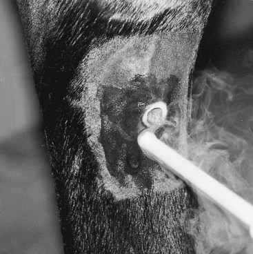
Fig. 89-2 Cryotherapy is being used to manage pain associated with an exostosis of the fourth metatarsal bone in a Standardbred racehorse. A double freeze-thaw cycle has been used in one site, and the probe is being applied at a distal site.
Cryotherapy is well tolerated by most horses, but sedation and a twitch are used. Local analgesia (a ring block proximal to the freeze site) may be necessary for highly strung or fractious horses. The area over the lesion is clipped with a No. 40 blade and cleaned of gross contamination with iodophor or chlorhexidine solution. The limb is dried, and a 0.5-cm–thick layer of water-soluble gel is applied generously over the site. This layer provides a template to ensure accurate positioning for the second freeze cycle.
The metal probe tip is applied to the skin for 60 seconds, then removed to allow thawing, and reapplied (see Figure 89-2). No benefit accrues from a third freeze-thaw cycle, and in fact a third cycle may lead to underlying bone necrosis. Freeze sites should be located 1 cm apart in staggered, parallel rows that cover the lesion site and extend 1.5 cm beyond the margins of the lesion. If freezing sites are closer than 1 cm, the areas of skin necrosis will coalesce and later slough.
After completion of the procedure, the leg is cleaned of lubricant and the sites are covered with an antibacterial spray. The leg should be left unbandaged, but it is kept dry, and antibacterial spray is applied daily until skin healing is complete. Local soft tissue swelling and edema develop and persist for 7 to 10 days and then gradually resolve. The administration of antiinflammatory agents should be avoided, because this medication limits the intended effect of cryotherapy. Percutaneous cryotherapy destroys melanocytes in skin cells, so hair regrowth will be white. This negative cosmetic effect should be considered when discussing complications of cryotherapy.
Treatment of Specific Lesions
Diagnosis always should be confirmed using diagnostic analgesia, because results obviously are improved if the painful area is identified correctly. Most horses return to full function and race performance within several days to weeks, but some (e.g., those treated for severe suspensory desmitis) require up to 120 days of rest after treatment. When managing horses with suspensory desmitis, rest is paramount but is a source of frustration for trainers and owners, who often want to rush a horse back to the races.
Splint Exostoses (Splints)
Horses with exostoses of the second, third, and fourth metacarpal and metatarsal bones should not be treated until acute inflammation has subsided, because cryotherapy may exacerbate periosteal reaction. Horses are given 2 weeks of rest during which a poultice is applied for 5 days, followed by the application of a cedar oil blister for 5 to 7 days. Scurf from the blister is removed by sweating before cryotherapy is performed. Horses with chronic splints can be managed with cryotherapy without this 2-week preparation period. After cryotherapy in any horse, stall rest is recommended for 3 to 5 days. The horse then may be jogged or galloped lightly and brought back into full training within 7 to 10 days after treatment. Prognosis is good for return to racing. The procedure may be repeated at the same site, or at additional sites, if pain recurs or new sites develop.
Second and Fourth Metacarpal or Metatarsal Bone Fractures
Ostectomy of distal fragments of the second or fourth metacarpal or metatarsal bones is ideal, but in some horses surgery is not an option and cryotherapy can provide analgesia. Acute inflammation must be resolved before treatment. A single freeze site is placed directly over the fracture site, and four more sites are frozen in a circular pattern around the fracture site. Prognosis and rest depend mostly on extent of associated suspensory desmitis, if present, because horses with simple splint bone fractures can return to work immediately. Horses with mild suspensory desmitis have a good prognosis for return to racing and usually receive 60 days of rest after cryotherapy. Horses with severe suspensory desmitis require a minimum of 120 days of rest and have a guarded prognosis.
Periostitis of the Third Metacarpal Bone (Bucked Shins)
Cryotherapy can be used to manage pain associated with bucked shins (see Chapter 102) once acute inflammation has subsided. A dorsal cortical fracture of the third metacarpal bone must be ruled out, because cryotherapy in horses with fracture is contraindicated. Once swelling and palpable pain resolve, cryotherapy is performed using three vertical rows of eight or nine freeze sites per row. After cryotherapy, 2 weeks of stall rest with handwalking followed by 4 weeks of turnout exercise is given. Prognosis is excellent, and few horses require additional treatments.
Suspensory Desmitis
Ultrasonography must be performed first to determine the severity and extent of desmitis (see Chapters 16 and 72). Before cryotherapy, acute inflammation is managed using rest, antiinflammatory medication, and local therapy, such as cold-water hosing and poultice application. Cryotherapy is then performed using a vertically oriented row of freeze sites, beginning 3 cm proximal and extending 3 cm distal to the lesion. It is crucial that freeze sites be 1 cm apart and that each site be located directly over the body of the suspensory ligament and not over the deep and superficial digital flexor tendons. Cryotherapy is performed over each affected branch. If suspensory body desmitis is present, the procedure is performed on the medial and lateral aspects. Horses are given a minimum of 2 weeks of stall rest with handwalking, followed by 2 weeks of turnout or swimming physiotherapy. The suspensory ligament and overlying tissue become fibrotic and rigid after treatment, and, although tissue may appear strong to laypersons and the horse may be sound at the walk and trot, it is essential to recommend a conservative return to training and racing. Prognosis always is guarded, but most Standardbred racehorses return to racing. Suspensory desmitis is often a compensatory condition caused by contralateral limb lameness, or lameness in a diagonal or ipsilateral limb, and management of the primary problem is critical in decreasing recurrence of suspensory desmitis. Cryotherapy is seldom effective for long-term management of suspensory desmitis if primary lameness continues. After developing suspensory desmitis, horses may drop in race class and value, but cryotherapy has been a useful adjunct in managing pain and allowing continued racing. The Editors believe that cryotherapy should not be performed for suspensory branch and body lesions in Thoroughbred racehorses because of the risk of catastrophic failure of the suspensory ligament (see Chapter 104).
Distal Hock Joint Pain (Bone or Jack Spavin, Cunean Tendonitis)
Cryotherapy is useful in managing horses with distal hock joint pain. A double horizontal row of freeze sites over the proximal and distal borders of the cunean tendon is used. The horse is given stall rest with handwalking for 2 weeks. Prognosis is excellent for return to racing in the Standardbred racehorse.
Osteoarthritis of the Proximal Interphalangeal Joint
Chronic pain from osteoarthritis of the proximal interphalangeal joint (see Chapter 35) may be alleviated partially by cryotherapy. Cryotherapy is most useful in horses in which palmar digital neurectomy fails to abolish all pain originating from the proximal interphalangeal joint, especially in those with severe bone lysis and proliferation and in which arthrodesis is not an option. Pain relief sometimes can be achieved by placing cryotherapy sites 1 cm apart, encircling the proximal interphalangeal joint proximal to the lesion, and directly over the sites of bony proliferation. It is important to avoid the superficial digital flexor and deep digital flexor tendons. After treatment, horses are given stall rest with handwalking for a minimum of 3 weeks. Prognosis is guarded.
Curb
Curb, a collection of injuries located along the distal, plantar aspect of the tarsus, can be managed successfully using cryotherapy (see Chapter 78). Acute inflammation must be resolved, a process that may take as long as 7 to 10 days. The number of cryotherapy sites depends on the size of the curb, but 8 to 12 sites are usually required. Horses are given stall rest with handwalking for 2 to 3 weeks, but further rest depends on the structure involved and severity of initial lameness. The technique works best on horses with curb as a result of inflammation of the peritendonous tissue or mild superficial digital flexor tendonitis (see Chapter 78). Prognosis is usually good, but some horses require additional treatment within 6 to 12 months.
Cryoneurectomy
Cryotherapy can be useful to provide analgesia to the palmar or plantar aspect of the digits, but the degree of cryoanalgesia depends on the technique used. Percutaneous cryoneurectomy has limited clinical use, because only temporary pain relief (3 to 4 weeks) is achieved.5 Cryoneurectomy has been found to be more effective in producing neuroanalgesia than simple transection, but the former procedure requires a surgical approach.10 The nerves are exposed and transected, and the proximal ends are frozen.
A modified approach can be used.5 The palmar or plantar digital nerves are exposed, and a double freeze-thaw technique is used to freeze the nerve within the perineurium, but the nerve is not transected. Analgesia is longer than with percutaneous cryotherapy, but having to expose the nerves limits the practical application of this technique.
Tendonitis
We feel that management of superficial digital flexor or deep digital flexor tendonitis with cryotherapy is contraindicated. After cryotherapy, fibrous tissue formation along the tendon fibers and possible scarring and adhesions of the digital flexor tendon sheath can occur and limit prognosis. Although we are aware of horses that have raced after cryotherapy for tendonitis, we suggest that other management options be explored (see Chapters 69 and 70).