PROGNOSIS.

In general, fractures in children heal in 4 to 6 weeks; in adolescents in 6 to 8 weeks; and in adults in 10 to 18 weeks. This process from fracture to full restoration of the bone will take weeks to months, depending on the type of fracture, location, vascular supply, health, and age of the individual. Nonunion or delayed union is more likely to occur in adults and occurs in up to 10% of all fractures (affecting nearly 500,000 people each year in the United States).409

Older adults who have suffered a hip fracture have the highest rate of nonunion complications (15% to 30%). These individuals are almost four times more likely to die in the first year after fracture compared with those without fracture. Delay until surgery after hip fracture increases mortality significantly.319 Older women (more than 65 years) who survive the first year after a hip fracture may be at increased risk of death up to 5 years after the injury.207,300

Many people are at high risk for premature death or loss of independence following fracture; mortality after fracture is higher among men than among women.41,46 Less than 50% of older adults with a hip fracture will regain their prior level of function.457 The inability to stand up, sit down, or walk 2 weeks after surgery is the strongest predictor for mortality among older adults with surgically repaired hip fractures.187,490

A person’s condition before fracture (especially that of older adults with hip fractures) has important prognostic implications. Older adults who fall within 6 months following a hip fracture are more likely to demonstrate poorer balance, slower gait speed, and greater decline in ADL from the prefracture level than those who do not fall.457

Healthy functional status contributes to faster recovery time with fewer complications and reduced medical expenses.88 Negative predictors for healing include medications such as calcium channel blockers and NSAIDs, renal or vascular insufficiency, smoking, alcoholism, and diabetes mellitus. Treatment can also affect healing via inadequate reduction, poor stabilization and fixation, distraction damage to blood supply, and postoperative infection.

Associated complications such as nerve injury can occur, and it can take up to 12 to 18 months before reinnervation of the motor endplate is complete. Return of function is dependent upon this factor. If there are no signs of improvement by 7 months, spontaneous recovery is unlikely.540 Exploratory surgery may be indicated at that time.

27-17   SPECIAL IMPLICATIONS FOR THE THERAPIST

Fracture

PREFERRED PRACTICE PATTERNS

Besides the obvious practice patterns, complications from the fracture or the treatment for the fracture can result in skin, vascular, neurologic, and joint involvement beyond the effects associated with bony injury. In such cases, other corresponding practice patterns may apply. Criteria for fractures of the skull may be best described by the neuromuscular practice patterns.

4G:

Impaired Joint Mobility, Muscle Performance, and Range of Motion Associated with Fracture

4H:

Impaired Joint Mobility, Motor Function, Muscle Performance, and Range of Motion Associated with Joint Arthroplasty

4I:

Impaired Joint Mobility, Motor Function, Muscle Performance, and Range of Motion Associated with Bony or Soft Tissue Surgery

7A:

Primary Prevention/Risk Reduction for Integumentary Disorders

There are excellent resources regarding fractures available for the therapist managing fractures, including special considerations, orthopedic intervention, rehabilitation considerations, precautions, goals, and therapeutic exercise with expected time frames for healing and rehabilitation.61,202,259,430,440

For the older adult, there are many potential consequences of fractures. These include biomechanical, functional, and psychologic effects that can limit function and result in considerable disability. Biomechanical consequences can include anorexia and weight loss, compression of abdominal contents and decreased lung function from kyphotic posture, and the risk of more fractures.

Chronic, debilitating pain and increased dependence on family and friends occur as part of the functional consequences. Often there is a significant decrease in the individual’s ability to perform ADLs because of impaired physical function. These factors combined with depression and anxiety (and for some people, sleep disorders) results in psychologic consequences.404 The therapist must remain alert to all of these potential consequences when evaluating each client and planning the best approach to clinical management.

Tobacco use, especially cigarette smoking (both the nicotine and the smoke itself) exposes individuals to toxins that can delay bone healing considerably. Nicotine occupies receptor sites on the stem cells’ surface that are intended for acetylcholine, a chemical that helps nerve cells communicate. Normally, stem cells turn into cartilage-forming cells needed to create the scaffold for callus development over the break.

Nicotine’s effect on stem cells is to cause them to produce too much cartilage while at the same time blocking nerve transmission and delaying or preventing bone healing. Therapists should review the hazards of smoking with clients who have fractures and encourage smoking cessation or reduction. Using nicotine patches or gum immediately after bone injury may have the same negative effect as continuing to smoke.

Fall (Fracture) Prevention

Physical therapists can have a major impact on fall prevention, contributing to the savings of high costs to the health care industry by assessing for risk factors and preventing falls that lead to fractures (see Boxes 27-19 and 27-20).

Given the risk for fracture and other complications and associated emotional and monetary costs, falls are of significant concern for older adults, their families, and the health care system. Complications caused by falls are the leading cause of death from injury in men and women older than 65 years439; a cluster of falls has been observed in older individuals during the months preceding death.160

INCIDENCE

Half of all older adults who fall die as a direct or indirect result of that fall; men are more likely to die after a fall than women. The fall rates and mortality rates from falls are on the rise in the United States.363,392 Other important statistics include the following:

• 13,700 people aged 65 years and older died from falls last year in the United States.73

• At least one out of every four adults aged 65 and older will fall at least once during the next year; it is likely that older adults are falling even more often than is generally reported.50

• Some sources say that one out of every three adults 65 years of age and older fall each year.181,203

• 14% of adults who fall return to the hospital within 2 weeks.

• Falls are the second leading cause of traumatic brain injury among persons age 65 and older.91,352

• Older adults who fall often sustain more severe head injuries than their younger counterparts.

• Falls are a major cause of intracranial lesions among older persons because of their greater susceptibility to subdural hematoma.91

• Even among older adults who do not sustain an injury during a fall, half cannot get up off the floor; this is a predictive factor for functional decline and/or death.500,503

• The complaint of dizziness is one of the most common reasons older adults visit the doctor; the incidence of dizziness doubles, triples, and quadruples decade by decade from 60 to 80 years of age.541

• Less than half (45%) of these cases (dizziness) are caused by vestibular problems.

RISK FACTORS AND RED FLAGS

Complex layers of skill are required to maintain balance in the upright position. Strength, coordination, endurance, flexibility, vision, vestibular control, and hearing are just a few of the skills involved.

At the same time, there are more than 400 risk factors identified for falls. It may be best to focus on the most common risk factors that are modifiable. Age is certainly a primary risk factor, and although age itself is not modifiable, we must be very aware of which adults are at risk requiring screening and intervention (Box 27-20). The rates of falls and fall injuries increase with age; adults 85 and older are four to five times more likely to injure themselves in a fall than adults ages 65 to 74; the risk of being seriously injured in a fall increases with age.477

Movement impairments, cognitive deficits, errors in judgment, and an unsafe environment are common hazards for the aging adult. Gait changes such as an increase in base of support or stride width increases the risk of falls.262 The use of an assistive device such as a cane or walker is a risk factor, especially when learning to use it for the first time. Incontinence (including functional incontinence) is a risk factor by itself but when combined with any of these other risk factors raises the risk even more.

Gait or balance instability combined with muscle weakness is among the highest risk factors.16 Combine any one of these with side effects of medication, the use of alcohol or other drugs (especially when combined with medications), and multiple comorbidities and the risk for falls increases disproportionately.

Many gait disturbances really are a reflection of underlying red flag histories and risk factors. For example, visual impairments can cause increased sway and increased stride width. A past history of falls is a predictor of fear of falling, and a fear of falling will lead to changes in gait such as shorter stride length and slower speed.

Past history of joint replacement (knee) increases the risk of tripping, which increases the risk of falling and injury.315 Painful feet from bunions, corns, overgrown or ingrown toenails, and other podiatric conditions are reported by 50% of older adults when asked. Loss of protective sensation in the feet occurs with diabetes or other peripheral neuropathies.

Neuromusculoskeletal impairments such as muscular weakness, loss of motion, and balance instability, especially when combined with comorbidities such as arthritis, Parkinson’s disease, multiple sclerosis, stroke, and so on, can result in positive Trendelenburg’s sign, uneven stride length, limping, uneven weight bearing, and many other changes in gait pattern.

Comorbidities such as osteoporosis, arthritis and other orthopedic problems, limb amputation, diabetes, dementia, chronic lung disease, stroke, and heart disease are actually more important than polypharmacy (use of four or more medications of any kind) as risk factors for falls, although polypharmacy is still an important risk factor.272

Many of the risk factors fall under the category of red flag histories, too. A past history of falls is one of the most important red flag histories. Obtaining an accurate history can be very challenging. Older adults may forget or deny problems with balance, coordination, and falls (or near falls). They may have lost self-confidence or experience postfall anxiety. Often they modify their behavior and activity level to avoid anything that might cause a fall, so they can honestly answer that they are not having any trouble with falls and have not fallen in the last weeks to months.

Meanwhile, their hygiene is poor because they are afraid to get in and out of the tub or shower. They may restrict and even eliminate community activities for fear of falling. Fear of falling (and especially the fear of not being able to get up) leads to self-imposed functional limitations such as prolonged sitting and all of the natural sequelae that come with an increasingly sedentary lifestyle. Predictors of fear of falling include the following:

• Past history of falls

• Use of assistive device

• Balance or gait instability

• Depression or anxiety

Gait and Movement Characteristics Associated with Falls

In the older adult, gait disturbances and gait changes are often an outward manifestation of an inward or other problem. The therapist should not seek to change the gait pattern until the underlying cause of the problem is known. Consider all of the following gait characteristics associated with falls and assess carefully. Here are just a few31:

• Decreased gait speed and stride length (linked with weak hip extensors and ankle plantar flexors, reduced push-off phase, reduced ability to propel the body forward during gait)

• Increased stride frequency (linked with muscle weakness and impaired balance requiring increased duration of double support)

• Increased (wider) stance (neurologic changes in diabetes or other conditions, muscle weakness)

• Unsteady gait (speed changes abruptly when unintended and/or person cannot adopt new movement patterns when the task requires it; alcohol or other drug use, dizziness, side effect of medication, decreased ankle/knee proprioception)

• Grabbing for support or stumbling

• Decreased medial-lateral sway (gluteus medius weakness)

• Unsafe or incomplete transfers

• Poor sitting balance; difficulty sitting or unsafe sitting down

• Difficulty rising from a seated position

Visual Impairment

Bifocal or multifocal eyeglasses combined with decreased contrast sensitivity and decreased depth perception lead to bending the head up and down to see the edge of a step. Decreased peripheral vision results in increased postural sway necessary to find center; with decreased reaction times, client cannot regain lost balance, resulting in an injurious fall.

Blindness and visual impairment are among the 10 most common causes of disability in the United States, associated with shorter life expectancy and lower quality of life.353 Visual impairment (especially combined with cognitive deficits) can lead to errors in judgment such as climbing up on counters, overreaching, and the use of step stools, resulting in falls.

The therapist can assist in identifying possible untreated visual impairment by asking a few simple visual screening questions and making an appropriate referral when indicated:

• How much difficulty would you have recognizing a friend across the street? [Choose one.]


No difficulty Some difficulty
Moderate difficulty Extreme difficulty

• How much difficulty do you have watching television? [Choose one.]

No difficulty Some difficulty
Moderate difficulty Extreme difficulty

• When was the last time you had an eye examination in which the pupils were dilated (you had to wear the wraparound sunglasses after the examination)?

SCREENING QUESTIONS TO IDENTIFY HISTORY OF FALLS

It may be best to avoid asking the question, Do you have a fear of falling? It is less threatening to ask about the degree of confidence in performing an activity, especially for people who view admitting fear as a sign of weakness.302,502 It may be better to ask questions with answers on a continuum from “no confidence” to “complete (100%) confidence.” For example:

• How confident are you when walking on sidewalks, grass, or uneven surfaces? [Choose one.]

Not confident at all Slightly confident
Moderately confident Completely confident

• How confident are you when getting in and out of a car? [Choose one.]

Not confident at all Slightly confident
Moderately confident Completely confident

• How confident are you when using a public restroom? [Choose one.]

Not confident at all Slightly confident
Moderately confident Completely confident

Follow-up questions may include the following:

• In the past month, have you had any falls?

• In the last month, have you slipped or tripped or lost your balance and almost fallen?

• In the last month, have you hit against furniture and bruised yourself?

• In the past month, have you landed on the floor? If yes, were you able to get up by yourself or without help?

TESTING

Tests and measures are important in identifying individuals at risk for falls and finding ways to reduce risk factors and prevent falls. The therapist should collect baseline data using validated, reliable tests, keeping in mind that many tests do not identify why the individual is at risk or what intervention strategies would be most effective.

Advances in technology are beginning to provide computerized assessments of sensory integration and motor control that can objectively identify and differentiate balance system disorders (e.g., NeuroCom, Biodex). Since these are not yet available in all facilities, other test measures must be relied upon. For example, the Semmes-Weinstein monofilament test (see Fig. 11-15) can be used to check the protective level of sensation in the feet; impairment of this peripheral system could be the primary risk factor for falls and subsequent fracture.

Decreased sensation in the feet associated with diabetic neuropathy can affect both the timing and quality of gait, requiring retraining of the somatosensory and vestibular systems to help compensate for the somatosensory deficit.398,399,541 A neurologic miscue can create footdrop and the toe hitting the ground can cause a fall, easily leading to a hip fracture or broken arm.

A loss of protective sensation and diminished information being received by the brain about how much muscle to use at corresponding sequences of toe-offs during gait can occur. Gait and strength training are important in the management of large fiber neuropathies when impaired vibration, depressed tendon reflexes, and shortening of the Achilles tendon occur.528

At the same time, keep in mind that diabetes gait may occur independent of sensory impairment. The increased joint movement, wider stance, and slower pace demonstrated in some individuals with type 2 diabetes may be neurologic in origin and not related to muscle weakness or loss of sensation in the feet.398,399

Test for ankle and knee proprioception. Determine the lowest threshold for detecting joint movement, determine the accuracy of joint position sense by comparing or matching each joint to the contralateral joint, and perform the joint repositioning test (test limb segment repositioning without the aid of vision).

Dizziness can be due to benign paroxysmal positional vertigo (BPPV) caused by particles containing calcium collected in a semicircular canal provoking episodes of spinning vertigo when the head is moved into certain positions. A canalith repositioning procedure (e.g., Epley maneuver, canalith repositioning treatment or CRT, liberatory maneuver) “cures” this problem by repositioning the person (from sitting to side lying or supine) to move particles out of the canal into the utricle, where they can be reabsorbed.

Test for BPPV by performing the Dix-Hallpike maneuver: assist the patient from sitting to supine with the head extended over the edge of the treatment table and rotated 45 degrees to the side of the suspected ear. A positive test result is present when nystagmus is induced after a delay of about 10 seconds. See further discussion in Chapter 38.

TEST FUNCTIONAL LIMITATIONS

The therapist should use a standardized fall assessment tool and use it consistently. An excellent resource for test tools is available: Falls Prevention for Older People: Resources: Screening Tools (http://www.fallsprevention.org.au/resources.htm; accessed July 17, 2007).

Tests for functional limitations are useful but do not necessarily identify the cause(s) of balance dysfunction. Many tests are available; the therapist should choose one that best matches the individual’s current level of functioning. Some examples include the following:

• Berg Balance Scale: 14-item test measures balance on a continuum from sitting to standing. Valid and reliable measure of balance in older adults; predictive of individuals at risk; helps goal setting and directs intervention; use with lower-functioning patients.

• Timed Up and Go Test (TUGT): used to screen individuals at risk for falls. The client must be able to follow directions. Score of 13.5 seconds indicates that the individual is at risk for falls.

• One-Legged Stance Test: measures postural stability needed to make turns, climb stairs, get dressed, get in a car, step into a bathtub, or step up onto a curb. Risk of falls increases two times if test score is less than 5 seconds; client is at risk if response is 12 seconds or less; marginal risk if response is 13 to 20 seconds; 20+ is considered “safe”; the individual must not sway more than 45 degrees to remain in the “safe” category.

• Sit-to-Stand: must be able to rise from a chair 10 times without using the arms in less than 20 seconds.

• Four Square Step Test (FSST): reliable, valid, easy to administer and score; timed test to assess the rapid change in direction while stepping over low objects and movement in four directions; requires higher level of cognitive function and ability to shift weight from one foot to the other while changing direction. Consider using with adults who report falls or near loss of balance as a result of hurrying. Correlates with Functional Reach Test and Timed Up and Go Test]; this means the tests measure similar constructs, so only one of the tests needs to be administered.105

Predictive fall risk information can also be obtained and can be useful when putting together the plan of care; fall risk assessment for use with the acute care inpatient population and assessing fear of falling may be appropriate (Box 27-21). No one scale best predicts falls risk in older adults. The Activities-Specific Balance Confidence (ABC) Scale and Falls Efficacy Scale (FES) are highly correlated with each other. These two tests are moderately correlated with Survey of Activities and Fear of Falling in the Elderly (SAFE).205

Box 27-21   PREDICTIVE FALL RISK ASSESSMENT

• Functional reach test: must be able to reach 6 or more inches

• Tinetti’s Performance-Oriented Mobility Assessment (POMA)/Tinetti’s Balance Index: objectify gait and balance; predict risk of falls; can be used with people who have an assistive device; does not detect small changes in gait deviation—Gait Abnormality Rating Scale (GARS) is better for assessing early, minute gait deviations

• Dynamic Gait Index: provides predictive fall-risk information

• Gait Abnormality Rating Scale—Modified (GARS-M): used to assess risk of falling in community-dwelling, frail older adults based on several gait variables

• Physiologic Profile Assessment (PPA): series of simple tests of vision, peripheral sensation, muscle force, reaction time, and postural sway that are easy to administer quickly with minimal equipment290

• Measurement of vital signs and assessment for postural hypotension: these are important assessment tools in predicting falls

For Use with Acute Care Inpatient Population—Fall Risk Assessment

• STRATIFY: St. Thomas’s Risk Assessment Tool in Falling Elderly Inpatients

• Hendrich II Fall Risk Model (http://www.hartfordign.org/publications/trythis/issue08.pdf)

Fear of Falling

• Falls Efficacy Scale (FES): good for adults who are frail

• Survey of Activities and Fear of Falling in the Elderly (SAFE): assesses 11 activities of daily living

• Activities-Specific Balance Confidence Scale (ABC): measure of balance confidence; good with higher functioning older adults; used in studies of amputee populations

The therapist should adopt a task-or functionoriented approach. This requires analysis of all the systems involved in the required movement. Rather than focus on individual exercise movements, identify the systems involved in the task and direct intervention to each of those systems.

For example, suppose the patient’s goal is to be able to pick up objects from the floor. Physical therapy goals are to improve reactive responses to external challenge during stance and ambulation activities as measured by the following: Goal 1—Increase Berg Balance Scale score from 34/56 to 50/56 in 6 weeks; Goal 2—Increase Tinetti’s Performance-Oriented Mobility Assessment (POMA) score from to 19/28 to 24/28.

The intervention should be customized for each individual, targeting the specific underlying impairments, which may include the following:

• Task-specific activities in sitting, standing, and walking (including reaching; weight shifting; progressing from flat surfaces to rocker board, curbs)

• Functional strengthening of the ankle, hip, and trunk muscles

• Range of motion in the trunk, hip, lower extremity (especially ankle)

• Static and dynamic balance activities with lateral sway, reach, grasp, manipulation of arms

• Treadmill training

Keep in mind that general strengthening is not enough. Specific muscle groups must be targeted and given enough resistance to build strength. The ankle is considered the most important joint governing balance.539 Older folks often have reduced ankle motion and strength; these deficits are linked with reduced balance.323 Check for loss of ankle motion; mobilize the calcaneus before stretching. Resistive exercises that produce overload of the hip extensors or ankle dorsiflexors are more effective in improving balance and reducing falls than general low-resistance exercises for the lower extremities.95

EXERCISE AND FALL PREVENTION

Three types of exercise that can help prevent falls and fractures are balance training (prevents falls), strength training (builds bone and muscle), and aerobic training (builds muscle and endurance).

Walking while performing an additional attention-demanding cognitive task is a means to measure whether the client is able to walk automatically. The therapist can assess for dual-task interference, which is the worsening of performance of the main task (e.g., walking) as a result of simultaneously performing an attention-demanding cognitive task (e.g., counting backward).

Automaticity of the main task is reflected by a low or absent dual-task interference effect. Nonautomaticity can have an impact on daily living; gait changes caused by performing an additional cognitive task while walking are associated with increased risk of falling among older adults.36,520

STEPS TO TAKE

Almost all hip fractures in older adults (over 65 years) are caused by falls, and very few are spontaneous, answering the question of whether falls are the result of fractures or vice versa.376 Hip fractures among older adults occur mainly in well-known environments, during everyday activities, and without overwhelming hazards, emphasizing again the importance of fall prevention/fracture reduction programs for all adults over age 65 years.

The CDC has recommended community-based falls prevention programs as an effective strategy based on research identifying interventions that can reduce falls. The therapist can be very instrumental in falls prevention by reviewing each of these with all clients age 65 and older. Four key fall prevention strategies are the following:

• Review of medications to reduce side effects and interactions

• Annual eye examination

• Regular exercise

• Reduction of fall hazards in every room of the home or facility

Modifying the environment is important; it may not prevent falls but it may reduce the severity of injury. The therapist should have the client and/or family complete an environmental safety check. Some studies show that this step is more effective when people are provided with a checklist and encouraged to make the changes themselves. An excellent checklist is available.371

Fall-proofing homes; fall prevention education; osteoporosis and fall assessment; osteoporosis prevention (see Chapter 24); and exercise programs to improve balance, coordination, flexibility, strength, endurance, breathing, and posture are all important components of fall prevention and subsequent fracture prevention. Exercise to address all these components is key in fall prevention.458 For example, lower extremity weakness or loss of motion, particularly at the ankle and knee, is significantly associated with recurrent falls in the older adult population.323,539

Specific intervention programs,504 exercise suggestions,162,170,450 and educational materials18,216 are available (Box 27-22). Sometimes even the simple step of teaching clients adequate hydration can improve their muscle strength, coordination, and balance, reducing risk for falls (see the section on Dehydration in Chapter 5).

Box 27-22   RESOURCES FOR FALLS ASSESSMENT AND PREVENTION

1. American Geriatrics Society, British Geriatrics Society, and American Academy of Orthopaedic Surgeons Panel on Falls Prevention: Guidance for prevention of falls in older persons, Ann Long Term Care 9:42-47, 2001. Available on-line at http://www.healthinaging.org/public_education/falls_index.php. Accessed June 1, 2008.

Falls: General information

Medical Evaluation of Falls

Choosing a Cane or Walker

Choosing and Starting an Exercise Program

Improve Your Balance in 10 Minutes/Day

Decrease Your Risk of Falling

Tips for Patients with Low Vision

2. Department of Health and Human Services (HHS), Bureau of Primary Health Care: Lower Extremity Amputation Prevention (LEAP) program. Available on-line at www.hrsa.gov/leap. Accessed June 1, 2008.

3. Falls Prevention Project for Older Adults offers program manuals and training resources. Available on-line at www.temple.edu/older_adult. Accessed June 1, 2008.

4. Fall Risk Assessment for Older Adults: The Hendrich II Model. Available on-line at http://www.hartfordign.org/publications/trythis/issue08.pdf. Accessed June 1, 2008.

5. Falls Prevention for Older People: Resources: Screening tools. Available on-line at http://www.fallsprevention.org.au/resources.htm. Accessed June 1, 2008. Excellent web page that describes various testing tools for falls.

6. National Center for Injury Prevention and Control (NCIPC): Preventing falls among older adults. Available on-line at http://www.cdc.gov/ncipc/duip/preventadultfalls.htm. Accessed June 1, 2008. Lots of great patient education materials, checklists, posters, prevention strategies.

7. National Safety Council: Falls in the home and community. Available on-line at http://www.nsc.org/issues/fallstop.htm. Accessed June 1, 2008.

8. Stevens JA: Falls among older adults—risk factors and prevention strategies. Falls free: promoting a national falls prevention action plan. Washington, DC, 2005, National Council on Aging. Brochures and posters available in English, Spanish, and Chinese are available at http://www.cdc.gov/ncipc/pub-res/toolkit/brochures.htm with additional information at http://www.cdc.gov/ncipc/pub-res/toolkit/toolkit.htm. Accessed June 1, 2008.

Computerized assessments of sensory integration and motor control can objectively identify and differentiate balance system disorders. Through advances in technology, therapists are now better able to identify the underlying impairments that may increase a person’s risk for falling.368,525 In addition, clinical risk factors predictive of fracture (e.g., age, gender, height, weight, use of walking aid, current smoking) have been identified. The presence of five or more of these factors increases the rate of fracture (tested in white women).531 These models have not been validated in other population groups.

Instituting and following a fall protocol in an extended or acute care facility may be able to reduce the incidence of repeated falls. The therapist can help staff set up a study to find out what time of day most falls occur and establish well-supervised small group activities during those times, especially for residents with dementia who are more likely to be put by themselves, get bored, and attempt to get up without assistance and fall. Residents of long-term care facilities may require individualized exercise interventions that can be adapted to their changing needs in reducing falls.375

Current use of physical restraints with acute care clients or residents of long-term care facilities is based largely on the assumption that these devices prevent falls and fall-related injuries. Numerous studies have reported a significant incidence of falls and injuries in restrained older adults rather than a lower risk of falls and injuries. With growing acceptance of restraint-free care as a standard of practice, therapists can be instrumental in educating personnel in these facilities of these findings when instituting a program.68,505

A THERAPIST’S PERSPECTIVE

The vestibular system controls head, neck, and eye movements and is certainly an important component of balance and falls prevention. The three semicircular canals positioned in each spatial plane accurately sense head position and rapidly send signals to the eyes to keep our vision stable. After the age of 40, the number and size of vestibular neurons decrease. In individuals over the age of 70, 40% of the vestibular sensory cells are gone.40,471

Vestibular impairment or dysfunction is certainly a possible cause of falls, and the therapist should test for this and intervene appropriately, but often the underlying etiologies are impairments of the peripheral systems feeding into the vestibular system. The vestibular system may react too little or inappropriately because external stimuli are deficient. Keep in mind that there may be other causes of vestibular system failure, which may not be failure at all but merely compensation for impairment or failure of other systems.

Take into consideration motor, visual, and especially oculomotor, vestibulomotor, and oculovestibular reflexes; it is not a single-system issue. The visual or ocular system determines movement and position in space and provides visual reference points. And the proprioceptive or somatosensory system consisting of pressure sensors in muscles, tendons, and joints (especially in the lower extremities) senses gravity and joint position. Any change in these systems can result in dizziness, fear of falling, and limited activity, leading to increased postural instability and contributing to further weakness and imbalance in a downward declining spiral of events.

Consider the effects of macular degeneration, loss of peripheral vision, and the loss of visual cueing mechanisms from these systems. A person may be able to compensate with one problem or system dysfunction, but when all three systems are impacted, it is much more difficult. The person starts to experience dizziness as the vestibular system tries to increase input. The person’s natural tendency is to sit more to avoid dizziness and falls.

Sometimes use of a simple cane to increase peripheral messages can make a big difference. But what is the typical response to this? “That will make me look old.” “I’d rather fall than use that thing.” Issues of pride enter in; the individual is unwilling to use a cane to help increase proprioceptive cues to make up for a loss of vestibular, visual, or motor input. You may need to have an honest, frank discussion with that person. Try to reframe the client’s view of the situation into something more acceptable; for example, help the individual see this as a short-term situation or even consider using a more direct approach. For instance, convey the following information to your patient/client:

Studies show that 50% of the people over 65 who fall fracture their hip and do not return home from the hospital. They often end up in a nursing home or extended care facility. Fifty percent of those people die within 1 year. Are you going to let a simple cane keep you from going home? If you do not want to use a cane, then how about getting a walking stick and letting me help you learn how to use it to your best advantage?

The therapist can also help older adults determine their own problem list and solutions in order to effect successful changes in behavior by asking the following questions271:

• What do you think is the problem?

• What can be done to change your situation?

Alternatively: What do you think will help?

• What needs to happen for you to be able to _________________ (repeat what the person just told you)?

• Can you do it? Can you _________________ (repeat back what the person said needs to happen).

• Will you do it?

If the patient/client hesitates, you can ask, “What is one thing you CAN do to improve matters?

The therapist can also make some changes in how he or she practices in regards to screening individuals for falls risk, reducing or modifying risk factors, and preventing falls leading to fractures. If implementing a falls screening and prevention program for your facility seems too overwhelming, then at least choose one test and perform it consistently for 6 months or 1 year. Consider providing and/or participating in health fairs offering balance and posture screening.

Put together a task force with the express purpose of developing a screening checklist to fit your population base. Complete the checklist for every patient/client and conduct appropriate test measures. Make an appropriate referral sooner rather than later.

Stress Reaction/Fracture

In the case of individuals reporting isolated or pinpoint pain of the lower extremity associated with overuse in the presence of negative radiographic examination (x-ray), the therapist may need to assess further for the possibility of a stress fracture. This is also true for the client who shows minimal improvement (or a worsening of symptoms) following therapy intervention.

Applying a translational, rotational, or impact stress to the bone often reproduces the symptoms when the stressed bone lies deep within the tissue. Testing may start by applying a striking (percussive/compressive) force through the heel as the client lies supine and observing for any associated increase in painful symptoms, especially at the hip or groin. Pull the leg into a position of internal rotation and resist as the person tries to externally rotate the leg; then move the leg into external rotation as the person tries to internally rotate the leg. For a differentiation of stress reactions of the femoral neck versus the pubic ramus, see Box 27-23.

Box 27-23   DIFFERENTIATION OF STRESS REACTION/FRACTURE

Pubic Ramus

• Adductors may be in spasm (adductor avulsion from stress fracture at the inferior ramus); limited abduction

• Negative heel strike/percussion test result

• Pain with activity that is relieved with rest, usually no pain at night

• Pain with resistance to the muscles of adduction and external rotation; positive Patrick’s or FABER (hip and knee flexion, hip abduction, hip external rotation) test (e.g., pain is reproduced as the person tries to internally rotate from a position of external rotation)

• Gait may be normal or antalgic depending on the fracture location

Femoral Neck

• Vague, nonspecific description and onset of pain; unrelieved by rest; aggravated at night by rolling onto that side

• Positive Trendelenburg’s sign (see Fig. 23-14) with compensated gait pattern

• Positive heel strike/percussion test; symptoms reproduced with hopping

• Positive test of femoral neck integrity (pain as person tries to externally rotate from an internally rotated position; may be unable to even assume test position in passive internal or medial rotation)*

• Noncapsular pattern (capsular pattern of the hip is defined as gross limitation of flexion, abduction, and medial rotation and slight limitation of extension with little or no limitation of lateral rotation)

• Localized tenderness at the greater trochanter unrelieved by treatment intervention for bursitis; pain may occur in the buttocks and/or groin

Tibia

• Painful pinpointed symptoms reproducible on palpation, frequently bilateral (one side more symptomatic than the other side); must be differentiated from shin splints

• Increased pain on weight bearing and walking (person assumes a wide-based waddling gait)

• May have a positive reaction to painful heel strike (more common in a hip fracture); symptoms reproduced with hopping

Adapted from Ozburn MS, Nichols JW: Pubic ramus and adductor insertion stress fractures in female basic trainees, Mil Med 146(5):332-334, 1981.


*This may be differentiated from trochanteric bursitis, which is characterized by painful hip abduction and lateral rotation (but not medial rotation).

Any suspicion of stress reaction/fracture warrants communication with the physician. Further imaging studies may be necessary. Any woman with a stress fracture should be evaluated for the female athlete triad (amenorrhea, eating disorder, osteoporosis). Treatment of the underlying factors that contribute to the injury is important, including these three factors.550

Rehabilitation will vary depending on the age, condition, and goals of the affected individual. For example, rehabilitation for military personnel will differ greatly from that prescribed for a postmenopausal woman. Specifics of progressing an exercise program are available.427

Acute Care and Complications

Complications of fractures require vigilance on the therapist’s part and possibly quick action. Significant swelling can occur around the fracture site, and if the swelling is contained within a closed soft tissue compartment, compartmental syndrome may occur (see the section on Soft Tissue Injuries in this chapter). Because of the progressively increased intracompartmental pressure, nerve and circulatory compromise can occur. This condition may be acute or chronic. The compartment becomes exquisitely painful.

A thorough sensory and motor examination may be warranted. If the therapist notes skin changes, decreased motor function, burning, paresthesia, or diminished reflexes, physician contact is necessary. Permanent damage and loss of function may result if this condition is not treated. The therapist’s examination may be helpful in establishing the extent of the injury and baseline function.99

Another complication associated with fractures is fat embolism, a potentially fatal event. The risk of developing this condition is related to fracture of long bones and the bony pelvis, which contain the most marrow. The fat globules from the bone marrow (or from the subcutaneous tissue at the fracture site) migrate to the lung parenchyma and can block pulmonary vessels, decreasing alveolar diffusion of oxygen.

The initial symptoms typically appear 1 to 3 days after injury, but this complication can occur up to a week later. Subtle changes in behavior and orientation occur if there are emboli in the cerebral circulation. There may also be complaints of dyspnea and chest pain, diaphoresis, pallor, or cyanosis. A rash on the anterior chest wall, neck, axillae, and shoulders may develop. The onset of any of these symptoms warrants immediate physician contact.

Individuals with acute VCFs (see Fig. 24-5) can be difficult to treat, because pain and fear can be severe. Even when extra care is taken with logrolling techniques, transitional movements can be exquisitely painful, with the client crying and begging the therapist to stop.

Arranging for premedication 45 to 60 minutes before treatment is advised, followed by modalities to modulate the pain and promote relaxation before attempting movement or exercise. Adaptive equipment, from wheelchair modifications to spinal orthotics to assistive devices (e.g., reachers/grabbers, stocking aids, raised toilet seats, bed grab bars), helps to improve posture, function, mobility, confidence, and independence.

The therapist must be alert to other complications that can occur following fracture, such as breakage of wires, displacement of screws, loss of fixation, refracture, delayed union and malunion, and infection.451 Anyone on bed rest is at risk for complications from immobility, including constipation, deep vein thrombosis, pulmonary embolism, and pneumonia.

Fracture Rehabilitation

Like medical treatment, fracture rehabilitation is shaped by fracture type, need for reduction, presence of instability after reduction, and functional requirements of the affected individual. Postoperative rehabilitation begins anywhere from immediately to within 1 week after surgery depending on the physician’s protocol.

There are some widely accepted guidelines and rehabilitation protocols for various types of fractures (e.g., Neer rehabilitation program for shoulder fractures, Vanderbilt program, Tinetti protocol of balance exercises for rehabilitation from hip fractures).280,501 The American Academy of Orthopaedic Surgeons offers guidelines for the rehabilitation of many different types of fractures. Several publications with fracture rehabilitation protocols are available specifically for the physical therapist.55,279,281,317,382

Mortality rates following hip fractures in older adults may be improved by a more intensive rehabilitation program immediately after the operation. The best predictor of mortality immediately after hip fracture up to 1 year after fracture is the inability to stand up, an indicator of frailty.187

Older adults admitted for care of a fall-related hip fracture should be evaluated early in their hospital stay to determine risk for falls following discharge. Indicators may include a previous history of falls and prefracture use of an assistive device for ambulation. The plan of care should include balance and mobility training to prevent future falls. A previous history of falls is a risk factor for future falls; poor balance, slow gait speed, and decline in ADLs have been identified in older adults who fall within 6 months following a hip fracture.457

Early mobilization accompanied by transfer training, and maintaining strength and range of motion after fracture surgery are essential to reduce the risk of deep vein thromboembolism, pulmonary or infectious complications, skin breakdown, and decline in mental status.

Following a fracture anywhere in the lower extremity (including the hip), some orthopedic surgeons advocate unrestricted weight bearing, advising the client to decide himself or herself how much weight to put on the leg. Stable fractures can usually tolerate weight bearing. Rotationally stable but potentially long unstable femur fracture may be allowed toe-touch weight bearing. Clients with vertically and rotationally unstable femoral fractures may be restricted to non–weight-bearing status using a wheelchair or electric scooter (if not in a spica hip cast).414

Although immediate weight bearing may cause initial bone loss, the long-term success of achieving bone growth remains unchanged,454 and the short-term benefits of functional recovery and quicker return to independent living that accompany unrestricted weight bearing are important.257,391

Again, depending on the type of fracture, some movements may be restricted to allow for proper fracture consolidation. Most importantly, a non–weight-bearing status can actually place greater forces on the hip as a result of the biomechanics involved in maintaining correct positioning of the lower extremity.208,357 In the case of femoral neck or intertrochanteric fractures, there is little biomechanical justification for restricted weight bearing; indeed, there is far greater pressure generated from performing a hip bridge while using a bedpan (almost equivalent to the effect of unsupported ambulation).257

Partial weight bearing is usually considered 30% to 50% of body weight. Touch or touch-down weight bearing is 10% of body weight, but this is a subjective decision that is not easily determined. Allowing for unrestricted weight bearing according to the client’s tolerance (WBAT) is less restrictive, but the therapist must assess for intact cognition and decision-making abilities, intact sensation, upper body strength, vestibular function and balance, and proprioception before allowing unsupervised WBAT.

Early repair and physical therapy have been shown to reduce hospital stays, increase chances of returning home (rather than being discharged to a nursing facility or rehabilitation facility), reduce complications, and improve functional mobility and independence at discharge,163,250 and are associated with higher rates of 6-month survival.88,198 Therapists are encouraged to share the results of studies such as these with hospital administrators when developing fall and fracture prevention programs.

The literature supports recommending follow-up for strength and functional assessment 7 to 9 months after fracture.438,454 Muscle strength around the hip remains weak after hip fracture, with joint arthroplasty requiring an exercise program for strengthening for 1 year or longer.218,456 Short-term intervention with a therapist can be very cost effective in reducing refracture rates.454,455

Older adults who receive physical therapy while still in the hospital following hip replacement for hip fracture are more likely to be discharged directly home rather than to a rehabilitation or assisted living facility.175 People with hip fractures who receive additional home health visits are less likely to be hospitalized and more likely to need fewer medical visits, which usually translates into lower Medicare costs.221

For clients with VCFs, the plan of care should include trunk extension strengthening and a cognitive-behavioral component to improve coping, especially for older adults. Improvements have been retained for at least 6 months in one randomized, controlled trial.150

Osteochondroses

A number of clinical disorders of ossification centers (epiphyses) in growing children share the common denominator of avascular necrosis and its sequelae. These disorders are grouped together and referred to as the osteochondroses430 with multiple synonyms (epiphysitis, osteochondritis, aseptic necrosis, ischemic epiphyseal necrosis). There are additional eponyms based on the name of the person or persons who described the disorder as well, such as Kohler’s disease (tarsal-navicular bone disease), Osgood-Schlatter disease, and Legg-Calvé-Perthes disease.

The underlying etiologic factors and pathogenesis are similar in all these entities, and the clinical manifestations are determined by the stresses and strains present. Most susceptible areas are the epiphyses, which are entirely covered by articular cartilage and therefore poorly vascularized.

Osteochondritis Dissecans

Osteochondritis dissecans (OCD) is a disorder of one or more ossification sites with localized subchondral necrosis followed by recalcification. This condition affects the subchondral bone and the layer of articular cartilage just above. A piece of articular cartilage and fragment of bone separate and pull away from the underlying bone. These fragments can become loose bodies in the joint; the most common site of involvement is the medial femoral condyle.

OCD is caused by repetitive microtrauma resulting in ischemia and disruption of the subchondral growth. The articular cartilage softens, and fragment separation leads to cartilage injury that can progress to form a crater. Activity-related pain, swelling, and giving way are common symptoms. Pain is increased with passive knee extension and tibial internal rotation and relieved with tibial external rotation (Wilson’s sign).

X-rays and MRI help confirm the diagnosis. Management varies with the person’s age and the severity of the lesion and includes nonoperative management (activity modification, protected weight bearing, immobilization for 4 to 6 weeks) and operative treatment. Quadriceps strengthening and gradual return to activities follow immobilization. If conservative care is unsuccessful in bringing about healing, then surgery may be needed (e.g., microfracture, implant tissue to stimulate cartilage and bone growth).

Osteonecrosis

Overview and Incidence

The term osteonecrosis refers to the death of bone and bone marrow cellular components as a result of loss of blood supply in the absence of infection. Avascular necrosis and aseptic necrosis are synonyms for this condition.

The femoral head is the most common site of this disorder (sometimes called Chandler’s disease), but other sites can include the scaphoid, talus, proximal humerus, tibial plateau, and small bones of the wrist and foot. Avascular necrosis is the underlying cause for 10% of total hip replacement surgeries362 and overall affects approximately 20,000 people annually, often between the second and fifth decades of life.270

Etiologic and Risk Factors

Osteocytic necrosis results from tissue ischemia brought on by the impairment of blood-conducting vessels. A minimum of 2 hours of complete ischemia and anoxia is necessary for permanent loss of bone tissue.224 The bony ischemia may be secondary to trauma disrupting the arterial supply or to thrombosis disrupting the microcirculation.

Bones or portions of bones that have limited collateral circulation and few vascular foramina are susceptible to avascular necrosis. Box 27-24 lists conditions associated with osteonecrosis. A number of these conditions are linked to osteonecrosis by the development of fat emboli (caused by altered fat metabolism) in the vascular tree of the involved bone.

Box 27-24   CONDITIONS ASSOCIATED WITH OSTEONECROSIS

• Idiopathic

• Trauma (e.g., fall)

• Systemic lupus erythematosus

• Pancreatitis

• Diabetes mellitus

• Hyperlipidemia

• Cushing’s disease

• Gout

• Sickle cell disease

• Alcoholism

• Obesity

• Pregnancy

• Medications

• Oral contraceptives

• Corticosteroids

• Bisphosphonates (under investigation)

• Organ transplantation (medication related)

• Human immunodeficiency virus (HIV) infection

• Radiation therapy (less common)

• Dysbaric disease (deep sea diving; rare)

The conditions associated with the development of fat emboli include alcoholism, obesity, pregnancy, pancreatitis, medications (e.g., oral contraceptives, corticosteroids), and unrelated fractures. Many cases of femoral head osteonecrosis are idiopathic (i.e., no known cause or risk factor can be identified).

Osteonecrosis has also been recognized as a complication in HIV-positive individuals; in fact, individuals who are HIV positive have a 100-fold greater risk of developing osteonecrosis than the general population.356 The exact mechanism for this remains unknown. It may be due to hyperlipidemia secondary to the use of protease inhibitors; however, avascular necrosis was reported before the era of highly active antiretroviral therapy (HAART).416 It does not appear to be related to the degree of immunodeficiency.

More recently, the use of bisphosphonates has been linked with osteonecrosis of the jaw (sometimes referred to as “dead jaw syndrome”), especially after trauma to the teeth or bones of the jaw such as occurs with dental surgery (e.g., tooth extraction). The reason this happens is not entirely clear. Scientists hypothesize that since the jaw has a high rate of bone renewal in response to stress via generation of an inflammatory response by the gums and teeth, bisphosphonates keep osteoclasts from reabsorbing damaged bone cells in the jaw. The damaged bone builds up and eventually results in osteonecrosis.

This phenomenon is most likely to occur in individuals treated for bone cancer with intravenous bisphosphonates. The dosage of intravenous bisphosphonates can be as much as 12 times more than the oral bisphosphonate dosage prescribed for osteoporosis. Individuals with cancer treated this way also undergo other bone-weakening treatments (e.g., chemotherapy, radiation therapy).

Pathogenesis

Certain bones are more vulnerable to osteonecrosis than others. These bones are covered extensively by cartilage, have few vascular foramina, and have limited collateral circulation. The femoral head is a prime example of a bone at risk. The superolateral two thirds of the femoral head receives its blood supply almost entirely from the lateral epiphyseal branches of the medial femoral circumflex artery (Fig. 27-26).

image

Figure 27-26 Blood supply to the femoral head in a child. (From Bullough PG: Orthopaedic pathology, ed 3, London, 1997, Mosby-Wolfe, p 263.)

The only other source of blood for the femoral head is the medial epiphyseal artery (contained within the ligamentum teres), which has limited anastomoses with the lateral epiphyseal vessels. Hip dislocation or fracture of the neck of the femur can compromise the precarious vascular supply to the head of the femur. The talus, scaphoid, and proximal humerus are also susceptible to osteonecrosis.

As the ischemia progresses, repair processes occur but are not capable of preventing necrosis and deformation of the bone, such as flattening and collapse of the femoral head. The articular cartilage and acetabulum are usually spared until late in the disease process, but the articular cartilage may be lifted off the underlying bone, resulting in irreparable damage to the joint.201 The entire process extends over many years, and unlike in osteochondrosis of immature bone (e.g., Legg-Calvé-Perthes disease), spontaneous healing never occurs.

Clinical Manifestations

Often no symptoms are observed during the initial development of osteonecrosis even though an ischemic condition of the bone exists.201 Hip pain is the usual initial presenting complaint, with a gradual onset, sometimes of many weeks’ duration, before diagnosis. The pain may be mild and intermittent initially but will progress to become severe, especially during weight-bearing activities.

If the femur is involved, the pain may be noted in the groin, thigh, or medial knee area. An antalgic gait is noted, and pain provocation occurs with weight-bearing activities and hip range-of-motion exercises, especially internal rotation and flexion and adduction. The affected individual will report a slowly progressive stiffening of the joint. When fracture occurs, it is usually at the junction between necrotic bone and reparative bone, possibly extending down through the reparative interface to the healthy inferior cortex of the femoral neck.340

Eventually degenerative joint changes and osteoarthrosis occur at the involved hip joint; the pathologic process is often relentless, with collapse of the femoral head imminent in spite of medical intervention.201

Osteonecrosis of the jaw is characterized by exposed bone in the mouth, numbness or heaviness in the jaw, pain, swelling, infection, and loose teeth. Delayed or poor wound healing after dental surgery may be the first indication of a problem. Crepitus as the jaw opens and closes may be present and is often described as like the sound of someone walking on ice.

MEDICAL MANAGEMENT

DIAGNOSIS.

Plain films may be normal initially. Bone scan, MRI, and CT scans are much more sensitive procedures and detect subtle bony changes.

TREATMENT.

The choice between conservative and surgical intervention depends on the size of the lesion, how early the diagnosis is made, and whether bony collapse has occurred. If surgery is not indicated, protected weight bearing is essential to prevent collapse of the lesion.

Surgical intervention may consist of core decompression for small lesions without evidence of structural collapse (most common procedure in early diagnosis) to relieve pain and delay or prevent structural collapse, hemiarthroplasty, or total joint replacement.292 Core decompression removes a core of bone from the femoral head and neck in an attempt to relieve intermedullary pressure, thereby promoting revascularization. This may be accompanied by bone grafting.475

Joint replacement may be required if femoral head collapse occurs or in order to prevent this complication. However, this procedure is limited by young age and high activity level as well as the limited life expectancy of the prosthesis.

New techniques for bone stimulation may be used, such as replacing the dead bone with living bone from the individual’s fibula to give added strength to the damaged area and possibly prevent or postpone joint arthroplasty in young individuals; see also the section on Fracture: Treatment in this chapter.

An osteotomy may be performed to shift the site to where maximal weight bearing occurs on a particular joint surface. Analgesics and NSAIDs are used for symptomatic relief of pain.

PROGNOSIS.

The prognosis depends on the extent of damage that has occurred before diagnosis in the case of nontraumatic disease. Unfortunately, many cases are diagnosed in an advanced stage of disease, when minimally invasive surgical procedures are no longer helpful.270 Early intervention (both surgical and nonsurgical) has definitely improved the outcome, but many people with femoral head osteonecrosis experience irreversible damage to the joint and will need total arthroplasty.

27-18   SPECIAL IMPLICATIONS FOR THE THERAPIST

Osteonecrosis

PREFERRED PRACTICE PATTERNS

4G:

Impaired Joint Mobility, Muscle Performance, and Range of Motion Associated with Fracture

4H:

Impaired Joint Mobility, Motor Function, Muscle Performance, and Range of Motion Associated with Joint Arthroplasty

4I:

Impaired Joint Mobility, Motor Function, Muscle Performance, and Range of Motion Associated with Bony or Soft Tissue Surgery

Therapists are always advised to obtain a thorough and complete history from clients, especially in the presence of musculoskeletal manifestations of apparently unknown cause. Because osteonecrosis is difficult to identify early, knowledge of causative factors (see Box 27-24) is important. Differential diagnosis of lumbar, hip, thigh, groin, or knee pain is essential, because osteonecrosis may present referred pain and symptoms as if coming from any one of these.

When treating people at risk for osteonecrosis, therapists must consider the possibility of fracture if there is a sudden worsening of pain complaints followed by a sudden, dramatic loss in range of motion. Once the diagnosis is made, close communication with the physician is important for safe progression of weight bearing and exercise.

Following surgical intervention, the usual postoperative precautions and indications apply for minimization of complications (e.g., deep vein thrombosis), early mobilization, assessment for gait-assistive devices, gait training, demonstration of motion restrictions, and pain management.

In the case of microvascular bone transplantation, some physicians caution clients to avoid high-impact activities such as jumping, skiing, competitive tennis, and carrying more than 100 lb, although long-term studies of these stresses on repaired or reconstructed bones have not been carried out.

Legg-Calvé-Perthes Disease

Definition and Overview

Legg-Calvé-Perthes disease, also known as coxa plana (flat hip) and osteochondritis deformans juvenilis, is epiphyseal aseptic necrosis (or avascular necrosis) of the proximal end of the femur. It is a self-limiting disorder characterized by avascular necrosis of the capital femoral epiphysis (the center of ossification of the femoral head). Complete revascularization of the avascular epiphysis occurs over a period of time without any treatment.

This condition occurs in approximately 1 in 1200 children, primarily boys (5: 1 ratio of boys to girls) between the ages of 3 and 12 years, making it the most common of the osteochondroses. Legg-Calvé-Perthes disease occurs 10 times more often in whites than in blacks.

Deformation of the epiphysis with changes in the shape of the femoral head and the acetabulum occur during the process of revascularization in a significant portion of affected individuals. This may lead to degenerative arthritis in young adult life.232 Changes in the shape of the acetabulum can be classified as type I (normal, concave acetabular margin), type II (flat, horizontal lateral acetabular margin), or type III (convex, sloping acetabular margin).161

Etiologic Factors

The direct cause is a reduction in blood flow to the joint, though what causes this is unknown. It may be that the artery of the ligamentum teres femoris closes too early, not allowing time for the circumflex femoral artery to take over. Genetic coagulopathy has been suggested,476 possibly triggered by exposure to cigarette smoke in utero and during childhood.149

Delay in bone age relative to the child’s chronologic age suggests a possible general disorder of skeletal growth with focal expression in the hip. Mechanisms proposed to explain the delay in bone maturation include genetic, endocrine, nutritional, and socioeconomic factors.274

Pathogenesis

The disease process consists of four stages lasting from 2 to 5 years (Table 27-3 and Fig. 27-27). Because the growth plate of the femoral head lies above the insertion of the capsule of the hip joint in children and because the epiphyseal plate acts as a firm barrier to blood flow between the metaphysis and epiphysis, the femoral head depends on vessels that track along the surface of the neck of the femur to enter the epiphysis above the growth plate.

Table 27-3

Stages of Legg-Calvé-Perthes Disease

image

image

Figure 27-27 Radiograph of lower pelvis in Legg-Calvé-Perthes disease after revascularization of the necrotic femoral head shows enlargement of the head, with the original necrotic ossification center seen as a “head within a head.” (From Bullough PG: Orthopaedic pathology, ed 3, London, 1997, Mosby-Wolfe, p 263.)

Injection studies have demonstrated that the most important vessels supplying the epiphysis are the lateral epiphyseal vessels. These vessels are vulnerable to interruption of blood flow by trauma or by increased intraarticular pressure. It is possible that in Legg-Calvé-Perthes disease the ischemic events are episodic in nature and result from increased intraarticular pressure.60

Delays in bone maturation observed with this disease are correlated with the stage of the disease. The decrease in bone age delay in the later stages of the disease indicates that as the disease progresses, bone maturation accelerates and tries to catch up with the chronologic age. This phenomenon is referred to as bone maturation acceleration. This process occurs earlier in the epiphyses of the lower ends of the radius and ulna and short bones of the hands compared to the carpal bones.274

Clinical Manifestations

The Legg-Calvé-Perthes condition is characterized by insidious onset, initially presenting as the intermittent appearance of a limp on the involved side with hip pain described as soreness or aching with accompanying stiffness. The pain may be present in the groin and along the entire length of the thigh following the path of the obturator nerve or referred pain just in the area of the knee. There is usually pinpoint tenderness over the hip capsule.

Painful symptoms are aggravated by activity and fatigue and relieved somewhat by rest. Mild Legg-Calvé-Perthes disease is characterized by partial femoral head collapse, retention of a full range of hip abduction and rotation, and lack of subluxation on radiographic examination.

Delay in bone maturation is a common feature of this condition. Skeletal development is unevenly timed in the growing bones, with the maximum delay occurring in the distal limb segments. As the condition progresses, there are decreases in active and passive range of motion, as well as limited physiologic (accessory) motion affecting walking and running.

Severe Legg-Calvé-Perthes disease begins later and involves collapse of the whole femoral head, stiffness, and subluxation. Atrophy of the thigh musculature and restriction of hip abduction and rotation may develop. Short stature may develop as a result of epiphyseal dysplasia, and in those individuals who are left untreated, a flat femoral head will develop that is prone to degenerative joint disease.413

Late complications in adults with a childhood history of Legg-Calvé-Perthes include early OA of the hip and acetabular labral tears. Hip, groin, or back pain may be the first symptom in affected adults. Postural asymmetry, leg length discrepancy, decreased range of motion, and decreased strength may be accompanied by an abnormal gait pattern.37

MEDICAL MANAGEMENT

DIAGNOSIS.

Physical examination, clinical history, and radiographic examination (Fig. 27-28) confirm the diagnosis. MRI is widely accepted as the imaging method of choice, allowing early diagnosis and providing staging information necessary for adequate management.

image

Figure 27-28 Legg-Calvé-Perthes disease. A, Anterior view of the pelvis demonstrates fragmentation and sclerosis of the right femoral epiphysis (arrow) in a 6-year-old male. B, Follow-up film obtained 8 years later shows continuing deformity resulting from osteonecrosis. C, The child developed significant degenerative arthritis by age 12. (From Mettler FA: Primary care radiology, Philadelphia, 2000, Saunders.)

There are several different classifications used to determine severity of disease and prognosis. The Catterall classification specifies four different groups defined by radiographic appearance during the period of greatest bone loss.

The Salter-Thomson classification simplifies the Catterall classification by reducing it down to two groups: group A (Catterall I, II), in which less than 50% of the ball is involved, and group B (Catterall III, IV), in which more than 50% of the ball is involved. Both classifications share the view that if less than 50% of the ball is involved, the prognosis is good, while more than 50% involvement indicates a potentially poor prognosis.

The Herring classification studies the integrity of the lateral pillar of the ball. In lateral pillar group A, there is no loss of height in the lateral one third of the head and little density change. In lateral pillar group B there is a lucency and loss of height of less than 50% of the lateral height. Sometimes the ball is beginning to extrude the socket. In lateral pillar group C there is more than 50% loss of lateral height.410a

Many doctors utilize these classifications as they provide an accurate method of determining prognosis and help in determining the appropriate form of treatment.

TREATMENT.

The goal of treatment is to limit deformity and preserve the integrity of the femoral head. Mild disease may not require intervention, but careful follow-up with radiographic examination every 3 months is needed to observe for deterioration and progression of the disease.327

Current methods of treatment attempt to prevent deformation of the femoral head and restore the spherical and congruent femoral head contour of the acetabulum. This is done by ensuring that the vulnerable anterolateral part of the avascular capital femoral epiphysis is contained within the acetabulum, a process called containment. The femoral head can be molded to a normal shape as it heals. The idea is to accomplish this while the bone is biologically plastic and before it is irreparably deformed.232

The closer to normal the femoral head is when growth stops, the better the hip will function in later life. The way that surgeons achieve this goal is through containment. In the past, weight bearing was minimized, but more recently, therapy allows the child to continue weight bearing with the femur in an abducted and internally rotated position. Keeping the head of the femur well seated in the acetabulum decreases focal areas of increased load and minimizes distortion, thereby maintaining range of motion and preventing deformity.

The femoral head must be held in the joint socket (acetabulum) as much as possible. It is better if the hip is allowed to move and is not held completely still in the joint socket. Joint motion is necessary for nutrition of the cartilage and for healthy growth of the joint. All treatment options for Legg-Calvé-Perthes disease try to position and hold the hip in the acetabulum as much as possible. This healing process can take several years.

Conservative care is usually continued for 2 to 4 years. A variety of splints, braces, and positional devices may be used to maintain the proper position. When lack of motion has become a problem, the child may be admitted to the hospital and placed in traction. Traction is used to quiet the inflammation. Antiinflammatory medications may be prescribed. Physical therapy is used to restore the hip motion as the inflammation comes under control. This process usually takes about a week. Home traction may also be an option.

In some cases, surgery will be required to obtain adequate containment. Sometimes, adequate motion cannot be regained with traction and physical therapy alone. If the condition is longstanding, the muscles may have contracted or shrunk and cannot be stretched back out.

To help restore motion, the surgeon may recommend a tenotomy of the contracted muscles. When a tenotomy is performed, the tendon of the muscle that is overly tight is cut and lengthened. This is a simple procedure that requires only a small incision. The tendon eventually scars down in the lengthened position, and no functional loss is noticeable.

Surgical treatment for containment may be best in older children who are not compliant with brace treatment or where the psychologic effects of wearing braces may outweigh the benefits. Surgical containment does not require long-term use of braces or casts. Once the procedure has been performed and the bones have healed, the child can pursue normal activities as tolerated.

Surgical treatment for containment usually consists of procedures that realign either the femur (thighbone), the acetabulum (hip socket), or both. Realignment of the femur is called a femoral osteotomy. This procedure changes the angle of the femoral neck so that the femoral head points more toward the socket.

To perform this procedure, an incision is made in the side of the thigh. The bone of the femur is cut and realigned in a new position. A large metal plate and screws are then inserted to hold the bones in the new position until the bone has healed. The plate and screws may need to be removed once the bone has healed.

Realignment of the acetabulum is called a pelvic osteotomy. This procedure changes the angle of the acetabulum (socket) so that it covers or contains more of the femoral head. To perform this procedure, an incision is made in the side of the buttock. The bone of the pelvis is cut and realigned in a new position. Large metal pins or screws are then inserted to hold the bones in the new position until the bone has healed. The pins usually must be removed once the bone has healed.

If there is a serious structural change in the anatomy of the hip, there may need to be further surgery to restore the alignment closer to normal. This is usually not considered until growth stops. As a child grows, there will be some remodeling that occurs in the hip joint. This may improve the situation such that further surgery is unnecessary.

In severe cases, both femoral osteotomy and pelvic osteotomy may be combined to obtain even more containment.

PROGNOSIS.

Legg-Calvé-Perthes disease may vary in severity from a mild self-healing problem with no sequelae to a condition that will destroy the hip unless serious action is taken. Early on, it may be difficult to determine which course the disease will follow.

Even though the disease is self-limiting, the prognosis varies according to the age of onset (better prognosis in children whose onset is before age 5 years). Children over the age of 8 years at the time of onset have a better outcome with surgical treatment than with nonoperative care.191 There is some evidence to suggest that early delay in bone age (stage I of the disease) is linked with more severe disease.274

Older age, complete involvement of the femoral head, and noncompliance with treatment contribute to a poorer prognosis. Although girls are less likely to develop Legg-Calvé-Perthes disease compared to boys, they often have a poorer prognosis. The reason for this difference is unknown.

A delay in bone age maturation of more than 2 years in stage I of the disease has been linked with greater severity of the disease. However, children with Legg-Calvé-Perthes disease have a normal onset of puberty, and by the time they are 12 to 15 years old, their stature and bone age are the same as those of their peers.262

27-19   SPECIAL IMPLICATIONS FOR THE THERAPIST

Legg-Calvé-Perthes Disease

PREFERRED PRACTICE PATTERNS

4B:

Impaired Posture

4D:

Impaired Joint Mobility, Motor Function, Muscle Performance, and Range of Motion Associated with Connective Tissue Dysfunction

4I:

Impaired Joint Mobility, Motor Function, Muscle Performance, and Range of Motion Associated with Bony or Soft Tissue Surgery

Therapists may be involved in gait training, aquatic therapy, and range of motion exercises during this period. It should be emphasized to the child and family that Legg-Calvé-Perthes disease is a long-term problem with treatment aimed at minimizing damage while the disease runs its course. Performing exercises daily is essential during the healing process to ensure that the femur and hip socket have a perfectly smooth interface. This will minimize the long-term effects of the disease. As sufferers age, problems in the knee and back can arise as a result of the abnormal posture and stride adopted to protect the affected joint.

Surgery may be performed to contain the femoral head in the acetabulum, especially in children older than 6 years with serious involvement of the femoral head. Hip replacements are relatively common during the sixth decade as the already damaged hip suffers routine wear; this varies from individual to individual.

See also Special Implications for the Therapist: Developmental Hip Dysplasia in Chapter 23. For specific intervention guidelines, the reader is referred to a more appropriate text.65,289,492

Osgood-Schlatter Disease

Overview

Osgood-Schlatter disease (osteochondrosis) results from fibers of the patellar tendon pulling small bits of immature bone from the tibial tuberosity. In the past, Osgood-Schlatter was considered a form of osteochondritis (inflammation of bone and cartilage), but more recent thinking suggests that the process is one part of the spectrum of mechanical problems related to the extensor mechanism. Rather than being an actual degenerative “disease,” Osgood-Schlatter is considered a form of tendinitis of the patellar tendon.

It is most commonly seen in active adolescent boys ages 10 to 15 years but can also affect girls ages 8 to 13 years. The ratio of boys to girls affected by OsgoodSchlatter disease is 3: 1.

Etiologic Factors and Pathogenesis

Osgood-Schlatter disease is probably the result of indirect trauma (force produced by the sudden, powerful contraction of the quadriceps muscle during an activity) or repetitive stress (repeated knee flexion against a tight quadriceps muscle) before complete fusion of the epiphysis to the main bone has occurred. It is further aggravated by the longitudinal traction associated with bone growth in adolescents and the presence of external tibial torsion. Other causes include local deficient blood supply and genetic factors.

Another possible cause of Osgood-Schlatter lesions is abnormal alignment in the legs. Children who are knock-kneed or flat-footed seem to be most prone to the condition. These postures put a sharper angle between the quadriceps muscle and the patellar tendon. This angle is called the Q angle. A large Q angle puts more tension on the bone growth plate of the tibial tuberosity, increasing the chances for an Osgood-Schlatter lesion to develop. A high-riding patella, called patella alta, is also thought to contribute to development of Osgood-Schlatter lesions.326

In young athletes, the tendon is attached to prebone, which is weaker than normal adult bone. With excessive stresses on the tendon from running and jumping, the structure becomes irritated and a tendinitis begins. Often fragments representing cartilage or bone formations are found on the surface of the patellar tendon and are a potential cause of pain. These patellar tendon fibers can actually pull fragments away from the tibial epiphysis (Fig. 27-29).

image

Figure 27-29 Clinical radiograph of the knee in a 12-year-old child shows fragmentation and avulsion of the tibial tubercle. Swelling below the knee and an enlarged tibial tuberosity may be observed clinically. This condition, known as Osgood-Schlatter disease, is probably posttraumatic. (From Bullough PG: Orthopaedic pathology, ed 3, London, 1997, Mosby-Wolfe, p 98.)

Clinical Manifestations

Clinically, clients report constant aching and pain at the site of the tibial tubercle (just below the kneecap), which is often enlarged on visual examination. Symptoms are aggravated by any activity that causes forceful contraction of the patellar tendon against the tubercle, such as active knee extension or resisted knee flexion (e.g., going up or down stairs, running, jumping, biking, hiking, kneeling, squatting).

Besides the obvious soft tissue swelling, there may be localized heat and tenderness, the latter elicited with direct pressure over the tibial tubercle. Many children with this condition also have significant tightness in the hamstrings, iliotibial band, triceps surae (bellies of the gastrocnemius and soleus), and quadriceps muscles. Tightness in these areas can potentially increase the flexion moment and subsequent stresses at the tibial tubercle.

MEDICAL MANAGEMENT

DIAGNOSIS.

On physical examination, the examiner forces the tibia into internal rotation while slowly extending the child’s knee from 90 degrees of flexion; at about 30 degrees of knee flexion pain is reproduced that can be relieved by externally rotating the tibia.

Clinical diagnosis may be confirmed by radiograph (or ultrasonography to avoid exposure to x-ray), since many conditions are very similar (e.g., patellar tendinitis, chondromalacia patella, synovial plica). Although the films may be normal, epiphyseal separation, soft tissue swelling, and bone fragmentation can be visualized in many cases.

TREATMENT AND PROGNOSIS.

Immobilization is no longer advocated with this condition, although rest from aggravating activities and/or activity modification is recommended until symptoms have subsided. This time frame ranges anywhere from 2 to 3 weeks in some individuals to 2 to 3 months or more in others. Enough time must be allowed for revascularization, healing, and ossification of the tibial tubercle before resumption of unrestricted athletic participation. NSAIDs and ice are used regularly.

Treatment should include exercises to address the mechanical inefficiencies of the extensor mechanism, stretching for any areas of inflexibility, and strengthening areas of weakness (e.g., ankle dorsiflexion, pain-free quadriceps strengthening).

Balance and coordination should be assessed and rehabilitation provided as appropriate. Support may be provided through the use of a knee sleeve, brace, or narrow strap around the leg placing pressure over the tibial tubercle. This latter device is used to reduce the pulling stresses of the patellar tendon on the tubercle and subsequently reduce pain.

About 90% of children with this condition respond well to nonoperative treatment. Complete recovery is expected with closure of the tibial growth plate.147 Conservative measures are usually sufficient to provide pain relief and resolution of local swelling. Some individuals experience mild discomfort in kneeling; activity restriction is imposed until the individual is symptom free.

When conservative care fails to resolve painful symptoms, full-extension immobilization of the leg through reinforced elastic knee support, cast, or splint may be prescribed for 6 to 8 weeks. In chronic, unresolved cases, surgery may be necessary to remove the epiphyseal ossicle that forms in the tendon. In extreme cases, the epiphysis may actually be removed or holes drilled into the tibial tubercle to facilitate revascularization of the area.

References

1. Adams, MA. What is intervertebral disc degeneration, and what causes it? Spine. 2006;31(18):2151–2161.

2. Afleck, G. Everyday life with osteoarthritis or rheumatoid arthritis: independent effects of disease and gender on daily pain, mood, and coping. Pain. 1999;83:601–609.

3. Ahuja, A, Brent, L. Revisting the spondyloarthropathies: a new era of treatment. J Musculoskelet Med. 2006;23(9):654–666.

4. Ala-Kokko, L. Genetic risk factors for lumbar disc disease. Ann Med. 2002;34:42–47.

5. Alarcon, GS. Unclassified or undifferentiated connective tissue disease. Baillieres Best Pract Res Clin Rheumatol. 2000;14(1):125–137.

6. Alexanderson, H. Benefits of intensive resistance training in patients with chronic polymyositis or dermatomyositis. Arthritis Rheum. 2007;57(5):768–777.

7. Alexanderson, H. Exercise: an important component of treatment in the idiopathic inflammatory myopathies. Curr Rheumatol Rep. 2005;7:115–124.

8. Alexanderson, H. Safety of a home exercise programme in patients with polymyositis and dermatomyositis: a pilot study. Rheumatology. 1999;38:608–611.

9. Alexanderson, H. The safety of a resistive home exercise program in patients with recent onset active polymyositis or dermatomyositis. Scand J Rheumatol. 2000;29:295–301.

10. Alpay, M, Cassem, EH. Mood disorders in rheumatic disease: evaluation and management. J Musculoskelet Med. 1999;16(11):643–650.

11. Altman, DA, Titus, L, Hair, GA, et al. Molecular biology and spinal disorders. Spine. 1999;24(7):723–730.

12. Altman, R. Development of criteria for the classification and reporting of osteoarthritis: classification of osteoarthritis of the knee, Diagnostic and Therapeutic Criteria Committee of the American Rheumatism Association. Arthritis Rheum. 1986;29:1039–1049.

13. Ambrosio, AM, Sahota, JS, Khan, Y, et al. A novel amorphous calcium phosphate polymer ceramic for bone repair: I. Synthesis and characterization. J Biomed Mater Res. 2001;58(3):295–301.

14. American College of Rheumatology (ACR) Seventieth annual meeting, Washington, DC, November 10-15, 2006. Presentations by Tom WJ Huizinga, MD, chairman of rheumatology, Leiden University Medical Center, Leiden, Netherlands; Steven B Abramson, MD, director of rheumatology, New York University and the Hospital for Joint Diseases, New York; Iain B McInnes, FRCP, PhD, professor of experimental medicine and rheumatology, University of Glasgow, Scotland; Cornelia F Allaart, MD, rheumatologist, Leiden University Medical Center, Netherlands

15. American College of Sports Medicine (ACSM). Guidelines for exercise testing and prescription, ed 7. Philadelphia: Lippincott Williams & Wilkins, 2005.

16. American Geriatrics Society, British Geriatrics Society, American Academy of Orthopaedic Surgeons Panel on Falls Prevention, Guidance for prevention of falls in older persons. Ann Long Term Care 2001;9:42–47. Available on-line at http://www.healthinaging.org/public_education/falls_index.php Accessed July 17, 2007.

17. American Physical Therapy Association (APTA). Guide to physical therapist practice, rev ed 2. Alexandria, VA: The Association, 2003.

18. American Physical Therapy Association (APTA). What you need to know about balance and falls: a physical therapist’s perspective. Alexandria, VA: The Association, 1999. [[Brochure.]].

19. Amin, S. Cigarette smoking and the risk for cartilage loss and knee pain in men with knee osteoarthritis. Ann Rheum Dis. 2007;66(1):18–22.

20. Anaya, JM. Genetic basis of Sjögren’s syndrome: how strong is the evidence? Clin Dev Immunol. 2006;13(2-4):209–222.

21. Andersson, GBJ. Intervertebral disc: clinical aspects. In: Buckwalter JA, Goldberg VM, Woo SL-Y, eds. Musculoskeletal soft tissue aging: impact on mobility. Rosemont, IL: American Academy of Orthopaedic Surgeons; 1993:331–347.

22. Arnett, FC, Chakraborty, R. Ankylosing spondylitis: the dissection of a complex genetic disease. Arthritis Rheum. 1997;40(10):1746–1748.

23. Arthritis Foundation (AF) Alternative treatments for arthritis: an A to Z guide. Marietta, GA: Longstreet Press; 2007. Available on-line at www.arthritis.org Accessed May 30, 2008.

24. Arthritis Foundation (AF) Arthritis self-help course. Atlanta: The Foundation; 2001. Available on-line at www.arthritis.org Accessed May 30, 2008.

25. Arthritis Foundation (AF) Exercise and your arthritis. Atlanta: The Foundation; 2007. Available on-line at http://ww2.arthritis.org/afstore/ProductSearchResults.asp Accessed July 13, 2007.

26. Arthritis Foundation (AF) Water exercise. Atlanta: The Foundation; 2007. Available on-line at http://www.arthritis.org/exercise-water.php Accessed July 13, 2007.

27. Azuma, Y, Ito, M, Harada, Y, et al. Low-intensity pulsed ultrasound accelerates rat femoral fracture healing by acting on the various cellular reactions in the fracture callus. J Bone Miner Res. 2001;16(4):671–680.

28. Baer, AN. Myotoxicity associated with lipid-lowering drugs. Curr Opin Rheumatol. 2007;19(1):67–73.

29. Balboni, TA. Heterotropic ossification: pathophysiology, clinical features, and the role of radiotherapy for prophylaxis. Int J Radiat Oncol Biol Phys. 2006;65(5):1289–1299.

30. Bandura, A. Self-efficacy: toward a unifying theory of behavioral change. Psychol Rev. 1977;84:191–215.

31. Barak, Y, et al. Characteristics of elderly people with a history of falls: a dynamic approach. Phys Ther. 2006;86(11):1501–1510.

32. Baron, JA, Karagas, M, Barrett, J, et al. Basic epidemiology of fractures of the upper and lower limb among Americans over 65 years of age. Epidemiology. 1996;7(6):612–618.

33. Bath Ankylosing Spondylitis Functional Index (BASFI) BASFI test. Available on-line at http://www.basdai.com/BASFI.php/. Accessed July 10, 2007

34. Battié, M.C. Lumbar disc degeneration: epidemiology and genetic influences. Spine. 2004;29:2679–2690.

35. Battié, M.C. 1995 Volvo Award in clinical sciences: determinants of lumbar disc degeneration: a study relating lifetime exposures and magnetic resonance imaging findings in identical twins. Spine. 1995;20:2601–2612.

36. Beauchet, O. Dual-task related gait changes in transitionally frail older adults: the type of the walking-associated cognitive task matters. Gerontology. 2005;51:48–52.

37. Beissel, MD. Acetabular labrum tears: a late complication of Legg-Calvé-Perthes disease. Orthop Pract. 2005;17(1):16–21.

38. Bekkering, WP, ten Cate, R, van Suijlekom-Smit, LW, et al. The relationship between impairments in joint function and disabilities in independent function in children with systemic juvenile idiopathic arthritis. J Rheumatol. 2001;28(5):1099–1105.

39. Bell, MJ, Lineker, SC, Wilkins, AL, et al. A randomized controlled trial to evaluate the efficacy of community based physical therapy in the treatment of people with rheumatoid arthritis. J Rheumatol. 1998;25(2):231–237.

40. Bergstrom, B. Morphology of the vestibular nerve: II. The number of myelinated vestibular nerve fibers in man at various ages. Acta Otolaryngol. 1973;76:173–179.

41. Bhattacharyya, T. Rate and risk factors for acute inpatient mortality after orthopaedic surgery. J Bone Joint Surg Am. 2002;84-A(4):562–572.

42. Bigos, S. Acute low back problems in adults. Rockville, MD: Agency for Healthcare Research and Quality, Public Health Service, U.S. Department of Health and Human Services, 1994. [Clinical practice guideline no. 14].

43. Birks, YF. Randomised controlled trial of hip protectors for the prevention of second hip fractures. Age Ageing. 2003;32(4):442–444.

44. Boden, S. Rheumatoid arthritis of the cervical spine. Spine. 1994;19:2275–2280.

45. Boden, SD. Abnormal magnetic-resonance scans of the lumbar spine in asymptomatic subjects: a prospective investigation. J Bone Joint Surg Am. 1990;72:403–408.

46. Bonnick, SL. Osteoporosis in men and women. Clin Cornerstone. 2006;8(1):28–39.

47. Boonen, A. The burden of ankylosing spondylitis. J Rheumatol Suppl. 2006;78:4–11.

48. Borchers, AT. Juvenile idiopathic arthritis. Autoimmun Rev. 2006;5(4):279–298.

49. Borenstein, D. Inflammatory arthritides of the spine: surgical versus nonsurgical treatment. Clin Orthop Relat Res. 2006;443:208–221.

50. Boulgarides, LK, et al. Use of clinical and impairment based tests to predict falls by community-dwelling older adults. Phys Ther. 2003;83(4):328–339.

51. Braun, J. First update of the international ASAS consensus statement for the use of anti-TNF agents in patients with ankylosing spondylitis. Ann Rheum Dis. 2006;65(3):316–320.

52. Bridges, SL. Spotting aggressive RA early: the physical examination, testing, and imaging. J Musculoskelet Med. 2006:S10–S14. [Suppl].

53. Brody, DM. Running injuries. Clin Symp. 1980;32(4):1–36.

54. Brody, DM. Running injuries: prevention and management. Clin Symp. 1987;39(3):1–36.

55. Brontzman, B. Clinical orthopaedic rehabilitation, ed 2. St Louis: Mosby, 2003.

56. Brophy, S, Calin, A. Ankylosing spondylitis: interaction between genes, joints, age at onset, and disease expression. J Rheumatol. 2001;28(10):2283–2288.

57. Brothers, A. Basic clinical management of muscle strains and tears. J Musculoskelet Med. 2003;20(6):303–307.

58. Buckwalter, JA. Spine update-aging and degeneration of the human intervertebral disc. Spine. 1995;20(11):1307–1314.

59. Buljina, AI, Taljanovic, MS, Avdic, DM, et al. Physical and exercise therapy for treatment of the rheumatoid hand. Arthritis Rheum. 2001;45(4):392–397.

60. Bullough, PG. Orthopaedic pathology, ed 3. London: Mosby-Wolfe, 1997.

61. Burr, DB, Milgrom, C. Musculoskeletal fatigue and stress fractures. Boca Raton, FL: CRC Press, 2000.

62. Calin, A. A new approach to defining functional ability in ankylosing spondylitis: the development of the Bath Ankylosing Spondylitis Functional Index. J Rheumatol. 1994;21(12):2281–2285.

63. Calin, A, Brophy, S, Blake, D. Impact of sex on inheritance of ankylosing spondylitis: a cohort study. Lancet. 1999;354(9191):1687–1690.

64. Cameron, MH. Physical agents in rehabilitation: from research to practice. Philadelphia: Saunders, 1999.

65. Campbell, SK. Physical therapy for children, ed 3. Philadelphia: Saunders, 2005.

66. Cannella, AC. Use of combination therapy for RA. J Musculoskelet Med. 2003;20(4):162–172.

67. Cantini, F, Salvarani, C, Olivieri, I, et al. Inflamed shoulder structures in polymyalgia rheumatica with normal erythrocyte sedimentation rate. Arthritis Rheum. 2001;44(5):1155–1159.

68. Capezuti, E, Evans, L, Strumpf, N, et al. Physical restraint use and falls in nursing home residents. J Am Geriatr Soc. 1996;44(6):727–728.

69. Carmona, L. Increased risk of tuberculosis in patients with rheumatoid arthritis. J Rheumatol. 2003;30:1436–1439.

70. Carragee, EJ. Clinical outcomes after lumbar discectomy for sciatica: the effects of fragment type and anular competence. J Bone Joint Surg. 2003;85-A(1):102–108.

71. Casavant, AM. Heterotopic ossification about the elbow: a therapist’s guide to evaluation and management. J Hand Ther. 2006;19(2):255–266.

72. Centers for Disease Control and Prevention (CDC). Arthritis prevalence and activity limitations-United States, 1990. MMWR Morb Mortal Wkly Rep. 1994;43:433–448.

73. Centers for Disease Control and Prevention (CDC) National Center for Health Statistics,: 2007. Available on-line at http://www.cdc.gov/nchs/ Accessed May 30, 2008

74. Centers for Disease Control and Prevention (CDC). Prevalence of disabilities and associated health conditions among adults-United States, 1999. MMWR Morb Mortal Wkly Rep. 2001;50:120–125.

75. Chao, ST. Treatment of heterotopic ossification. Orthopedics. 2007;30(6):457–466.

76. Charrois, O. Rapid chondrolysis after arthroscopic external meniscectomy. Rev Chir Orthop Reparatrice Appar Mot. 1998;84:88–92.

77. Chen, AL. Rheumatoid arthritis of the shoulder. J Am Acad Orthop Surg. 2003;11(1):12–24.

78. Chikanza, I, Fernandes, L. Novel strategies for the treatment of osteoarthritis. Expert Opin Investig Drugs. 2000;9(7):1499–1510.

79. Christopher-Stine, L. Statin myopathy: an update. Curr Opin Rheumatol. 2006;18(6):647–653.

80. Chung, KC, Spilson, SV. The frequency and epidemiology of hand and forearm fractures in the United States. J Hand Surg Am. 2001;26(5):908–915.

81. Chung, YL. Creatine supplements in patients with idiopathic inflammatory myopathies who are clinically weak after conventional pharmacologic treatment: six-month, double-blind, randomized, placebo-controlled trial. Arthritis Rheum. 2007;57(4):694–702.

82. Cimen, B, Deviren, SD, Yorgancloglu, ZR. Pulmonary function tests, aerobic capacity, respiratory muscle strength and endurance of patients with rheumatoid arthritis. Clin Rheumatol. 2001;20(3):168–173.

83. Claeys, V. Sjögren’s syndrome: a review of the literature and a case report. Rev Belge Med Dent. 2006;61(3):161–172.

84. Clarke, MJ. Long-term incidence of subaxial cervical spine instability following cervicalarthrodesis surgery in patients with rheumatoid arthritis. Surg Neurol. 2006;66(2):136–140.

85. Clarkson, PM. Exertional rhabdomyolysis and acute renal failure in marathon runners. Sports Med. 2007;37(4-5):361–363.

86. Clegg, DO. Glucosamine, chondroitin sulfate, and the two in combination for painful knee osteoarthritis. N Engl J Med. 2006;354(8):795–808.

87. Cohen, M. Anterior cruciate ligament reconstruction after 10 to 15 years: association between meniscectomy and osteoarthrosis. Arthroscopy. 2007;23(6):629–634.

88. Cooney, LM. Hip fracture outcomes. Arch Intern Med. 1997;157:485–486.

89. Cooper, C. Fracture risk in patients with ankylosing spondylitis: a population based study. J Rheumatol. 1994;21:1877–1882.

90. Crock, HV. Internal disc disruption: a challenge to disc prolapse fifty years on. Spine. 1986;11:650–653.

91. Cross, J, et al. Public health and aging: nonfatal fall-related traumatic brain injury among older adults. MMWR Morbid Mortal Wkly Rep. 2003;52(13):276–278.

92. Csizy, M, Buckley, RE, Rennell, C. Benign calcaneal bone cyst and pathologic fracture-surgical treatment with injectable calcium-phosphate bone cement (Norian). Foot Ankle Int. 2001;22(6):507–510.

93. Dagfinrud, H. The Cochrane review of physiotherapy interventions for ankylosing spondylitis. J Rheumatol. 2005;32(10):1899–1906.

94. Dagfinrud, H, Hagen, K. Physiotherapy interventions for ankylosing spondylitis (Cochrane review). Cochrane Database Syst Rev. 2001;4:CD002822.

95. Daubney, ME. Lower-extremity muscle force and balance performance in adults aged 65 years and older. Phys Ther. 1999;79(12):1177–1185.

96. Davidson, MH. Safety of aggressive lipid management. J Am Coll Cardiol. 2007;49(17):1753–1762.

97. De Kleer, IM. Autologous stem cell transplantation for refractory juvenile idiopathic arthritis: analysis of clinical effects, mortality, and transplant related morbidity. Ann Rheum Dis. 2004;63(10):1318–1326.

98. Deane, K. Managing comorbidities in RA. J Musculoskelet Med. 2006:S24–S31. [Suppl].

99. DeFranco, MJ. Radial nerve injuries associated with humeral fractures. J Hand Surg. 2006;31A(4):655–663.

100. deJong, Z. Is a long-term high-intensity exercise program effective and safe in patients with rheumatoid arthritis? Results of a randomized controlled trial. Arthritis Rheum. 2003;48(9):2415–2424.

101. Deyle, GD. Physical therapy treatment effectiveness for osteoarthritis of the knee: a randomized comparison of supervised clinical exercise and manual therapy procedures versus a home exercise program. Phys Ther. 2005;85(12):1301–1317.

102. Deyle, GD, Henderson, NE, Matekel, RL, et al. Effectiveness of manual physical therapy and exercise in osteoarthritis of the knee: a randomized, controlled trial. Ann Intern Med. 2000;132(3):173–181.

103. Dieppe, P. Osteoarthritis: time to shift the paradigm. BMJ. 1999;318:1299–1300.

104. Distler, J. Evidence-based practice: review of clinical evidence on the efficacy of glucosamine and chondroitin in the treatment of osteoarthritis. J Am Acad Nurse Pract. 2006;18(10):487–493.

105. Dite, W, Temple, VA. A clinical test of stepping and change of direction to identify multiple falling older adults. Arch Phys Med Rehabil. 2002;83(11):1566–1571.

106. Divine, JG. Viscosupplementation for knee osteoarthritis. Clin Orthop Relat Res. 2007;455:113–122.

107. Doran, MR. Frequency of infection in patients with rheumatoid arthritis compared to controls: a population-based study. Arthritis Rheum. 2002;46:2287–2293.

108. Dreyer, SJ, Boden, SD. Natural history of rheumatoid arthritis of the cervical spine. Clin Orthop. 1999;366:98–106.

109. Dunkin, MA. Juvenile rheumatoid arthritis. Arthritis Today. 2000:32–33.

110. Elliott, JM, Grainger, AJ, Grigorian, MA, et al. Rheumatoid arthritis: a guide to imaging studies. J Musculoskelet Med. 1999;16(9):507–514.

111. Ensrud, KE. Prevalent vertebral deformities predict mortality and hospitalization in older women with low bone mass, Fracture Intervention Trial Research Group. J Am Geriatr Soc. 2000;48:241–249.

112. Ernst, E. Complementary or alternative therapies for osteoarthritis. Nat Clin Pract Rheumatol. 2006;2(2):74–80.

113. Esselman, PC. Burn rehabilitation: state of the science. Am J Phys Med Rehabil. 2006;85(4):383–413.

114. Ettinger, WH, Burns, R, Messier, SP, et al. A randomized trial comparing aerobic exercise and resistance exercise with a health education program in older adults with knee osteoarthritis. JAMA. 1997;277:25–31.

115. Falkenbach, A. Disability motivates patients with ankylosing spondylitis for more frequent physical exercise. Arch Phys Med Rehabil. 2003;84(3):382–383.

116. Feldman, M. Rheumatoid arthritis. Cell. 1996;85(3):307–310.

117. Feldtkeller, E. Prevalence and annual incidence of vertebral fractures in patients with ankylosing spondylitis. Rheumatol Int. 2006;26(3):234–239.

118. Felson, DR. An update on the pathogenesis and epidemiology of osteoarthritis. Radiol Clin North Am. 2004;42:1–9.

119. Felson, DT, Lawrence, RC, Dieppe, RA, et al. Osteoarthritis: new insights: I. The disease and its risk factors. Ann Intern Med. 2000;133(8):635–646.

120. Felson, DT, Lawrence, RC, Hochberg, MC, et al. Osteoarthritis: new insights: II. Treatment approaches. Ann Intern Med. 2000;133(9):726–737.

121. Fernandez-de-Las-Penas, C. One-year follow-up of two exercise interventions for the management of patients with ankylosing spondylitis: a randomized controlled trial. Am J Phys Med Rehabil. 2006;85(7):559–567.

122. Fernandez-de-Las-Penas, C. Two exercise interventions for the management of patients with ankylosing spondylitis: a randomized controlled trial. Arch Phys Med Rehab. 2005;84(6):407–419.

123. Fiatarone, MA. High-intensity strength training in nonagenarians: effects on skeletal muscle. JAMA. 1990;263(22):3029–3034.

124. Fisher, N, Exercise improves juvenile arthritis. Univ Buffalo Res Q. 2000;10(1). Available on-line at http://www.research.buffalo.edu/quarterly/vol10/num01/f2.shtml Accessed July 12, 2007.

125. Fitzgerald, GK. Role of physical therapy in management of knee osteoarthritis. Curr Opin Rheumatol. 2007;16(2):143–147.

126. Focht, BC. Exercise, self-efficacy, and mobility performance in overweight and obese older adults with knee osteoarthritis. Arthritis Rheum. 2005;53(5):659–665.

127. Focht, BC. The unique and transient impact of acute exercise on pain perception in older, overweight, or obese adults with knee osteoarthritis. Ann Behav Med. 2002;24(3):201–210.

128. Foldes, K, Balint, P, Gaal, M, et al. Nocturnal pain correlates with effusion in diseased hips. J Rheumatol. 1992;19:1756–1758.

129. Foley, A. Does hydrotherapy improve strength and physical function in patients with osteoarthritis-a randomized controlled trial comparing a gym based and a hydrotherapy based strengthening programme. Ann Rheum Dis. 2003;62(12):1162–1167.

130. Fox, PC. Autoimmune diseases and Sjögren’s syndrome: an autoimmune exocrinopathy. Ann N Y Acad Sci. 2007;1098:15–21.

131. Fransen, M, Crosbie, J, Edmons, J, et al. Physical therapy is effective for patients with osteoarthritis of the knee: a randomized controlled clinical trial. J Rheumatol. 2001;28:156–164.

132. Franzen, C, Salzberger, B, Fatkenheuer, G. Avascular necrosis of both femoral heads in an HIV-infected patient receiving protease inhibitors. Eur J Med Res. 2001;6(2):83–84.

133. Frech, TM. The utility of nutraceuticals in the treatment of osteoarthritis. Curr Rheumatol Rep. 2007;9(1):25–30.

134. Fujiwara, K. Cervical lesions related to the systemic progression in rheumatoid arthritis. Spine. 1998;23(19):2052–2056.

135. Fye, KH. Recognizing the extra-articular manifestations of RA. J Musculoskelet Med. 2002;19(8):307–316.

136. Gannotti, ME. Can exercise influence low bone mineral density in children with juvenile rheumatoid arthritis? Pediatr Phys Ther. 2007;19(2):128–139.

137. Garabrant, DH, Dumas, C. Epidemiology of organic solvents and connective tissue disease. Arthritis Res. 2000;2(1):5–15.

138. Garcia-Carrasco, M. Pathophysiology of Sjögren’s syndrome. Arch Med Res. 2006;37(8):921–932.

139. Gardam, M. Rheumatoid arthritis and tuberculosis: time to take notice (editorial). J Rheumatol. 2003;30:1397–1399.

140. Gautschi, OP. Osteoinductive effect of cerebrospinal fluid from brain-injured patients. J Neurotrauma. 2007;24(1):154–162.

141. Gelber, AC, Hochberg, MC, Mead, LA, et al. Joint injury in young adults and risk for subsequent knee and hip osteoarthritis. Ann Intern Med. 2000;133:321–328.

142. Genant, HK, Li, J, Wu, CY, et al. Vertebral fractures in osteoporosis: a new method for clinical assessment. J Clin Densitom. 2000;3(3):281–290.

143. Gennari, L. Osteoporosis in men. Endocrinol Metab Clin North Am. 2007;36(2):399–419.

144. Gerber, L. Exercise and arthritis. Bull Rheum. 1990;39:1–9.

145. Gerwin, RD. A review of myofascial pain and fibromyalgia-factors that promote their persistence. Acupunct Med. 2005;23(3):121–134.

146. Gerwin, RD. An expansion of Simons’ integrated hypothesis of trigger point formation. Curr Pain Headache Rep. 2004;8(6):468–475.

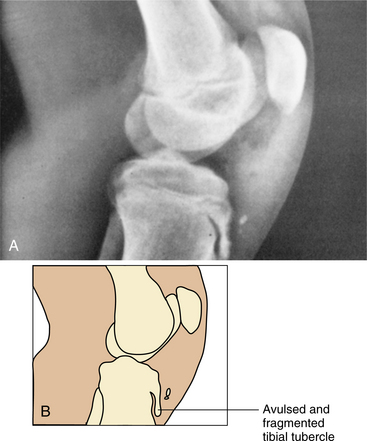
147. Gholve, PA. Osgood Schlatter syndrome. Curr Opin Pediatr. 2007;19(1):44–50.

148. Ghose, RR. Vitamin D deficiency and muscle weakness in the elderly. N Z Med J. 2005;118(1219):U1582.

149. Glueck, CJ, Freiberg, RA, Crawford, A, et al. Secondhand smoke, hypofibrinolysis, and Legg-Perthes disease. Clin Orthop. 1998;352:159–167.

150. Gold, DT. Group treatment improves trunk strength and psychological status in older women with vertebral fractures: results of a randomized, clinical trial. J Am Geriatr Soc. 2004;52(9):1471–1478.

151. Gold, L. Levofloxacin-induced tendon rupture: a case report and review of the literature. J Am Board Fam Pract. 2003;16(5):458–460.

152. Goldberg, SH. Pharmacologic therapy for osteoarthritis. Am J Orthop. 2002;31(12):673–680.

153. Goldman, AB, Freiberger, RH. Localized infectious and neuropathic disease. Semin Roentgenol. 1979;14:19–32.

154. Goldstein, SA. Tissue engineering solutions for traumatic bone loss. J Am Acad Orthop Surg. 2006;14(10):S152–S156.

155. Gonzalez-Gay, MA. Genetic epidemiology: giant cell arteritis and polymyalgia rheumatica. Arthritis Res. 2001;3(3):154–157.

156. Goodman, CC, Snyder, TE. Differential diagnosis for physical therapists: screening for referral, ed 4. Philadelphia: Saunders, 2007.

157. Green, MJ, Deodhar, AA. Bone changes in early rheumatoid arthritis. Baillieres Best Pract Res Clin Rheumatol. 2001;15(1):105–123.

158. Greenspan, A. Evaluating suspected stress fracture. J Musculoskelet Med. 1999;16(3):144.

159. Greenspan, SL, Myers, ER, Maitland, LA, et al. Fall severity and BMD as risk factors for hip fracture in ambulatory elderly. JAMA. 1994;271:128–133.

160. Gryfe, CI, Amies, A, Ashley, MJ. A longitudinal study of falls in an elderly population: I. Incidence and morbidity. Age Ageing. 1977;6:201–210.

161. Grzegorzewski, A. The role of the acetabulum in Perthes disease. J Pediatr Orthop. 2006;26(3):316–321.

162. Guccione, AA. Geriatric physical therapy, ed 2. St Louis: Mosby, 2000.

163. Guccione, AA, Fagerson, TL, Anderson, JJ. Regaining functional independence in the acute care setting following hip fracture. Phys Ther. 1996;76(8):818–826.

164. Günther, KP. Prevalence of generalised osteoarthritis in patients with advanced hip and knee osteoarthritis: the Ulm Osteoarthritis Study. Ann Rheum Dis. 1998;57:717–723.

165. Gür, H. Muscle mass, isokinetic torque, and functional capacity in women with osteoarthritis of the knee. Arch Phys Med Rehabil. 2003;84:1534–1541.

166. Guthrie, B. Review of medications used in juvenile rheumatoid arthritis. Pediatr Emerg Care. 2007;23(1):38–46.

167. Häkkinen, A. Effectiveness and safety of strength training in rheumatoid arthritis. Curr Opin Rheumatol. 2004;16(2):132–137.

168. Häkkinen, A. Effects of concurrent strength and endurance training in women with early or longstanding rheumatoid arthritis: comparison with healthy subjects. Arthritis Rheum. 2003;49(6):789–797.

169. Häkkinen, A, Sokka, T, Kotaniemi, A, et al. A randomized-two year study of the effects of dynamic strength training on muscle strength, disease activity, functional capacity, and bone mineral density in early rheumatic arthritis. Arthritis Rheum. 2001;44(3):515–522.

170. Hall, CM, Brody, LT. Therapeutic exercise: moving towards function, ed 2. Philadelphia: Lippincott Williams & Wilkins, 1998.

171. Hall, T, Zusman, M, Elvey, R. Adverse mechanical tension in the nervous system? Analysis of straight leg raise. Man Ther. 1998;3(3):140–146.

172. Hannallah, D, Kange, J. Use of growth factors for bone repair. J Musculoskelet Med. 2003;20(8):365–372.

173. Hansen, BP. Postarthroscopic glenohumeral chondrolysis. Am J Sports Med. 2007;35(10):1628–1634.

174. Hanten, WP, Olson, SL, Butts, NL, et al. Effectiveness of a home program of ischemic pressure followed by sustained stretch for treatment of myofascial trigger points. Phys Ther. 2000;80(10):997–1003.

175. Harada, ND. Patterns of rehabilitation utilization after hip fracture in acute hospitals and skilled nursing facilities. Med Care. 2000;38(11):1119–1130.

176. Harkcom, TM, Lampman, RM, Banwell, BF, et al. Therapeutic value of graded aerobic exercise training in rheumatoid arthritis. Arthritis Rheum. 1985;28:32–39.

177. Harris, E. Rheumatoid arthritis: pathophysiology and implications for therapy. N Engl J Med. 1990;322:1277–1289.

178. Harris ED, ed. Kelley’s textbook of rheumatology, ed 7, Philadelphia: Saunders, 2005.

179. Harrison, AL. The influence of pathology, pain, balance, and self-efficacy on function in women with osteoarthritis of the knee. Phys Ther. 2004;84(9):822–831.

180. Hartman, CA, Manos, TM, Winter, C, et al. Effects of T’ai Chi training on function and quality of life indicators in older adults with osteoarthritis. J Am Geriatr Soc. 2000;48(12):1553–1559.

181. Hausdorff, JM, et al. Gait variability and fall risk in community-living older adults: a 1-year prospective study. Arch Phys Med Rehabil. 2001;82(8):1050–1056.

182. Hazes, JMW, Dijkmans, BAC, Vandenbroucke, JP, et al. Pregnancy and the risk of developing rheumatoid arthritis. Arthritis Rheum. 1990;33:1770–1775.

183. Headley, B. Myofascial pain syndrome. Fibromyalgia Netw. 2007;76:2–6.

184. Headley, BJ. For stabilization, timing is everything. Adv Phys Ther PT Assist. 2005;16(21):54.

185. Healthy people 2010: arthritis objectives 2001. Available on-line at www.health.gov/healthypeople/ Accessed May 30, 2008

186. Heikkila, S, Viitanen, JV, Kautiainen, H, et al. Sensitivity to change of mobility tests: effect of short term intensive physiotherapy and exercise on spinal, hip, and shoulder measurements in spondyloarthropathy. J Rheumatol. 2000;27(5):1251–1256.

187. Heinonen, M. Post-operative degree of mobilization at two weeks predicts one-year mortality after hip fracture. Aging Clin Exp Res. 2004;16(6):476–480.

188. Henroitin, Y, Labasse, A, Zheng, SX, et al. Strontium ranelate increases cartilage matrix formation. J Bone Miner Res. 2001;16(2):299–308.

189. Herkowitz, HN. Spine update: degenerative lumbar spondylolisthesis. Spine. 1995;20:1084–1090.

190. Herrero-Beaumont, G. Glucosamine sulfate in the treatment of knee osteoarthritis symptoms: a randomized, double-blind, placebo-controlled study using acetaminophen as a side comparator. Arthritis Rheum. 2007;56(2):555–567.

191. Herring, JA. Legg-Calvé-Perthes disease: Part II: Prospective multicenter study of the effect of treatment on outcome. J Bone Joint Surg Am. 2004;86-A(10):2121–2134.

192. Hicks, JE. Rehabilitating patients with idiopathic inflammatory myopathy. J Musculoskelet Med. 1995;12(4):41–54.

193. Hicks, JE. Role of rehabilitation in the management of myopathies. Curr Opin Rehabil. 1998;10(6):548–555.

194. Hicks, JE, Gerber, L. Rehabilitation of the patient with arthritis and connective tissue disease. In: Delisa J, ed. Rehabilitation medicine. ed 2. Philadelphia: Lippincott; 1993:1047–1081.

195. Hicks, JE, Miller, F, Plotz, P, et al. Isometric exercise increases strength and does not produce sustained creatine phosphokinase increases in a patient with polymyositis. J Rheumatol. 1993;20:1399–1402.

196. Hinman, RS. Aquatic physical therapy for hip and knee osteoarthritis: results of a single-blind randomized controlled trial. Phys Ther. 2007;87(1):32–43.

197. Hitchon, PW. Fractures of the thoracolumbar spine complicating ankylosing spondylitis. J Neurosurg. 2002;97:218–222.

198. Hoenig, H, Rubenstein, LV, Sloane, R, et al. What is the role of timing in the surgical and rehabilitative care of community-dwelling older persons with acute hip fracture? Arch Intern Med. 1997;157:513–520.

199. Hoffman, RW, Greidinger, EL. Mixed connective tissue disease. Curr Opin Rheumatol. 2000;12(5):386–390.

200. Hong, CZ, Simons, DG. Pathophysiologic and electrophysiologic mechanisms of myofascial trigger points. Arch Phys Med Rehabil. 1998;79:863–872.

201. Hoover, RT. Avascular necrosis of the femoral head and hemiarthroplasty. Phys Ther Case Rep. 2000;3(1):25–27.

202. Hoppenfeld, S, Murthy, VL. Treatment and rehabilitation of fractures. Philadelphia: Lippincott Williams & Wilkins, 2000.

203. Hornbrook, MC, et al. Preventing falls among community-dwelling older persons: results from a randomized trial. Gerontologist. 1994;34(1):16–23.

204. Horowitz, S, Evinson, B, Borer, A, et al. Mycoplasma fermentans in rheumatoid arthritis and other inflammatory arthritides. J Rheumatol. 2000;27(12):2747–2753.

205. Hotchkiss, A, et al. Convergent and predictive validity of three scales related to falls in the elderly. Am J Occup Ther. 2004;58(1):100–103.

206. Hsu, S. A new approach to managing oral manifestations of Sjögren’s syndrome and skin manifestations of lupus. J Biochem Mol Biol. 2006;39(3):229–239.

207. Hudson, JI, Kenzora, JE, Hebel, JR, et al. Eight-year outcome associated with clinical options in the management of femoral neck fractures. Clin Orthop. 1998;348:59–66.

208. Hughes, MA, Cooperman, J, Peterson, C, et al. Partial weight-bearing in the older person. Top Geriatr Rehabil. 1996;11(3):1–8.

209. Hund, E. Critical illness polyneuropathy. Curr Opin Neurol. 2001;14(5):649–653.

210. Hunder, GG, Kaye, RL, Lane, NE. Osteoporosis, fibromyalgia, Lyme disease, polymyalgia, lupus, and gout. J Musculoskelet Med. 1999;16(1):12–16. [31-32].

211. Hunt, JL. Heterotopic ossification revisited: a 2-year experience. J Burn Care Res. 2006;27(4):535–540.

212. Hunt, SA. Arthroscopic management of osteoarthritis of the knee. J Am Acad Orthop Surg. 2002;10:356–363.

213. Hunter, DJ. Knee alignment does not predict incident osteoarthritis: the Framingham osteoarthritis study. Arthritis Rheum. 2007;56(4):1212–1218.

214. Hunter, DJ. Patella malalignment, pain and patellofemoral progression: the Health ABC Study. Osteoarthritis Cartilage. 2007;15(10):1120–1127.

215. Hunter, DJ, Felson, DT. Osteoarthritis: clinical review. BMJ. 2006;332:638–642.

216. Hutton, JT. Preventing falls: a defensive approach. New York: Prometheus Books, 2000.

217. Hyrich, KL, Inman, RD. Infectious agents in chronic rheumatic diseases. Curr Opin Rheumatol. 2001;13(4):300–304.

218. Iaria, J, Houseknecht, H, Craik, R. Effects of hip fracture on impairment and function in older persons. Phys Ther. 2001;81(5):A17. [(abstract)].

219. Ince, G. Effects of a multimodal exercise program for people with ankylosing spondylitis. Phys Ther. 2006;86(7):924–935.

220. Inman, RD. Mechanisms of disease: infection and spondyloarthritis. Nat Clin Pract Rheumatol. 2006;2(3):163–169.

221. Intrator, O. Benefits of home health care after inpatient rehabilitation for hip fracture. Arch Phys Med Rehabil. 1998;79(10):1195–1199.

222. Ismail, AA. Prevalent vertebral deformity predicts incident hip though not distal forearm fracture: results from the European Prospective Osteoporosis Study. Osteoporosis Int. 2001;12:85–90.

223. Iversen, MD. Physical therapy for management of RA. J Musculoskelet Med. 2002;19(9):352–362.

224. James, J, Steijn-Myagkaya, GL. Death of osteocytes: electron microscopy after in vitro ischemia. J Bone Joint Surg Br. 1986;68:620–624.

225. Janowsky, EC, Kupper, LL, Hulka, BS. Meta-analysis of the relation between silicone breast implants and the risk of connective-tissue diseases. N Engl J Med. 2000;342:781–790.

226. Järvinen, M. The effect of early mobilization and immobilization on the healing process following muscle injuries. Sports Med. 1993;15:78–89.

227. Järvinen, TAH. Muscle injuries: biology and treatment. Am J Sports Med. 2005;33(5):745–764.

228. Jensen, MC. Magnetic resonance imaging of the lumbar spine in people without back pain. N Engl J Med. 1994;331:69–73.

229. Johnson, EO. Hypothalamic-pituitary-adrenal axis function in Sjögren’s syndrome: mechanisms of neuroendocrine and immune system homeostasis. Ann N Y Acad Sci. 2006;1088:41–51.

230. Johnson, EO, Skopouli, FN, Moutsopoulos, HM. Neuroendocrine manifestations in Sjögren’s syndrome. Rheum Dis Clin North Am. 2000;26(4):927–949.

231. Jonssou, B, Stromquist, B. Symptoms and signs in degeneration of the lumbar spine. J Bone Joint Surg Br. 1993;75:381–385.

232. Joseph, B. Natural evolution of Perthes disease: a study of 610 children under 12 years of age at disease onset. J Pediatr Orthop. 2003;23:590–600.

233. Min, JH, Jang, JS, Lee, SH. Comparison of anterior-and posterior-approach instrumented lumbar interbody fusion for spondylolisthesis. J Neurosurg Spine. 2007;7(1):21–26.

234. Kalichman, L, et al. The association between patellar alignment and patellofemoral joint osteoarthritis features-an MRI study. Rheumatology (Oxford). 2007;9(2):R26.

235. Kalichman, L. The association between patellar alignment on magnetic resonance imaging and radiographic manifestations of knee osteoarthritis. Arthritis Res Ther. 2007;9(2):R26.

236. Kannus, P. Prevention of hip fracture with hip protectors. Age Ageing. 2006;35(suppl 2):ii51–ii54.

237. Kapural, L. Novel intradiscal biacuplasty (IDB) for the treatment of lumbar discogenic pain. Pain Pract. 2007;7(2):130–134.

238. Kauppi, M, Anttila, P. A stiff collar can restrict atlantoaxial instability in rheumatoid cervical spine in selected cases. Ann Rheum Dis. 1995;54:305–307.

239. Kauppi, M, Anttila, P. A stiff collar for the treatment of rheumatoid atlantoaxial subluxation. Br J Rheumatol. 1996;35:771–774.

240. Kauppila, LI. MR aortography and serum cholesterol levels in patients with long-term nonspecific lower back pain. Spine. 2004;29(19):2147–2152.

241. Keating, RM, Vyas, A. Reactive arthritis following Clostridium difficile colitis. West J Med. 1995;162(1):61–63.

242. Kellgren, J, Lawrence, J. Radiologic assessment of osteoarthritis. Ann Rheum Dis. 1957;16:494–501.

243. Kellgren, JH. Observations of referred pain arising from muscle. Clin Sci. 1938;3:175.

244. Kelly, AKW. Managing stress fractures in athletes. J Musculoskelet Med. 2005;22(9):463–472.

245. Kerrigan, DC. Effectiveness of a lateral-wedge insole on knee varus torque in patients with knee osteoarthritis. Arch Phys Med Rehabil. 2002;83(7):889–893.

246. Kiebzak, GM. Undertreatment of osteoporosis in men with hip fracture. Arch Intern Med. 2002;162(19):2217–2222.

247. Kiebzak, GM. Vitamin D status of patients admitted to a hospital rehabilitation unit: relationship to function and progress. Am J Phys Med Rehabil. 2007;86(6):435–445.

247a. Kim, DH. Factors influencing segmental range of motion after lumbar total disc replacement using the ProDisc II prosthesis. J Neurosurg Spine. 2007;7(2):131–138.

248. Kim, DH. Rheumatoid arthritis in the cervical spine. J Am Acad Orthop Surg. 2005;13(7):463–474.

249. Kim, M. Transverse sacral fractures: case series and literature review. Can J Orthop Surg. 2001;44:359–363.

250. Kirk-Sanchez, NJ, Roach, KE. Factors predicting mobility at discharge following rehabilitation for femur fracture. Phys Ther. 2001;81(5):A16. [(abstract)].

251. Kirkaldy-Willis, WH, Hill, RJ. A more precise diagnosis for low back pain. Spine. 1979;4:102–109.

252. Kirschner, RE, Karmacharya, J, Ong, G, et al. Synthetic hybrid grafts for craniofacial reconstruction: sustained gene delivery using a calcium phosphate bone mineral substitute. Ann Plast Surg. 2001;46(5):538–545.

253. Knop, C, Lange, U, Bastian, L, et al. Biomechanical compression tests with a new implant for thoracolumbar vertebral body replacement. Eur Spine J. 2001;10(1):30–37.

254. Koch, AE. Targeting cytokines and growth factors in RA. J Musculoskelet Med. 2005;22(3):130–136.

255. Komatireddy, GR, Leitch, RW, Cella, K, et al. Efficacy of low load resistive training in patients with rheumatoid arthritis functional class II and III. J Rheumatol. 1997;24(8):1531–1539.

256. Konig, A. Insufficiency fractures of the ileum and sacrum. Z Rheumatol. 2000;59(5):343–347.

257. Konttinen, YT. Immunohistology of Sjögren’s syndrome. Autoimmun Rev. 2006;6(1):16–20.

258. Kostopoulos, D, Rizopoulos, K, Kurman, RJ, et al. The manual of trigger point and myofascial therapy. Thorofare, NJ: Slack, 2001.

259. Koval, KJ, Zuckerman, JD. Hip fractures: a practical guide to management. New York: Springer-Verlag, 2000.

260. Kraemer, J. Natural course and prognosis of intervertebral disc diseases. Spine. 1995;20:635–639.

261. Krebs, DE, et al. Is base of support greater in unsteady gait? Phys Ther. 2002;82(2):138–147.

262. Kristmundsdottir, F. Delayed skeletal maturation in Perthes’ disease. Acta Orthop Scand. 1987;58(3):277–279.

263. Kruithof, E. Identification of synovial biomarkers of response to experimental treatment in early-phase clinical trials in spondyloarthritis. Arthritis Rheum. 2006;54(6):1733–1735.

264. Kujala, UM. Hamstring injuries: current trends in treatment and prevention. Sports Med. 1997;23:397–404.

265. Kurowski, P. The relationship of degeneration of the intervertebral disc to mechanical loading on lumbar vertebrae. Spine. 1986;11:726–731.

266. Lacomis, D. Causes of neuromuscular weakness in the intensive care unit: a study of 92 patients. Muscle Nerve. 1998;21(5):610–617.

267. Lacomis, D. Critical illness myopathy. Curr Rheumatol Rep. 2002;4(5):403–408.

268. Laing, TJ, Schottenfeld, D, Lacey, JV, et al. Potential risk factors for undifferentiated connective tissue disease among women: implanted medical devices. Am J Epidemiol. 2001;154(7):610–617.

269. Laurencin, CT, Attawia, MA, Lu, LQ, et al. Poly/hydroxyapatite delivery of BMP-2-producing cells: a regional gene therapy approach to bone regeneration. Biomaterials. 2001;22(11):1271–1277.

270. Lavernia, CJ, Sierra, RJ, Grieco, FR. Osteonecrosis of the femoral head. J Am Acad Orthop Surg. 1999;7(4):250–261.

271. Lawley, J, Thompkins, P. Metaphors in mind. Highgate, London: Developing Company Press, 2000.

272. Lawlor, DA, et al. Association between falls in elderly women and chronic disease and drug use: cross sectional study. BMJ. 2003;327(7417):712–717.

273. LeBoff, MS, Kohlmeier, L, Hurwitz, S, et al. Occult vitamin D deficiency in postmenopausal US women with acute hip fracture. JAMA. 1999;281(16):1505–1511.

274. Lee, S. Bone age delay patterns in Legg-Calvé-Perthes disease. J Pediatr Orthop. 2007;27(2):198–203.

275. Leech, JA. Relationship of lung function to severity of osteoporosis in women. Am Rev Respir Dis. 1990;141:951–956.

276. Leoux, JL. Sacral insufficiency fractures presenting as acute low-back pain-biomechanical aspects. Spine. 1993;18(12):2502–2506.

277. Lephart, SM. Proprioception and neuromuscular control in joint stability. Champaign, IL: Human Kinetics, 2000.

278. Levine, WN. Chondrolysis following arthroscopic thermal capsulorrhaphy to treat shoulder instability. J Bone Joint Surg Am. 2005;87:616–621.

279. Lewis, C. Discovering best treatments for hip fractures. Adv Phys Ther PT Assist. 2002;13(11):6–7.

280. Lewis, C. PTs should be hip to orthopedic rehab. Adv Phys Ther PT Assist. 2003;14:6, 49.

281. Lewis, C. Shoulder fracture. Adv Phys Ther PT Assist. 2003;14(18):6–7.

282. Lewis, R. Ankylosing spondylitis: early diagnosis and management. J Musculoskelet Med. 2003;20(4):184–198.

283. Lewit, D, Simons, DG. Myofascial pain: relief by post-isometric relaxation. Arch Phys Med Rehabil. 1984;65:452–456.

284. Lie, H. Mobilizing or stabilizing exercise in degenerative disk disease in the lumbar region? Tidsskr Nor Laegeforen. 1999;119(14):2052–2053.

285. Lin, DH. Comparison of proprioceptive functions between computerized proprioception facilitation exercise and closed kinetic chain exercise in patients with knee osteoarthritis. Clin Rheumatol. 2007;26(4):520–528.

286. Lindsay, R. Risk of new vertebral fracture in the year following a fracture. JAMA. 2001;285:320–323.

287. Lineker, SC, Bell, MJ, Wilkins, AL, et al. Improvements following short term home based physical therapy are maintained at one year in people with moderate to severe rheumatoid arthritis. J Rheumatol. 2001;28(1):165–168.

288. Lo, TC. Heterotopic bone formation after hip surgery: prevention with single-dose postoperative hip irradiation. Radiology. 1988;168:851–854.

289. Long, T. Handbook of pediatric physical therapy. Philadelphia: Lippincott Williams & Wilkins, 2001.

290. Lord, SR. A physiological profile approach to falls risk assessment and prevention. Phys Ther. 2003;83(3):237–252.

291. Lorig, KR, Mazonson, PD, Holman, HR. Evidence suggesting that health education for self-management in patients with chronic arthritis has sustained health benefits while reducing health care costs. Arthritis Rheum. 1993;36:439–446.

292. Lotke, PA, Battish, R, Nelson, CL. Treatment of osteonecrosis of the knee. Instr Course Lect. 2001;50:483–488.

293. Lovell, DJ. Update on treatment of arthritis in children: new treatments, new goals. Bull N Y U Hosp Jt Dis. 2006;64(1-2):72–76.

294. Lowe, JC, Honeyman-Lowe, G. Ultrasound treatment of trigger points: differences in technique for myofascial pain syndrome and fibromyalgia. Myalgia ’99. 1999;2:12–15. [Available from the Fibromyalgia Research Foundation, PO Box 396, Tulsa, OK 74101-0396].

295. Lozada, CJ. Glucosamine in osteoarthritis: questions remain. Cleve Clin J Med. 2007;74(1):65–71.

296. Lundberg, IE. Technology insight: tools for research, diagnosis and clinical assessment of treatment in idiopathic inflammatory myopathies. Nat Clin Pract Rheumatol. 2007;3(5):282–290.

297. Luu, HH. Distinct roles of bone morphogenetic proteins in osteogenic differentiation of mesenchymal stem cells. J Orthop Res. 2007;25(5):665–677.

298. MacDonald, CW, Whitman, JM, Cleland, JA, et al. Clinical outcomes following manual physical therapy and exercise for hip osteoarthritis: a case series. J Orthop Sports Phys Ther. 2006;36(8):588–599.

299. Maddison, PJ. Mixed connective tissue disease: overlap syndromes. Baillieres Best Pract Res Clin Rheumatol. 2000;14(1):111–124.

300. Magaziner, J, Lydick, E, Hawkes, W, et al. Excess mortality attributable to hip fracture in white women aged 70 years and older. Am J Public Health. 1997;87(10):1630–1636.

301. Majlesi, J. High-power pain threshold ultrasound technique in the treatment of active myofascial trigger points: a randomized, double-blind, case-control study. Arch Phys Med Rehabil. 2004;85(5):833–836.

302. Maki, BE, et al. Fear of falling and postural performance in the elderly. J Gerontol. 1991;46:M123–M131.

303. Malm, C. Immunological changes in human skeletal muscle and blood after eccentric exercise and multiple biopsies. J Physiol. 2000;529:243–262.

304. Mandelbaum, B. Etiology and pathophysiology of osteoarthritis. Orthopedics. 2005;28(suppl 2):S207–S214.

305. Mangione, KK. The effects of high-intensity and low-intensity cycle ergometry in older adults with knee osteoarthritis. J Gerontol A Biol Sci Med Sci. 1999;54(4):M184–M190.

306. Mannion, AF. A randomized clinical trial of three active therapies for chronic low back pain. Spine. 1999;24:2435–2448.

307. Mansi, IA. Rhabdomyolysis in response to weight-loss herbal medicine. Am J Med Sci. 2004;327(6):356–357.

308. March, LM. Economics of osteoarthritis: a global perspective. Baillieres Clin Rheumatol. 1997;11:817–834.

309. Marinelli, WA. Neuromuscular disorders in the intensive care unit. Crit Care Clin. 2002;18(4):915–929.

310. Martin, TM, Smith, JR, Rosenbaum, JT. Anterior uveitis: current concepts of pathogenesis and interactions with the spondyloarthropathies. Curr Opin Rheumatol. 2002;14:337–341.

311. Martinez-Taboada, VM, Blanco, R, Rodriquez-Valverde, V. Polymyalgia rheumatica with normal erythrocyte sedimentation rate: clinical aspects. Clin Exp Rheumatol. 2000;18(4 suppl 20):S34–S37.

312. Masuko Hongo, K, Kato, T. The association between autoimmune thyroid diseases and rheumatic diseases: a review. Nippon Rinsho. 1999;57(8):1873–1877.

313. Matsumoto, T. Cyclic mechanical stretch stress increases the growth rate and collagen synthesis of nucleus pulposus in vitro. Spine. 1999;24:315–319.

314. Mattila, VM, Niva, M, Kiuru, M, et al. Risk factors for bone stress injuries: a follow-up study of 102,515 person-years. Med Sci Sports Exerc. 2007;39(7):1061–1066.

315. Mauer, AC, et al. Bilateral total knee arthroplasty increases the propensity to trip on an obstacle. Clin Orthop Relat Res. 2005;433:160–165.

316. Mavragani, CP. The management of Sjögren’s syndrome. Nat Clin Pract Rheumatol. 2006;2(5):252–261.

317. Maxey, L, Magnusson, J. Rehabilitation for the postsurgical orthopedic patient, ed 2. St Louis: Mosby, 2006.

318. McDonough, A, Saidoff, D. Clinical pathways in therapeutic intervention: upper extremity. St Louis: Mosby, 1997.

319. McGuire, KJ. The 2004 Marshall Urist Award: delays until surgery after hip fracture increases mortality. Clin Orthop Relat Res. 2004;428:294–301.

320. McKenzie, R. The lumbar spine: mechanical diagnosis and therapy. Waikanae, New Zealand: Spinal Publications, New Zealand, 1981.

321. McLain, RF. Determining treatment of lumbar disk protrusion and disk extrusion. J Musculoskelet Med. 2005;22(1):21–28.

322. McNair, PJ, Marshall, RN, Maguire, K. Swelling of the knee joint: effects of exercise on quadriceps muscle strength. Arch Phys Med Rehabil. 1996;77(9):896–899.

323. Mecagni, C, Smith, JP, Roberts, KE, et al. Balance and ankle range of motion in community-dwelling women aged 64 to 87 years: a correlational study. Phys Ther. 2000;80(10):1004–1011.

324. Medical Multimedia Group (MMG) A patient’s guide to lumbar artificial disc replacement, 2007. Available on-line at http://www.eorthopod.com. Accessed July 11, 2007

325. Medical Multimedia Group (MMG) A patient’s guide to lumbar degenerative disc disease, 2002. Available on-line at http://www.eorthopod.com. Accessed July 11, 2007

326. Medical Multimedia Group (MMG) A patient’s guide to Osgood-Schlatter lesion of the knee. Available on-line at http://www.eorthopod.com. Accessed July 10, 2007

327. Medical Multimedia Group (MMG). A patient’s guide to Perthes disease of the hip. Missoula, MT: MMG, 2005.

328. Meisel, HJ. Clinical experience in cell-based therapeutics: disc chondrocyte transplantation: a treatment for degenerated or damaged intervertebral disc. Biomol Eng. 2007;24(1):5–21.

329. Melaragno, PG, Cheney, RA, Bennett, GL. Lower leg compartment syndromes: when to suspect and how to diagnose. J Musculoskelet Med. 1996;13(11):14–27.

330. Melton-Rogers, S, Hunter, G, Walter, J, et al. Cardiorespiratory responses of patients with rheumatoid arthritis during bicycle riding and running in water. Phys Ther. 1996;76(10):1058–1065.

331. Mense, S, Simons, DG, Russell, IJ. Muscle pain: understanding its nature, diagnosis, and treatment. Philadelphia: Lippincott Williams & Wilkins, 2000.

332. Messier, SP. Exercise and dietary weight loss in overweight and obese older adults with knee osteoarthritis: the Arthritis, Diet, and Activity Promotion Trial. Arthritis Rheum. 2004;50(5):1501–1510.

333. Messier, SP, Royer, TD, Craven, TE, et al. Long-term exercise and its effect on balance in older, osteoarthritic adults: results from the Fitness, Arthritis, and Seniors Trial (FAST). J Am Geriatr Soc. 2000;48(2):131–138.

334. Messinger-Rapport, BJ. Prevention for the older woman: mobility: a practical guide to managing osteoarthritis and falls. Geriatrics. 2003;58(7):22–29.

335. Michlovitz, SL. Thermal agents in rehabilitation, ed 3. Philadelphia: FA Davis, 1996.

336. Mikuls, TR. Coffee, tea, and caffeine consumption and risk of rheumatoid arthritis: results from the Iowa Women’s Health Study. Arthritis Rheum. 2002;46(1):83–91.

337. Miller, D. Endothelial dysfunction and decreased vascular responsiveness in the anterior cruciate ligament-deficient model of osteoarthritis. J Appl Physiol. 2007;102(3):1161–1169.

338. Miller, GD. Intensive weight loss program improves physical function in older obese adults with knee osteoarthritis. Obesity. 2006;14(7):1219–1230.

339. Miller, JA. Lumbar disc degeneration: correlation with age, sex, and spine level in 600 autopsy specimens. Spine. 1988;13:173–178.

340. Min, BW, Koo, KH, Song, HR, et al. Subcapital fractures associated with extensive osteonecrosis of the femoral head. Clin Orthop. 2001;390:227–231.

341. Minden, K. What are the costs of childhood-onset rheumatic disease? Best Pract Res Clin Rheumatol. 2006;20(2):223–240.

342. Minor, MA. Exercise in the treatment of osteoarthritis. Rheum Dis Clin North Am. 1999;25(2):397–415.

343. Minor, MA. Exercise proves a vital tool against rheumatoid arthritis. Arthritis Today. 1997:36–37.

344. Minor, MA, Johnson, JC. Reliability and validity of a submaximal treadmill test to estimate aerobic capacity in women with rheumatic disease. J Rheumatol. 1996;23(9):1517–1523.

345. Minor, MA, Lane, NE. Recreational exercise in arthritis. Rheum Dis Clin North Am. 1996;22(3):563–577.

346. Minor, MA, Sanford, MK. The role of physical therapy and physical modalities in pain management. Rheum Dis Clin North Am. 1999;25(1):233–248.

347. Moder, KG. A working guide to joint examination in rheumatoid arthritis. J Musculoskelet Med. 1995;12(11):17–34.

348. Mohler, R. Painless hand mass in an elderly man. J Musculoskelet Med. 2001;18(3):153–156.

349. Monauni, T, Zenti, MG, Cretti, A, et al. Effects of glucosamine infusion on insulin secretion and insulin action in humans. Diabetes. 2000;49(6):926–935.

350. Monu, JU. Gout: a clinical and radiologic review. Radiol Clin North Am. 2004;42(1):169–184.

351. Moorthy, LN. Juvenile idiopathic arthritis: making the diagnosis. J Musculoskelet Med. 2004;21(11):581–588.

352. Morbidity Mortality Weekly Report (MMWR). Fatalities and injuries from falls among older adults, 2005. MMWR Morbid Mortal Wkly Rep. 2006;55(45):1221–1224.

353. Morbidity Mortality Weekly Report (MMWR). Visual impairment and eye care among older adults, 2005. MMWR Morbid Mortal Wkly Rep. 2006;55(49):1321–1325.

354. Morehead, K. Early diagnosis of RA: the therapeutic implications. J Musculoskelet Med. 2005;22(11):599–606.

355. Moreland, LW. Etanercept therapy in rheumatoid arthritis: a randomized, controlled trial. Ann Intern Med. 1999;130(6):478–486.

356. Morse, CG. The incidence and natural history of osteonecrosis in HIV-infected adults. Clin Infect Dis. 2007;44(5):739–748.

357. Munin, MC, Hockenberry, PS, Flynn, PG, et al, Rehabilitation. Callaghan, JJ, Rosenberg, AG, Rubash, HE, eds. The adult hip, II. Philadelphia: Lippincott-Raven, 1998.

358. Mustafa, K, Khan, MA. Recognizing and managing reactive arthritis. J Musculoskelet Med. 1996;13(6):28–41.

359. Myer, GD. Specialized neuromuscular training to improve neuromuscular function and biomechanics in a patient with quiescent juvenile rheumatoid arthritis. Phys Ther. 2005;85(8):791–802.

360. Nannapaneni, R. Surgical outcome in rheumatoid Ranawat class IIIb myelopathy. Neurosurgery. 2005;56(4):706–715.

361. National Ankylosing Spondylitis Society (NASS) The Bath Indices, 2004. Available on-line at http://www.mc.uky.edu/rheumatology/education/nass-bath-indices.pdf Accessed July 10, 2007

362. National Center for Health Statistics. 1990 and 1991 National hospital discharge survey. Rosemont, IL: American Academy of Orthopedic Surgeons Department of Research and Scientific Affairs, 1994.

363. National Center for Injury Prevention and Control (NCIPC) Preventing falls among older adults. Available on-line at http://www.cdc.gov/ncipc/duip/preventadultfalls.htm Accessed December 26, 2006. Lots of great patient education materials, checklists, posters, prevention strategies

364. National Institutes of Health (NIH). Consensus statement: osteoporosis prevention, diagnosis, and therapy. Bethesda, MD: The Institutes, 2000.

365. National Osteoporosis Foundation Important disease facts, 2001. Available on-line at www.nof.org/other/statistics.html Accessed May 30, 2008

366. Nativ, A. Stress fractures and bone health in track and field athletes. J Sci Med Sport. 2000;3(3):268–279.

367. NeuroCom Assessment Protocols. NeuroCom International, Clackamas, O.R., (800) 767-6744. Available on-line at www.onbalance.com Accessed May 30, 2008. (Reference to this product does not represent endorsement.)

368. Neva, MH. Mortality associated with cervical spine disorders: a population-based study of 1,666 patients with rheumatoid arthritis who died in Finland in 1989. Rheumatology (Oxford). 2001;40:123–127.

369. Nevitt, MC. Risk factors for a first-incident radiographic vertebral fracture in women greater than or equal to 65 years of age: the study of osteoporotic fractures. J Bone Miner Res. 2005;20(1):131–140.

370. Newcomer, K, Jurrison, ML. Rheumatoid arthritis: the role of physical therapy. J Musculoskelet Med. 1994;11:14–26.

371. Newton RA, Fall Prevention Project, Temple University Falls and Fall Prevention, 1999. Available on-line at www.temple.edu/older_adult Accessed July 17, 2007

372. Nicola, PJ. The risk of congestive heart failure in rheumatoid arthritis: a population based study over 46 years. Arthritis Rheum. 2005;52:412–420.

373. Niva, MH. Bone stress injuries causing exercise-induced knee pain. Am J Sports Med. 2006;34(1):78–84.

374. Noonan, TJ. Thermal effects on skeletal muscle tensile behavior. Am J Sports Med. 1993;21:517–522.

375. Norwalk, MP, Prendergast, JM, Bayles, CM, et al. A randomized trial of exercise programs among older individuals living in two long-term care facilities: the FallsFREE program. J Am Geriatr Soc. 2001;49(7):859–865.

376. Nyberg, L, Gustafson, Y, Berggren, D, et al. Falls leading to femoral neck fractures in lucid older people. J Am Geriatr Soc. 1996;44(2):156–160.

377. Oda, T, Fujiwara, K, Yonenobu, K, et al. Natural course of cervical spine lesions in rheumatoid arthritis. Spine. 1995;20:1128–1135.

378. O’Dell, JR, Blakely, KW, Mallek, JA, et al. Treatment of early seropositive rheumatoid arthritis: a two-year, double-blind comparison of minocycline and hydroxychloroquine. Arthritis Rheum. 2001;44(10):2235–2241.

379. O’Halloran, PD. Factors affecting adherence to use of hip protectors amongst residents of nursing homes-a correlation study. Int J Nurs Stud. 2007;44(5):672–686.

380. Olsen, NJ. Initial diagnosis and early management of RA. J Musculoskelet Med. 2005;22(10 suppl):S6–S11.

381. Ostensen, M. Sex, reproduction, and gynecological disease in young adults with a history of juvenile chronic arthritis. J Rheumatol. 2000;27:1783–1787.

382. O’Sullivan, SB. Physical rehabilitation, ed 5. Philadelphia: FA Davis, 2006.

383. Ottawa Panel evidence-based clinical practice guidelines for electrotherapy and thermotherapy interventions in the management of rheumatoid arthritis in adults. Phys Ther. 2004;84(11):1016–1043.

384. Ottawa Panel evidence-based clinical practice guidelines for therapeutic exercises in the management of rheumatoid arthritis in adults. Phys Ther. 2004;84(10):934–972.

385. Paap, E. Physiologic response of the six-minute walk test in children with juvenile idiopathic arthritis. Arthritis Rheum. 2005;53(3):351–356.

386. Paget, SA. Modern-day RA management: resetting the inflammatory thermostat. J Musculoskelet Med. 2006:S3. [Suppl].

387. Paimela, L. Progression of cervical spine changes in patients with early rheumatoid arthritis. J Rheumatol. 1997;24(7):1280–1284.

388. Parker, MJ, Gillespie, LD, Gillespie, WJ. Hip protectors for preventing hip fractures in the elderly. Cochrane Database Syst Rev. 2000;4:CD001255.

389. Parkkari, J, Kannus, P, Palvanen, M, et al. Majority of hip fractures occur as a result of a fall and impact on the greater trochanter of the femur: a prospective controlled hip fracture study with 206 consecutive patients. Calcif Tissue Int. 1999;65(3):183–187.

390. Pate, RR, Pratt, M, Blair, SN, et al. Physical activity and public health: a recommendation from the Centers for Disease Control and Prevention and the American College of Sports Medicine. JAMA. 1995;273(5):402–407.

391. Paterno, MV. Early rehabilitation following surgical fixation of a femoral shaft fracture. Phys Ther. 2006;86(4):558–572.

392. Paulozzi, LJ, et al. Recent trends in mortality from unintentional injury in the U.S. J Safety Res. 2006;37(3):277–283.

393. Pavlica, P, Barozzi, L, Salarani, C, et al. Magnetic resonance imaging in the diagnosis of PMR. Clin Exp Rheumatol. 2000;18(4 suppl 20):S38–S39.

394. Pawl, RP. Chronic neck syndromes: an update. Compr Ther. 1999;25(5):278–282.

395. Penninx, BW, Messier, SP, Rejeski, J, et al. Physical exercise and the prevention of disability in activities of daily living in older persons with osteoarthritis. Arch Intern Med. 2001;161(19):2309–2316.

396. Pépin, JL. Sleep apnoea syndrome secondary to rheumatoid arthritis. Thorax. 1995;50:692–694.

397. Peri, P. Stress fractures. Best Pract Res Clin Rheumatol. 2003;17(6):1043–1061.

398. Petrofsky, J, Lee, S, Bweir, S. Gait characteristics in people with type 2 diabetes mellitus. Eur J Appl Physiol. 2005;93(5-6):640–647.

399. Petrofsky, J, Lee, S, Macnider, M, et al. Autonomic, endothelial function and the analysis of gait in patients with type 1 and type 2 diabetes. Acta Diabetol. 2005;42(1):7–15.

400. Petty, DH. Glenohumeral chondrolysis after shoulder arthroscopy. Am J Sports Med. 2004;32:509–515.

401. Phelan, JD. Genomic progress in pediatric arthritis: recent work and future goals. Curr Opin Rheumatol. 2006;18(5):482–489.

402. Pill, SG. Managing and preventing overuse injuries in young athletes. J Musculoskelet Med. 2003;20(9):434–442.

403. Pinals, RS. Preliminary criteria for clinical remission in rheumatoid arthritis. Arthritis Rheum. 1981;24:1308–1315.

404. Podichetty, VK. Managing vertebral compression fractures in older patients. J Musculoskelet Med. 2004;21(7):372–378.

405. Pollintine, P. Neural arch load-bearing in old and degenerated spines. J Biomech. 2004;37:197–204.

406. Pongratz, DF. Soft tissue pain syndromes: clinical diagnosis and pathogenesis. Binghamton, NY: Haworth Press, 2006.

407. Pope, JE, Stevens, A, Howson, W, et al. The development of rheumatoid arthritis after recombinant hepatitis B vaccination. J Rheumatol. 1998;25:1687–1693.

408. Pope, RM. Rheumatoid arthritis: pathogenesis and early recognition. Am J Med. 1996;100(suppl 2A):3–9.

409. Praemer, A. Musculoskeletal conditions in the United States, ed 2. Rosemont, IL: American Academy of Orthopaedic Surgeons, 1999.

410. Prescott, K. Ankylosing spondylitis: the difficulty and importance of early diagnosis. J Musculoskelet Med. 2007;24(4):163–173.

410a. Price, CT. The lateral pillar classification for Legg-Calvé-Perthes disease. J Pediatr Orthop. 2007;27(5):592–593.

411. Puett, DW, Griffen, MR. Published trials of non-medicinal and non-invasive therapies for hip and knee osteoarthritis. Ann Intern Med. 1994;121:133–140.

412. Racette, SB. Creatine supplementation and athletic performance. J Orthop Sports Phys Ther. 2003;33(10):615–621.

413. Rang, M. Management of Legg-Calvé-Perthes disease varies with severity. J Musculoskelet Med. 1996;13(4):10–11.

414. Rathjen, KE. Stainless steel flexible intramedullary fixation of unstable shaft fractures. J Pediatr Orthop. 2007;27(4):432–441.

415. Ravelli, A. Juvenile idiopathic arthritis. Lancet. 2007;369(9563):767–778.

416. Reddy, R. Avascular necrosis and protease inhibitors. J Natl Med Assoc. 2005;97(11):1543–1546.

416a. Reichenbach, S. Meta-analysis: chondroitin for osteoarthritis of the knee or hip. Ann Intern Med. 2007;146(8):580–590.

417. Reginster, JY. Current role of glucosamine in the treatment of osteoarthritis. Rheumatology (Oxford). 2007;46(5):731–735.

418. Reginster, JY, Deroisy, R, Rovati, LC, et al. Long-term effects of glucosamine sulfate on osteoarthritis progression: a randomized, placebo-controlled clinical trial. Lancet. 2001;357(9252):247–248.

419. Reginster, JY, Gillot, VV, Bruyere, O, et al. Evidence of nutraceutical effectiveness in the treatment of osteoarthritis. Curr Rheumatol Rep. 2000;2(6):472–477.

420. Reiter, MF, Boden, SD. Inflammatory disorders of the cervical spine. Spine. 1998;23(24):2755–2766.

421. Robbins, L. Clinical care in the rheumatic diseases. Atlanta: American College of Rheumatology, 2001.

422. Robin, DW, Hasan, SS, Edeki, T, et al. Increased baseline sway contributes to increased losses of balance in older people following triazolam. J Am Geriatr Soc. 1996;44(3):300–304.

423. Robinson, CP, Yamachika, S, Bounous, DI, et al. A novel NOD-derived murine model of primary Sjögren’s syndrome. Arthritis Rheum. 1998;41:150–156.

424. Robinson, E. Weakness after critical illness-just deconditioning? Or something more? Acute Care Perspect. 2006;15(3):7–9.

425. Roos, EM. Joint injury causes knee osteoarthritis in young adults. Curr Opin Rheumatol. 2005;17(2):195–200.

426. Roos, EM. Positive effects of moderate exercise on glycosaminoglycan content in knee cartilage: a four-month, randomized, controlled trial in patients at risk of osteoarthritis. Arthritis Rheum. 2005;52(11):3507–3514.

427. Rosenthal, MD. Diagnosis of medial knee pain: atypical stress fracture about the knee joint. J Orthop Sports Phys Ther. 2006;36(7):526–534.

428. Russek, LN. Examination and treatment of a patient with hypermobility syndrome. Phys Ther. 2000;80(4):386–398.

429. Safran, MR. Warm-up and muscular injury prevention. Am J Sports Med. 1989;16:123–129.

430. Salter, RB. Textbook of disorders and injuries of the musculoskeletal system, ed 3. Baltimore: Williams & Wilkins, 1999.

431. Saltzman, CL, Vogelgesang, S. Clinical evaluation of the rheumatoid foot. J Musculoskelet Med. 1997;14(8):27–42.

432. Salvarani, C, Cantini, F, Olivieri, I. Distal musculoskeletal manifestations in polymyalgia rheumatica. Clin Exp Rheumatol. 2000;18(4 suppl 20):S51–S52.

433. Salvarani, C, Gabriel, SE, O’Fallon, WM, et al. Epidemiology of polymyalgia rheumatic in Olmsted County, Minnesota, 1970-1991. Arthritis Rheum. 1995;38:369–373.

434. Sambrook, PN. Genetic influences on cervical and lumbar disc degeneration: a magnetic resonance imaging study in twins. Arthritis Rheum. 1999;42:366–372.

435. Sanchez-Sotelo, J, Munuera, L, Madero, R. Treatment of fractures of the distal radius with a remodelable bone cement: a prospective, randomized study using Norian SRS. J Bone Joint Surg Br. 2000;82(6):856–863.

436. Santos, H, Brophy, S, Calin, A. Exercise in ankylosing spondylitis: how much is optimum? J Rheumatol. 1998;25:2156–2160.

437. Santos, H, Brophy, S, Calin, A. Exercise in ankylosing spondylitis: how much is optimum? J Rheumatol. 1998;25(11):2156–2160.

438. Sashika, H, Matsuba, Y, Watanabe, Y. Home program of physical therapy: effect on disabilities of patients with total hip arthroplasty. Arch Phys Med Rehabil. 1996;77(3):273–277.

439. Sattin, RW. Falls among older persons: a public health perspective. Annu Rev Public Health. 1992;13:489–508.

440. Saunders, SR. Physical therapy management of hand fractures. Phys Ther. 1989;69(12):1065–1076.

441. Sawka, AM. Do hip protectors decrease the risk of hip fracture in institutional and community-dwelling elderly? A systematic review and meta-analysis of randomized controlled trials. Osteoporos Int. 2005;16(12):1461–1474.

442. Sawka, AM. Hip protectors decrease hip fracture risk in elderly nursing home residents: a Bayesian meta-analysis. J Clin Epidemiol. 2007;60(4):336–344.

443. Scher, JU. Managing osteoarthritis: NSAIDs past, present, and future. J Musculoskelet Med. 2006;23(11):779–787.

444. Schnitzer T Advances in osteoarthritis research: investigating subchondral bone as etiologic agent and therapeutic target. Medscape, 2004. Available on-line at www.medscape.com/viewprogram/2892_pnt Accessed July 17, 2007

445. Schreiber, DH. Statin-induced rhabdomyolysis. J Emerg Med. 2006;31(2):177–180.

446. Scribner, AN, Troia-Cancio, PV, Cox, BA, et al. Osteonecrosis in HIV: a case-control study. J Acquir Immune Defic Syndr Hum Retrovirol. 2000;25(1):19–25.

447. Seeger, JD. Achilles tendon rupture and its association with fluoroquinolone antibiotics and other potential risk factors in a managed care population. Pharmacoepidemiol Drug Saf. 2006;15(11):784–792.

448. Serhal, D, Calabrese, LH. Diagnosing and managing rheumatic disorders in HIV-infected persons. J Musculoskelet Med. 2000;17(10):606–620.

449. Shakoor, N. Osteoarthritis: 20 clinical pearls. J Musculoskelet Med. 2003;20(4):173–182.

450. Shankar, K. Exercise prescription. Philadelphia: Hanley & Belfus, 1999.

451. Shankman, G. Fundamental orthopedic management for the physical therapist assistant. St Louis: Mosby, 1997.

452. Sharma, L. Quadriceps strength and osteoarthritis progression in malaligned and lax knees. Ann Intern Med. 2003;138(8):613–619.

453. Shen, M, Kim, Y. Osteoporotic vertebral compression fractures: a review of current surgical management techniques. Am J Orthop. 2007;36(5):241–248.

454. Sherrington, C, Lord, S. Home exercises to improve strength and walking velocity after hip fracture. Arch Phys Med Rehabil. 1997;78(2):208–212.

455. Sherrington, C, Lord, S. Increased prevalence of fall risk factors in older people following hip fracture. Gerontology. 1998;44(6):340–344.

456. Shih, CH, Du, YK, Lin, YH, et al. Muscular recovery around the hip joint after total hip arthroplasty. Clin Orthop. 1994;302:115–120.

457. Shumway-Cook, A. Incidence of and risk factors for falls following hip fracture in community-dwelling older adults. Phys Ther. 2005;85(7):648–655.

458. Shumway-Cook, A, Gruber, W, Baldwin, M, et al. The effect of multidimensional exercises on balance, mobility, and fall risk in community-dwelling older adults. Phys Ther. 1997;77(1):46–57.

459. Siegel, LB, Gall, EP. The systematic approach to the physical examination in RA: I. J Musculoskelet Med. 1999;16(6):329–335.

460. Siegel, LB, Gall, EP. The systematic approach to the physical examination in RA: II. J Musculoskelet Med. 1999;16(7):392–400.

461. Sieper, J. Ankylosing spondylitis: an overview. Ann Rheumatol Dis. 2002;61(suppl 3):iii8–iii18.

462. Simons, DG, Travell, JG, Simons, LS. ed 2. Myofascial pain and dysfunction. Baltimore: Williams & Wilkins; 1999;1.

463. Singh-Grewal, D. Pilot study of fitness training and exercise testing in polyarticular childhood arthritis. Arthritis Rheum. 2006;55(3):364–372.

464. Skopouli, FN, Kassan, SS, Moutsopoulos, HM. Sjögren’s syndrome: causes, detection, and management. J Musculoskelet Med. 2000;17(1):33–42.

465. Skovgaard, D, Feldt-Rasmussen, B, Nimb, L, et al. Bilateral Achilles tendon rupture in kidney-transplanted individuals. Dan Med Bull. 1997;159:57–58.

466. Slemenda, C. Quadriceps weakness and osteoarthritis of the knee. Ann Intern Med. 1997;27:97–104.

467. Slemenda, C. Reduced quadriceps strength relative to body weight: a risk factor for knee osteoarthritis in women? Arthritis Rheum. 1998;41:1951–1959.

468. Soubrier, M. Polymyalgia rheumatica: diagnosis and treatment. Joint Bone Spine. 2006;73(6):599–605.

469. Spackey, J. Diagnosis: gout. Orthopedics. 2007;30(5):405–408.

470. Spector, TD, Roman, E, Silman, AJ. The pill, parity and rheumatoid arthritis. Arthritis Rheum. 1990;33:782–789.

471. Spirduso, W. Physical dimensions of aging. Champaign, IL: Human Kinetics, 1995.

472. Spondylitis Association of America. Facts about ankylosing spondylitis. The Association: Sherman Oaks, CA, 2007:777–8189. (800) Available on-line at www.spondylitis.org Accessed July 19, 2007.

473. Srbely, JZ. Randomized controlled study of the antinociceptive effect of ultrasound on trigger point sensitivity: novel applications in myofascial therapy? Clin Rehabil. 2007;21(5):411–417.

474. Stahl, CE. No pain, no gain-exercise-induced rhabdomyolysis associated with the performance herbal supplement ephedra. Med Sci Monit. 2006;12(9):CS81–CS84.

475. Steinberg, ME, Larcom, PG, Strafford, B, et al. Core decompression with bone grafting for osteonecrosis of the femoral head. Clin Orthop. 2001;386:71–78.

476. Stevens, DB, Tao, SS, Glueck, CJ. Recurrent Legg-Calvé-Perthes disease: case report and long-term followup. Clin Orthop. 2001;385:124–129.

477. Stevens, JA. Fatalities and injuries from falls among older adults-United States. MMWR Morbid Mortal Wkly Rep. 2005;55(45):1221–1224.

478. Stitik, TP. Nonpharmacologic management of knee and hip osteoarthritis. J Musculoskelet Med. 2005;22(2):61–70.

479. Strömbeck, B. The role of exercise in rehabilitation of patients with systemic lupus erythematosus and patients with primary Sjögren’s. Curr Opin Rheumatol. 2007;19(2):197–203.

480. Subbaro, JV. Heterotopic ossification: diagnosis and management, current concepts and controversies. J Spinal Cord Med. 1999;22:273–283.

481. Sullivan, DH, Wall, PT, Bariola, JR, et al. Progressive resistance muscle strength training of hospitalized frail elderly. Am J Phys Med Rehabil. 2001;80:503–509.

482. Sullivan, KE. The role of bone marrow transplantation in pediatric rheumatic diseases. J Rheumatol. 2000;58:49–52.

483. Summers, B. Low back pain on passive straight leg raising: the anterior theca as a source of pain. Spine. 2005;30(3):L342–L345.

484. Suomi, R. Effects of arthritis exercise programs on functional fitness and perceived activities of daily living measures in older adults with arthritis. Arch Phys Med Rehabil. 2003;84:1589–1594.

485. Symmons, DPM, Jones, MA, Scott, DL, et al. Long-term mortality outcome in patients with rheumatoid arthritis: early presenters continue to do well. J Rheumatol. 1998;25:1072–2077.

486. Takikawa, S, Matsui, N, Kokubu, T, et al. Low-intensity pulsed ultrasound initiates bone healing in rat nonunion fracture model. J Ultrasound Med. 2001;20(3):197–205.

487. Takken, T. Aquatic fitness training for children with juvenile idiopathic arthritis. Rheumatology (Oxford). 2003;42(11):1408–1414.

488. Takken, T. Physical activity and health related physical fitness in children with juvenile idiopathic arthritis. Ann Rheum Dis. 2003;62(9):885–889.

489. Tan, V, Dormans, JP. A persistent lesion following forearm fracture. J Musculoskelet Med. 1999;16(7):429–430.

490. Tanaka, J. Outcomes of hip fracture surgery in patients aged > 90 years. Orthopedics. 2003;26(1):55–58.

491. Tanaka, M. A pathologic study of discs in the elderly: separation between cartilaginous endplate and the vertebral body. Spine. 1993;18:1456–1462.

492. Tecklin, JS. Pediatric physical therapy, ed 3. Philadelphia: Lippincott Williams & Wilkins, 1999.

493. Terrono, AL, Horner, G. Carpometacarpal pain: is it osteoarthritis? J Musculoskelet Med. 2000;17:744–753.

494. Thelander, U. Straight leg raising test versus radiologic size, shape, and position of lumbar disc hernias. Spine. 1992;17:395–399.

495. Theodosakis, J. The arthritis cure. New York: St Martin’s Press, 2004.

496. Theodosakis, J, Adderly, B, Fox, B, et al. Maximizing the arthritis cure: a step by step program to faster stronger healing during any stage of the cure. New York: St Martin’s Press, 1999.

497. Thigpen, MT, Light, KE, Creel, GL, et al. Turning difficulty characteristics of adults aged 65 years or older. Phys Ther. 2000;80(12):1174–1187.

498. Thomas, E. National study of cause-specific mortality in rheumatoid arthritis, juvenile chronic arthritis, and other rheumatic conditions: a 20 year followup study. J Rheumatol. 2003;30:958–965.

499. Tinetti, ME. Clinical practice: preventing falls in elderly persons. N Engl J Med. 2003;348(1):42–49.

500. Tinetti, ME. Instability and falling in elderly patients. Semin Neurol. 1989;9:39–45.

501. Tinetti, ME. Risk factor abatement strategy for fall prevention. J Am Geriatr Soc. 1993;41:315–320.

502. Tinetti, ME, et al. Falls efficacy as a measure of fear of falling. J Gerontol. 1990;45:239–243.

503. Tinetti, ME, et al. Prevention of falls among the elderly. N Engl J Med. 1989;320:1055–1059.

504. Tinetti, ME, Baker, D, McAvay, G. A multifactorial intervention to reduce the risk of falling among elderly people living in the community. N Engl J Med. 1994;331(13):821–827.

505. Tinetti, ME, Liu, WL, Ginter, SF. Mechanical restraint use and fall-related injuries among residents of skilled nursing facilities. Ann Intern Med. 1992;116:369–374.

506. Tinetti, ME, Richman, D, Powell, L. Falls efficacy as a measure of fear of falling. J Gerontol Psychol Sci. 1990;45(6):P239–P243.

507. Tough, EA. Variability of criteria used to diagnose myofascial trigger point pain syndrome-evidence from a review of the literature. Clin J Pain. 2007;23(3):278–286.

508. Travell, J, Rinzler, SH. The myofascial genesis of pain. Postgrad Med. 1952;11:425–431.

509. Travell, JG, Simons, DG. Myofascial pain and dysfunction. Baltimore: Williams & Wilkins; 1992;2.

510. Tropiano, P. Lumbar total disc replacement. J Bone Joint Surg. 2005;87-A(3):490–497.

511. Uhrin, Z, Kuzis, S, Ward, MM. Exercise and changes in health status in patients with ankylosing spondylitis. Arch Intern Med. 2000;160(19):2969–2975.

512. Unwin, B. Polymyalgia rheumatic and giant cell arteritis. Am Fam Phys. 74(9), 2006. [1547-1154].

513. Urquhart, N, Stanish, WD. Drugs for knee osteoarthritis: what primary care physicians need to know. J Musculoskelet Med. 2007;24(4):178–187.

514. van Baar, ME, Assendelft, WJJ, Dekker, J, et al. Effectiveness of exercise therapy in patients with osteoarthritis of the hip or knee: a systematic review of randomized clinical trials. Arthritis Rheum. 1999;42:1361–1369.

515. Vanden Bossche, L. Heterotopic ossification: a review. J Rehabil Med. 2005;37(3):129–136.

516. van den Ende, CH, Breedveld, FC, le Cessie, S, et al. Effect of intensive exercise on patients with active rheumatoid arthritis: a randomized clinical trial. Ann Rheum Dis. 2000;59(8):615–621.

517. Van der Esch, M. Joint proprioception, muscle strength, and functional ability in patients with osteoarthritis of the knee. Arthritis Rheum. 2007;57(5):787–793.

518. van der Linden, PD. Increased risk of Achilles’ tendon rupture with quinolone antibacterial use especially in elderly patients taking oral corticosteroids. Arch Intern Med. 2003;163(15):1801–1807.

519. van der Linden, S. Physiotherapy in ankylosing spondylitis: what is the evidence? Clin Exper Rheum. 2002;6(suppl 28):S60–S64.

520. Van Dillen, LR. Symmetry of timing of hip and lumbopelvic rotation motion in two different subgroups of people with low back pain. Arch Phys Med Rehabil. 2007;88(3):351–360.

521. van Tulder, MW. Conservative treatment of acute and chronic nonspecific low back pain: a systematic review of randomized controlled trial of the most common interventions. Spine. 1997;22:2128–2156.

522. Velazquez V, Fisher NM, Venkatraman JT, et al The effect of a lower extremity resistance exercise rehabilitation program on TNF-alpha and TNF receptors in juvenile arthritis. Presentation at the Annual Scientific Meeting of the Association of Rheumatology Health Professionals and the American College of Rheumatology, Boston, November 16, 1999

523. Venning, G. Recent developments in vitamin D deficiency and muscle weakness among elderly people. BMJ. 2005;330(7490):524–526.

524. Vestergaard, P, Rejnmark, L, Mosekilde, L. Hip fracture prevention: cost-effective strategies. Pharmacoeconomics. 2001;19(5 pt 1):449–468.

525. Vestibular Autorotation Test (VAT). Western Systems Research: Pasadena, CA, 2001:578–7363. (626) Available on-line at www.4wsr.com Accessed May 30, 2008. (Reference to this tool does not imply endorsement.).

526. Videman, T. Magnetic resonance imaging findings and their relationships in the thoracic and lumbar spine: insights into the etiopathogenesis of spinal degeneration. Spine. 1995;20:928–935.

527. Videman, T, Levälahti, E, Battié, M.C. The effects of anthropometrics, lifting strength, and physical activities in disc degeneration. Spine. 2007;32(13):1406–1413.

528. Vinik, AI, Mehrabyan, A. Diabetic neuropathies. Med Clin North Am. 2004;88(4):947–999.

529. Vivino, FB, Sjögren’s syndrome: a disease in evolution. Medscape Rheum. 2004;5(2). Available on-line at www.medscape.com/viewarticle/493493 Accessed July 11, 2007.

530. Vlad, SC. Glucosamine for pain in osteoarthritis: why do trial results differ? Arthritis Rheum. 2007;56(7):2267–2277.

531. Vogt, TM, Ross, PD, Palermo, L, et al. Tool identifies women at risk for vertebral fracture. Mayo Clin Proc. 2000;75:888–896.

532. Wallace, CA. Current management of juvenile idiopathic arthritis. Best Pract Res Clin Rheumatol. 2006;20(2):279–300.

533. Wang, TJ. Effects of aquatic exercise on flexibility, strength, and aerobic fitness in adults with osteoarthritis of the hip or knee. J Adv Nurs. 2007;57(2):141–152.

534. Warden, SJ, Bennell, KL, McMeeken, JM, et al. Acceleration of fresh fracture repair using the sonic accelerated fracture healing system (SAFHS): a review. Calcif Tissue Int. 2000;66(2):157–163.

535. Warden, SJ, Favaloro, JM, Bennell, KL, et al. Low-intensity pulsed ultrasound stimulates a bone-forming response in UMR-106 cells. Biochem Biophys Res Commun. 2001;286(3):443–450.

536. Weinblatt, ME. A trial of etanercept, a recombinant tumor necrosis factor receptor: Fc fusion protein, in patients with rheumatoid arthritis receiving methotrexate. N Engl J Med. 1999;340(4):253–259.

537. Welsing Paco, MJ. Is the disease course of rheumatoid arthritis becoming milder? Time trends since 1985 in an inception cohort of early RA. Arthritis Rheum. 2005;52(9):2616–2624.

538. Wetzel, ET. The role of repeated end-range/pain response assessment in the management of symptomatic lumbar discs. Spine J. 2003;3(2):146–154.

539. Whipple, RH, Wolfson, LI, Amerman, P. The relationship of knee and ankle weakness to falls in nursing home residents: an isokinetic study. J Am Geriatr Soc. 1987;35:13–20.

540. White, JH. Imaging of sacral fractures. Clin Radiol. 2003;58(12):914–921.

541. Whitney, SL. Management of the elderly person with vestibular dysfunction. In Herdman SJ, ed.: Vestibular rehabilitation, ed 3, Philadelphia: FA Davis, 2007.

542. Will, R, Kennedy, G, Elswood, J, et al. Ankylosing spondylitis and the shoulder: commonly involved but infrequently disabling. J Rheumatol. 2000;27(1):177–182.

543. Wlodarski, KH. Bone histogenesis mediated by nonosteogenic cells. Clin Orthop. 1991;272:8–15.

544. Wolfe, F. Increase in cardiovascular and cerebrovascular disease prevalence in rheumatoid arthritis. J Rheumatol. 2003;30:36–40.

545. Yacyshyn, EA, Cohen, MD. Ankylosing spondylitis: early detection and management. J Musculoskelet Med. 1999;16(12):703–711.

546. Yang, YX. Long-term proton pump inhibitor therapy and risk of hip fracture. JAMA. 2006;296(24):2947–2953.

547. Yarbrough, R. Chondrolysis: an update. J Pediatr Orthop. 2005;25(5):702–704.

548. Yazici, Y. Bright future for RA therapies. J Musculoskelet Med. 2006:32–35. [Suppl].

549. Young, JWR. Fracture of the pelvis: current concepts of classification. AJR Am J Roentgenol. 1990;155:1169–1175.

550. Zeni, AI, Street, CC, Dempsey, RL, et al. Stress injury to the bone among women athletes. Phys Med Rehabil Clin North Am. 2000;11(4):929–947.