Cutaneous.: The visible rheumatoid nodule is a characteristic skin finding in RA, occurring in approximately 25% of all cases. These granulomatous lesions usually occur in areas of repeated mechanical pressure, such as over the extensor surface of the elbow, Achilles tendon, and extensor surface of the fingers (Fig. 27-13). Nodules are usually asymptomatic, but they can become tender or cause skin breakdown and become infected. Nodules that cannot be seen visibly can also occur in the heart, lungs, and gastrointestinal tract, causing serious problems such as heart arrhythmias and respiratory failure.

image

Figure 27-13 A, Rheumatoid nodules may be firm, raised, nontender bumps over which the skin slides easily. Common sites are in the olecranon bursa (elbow), along the extensor surface of the forearm, and behind the heel (calcaneus). B, These nodules are also associated with rheumatoid arthritis and are firm, nontender, and freely movable. These are most common in people with severe arthritis, high-titer rheumatoid factor, or rheumatoid vasculitis. C, Multiple rheumatoid nodules of the digits with typical ulnar deviation deformity from long-standing rheumatoid arthritis. Histologically identical lesions have been found in the sclera (eye), larynx, heart, lungs, and abdominal wall. The lesions develop insidiously and may regress spontaneously but usually persist. (A, from Callen JP, Greer KE, Hood AF: Color atlas of dermatology, Philadelphia, 1993, Saunders, p 130. B and C, from Callen JP, Jorizzo J, Greer KE: Dermatological signs of internal disease, Philadelphia, 1988, Saunders, pp 41, 42.)

Neurologic.: One third of adults with RA have cervical spine involvement leading to compressive cervical myelopathy presented as neck pain and stiffness, Lhermitte’s sign, weakness of the upper or lower extremities, hyperactive distal tendon reflexes, and presence of the Babinski’s sign. In severe cases, urinary and fecal incontinence and paralysis can occur.135

Chronic inflammation of the atlantoaxial joint can lead to laxity of the transverse ligament, which normally keeps the dens closely abutted against the anterior arch of the atlas. With loss of integrity of the ligament, the dens moves backward and presses against the spinal cord during forward neck flexion. The individual experiences a shocklike sensation and numbness down the arms with forward flexion of the neck (Lhermitte’s sign, previously mentioned). Arthritic changes with erosive involvement of the lower cervical spine facet (zygapophyseal) joints can also lead to compressive myelopathy or radiculopathy.

Peripheral neuropathies are common as the nerves become compressed by inflamed synovia in tight compartments. Pain, dysesthesias, motor loss, and muscle atrophy can occur, leading to dysfunction and disability. Rheumatoid vasculitis involving medium-sized arteries to the muscles can lead to mononeuritis multiplex, while small-vessel vasculitis causes stocking-glove peripheral neuropathy.135

Extraarticular.: The extraarticular manifestations are numerous and affect men and women equally (see Box 27-9 and Fig. 27-14). Many of these manifestations impair cardiopulmonary function, restrict activity, decrease endurance, and are disabling; some are life-threatening. They could easily hamper rehabilitation efforts, delaying or preventing progress. See Chapters 12 and 15 for descriptions of the cardiovascular and pulmonary manifestations, respectively.

image

Figure 27-14 Vasculitis splinter infarction around the finger of a person with systemic vasculitis associated with rheumatoid arthritis (extraarticular manifestation). Clinical features are diverse, because virtually any blood vessel anywhere in the body can be affected. (From Moots RJ, Bacon PA: Extra-articular manifestations of rheumatoid arthritis, J Musculoskelet Med 11:10-23, 1994.)

Sjögren’s syndrome (discussed in this chapter) is marked by lymphocytic and plasma cell infiltration of the lacrimal and parotid glands. This can result in diminished salivary and lacrimal secretions. Felty’s syndrome is marked by splenomegaly and leukopenia. Mood disorders, especially depression, are common (see the section on Depression in Chapter 3).10

Individuals with RA are also at increased risk for severe infection, including tuberculosis, requiring hospitalization.69,107,139 There is also a greater risk of cardiovascular and cerebrovascular morbidity and mortality among adults with RA compared to adults with OA.

The increased risk of myocardial infarction, congestive heart failure, and cerebrovascular accident is not explained by traditional cardiovascular risk factors, but the mechanism for this association is unknown at this time. Altered immunologic function may possibly explain the increased association, but other factors, such as the new biotherapies for RA (e.g., TNF-α blockers), may be at work as well.107,372,544

MEDICAL MANAGEMENT

PREVENTION.

As mentioned in the section on OA, there is a need to implement interventions such as supervised exercise programs, weight loss, and self-education courses such as the Arthritis Self-Help Course,24 which have been shown to reduce pain and physician visits.24,291

DIAGNOSIS.

In the early stages of RA the diagnosis can be difficult because of the gradual, subtle onset of the complaints. The symptoms may wax and wane, delaying the visit to a physician’s office. Early diagnosis can help prevent or reduce erosive and irreversible joint damage as well as reduce morbidity and mortality associated with this chronic disease. The diagnosis is ultimately based on a combination of history, physical examination, imaging studies, and laboratory tests, with careful exclusion of other disorders.

Table 27-2 lists the diagnostic criteria for RA proposed by the American College of Rheumatology. Although the criteria require that signs and symptoms be present for at least 6 weeks before a definitive diagnosis can be made and this period of time represents a delay in diagnosis, the truth is that many individuals suffer symptoms much longer than this before seeing a physician. At least half of all adults with RA are not referred for rheumatologic consultation until they have had their disease for at least 6 months (sometimes more than 1 year).354

Table 27-2

Criteria for the Classification of Acute Rheumatoid Arthritis*

image

PIP, Proximal interphalangeal; MCP, metacarpophalangeal; MTP, metatarsophalangeal.

*For classification purposes, a person shall be said to have rheumatoid arthritis if he/she has satisfied at least four or these seven criteria. Criteria 1 through 4 must have been present for at least 6 wk.

Data from Arnett FC: The American Rheumatism Association 1987 revised criteria for the classification of rheumatoid arthritis, Arthritis Rheum 31:315-324, 1988.

The presence of serum rheumatoid factor supports the diagnosis but can also be found in healthy persons. Synovial fluid analysis will reveal an elevated white blood cell count and protein content. Also, a decrease in synovial fluid volume and poor viscosity and an increased turbidity may be noted (see Table 27-1).

C-reactive protein, an acute-phase reactant, may be helpful when obtained in a series over time to predict those individuals who are at increased risk for joint deterioration and as a measure of response to treatment. Persistent elevation in C-reactive protein is a predictive factor for cervical spine subluxation.134,387

Conventional radiography, ultrasonography, and MRI studies are allowing more accurate diagnosis of RA. The earliest joint changes (periarticular swelling and cortical thinning with erosion at the margins of the articular cartilage and joint space narrowing) are seen on plain radiographs (Fig. 27-15). Screening cervical spine radiographs should be considered for all individuals with RA but especially for those with advanced peripheral joint disease.248

image

Figure 27-15 A, Radiograph of normal hips and pelvis. B, Radiograph of rheumatoid arthritis of the hips. Note the narrowed joint space (loss of articular cartilage) and periarticular bone density changes. (From McKinnis LN: Fundamentals of radiology for physical therapists. In Richardson JK, Iglarsh ZA, eds: Clinical orthopaedic physical therapy, Philadelphia, 1994, Saunders, p 673.)

MRI is more sensitive than conventional radiography for detecting early RA and can show lesions of the synovium and cartilage, and joint effusions.110 MRI has the ability to visualize synovitis and detect bone edema, which is emerging as a predictor of future erosive bone changes.52

Ultrasonography now allows the visualization of small superficial structures and can reveal synovial inflammation and tenosynovitis as well as effusions and bone erosions.

TREATMENT.

Early treatment of RA is critical to improving long-term outcomes, since clinical evidence has clearly shown that joint destruction in RA begins early in the disease.408 The treatment goals for someone with RA are to reduce pain, maintain mobility, and minimize stiffness, edema, and joint destruction. Aggressive combination drug therapy (Box 27-10) in conjunction with other management techniques, including physical therapy, is the mainstay of treatment.

Box 27-10   PHARMACOTHERAPY FOR RHEUMATOID ARTHRITIS

Analgesics

• Various over-the-counter and prescription drugs, including acetaminophen (Tylenol), tramadol (Ultram), and codeine

Nonsteroidal Antiinflammatory Drugs (NSAIDs)

• Over-the-counter and prescription formulas, including aspirin, ibuprofen (Advil, Motrin), naproxen (Aleve, Anaprox, Naprelan, Naprosyn), ketoprofen (Orudis, Oruvail), diclofenac (Voltaren), diflunisal (Dolobid), indomethacin (Indocin)

Corticosteroids

• Oral or injection formulas, including prednisone (Cortan, Deltasone, Meticorten), methylprednisolone (Medrol)

Disease-Modifying Antirheumatic Drugs (DMARDs)

• Antimalarials (hydroxychloroquine [Plaquenil])

• Antibiotics (sulfasalazine [Azulfidine], minocycline [Minocin, Dynacin])

• Injectable and oral gold (Ridaura)

• D-penicillamine (Depen, Cuprimine)

• Methotrexate (Amethopterin, Rheumatrex)

• Immunosuppressants (azathioprine [Imuran], cyclophosphamide [Cytoxan], cyclosporine [Neoral, Sandimmune], Leflunomide [Arava])

Biologic Response Modifiers (BRMs)

Cytokine Inhibitors*

• Tumor necrosis factor (TNF) inhibitors (etanercept [Enbrel], infliximab [Remicade], adalimumab [Humira])

• Interleukin-1 inhibitor (anakinra [Kineret])

Lymphocyte Inhibitors

• Rituximab (Rituxan) (antibody originally developed for the treatment of B-cell lymphoma)

• Abatacept (Orencia) (interrupts the activation of T cells, leading to T-cell anergy and apoptosis)

Investigational Drugs (in Clinical Trials)

• HuMax-CD20 (antibody that targets B cells)

• Belimumab (LymphoStat-B; inhibits B-cell growth and survival)

• Atacicept (inhibits B-cell growth and survival)

• Tocilizumab (anti–interleukin-6 receptor monoclonal antibody)

• Certolizumab (human monoclonal antibody to TNF-α)

• Golimumab (human monoclonal antibody to TNF-α)

Data from Yazici Y: Bright future for RA therapies, J Musculoskelet Med Suppl:32-35, November 2006.


*PEGylation is a new way to deliver anti–TNF-α agents that is site specific. Polyethylene glycol is added to enhance the pharmacokinetic properties of a molecule, decreasing its volume of distribution and clearance and increasing its half-life.

May also be referred to as TNF-α antagonists.

The management approach is individualized, especially in the presence of extraarticular manifestation. The physician has a challenging task trying to optimize the pharmacologic management when there is no way to identify which people will need aggressive therapy. The physician must balance the need for conservative care without being too conservative to avoid unchecked inflammation and joint damage, while at the same time avoiding being too aggressive, leading to exposure to potentially toxic medications when less expensive, safer drugs would have been as effective.52

The chronic nature of the disease makes client education and continual adherence to the treatment program vital. Since the inflammatory process results in progressive joint destruction, controlling inflammation is a primary goal. Medications, rest, ambulatory assistive devices, orthoses, and ice can be used during the acute phase.

Pharmacotherapy.

Many medications are available now to help in the management of RA (see Box 27-10). Analgesics are used to help relieve pain. NSAIDs reduce pain, swelling, and inflammation. Corticosteroids help reduce inflammation and pain and can slow joint damage. Biotechnology has made new pharmacologic agents possible with genetically engineered products that can relieve symptoms and slow the progression of this disease. These new agents can “reset the inflammatory thermostat” and avoid joint damage.386

Disease-modifying antirheumatic drugs (DMARDs) and biologic response modifiers (BRMs) are two examples of newer types of drugs used in combination with analgesics, antiinflammatories, and steroids to alter the course and clinical presentation of RA. Some DMARDs block the activity of a protein (TNF) that triggers and prolongs the inflammatory process, leading to joint destruction. Others block IL-1, a protein present in excess in people with RA, thus inhibiting inflammation and cartilage damage.

DMARDs, often used in combination, are started as early as possible to reduce or prevent joint damage. These drugs take weeks to months to begin working and must be monitored carefully for adverse side effects. DMARDs are often given along with a steroid, which quiets inflammation and improves symptoms while the individual is waiting for the DMARD to take effect. Then the steroid is withdrawn slowly.

Immunosuppressants, such as methotrexate (MTX; Rheumatrex), azathioprine (Imuran), and cyclophosphamide (Cytoxan), may be used. MTX is currently the most widely used immunosuppressant for RA management because of its long-term efficacy. Although its exact mechanism remains unknown, it has been shown to alleviate pain and morning stiffness. Effects tend to plateau after 6 months, and side effects can be numerous; regular serum monitoring of liver and renal function is required.

NSAIDs are effective for pain and swelling associated with inflamed joints caused by RA, but these pharmacologic agents do not affect disease progression. Corticosteroids may be prescribed in addition to DMARDs and NSAIDs to relieve pain and in clients with unremitting disease with extraarticular manifestations. Intraarticular injections can provide relief of acute inflammation. Administration of these drugs for periods of 3 to 6 months is often necessary for benefit to be noted.

The development of cytokine inhibitors has had an important role in treating individuals with RA. Targeting TNF-α, an important proinflammatory cytokine present in the rheumatoid synovium, has been a great help in treating this disease. IL-1 has several actions that overlap those of TNF-α; TNF-α appears to be more important in early inflammation, while IL-1 may be more important in erosive arthritis. Cytokine targets such as IL-6, IL-12, and IL-18 remain under investigation.254

BRMs were developed to target the interaction sites in the pathologic pathway blocking the action of TNF-α, which initiates the inflammatory response, thereby suppressing inflammation more effectively. Etanercept (Enbrel) is a genetically engineered (recombinant) version of a receptor for TNF that helps bind and inactivate excess TNF, thereby reducing the inflammatory response (cytokine inhibitor). When used in combination with MTX, results are superior to those of MTX alone.355,536

In fact, some people treated early with MTX and infliximab (Remicade) are experiencing drug-free remission. Two landmark studies have been reported: the first study (called PROMPT) and the second study (the BeSt study) found less radiographic evidence of joint damage, indicating an ability in the first group to prevent the disease from progressing to full-blown RA and in the second group, the ability to stop taking infliximab without relapsing and then taper off the use of MTX.14

Other drugs under investigation block protein signals that cause inflammation. Reducing the number of these proteins or blocking the receptors that receive their signals might help control RA. Researchers are investigating a number of targets, including IL-6. One investigational drug, tocilizumab, attaches to the receptors that accept signals from IL-6, so the messages cannot get through. Early studies show promise, but side effects such as elevated cholesterol levels and increased risk of infection have been observed.

Drugs that stop B cells from causing inflammation, such as rituximab, intercept B cells and stop them from completing their tasks. Several other approaches to stopping B cells are under investigation. One investigational drug, belimumab (LymphoStat-B), blocks signals that drive B cells. Early studies have had mixed results.548

The additional use of bone-active agents can reduce the rate of bone loss (e.g., OPG, a regulator of osteoclast formation, may prevent bone erosion without inhibiting the inflammatory process necessary in other parts of the body).157 Minocycline, an antibiotic, is another effective DMARD that can be used in people with early seropositive RA, making it possible to reduce the amount of corticosteroids used.378

Surgery.

Surgery may be indicated if conservative care is insufficient in achieving acceptable pain control and level of function. Synovectomy to reduce pain and joint damage is the primary operation for the wrist. Total joint replacement procedures are performed at the shoulder, hip, knee, wrist, and fingers. The most common soft tissue procedure is tenosynovectomy of the hand. Studies support prophylactic stabilization of the rheumatoid cervical spine to prevent paralysis in high-risk individuals.84,360

Complementary and Alternative Medicine/Complementary and Integrative Medicine (CAM/CIM).

Some complementary approaches have been advocated by some in the treatment of RA, including fish and plant oils; vitamin, mineral, and other supplements (e.g., S-adenosylmethionine, or SAMe). Safety and effectiveness are not proven yet in long-term studies, but complementary approaches show promise.

PROGNOSIS.

There is no known cure for RA at this time, and joint changes are usually irreversible. Restrictions in the ability to perform specific actions and difficulty in performing ADLs can result in functional limitations and disability. It is now established that the longer a person has RA, the greater the likelihood of having cervical spine disease.377

More specific predictors of neurologic recovery from brainstem or spinal cord compression include location of the disease (basilar invagination has a poorer prognosis than isolated atlantoaxial or subaxial instability), degree of preoperative neurologic deficit, and spinal canal diameter.420

Knowledge of the natural history of RA affecting the cervical spine is limited. Studies are small in size and limited in scope. Transition from reducible subluxation to irreducible subluxation often accompanies atlantoaxial impaction an average of 6 years after atlantoaxial subluxation. Up to 80% of individuals with rheumatoid subluxations demonstrate radiologic progression but may not experience corresponding clinical symptoms.248

There is a high rate of sudden death linked with untreated myelopathy. The presence of myelopathy increases the risk of mortality dramatically; without surgery, most people die within 1 year. Even with surgery to stabilize the spine, death from cord compression does occur.368

Quality-of-life issues are central to this disease when people who expected to be active and productive are severely capacitated in early adulthood. The natural history of RA varies considerably, but people who present at an early stage and receive early intervention continue to do well years later, with reduced joint pain and inflammation and preservation of function.485

Mortality in adults with extraarticular manifestations of RA is significantly greater than in those whose disease is limited to the joints; in many people, the extraarticular manifestations are more debilitating than the arthritis itself.135 Death from complications associated with RA and its treatment can occur. These complications include subluxation of the upper cervical spine; infections; gastrointestinal hemorrhage and perforation; and renal, heart, and lung disease. The same factors that contribute to joint inflammation also accelerate atherosclerosis and heart disease; early death resulting from coronary artery disease will be the focus of future treatment efforts.98

As the complex pathogenesis is better understood, new drugs that can interrupt tissue and joint destruction without interfering with host defense mechanisms or causing other adverse effects may be developed to stop the progression of this disease.

27-9   SPECIAL IMPLICATIONS FOR THE THERAPIST

Rheumatoid Arthritis

PREFERRED PRACTICE PATTERNS

4A:

Primary Prevention/Risk Reduction for Skeletal Demineralization (osteopenia, osteoporosis)

4B:

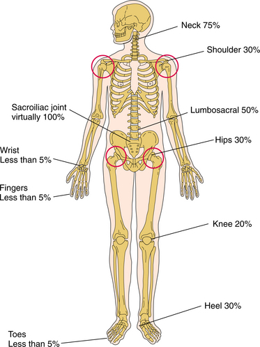
Impaired Posture (cervical involvement)

4C:

Impaired Muscle Performance

4D:

Impaired Joint Mobility, Motor Function, Muscle Performance, and Range of Motion Associated with Connective Tissue Dysfunction

4H:

Impaired Joint Mobility, Motor Function, Muscle Performance, and Range of Motion Associated with Joint Arthroplasty

4I:

Impaired Joint Mobility, Motor Function, Muscle Performance, and Range of Motion Associated with Bony or Soft Tissue Surgery (tenosynovectomy, tendon reconstruction)

5H:

Impaired Motor Function, Peripheral Nerve Integrity, and Sensory Integrity Associated with Nonprogressive Disorders of the Spinal Cord (cervical spine)

6B:

Impaired Aerobic Capacity/Endurance Associated with Deconditioning

Medical Screening of Joint Pain

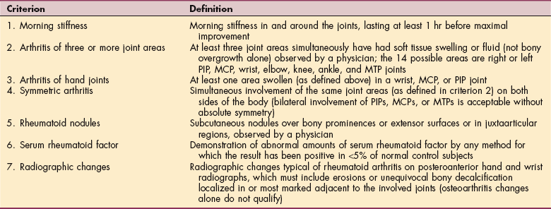
Therapists see many people with joint pain. Most of these people have joint pain secondary to degenerative OA as opposed to rheumatic joint disease, but the therapist must be aware of the symptoms and signs associated with RA. The therapist can ask a series of questions to help identify the cause of joint pain.156 An abbreviated version of this list can include the following:

• Are you stiff in the morning? If yes, how long does this last?

• Does your stiffness increase after sitting?

• Where do you think the pain is coming from?

• If joints, which ones are involved?

• Do you ever notice any joint swelling or other changes?

• Does anyone else in your family have RA or other kinds of arthritis?

• Does aspirin or ibuprofen help you feel better?

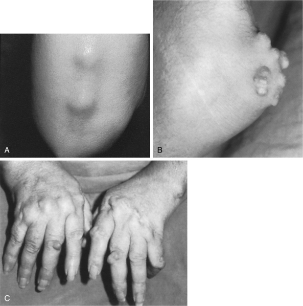
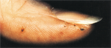
Being aware of the clinical signs and symptoms of RA will help the therapist make an early referral. The distribution of joint involvement is an important clue. RA usually affects the small joints of the feet and hands symmetrically; generalized pain (“I hurt all over”) is not characteristic of RA.

Quick, aggressive medical treatment is necessary to minimize joint destruction. Unexplained joint pain for 1 month or more, especially accompanied by systemic complaints, skin rash, or extensor nodules, no matter how mild, should raise concern on the therapist’s part. Cervical pain with reports of urinary retention or incontinence warrants immediate medical evaluation. Also, insidious onset of polyarthritis or joint pain within 6 weeks of taking a medication should raise suspicion regarding the nature of the pain complaints. Any of these red flags suggests the need for a medical referral if a physician has not recently evaluated the affected individual.

Anyone with known RA should be instructed in recognizing the signs and symptoms of progression of disease (e.g., increased duration of morning stiffness, increased number of tender and painful joints, increased intensity of joint pain, increased fatigue, increased gait disturbances, worsening of visible deformities) so that the intervention plan can be modified to meet the individual’s needs.

When distinguishing articular pain from periarticular involvement, remember that true arthritis produces pain and limitation during both active and passive range of motion, while limitation from tendinitis is much worse during active than during passive motion. Inflammatory joint involvement typically produces warmth, erythema, and tenderness. Frequently, there is bogginess related to underlying synovitis or effusion. These indicators are not present with joint pain of a mechanical cause.

Patient/Client Education

Helping individuals affected by RA understand the disease, disease process, treatment, possible outcomes, and role of exercise and self-care is a major part of the therapist’s task. Self-management includes learning pacing, joint protection, and energy conservation; monitoring symptoms; and maintaining or progressing an exercise program.

Each person must be taught ways to minimize trauma to inflamed joints by unloading joints and reducing mechanical joint stresses. This can be done through modification of activities such as using assistive devices to open doors and jars, and avoiding excessive weight bearing on inflamed joints by reducing movements such as bending and stooping. Energy conservation (see Box 9-8) should become a way of life for anyone with acute or subacute disease. The systemic nature of this disease produces global fatigue; the demand for energy to move joints may increase if biomechanics are altered.223

The need for frequent rest breaks, change in level of activity, and change in positions throughout the day should be taught and their use encouraged, but they should be balanced by the need to avoid muscle wasting and weakness from immobilization. Range of motion, stretching, and isometric exercises must be taught, monitored, and reinforced for as long as the therapist follows the individual, always teaching the client how to modify the program during periods of active inflammation.

Cervical Spine Involvement

Cervical collars may be used for comfort but do not protect against progressive subluxation or neurologic compromise. Rigid cervical collars can partially limit anterior atlantoaxial subluxation, but they also prevent reduction of the deformity in extension.238,239 Rigid orthoses are also poorly tolerated in these individuals because of skin sensitivity and temporomandibular joint involvement. Anyone with cervical spine involvement should be taught to avoid cervical flexion. The therapist can focus on isometric strengthening of neck muscles and overall postural training.

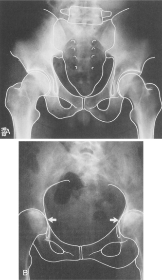
In the case of conservative intervention with nonsurgical management, the therapist must observe for (and teach the client to observe for) gradual deterioration in function that may indicate the development of subtle myelopathy (see Clinical Manifestations in this section). Radiographic evaluation is important in observing for impending neurologic compromise.420

Rehabilitation

RA is a chronic, progressive disease requiring an interdisciplinary team approach that is individual to the client and comprehensive, with long-range planning that extends beyond the initial acute phase. The role of the physical therapist as an integral part of the management of RA has been well established,59,287,370 with a renewed focus on outcome-based intervention.

The American College of Rheumatology has developed criteria for classification of functional status for individuals with RA that may help guide the therapist in designing and monitoring the results of an appropriate plan of care (Box 27-11).

Box 27-11   ACR CRITERIA FOR CLASSIFICATION OF FUNCTIONAL STATUS IN RHEUMATOID ARTHRITIS

Class 1: Completely able to perform usual activities of daily living (self-care, vocational, avocational)

Class 2: Able to perform usual self-care and vocational activities, but limited in avocational activities

Class 3: Able to perform usual self-care activities, but limited in vocational and avocational activities

Class 4: Limited in ability to perform usual self-care, vocational, and avocational activities

ACR, American College of Rheumatology.

The Ottawa Panel has identified nine goals in the rehabilitation of individuals with RA, including decreasing pain, effusion (swelling), and stiffness; correcting or preventing joint deformity; increasing motion and muscle force (decreasing weakness); improving mobility and walking; increasing physical fitness; reducing fatigue; and increasing functional status.383

Before initiating any rehabilitation program for this group of individuals, a thorough limb and joint examination must be done to provide an objective way to assess and document disease activity and progression or, conversely, remission and improved function. Numerous resources are available to assist the clinician in carrying out a thorough clinical assessment of the individual with RA, including a helpful joint-by-joint guide (describing where and how to palpate).347,431,459,460 Physical therapy examination should also include observation of functional performance, limitations, impairments, and a systems review as outlined in the Guide to Physical Therapist Practice.17

Complete bed rest is rarely indicated and is saved for those with severe, uncontrolled inflammation. For many people, a rest period of up to 2 hours during the day is important for dealing with general body fatigue and protection of involved joints. Splints can be applied to rest involved joints, prevent excessive movement, and reduce mechanical stresses. Crutches, canes, or walkers can be used to reduce weight-bearing stresses and enhance balance.

Adaptations may be necessary (e.g., platform crutches) because of upper extremity involvement. A home program of self-management will include instruction in proper body mechanics, positioning, joint protection, and energy conservation (see Box 9-8). Adaptive equipment designed to make tasks easier may include large key handle attachments allowing the person to use the whole palm to turn a key, spring-open scissors with big loops for anyone with hand involvement, jar openers and electric can openers, clip-on bottle openers, and ergonomic kitchen utensils with large handles and ergonomically angled handles.318

Instability at any joint, particularly at the atlantoaxial segment, requires caution on the therapist’s part. Such a joint may present with a marked reduction in range of motion, such as the shoulder or neck feeling stuck or caught with a certain movement. A history of periods of significant loss of range of motion alternating with full range of motion suggests joint hypermobility. Restoration of mobility is an important goal, but choosing techniques that are gentle or applying traction while stretching is necessary.

The extraarticular problems may affect the rehabilitation program. For example, if fatigue is present, the therapist may have to allow periods of rest during the treatment session. During periods of symptom exacerbation there is a fine line between overextending the client and maximizing activity. There are times when active exercise may have to be curtailed, but passive stretching remains important to prevent contractures.

Splenomegaly may account for tenderness on palpation and fullness or increased resistance of the left upper abdominal quadrant. Deep soft tissue techniques are contraindicated in this area. Percussion techniques may help the therapist delineate the caudal boundaries of the spleen.

Remission

Whether or not remission from RA was possible was a point of discussion and debate in 1981 when preliminary criteria for remission were first proposed.403 These criteria have been modified over the years to reflect current treatment, trends, and outcomes. For example, the complete absence of tender or swollen joints is the most important sign of remission.380

Changes must last for more than 2 months, but a definitive time line has not been established. Today even some people with long-standing disease may achieve remission, although this is not possible for everyone. After remission, researchers hope to be able to effect a curative approach.

Postoperative Care

Surgical treatment of RA is often complicated by the client’s generalized debilitated condition. People with RA tend to have poor skin condition, poorly healing wounds, and osteopenic bone. Generalized bone loss occurs early in the course of RA and correlates with disease activity. This condition is further affected by the use of corticosteroids.

Poor nutritional status has been associated with higher complication rates following surgery, including infection. Following Standard Precautions with adequate handwashing is important. Likewise, promoting respiration with good breathing techniques is an important component of the therapist’s postoperative intervention.

Anyone with RA should be taught early on that, if surgery is ever indicated, a program of isometric exercises and range of motion before surgery is advised. Review dislocation precautions and restrictions prior to the surgical procedure. After arthroplasty, correction of deformity and relief of pain are typical, but recurrence of deformity can occur even with appropriate rehabilitation. Many clients are still very satisfied with the improved cosmesis, reduced pain, and improved function. Maximum benefit from arthroplasty may not occur for up to 1 year after surgery.77

The postoperative rehabilitation regimen must be tailored to the specific needs of each individual. The surgeon’s intraoperative assessment of the quality of tissues, component stability, and any associated repairs is critical to the rehabilitation protocol selected. Specific motion limitations vary depending on intraoperative repairs made, complications, and adverse events (e.g., infection, wound dehiscence, dislocation, implant fracture or failure, nerve damage).

Exercise and Rheumatoid Arthritis

A group of experts from the University of Ottawa reviewed comparative controlled trials and compiled evidence to suggest and support the conclusion that therapeutic exercises, including specific functional strengthening and whole-body functional strengthening, are a beneficial intervention for individuals with RA. The benefit may vary depending on the stage of disease (acute, subacute, inactive) but includes reduced pain, improved overall function, and decreased number of sick leaves.384

There is a need for more research to investigate the impact and effects of exercise on individuals with RA. Whereas rest has often been the treatment of choice, a balance must be attained between rest required during acute flare-ups and activity necessary to prevent the deconditioning effects of prolonged rest, immobilization, and inactivity. Education as to the efficacy of exercise and its proper use in self-management of RA has been shown effective in reducing stiffness and improving function in as little as 4 hours of a community-based physical therapy intervention delivered over a period of 6 weeks.39

To date, there is no evidence that active exercise beneficially affects the inflammatory processes associated with adult RA, but it has been shown that a short-term intensive exercise program in active RA is more effective in improving muscle strength than a conservative exercise program and does not have deleterious effects on disease activity.516

Individual studies of long-term intensive exercise have not shown an increase in joint swelling or pain with such a regimen. In fact, those who exercise rigorously at least twice a week for an hour show more improvement in physical abilities such a stair climbing and reduced psychologic distress compared to those who received standard care.100

The beneficial effect of exercise in lowering cytokine levels and increasing antiinflammatory compounds in plasma has been demonstrated in children with juvenile RA.522 Other effects of exercise on the immune system are discussed in the section on Exercise Immunology in Chapter 7.

GENERAL CONCEPTS

Exercises to prevent contractures, improve strength and flexibility, and enhance cardiorespiratory or aerobic conditioning are important components of the rehabilitation program.176,255,370 Joint pain leads to a reflex inhibition of muscle surrounding the joints, causing disuse atrophy of these muscles. Use of corticosteroids may lead to an additional decrease in strength and function.370

The feet are often overlooked in people with RA, but foot involvement occurs frequently, can impair gait, and can prevent safe participation in an exercise program. Foot involvement resembles that of the hand, with one important difference being that alterations in biomechanics may cause excessive stress to proximal lower extremity joints and to the spine areas forced to compensate for the altered gait.

Careful assessment of the feet may reveal uneven or pathologic weight-bearing patterns. Gait analysis and assessment of shoe wear can provide additional significant information regarding altered biomechanics. Providing assistive devices or orthotics before initiating an exercise program may be essential.431,459,460

Avoid overloading and overtraining. For the individual with active (acute) disease, adequate sleep is essential. Encourage 8 to 10 hours of rest each night. Range-of-motion exercises should begin with low repetitions several times throughout the day. Isometric exercises with short holds (4 to 6 seconds) have been suggested, once again using low repetitions (start with one or two and build up gradually to four to six).223

Range-of-motion exercises can be increased up to 8 to 10 repetitions in subacute cases, with the addition of dynamic strengthening exercises. Stable, quiescent, or inactive disease makes it possible to add an aerobic component such as walking, aquatics, or biking for at least 15 minutes each day three times a week. Range of motion and strengthening can be continued and monitored.223

EXERCISE PRESCRIPTION

A helpful guide in establishing the level of acceptable exercise intensity is as follows: acute pain during exercise indicates a need to modify the program; if joint pain persists for more than 1 hour after exercise is completed, the exercise was probably excessive.144 Recent research indicates that it is not necessary to work out as vigorously as once thought to derive benefits from exercise.

Engaging in moderate-level exercise for 30 min/day 4 or 5 days/wk appears to substantially increase physical fitness, even for older adults.484 Exercise spaced out over the course of the day can help loosen up stiff, achy joints while still providing cardiovascular benefits.343,390

The Arthritis Foundation provides a 24-page booklet (Exercise and Your Arthritis) describing the benefits and types of exercises, how to start, and how to keep going. This publication educates people about the possible effects of not exercising (e.g., increased joint stiffness, muscle weakness, muscle atrophy, increased risk of fracture and deformity). The foundation also provides valuable exercise tips for before, during, and after exercise.25

Currently the National Arthritis Foundation offers the largest standardized exercise program to individuals with arthritis through two community-based programs: an on-land exercise program called People with Arthritis Can Exercise (PACE) and an aquatic exercise program called the Arthritis Foundation Aquatic Program (AFAP).

The PACE program involves 72 exercises to improve motion, strength, endurance, balance, coordination, posture, and body mechanics. Other recommendations and guidelines for including conditioning exercise in a comprehensive management program for RA are available.15,26,345

STRENGTH TRAINING

Dynamic strengthening (gradually increasing resistance) through the full available range of motion helps stabilize joints, reducing erosive wear and tear on the structures. Regular, dynamic strength training combined with endurance-type physical activities improves muscle strength and physical function but not bone mineral density (BMD) in adults with early and long-standing RA, without causing detrimental effects on disease activity.168,169

Low-load resistive muscle training has been shown to increase functional capacity and is a clinically safe form of exercise in mild to moderate RA.255 Other studies report that moderate-or high-intensity strength training programs have better training effects on muscle strength in RA. It is essential to maintain the training routine to obtain long-term benefits.167

Strengthening in some cases of RA can be difficult, because exercise may lead to increased joint swelling and subsequent joint destruction. Before initiating a strengthening program, the physical therapist must be sure that pain is controlled, range of motion is optimized, and contractures are minimized.370 Paying attention to biomechanics, deformities, and muscle imbalances is important. Exercising misaligned joints without properly distributing the load can place too much pressure on vulnerable joints.452

AEROBIC EXERCISE

Aerobic exercise in this population is necessary to help reduce weight and improve cardiovascular fitness without increasing pain.330 Aerobic exercises are safe to perform during the subacute and inactive stages of RA. Aerobic capacity can be estimated using a single-stage submaximal treadmill test.344 Training programs begin at 50% (and work towards 80%) of maximal oxygen uptake based on the baseline test results. Without baseline testing, the therapist can rely upon (and teach the client to use) the Borg scale for rate of perceived exertion (RPE; see Table 12-13). Heart rate monitors are also helpful in enabling clients to track their cardiovascular responses.223

Screening for unknown coronary artery disease is recommended before initiating resistance or aerobic exercise. RA can also affect the bony structures of the rib cage and cause a decrease in chest expansion. The usual precautions for cardiopulmonary screening still apply based on the individual’s age, risk factors, and health history (especially heart health history).

People with RA may have normal pulmonary function tests but reduced respiratory muscle strength and endurance with reduced aerobic capacity compared to adults without RA.82 Assessment of respiration and intervention to improve breathing patterns are important components of the rehabilitation program.

AQUATIC THERAPY

Aquatic therapy may be beneficial for conditioning, strengthening, and flexibility while reducing mechanical stress on the joints. Water exercise provides the means by which people with RA can reach needed training levels in a comfortable environment. The AFAP program consists of 72 exercises similar to the PACE program that can be done in water with decreased joint loading, a reduced effect of gravity, increased buoyancy, and increased circulation.

Modalities and Rheumatoid Arthritis

Various modalities provide temporary pain relief and may be used in effectively and safely controlling symptoms of the acute inflammatory phase of RA. Information on the rationale for use and effectiveness of the various physical modalities is available.64,335,346

Although cold may be more suitable in acute inflammation, people with RA usually prefer heat. Superficial heat (e.g., paraffin baths, moist hot packs, hydrotherapy or aquatic therapy) is recommended, whereas prolonged or deep heat is contraindicated, since it may increase intraarticular temperature, leading to increased collagenase activity, possibly contributing to joint destruction.194

Electrotherapeutic modalities and thermotherapy physical agents are often used as part of a rehabilitation program mainly for pain relief, to control inflammation, and to reduce joint stiffness.

The Ottawa Panel recommends the use of low-level laser therapy, therapeutic ultrasound, thermotherapy, electrical stimulation, and transcutaneous electrical nerve stimulation for the management of RA. This recommendation is based on the analysis of systematic and literature reviews. Specifics of studies reviewed and a summary of the findings have been published in our own journal (Physical Therapy).383

The Ottawa Panel reported on available systematic reviews on the efficacy of ultrasound in the management of RA and noted that these are limited and do not offer conclusive evidence-based support for the use of ultrasound alone or combined with exercise.

Medications

Because of the long-term nature of RA, intervention is usually an ongoing process. Aspirin and NSAIDs are potentially ulcerogenic, and prolonged use of corticosteroids can lead to osteoporosis.

The analgesics and slow-acting antirheumatic drugs such as gold and penicillamine can impair renal function. Periodic screening of each of the body systems is imperative when working with this population; anyone taking DMARDs but still having joint pain and swelling should be reevaluated by the rheumatologist.

Numerous other side effects can occur with any of the pharmacologic agents used in the management of RA. The therapist should be aware of the potential side effects with any of the medications each client is taking.

For the individual with Felty’s syndrome (RA in combination with low white blood cell count or leukopenia and an enlarged spleen), there is an increased risk of infection, even when treatment with DMARDs raises the white blood count. Careful handwashing and Standard Precautions are always warranted, but especially in the case of this syndrome.

Juvenile Idiopathic Arthritis

Overview and Incidence.: Although the term juvenile idiopathic arthritis (JIA; formerly juvenile rheumatoid arthritis [JRA]) brings to mind a single disease similar to adult RA, it is actually an umbrella term for a heterogeneous group of arthritides (Box 27-12) of unknown cause that begin before 16 years of age and occur in all races. Each subtype has a different presentation, genetic background, and prognosis.48,415

Box 27-12   SUBCATEGORIES OF JUVENILE IDIOPATHIC ARTHRITIS (JIA)

• Pauciarticular JIA (PaJIA; oligoarthritis)

• Polyarthritis JIA (PoJIA RF+)

• Polyarthritis JIA (PoJIA RF-)

• Systemic-onset JIA (SoJIA)

• Psoriatic JIA

• Enthesitis-related arthritis

• Other (undefined)

Data from Petty RE: ILAR classification of JIA: second revision, Edmonton 2001, J Rheumatol 31(2):390, February 2004.

RF+, Rheumatoid factor positive; RF-, rheumatoid factor negative.

Many other forms of arthritis (e.g., SLE, dermatomyositis, scleroderma) that affect adults occur less frequently in children. Approximately 30,000 to 50,000 children in the United States are affected by one of the subtypes discussed here (Fig. 27-16). The general classification of JIA is based on the number of involved joints and the presence of systemic signs and symptoms.

image

Figure 27-16 Breakdown in types of juvenile idiopathic arthritis (JIA).

Pauciarticular JIA, (PaJIA; also known as oligoarthritis), meaning “few joints,” generally affects four or fewer joints, usually in an asymmetric pattern, and most commonly involves the knees, elbows, wrists, and ankles. Girls are affected more often than boys and usually between the ages of 1 and 5 years. This type of JIA is relatively mild with few extraarticular features. Parents may notice a swollen joint and limp or abnormal gait, usually early after the child wakes up in the morning. Leg length discrepancy is common. Pain is not a central feature at first, and the disease rarely manifests any constitutional symptoms.351

PaJIA is the most common type of JIA, comprising one half of all JIA cases, and has three subtypes. The first is characterized by the presence of antinuclear antibodies (ANAs; see discussion in Chapter 40) and a high risk of uveitis, a potentially dangerous inflammation of the eye resulting in irreversible damage and blindness. The second subtype affects the spine as well as other joints, although spinal involvement may not occur until the child reaches late adolescence. Children with this subtype may test positive for the HLA-B27 gene (see Table 40-20), which is common in adults with AS (this subtype is sometimes referred to as juvenile AS). In the third subtype, joint involvement is the extent of the disease. Usually PaJIA runs a benign course; recurrences occur in up to 20% of children, most often during the first year but possibly delayed by as much as 5 years after the initial diagnosis. There are some children who develop persistent joint disease, referred to as extended oligoarthritis.351

Polyarticular JIA (PoJIA) affects five or more joints, most commonly including the large and small joints (wrists, cervical spine, temporomandibular joint, small joints of the hands and feet, as well as the knees, ankles, and hips). Joint involvement is usually symmetric and is most like that of adult RA, with the potential for severe, destructive arthropathy.

PoJIA comprises 40% of all cases of JIA and affects girls more than boys. There are two subtypes depending on whether children are rheumatoid factor positive or negative. The rheumatoid factor–positive subtype is characterized by the presence of rheumatoid factor (a type of autoantibody found in the blood of adults with RA) and the DR4 genetic type, also common in adults with RA. Subcutaneous nodules, cervical spine fusion, chronic uveitis, and destructive hip disease can occur in this type of PoJIA.351

The second subtype is characterized only by joint involvement, usually less severe. Children with this subtype do not test positive for rheumatoid factor.109 Morning stiffness and fatigue with possible low-grade fever are common clinical manifestations of this type of JIA.

Systemic-onset JIA (SoJIA; also called Still’s disease; sometimes also affecting adults, although rare) affects boys and girls equally with involvement of any number of joints. This subtype has the most severe extraarticular manifestations, affecting many body systems, and comprises 10% of all cases of JIA.

It often begins with a high-spiking fever and chills that appear intermittently for weeks and may be accompanied by a rash on the thighs and chest that often goes away within a few hours (Fig. 27-17). The fever pattern is marked by spikes exceeding 102° F (39° C) and periods between the spikes during which the child feels much better.

image

Figure 27-17 A and B, Skin rash associated with juvenile idiopathic arthritis. (A, from Paller AS, Mancini AJ: Hurwitz clinical pediatric dermatology: a textbook of skin disorders of childhood adolescence, ed 3, Philadelphia, 2006, Saunders. B, from James WD, Berger T, Elston D: Andrews’ diseases of the skin: clinical dermatology, ed 10, Philadelphia, 2006, Saunders.)

Inflammatory arthritis typically develops at some point, and 95% of the children have joint complaints within 1 year of the initial presenting symptoms. Approximately half of the children who have SoJIA recover almost entirely; unfortunately, one third of the children remain ill, with persistent inflammation manifesting as fever, rash, and chronic destructive arthritis.351

In addition to inflamed joints, the child may experience enlargement of the spleen (hepatosplenomegaly) and lymph nodes (lymphadenopathy); inflammation of the liver, heart, and surrounding tissues; and anemia.44 Box 27-13 lists clinical manifestations associated with Still’s disease.

Box 27-13   CLINICAL MANIFESTATIONS ASSOCIATED WITH STILL’S DISEASE

Systemic

• Fever

• Rash

• Lymphadenopathy

• Polyarthritis

• Pericarditis

• Pleuritis

• Peptic ulcer disease

• Hepatitis

• Anemia

• Anorexia

• Weight loss

Musculoskeletal

• Polyarthritis, polyarthralgias

• Myalgia, myositis

• Tenosynovitis

• Skeletal growth disturbances (short stature, failure to thrive)

Psoriatic JIA presents with psoriasis, arthritis, and at least two of the following: dactylitis, nail abnormalities, and a family history of psoriasis. Treatment with aggressive immunosuppressives may be required; uveitis is a feature in some cases.

Enthesitis-related arthritis presents as inflammation of the tendon attachments to the bone, especially along the spine and Achilles tendon, along with arthritis and any two of the following: sacroiliac joint tenderness, inflammatory spinal pain, the presence of HLA-B27, positive family history, acute uveitis, and pauciarticular or polyarticular arthritis in boys older than 8 years.

Etiologic and Risk Factors and Pathogenesis.: The cause is still poorly understood, but JIA may be triggered by environmental factors and infection (viral or bacterial) in children with a genetic predisposition. Genomic studies hope to identify genetic traits that will predict disease risk and other characteristics such as disease course, age of onset, and disease severity. Eventually, researchers may be able to identify molecular biomarkers to help diagnose and treat this group of arthritides.401

JIA can occur in boys or girls of any age (girls more often than boys) and most commonly begins during the toddler or early adolescent period. The pathogenesis is similar to that of adult RA, with immune cells mistakenly attacking the joints and organs, causing inflammation, destruction, fatigue, and other local and systemic effects.

TNF and the interleukins (IL-1 and IL-6) seem to be the primary cytokines responsible for many systemic features. These cytokines increase collagenase activity, osteoclast activation, body temperature, and muscle and fat breakdown, as well as acute-phase reactants.

MEDICAL MANAGEMENT

DIAGNOSIS.

Early disease recognition is needed to help improve the clinical outcome, but symptoms of rheumatic disease are often mistaken for “growing pains,” delaying diagnosis by many months. Diagnosis involves a medical history, physical examination, and laboratory tests, including serum evaluation to measure inflammation and to detect ANAs, rheumatoid factor, or sometimes HLA-B27.

For a diagnosis of JIA, objective arthritis must be seen in one or more joints for at least 6 weeks in children younger than 16 years; it may take up to 6 months to determine which subtype is present. Pain is often dull and aching and less severe but presents in the morning and early during the day rather than the more common presentation of growing pains at night. The systemic features in SoJIA are more readily diagnosed.

TREATMENT AND PROGNOSIS.

Some of the immunomodulatory medications used in adult RA can be used in cases of JIA, but none of the current medications used has a curative potential. The goal of treatment is to control pain, preserve joint motion and function, minimize systemic complications, and assist in normal growth and development.

Early aggressive combination medications are replacing the previous gradual add-on approach to treatment (i.e., start with one drug and slowly add another and another to gain the desired effects without too many side effects).532 Medications are administered to control the systemic and articular complaints and, in some cases, halt the progression of the disease.

These agents may include immunosuppressives, DMARDs (e.g., MTX), and biologic agents such as TNF inhibitors (etanercept [Enbrel], infliximab [Remicade]).166

Adverse effects from taking anti-TNF agents (e.g., neurologic disorders, weight gain, severe infection, hemorrhagic diarrhea) have been reported and should be monitored for carefully. In systemic JIA, approximately 50% respond to anti-TNF agents, but in many children, the response is not sustained.293 Corticosteroids are indicated if severe anemia, unrelenting fever, or vasculitis is present. Bone marrow transplantation may be used in cases of JIA that are resistant to standard medical management.482

The prognosis has greatly improved as a result of substantial progress in disease management, especially the use of anticytokine agents in children who are resistant to conventional antirheumatic agents. Early-onset, progressive forms of JIA have a guarded prognosis. Between 25% and 70% of children with JIA will still have active arthritis 10 years after disease onset; over 40% enter adulthood with active arthritis.293

The mortality risk for children with JIA has been estimated to be three to five times higher than in the general population.498 Morbidity and mortality may be improved (including increased rates of disease remission) with the changes in treatment approaches, but this has not been documented as yet.293

Autologous stem cell transplantation (ASCT) is used for some individuals whose disease is refractory to MTX and other DMARDs. Complete remission is possible for up to half of the individuals receiving ASCT, with improvement reported in those individuals who are not resistant. Infection is a common morbidity associated with this treatment, observed in up to 71% of cases, in addition to an associated death rate of 15%.97

As the immune mechanisms and inflammatory processes are better understood, new drugs able to inhibit single molecules or pathways will be developed.415 Investigations continue to study biologic therapies that block IL-1 and IL-6 in systemic JIA.293

27-10   SPECIAL IMPLICATIONS FOR THE THERAPIST

Juvenile Idiopathic Arthritis

PREFERRED PRACTICE PATTERNS

See also appropriate practice patterns outlined for adult RA.

4A:

Primary Prevention/Risk Reduction for Skeletal Demineralization (low bone mineral density)

4E:

Impaired Joint Mobility, Motor Function, Muscle Performance, and Range of Motion Associated with Localized Inflammation

5B:

Impaired Neuromotor Development

Children with JIA may have no disability, especially if they have the oligoarticular form of the disease. Severe disability is seen most often in cases of rheumatoid factor–positive polyarticular and systemic disease, followed by rheumatoid factor–negative polyarthritis, enthesitis-related arthritis, and psoriatic arthritis. Physical therapy is an important adjunctive therapy for JIA.48

Efforts have been made to identify early predictive clinical, laboratory, demographic, genetic, or treatment-related factors for functional disability. It appears that there are complex interactions between disease subtypes but with specific predictors of outcome for each disease subtype. More study is needed to completely define predictive factors.

Physical therapy and occupational therapy are utilized for pain control, facilitation of mobility, and function. Equally important is the role of exercise in improving strength, endurance, and aerobic capacity. In children with JIA, resistive exercise produces a change in the immune response, with significantly lower levels of cytokines and higher levels of antiinflammatory compounds compared with those who did not exercise.522

With respect to joint impairment, loss of joint motion is the strongest indicator of functional disability in children with systemic JIA, and loss of joint motion has a greater effect on lower limb function than on upper limb function.38 By the time children become adults, only 20% of them have moderate to severe limitations.

The role of the therapist in preventing joint loss should not be underestimated. Cervical spine involvement can occur in PoJIA and systemic JIA, with cervical stiffness as the most common finding. Neck pain is uncommon, and neurologic complications are less likely to develop in JIA than in adult RA.

The therapist should keep in mind the significant impact JIA has on the children and their families. The cost of this disease can be staggering, with pharmacotherapy, hospitalizations, medical visits, and other professional services, including physical and occupational therapy.

Psychosocial-spiritual and quality-of-life issues should also be addressed. Appearance and body image are affected directly by JIA (e.g., generalized growth failure, local growth anomalies such as micrognathia), side effects of drug therapy, surgical scars, and severity of pain and fatigue. The therapist may be the first one to recognize overall problems with adjustment reflected by anxiety, depression, and/or social withdrawal.48

For those children with JIA who reach adulthood, significant problems can result from arthritis and uveitis, medication morbidity, and lifelong disability.341 Young women may face the issue of pregnancy and childbirth with fears of transmitting JIA to the offspring, and possible reduced ability to conceive along with increased rates of miscarriages have been reported by some.381

If the therapist is seeing a young child with joint pain who has not been diagnosed, sensitivity to soft tissue manipulation despite improved or restored joint range of motion warrants further medical investigation. For children with a known diagnosis, the use of physical modalities may be appropriate but must be used with caution as children are not always able to perceive and/or report discomfort.

Exercise and Juvenile Idiopathic Arthritis

Very few studies have been done to determine the safety and efficacy of exercise for children with JIA. Studies that have been done have a small sample size, and results cannot be generalized to all children with this condition. For example, the results of at least one study of nine children suggest that bicycle and treadmill exercise is safe and feasible for children who do not have severe hip disease.463

Aquatic physical therapy is an excellent way to complete exercises while providing joint protection and engaging the child in a fun activity. One study engaged 54 children with JIA ages 5 to 13 in a supervised aquatic training program 1 hr/wk for 20 weeks. Measures of functional ability, health-related quality of life, joint status, and physical fitness showed no improvement, but there were also no signs of worse health status, suggesting that swimming is a safe exercise program.487 More studies are needed to determine at what frequency, level, intensity, and duration a swimming training program would make a significant difference (improvement) in the areas tested and measured.

Strengthening programs for children with rheumatic disease can be part of the exercise program, even for children under 6 years of age. Twice-daily sessions of 15 to 20 minutes are advised.421 Research at the University of Buffalo showed significant improvement in function and better strength, endurance, and aerobic capacity. Equally important, pain, disability, and use of medications decreased significantly. The researchers found that the children who participated in the exercise program had significantly lower levels of cytokines (proinflammatory proteins) and higher levels of antiinflammatory compounds in their plasma than those who did not exercise.124

Active exercise is not advised during flare-ups when joints are inflamed, but most children usually self-limit their physical activity according to symptoms. Passive stretching and modified aquatic physical therapy are better choices during exacerbations. Parents and children should be educated on the importance of avoiding forced or deep flexion of inflamed joints. Some activities may need to be modified or avoided.

Children with quiescent JIA can participate in some sports activities. With improved medical therapies, children with JIA are able to lead a more active lifestyle compared with similar children even 10 years ago. The therapist can be helpful in assessing each child for abnormal biomechanics that can place him or her at increased risk for injury or future articular damage. Neuromuscular training may be needed to improve neuromuscular function and biomechanics. Proper technical performance during athletics may allow children with JIA to use joint-loading techniques (e.g., during jumping and landing) in a safe and controlled manner.359

Low BMD is a common secondary condition associated with JIA. Weight-bearing exercise programs to reduce the risk of low BMD are safe and effective for children with JIA who are healthy and should be included in the plan of care. More research is needed to determine the amount, duration, and frequency of weight-bearing activity needed to reduce the risk for low BMD in this population.136

The role of physical activity and an active lifestyle in cardiorespiratory fitness has been documented, but long-term follow-up is still needed to show if such a program protects from loss of aerobic fitness in this population group.488 The 6-minute walk test has been shown to be a good test for measuring functional exercise capacity in a small study of children (boys and girls) with JIA ages 7 to 17 years.385 This might be a good place to begin baseline studies and evaluate response to the aerobic component of the plan of care for individuals with JIA.

Spondyloarthropathies

Spondyloarthropathies (SpAs), a group of disorders formerly considered variants of RA, are in fact distinct entities with similar features affecting the spine (Box 27-14). SpAs are characterized by inflammation of the joints of the spine and include several distinct entities: AS, Sjögren’s syndrome, psoriatic arthritis, and reactive arthritis, including arthritides that accompany inflammatory bowel disease (IBD; known as enteric arthritides), and Reiter syndrome. Inflammatory eye disease (e.g., uveitis, conjunctivitis, iritis) occurs in approximately 25% of clients.

Box 27-14   COMMON FEATURES OF THE SPONDYLOARTHROPATHIES

• Chronic inflammation of the axial skeleton and sacroiliac joints

• Asymmetric involvement of a small number of peripheral joints

• Young males (late teens, early adulthood) most commonly affected

• Familial predisposition

• Inflammation at sites of ligament, tendon, and fascial insertion into bone

• Seronegativity for rheumatoid factor, but an association with histocompatibility antigens, including HLA-B27

• Extraarticular involvement of eyes, skin, genitourinary tract, cardiac system

Enteropathic arthritis (arthritis associated with IBD) occurs in about 20% of clients who have IBD (e.g., Crohn’s disease, ulcerative colitis) and is discussed further in Chapter 16. Arthritic symptoms flare with IBD and usually affect the lower extremities in an asymmetric pattern. Vasculitis, clubbing of the fingers, and skin changes may be present.

New biomarkers for SpAs have been reported recently. Changes in synovial tissue may be good biomarkers to assess the effectiveness of treatment. Specific changes in the cells of the synovial tissue correspond to treatment with TNF blockers. Biopsy specimens of synovial tissue taken after treatment showing changes in synovial macrophages, polymorphonuclear leukocyte (PMN) levels, and expression of metalloproteinases (MMP-3) thought to play a role in the degradation of collagen may lead scientists to use these same biomarkers for early identification and treatment of RA.263

Ankylosing Spondylitis:

Overview and Incidence.: AS, sometimes referred to as Marie-Strümpell disease, is an inflammatory arthropathy of the axial skeleton, including the sacroiliac joints, apophyseal joints, costovertebral joints, and intervertebral disk articulations.

Approximately one third of those with AS have asymmetric involvement of the large peripheral joints, including the hip, knee, and shoulder. Fig. 27-18 shows the most commonly involved joints. The disorder can ultimately lead to fibrosis, calcification, and ossification with fusion of the involved joints. The pain, resultant postural deformities, and complications associated with this disease can be disabling.

image

Figure 27-18 Joints most commonly involved in ankylosing spondylitis and incidence of involvement. Not shown: jaw 15%, eye 20%, ribs 20%, costovertebral junction 70%. (From Ramanujan T, Schumacher HR: Ankylosing spondylitis: early recognition and management, J Musculoskelet Med 1[1]:75-91, 1992.)

Prevalence of AS is 0.1% to 0.2% in the U.S. general population. Nearly 2 million people in the United States have this condition, making it almost as common as RA. It is higher in Caucasians and some Native Americans than in African Americans, Asians, or other nonwhite groups.3

AS typically affects young people, beginning between the ages of 15 and 30 years (rarely after age 40 years). This differs from back pain of mechanical origin, which is much more likely to develop between 30 and 65 years of age. Men are affected two to three times more often than women, although this disorder may be just as prevalent in women but diagnosed less often because of a milder disease course with fewer spinal problems and more involvement of joints such as the knees and ankles.519,545 Overall sibling risk is about 5.9%.282

Etiologic and Risk Factors.: Although the molecular basis of AS remains unclear, evidence points to a genetic or environmental link. Approximately 90% of those with AS are HLA-B27 positive, but of all of the people with this antigen only 2% develop AS. Proof that this disorder is an autoimmune disease attributable to cross-reactivity between bacteria and HLA-B27 is still lacking.220 However, approximately 20% of people who develop AS have a first-degree relative who is HLA-B27 positive.

Gender, age, race, and family history are all important factors related to the risk of developing AS. Although it is more prevalent in males, there is significantly less disparity in incidence between the genders than was once thought. The belief is that the disorder has been grossly underdiagnosed in women because the disease tends to be milder and peripheral joint involvement is more common, confusing the clinical picture.

No direct linkage of the X chromosome with susceptibility to AS has been found, but the influence of female gender is greater than that of male gender in determining increased susceptibility to AS in children. The striking maternal effect is greatest for women with young age at onset, which is not seen in men.63 It has been hypothesized that women tend to have a milder form of AS because they have the susceptibility genes, which they pass on to their children, whereas men have the severity genes.56

Studies under way using the results of the Human Genome Project are searching for other genes that in combination with HLA-B27 constitute risk for AS. The results of the first genome scan show areas where there appear to be some other genes that have been implicated in Crohn’s disease and psoriasis, suggesting a shared genetic basis in these conditions. Other researchers have demonstrated that environmental factors, such as infectious microbes, are essential for the development of AS, in particular normal bacteria in the bowel.22,217,282

Pathogenesis.: The pathogenesis of AS is poorly understood, but the fundamental lesion appears to be chronic inflammation at sites of attachment of cartilage, tendons, ligaments, and synovium to the bone.410 AS is marked by a chronic nongranulomatous inflammation at the area where the ligaments attach to the vertebrae (an area called the enthesis), initially in the lumbar spine and then in the sacroiliac joint.

Disruption of this ligamentous-osseous junction results, and reactive bone formation occurs as part of the repair process. Cartilage of the sacroiliac joints may also be involved (Fig. 27-19). The replacement of inflamed cartilaginous structures by bone contributes to progressive ossification with bony growth between the vertebrae, leading to a fused, rigid, or bamboo spine, characteristic of end-stage disease (see Fig. 27-20).

image

Figure 27-19 Progression of ankylosing spondylitis of the sacroiliac joints. A, Normal sacroiliac joints. B, Fusion of sacroiliac joint spaces; the sclerosis has resorbed, and there is slight narrowing of the left hip joint. C, Advanced ankylosing spondylitis with generalized osteoporosis and fusion of the spinous process, intervertebral disks, sacroiliac joints, and symphysis pubis. The entire skeletal unit has been transformed into one continuous osseous mass. (A, from Magee D: Orthopedic physical assessment, ed 2, Philadelphia, 1992, Saunders, p 329. B, from Rothman RH, Simeone FA: The spine, Philadelphia, 1982, Saunders, p 921. C, from Bullough PG: Orthopaedic pathology, ed 3, London, 1997, Mosby-Wolfe, p 300.)

image

Figure 27-20 Radiograph of a sagittal vertebral column in a person with ankylosing spondylitis. There is complete fusion of the spine, accentuated kyphosis, and loss of lumbar and cervical lordosis. There is also complete fusion of the intervertebral disk spaces. (From Bullough PG: Orthopaedic pathology, ed 3, London, 1997, Mosby-Wolfe, p 301.)

Clinical Manifestations.: Insidious onset of low back, buttock, or hip pain and stiffness lasting for at least 3 months are often the initial presenting complaints. Early onset of back pain, stiffness, and fatigue during childhood often goes unrecognized for what it is. Onset of symptoms leading to a diagnosis occurs most often during early adulthood.410

At first the pain is described as a dull ache that is poorly localized, but it can be intermittently sharp. Over time, pain can become severe and constant, increased by prolonged rest or immobility and decreased by active movement. Coughing, sneezing, and twisting may worsen the pain.282

Pain may radiate to the thighs but does not usually go below the knee.3 Buttock pain is often unilateral but may alternate from side to side. Significant morning stiffness, lasting more than 1 hour, is often present. There may be tenderness over the spinous processes and sacroiliac areas with associated paraspinal spasms.

Enthesitis (inflammation of the tendons, ligaments, and capsular attachments to bone) may produce tenderness, pain and/or stiffness, and restricted mobility in the costosternal, costovertebral, and manubriosternal joints, iliac crest, ischial tuberosities, greater trochanters, spinous processes, or ligamentous attachments at the calcaneus.

Other clinical features include early loss of normal lumbar lordosis with accompanying increased kyphosis of the thoracic spine, painful limitation of cervical joint motion, and loss of spine mobility (flexibility) in all planes of motion.

In some cases the initial complaints may occur in the extremities (e.g., hips or knees) with back symptoms appearing on average 3 years after onset of the peripheral involvement. Shoulder symptoms and loss of shoulder mobility are common but are rarely disabling. Involvement of the shoulder joint in AS correlates with involvement of other peripheral joints as well as the extent of radiographic change on shoulder films.542

Hip flexion contractures are often present bilaterally and can be assessed using the Thomas test (in the supine position, ask the individual to maximally flex the contralateral hip joint; observe for loss of lumbar lordosis and flexion of the opposite leg, signaling a positive Thomas test result for the presence of a hip flexion contracture; repeat on the other side). Loss of hip mobility can result in reduced functional ability.

Loss of chest wall excursion is an indicator of decreased axial skeleton mobility because of involvement of the thoracic spine and the costovertebral and costosternal joints. If the ligaments that attach the ribs to the spine (costovertebral junction) become ossified, the chest may be unable to expand, compromising breathing. Circumferential measurements taken at the fourth intercostal space (or just below the breasts in women) before and at the end of inspiration should reveal an excursion of 4 to 5 cm. Excursion less than 4 cm, and especially anything less than 2.5 cm, is a suspicious finding.

COMPLICATIONS.: Long-standing disease is associated with multiple complications. Skeletal complications include osteoporosis, fracture, atlantoaxial subluxation, and spinal stenosis. In the most severe cases, the spine becomes so completely fused that the person may become locked in a rigid upright or stooped position, unable to move the neck or back in any direction. Flexion contractures, rigid gait, and flexing at the knees in order to maintain an upright position are not uncommon findings.

The stiff and osteoporotic spinal column is prone to fracture from even a minor insult; a significant proportion of individuals with AS experience vertebral fracture during the course of the disease. Fracture sites range from T7 to S1.117,197 In fact, the incidence of thoracolumbar fractures in AS is four times higher than in the general population.89,197

Fractures can occur in the lower cervical spine. The atlantoaxial segment is among the last areas of the axial skeleton to fuse. Because of the inherent mobility of C1 and C2 and the immobility of the remainder of the cervical spine secondary to the disease, attempts to move the head could result in subluxation. Spinal stenosis can result from the proliferation of bony tissue from the spinal ligaments and facet joints. Stenosis may cause neurogenic claudication and cauda equina syndrome.

AS is a systemic disease with widespread effects. In addition to arthritis in the spine, arthritis in other joints may be accompanied by inflammatory bowel syndrome with fever, fatigue, loss of appetite, weight loss, and other extraarticular complications. These clinical features distinguish AS from mechanical pain.

The most common extraarticular manifestation is uveitis, occurring in 20% to 30% of affected individuals. Cardiomegaly, pericarditis, aortic regurgitation or insufficiency, amyloidosis (rare), and pulmonary complications may also occur. Pulmonary problems include upper lobe fibrosis and decreased total lung capacity and vital capacity (late stages of AS).310

MEDICAL MANAGEMENT

DIAGNOSIS.

AS is not easily detectable in the early stages without an MRI and only then with specialized MRIs, which are not routinely ordered. Early, accurate diagnosis allows treatment to start before the onset of permanent rigidity and deformity.

Intraarticular inflammation, early cartilage changes, and underlying bone marrow edema and osteitis can be seen with a specific MRI technique called short tau inversion recovery (STIR) sequences.410 Diagnosis is usually based on identification of the clinical manifestations and radiographic findings.3 History, physical examination, radiography, and laboratory tests are all used in the diagnosis.

In the physical examination, range-of-motion tests provide important information. The Schober test, chest expansion (measured at the fourth intercostal space or just below the breasts in women on inspiration after forced maximal expiration), and military stand against the wall are clinical tests used most often. A distance of less than 15 cm for the Schober test and less than 2.5 cm for chest expansion on inspiration are suspicious findings.

Clinical manifestations alone are not diagnostic, but when taken along with the history and radiographic results, they become significant for the diagnosis. Radiographs may be negative in the early stages of the disease; MRI enables the examiner to identify sacroiliitis earlier than plain radiography.

Radiographic findings of symmetric, bilateral sacroiliitis include blurring of joint margins, juxtaarticular sclerosis, erosions, and joint space narrowing. The replacement of ligamentous tissue by bone at the site where the annulus fibrosus of the intervertebral disk inserts into the vertebral body results in a characteristic square-shaped vertebral body. In addition, as bony tissue bridges the vertebral bodies and posterior arches, the thoracic and lumbar spine takes on the appearance of a bamboo spine on radiographs (Fig. 27-20).

No laboratory test is diagnostic of AS; laboratory tests assist primarily by ruling out other diseases. The presence of the HLA-B27 antigen is a useful adjunct to the diagnosis but is not diagnostic itself, since so many people with other causes of back pain also are HLA-B27 positive. ESR and C-reactive protein are elevated in 75% of affected individuals and may correlate with disease activity in some people, but these levels can be normal even in individuals with active disease.410,461

TREATMENT.

The primary focus of intervention is to reduce inflammation and stiffness in the joints, maintaining mobility and proper postural alignment of the spine to prevent structural damage, while providing pain relief. Effective education is essential, because much of the management requires lifestyle adjustments and cooperation, especially compliance with the exercise program.282

Joint involvement can be managed with NSAIDs (including the new COX-2–specific inhibitors), but in some cases disease-modifying drugs (DMARDs) such as methotrexate (MTX) or sulfasalazine (SSZ) may be used for peripheral disease.

Persistent symptoms of spinal involvement, peripheral arthritis, or enthesitis unresponsive to NSAIDs or DMARDs can be managed with biologic agents such as TNF-α antagonists (e.g., etanercept, infliximab, adalimumab). These agents have been shown effective in preventing the progression of disease by reducing disease activity, decreasing inflammation, and improving spinal mobility.3,51

Other targeted therapies may be needed to treat specific organ involvement, such as eye inflammation. To avoid long-term complications associated with severe postural deformities, a lifetime commitment to exercise is important.

Surgery has a limited role in the management of AS and is most appropriate for individuals with severe deformity that impedes vision, walking, eating, abdominal expansion, or respiratory function. Spondylodiskitis or spinal fracture may require surgical intervention.49 Spinal fusion may be needed but is not routinely recommended. The most valuable surgical intervention is total joint arthroplasty, especially a total hip replacement.282 It is expected that with the new biologic therapies, the need for surgical intervention will become a rare event.49

PROGNOSIS.

The extent of disability in persons with AS varies considerably, but fewer than 1% experience complete remission and more than 80% who are ill for longer than 20 years still have daily pain.24,545 Periods of exacerbation and remission are common during the course of the disease.

The severity of symptoms during the first decade indicates the long-term severity and disabling nature of the disorder. Severe disease is usually marked by peripheral joint and extraarticular manifestations. The onset of hip disease in anyone with AS at any stage of the disease is a major prognostic marker for long-term severe disease and is more common in people with onset at a young age.56

Individuals with AS have an increased mortality rate. The impact of this disease can be seen in various aspects of workforce participation, such as needing more assistance, withdrawal from the workforce, and reduced quality of life. Early diagnosis and management will likely help prevent functional disability and improve outcomes.47

27-11   SPECIAL IMPLICATIONS FOR THE THERAPIST

Ankylosing Spondylitis

PREFERRED PRACTICE PATTERNS

4A:

Primary Prevention/Risk Reduction for Skeletal Demineralization (osteopenia, osteoporosis)

4B:

Impaired Posture

4E:

Impaired Joint Mobility, Motor Function, Muscle Performance, and Range of Motion Associated with Localized Inflammation

4F:

Impaired Joint Mobility, Motor Function, Muscle Performance, Range of Motion, and Reflex Integrity Associated with Spinal Disorders (spinal stenosis)

6B:

Impaired Aerobic Capacity/Endurance Associated with Deconditioning (other cardiovascular/pulmonary patterns may apply depending on clinical presentation)

Whenever someone presents with new onset of back, sacroiliac, or hip pain in the absence of trauma or overuse that is accompanied by associated signs and symptoms of systemic disease (e.g., fever, fatigue, respiratory compromise) the therapist must recognize that these are not symptoms typically associated with mechanical causes of back pain. If present, these features are considered red flags that should raise concern regarding the underlying cause of the complaints.

The therapist can assess for limitation of spinal mobility, most evident as a decrease in forward flexion of the spine, using the Schober test. In the standing position, place a mark at the lumbosacral junction, which is represented by the intersection of a line joining the dimples at the posterosuperior iliac spines (PSISs). Place a second mark 5 cm below and 10 cm above the lumbosacral junction. The client is then asked to bend forward and attempt to touch the toes.

Now remeasure the distance between the upper and lower marks. The distraction between these two marks has been found to correlate very closely with anterior flexion measured radiologically. The distance increases more than 5 cm in the absence of AS and less than 5 cm when AS is present.

Alternatively, measure the horizontal distance between the posterior inferior iliac spine in both the prone and sitting positions, observing for a change in the distance with a change in position. Expect to see the posterior inferior iliac spine ½ inch closer in the prone position compared to sitting in the normal young adult and suspect AS if there is no change.

Treating a client with a diagnosis of AS requires that the therapist entertain different considerations. If the client complains of sharp pain associated with a fall, sneeze, or lifting a moderately heavy object, one must consider the possibility of a fracture. With osteoporosis being a potential complication, technique modification is warranted. See Special Implications for the Therapist box in the section on Osteoporosis in Chapter 24 for further discussion.

Management should follow guidelines similar to those outlined for fibromyalgia (see Chapter 7). With the fragility of the spine in AS and the risk of fractures from even a minor injury or fall, falls prevention is also important. This is an area for careful attention, because a number of people with AS suffer neurologic deficit after injury, especially spinal fracture.197

Trunk range-of-motion and strengthening exercises to minimize thoracic kyphosis are essential. The more severe the kyphosis, the more hindered pulmonary function will be and the more pronounced the compensatory forward head posture. Postural deformities contribute to cervical pain and headaches and may also affect balance. Avoiding obesity is recommended to reduce stress on weight-bearing joints and the cardiopulmonary system.

Finally, smoking should be discouraged because of its adverse effects on the cardiopulmonary system. The therapist can also monitor clients taking NSAIDs for early signs of gastrointestinal bleeding or other adverse side effects (see Chapter 6).

Exercise and Ankylosing Spondylitis

Therapists play a potentially important role in the rehabilitation of this population. Although more studies are available now than ever before in this area, there still is not sufficient evidence to base recommendations for or against specific physical therapy interventions. Continued research is needed to address those modalities and applications commonly used in practice.94 A summary of the available scientific evidence on the effectiveness of physical therapy interventions in the management of AS is presented here and elsewhere.519

GENERAL CONCEPTS

Although exercise is a commonly recommended intervention for AS, little is known about the effectiveness of unsupervised recreational and back exercises. Findings from one study511 suggest that exercise improves pain, stiffness, and function when performed at least 30 min/day and back exercises at least 5 days/week, although these effects vary with the duration of the AS. There may be an optimal duration for exercise performed independently over a weekly period, but consistency rather than quantity appears to be a much more significant variable. Although there is no difference in disease activity with a regular program of exercise, improved function has been demonstrated.437

Interpreting the results of some studies can be difficult. For example, one study showed that younger individuals with AS who had a shorter duration of disease and were less disabled also exercised less compared to older, more disabled adults who exercised more often. Without careful analysis and understanding of the results, the conclusion could be made that those people who exercised more frequently were more disabled, when in fact, affected individuals did not get serious about exercising consistently until the disease had progressed.115

EXERCISE PRESCRIPTION

The question of what type of exercise is best for this condition has not been adequately answered. Exercising has been shown to benefit people with AS, but again, consistency, not quantity, seems to be the key factor in influencing posture, strength, motion, mobility, fitness, and overall health.436 Home programs and group programs have both been shown to be beneficial for individuals with AS.93,122

A multimodal physical therapy program including aerobic, stretching, and pulmonary exercises along with routine medical management has been shown to yield greater improvements in spinal mobility, work capacity, and chest expansion compared with medical care alone. Individuals in a 50-minute, three-times-a-week multimodal exercise program were significantly improved after 3 months in chest expansion, chin to chest distance, occiput to wall distance, and the modified Schober flexion test.219

Functional and breathing capacity as well as balance should be assessed and developed. Stretching of the shortened muscles and chest expansion exercises should be encouraged. Improving and/or maintaining cardiovascular fitness is important.

Strengthening of the trunk extensors is equally important, so that if and when spinal fusion occurs, the spine is aligned in the most functional position possible. This requires a coordinated effort among all team members, including the affected individual and family.

High-impact and flexion exercises should be avoided, whereas low-impact, aerobic exercise with extension and rotational components can be emphasized. Each individual will need to identify personal limitations and safe levels of participation.

In general, contact sports and high-risk activities such as downhill skiing, horseback riding, boxing, football, soccer, and water skiing should be avoided; aquatic therapy is an excellent option for most people provided extension principles are emphasized. In the presence of spinal fusion and osteoporosis, other activities requiring high levels of balance, agility, and coordination (e.g., bicycling, ice skating, rollerblading) can result in falls and fractures.

Overexercising can be potentially harmful and can exacerbate the inflammatory process; principles of relaxation, proper body mechanics, and energy conservation should be a part of the education program offered (see Box 9-8), including assessment of ADLs and providing necessary aids or devices such as long-handled reaching tools, adaptations for the car, special garden tools, or elastic shoelaces. Learning and using proper breathing techniques throughout all activities will help maintain chest expansion, improve oxygenation, and minimize muscle fatigue.

At present, aggressive but careful stretching to address the areas of hypomobility and the muscle imbalances is purported to help maintain as optimal a posture as possible. An exercise program focusing on specific strengthening and flexibility exercises of the shortened muscle chains offers promising short- and long-term results in the management of this condition.121

OUTCOMES MEASUREMENTS

Mobility tests to establish a baseline and measure the effectiveness of short-term intensive physical therapy and exercise on spinal, hip, and shoulder motion have been evaluated.

Finger to floor distance, thoracolumbar rotation, and thoracolumbar lateral flexion are the most sensitive tests to detect improvements (or progression) in short-term clinical trials, whereas the Schober test, thoracolumbar flexion, and occiput-wall distance are insensitive measures of improvement. Hip internal rotation, shoulder flexion, and abduction measurements are also sensitive, although more suitable for individuals with articular symptoms. Thoracolumbar rotation and hip rotation are the only measurements that correlate with disease duration but not with age.74,186

The Bath Ankylosing Spondylitis Functional Index (BASFI), along with the Dougados Functional Index score, Bath Ankylosing Spondylitis Metrology Index, and Bath Ankylosing Spondylitis Disease Activity Index are self-assessment tools that can be used both by the therapist and by the client to establish a baseline and document improvement or decline.33,62,361

The therapist must be vigilant for an exacerbation of the inflammatory process. Assess (and periodically reassess) the spine and peripheral joints for mobility, range of motion, and strength. Modalities such as ultrasound, heat and cold, and electrotherapy may be effective during the acute phase if used judiciously.

Aggressive stretching must be avoided during these phases; teaching the individual a progressive relaxation program may be helpful. Communication with the physician is necessary for the development of a proper medication regimen.

POSITIONING

The affected individual may need help modifying home or work situations. Appropriate footwear advice should be provided; some people benefit from functional foot orthoses. Resting in the prone position is advised to help avoid hip and spine flexion contractures. The Spondylitis Association of America472 recommends a firm, supportive sleeping surface to maintain good spinal alignment. Soft mattresses or waterbeds can contribute to excessive flexion and stooped postures.

The therapist can provide additional recommendations for the use of pillows or towel rolls for proper alignment in the various positions. The person may not stay positioned all night, but with time and training, changing positions and realigning props can be incorporated into the sleep cycle at least part of the time.

Proper lifting techniques can be demonstrated, with return demonstration provided by the client. The therapist should assess the work area or instruct the individual in appropriate ergonomics given the diagnosis of AS.

Sjögren’s Syndrome

Overview.: Sjögren’s syndrome is a chronic arthritis-related disease that can affect several organs, most commonly the moisture-producing glands (e.g., mouth, eyes) but also joints, lungs, kidneys, or liver. Sjögren’s syndrome is an autoimmune disease, sometimes also considered as a connective tissue disease, that is characterized by the body’s inability to distinguish healthy cells from foreign substances.

In Sjögren’s, the immune system mistakenly attacks its own moisture-producing glands and other organs. It may be a primary condition occurring alone or secondary to other autoimmune diseases, such as RA or lupus.83

Incidence and Risk Factors.: Sjögren’s syndrome is the second most common autoimmune rheumatic disease, affecting an estimated 2 to 4 million Americans, developing most often in postmenopausal women. It can occur in children and men or women of any age; women are affected nine times more often than men.130

Information from rheumatology clinics suggests that primary Sjögren’s syndrome is as common as SLE and that approximately 30% of people with RA or systemic sclerosis have histologic evidence of Sjögren’s. Other risk factors include having another autoimmune disease or having a family member with Sjögren’s.

Etiologic Factors and Pathogenesis.: The primary symptoms of Sjögren’s syndrome are the result of exocrine gland (mainly salivary and lacrimal gland) destruction by focal T-lymphocytic infiltrates. The infiltrating T and B cells interfere with glandular function at several points.138 Additional potential contributing factors are B-cell hyperreactivity (these locally produce immunoglobulins having autoantibody reactivity) and long-term immune system stimulation.464

Evidence supports a genetic component in its etiology, but there is no strong evidence for a specific candidate gene.20 Neurogenic regulation of the salivary gland is impaired, with structural abnormalities of the secretory acinar apparatus. The acinar basement membrane is abnormal, as it lacks the laminin α1 chain; this loss may impair its ability to induce stem cells to differentiate into acinar cells.257

Organ-specific autoantibodies are present, but the role of the autoantibodies in the disease process is not clear, and it is unknown whether they contribute to tissue dysfunction before tissue inflammation is observed.423 Researchers suspect that a common immunologic mechanism (e.g., infiltration by activated T cells and expression of HLA-class molecules on epithelial cells) is involved in the development of autoimmune disorders, especially autoimmune thyroid diseases and Sjögren’s syndrome, but the details remain unknown.

The interactions between the neuroendocrine and immune systems as these relate to autoimmune diseases such as Sjögren’s syndrome are the topic of numerous research studies.230 Significantly lower basal adrenocorticotrophic hormone (ACTH) and cortisol levels have been found in individuals with Sjögren’s, associated with a blunted pituitary and adrenal response to ovine corticotropin-releasing factor (oCRH) compared to normal controls. Research findings suggest both adrenal axis hypoactivity as well as adrenal and thyroid axes dysfunction.229

Clinical Manifestations.: Clinical manifestations vary according to the systemic problems present from integumentary, respiratory, renal, hepatic, neurologic, and vascular involvement. Associated symptoms may include extremely dry throat, esophagitis, gastritis, and dental cavities from a lack of saliva; vaginal dryness with painful sexual intercourse; fatigue; joint and muscle pain; joint and muscle stiffness; swelling; rashes (vasculitis); numbness (peripheral neuropathy as a consequence of small-vessel vasculitis); Raynaud’s phenomenon; B-cell lymphoma; and inflammation of the lungs, kidneys, or liver.

The hallmark symptoms of Sjögren’s syndrome are dry eyes and dry mouth. This syndrome may also cause dryness in other areas, such as the kidneys, gastrointestinal tract, blood vessels, sinuses, respiratory tract, liver, pancreas, and central nervous system.

Some of the problems (e.g., recurrent bronchitis or sinusitis) arise from exocrine dysfunction in other organs, while other problems (e.g., interstitial lung disease, interstitial nephritis) occur as a result of extraglandular spread of lymphocytic infiltration discussed in the pathogenesis of this disease.529 Primary Sjögren’s causes salivary gland swelling and tenderness. The dry eyes (keratoconjunctivitis sicca) are described as the feeling of sand or a burning sensation in the eyes with decreased secretion of tears. Dry mouth (xerostomia) and dry cough make it difficult for affected individuals to chew and swallow food or speak continuously.

Depression, anxiety, thyroiditis, and fibromyalgia are frequent comorbid illnesses requiring a comprehensive management approach to this condition. Quality of life is decreased by complications such as sleep loss, loss of teeth and poorly fitting dentures, loss of vision, profound fatigue, musculoskeletal pain, morning stiffness, and so on.

MEDICAL MANAGEMENT

DIAGNOSIS.

Many conditions present similarly to Sjögren’s syndrome with dry eyes and dry mouth, such as lupus, vasculitis, thyroid disease, and scleroderma; side effects of some medications (e.g., tricyclic antidepressants, antihistamines, radiation treatments of the head and neck) can mimic Sjögren’s. Sjögren’s is a systemic disease with the potential to affect almost every organ system in the body, so the proper diagnosis is important.

Diagnosis is based on a complete physical examination; medical history; and specific tests such as a slit-lamp test to detect damage to the surface of the eye by using a dye that exposes eroded areas of the conjunctiva (the membrane that covers the eye and lines the inside of the eyelids), Schirmer’s test to assess degree of dryness in the eyes, lip biopsy to show inflammation of the salivary glands, and blood tests to detect antibodies (e.g., rheumatoid factor, ANA, anti-SSA, anti-SSB) that are associated with primary Sjögren’s.

Many serum and salivary biomarkers for Sjögren’s have been proposed, but none has been specific enough for diagnostic purposes or correlated with disease activity measures. Modern genomic investigation is looking for candidate biomarkers and possible etiopathologic mechanisms underlying this disorder.130

TREATMENT.

There is no cure for Sjögren’s, but it can be managed effectively. Ocular involvement is managed with local and systemic stimulators of tear secretion. Treatment of oral manifestations includes intense oral hygiene and prevention and treatment of oral infections. The use of saliva stimulants and mouth lubricants can help with the dryness.316 Avoiding situations and activities that contribute to dryness and moisturizing other areas of dryness such as the skin and vagina (women) are advised.

Intervention typically involves medications (e.g., corticosteroids such as prednisone, NSAIDs, or hydroxychloroquine [Plaquenil]) to help reduce joint pain and stiffness and ease fatigue and muscle pain as well as other palliative measures for symptomatic relief. Exercise and proper nutrition may help with the fatigue and joint symptoms.

Mild cases of peripheral neuropathy can remit spontaneously, but usually symptomatic treatment (e.g., gabapentin) is needed. More severe involvement affecting ambulation may require the use of steroids, azathioprine, or intravenous gamma globulin or cyclophosphamide.529 Anti–B-cell therapy is a new potential therapy for glandular and extraglandular manifestations such as glomerulonephritis or vasculitis. Gene transfer has been attempted in animal models with promising results.316 The use of green tea polyphenols (GTPs), which have both antiinflammatory and antiapoptotic properties, is also under investigation based on the knowledge that the incidence of Sjögren’s is much lower in China and Japan, two leading green tea–consuming countries. Animal studies show that GTPs could provide protective effects against autoimmune reactions in skin and salivary glands.206

PROGNOSIS.

Sjögren’s syndrome progresses slowly, with the interval between first symptoms and diagnosis ranging from 2 to 8 years. Left untreated, dryness of the eyes can lead to eye infections and may result in damage to the cornea and visual loss.

Sjögren’s is a benign disease that affects quality of life. When extraoral and extraocular exocrine gland dysfunction or lymphocyte-mediated tissue destruction involves other organs, significant morbidity and mortality can occur. There is a high risk of malignant transformation that requires close follow-up.

27-12   SPECIAL IMPLICATIONS FOR THE THERAPIST

Sjögren’s Syndrome

Special implications and preferred practice patterns are determined by the presenting clinical features but follow the general guidelines for RA. See Special Implications for the Therapist box in the section on Rheumatoid Arthritis.

Physical capacity is reduced in Sjögren’s, and fatigue is a dominating and disabling symptom. Evidence-based studies on the effect of exercise in Sjögren’s are limited, with small sample sizes. The available studies indicate that clients with Sjögren’s can benefit from moderate-to high-intensity levels of exercise with positive effects on aerobic capacity, fatigue, physical function, and depression (mood).

Further research is needed to evaluate the effect of exercise on groups with varying degree of disease severity and to document the long-term impact on the disease.479

Psoriatic Arthritis

Overview and Incidence.: Psoriatic arthritis is a seronegative inflammatory joint disease afflicting a small percentage of people who have psoriasis. This joint disorder is associated with radiographic evidence of periarticular bone erosions and occasional significant joint destruction. Psoriatic arthritis tends to progress slowly, and for most of those affected it is more a nuisance than a disabling condition.

Approximately 1% of the population of the United States has psoriasis. Psoriatic arthritis occurs in about 20% of persons with psoriasis and more often in those with severe psoriasis. Uncomplicated psoriasis typically presents during the second and third decades of life, with the onset of the arthritis occurring up to 20 years later. The disease can occur in children, with onset typically between the ages of 9 and 12 years. Psoriatic arthritis does not appear to have a strong predilection for one gender.

Etiologic and Risk Factors.: A strong familial association has been noted with this disease. Although specific marker genes have not been discovered, there is general agreement that a genetic predisposition exists for psoriatic arthritis. There is approximately an 80% to 90% chance of contracting psoriatic arthritis if one has a first-degree relative with the disorder.

Pathogenesis.: An inflammatory synovitis results in the joint changes associated with psoriatic arthritis. Lymphocyte infiltration into the synovium occurs. Initially the synovium is pale, with edematous granulation tissue extending along the contiguous bone. The synovium later becomes thickened with villous hypertrophy. Eroded articular margins begin to appear at this time. In severe cases the joint space tends to be filled in with dense fibrous tissue.

Clinical Manifestations.: The arthritis can be oligoarticular or polyarticular. There is a predilection for the distal interphalangeal joints of the hands. Other joints of the digits may be involved. The joint changes may lead to significant hand deformities, including claw deformity. The digital joint changes and associated flexor tenosynovitis can result in an edematous, thickened digit.

Joints of the axial skeleton can also be affected but typically become involved several years after the onset of the peripheral joint disease. The sacroiliitis is usually unilateral, unlike that in AS. Sacroiliitis can occur in 20% to 40% of clients.

Although not as common as in RA or Reiter syndrome, extraarticular manifestations can occur with psoriatic arthritis. Inflammatory eye disease, including conjuncti- vitis and iritis; renal disease; mitral valve prolapse; and aortic regurgitation have been associated with this disorder. Pitting of the nails and onycholysis (Fig. 27-21) are also commonly associated with psoriatic arthritis.

image

Figure 27-21 Nail changes associated with various forms of arthritis. A, Pitting of the nail beds associated with psoriasis. B, Onycholysis associated with reactive arthritis, a separation of the nail plate from the nail bed beginning at the free margin and progressing inward. (A, from James WD, Berger T, Elston D: Andrews’ diseases of the skin: clinical dermatology, ed 10, Philadelphia, 2006, Saunders. B, from Arndt KA: Primary care dermatology, Philadelphia, 1997, Saunders.)

There are some differences in the manifestations of this disease between children and adults. A slight predilection for females is noted in children. In addition, the arthritis may appear before the skin manifestations in a number of children. Compared with adults, the onset of the arthritis tends to be more acute in children, with the involvement of multiple asymmetric joints. The hip joint is much more commonly involved in children.

MEDICAL MANAGEMENT

DIAGNOSIS.

The diagnosis of psoriatic arthritis is usually easily made because of the onset of inflammatory arthritis in the presence of obvious psoriasis. Differential diagnosis can be difficult, however, if the psoriasis is absent or equivocal. Laboratory tests do not help except to rule out RA. Box 27-15 lists common radiographic findings in psoriatic arthritis.

Box 27-15   RADIOGRAPHIC FEATURES OF PSORIATIC ARTHRITIS

• Asymmetric oligoarticular distribution of disease

• Relative absence of osteopenia

• Involvement of distal interphalangeal joints

• Involvement of sacroiliac joint (unilateral)

TREATMENT AND PROGNOSIS.

There is currently no cure for psoriasis or psoriatic arthritis. People with mild arthritis are treated symptomatically with NSAIDs. If there is an acute flare of only one or two joints, local corticosteroid injections may help.

Anyone with more aggressive disease may benefit from DMARD therapy with MTX, SSZ, and the TNF-α antagonists).

Because of the association between severe skin involvement and severe arthritis, treatment of the psoriasis is emphasized with the hope of reducing the arthritis. Multiple medications have been used in an attempt to control progressive psoriatic arthritis but with equivocal results. As noted earlier, for most persons with psoriatic arthritis, the disease is mild, not destructive.

27-13   SPECIAL IMPLICATIONS FOR THE THERAPIST

Psoriatic Arthritis

PREFERRED PRACTICE PATTERNS

See Special Implications for the Therapist box in the section on Rheumatoid Arthritis.

If a flare-up of the skin condition is noted, encourage the client to see her or his physician. If the joint inflammation worsens, prompt communication with the physician should occur so the client can be placed on an appropriate medication regimen.

Reactive Arthritis

Overview.: This section is confined to the discussion of reactive arthritis, which differs from bacterial arthritis (discussed in Chapter 25) in several ways. Reactive arthritis is defined as the occurrence of an acute, aseptic, inflammatory arthropathy arising after an infectious process but at a site remote from the primary infection, whereas bacterial arthritis may be a local response with joint destruction and sepsis.

The borderline between reactive arthritis and true septic arthritis may be obscure, since several organisms can cause both, with overlapping symptoms and laboratory features. Other infectious causes of arthritis are discussed in other chapters (e.g., HIV in Chapter 7; Lyme disease and Epstein-Barr virus in Chapter 8; rheumatic fever in Chapter 12).

Etiologic and Risk Factors and Pathogenesis.: Reactive arthritis is a recognized sequela of infection with a number of enteric pathogens, such as Campylobacter jejuni (gastrointestinal tract), Salmonella typhimurium, Shigella (dysentery), Chlamydia trachomatis (genitourinary tract), Chlamydia pneumoniae (respiratory tract), Yersinia, Mycoplasma fermentans,204 and Clostridium difficile (colitis associated with antibiotic therapy; see discussion in Chapter 16).

The overall prevalence of reactive arthritis has declined, although an increase has been seen in a small population group composed of intravenous drug users with acquired immunodeficiency syndrome. Reactive arthritis is most common in young, sexually active adults, especially men who have been infected with C. trachomatis. However, children and older adults of both genders are affected by the postenteric form. Reactive arthritis following urogenital infection is underdiagnosed in women. The tendency for chlamydial infection to be subclinical or asymptomatic and the relative infrequency of pelvic examinations are contributing factors.358

A particular MHC class I antigen, HLA-B27, is well recognized as a genetic marker of susceptibility to reactive arthritis (see Table 40-20). Bacteria in the joint may stimulate the immune system to produce antibodies and protein factors (cytokines), several of which produce local inflammation and tissue damage, leading to an arthritic joint.

Clinical Manifestations.: The arthritis first manifests 1 to 4 weeks after the infectious insult and is usually asymmetric affecting more than one joint, typically the large and medium joints of the lower extremities. Sacroiliac joint involvement occurs in about 10% of acute cases and 30% of chronic cases. The clinical picture varies from mild arthralgia and arthritis to incapacitating illness that may result in bed rest for several weeks. Joint pain may be minimal with no signs of inflammation, but stiffness, pain, tenderness, and loss of motion are often present.358

Associated findings may include uveitis, enthesitis (inflammation involving the sites of bony insertion of tendons and ligaments), sacroiliitis, urethritis, and conjunctivitis. Reactive arthritis encompasses a subgroup that demonstrates the classic clinical triad of arthritis, urethritis, and conjunctivitis, which is called Reiter syndrome (see further discussion in the next section). Reactive arthritis is a broader category that includes some but not all of the more restrictive features associated with Reiter syndrome. The distinction between these two conditions is somewhat arbitrary.241

Extraarticular manifestations of reactive arthritis may include onycholysis of the fingernails or toenails, dactylitis (sausagelike swelling of the toes and fingers because of joint and tenosynovium inflammation), painless mucosal ulcers in the mouth, discharge from the vagina or penis, urologic symptoms (urgency, frequency, difficulty starting or continuing a flow of urine), or various types of skin lesions. Rarely, neurologic or cardiac involvement occurs secondary to inflammatory and fibrotic lesions.

MEDICAL MANAGEMENT

DIAGNOSIS.

There is considerable clinical overlap among the various types of inflammatory arthritides. Usually, a careful clinical and family history and physical examination will lead to the diagnosis. Laboratory evaluation, synovial fluid aspiration, cultures for bacteria, antibody testing, measurement of serum immunoglobulin, and imaging studies contribute to the differential diagnosis.

TREATMENT AND PROGNOSIS.

NSAIDs and disease-modifying drugs are the basis of medical management. A short course of corticosteroids may be necessary in some cases, and antirheumatic agents may be beneficial in chronic reactive arthritis. Antibiotics are recommended if the infection is identified.

The overall prognosis for reactive arthritis is good even in severe cases, but full recovery does not always occur. Many people will experience some form of persisting symptoms that can lead to chronic disability.

Recurrence is possible, and a chronic form of this condition can develop, characterized by recurring arthritis that is accompanied by tendinitis or tenosynovitis. Sacroiliitis and spondylitis may not resolve but may persist, with ongoing pain and stiffness of the neck and back.358

27-14   SPECIAL IMPLICATIONS FOR THE THERAPIST

Reactive Arthritis

PREFERRED PRACTICE PATTERNS

See Special Implications for the Therapist box in the section on Rheumatoid Arthritis.

The relationship of infections of the gastrointestinal or genitourinary system to the joint is well documented (see the section on Arthritis and Inflammatory Intestinal Diseases in Chapter 16), so that anyone with new onset of joint involvement must be medically evaluated for an underlying bacterial or infectious cause.

Past medical history may reveal a recent infectious process, use of antibiotics, presence of a sexually transmitted disease, or bowel disease to alert the physician. The presence of joint involvement accompanied by (or alternating with) gastrointestinal signs and symptoms such as diarrhea, abdominal pain or bloating, constitutional symptoms (e.g., fever, night sweats), or positive iliopsoas or obturator sign (see Figs. 16-14 and 16-15) must be reported to the physician.

Anyone taking NSAIDs for reactive arthritis must take them as prescribed and not just for analgesia or on occasion. The therapist can help educate affected individuals that a stoic attitude of enduring the pain and restricted mobility with a refusal to “take pills” will result in less optimal and delayed recovery.

Physical therapy intervention is very valuable during convalescence to regain full motion, strength, and function. Temporary splinting may be advised in the most painful cases, but muscle atrophy can be rapid, and therefore immobilization should be minimized.358

If new symptoms develop or the person does not respond to therapy, medical evaluation is advised; modification of medications may be needed.

Reiter Syndrome

Overview.: Reiter syndrome is one of the most common examples of reactive arthritis. Reiter syndrome usually follows venereal disease or an episode of bacillary dysentery (enteric infection) and is associated with typical extraarticular manifestations.

The prevalence and incidence of Reiter syndrome are difficult to establish because of (1) the lack of consensus regarding diagnostic criteria, (2) the nomadic nature of the young target population, (3) the underreporting of venereal disease, and (4) the asymptomatic or milder course in affected women.

Etiologic and Risk Factors.: The most common microbial pathogens are Shigella, Salmonella, Yersinia, Campylobacter, and Chlamydia species. Age, gender, and medical history are important risk factors associated with Reiter syndrome. The peak onset of this disorder occurs during the third decade of life, although children and older adults can also develop this disease.

Males are more commonly affected than females but not to the extent once thought. The incidence in women is potentially underestimated because their clinical manifestations are less severe than men’s and women are more prone to occult genitourinary disease, leading to misdiagnosis.

A history of infection, especially venereal or dysenteric, is associated with increased risk of developing this condition. Men and women are equally affected by enteric infections. Reiter syndrome is the most common form of reactive arthritis observed in HIV-infected adults and appears to be more strongly associated with male homosexuality than with injection drug use or other risky behaviors.448

Pathogenesis.: Reiter syndrome is primarily marked by inflammatory synovitis and inflammatory erosion at the insertion sites of ligaments and tendons (enthesitis). Heterotopic bone formation can occur at these sites. Synovial findings include edema, cellular invasion (lymphocytes, neutrophils, plasma cells), and vascular changes. Extensive pannus formation is rare, unlike in RA.

Clinical Manifestations.: The triad of symptoms classically associated with Reiter syndrome includes urethritis, conjunctivitis, and arthritis. The urethritis and conjunctivitis often occur early in the disease. Other ocular manifestations include uveitis and keratitis (fungal infection of the cornea).

Three musculoskeletal manifestations are acute inflammatory arthritis, inflammatory back pain, and enthesitis. Only about one third of individuals affected by Reiter syndrome have all three. As discussed in the previous section, the arthritis is usually asymmetric, is often acute, and typically involves joints of the lower extremity, including the knees, ankles, and first metatarsophalangeal joint. Isolated hand joints can be involved. Although most of the symptoms and signs disappear within days or weeks, the arthritis may last for months or years.

Extraarticular manifestations are as previously mentioned for reactive arthritis. The skin lesions may be indistinguishable from those of psoriasis. Low back pain is also a common complaint. The arthritis can progress and spread to the spine and even to the upper extremities.

MEDICAL MANAGEMENT

DIAGNOSIS.

The diagnosis of Reiter syndrome may require months to establish, because the various manifestations can occur at different times. The combination of peripheral arthritis with urethritis lasting longer than 1 month is necessary before the diagnosis can be confirmed.

Laboratory tests typically reveal an aggressive inflammatory process. Elevated ESR and C-reactive protein are detected, and thrombocytosis and leukocytosis are common findings. Urine samples, genital swabs, and stool cultures are useful laboratory tests for identifying the triggering infection.

Up to 70% of those with established Reiter syndrome may have radiographic abnormalities, including (1) asymmetric involvement of the lower extremity diarthroses, amphiarthroses, symphyses, and entheses; (2) ill-defined bony erosions with adjacent bony proliferation; and (3) paravertebral ossification.

TREATMENT AND PROGNOSIS.

Although Reiter syndrome is precipitated by an infection, there is no evidence that antibiotic therapy changes the course of the disorder. Treatment in general is largely symptomatic, with NSAIDs being the primary intervention.

If the arthritis persists, joint protection and maintenance of function become important. Immobilization and inactivity are usually discouraged, whereas range-of-motion and stretching exercises are emphasized. TNF-α antagonists may improve the outcome, but no controlled trials have been performed. Typically the arthritis resolves in 3 to 12 months but can recur. Chronic articular or spinal disease affects 30% of the population affected; severe disability occurs in less than 15% of those afflicted.3

27-15   SPECIAL IMPLICATIONS FOR THE THERAPIST

Reiter Syndrome

PREFERRED PRACTICE PATTERNS

See Special Implications for the Therapist box in the section on Rheumatoid Arthritis.

Questions related to the presence and treatment of infection, current and past, should be asked during the history taking. Also inquire into the person’s general health, both current and before the onset of the presenting pain complaints. Typically, the onset of joint pain associated with concurrent systemic complaints would raise suspicion.

New-onset inflammatory joint disease with a history of recent enteric or venereal infection or new sexual contact strongly suggests a systemic origin of symptoms. Reiter syndrome is one condition in which past medical history and general health status may provide the most important information.

If the client is undiagnosed or has not yet been seen by a physician, medical evaluation is required. See also the previous section on Reactive Arthritis, including the Special Implications for the Therapist box.

Gout

Overview.: Gout represents a heterogeneous group of metabolic disorders marked by an elevated level of serum uric acid and the deposition of urate crystals in the joints, soft tissues, and kidneys. Gout is the most common crystalopathy in the United States.

Hyperuricemia and gout are generally classified into one of three groups. Primary hyperuricemia is an inherited disorder of uric acid metabolism. Secondary hyperuricemia occurs as a result of some other metabolic problem, such as glucose-6-phosphatase dehydrogenase (G6PD) deficiency, reduced renal function (from any number of causes), certain medications that block uric acid excretion, or neoplasms. The third category, idiopathic hyperuricemia, encompasses conditions that do not fit into either of the other categories.

Although gout is a metabolic disorder and could be presented in Chapter 24 as such, it is so predominantly viewed as a form of arthritis because of its clinical presentation (gout can be manifested as a joint disorder characterized by acute or chronic arthritis) that it is included here instead. Crystals other than uric acid crystals can also form inside joints, such as occurs in a condition called pseudogout when calcium pyrophosphate dihydrate crystals are present.

The presence of calcium pyrophosphate dihydrate crystals in the synovial fluid can cause symptoms identical to those of acute gout. Unlike gout, however, calcium pyrophosphate dihydrate most often affects the knees of older women and may have polyarticular involvement. Pseudogout, also known as chondrocalcinosis, is associated with a number of metabolic disorders, such as hypothyroidism, hemochromatosis, hyperparathyroidism, and diabetes mellitus.

Incidence.: Primary gout is predominantly associated with middle-aged men, with a peak incidence during the fifth decade of life. It is the most common inflammatory disease in men older than 30 years, affecting 2.1 million people in the United States, generally becoming symptomatic after a period of hyperuricemia lasting 10 to 20 years.348

Gout is rare in children, and less than 10% of the cases occur in women. Most women with gout are 15 years or more postmenopausal (later for women taking hormone replacement therapy; a few years of estrogen deficiency are necessary before gout becomes evident in this population).210

Risk Factors and Etiologic Factors.: A family history of gout increases the risk of developing the disorder. The prevalence of gout increases with increasing serum urate concentration and age; with the aging of the American population, decreased renal function is becoming more prevalent, accompanied by a rise in the number of cases of gout.

Secondary hyperuricemia (gout) can be a result of urate overproduction or decreased urinary excretion of uric acid. People at risk for urate overproduction are those with a history of leukemia, lymphoma, psoriasis, or hemolytic disorders and those receiving chemotherapy for cancer.

Heavy alcohol consumption, obesity, fasting, medications (e.g., thiazide diuretics, levodopa, salicylates), renal insufficiency, hypertension, hypothyroidism, and hyperparathyroidism can all lead to decreased excretion of uric acid. Among the associated factors, age, duration of hyperuricemia, genetic predisposition, heavy alcohol consumption, obesity, thiazide drugs, and lead toxicity contribute the most to the conversion from asymptomatic hyperuricemia to acute gouty arthritis.469

A diet rich in purines (nitrogen-containing compounds found in foods such as shellfish, trout, sardines, anchovies, meat [especially organ meats], asparagus, beans, peas, spinach) can increase the risk of gout or make gout attacks more severe. Conversely, there is a lower prevalence of gout in vegetarians.210

In many cases of primary gout the specific biochemical defect responsible for the hyperuricemia is unknown. A majority of cases probably result from an unexplained impairment in uric acid excretion by the kidneys. This impairment could result from decreased renal filtration, increased reabsorption, or decreased urate excretion by the renal tubules.

Pathogenesis.: Uric acid is a substance that normally forms when the body breaks down cellular waste products called purines. In healthy people, uric acid dissolves in the blood, passes through the kidneys, and is then excreted through the urine. If the body produces more uric acid than the kidneys can process or if the kidneys are unable to handle normal levels of uric acid, then the acid level in the blood rises.

When the uric acid in the blood reaches high levels, it may precipitate out and accumulate in body tissues, forming supersaturated body fluids, including in the joints and kidneys. These crystals frequently collect on articular cartilage, epiphyseal bone, and periarticular structures. The crystal aggregates trigger an inflammatory response, resulting in local tissue necrosis and a proliferation of fibrous tissue secondary to an inflammatory foreign-body reaction.

Clinical Manifestations.: The disease occurs in four stages: asymptomatic hyperuricemia (defined as serum urate of more than 7 mg/dl), acute gouty arthritis, intercritical gout, and chronic tophaceous gout.350 Many people with elevated uric acid levels for prolonged period of time never develop signs or symptoms.

The most common clinical presentation is the acute, monoarticular, inflammatory arthritis manifested by exquisite joint pain, occurring suddenly at night. Although the first metatarsophalangeal joint (i.e., the big toe) is a common site of pain, the ankle, instep, knee, wrist, elbow (olecranon bursa), and fingers can all be the site of the initial attack (Fig. 27-22). Besides local, intense pain of quick onset, erythema, warmth, and extreme tenderness and hypersensitivity are typically present. Chills, fever, and tachycardia may accompany the joint complaints.

image

Figure 27-22 Tophaceous gout. A to C, Chronic gouty arthritis with tophaceous destruction of bone and joints. D, Tophaceous deposits in the digital pad of a 28-year-old man with systemic lupus erythematosus. E, Tophaceous enlargement of the great toe in a 44-year-old man with a 4-year history of recurrent gouty arthritis. (From Goldman L: Cecil textbook of medicine, ed 22, Philadelphia, 2004, Saunders.)

After recovering from the initial episode the person enters an asymptomatic phase called the intercritical period. This period can last months or years despite persistent hyperuricemia and synovial fluid that contains monosodium urate crystals.350

The gouty attacks return suddenly with increasing frequency and severity and often in different joints. These attacks may be precipitated by trauma, surgery, alcohol consumption, or overindulgence in foods with high purine content. The arthritis can enter the chronic phase up to a decade after the initial attack, characterized by joint damage, functional loss, and disability. Deposits of monosodium urate crystals in soft tissue (tophi) and bone abnormalities are the hallmarks of chronic disease (Fig. 27-23).469 Tophi can be located in tendons, ligaments, cartilage, subchondral bone, bursae, synovium, and subcutaneous tissue around the joints. Common sites of these hard, sometimes ulcerated masses that extrude chalky material include the helix of the ear, forearm, knee, and foot.350

image

Figure 27-23 Tophus, a chalky deposit of sodium urate present in the Achilles tendon and foot, occurs in cardiac transplant recipients who have an associated history of gout. These tophi form most often around the joints in cartilage, bone, bursae, and subcutaneous tissue, producing a chronic foreign-body inflammatory response. Tophi are not clinically significant for the therapist but indicate an underlying condition that requires medical attention. (From Howe S, Edwards NL: Controlling hyperuricemia and gout in cardiac transplant recipients, Musculoskelet Med 12:15-24, 1995.)

MEDICAL MANAGEMENT

DIAGNOSIS.

Often termed “the great imitator,” gout may masquerade as septic arthritis, RA, or neoplasm. The diagnosis can be delayed for weeks or months. A definitive diagnosis of gout is made when monosodium urate crystals (tophi) are found in synovial fluid, connective tissue, or articular cartilage.

Serum uric acid levels are elevated in approximately 10% of the affected population (more than 7 mg/dl); the presence of hyperuricemia alone does not equal a diagnosis of gout, nor does a normal serum level exclude its presence. The diagnosis is made most often on the basis of the triad of acute monoarticular arthritis, hyperuricemia, and prompt response to drug therapy.350

Bone abnormalities seen on imaging studies (e.g., calcification, overhanging edges of bone erosions with sclerotic margins but with normal bone density) may be present in a small number of affected individuals. These are usually late findings in the disease process, occurring most often in the chronic phase.

TREATMENT AND PROGNOSIS.

The goals of intervention are twofold: (1) to end acute attacks and prevent recurrent attacks and (2) to correct the hyperuricemia. NSAIDs are effective in treating the pain and inflammation of an acute attack. Occasionally intraarticular injection of corticosteroids is used to manage acute attacks. Allopurinol can prevent or lessen future gout attacks by slowing the rate at which the body makes uric acid in cases of excess uric acid production.

Other medications can be used to lower uric acid levels in the blood by increasing the amount of uric acid passed in the urine. These pharmacologic agents must be taken on a continuous basis to maintain a lower concentration of uric acid in the blood. Colchicine is another medication given during the acute phase but is less commonly used now because of its narrow therapeutic range and numerous side effects. Involved joints should also be rested, elevated, and protected (e.g., crutches, foot cradle, assistive devices, orthotics, proper shoe wear).

Once the acute attack has been relieved, the hyperuricemia may be treated, especially in the case of recurrent attacks of acute gouty arthritis or chronic gout. This requires lifelong management, and compliance is absolutely necessary. Dietary changes, weight loss, and moderation of alcohol intake are all important. Controlling the hyperuricemia is the key to preventing this disease from becoming chronic and disabling.348

27-16   SPECIAL IMPLICATIONS FOR THE THERAPIST

Gout

PREFERRED PRACTICE PATTERN

4E:

Impaired Joint Mobility, Motor Function, Muscle Performance, and Range of Motion Associated with Localized Inflammation

An onset of severe joint pain with a swollen, hot joint should always concern the therapist. Gout, infection, and hemarthrosis are all conditions that could account for this clinical scenario. Gout may be associated with fever and malaise, making it difficult to distinguish clinically from a septic joint.

A septic joint is an orthopedic emergency so anytime a red, hot painful joint is observed without prior medical diagnosis, immediate medical evaluation is necessary. Quick diagnosis and initiation of intervention are necessary to control or prevent damage to the joint structures.

Neuroarthropathy

Neuroarthropathy, or neuropathic arthropathy, is an articular abnormality related to neurologic deficits, regardless of the nature of the primary disease. Other terms applied to this disorder are Charcot’s joint, neurotropic or neuropathic joint disease, and neuropathic osteoarthropathy. Many underlying diseases or conditions can cause neuropathy, such as syphilis, syringomyelia, meningomyelocele, injury or trauma, multiple sclerosis, congenital vascular anomalies, diabetes mellitus, alcoholism, amyloidosis, infection (e.g., tuberculosis, leprosy), pernicious anemia, and intraarticular or systemic administration of corticosteroids.153 See individual discussion of each condition.

Early joint changes as seen on imaging studies may look very similar to those of OA. Advanced neuropathy is more clearly defined, with enlarging and persistent effusion and minimal subluxation, fracture, or fragmentation. Microfractures can progress quickly into gross fragmentation, and the joint may appear to deteriorate quickly over a period of days to weeks.

Malalignment with angular deformity, subluxation, or dislocation leads to increased stress on the articular bone, contributing to sclerosis and fractures. Fracture lines can originate in the subchondral region and extend in an extraarticular direction. Management with arthrodesis or arthroplasty is often unsuccessful. More specific intervention approaches are discussed with each individual underlying condition.

BONE

Fracture

Overview

A fracture is any defect in the continuity of a bone, ranging from a small crack to a complex fracture with multiple segments. Fractures can be classified into four general categories: (1) fracture by sudden impact (traumatic), (2) stress or fatigue fracture, (3) insufficiency fracture, and (4) pathologic fracture.

A stress or fatigue fracture, sometimes referred to as a stress reaction or bone stress injury, is defined as a partial break (reaction) or complete break (fracture) in the bone caused by the bone’s inability to withstand stress applied in a rhythmic, repeated, microtraumatic fashion. More simply stated, a fatigue fracture occurs if normal bone is exposed to repeated abnormal stress, and an insufficiency fracture occurs if normal stress is applied to abnormal bone.

These types of overuse stress or fatigue fractures are most common in track and field athletes, distance runners, and soldiers in training. Most occur in the lower extremity and affect the tibial shaft and metatarsal bones, but they can also occur at the pubic ramus, femoral neck, or fibula; an increasing number of stress fractures have been reported in the knee (tibial plateau, proximal tibial shaft, femoral condyles).374,427

The two kinds of stress fractures are compressive and distractive. Compressive stress fractures occur as a result of forceful heel strike during prolonged marching or running. Distractive stress reactions occur as a result of muscle pull and can become more serious if displacement occurs.

Insufficiency fractures (sometimes referred to as insufficiency stress fractures) result from a normal stress or force acting on bone that has deficient elastic resistance or has been weakened by decreased mineralization. Reduced bone integrity can result from many factors but occurs most commonly from the effects of radiation, postmenopausal or corticosteroid-induced osteoporosis, or other underlying metabolic bone disease (e.g., hyperparathyroidism, osteomalacia, rickets, osteodystrophy). Insufficiency fractures arise insidiously or as a result of minor trauma. It has been proposed that weight bearing alone can be enough “trauma” to transmit a traumatic force to the compromised spine.276

Pathologic fracture is a term used to describe a fracture that occurs in bone rendered abnormally fragile by neoplastic or other disease conditions. Insufficiency fractures can be thought of as a subset of pathologic fractures, occurring in bones with structural alterations due to osteopenia, osteoporosis, or disorders of calcium metabolism.

Fractures can also be further classified as displaced or nondisplaced, depending on whether the bone has moved on either side of the fracture, and as open (also called compound) or closed, depending on whether the skin is breached or not. Figs. 27-24 and 27-25 show the different types of fractures (Box 27-16). Displaced, open fractures are more likely to be unstable. Compressive or shear forces can cause stable fractures to shift, becoming unstable. Unstable fractures are more likely to require surgery to stabilize them.

Box 27-16   TYPES AND DEFINITIONS OF SOME FRACTURES

• Colles’ fracture: fracture of the distal radius and ulnar styloid in which the lower fragment is displaced posteriorly, usually from a fall on an outstretched hand

• Galeazzi’s fracture: fracture of the middle third and distal third of the radius accompanied by dislocation of the distal radioulnar joint at the wrist

• Jones fracture: fracture of the base of the fifth metatarsal

• Maisonneuve fracture: tear of the anterior and interosseous tibiofibular ligaments and a fracture (usually oblique) of the fibula 3 or 4 inches above the ankle mortise

• Monteggia’s fracture: fracture of the proximal third of the ulna with dislocation of the radial head

• Nightstick fracture: fracture of the ulna alone, usually midshaft

• Piedmont fracture: fracture of the radial shaft (rare)

• Pott’s fracture: oblique fracture of the lateral malleolus and transverse fracture of the medial malleolus; the talus may be displaced posteriorly (avulsion)

• Torus fracture: sometimes referred to as a “buckle” fracture; a fracture in which there is localized cortical expansion but little or no displacement; most common in young children when a compression fracture may merely “buckle” the thin cortex surrounding the cancellous bone

image

Figure 27-24 Classification of fractures. A complete fracture extends through the entire bone; a greenstick fracture does not. A greenstick fracture often has to be completed before effective healing occurs. Other incomplete fractures may be called torus (or buckle), crack, or hairline fractures. In a transverse fracture, the fracture line is at a right angle to the long axis of the bone; this fracture is usually produced by shearing force. An oblique or spiral fracture occurs following a twisting or torsional force; fragments displace easily in the oblique fracture, whereas nonunion rarely occurs in a spiral fracture because of the wide area of surface contact. A fracture is comminuted if the bone is broken into more than two fragments and segmental if a fragment of the free bone is present between the main fragments. The separation of a wedge-shaped piece of bone is called a butterfly fracture. See Box 27-16 for other types of fractures and their definitions.

image

Figure 27-25 Types of patella fracture. Patella fractures are classified as transverse, stellate, or vertical. These three categories can be further divided into displaced and undisplaced. The arterial blood supply to the patella is derived from two systems of vessels from branches of the geniculate arteries. These two systems supply the middle third and apex of the patella. In cases of displaced transverse fractures, the proximal blood supply may be compromised, leading to avascular necrosis of the proximal segment. (From Shankman G: Fundamental orthopedic management for the physical therapist assistant, St Louis, 1997, Mosby.)

An epiphyseal fracture occurs in the growth centers of children and adolescents, located in the long bones. Growth can be arrested or altered in this type of fracture, and immediate intervention is required. An articular fracture occurs on or near a joint and is described by the course of the fracture line (e.g., T or Y shaped, transcondylar, supracondylar, intercondylar).

Pelvic and sacral fractures were traditionally classified according to stability, but with improvements in orthopedic procedures, these types of fractures are more often classified based on causative force vectors; this system is more appropriate as they direct surgical fixation.549 The mechanisms of force vectors from the injury include anteroposterior compression, lateral compression, vertical shear, and combined/mixed mechanisms.

Pelvic and sacral fractures may include a single pubic or ischial ramus, ipsilateral pubic and ischial rami, pelvic wing of the ilium (Duverney’s fracture), or fracture of the sacrum or coccyx. If the injury only results in a slight widening of the symphysis pubis or the anterior sacroiliac joint and the pelvic ligaments are intact, the fracture is considered stable. Unstable pelvic fractures can cause rotational instability, vertical instability, or both.

Vertically unstable pelvic fractures occur when a vertical force is exerted on the pelvis such as occurs when an individual falls from a height onto extended legs or is struck from above by a falling object. Disruption of the ligaments (posterior sacroiliac, sacrospinous, sacrotuberous) is usually complete, and the hemipelvis is displaced anteriorly and posteriorly through the symphysis pubis.

Sacral fractures occur from stress transmitted through the pelvic ring to the sacrum. Lateral compression fractures are seen most often in motor vehicle accidents. Direct stress to the sacrum from a high fall onto the buttocks occurs less often and produces a transverse, rather than vertical fracture.249

Vertebral compression fracture (VCF) is one of the most common osteoporosis-related fragility fractures. VCFs often occur with only minor trauma. Only 20% to 25% of people who sustain a VCF develop symptoms severe enough to seek medical attention.286 VCFs are classified as wedge, crush, or biconcave according to their morphologic appearance.222 The greater prevalence of wedge fractures may be related to DDD, a condition that causes normal intradiskal pressure to shift and concentrate load to the peripheral aspects of the vertebral body.265

Etiology

Bone mass is known to reach its maximum size and density (peak bone mass) by the time an adult reaches age 30. Women have a tendency to lose bone mass sooner than men, often beginning in their late thirties during the perimenopausal years. Bone loss is accelerated for women during and after menopause; men are more likely to experience bone loss in their mid to late sixties.

Cancellous bones have a greater percentage of trabecular bone (e.g., spine, ribs, jaw, wrist) and are more porous with a greater surface area and are therefore more susceptible to bone loss and fractures. But low bone mass does not always result in fractures. Scientists are actively studying the differences between bones that fracture and those that do not, especially among people of different ethnic backgrounds. It appears that differences in bone structure and repair capability are two major factors to explain differences in fracture rates.

Risk Factors and Incidence

By far the most common traumatic fractures are those associated with sudden impact, such as occurs with assault, abuse, traumatic falls, or motor vehicle accidents.

Motor vehicle accidents involve fractures of the skull, nasal bone, and mandible most often; high-velocity injuries including automobile or motorcycle accidents often result in open fractures of the lower extremity. In the general population, radius and/or ulna fractures comprise the largest proportion of upper extremity fractures. The most affected age group is children ages 5 to 14 years as a result of accidental falls at home.80

Age is an important risk factor for fractures. The rate of hip fracture increases at age 50, doubling every 5 to 6 years. Increasing age and low BMD are the two most important independent risk factors for an initial vertebral or nonvertebral fracture.46

Decreased BMD associated with osteoporosis accounts for the largest number of fractures among the older adult population (see the section on Osteoporosis in Chapter 24). In fact, a fracture may be the first sign of an underlying diagnosis of osteoporosis, and a serious fracture is a risk factor itself for future fractures in high-risk groups. There are an estimated 1.5 million osteoporosis-related fragility fractures in the United States each year.

VCFs are the most common osteoporosis-related fractures, accounting for approximately 700,000 injuries. The incidence increases with age and with decreasing bone density. Factors that increase the risk of a first vertebral fracture include previous nonspine fracture, low BMD at all sites, low body mass index, current smoking, low milk consumption during pregnancy, low levels of daily physical activity, previous fall(s), and regular use of aluminum-containing antacids.369

A woman’s risk of developing a hip fracture is equal to her combined risk of developing breast, uterine, and ovarian cancer.365 Data collected from the U.S. Medicare population over age 65 years revealed a pattern of rapidly rising rates with age for fractures of the pelvis, hip, and other parts of the femur among women.

Fractures at the hip were most common, accounting for 38% of the fractures identified. The proximal humerus, distal radius/ulna, and ankle also were common fracture sites. Fractures distal to the elbow or knee had only small increases in incidence with age over 65 years. Women have higher fracture rates than men of the same race, and whites generally have higher rates than blacks of the same gender.32

Men are less likely to develop osteoporosis and subsequent fracture, but they are not immune to this condition and are frequently undertreated for osteoporosis even after a fracture.246 Epidemiologic studies have confirmed that osteoporosis in men is an increasing health problem, possibly attributable to increased longevity and increased awareness of the problem.143 Bone loss due to hypogonadism associated with erectile dysfunction or induced by androgen deprivation therapy in the treatment of prostate cancer increases the risk of osteoporosis and thus fracture for some men.

Fracture risk has been consistently associated with a history of falls, including falls to the side, and attributes of bone geometry, such as tallness, hip axis, and femur length.364 The way a person falls, laterally landing directly on the trochanter versus falling backward, is an independent risk factor for hip fractures.159,389

Other risk factors for fracture are listed in Box 27-17; see also Box 27-20. Some risk factors for fracture, such as age, low body mass index, and low levels of physical activity, probably affect fracture incidence through their effects on bone density and propensity to fall and inability to absorb impact.364 Vitamin D deficiency and its link with generalized muscle weakness leading to falls and fractures is likely more prevalent among older adults than previously thought.148,523

Box 27-17   RISK FACTORS FOR FRACTURES

• Trauma

• Motor vehicle accidents

• Industrial or work-related accidents

• Assault

• History of falls; risk factors for falls (see also Boxes 27-19 and 27-20)

• Overuse (marathon runners, military); sudden changes in training (duration, intensity)

• Participation in sports, including dance (recreational or competitive)

• Advanced age

• Women: postmenopausal osteoporosis; military: stress fractures

• Men: hypogonadism (erectile dysfunction, prostate cancer)

• Any insufficiency* or fragility fractures, especially vertebral fractures

• Residence in a long-term care facility

• Poor self-rated health

• Low physical function

• Slow gait speed; gait disorders or movement dysfunction; low levels of physical activity

• Difficulty in turning while walking; inability to pivot

• Use of a walking aid (cane, walker)

• Decreased quadriceps strength (e.g., inability to rise from chair without using arms)

• Increased postural (body) sway422,455

• Impaired cognition, dementia

• Physical attributes

• Low physical fitness

• Decreased bone mineral density (BMD)

• Bone geometry (see text description)

• Leg length discrepancy

• Height

• Low body mass index (BMI); low muscle mass

• Poor nutrition; eating disorder; vitamin D deficiency

• Alcohol and/or substance use

• Other diseases or conditions

• Osteoporosis; failure to treat or undertreatment of osteoporosis

• Osteogenesis imperfecta

• Osteonecrosis

• Neoplasm; skeletal metastases; surgical resection for tumor

• Radiation treatment

• High-dose, long-term use of proton pump inhibitors (PPIs)


*Fracture in bones with nontumorous disease (e.g., rheumatoid arthritis, osteoporosis, following radiation) at normal load.256

Postural sway is a corrective mechanism associated with staying upright and can be used as a measure of balance. Postural sway increases with age (reflecting decreased balance) and with the use of benzodiazepines.422

Box 27-20   RISK FACTORS FOR FALLS

Age Changes

• Muscle weakness or imbalance

• Decreased balance

• Age-related changes in peripheral vestibular mechanisms

• Impaired proprioception or sensation

• Delayed muscle response, increased reaction time

Pathologic Conditions

• Pathologic fractures

• Vestibular disorders

• Orthostatic hypotension (especially before breakfast)

• Dehydration

• Neuropathies

• Osteoporosis

• Arthritis

• Amputation

• Visual or hearing impairment

• Cardiovascular disease

• Urinary incontinence

• Central nervous system disorder (e.g., stroke, Parkinson’s disease, multiple sclerosis, traumatic brain injury, spinal cord injury, amyotrophic lateral sclerosis)

• Altered neuromuscular reflexes

• Depression; dementia or other cognitive impairments

• Anemia

Medications

• Antihypertensives

• Sedatives-hypnotics

• Tricyclic antidepressants

• Diuretics

• Narcotics

• Benzodiazepines (antianxiety)

• Phenothiazines

• Polypharmacology (use of more than four medications)

Environmental

• Poor lighting

• Throw rugs, loose carpet, complex carpet designs

• Cluster of electrical wires or cords

• Stairs without handrails

• Bathrooms without grab bars

• Slippery floors (water, urine, floor surface)

• Restraints

• Footwear, especially slippers

Other

• History of falls (past month)

• Abuse or assault of older persons

• Sedentary lifestyle

• Nonambulatory status requiring transfers

• Gait changes or limitations

• Decreased stride length or speed

• Increased stride width

• Muscular weakness

• Postural instability

• Fear of falling506

• Use of assistive devices (e.g., walkers, cane)

• Use of alcohol or other drugs

• Sleep disturbance, sleep disorder, or insomnia

Low vitamin D status has been linked with decreased functional status and progress during inpatient rehabilitation in men and women (mean age 70 years) with a variety of diagnoses.247

The long-term use of high-dose proton pump inhibitors (PPIs) such as Prilosec, Protonix, Prevacid, Aciphex, and Nexium used to reduce stomach acid has also been linked with hip fractures. The presumed mechanism is reduced bone density via interference with calcium absorption. A certain amount of acid is needed to absorb most forms of calcium. PPIs may also inhibit another proton pump important in bone remodeling. Until further information is known about this effect, individuals at risk for fractures who are also taking PPIs should talk with their physicians about fracture prevention.546

Stress Fractures.: In the case of stress reactions and fractures, an abrupt increase in the intensity or duration of training (i.e., military trainees, athletes preparing for marathons) is often an additional risk factor.374 Female recruits are at increased risk for pelvic and sacral stress fractures. The generally increased risk of bone stress injuries among females has been explained by anatomic (wide pelvis, coxa vara, genu valgum), hormonal, and nutritional factors.314

Leg length discrepancy may also increase the risk of stress fracture, especially in female athletes. Decreased muscle mass and strength may play a role in the developing stress fractures by absorbing less of the force and distributing or exerting more load to the bone. Good muscle strength may decrease the strain on bone and delay muscle fatigue. Muscle fatigue may cause alterations in running mechanics that could increase ground reaction forces exerted on the bone.244,314

Pathogenesis

The repair or regeneration of bone involves a complex sequence of cellular activities, beginning with acute hematoma formation and early inflammatory response and followed by granulation tissue infiltration, recruitment, proliferation, and differentiation of osteogenic and often chondrogenic cells; matrix formation and mineralization; and eventual remodeling.154

The process is orchestrated and guided by a series of biologic and mechanical signals. Molecular signaling cascades and nutrition are key factors in the success of bone repair or regeneration. Bone response to injury and the phases of the reparative process are discussed in greater detail in Chapter 6 (see also Fig. 6-21).

When a bone is fractured, its normal blood supply is disrupted. Osteocytes (bone cells) will die from the trauma and the resulting ischemia. Bone macrophages will remove the dead bone cells and the damaged bone. A precursor fibrocartilaginous growth of tissue occurs before the laying down of primary bone, eventually followed by the laying down and remodeling of normal adult bone.

This complex process of fracture healing can be broken down into five stages: (1) hematoma formation, (2) cellular proliferation, (3) callous formation, (4) ossification, and (5) consolidation and remodeling. Some resources describe the phases of bone healing more succinctly as inflammatory, reparative, and remodeling.202

During the initial 48 to 72 hours after fracture, hematoma formation occurs as clotting factors from the blood initiate the formation of a fibrin meshwork. This meshwork is the framework for the ingrowth of fibroblasts and capillary buds around and between the bony ends.

During the cellular proliferation phase, osteogenic cells proliferate and eventually form a fibrocartilage collar around the fracture site. Eventually the collars and the ends of the bones unite. The cartilage is eventually replaced by bone as osteoblasts continue to move into the site (callous formation and ossification). Finally, the excessive bony callus is resorbed and the bone remodels in response to the mechanical stresses placed on it.

Clinical Manifestations

The primary manifestations of fracture are listed in Box 27-18. Point tenderness over the site of the fracture is usually present, but not all fractures are equally painful. Insufficiency fractures of the spine, pelvis, or sacrum often present with nonspecific low back, groin, or pelvic pain, mimicking other clinical conditions such as local tumor or metastatic disease or disk disease.

Box 27-18   CLINICAL MANIFESTATIONS OF FRACTURES

• Pain and tenderness

• Increased pain on weight bearing

• Edema

• Ecchymosis

• Loss of general function

• Loss of mobility

With many fractures, attempts to move the injured limb will provoke severe pain, but in the presence of a fatigue fracture (stress reaction) active movement is typically painless. Resistive motions or repetitive weight bearing will cause pain, and the area will be exquisitely tender to local palpation. There may be edema observed in the area of the fracture. Clinical manifestations are most severe when the fracture is unstable.

In the presence of a compression fracture of a thoracic vertebra, the initial pain may be sharp and severe, but after a few days it may become dull and achy. The pain may be reproducible on examination with pressure over the spinous process of the involved level. Pain associated with VCFs tends to be postural (i.e., worse with spinal extension or even standing up straight); it can be debilitating enough to confine some older adults to a wheelchair or bed.

Complications.: The deformity associated with an extremity fracture is often obvious, but the deformity of a spinal fracture is not always so. For example, a compression fracture of a thoracic vertebral body may result in an anterior wedging of the body but only a mildly accentuated thoracic kyphosis. When thoracic kyphosis does occur, decreased trunk strength and decreased pulmonary function are possible.275

Older adults with VCF are two to three times more likely to die secondary to pulmonary causes (e.g., congestive heart failure, pneumonia) and have an increased risk for hospitalization and mortality.111 Urinary retention and gastrointestinal symptoms are also common manifestations in people with VCFs. Neurologic deficits can also occur, but these symptoms usually resolve; less than 5% of affected individuals need surgical decompression.453

Occasionally, in an adolescent or young adult who has not achieved mature bone growth, a persistent but painless prominence may occur 1 to 3 months after a minimally displaced fracture. It is located on the compression side of the fracture within the newly formed subperiosteal bone (intracortical) as a result of encapsulation or calcification of a hematoma. This transient postfracture cyst is benign but must be medically diagnosed as such, since it cannot be distinguished clinically from infection or tumors.489

The healing of a fracture can be abnormal in one of several ways. The fracture may heal in the expected amount of time but in an unsatisfactory position with residual bony deformity called malunion. The fracture may heal, but this may take considerably longer than the expected time (delayed union); or the fracture may fail to heal (nonunion) with resultant formation of either a fibrous union or a false joint (pseudoarthrosis).

Loss of blood supply to the fracture fragments may impede healing by preventing adequate revascularization. Motion at the fracture site or an excessively wide gap can also contribute to nonunion. Individuals with nonunion often have pain, heat, and tenderness at the fracture site.

Other complications may include associated soft tissue injury, complications secondary to treatment, infection, skin ulceration, growth disturbances, posttraumatic degenerative arthritis, soft tissue or connective tissue adhesions, arthrodesis, myositis ossificans, osteomyelitis, refracture, nerve injury and neurologic complications, and vascular compromise.430

MEDICAL MANAGEMENT

PREVENTION.

Therapists have a key role in the prevention of falls. Education and risk evaluation are two important variables in preventing fractures from occurring (Box 27-19). Combining BMD with fracture assessment (e.g., use of dual x-ray absorptiometry [DXA] to assess vertebral fractures) has a positive impact on lowering repeat fractures.142

Box 27-19   PREVENTION OF FALLS

• Wear low-heeled, closed footwear with rubber soles or good gripping ability; avoid smooth-bottomed shoes or boots. This applies to slippers; wear slippers or shoes when getting out of bed at night.

• Provide adequate lighting for hallways, stairways, bathrooms; use a flashlight outdoors. Wear glasses at night when getting out of bed for any reason.

• Conduct a home safety evaluation. Remove loose cords, slippery throw rugs; repair uneven stairs, steps, sidewalks.

• Avoid oversedation; carefully monitor medications (especially sleep medications, antidepressants) and drink alcohol in moderation (never drink alcohol if taking medications without your physician’s approval).

• Provide sturdy handrails on both sides of stairways.

• Provide grab bars on bathroom walls and nonskid strips on mats in tub or shower and beside tub or shower.

• Avoid going outdoors when it is wet, icy, or slippery; wear footwear with good traction or clip-on ice grippers; avoid walking on wet leaves or garden or yard clippings or debris.

• Carry items close to the body and leave one hand free to grasp railings or for balance.

• Know the location of pets before walking through a room or area of the house or apartment; maintain floors free of clutter and small objects.

• Put aside pride and use an appropriate assistive device as recommended by the therapist (e.g., cane, walking stick, walker); walkers equipped with a seat work well for people with limited endurance.

• Encourage a program of physical activity and exercise that is attainable.

• Avoid changing position quickly, such as when getting out of a chair or bed. Stand for a moment to see if you are dizzy so that you can sit down again if necessary. See discussion of postural hypotension for prevention strategies (Chapter 12).

• Keep items on shelves in the kitchen and elsewhere within reach. Do not stand on a chair or stepladder to reach items. Consider the consequences of a fall and broken hip if you are tempted and if you are thinking, “Nothing will happen, I will be fine.”

Studies are under way to determine the most cost-effective strategy for fracture prevention. In the case of hormone replacement therapy, treating those people with low BMD levels (secondary prevention) seems to be more cost effective than general treatment (primary prevention).

High-risk groups can be identified (e.g., long-term care residents) and treated with low-cost interventions (e.g., calcium plus vitamin D or external padded hip protectors). Use of hip protectors (padded, convex plastic shields worn inside specially designed undergarments) to prevent hip fracture for those people at risk has met with mixed results.43,236,388,442 Problems such as insufficient supply, discomfort while sleeping, functional incontinence, correct positioning of the shield, and ease of application for anyone who is overweight or obese and/or has arthritis has made the use of hip protectors less than optimal. Instead of relying on hip protectors, older adults should be encouraged to increase bone mass through nutrition and physical activity and take extra care with medications that cause dizziness. The use of hip protectors has been advocated for institutionalized individuals. Cognitive impairment is actually helpful in terms of compliance and positive results.379,441

Fall prevention is important in adults over 60 years of age (see Box 27-19). Further studies comparing different preventive regimens are needed.524 Fracture prevention in the athlete begins with assessment of the athlete’s past history, training variables, biomechanical factors, and shoe wear. In the military population, most bone stress injuries occur during the 8-week basic training period; injury-prevention programs to target this group are advised.314 The reader is referred to other sources for more specific assessment techniques.15,53,54

DIAGNOSIS.

Fractures are often diagnosed by visual inspection and confirmed by plain radiographs. Many VCFs are detected incidentally on chest radiographs. Fractures can often involve surrounding soft tissue, vascular, and neurologic structures, requiring careful assessment at the time of injury.

In the case of stress reactions (stress fractures) conventional radiographic studies (x-rays) are usually inadequate; often the lag time between manifestation of symptoms and detection of positive radiographic findings ranges from 1 week to several months. Up to 35% of sacral fractures are undetected on plain radiographs; cross-sectional imaging such as CT or MRI may be needed to identify and confirm sacral fractures. MRI is as sensitive as scintigraphy for identifying bone stress injuries of the lower extremities, especially during the early stages of developing injury. CT is the imaging technique of choice to identify pathologic fractures.314,540

Radionuclide bone scanning (scintigraphy) has become a useful imaging study because it can demonstrate subtle changes in bone metabolism long before conventional radiography. MRI is also sensitive for detecting pathophysiologic changes associated with stress injuries but is more expensive and is reserved for cases in which other imaging findings are indeterminate.158

TREATMENT.

The medical approach to management of fractures is based on the location of the fracture, assessment of fracture type, need for reduction, presence of instability after reduction, and functional requirements of the affected individual. For example, stress fractures are usually uncomplicated and can be managed by rest and restriction from activity,308 whereas an unstable fracture of any bone may require immediate surgical intervention.

Individual factors such as age, activity level, the person’s general health and overall condition, and the presence of any other injuries must also be taken into consideration. The goal of treatment is to promote hemostasis, hemodynamic stability, comfort, and early mobilization to prevent potential complications from immobility (e.g., constipation, deep vein thrombosis, pulmonary embolism, pneumonia). In the case of stress fractures, the initial period of rest is followed by a gradual return to activity. The progression of return to sports is based on symptomatic response to increasing activity.

The presence of osteoporosis complicates the need for immobilization or spinal fusion. Nonoperative treatment for VCFs includes activity modification, bracing, assistive devices, pharmacology (e.g., narcotic analgesics, calcitonin), and physical therapy. Hospital admission and bed rest is required for up to 20% of the population for whom conservative care is not possible or adequate.

The debilitating effects of immobilization and keeping older adults bed bound is well recognized, with increased risks for developing pulmonary complications, pressure ulcers, deep vein thrombosis, and urinary tract infections. And BMD is further reduced by immobility and bed rest, thereby increasing the risk of additional VCFs and other fragility fractures.453

Surgery.

Surgical intervention may be required for VCFs, including bone grafts or bone graft substitutes, internal fixation (e.g., metal plating, wiring, screws), traction, or reduction and casting or immobilization. VCFs also may be treated by surgical decompression and fusion, vertebroplasty, and kyphoplasty. Analgesic therapy is effective for most people with VCFs from bone metastases.404

Minimally invasive procedures for the management of acute vertebral fracture have been developed. Injection of FDA-approved polymethylmethacrylate bone cement into the fractured vertebra is being used around the United States in procedures known as vertebroplasty or kyphoplasty.

In kyphoplasty, using a fluoroscope the surgeon locates the spinal fracture, inserts a needle into that vertebra, and inflates a tiny balloon at the tip of the needle, pushing the vertebral body as close to its normal position as possible and leaving a defined cavity that can be filled. Once the collapsed portion of the vertebra has been raised, the balloon is deflated and removed and bone cement is injected through the same needle into the vertebral body. The cement hardens, quickly sealing the fracture. No postsurgery bracing is required.

Reports of acute pain relief have been documented, but the long-term effect of one or more reinforced rigid vertebrae on the risk of fracture of adjacent vertebrae remains unknown at this time.364 Another new technique under investigation is the use of a titanium implant for vertebral replacement. Data are very limited on this intervention modality at this time.253

A similar technique is being developed for the treatment of a fractured distal radius with calcium phosphate bone cement injected into the trabecular defect of the fracture site. Using a gene transfer vector, this remodelable bone cement allows for earlier removal (at 2 weeks instead of 6 weeks or more) of the cast or splint and early mobilization.

Results have been very encouraging, with better clinical and radiologic results than with conventional treatment.435 In the United States, other researchers are experimenting with the use of this cement on other bones, such as the calcaneus, and for use in cranial reconstruction.92,252 Whereas this product is approved only for limited use in the United States, it is currently used on fractures of the lower extremities and hips in Europe.

Rehabilitation and Fractures.

With or without surgical intervention, following bone fracture there is usually a period of immobilization (casting or splinting, fracture brace) to remove longitudinal stress. This period allows for the phagocytic removal of necrotic bone tissue and the initial deposition of the fibrocartilaginous callus.

For any type of fracture, management during the perifracture period is directed toward blood clot prevention (mechanical and/or pharmacologic), the avoidance of substances that inhibit fracture repair (e.g., nicotine, corticosteroids), and the possible need for supplemental caloric intake. Treatment should be initiated for anyone with osteoporosis, including calcium and vitamin D supplements, oral bisphosphonates, selective estrogen receptor modulators, calcitonin, and teriparatide (see discussion of treatment for osteoporosis in Chapter 24).

Gradually progressive stress will be applied to stimulate fracture callus formation and healing. In the case of pelvic or lower extremity fractures, the timing and extent of mobilization depend on the type of fixation used. For example, if an external fixation is applied for fracture stabilization, mobilization can occur within tolerance of the person’s symptoms almost immediately.

Modalities and Fractures.

Many studies carried out on the effect of ultrasound waves on fracture healing show that bone heals faster when it responds to applied pressure. Low-intensity (0.1 W/cm2), pulsed ultrasound (2-msec bursts of sine waves of 1.0 MHz [frequency]; duration of 20 minutes daily) is an established therapy for fracture repair.535

In both animal and human trials, such ultrasound has been shown to facilitate fresh fracture repair and initiate healing in fractures with repair defects. However, the mechanism by which ultrasound achieves these outcomes is not clear. One possible mechanism is the direct stimulation of bone formation. Ultrasound has a direct effect on blood flow distribution around a fracture site, resulting in greater callous formation. This increased circulation serves as a principal factor facilitating the acceleration of fracture healing by ultrasound.27,486,535

Ultrasound and electromagnetic stimulation are used most often for fracture healing where physicians anticipate healing problems or where nonunion has already occurred. A relatively new fracture management tool that incorporates the application of a specifically modified diagnostic ultrasound unit to heal fractures with the intention of accelerating repair is available.534 Therapists in some parts of the United States are involved in the use and study of this modality.

Bone Grafting.

Bone grafting to enhance bone repair can be applied during the repair stage of bone formation. Autogenous bone grafting takes bone from another part of the body and implants it in the bony defect that requires healing. The graft is most often taken from the iliac crest or fibula and contains all the components needed for bone healing. Donor site pain is a common complaint and the primary reason why some people prefer to use allogenic bone graft material from a donor (bone bank).

Tissue engineering of bone has emerged as a new treatment alternative in bone repair and regeneration. The use of biodegradable plastics has been developed to provide scaffolding for the regrowth of tissue with the potential for healing fractures and repairing bone lost to tumors, osteoporosis, trauma, and other disorders.

Commercially available demineralized bone matrix can be used to enhance bone healing, especially in people with nonunions or after the removal of bone cysts or fibrous lesions. Demineralized bone matrix still retains some of the original trabecular structure, which can function as a scaffold for osteoconduction.172

The addition to this scaffolding of growth hormones or other bioactive molecules that enhance bone repair to create a “smart matrix” has the potential of speeding up the healing of fractures and repair of more serious crush injuries or nonunion of bone. Further development of the concept includes gene transfer as a cellular vehicle for delivery of BMP to promote bone formation.13,269

Gene Therapy.

Gene therapy involves the introduction of DNA into cells (exogenous or endogenous) in an effort to direct them to overexpress a selected biofactor and thus promote bone repair. Gene transfer may be accomplished in one of several different ways. Cells may be grown in culture and reimplanted into the wound. DNA may be mixed into bone marrow during surgery and then implanted. Or DNA may be injected directly into the wound site. Gene-based strategies are still in the laboratory phase. Introduction into the clinic is expected in the next decade.154