Atherosclerosis.: Because of the hyperglycemia and increased fat metabolism associated with type 1 DM, atherosclerosis begins earlier and is more extensive among people with diabetes than in the general population. Atherosclerotic changes in large blood vessels, caused by lipid accumulation and thickening of vessel walls, result in decreased vessel lumen size, compromised blood flow, and ischemia to adjacent tissues. As a consequence, people with diabetes have a much higher risk of myocardial infarction, stroke, and limb amputation.
Atherosclerosis and the accompanying large-vessel changes result in cardiovascular and cerebrovascular changes, skin and nail changes, poor tissue perfusion, decreased or absent pedal pulses, and impaired wound healing. Atherosclerosis combined with peripheral neuropathy and the subsequent foot deformities increases the risk for ulceration of skin and underlying tissues and limb amputation.
Individuals with undiagnosed type 2 DM are at significantly higher risk for CAD, stroke, and peripheral vascular disease than the population without diabetes. Screening of the type 2 at-risk population is essential in the prevention and treatment of diabetes-related complications. In addition, all individuals with diabetes should be aware of the strong and consistent data regarding the risks of smoking and the exacerbation of atherosclerosis-related diabetic complications.
Clients and families should be consistently and continuously counseled and encouraged in smoking cessation. The combination of smoking and diabetes dramatically increases the risks related to atherosclerotic vessel disease, impaired wound healing, and the associated morbidity and mortality rates.5
Cardiovascular Complications.: CVD is the leading cause of mortality and morbidity in diabetes and accounts for approximately two-thirds of all deaths among the diabetic population.41 People with diabetes have 1.5-to 4-fold increased risk of having CAD, stroke, and myocardial infarction.41 Although diabetes has long been recognized as a potent and prevalent risk factor of ischemic heart disease caused by coronary atherosclerosis, only recently has diabetes become associated with left ventricular dysfunction independent of hypertension and CAD. This is a disease of a cardiac muscle itself and is called diabetic cardiomyopathy.71
Left ventricular diastolic and systolic dysfunction, left ventricular hypertrophy, and alterations in the coronary microcirculation have all been observed in diabetic cardiomyopathy and are not fully explained by the cellular effects of hyperglycemia alone. The most important mechanisms of diabetic cardiomyopathy are metabolic disturbances, myocardial fibrosis, small vessel disease, cardiac autonomic neuropathy, and insulin resistance.71 Because of the presence of autonomic neuropathy, people with diabetes may have what is called “silent ischemia” or silent heart attack. They do not experience typical pain because of the damage to nerves that occurs in diabetes.
The cardiovascular and renal systems are intricately connected and affected by diabetes. Low blood flow to the kidney causes a release of renin, which in turn triggers a cascade of events as angiotensin is converted to angiotensin I then to angiotensin II, resulting in large increases in blood pressure. The risk of myocardial infarction and stroke increases as well.
Retinopathy and Nephropathy.: Diabetic retinopathy is a highly specific vascular complication in persons with both type 1 and type 2 DM and its prevalence is correlated closely with duration and control of high blood glucose levels. After 20 years with DM, nearly all individuals with type 1 DM and more than 60% of type 2 DM have some degree of retinopathy.
Diabetic retinopathy poses a serious threat to vision. Underlying microvascular occlusion of the retina resulting in progressive areas of retinal ischemia and tissue death causes diabetic retinopathy. Studies have established that intensive management of blood glucose level control to consistent near-normal levels can prevent and delay the progression of diabetic retinopathy.55
Diabetes is now the leading cause of end-stage renal disease (ESRD), which is kidney failure requiring dialysis or transplantation, in the United States and Europe.171 Hardening and thickening of the glomerular basement membrane, which result in eventual destruction of critical renal filtration structures, cause diabetic nephropathy. The presence of small amounts of albumin in the urine is the earliest clinical evidence of nephropathy. The eventual destruction of the filtering ability of the kidney causes chronic renal failure and the need for permanent dialysis or renal transplantation.
Renal destruction, as with retinopathy, can be slowed significantly with early detection and monitoring, tight glucose control, early treatment of hypertension (particularly with angiotensin-converting enzyme [ACE] inhibitors), careful monitoring of dietary protein, and strong encouragement of cessation of smoking.55,60,102 Hypertension is managed with ACE inhibitors initially and if blood pressure is not less than 130/85 mm Hg, a β-blocker may be added. However, combining a β-blocker with a diuretic can blunt awareness and symptoms of low glucose, so this combination usually is not recommended.
Infection.: Chronic, poorly controlled diabetes mellitus can lead to a variety of blood vessel and tissue changes that result in impaired wound healing and markedly increased risk for infections. Impaired vision and peripheral neuropathy contribute to the decreased ability of the person with diabetes to feel or see breaks in skin integrity and developing wounds. Vascular disease contributes to tissue hypoxia, which further decreases healing ability.
In addition, once pathogens are inside the body, they multiply rapidly because the increased glucose content in body fluids and tissues fosters bacterial growth. Because the blood supply to tissues is already compromised, white blood cells are not mobilized to the affected areas efficiently or adequately. Diabetes results in higher incidences of skin, urinary tract, vaginal, and other types of tissue infections.111
Musculoskeletal Problems.: Musculoskeletal complications are common, often involving the hands, shoulders, spine, and feet. Carpal tunnel syndrome, Dupuytren’s contracture, trigger finger, and adhesive capsulitis occur four times more often in people with diabetes compared with those who do not have diabetes.30,32 Available data show that more than 30% of people with type 1 or type 2 DM have some kind of hand or shoulder disease. More people with type 1 DM have musculoskeletal disorders than those with type 2 DM and the degree of stiffness is greater with this type of diabetes. The exact mechanism by which the specific metabolic abnormalities of diabetes are linked to rheumatic manifestations remains unclear.31
Although these disorders are not life-threatening, they can add significant functional impairment to a person’s life. See also the discussion of orthopedic problems that can develop secondary to sensory and motor neuropathy in the section on Sensory, Motor, and Autonomic Neuropathy in this chapter.
UPPER EXTREMITY.: In the hand, the syndrome of limited joint mobility (SLJM or LJM) and the stiff hand syndrome are unique to diabetes. SLJM is characterized by painless stiffness and limitation of the finger joints (Fig. 11-10). Flexion contractures typically progress to result in loss of dexterity and grip strength. The SLJM is an underdiagnosed complication of diabetes, largely because this type of loss of hand range of motion is considered a common normal sign of aging.89 The severity of this syndrome in diabetes is correlated with the duration of disease, duration and quantity of insulin therapy, and smoking. Joint contractures also may develop in larger joints, such as the elbows, shoulders, knees, and spine.

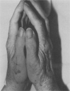
Figure 11-10 The prayer sign. The individual is unable to press the palms flat against each other, a diagnostic sign for the syndrome of limited joint mobility in diabetic persons. Other conditions also may result in loss of extension with a positive prayer sign. (From Kaye T: Watching for and managing musculoskeletal problems in diabetes, J Musculoskel Med 11:25-37, 1994.)
The stiff hand syndrome often is confused with or included in SLJM, but it has a distinct pathogenesis and clinical presentation. The stiff hand syndrome occurs uniquely with diabetes and is seen more frequently with type 1 DM and poor blood glucose control. Paresthesias, which eventually become painful, are accompanied by subcutaneous tissue changes such as stiffness and hardness. Vascular insufficiency may be the underlying cause or may be secondary to neuropathy, nodular tenosynovitis, and osteoarthritis.
Dupuytren’s contracture is characterized by the formation of a flexion contracture, palmar nodules, and thickening band or cord of palmar fascia (Fig. 11-11), usually involving the third and fourth digits in the population with diabetes (rather than the fourth and fifth digits in the population without diabetes). Pain and decreased range of motion are the primary presentation. Painless nodules develop in the distal palmar crease, often in line with the ring finger, which slowly mature into a longitudinal cord that is readily distinguishable from a tendon. The skin overlying the nodules is usually puckered.

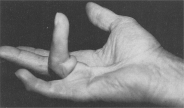
Figure 11-11 Dupuytren’s contracture. Painless nodules develop in the distal palmar crease, often in line with the ring finger, that slowly mature into a longitudinal cord that is readily distinguishable from a tendon. The skin overlying the nodules is usually puckered. The contracture may be symptomatic (painful), but with or without pain it results in impaired hand function. (From Kaye T: Watching for and managing musculoskeletal problems in diabetes, J Musculoskel Med 11:25-37, 1994.)
In some cases, regression of symptoms does occur without intervention, although the underlying mechanism for this phenomenon remains unknown. Surgical excision has not been shown to be a reliable cure for the disease and is not recommended unless there is a contracture that is bothersome. It has been reported that if the disease recurs after surgical excision, the rate of progression may be faster.204
Flexor tenosynovitis (also called chronic stenosing tenosynovitis) is another rheumatologic condition seen more commonly in persons with diabetes. Tenosynovitis is caused by accumulation of fibrous tissue in the tendon sheath and can cause aching, nodularity along the flexor tendons, and contracture. Locking of the digit, called trigger finger, can occur in flexion or extension and may be associated with crepitus or pain. In the population with diabetes, tenosynovitis is found predominantly in women and affects the thumb, middle, and ring fingers most often.
Diabetes is the systemic disease most often seen in connection with peripheral neuropathy of the hand, including CTS. The clinical presentation of CTS is the same for the person with diabetes as for the person without diabetes, although in diabetes CTS can be either a neuropathic process or an entrapment problem. Both neuropathy and compression within the carpal tunnel may exist together.
Adhesive capsulitis (also known as periarthritis or frozen shoulder) is characterized by diffuse shoulder pain and loss of motion in all directions, often with a positive painful arc test and limited joint accessory motions. The pattern is slightly different from that of typical adhesive capsulitis, in which regional tightness in the anteroinferior joint capsule primarily compromises external rotation, followed by loss of abduction and less often, internal rotation and flexion.
The pattern in diabetes is one of significant global tightness with external and internal rotation equally limited in the dominant shoulder, followed by limitations in abduction and hyperextension. External rotation and hyperextension are most limited in the nondominant shoulder, followed by internal rotation and abduction. The pathogenesis of the capsular thickening and adherence to the humeral head remains unknown. The long head of the biceps tendon may become glued down in its tendon sheath on the anterior humeral head.239
Adhesive capsulitis may be accompanied by vasomotor instability of the hand previously referred to as reflex sympathetic dystrophy (RSD) but now classified as the complex regional pain syndrome (CRPS). This condition is characterized by severe pain, swelling, and trophic skin changes of the hand (e.g., thinning and shininess of the skin with loss of wrinkling, sometimes with increased hair growth).
Skin changes in diabetic hand arthropathy, in addition to changes caused by CRPS, may occur in association with adhesive capsulitis. Other skin changes associated with diabetes include scleroderma diabeticorum, an asymptomatic thickening of the skin that may lead to a peau d’orange appearance, which usually involves the posterior neck, upper back, and shoulders.27
Skin and subcutaneous tissue atrophy and tendon flexion contractures develop. The natural history of this condition ranges from spontaneous remission to perma- nent loss of function. (See the section on Complex Regional Pain Syndrome in Chapter 39.)
Tendinopathy with thickening of the plantar fascia and Achilles tendon and tendo-Achilles tightening occurs as glucose deposits in tendons and ligaments result in loss of flexibility and rigid foot. In the diabetic population, loss of Achilles tendon flexibility, especially when combined with a flatfoot, increases pressure under the foot, adding to the compressive forces that contribute to ulcer formation.82
SPINE.: Diffuse idiopathic skeletal hyperostosis (DISH; also known as ankylosing hyperostosis or Forestier disease) is a condition of the spine seen most often in people with type 2 DM, although it can occur in a person who does not have diabetes. In DISH, osteophytes develop into bony spurs, typically right-sided syndesmophytes that may join to form bridges (Fig. 11-12). The thoracic spine most commonly is involved. In contrast to ankylosing spondylitis, the sacroiliac joints are spared, and vertebral body osteoporosis is absent. Calcaneal and olecranon spurs may develop, and new bone may form around hips, knees, and wrists.

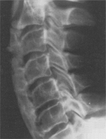
Figure 11-12 Diffuse idiopathic skeletal hyperostosis (DISH), or ankylosing hyperostosis, associated with type 2 diabetes mellitus (DM). DISH can occur with other conditions such as ankylosing spondylitis. Although the dense anterior bony bridging of the cervical vertebrae is pictured on this lateral roentgenogram, the thoracic spine most commonly is involved in diabetes. This type of DISH can be distinguished from ankylosing spondylitis by the preservation of sacroiliac joints, a site of typical involvement in ankylosing spondylitis. (From Kaye T: Watching for and managing musculoskeletal problems in diabetes, J Musculoskel Med 11:25-37, 1994.)
People with DISH may be asymptomatic or they may experience back pain and stiffness without limitations in range of motion. Dysphagia may develop if extensive cervical spine involvement occurs. The pathogenesis of DISH is unknown, and apparently no correlation exists between the degree of diabetic control and the extent of hyperostosis.
OSTEOPOROSIS.: Generalized osteoporosis usually develops within the first 5 years after the onset of DM and is more severe in persons with type 1 DM. It is hypothesized that bone matrix formation may be inadequate in the absence of normal circulating insulin levels. Results of bone density studies in persons with type 2 DM are conflicting, with some studies demonstrating decreased bone density and others indicating increased bone density. People with type 2 DM have decreased circulating insulin levels because of beta-cell exhaustion, and others are hyperinsulinemic because of insulin resistance.
As in any case of osteoporosis, regardless of the underlying cause, this condition places the person at greater risk for fractures. With the additional loss of sensation associated with diabetes, minor trauma easily produces injury. Microfractures can occur in already weakened bone and cartilage and may remain unrecognized because of the lack of pain appreciation. A vicious circle is started, leading to further damage.
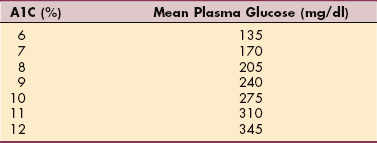
Sensory, Motor, and Autonomic Neuropathy.: Sensory, motor, and autonomic neuropathy associated with DM is a common phenomenon with known risk factors (e.g., duration of diabetes, current glycated hemoglobin value [HbA1c, also written as HbA1c], BMI, smoking, hypertension, and high triglycerides). The presence of CVD doubles the risk of neuropathy.242
Neuropathy may affect the CNS, peripheral nervous system, or autonomic nervous system (see Box 39-5). The most common form of diabetic neuropathy is a sensory polyneuropathy, usually affecting the hands and feet and causing symptoms that range from mild tingling, burning, numbness, or pain to a complete loss of sensation (usually feet) and foot drop. See further discussion of diabetic neuropathy in Chapter 39.
SENSORY NEUROPATHY.: Many people with diabetes suffer from diabetic peripheral neuropathic pain (DPNP) associated with nerve damage. Spontaneous pain, allodynia (painful response to benign stimuli), hyperalgesia, and other unpleasant symptoms are common with DPNP. Neuropathic pain often progressively increases in intensity throughout the day and is worse at night, significantly impairing sleep. Some individuals experience painful neuropathy called insulin neuritis syndrome at the beginning of therapy for diabetes; the feet are affected more often than the hands and it is usually self-limiting.256
The loss of sensation in diabetic neuropathy predisposes joints to repeated trauma and progressive joint destruction. Chronic progressive degeneration of the stress-bearing portion of a joint associated with loss of proprioceptive sensation in the joint produces a condition called Charcot’s disease, Charcot’s arthropathy, neuroarthropathy, or neuropathic arthropathy. Diabetes is the most common cause of neuropathic joints.
Several stages of neuropathic arthropathy (Charcot’s foot) occur involving bone destruction and absorption leading to dislocation and an unstable joint. Bone fragments and debris are deposited in the affected joint. Subluxation of the tarsal and metatarsal joints commonly results in a rocker-bottom foot deformity (see Fig. 23-9) and a redistribution of pressure on the plantar surface of the foot with progressive ulceration. An acute neuropathic joint is swollen, warm, and edematous, but pain may be minimal because of the underlying altered sensation.
Left untreated, neuropathic changes can progress to complete destruction of the joint. The presence of autonomic neuropathy may hasten this process as the blood vessels are unable to respond appropriately (e.g., vasoconstrict) to even minor trauma. Prolonged and unregulated hyperemia in the foot may lead to excessive bone resorption resulting in decreased bone mineral density, further increasing the risk of bone and joint destruction.100
Joints with less movement transmit abnormal forces through the foot to injure already damaged joints. This is especially true during walking, when large forces are placed on the midtarsal and tarsometatarsal joints. Obesity further increases these forces, and in the presence of any preexisting gait abnormalities or deformities, both create additional stress that compounds the condition.
Assessment of the underlying problem is important in planning the appropriate treatment intervention. For example, improving circulation may be a goal with macrovascular or peripheral vascular disease, whereas foot care and orthoses are more appropriate treatments for microvascular-caused neuropathy. The underlying neurologic disorder should be treated but this has no effect on the existing arthropathy. Reduction of weight bearing, joint immobilization, and joint protection are important conservative treatment tools. Surgical fusion can be performed if all else fails, but joint replacement is contraindicated in this condition.36,212
MOTOR NEUROPATHY.: Motor neuropathy is more common with long-standing disease and produces weakness and atrophy; bilateral but asymmetric proximal muscle weakness is called diabetic amyotrophy. Diabetic amyotrophy leads to bony deformities (e.g., claw toes, severe flatfoot with valgus of the midfoot, or collapse of the longitudinal arch) that contribute to biomechanical changes in foot function resulting in abnormal patterns of loading. Pain and erythema of the forefoot may constitute forefoot osteolysis, which is sometimes considered another form of neuropathy distinguished from cellulitis or osteomyelitis by laboratory values (leukocyte count) and roentgenographic appearance.
AUTONOMIC NEUROPATHY.: Autonomic neuropathy is sometimes referred to as diabetic autonomic neuropathy (DAN) and affects nerves that innervate heart, lung, stomach, intestines, bladder, and reproductive organs. It may manifest itself through the loss of control of blood pressure, blood glucose levels, temperature, regulation of sweating (skin becomes dry and cracked with buildup of callus), and blood flow in the limbs. Skin changes such as these can create more openings for bacteria to enter. The combination of all three types of neuropathy can ultimately lead to gangrene and possible amputation, largely preventable with proper care (see Special Implications for the Therapist: Diabetes and Foot Care in this section).
Cardiovascular autonomic neuropathy (CAN) is manifested by the lack of heart rate variability in response to deep breathing and exercise, exercise intolerance, persistent sinus tachycardia, bradycardia, and postural hypotension. Stress testing should be considered before starting an exercise program, especially in the older adult.8 CAN may also result in reduced perception of ischemic pain, making a person with diabetes unaware of having a heart attack. This may delay appropriate medical treatment and lead to death.255
DAN may lead to hypoglycemia without awareness because of loss of the warning signs of hypoglycemia such as sweating and palpitations. Being unaware of hypoglycemia and unresponsive to it are troublesome metabolic complications because they impair the person’s ability to manage the disease and may result in death. Other forms of autonomic neuropathy include gastroparesis (decreased gastrointestinal motility accompanied by diarrhea and fecal incontinence), constipation, urinary tract infections (nerve damage can prevent the bladder from emptying completely, allowing bacteria to grow in the bladder and kidneys), urinary incontinence, and sexual (erectile) dysfunction.
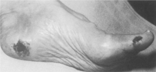
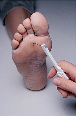
ULCERATION.: Sensory neuropathy, occurring as a result of improper glucose metabolism and diminished vascular perfusion to nerve tissues, places the diabetic person at risk for the development of ulcers. Diabetic foot ulcers are caused primarily by repetitive stress on the insensitive skin with increased pressure and/or horizontal (shear) stress. Body weight and activity level increase the force that the foot must transmit, and this also may increase pressure and shear force, especially in the presence of an underlying bony prominence or foot imbalance. In addition, previously healed ulcers leave scars that transmit force to underlying tissues in a more concentrated manner and hold the fat pad locally so that it cannot function physiologically. As a result, it cannot transmit shear forces, and it becomes damaged easily.
The loss of autonomic nerve function eliminates the production of sweat, leaving the skin dry and inelastic. Changes in pressure and gait, fat atrophy, and muscle weakness are mechanical factors that, along with sensory neuropathy, influence the development of plantar skin abnormalities, especially ulceration.21,229 Diabetes-induced changes in the skin are likely to contribute to ulceration because the collagen and keratin (a protein that is the principal constituent of epidermis, hair, and nails) may be glycosylated (saturated with glucose) with increased cross-linking, which makes the skin stiff. Keratin builds up in response to the increased pressure, covering the openings of unhealed ulcers, and cannot be removed as readily as normal keratin.
The areas most commonly affected by foot ulcers are the plantar areas of the metatarsal heads, the toes, and the plantar area of the hallux (Fig. 11-13). In the Charcot foot, the incidence of ulceration beneath the talus and navicular bones becomes more common because of the rigid rocker-bottom deformity.
Prevention of obesity-related health problems, including type 2 diabetes, is a key focus of the medical community. Therapists play an important role in providing education on the beneficial effects of exercise combined with proper nutrition. Studies have clearly shown that people who incorporate physical activity and exercise into their daily lives are less likely to develop type 2 diabetes no matter what their initial weight. Adopting an activity program of 150 minutes weekly of moderate intensity activity (e.g., brisk walking) similar to what the Surgeon General advises is a key prevention strategy.133-135
Studies using liposuction in the overall treatment of obesity point to the possibility of this treatment option to disrupt the pathway that brings about insulin insensitivity in the obese individual and thus prevent diabetes from developing. Fat removal by liposuction has been linked with modification of cardiovascular risk and vascular inflammatory markers in the obese individual with beneficial effects on insulin resistance as well.67,188
In response to the statistic that one-fourth of all new cases of DM under age 20 are diagnosed as type 2 DM, the Centers for Disease Control and Prevention (CDC) now recommends diabetes testing begin at age 25 years. The American Diabetes Association recommends universal screening at age 45 (earlier for high-risk groups such as non-Caucasians, obese individuals, and those with a family history of type 2 DM in a first-degree relative).
Diagnostic assessment may include a variety of testing procedures, such as plasma glucose, glucose tolerance test, (see Table 11-13), and urine ketone levels, to name just a few (see Table 40-4). A diagnosis of diabetes is confirmed by symptoms of hyperglycemia and blood and urine glucose and ketone abnormalities. Current defined criteria for definitive diagnosis of diabetes mellitus are the following41:
Classic symptoms of diabetes (polyuria, polydipsia, and unexplained weight loss) plus a casual plasma glucose concentration of ≥200 mg/dl. (Casual is defined as any time of day without regard to time since last meal.)
Fasting plasma glucose (FPG) ≥100 mg/dl after no caloric intake for at least 8 hours. (If the FPG is repeated and continues to be 100 mg/dl on a subsequent day, a GTT does not need to be done to confirm diagnosis.)
Glucose monitoring is not as crucial for a person who has an established pattern of activities and/or exercise. When a new activity is introduced, such as occurs in an exercise or rehabilitation program, monitoring blood glucose levels is recommended until the individual’s response to the change is known and predictable in maintaining stable blood glucose levels. New insulins and easier blood glucose monitoring have improved the ability to obtain much tighter control of blood glucose levels with fewer fluctuations and reduced risk of hypoglycemia. There are several methods used to monitor glucose immediately and over time.
Frequent self-monitoring by performing a direct blood sampling (fingerstick or laser technique) provides immediate monitoring of blood glucose levels and is an important management tool in the long-term treatment of this disease. Early screening and assessment of people at risk for diabetes are critical so that prevention and treatment of complications can be initiated before the onset of significant blood vessel and tissue damage.
The development of noninvasive testing methods to monitor glucose levels without the use of fingersticks is underway. One device already commercially available (the GlucoWatch G2 Biographer [GW2B]) is worn like a watch and uses electrical currents to obtain interval measurements of glucose levels in the skin. This type of reverse iontophoresis monitoring device prevents the invasive and often painful skin punctures by needle or laser, now in use for blood glucose monitoring.56,175,223 Use of the GW2B has not been shown to improve glycemic control or reduce the frequency of severe hypoglycemia. Skin reactions and other problems may lead to decreasing use over time.38 At least one study advised against relying on the GW2B during moderate to intense exercise.179
A handheld, infrared meter with voice activation that gives verbal directions and a verbal read out for the visually impaired is also under investigation. Other technology in use or being actively researched includes nocturnal alarms using a real-time glucose sensor to alert parents and children of hypoglycemic and hyperglycemic events while sleeping or for adolescents and adults, glucose-sensing skin patches, tattoos, or contact lenses. The contact lens would allow the individual with diabetes to see changes in the color of the contact lens to give an indication of blood glucose levels.13
Glycated (glycosylated) HbA1c level is used to monitor blood glucose control over time (Table 11-14). A1C is not used to diagnose diabetes. It is taken every 3 months and provides an average of the person’s overall glucose control. The American Diabetes Association recommends a target A1c level as 7% or less. According to the U.K. Prospective Diabetes Study, a 1% reduction of the A1c level reduces the risk of heart attack by 14% or more. People with HbA1c concentrations less than 5% had the lowest rates of CVD and mortality.121
Table 11-14
Correlating A1C to Mean Plasma Glucose Levels.

Normal reference range for A1C is 4%-6%. The goal for clients with diabetes is below 7% (target level is 6.5); higher levels are linked with greater risk of diabetes-related complications. A1C level of 7 correlates to an average daily plasma glucose level below 170 mg/dl. It is not used to diagnose diabetes and should not be measured too often in those who are using it to measure glucose control. Two measurements a year are sufficient in anyone who is meeting goals of treatment and who has stable control, and a maximum of 4-6 measurements a year is sufficient in people whose treatment has changed, or who are not meeting treatment goals.205 Data from American Diabetes Association, 2006.
Another monitoring tool used by individuals with diabetes includes fructosamine testing. Although the A1C is more popular and more widely accepted, the American Diabetes Association recognizes the fructosamine test as helpful in situations where A1C cannot be measured reliably. Situations in which fructosamine may be a better monitoring choice than A1c include the following4a:
• Rapid changes in diabetes treatment—fructosamine allows the effectiveness of diet or medication adjustments to be evaluated after a couple of weeks rather than months.
• Diabetic pregnancy—good control is essential during pregnancy and the needs of the mother frequently change during gestation; fructosamine measurements may be ordered with glucose levels to help monitor and accommodate shifting glucose and insulin requirements.
• Red blood cell (RBC) loss or abnormalities—an A1C test will not be accurate when an individual has a condition, such as hemolytic anemia or blood loss, that affects the average age of RBCs present. The presence of some hemoglobin variants may affect certain methods for measuring HbA1c. In these cases, fructosamine can be used to monitor glucose control.
• Individuals on dialysis when an average glucose level over the past 2 to 3 weeks is needed.
The fructosamine test is not useful as a screen for diabetes, since the fructosamine concentrations of well-controlled diabetics may overlap with those of individuals who do not have diabetes.
A standard reference range is not available for the fructosamine test. Because reference values are dependent on many factors, including age, gender, sample population, and test method, numeric test results have different meanings in different laboratories. The laboratory report will include the specific reference range for the test and the client’s results in comparison.
An elevated fructosamine level is an indication of higher than average blood glucose level. Like the A1C, the value of this test is in looking at trends over time but over a shorter period of time compared to A1C. Fructosamine results are very useful when monitoring change in glucose levels associated with alterations in diet or medications.
There is no widely available cure for diabetes. The goal of overall care for persons with diabetes is control or regulation of blood glucose. Many large-scale studies have shown that tight glucose control reduces the risk of vascular complications in both type 1 and type 2 diabetes. Early identification and intervention are strongly linked with risk reduction of late complications.141 Three key standards and goals in the treatment and self-management of DM include the following:
• Blood pressure less than 130/80 mm Hg
• Total cholesterol less than 200 mg/dl (low density lipoprotein [LDL] less than 100 mg/dl)
Data from the National Center of Health Statistics show that only 7.3% of adults with diabetes have achieved all 3 targets.213 To help people with diabetes reach these goals, the National Diabetes Education Program has started an education program called Control the ABCs, in which A is HbA1c, B is blood pressure, and C is cholesterol). Education materials are available in English and Spanish and for Asian Americans and Pacific Islanders.176 Therapists can help reinforce these concepts as part of their client education programs.
Data suggest that atherogenic and inflammatory mediators contributing to microvascular and macrovascular complications are elevated even before the onset of diabetes. There may even be a “metabolic memory” associated with these early changes. Comprehensive metabolic control instituted early may alter the natural history of diabetic complications by affecting this metabolic memory.141
Researchers continue to investigate drugs that would prevent the formation of fat cells, thereby reducing the problem of obesity before type 2 DM can develop. Studies of the use of gene therapy as a treatment for both types of diabetes are ongoing, utilizing a variety of approaches, such as direct delivery of the insulin gene to non–beta-cells, improving insulin secretion from existing beta-cells, and implanting genetically modified cells.83,84,116 Experimental research is underway in the development of a vaccine for type 1 DM that may help stop the immune system attack of the insulin-producing beta-cells of the pancreas.258
Type 1 Diabetes Mellitus.: Type 1 DM requires exogenous insulin administration and dietary management to achieve tight (near normal) blood glucose control. With no circulating endogenous insulin, the effect of aerobic exercise in providing increased glycemic control for the person with type 1 DM may be limited. To date, studies of the effect of aerobic exercise in type 1 DM have shown mixed results. Regardless, exercise should be taken into account as part of the total picture in order to minimize the complications associated with diabetes.
The insulin dosage schedule varies depending on the individual’s age, level of compliance, and severity of diabetes (Table 11-15). Control over blood glucose levels dictates how “brittle” the diabetes is. Brittle diabetes (also known as labile or unstable diabetes) is a term used when a person’s blood glucose level often swings quickly from high to low and from low to high. The individual with wide glucose excursions is considered very brittle.
Poorly controlled diabetes is ideally treated with more frequent administration of insulin (e.g., four times per day), whereas other individuals may receive insulin once or twice daily, sometimes mixing different types of insulin (e.g., rapid-acting [human analog; Humalog]; short-acting [regular] with intermediate-acting [NPH] insulin). Humalog (Lispro) is a type of insulin that has rapid action. It works faster than short-acting insulin and must be taken with a meal to prevent hypoglycemia.115 From a therapist’s point of view, the client receiving more frequent dosages is less likely to develop hypoglycemia, especially when beginning an exercise program.
Insulin Pump.: An insulin pump also known as continuous subcutaneous insulin infusion (CSII) is now available to deliver fixed amounts of regular insulin continuously, thereby more closely imitating the release of the hormone by the islet cells. This lightweight, pager-sized device is worn conveniently in a pocket or on a belt clip (Fig. 11-14); a waterproof design makes swimming possible.

Figure 11-14 The programmable insulin pump. Compact and worn like a pager, the programmable insulin pump delivers fixed amounts of insulin continuously, based on blood glucose levels determined by regular fingerstick glucose monitoring. The device includes the pump itself (including controls, processing module, and batteries), a disposable reservoir for insulin (inside the pump), and a disposable infusion set, including a cannula for subcutaneous insertion (under the skin) and a tubing system to interface the insulin reservoir to the cannula. (Courtesy Mini-Med, Sylmar, CA, 2000.)
The insulin pump offers many advantages such as flexible eating and exercising schedules, fewer episodes of severe hypoglycemia (especially at night and for teenagers who sleep longer hours), and convenience of taking insulin without the social consequences of public injections, to name a few.43 Although this type of insulin administration provides better control, it has some disadvantages. It cannot detect and respond to changes in the blood glucose level so the individual must continue to monitor glucose levels and make dosage adjustments. There is no long-term backup supply of insulin such as is available with long-acting insulin injections. If the pump malfunctions and the person is unaware of it, blood glucose levels can rise quickly, potentially leading to ketoacidosis. The pump wearer must still monitor blood glucose levels at regular intervals.
It cannot be removed for more than 1 hour, reactions to the needle are common, bleeding can occur at the sensor insertion site, and like any other mechanical device, it is subject to malfunction. Insulin pump technology is improving every year; new “smart” features are added to the designs to simplify the tasks involved in delivering an insulin bolus. Implantable pump options that can dispense insulin in constant, steady pulses throughout the day are being tested. This type of pump would eliminate the need for an open needle site in the skin. Penlike injection cartridges also are in use.126
Inhaled Insulin.: In 2006, the Food and Drug Administration (FDA) approved the first inhaled insulin combination product, Exubera, an inhaled powder form of recombinant human insulin (rDNA), for the treatment of adults with type 1 and type 2 diabetes. The inhaled form of insulin offers some people with diabetes an alternative to the many daily injections. It is a short-acting insulin and does not eliminate insulin injections for type 1 diabetes, but it can reduce the daily number required.
The onset of action for inhaled insulin is 10 to 20 minutes, which is more rapid than that of either regular or lispro subcutaneous insulin, so the individual is instructed to take it no more than 10 minutes before meals to prevent hypoglycemia. Inhaled insulin reaches peak effectiveness within 2 hours and remains effective for up to 6 hours.268
Its use is contraindicated in cigarette smokers or anyone who has smoked in the past 6 months, since it is inhaled directly into the lungs rather than into the blood stream. Smokers absorb much more insulin this way than nonsmokers do, leaving them at risk of dangerously low glucose levels.241
Inhaled insulin is not recommended for people with pulmonary disorders such as asthma, bronchitis, or emphysema. Potential disadvantages include erratic absorption, poor postprandial glucose control, and side effects such as cough, dyspnea, sinusitis, and pharyngitis.139 Long-term effects remain unknown but are under investigation.
Research continues to find other forms of a pain-free insulin delivery system. Insulin pills, an insulin patch, and an oral (mouth) spray are under investigation.
Type 2 Diabetes Mellitus.: Type 2 DM is most often treated with diet and exercise, sometimes in conjunction with oral hypoglycemic drugs (OHDs); insulin occasionally is required. Exercise is a recognized therapy for the prevention of complications in type 2 DM. Numerous studies have shown a consistent positive effect of regular exercise training on carbohydrate metabolism and insulin sensitivity. Some of the beneficial effects include decreased need for insulin, prevention of CVD and obesity, management of hypertension, and reduction in very LDL (VLDL) cholesterol.41,161
A plant-based diet is becoming more widely known for its potential effects and benefits in the prevention and treatment of type 2 DM. The use of whole-grain or traditionally processed cereals and legumes has been associated with improved glycemic control in individuals with diabetes and in individuals who are insulin-resistant. Long-term studies have shown that whole-grain consumption reduces the risk of both type 2 diabetes and CVD.114
The combination of diet and exercise is more powerful than either one alone and may be even more effective than drugs for preventing type 2 DM. A low-fat, low-calorie diet with moderate exercise (30 minutes 5 times a week) has been shown to reduce new diabetes cases by 58% over a 3-year period. By contrast, the drug metformin, which boosts insulin sensitivity, reduced new cases by only 31%.127 The National Institutes of Health (NIH) is conducting a 12-year, 5000 patient study to further test the additive effects of diet and exercise on diabetes.
Several types of OHDs are available, including the following:
1. OHDs used to stimulate islet cells to increase endogenous insulin secretion and enhance insulin-receptor binding (sulfonylureas).
2. OHDs that act by slowing the digestion of sugars in the intestine (acarbose).
3. OHDs referred to as insulin-resistance reducers or “insulin sensitizers” (thiazolidinediones such as pioglitazone [Actos] and rosiglitazone [Avandia]). Insulin sensitizers improve the action of liver, muscles, and fat tissues and do not cause liver problems as seen with troglitazone (Rezulin), which was removed from the market in March 2000.
4. OHDs used to improve hepatic and peripheral tissue sensitivity to insulin (metformin [Glucophage]), thereby increasing the effectiveness of insulin found in the body.
The advantage of this last OHD is that it does not stimulate activity with concomitant weight gain. However, diarrhea develops for 2 to 3 weeks in approximately one-third of people using this drug. One potentially serious but rare side effect is lactic acidosis, a life-threatening buildup of lactic acid in the blood. This condition can be fatal in people with kidney or liver disease or alcoholism. The main clinical feature of lactic acidosis is hyperventilation.
Treatment of Long-Term Complications.: Prevention of long-term complications is the goal for all clients with DM. Risk of complications is associated independently and additively with hyperglycemia and hypertension. Intensive treatment of both these risk factors is required to prevent and minimize the incidence of most complications.237
Medical treatment of long-term diabetic complications may include dialysis or kidney transplantation for renal failure and vascular surgery for large vessel disease. Currently, the American Diabetes Association advises that people with diabetes take low-dose aspirin (75 to 162 mg) daily to help minimize risks such as heart attacks and strokes. Prophylactic aspirin therapy is recommended in both men and women with diabetes who are 40 years and older, although some diabetologists suggest that aspirin prophylaxis should begin at age 30.41
New treatment guidelines from the American College of Physicians recommend the use of statins (cholesterol-lowering drugs such as Crestor, Lipitor, Zocor, Mevacor, or Pravachol) for anyone with diabetes and diagnosed CAD—even if their cholesterol levels are normal.
Review of available data shows that statins reduced heart attacks and strokes by 22% to 44% in people with diabetes.231 People with diabetes and normal cholesterol who have other cardiac risk factors, such as hypertension, obesity, smoking, age over 55, and chronic physical inactivity, may benefit from a nonstatin cholesterol medication such as gemfibrozil (Lopid).
Diabetic Ulcers.: The therapist often is involved in prevention and wound care for diabetic ulcers, which may help prevent amputation. Early recognition and prompt management of wounds, ulceration, and Charcot foot can facilitate healing. For example, a CDC study showed that people with diabetes who wore proper shoe protection had only a 20% recurrence rate of ulceration compared with an 80% rate for those without offloading.232
Offloading or pressure reduction is a key component for healing ulcers and preventing recurrence. The normal response to damaged areas is to spare them from pressure because they are painful. However, in the insensitive foot of a person with diabetes, this normal alteration of weight-bearing surface, pressure, and duration does not take place, resulting in repetitive stress and injury with subcutaneous and cutaneous necrosis and skin breakdown.
A marked improvement in the rate of healing for plantar ulcers has been reported using a combination of total-contact cast (TCC) and tendo-Achilles lengthening (TAL; percutaneous heel cord lengthening), as opposed to TCC alone.10,143,172,211
The results of at least one study show TAL should not be done in anyone with complete anesthesia of the heel pad; increased dorsiflexion can increase the risk of heel ulceration. This procedure is advised only in a multidisciplinary setting able to provide adequate nutrition, wound care, surveillance, treatment of complications and other biomechanical abnormalities and intervene early in any developing ulcerations.106
Other interventions include debridement, infection control, protective dressings, revascularization, proper nutrition, and client education. Active dressings, such as growth factors and living skin, are also in use. Topical application of growth factors on wounds without infection and with at least a minimal level of vascularization was introduced in the early 1990s and has progressed to include new techniques in skin transplantation (see the section on Skin Transplantation in Chapter 21).
Infrared light therapy, such as monochromatic near-infrared photo energy (MIRE), has been applied using the anodyne therapy system (ATS) to improve sensory impairment, reduce pain, and prevent and heal ulcers. When used in conjunction with other physical therapy interventions, MIRE has been shown to improve balance in clients with diabetic peripheral neuropathy. ATS has been shown to be effective in reversing the loss of protective sensation by improving circulation. Light absorbed by hemoglobin in the blood causes the release of nitric oxide, resulting in vasodilation and improved collateral circulation. Long-term studies are still needed to show whether the results can be sustained.128,129,140,198
The use of cool laser therapy as a revascularization therapy is now available. Cool laser revascularization for peripheral artery therapy (CliRpath) uses a cool excimer laser and catheter system to vaporize arterial blockages, restoring blood flow and promoting wound healing. Reduction in pain, improved circulation, and facilitation of wound healing may help prevent limb loss in this population.
Treatment of diabetic peripheral neuropathic pain (DPNP) has not been successful using any one single intervention technique. The ideal treatment is correcting the underlying condition of chronic hyperglycemia. Many methods have been employed (e.g., capsaicin topical cream, acupuncture, and electrical stimulation) to address the painful symptoms of DPNP with limited and variable results. Medications aimed at chronic neuropathic pain have included tricyclic antidepressants (e.g., amitriptyline, nortriptyline, or imipramine), but anticholinergic effects, such as dry mouth, blurred vision, constipation, cardiac arrhythmias, and orthostatic hypotension, often limit their use.
More recently, off-label use of anticonvulsants, such as gabapentin (Neurontin) and pregabalin (Lyrica), have met with greater success. Serotonin-norepinephrine reuptake inhibitors, such as duloxetine (Cymbalta), can be used by some individuals to treat painful DPNP. By inhibiting the reuptake of these neurotransmitters, descending inhibitory pathways in the spinal cord are activated and block ascending pain signals to the brain.254
Transplantation.: Research is being conducted on the use of transplanted pancreatic islet cells rather than the entire pancreas. The transplant recipient receives one or more infusions of pancreatic islet cells that include insulin-producing beta-cells. Almost 500 people with type 1 diabetes have received islet transplants at 43 institutions worldwide in the last 5 years.222
High rates of insulin independence have been reported at 1 year in the leading islet transplant centers. Loss of insulin independence by 5 years occurs in the majority of recipients. Life-long immunosuppression and its complications limit this treatment to candidates who have the most severe, unstable glycemic control despite optimal insulin therapy.222
Stem cell research may find a way for people to use their own stem cells to develop them into islet cells and allow infusions without cell-rejection complications and the need for life-long immunosuppression. In addition, artificial pancreata provide hope for future treatment without repeated injections for the person with type 1 diabetes. An artificial pancreas contains a reservoir for insulin (which must be filled by the affected individual, typically through a tube in the abdomen), and an internal glucose monitor that continuously determines the plasma glucose level, automatically releasing the appropriate amount of insulin. Such instruments are expected to reach the market shortly. The use of wireless communication between a continuous glucose sampling device and an insulin pump would give people with diabetes a closed-loop system or electromechanical artificial pancreas.63
Diabetes control depends on the proper interaction between the following three factors: (1) food, (2) insulin or oral medication to lower blood glucose, and (3) activity (e.g., sedentary or exertional) or exercise. When diabetes is regulated successfully, complications of hyperglycemia and hypoglycemia can be avoided with minimal disruption to a normal lifestyle. However, diabetes can be fatal even with medical treatment, or it can cause major permanent disabilities and seriously impair functional abilities. Studies have shown that type 2 DM raises a person’s risk of dying from heart disease by 2 to 3 times.234 In fact, about 50% of myocardial infarctions and 75% of strokes are attributable to diabetes. Diabetes is the leading cause of new blindness and is a contributory cause to renal failure and peripheral vascular disease.
Regardless of the modality of treatment used for the person with type 1 or type 2 DM, recent studies have shown clearly that tight glucose control (plasma glucose levels consistently within normal limits, approximately 100 mg/dl) delays onset and progression of diabetic complications. The only apparent danger in maintenance of tight control is the greater possibility of hypoglycemia, particularly in those people with type 1 DM who receive frequent exogenous insulin administration.41
Insulin resistance refers to the phenomenon of having high levels of both circulating insulin and glucose in the bloodstream, but the insulin molecules cannot bind properly to the insulin receptor sites on the surface of the cell to allow glucose to enter the cells and be used for energy. A syndrome of insulin resistance has been proposed to explain the frequent association of hypertension, carbohydrate intolerance, abdominal obesity, dyslipidemia, and accelerated atherosclerosis associated with type 2 DM.
Although a primary insufficiency of insulin secretion is the pathology in the development of type 2 DM, obesity is a major risk factor for the development of this type of DM, caused in part by the associated insulin resistance. In 1988 the combination of hypertension, glucose intolerance, hyperinsulinemia, and dyslipidemia was called syndrome X by Gerald Reaven, MD, a diabetes expert, who predicted an increased incidence of coronary heart disease.202
In the following years, syndrome X was redefined as a prediabetic state and called metabolic syndrome. Criteria for metabolic syndrome include abdominal obesity; elevated triglyceride levels, and low HDLs, hypertension, insulin resistance (with or without glucose intolerance), and proinflammatory markers such as increased C-reactive protein levels or coagulation factors (see Box 12-2).
Even this definition has been through numerous iterations and is still hotly debated. The National Cholesterol Education Program, the World Health Organization, and the American Heart Association all have slightly different versions of the criteria for metabolic syndrome. In contrast, the American Diabetes Association and the European Association for the Study of Diabetes published position statements concluding that there is no disorder called metabolic syndrome. They state that, “there is no solid evidence that any of the metabolic syndrome health factors contribute more together than they do individually. In other words, the whole is not greater than the sum of its parts.”117 The issue is still under debate; see Chapter 12 for further discussion of metabolic disorder.
Obesity and insulin resistance are the underlying factors responsible for the diagnosis of metabolic syndrome. Several definitions of metabolic syndrome have been proposed, but all include insulin resistance or glucose intolerance, hypertension, dyslipidemia, and central obesity. For this reason, the term insulin resistance syndrome (IRS) was suggested by the American College of Endocrinology and the American Association of Clinical Endocrinologists to more aptly describe the prediabetic state.64 While IRS has many of the same characteristics of metabolic syndrome, diagnosis is based on a fasting glucose level (100 mg/dl < IRS < 126 mg/dl).
Insulin resistance, a generalized metabolic disorder in which the body cannot use insulin efficiently, appears to play a key role in metabolic syndrome. Although not everyone with insulin resistance has metabolic syndrome, most people with metabolic syndrome are also resistant to the action of insulin.
Regardless of the definition or criteria, most agree that obesity is the single modifiable factor that sets off the cascade. The syndrome is associated with alterations in the abdominal fat cells. With increased fat storage, these cells become distorted in shape, and the receptor site for insulin becomes “warped” or out of proper alignment, so the insulin molecule “key” no longer fits in the receptor. Insulin resistance makes it more difficult to lose weight because the cells are not getting enough fuel and the individual perceives hunger when adequate amounts of circulating glucose exist.
The affected individual may develop elevated blood pressure and problems with reactive hypoglycemia. When the excess insulin is suddenly used, glucose rushes into the cells and the blood glucose drops suddenly. This sequence creates intense sweet cravings, and the cycle repeats itself with increasing insulin resistance.257
The most important implications of recent research indicate that a diagnosis with a syndrome is not necessary in order to treat the individual risk factors. At this prediabetic stage, changes in lifestyle will have the greatest impact on halting any disease progression. In fact, it may be the only time in the disease progression where changes in daily activity levels and nutritional status may have an impact.
Two primary life-threatening metabolic conditions, DKA and hyperosmolar hyperglycemic state (HHS), can develop if uncontrolled or untreated DM progresses to a state of severe hyperglycemia (greater than 300 mg/dl).124 Between DKA and HHS is a continuum of metabolic abnormalities.
Definition and Overview.: DKA is most commonly seen in type 1 diabetes when complications develop from severe insulin deficiency. About one-half of the people who require hospitalization for DKA develop this hyperglycemic emergency secondary to an acute infection or failure to follow their prescribed dietary or insulin therapy.259
Most episodes of DKA occur in persons with previously diagnosed type 1 DM. However, the condition may occur in new cases of type 1 and in persons with type 2 DM (under stressful conditions in the latter such as during a myocardial infarction). It is characterized by the triad of hyperglycemia, acidosis, and ketosis.230
Etiologic Factors.: Any condition that increases the insulin deficit in a person with diabetes can precipitate DKA. Causes of DKA commonly include taking too little insulin; omitting doses of insulin; failing to meet an increased need for insulin because of surgery, trauma, pregnancy, stress, puberty, or infection; and development of insulin resistance caused by insulin antibodies. Other precipitating causes are listed in Box 11-8.
The most common precipitating factor is infection, which occurs in up to half of all cases and may seem like a trivial condition such as mild cellulitis or upper respiratory tract infection. Omission of insulin, either because of noncompliance or because people mistakenly believe that insulin is not required on sick days when they are not eating well, is another important and preventable cause of DKA.
In young individuals with type 1 DM, psychologic problems complicated by eating disorders may be a contributing factor in 20% of recurrent ketoacidosis. Factors that may lead to insulin omission in younger people include fear of weight gain with improved metabolic control, fear of hypoglycemia, rebellion from authority, and stress of chronic disease. In approximately 15% to 30% of cases, no identifiable cause of DKA can be determined.230
Pathogenesis.: The initiating metabolic defect in DKA is an insufficient or absent level of circulating insulin. Insulin may be present, but not in a sufficient amount for the increase in glucose resulting from the stressor (see Box 11-8). Inadequate insulin creates a biologic state of starvation, which triggers the excess secretion of counterregulatory hormones, particularly glucagon, in an attempt to get more glucose to the cells and tissues. The abnormal insulin-to-glucagon ratio, with excess circulating cate- cholamine, cortisol, and GH levels, initiates a host of complex metabolic reactions, leading to hyperglycemia, acidosis, and ketosis.
When the body lacks insulin and cannot use carbohydrates for energy, it resorts to fats and proteins. The process of catabolizing fats for fuel gives rise to incomplete lipid metabolism, dehydration, metabolic acidosis, and electrolyte and acid-base imbalances. (See more complete discussion in the section on Pathogenesis of Diabetes Mellitus in this chapter.)
Clinical Manifestations.: The signs and symptoms of DKA vary, ranging from mild nausea to frank coma (Table 11-20). Common symptoms are thirst, polyuria, nausea, and weakness that have progressed over several days. This condition also may develop quickly, with symptoms progressing to coma over the course of only a few hours. Other symptoms may include dry mouth; hot, dry skin; fruity (acetone) odor to the breath, indicating the presence of ketones; overall weakness, possible paralysis; confusion, lethargy, or coma; and deep, rapid respirations (Kussmaul’s respirations). Fever is seldom present even though infection is common, primarily a result of peripheral vasodilation. Severe abdominal pain, possibly accompanied by nausea and vomiting, easily mimics an acute abdominal disorder.
Table 11-20
Clinical Symptoms of Life-Threatening Glycemic States

*Less gradual than HHS.
Modified from Goodman CC, Snyder TE: Differential diagnosis for physical therapists, ed 4, Philadelphia, 2007, WB Saunders.
DIAGNOSIS, TREATMENT, AND PROGNOSIS.
Prevention of DKA through client education is the key to avoiding this serious condition. Once DKA is suspected, the diagnosis must be established quickly, with immediate treatment after diagnostic confirmation (blood glucose level >250 mg/dl, pH <7.3, bicarbonate level <18 mEq/L, and serum ketones).
Treatment includes fluid administration, insulin therapy, and correction of metabolic abnormalities (potassium, bicarbonate, and phosphate), in addition to correction of any underlying illnesses (e.g., infection). Before the discovery of insulin in the 1920s, DKA was almost universally fatal. This complication is still potentially lethal with an average mortality rate between 5% and 10%.
Hyperosmolar Hyperglycemic State.: HHS is another acute complication of diabetes, a variation of DKA. HHS is characterized by extreme hyperglycemia (800 to 2000 mg/dl), mild or undetectable ketonuria, and the absence of acidosis. It is seen most commonly in older adults with type 2 DM.111,124
The precipitating factors of HHS may be similar to those for DKA such as infections, inadequate fluid intake, medications (see Box 11-8), or stress. HHS may be the first indication of undiagnosed diabetes, and it may occur in the case of someone who is receiving total parenteral nutrition (hyperalimentation) or who is on renal dialysis and receiving solutions containing large amounts of glucose.
The major difference between HHS and DKA is the lack of ketosis with HHS. Because some residual ability exists to secrete insulin in type 2 DM, the mobilization of fats for energy is avoided. When adequate insulin is lacking, blood becomes concentrated with glucose. Because glucose molecules are too large to pass into cells, osmosis of water occurs from the interstitial spaces and cells to dilute the glucose in the blood. Osmotic diuresis occurs, and eventually the cells become dehydrated. If not treated promptly, the severe dehydration leads to vascular collapse and death.
Clinical manifestations of HHS are polyphagia, polydipsia, polyuria, glucosuria, dehydration, weakness, changes in sensorium, coma, hypotension, and shock (see Table 11-20). Lactic acidosis also can develop if tissue perfusion is compromised.
Treatment is with short-acting insulin, electrolyte replacement, and careful fluid replacement to avoid congestive heart failure and intercerebral swelling in older adults, who often have other cardiovascular or renal disorders.
As noted earlier, the endocrine system works with the nervous system to regulate and integrate the body’s metabolic activities. Metabolism is the physical and chemical (physiologic) processes that allow cells to utilize food to continually rebuild body cells and transform food into energy. Metabolism is broken down into two phases: the anabolic (tissue-building) and catabolic (energy-producing) phases. The anabolic phase converts simple compounds derived from nutrients into substances the body cells can use, whereas the catabolic phase is a consumptive phase when these organized substances are reconverted into simple compounds with the release of energy necessary for the proper functioning of body cells.95
The body gets most of its energy by metabolizing carbohydrates, especially glucose. A complex interplay of hormonal and neural controls regulates the homeostasis of glucose metabolism. Hormone secretions of five endocrine glands dominate this regulatory function (see Table 11-10). The rate of metabolism can be increased by exercise, elevated body temperature (e.g., high fever or prolonged exertional exercise), hormonal activity (e.g., thyroxine, insulin, or epinephrine), and increased digestive action after the ingestion of food.
Fluid and electrolyte balance is a key component of cellular metabolism. Homeostasis, maintaining the body’s chemical and physical balance, involves the proper functioning of body fluids to preserve osmotic pressure, acid-base balance, and anion-cation balance. The goal of metabolism and homeostasis is to maintain the complex environment of body fluid that nourishes and supports every cell.
Body fluids, classified as intracellular and extracellular, contain two kinds of dissolved substances: those that dissociate (separate) in solution (electrolytes) and those that do not. For example, when dissolved in water, glucose does not break down into smaller particles but sodium chloride dissociates into sodium cations (positively charged) and chloride anions (negatively charged).
The composition of these electrolytes in body fluids is electrically balanced, so the positively charged cations (sodium, potassium, calcium, and magnesium) equal the negatively charged anions (chloride, bicarbonate, sulfate, phosphate, and carbonic acid). Although these particles are present in relatively low concentrations, any deviation from their normal levels can have profound physiologic effects.
Because many situations in the body cause both normal and abnormal fluid shifts, it is important to have a clear understanding of fluid compartments. The recognition of pathologic conditions, such as edema, dehydration, ketoacidosis, and various types of shock, can depend on the understanding of these concepts.
In the healthy body, fluids and electrolytes are constantly lost or exchanged between compartments. This balance must be maintained for the body to function properly. The amount used in these functions depends on such factors as humidity; body and environmental temperature; physical activity; metabolic rate; and fluid loss from the GI tract, skin, respiratory tract, and renal system. Normal balance is achieved through fluid intake and dietary consumption. Alterations in fluid and electrolyte balance are discussed more completely in Chapter 5.
The proper balance of acids and bases in the body is essential to life. The body maintains the pH of extracellular fluid (fluid found outside cells) between 7.35 and 7.45 through a complex chemical regulation of carbonic acid by the lungs and base bicarbonate by the kidneys. The pH is essentially a measure of hydrogen ion concentration in body fluid. Nutritional deficiency or excess, disease, injury, or metabolic disturbance may interfere with normal homeostatic mechanisms and cause a lowering of pH called acidosis or a rise in pH called alkalosis.
Various bodily functions operate to keep the pH at a relatively constant level. Acid-base regulatory mechanisms include chemical buffer systems, the respiratory system, and the renal system. These systems interact to maintain a normal acid-base ratio of 20: 1 bicarbonate to carbonic acid. The consequences of an acid-base metabolism disorder can result in many signs and symptoms encountered by the therapist. These conditions are discussed more completely in Chapter 5.
Aging as measured by loss of physiologic function has not yet been defined precisely, so the distinction between usual, normal, and ideal metabolic changes remains undetermined. Studies of the aging population have shown that several physiologic parameters, such as body weight, basal metabolism, renal clearance, and cardiovascular function, decline with age. Protein-calorie nutritional status has pervasive effects on metabolic regulatory systems; nutritional status often declines with age, which contributes to metabolic dysfunction.118
Because the respiratory and renal systems are largely responsible for maintaining acid-base balance, changes in these systems associated with aging also have an impact on metabolic function. A common measure for metabolic loss in tissues is the decline in VO2max, the maximum oxygen extraction capacity of the lungs.
Loss of muscle mass associated with aging can affect stroke volume capacity and oxidative metabolism.186 The low level metabolic acidosis that appears to occur in many people with advancing age may play a role in age-associated bone loss, a factor that has received little attention from those who study bone loss and aging.156
Oxidative stress has been implicated in the pathogenesis of a number of diseases and has been labeled the free radical theory of aging (discussed in Chapters 2 and 6); studies indicate that protection from the consequences of excess metabolic activity results in a slowing of the aging process, particularly in the postreproductive period of life.34,77 Links between oxidative stress and aging focus on mitochondria in a theory called the mitochondrial theory of aging. Mitochondria, the principal site of adenosine triphosphate (ATP) synthesis (also containing DNA and RNA), is the cellular site of energy production from oxygen and the principal site of free radical damage.51
Free radical derivatives of oxygen are generated as a result of normal metabolic activity, producing destructive oxidation of membranes, proteins, and DNA. These free radicals (unstable oxygen molecules robbed of electrons) attempt to replace their missing electrons by scavenging the body and taking electrons from healthy cells, causing a chain reaction called oxidation (see Fig. 6-2).
The formation of free radicals can be triggered by many exogenous (outside) factors such as cigarette smoke, air pollution, anticancer drugs, ultraviolet lights, pesticides and other chemicals, uncontrolled diabetes, radiation, and emotional stress. The major defenses against these destructive byproducts of normal metabolism are the protective enzymes, which remove the free radicals and remove, repair, and replace cell constituents.
Impairment of cellular function and metabolism occurs as proteins and DNA (which turn over slowly or not at all) are damaged over time.61 The use of antioxidants found naturally in fruits and vegetables or ingested as a nutritional supplement to counteract this process is believed to increase longevity but remains under scientific investigation.167,168
Clinical manifestations of metabolic disorders vary, depending on the specific pathology present. Fluid and electrolyte disorders, disorders of acid-base metabolism leading to metabolic (nonrespiratory) alkalosis or acidosis, and their associated signs and symptoms are discussed in Chapter 5.
Metabolic bone disease is discussed in Chapter 24, and disorders of purine and pyrimidine metabolism resulting in gout and pseudogout are discussed in Chapter 27.
Metabolic disorders involving the connective tissue may result in pathologic loss of bone mineral density, such as occurs in osteomalacia or osteoporosis, or acceleration of both deposition and resorption of bone, as seen in Paget’s disease. These disorders differ in pathogenesis and treatment and are discussed in Chapter 24.
Metabolic neuronal diseases are rare and are not likely seen in a therapy practice. Phenylketonuria (PKU), Wilson’s disease, and porphyrias are the three most often encountered and are briefly discussed in this section.
PKU is an autosomal recessive disease resulting from a genetic defect in the metabolism of the amino acid phenylalanine (Phe). This condition is transmitted recessively through apparently healthy parents, who show signs of the disease only on testing. The lack of an enzyme (phenylalanine hydroxylase) necessary for the conversion of the amino acid Phe into tyrosine results in an accumulation of Phe in the blood with excretion of phenylpyruvic acid in the urine. If untreated, the condition results in mental retardation and other manifestations such as tremors, poor muscular coordination, excessive perspiration, mousy odor (resulting from skin and urinary excretion of phenylpyruvic acid), and seizures.
Although PKU cannot be cured, a simple screening test for PKU can be administered to newborns and is required by law in most states in the United States and in all provinces in Canada. Currently, between 160 and 400 of the 4 million babies born in the United States each year are affected. The practice of discharging newborns in 24 hours is resulting in an increase in the number of babies at risk of PKU.
Treatment is primarily through Phe restriction of the infant’s diet to control the effects of PKU and is prescribed on an individual basis with the additional administration of a dietary protein substitute. The start of newborn screening for PKU during the early 1970s has given rise to an increasing number of people who have been identified and successfully treated for the disease in childhood. Initiation of nutritional therapy before conception for women assures a successful pregnancy outcome.123 A need remains for maternal screening before pregnancy to identify undiagnosed maternal PKU and subsequent prophylactic treatment to prevent maternal PKU syndrome.96
The prognosis for people with PKU has improved greatly with early institution of treatment after birth. However, hyperphenylalaninemia can cause white matter abnormalities, psychiatric illness, and decreased performance on neuropsychologic tests for people with PKU compared with subjects without PKU. It has been shown that the diet necessary to reduce Phe levels cannot be terminated after adolescence without elevation of plasma levels resulting in poor neuropsychologic performance.52
Wilson’s disease, also known as hepatolenticular degeneration, is a progressive disease inherited as an autosomal recessive trait (both parents must carry the abnormal gene). This condition produces a defect in the metabolism of copper, with accumulation of copper in the liver, brain, kidney, cornea, and other tissues. Although the pathogenesis of Wilson’s disease is still uncertain, it seems likely that defective biliary excretion of copper is involved.
The disease is characterized by the presence of Kayser-Fleischer rings around the iris of the eye (from copper deposits), cirrhosis of the liver (see Chapter 17), and degenerative changes in the brain, particularly the basal ganglia. Liver disease is the most likely manifestation in the pediatric population and neurologic disease is most common in young adults. Cerebellar intoxication from deposition of copper in the brain results in athetoid movements and an unsteady gait.
Other CNS symptoms may include pill-rolling tremors in the hands, facial and muscular rigidity, dysarthria, and emotional and behavioral changes. Musculoskeletal effects occur in severe disease and may include muscle atrophy and wasting, contractures, deformities, osteomalacia, and pathologic fractures.230
Treatment is pharmacologic (e.g., lifetime administration of vitamin B6 and D-penicillamine) and is aimed at reducing the amount of copper in the tissues by promoting its urinary excretion. Managing hepatic disease is also important; if left untreated, Wilson’s disease progresses to fatal hepatic failure.
Porphyrias are a group of hereditary and sometimes acquired diseases characterized by enzymatic abnormalities in biosynthesis of the heme molecule. Normally, porphyrins and their precursors are necessary for the synthesis of the heme molecule. In porphyrias, because of enzyme deficiencies, an accumulation of excessive amounts of porphyrins and their precursors occurs. This accumulation results in generalized clinical symptoms.
Neurologic abnormalities, acute abdominal pain, skin fragility, and photosensitivity and psychiatric problems are symptoms that characterize the porphyrias. Various drugs and chemicals can cause porphyria (e.g., large amounts of alcohol, hemodialysis, or other chemical toxins) or can trigger acute, potentially life-threatening attacks in susceptible individuals. Diagnosis is suspected when clinical symptoms are combined with substantial increases in porphyrins or porphyrin-precursors in the blood and urine.39
1. Adili, F. Diabetic patients: psychological aspects. Ann NY Acad Sci. 2006;1084:329–349.
2. Albers, JW, Brown, MB, Sima, AA, et al. Frequency of median mononeuropathy in patients with mild diabetic neuropathy in the early diabetes intervention trial (EDIT). Tolrestat study group for EDIT. Muscle Nerve. 1996;19:1504–1505.
3. American College of Sports Medicine (ACSM). ACSM’s guidelines for exercise testing and prescription, ed 7. Philadelphia: Lippincott, Williams & Wilkins, 2005.
4. American College of Sports Medicine (ACSM). ACSM’s resource manual for guidelines for exercise testing and prescription, ed 5. Philadelphia: Lippincott, Williams & Wilkins, 2004.
4a. American Diabetes Association (ADA): All about diabetes. Available on-line at http://www.diabetes.org/home.jsp Accessed July 6, 2007.
5. American Diabetes Association (ADA). Position statement: smoking and diabetes. Diabetes Care. 2004;27(suppl 1):S74–S75.
6. American Diabetes Association (ADA). Smoking and diabetes. Diabetes Care. 2000;23:93–94.
7. Argoff, CE. Consensus guidelines: treatment planning and options. Diabetic peripheral neuropathic pain. Mayo Clin Proc. 2006;81(Suppl 4):S12–25.
8. Aring, AM, Jones, DE, Falko, JM. Evaluation and prevention of diabetic neuropathy. Am Fam Phys. 2005;71(11):2123–2128.
9. Ariza-Andraca, CR, Altamirano-Bustamante, E, Frati-Munari, AC, et al. Delayed insulin absorption due to subcutaneous edema. Arch Invest Med. 1991;22(2):229–233.
10. Armstrong, D. Effect of Achilles tendon lengthening on neuropathic plantar ulcers. Foot Ankle Q. 2005;17:2.
11. Armstrong, D, Lavery, L, Liswood, P, et al. Infrared dermal thermometry for the high risk diabetic foot. Phys Ther. 1997;77:169–175.
12. Attallah, H, Freidlander, AL, Hoffman, AR. Visceral obesity, impaired glucose tolerance, metabolic syndrome, and growth hormone therapy. Growth Horm IGF Res. 2006;16:S62–S67.
13. Badugu, R, Lakowicz, JR, Geddes, CD. A glucose-sensing contact lens: from bench top to patient. Curr Opin Biotechnol. 2005;16(1):100–107.
14. Baillargeon, J, Rose, DP, Obesity, adipokines and prostate cancer. Int J Oncol. 2006(28):737–745.
15. Barnes, D. ACSM’s action plan for diabetes. Champaign, IL: Human Kinetics, 2004.
16. Bastard, JP, Maachi, M, Lagathu, C, et al, Recent advances in the relationship between obesity, inflammation, and insulin resistance. Eur Cytokine Netw. 2006(17):4–12.
17. Bergholdt, R, Eising, S, Nerup, J, et al. Increased prevalence of Down’s syndrome in individuals with type 1 diabetes in Denmark: a nationwide population-based study. Diabetologia. 2006;49(6):1179–1182.
18. Berne, RM, Levy, MN. Principles of physiology, ed 3. St Louis: Mosby, 1999.
19. Birch, C. Nursing care of clients with adrenal, pituitary and gonadal disorders. In Black JM, Matassarin-Jacobs E, eds.: Medical-surgical nursing: clinical management for continuity of care, ed 5, Philadelphia: WB Saunders, 1997.
20. Birch, C, Greear, K. Nursing care of clients with endocrine disease of the pancreas. In Black JM, Matassarin-Jacobs E, eds.: Medical-surgical nursing: clinical management for continuity of care, ed 5, Philadelphia: WB Saunders, 1997.
21. Birke, JA, Patout, CA, Foto, JG. Factors associated with ulceration and amputation in the neuropathic foot. J Orthop Sports Phys Ther. 2000;30(2):91–97.
22. Bojunga, J. Molecular detection of thyroid cancer: an update. Clin Endocrinol. 2004;61(5):523–530.
23. Boyko, EJ, Fihn, SD, Scholes, D, et al. Risk of urinary tract infection and asymptomatic bacteriuria among diabetic and nondiabetic postmenopausal women. Am J Epidemiol. 2005;161(6):557–564.
24. Brandon, LJ, Gaasch, DA, Boyette, LW, et al. Effects of long-term resistive training on mobility and strength in older adults with diabetes. J Gerontol A Biol Sci Med Sci. 2003;58(8):740–745.
25. Brankston, GN. Resistance exercise decreases the need for insulin in overweight women with gestational diabetes mellitus. Am J Obstet Gynecol. 2004;190(1):188–193.
26. Brownlee, M. Banting lecture. The pathobiology of diabetic complications: a unifying mechanism. Diabetes. 2005;54(6):1615–1625.
27. Bruen, J, Blair, K, Haynie, S. Addressing risk factors and functional mobility in diabetic wound care. Phys Ther Case Rep. 1998;1(5):227–237.
28. Bub, JD, Miyazaki, T, Iwamoto, Y, Adiponectin as a growth inhibitor in prostate cancer cells. Biochem Biophys Res Commun. 2006(340):1158–1166.
29. Bunevicius, R. Mood and anxiety disorders in women with treated hyperthyroidism and ophthalmopathy caused by Graves’ disease. Gen Hosp Psychiatry. 2005;27(2):133–139.
30. Bunker, TD, Anthony, PP. The pathology of frozen shoulder, a Dupuytren-like disease. J Bone Joint Surg. 1995;77B:677–683.
31. Cagliero, E. Rheumatic manifestations of diabetes mellitus. Curr Rheumatol Rep. 2003;5(3):189–194.
32. Cagliero, E, Apruzzese, W, Perlmutter, GS, et al. Musculoskeletal disorders of the hand and shoulder in patients with diabetes mellitus. Am J Med. 2002;112(6):487–490.
33. Cannon, B, Nedergaard, J, Brown adipose tissue. Function and physiological significance. Physiol Rev. 2004(84):277–359.
34. Carlson, JC, Riley, JC. A consideration of some notable aging theories. Exp Gerontol. 1998;33(1-2):127–134.
35. Carr, MC. The emergence of the metabolic syndrome with menopause. J Clin Endocrinol Metab. 2003;88(6):2404–2411.
36. Chan, A, Meyer, C. Right ankle pain and foot swelling. J Musculoskel Med. 1999;16(7):416–417.
37. Charness A: Personal communication (past president, Aquatics Section, American Physical Therapy Association; faculty, Medical College of Pennsylvania, Philadelphia), 2000.
38. Chase, HP. A randomized multicenter trial comparing the GlucoWatch Biographer with standard glucose monitoring in children with type 1 diabetes. Diabetes Care. 2005;28(5):1101–1106.
39. Chernecky, C, Berger, B. Laboratory tests and diagnostic procedures, ed 2. Philadelphia: WB Saunders, 1997.
40. Ciccone, C. Evidence in practice. Phys Ther. 2003;83(1):68–74.
41. Clinical Practice Recommendations. American Diabetes Association. Diabetes Care. 2006;29(suppl 1):S1–S85.
42. Colberg, SR. Use of heart rate reserve and rating of perceived exertion to prescribe exercise intensity in diabetic autonomic neuropathy. Diabetes Care. 2003;26(4):986–990.
43. Colberg, SR, Walsh, J. Pump insulin during exercise. Phys Sports Med. 2002;30(4):33–38.
44. Colton, P. Disturbed eating behavior and eating disorders in preteen and early teenage girls with type 1 diabetes: a case-controlled study. Diabetes Care. 2004;27(7):1654–1659.
45. Cooper, DS. Management guidelines for patients with thyroid nodules and differentiated thyroid cancer. Thyroid. 2006;16(12):109–142.
46. Cowie, CC. Prevalence of diabetes and impaired fasting glucose in adults in the U.S. population: National Health and Nutrition Examination Survey 1999-2002. Diabetes Care. 2006;29(6):1263–1268.
47. Cowley, SI, Quast, M, Belarcazar, IM, et al, Abdominal obesity, insulin resistance and colon carcinogenesis are increased in mutant mice lacking gastrin gene expression. Cancer. 2005(103):2643–2653.
48. Cryer, PE, Davis, SN, Shamoon, H. Hypoglycemia in diabetes. Diabetes Care. 2003;26(6):1902–1912.
49. Cutolo, M. Macrophages as effectors of the immunoendocrinologic interactions in autoimmune rheumatic diseases. Ann N Y Acad Sci. 1999;22:32–41.
50. Davies, J. Aging and the endocrine system. In Tallis R, Fillit H, Brocklehurst JC, eds.: Brocklehurst’s textbook of geriatric medicine and gerontology, ed 5, London: Churchill Livingstone, 1998.
51. de Grey, AD. The reductive hotspot hypothesis: an update. Arch Biochem Biophys. 2000;373(1):295–301.
52. de Valk, HW, de Sonneville, LM, Duran, M, et al. Phenylketonuria: a children’s disease in adulthood. Ned Tijdschr Geneeskd (German). 2000;144(1):11–15.
53. Delp, MD, McAllister, RM, Laughlin, MH. Exercise training alters aortic vascular reactivity in hypothyroid rats. Am J Physiol. 1995;268(4 pt 2):H1428–1435.
54. Department of Health and Human Services (HHS), Bureau of Primary Health Care: Lower Extremity Amputation Prevention (LEAP) program. Available on-line at http://www.bphc.hrsa.gov/leap/leapprograminfo.htm Accessed June 13, 2006.
55. Diabetes Control and Complications Trial Research Group. The effect of intensive treatment of diabetes on the development and progression of long-term complications in insulin-dependent diabetes mellitus. N Engl J Med. 1993;329(14):977–986.
56. Diabetes Research in Children Network (DirecNet) Study Group. Accuracy of the GlucoWatch G2 Biographer and the continuous glucose monitoring system during hypoglycemia. experience of the Diabetes Research in Children Network. Diabetes Care. 2004;27(3):722–726.
57. DiPietro, L, Dziura, J, Yeckel, CW, et al. Exercise and improved insulin sensitivity in older women: evidence of the enduring benefits of higher intensity training. J Appl Physiol. 2006;100(1):142–149.
58. Dorman, JS, Steenkiste, AR, Foley, TP, et al. Menopause in type 1 diabetic women: is it premature? Diabetes. 2001;50(8):1857–1862.
59. Ebenbichler, GR. Ultrasound therapy for calcific tendinitis of the shoulder. N Engl J Med. 1999;340:1533–1538.
60. Eberhard, R, Orth, S. Nephropathy in patients with type 2 diabetes mellitus. N Engl J Med. 1999;341(15):1127–1133.
61. Ebersole, P, Hess, P. Toward healthy aging: human needs and nursing response, ed 5. St Louis: Mosby, 1998.
62. Ehling, A, Schaffler, A, Herfarth, H, et al, The potential of adiponectin in driving arthritis. J Immunol. 2006(176):4468–4478.
63. Einhorn, D, Advances in diabetes for the millennium: insulin treatment and glucose monitoring. MedGenMed. 2004;6(3s):8. Available on-line at http://www.medscape.com/viewprogram/3429
64. Einhorn, D, Reaven, GM, Cobin, RH, et al. ACE position statement on the insulin resistance syndrome. Endocr Pract. 2003;9(3):240–252.
65. Ellis, JM, Folkers, K, Minadea, M, et al. A deficiency of vitamin B6 is a plausible molecular basis of the retinopathy of patients with diabetes mellitus. Biochem Biophys Res Commun. 1991;179:615–619.
66. Erkintalo, M, Bendahan, D, Mattei, JP, et al. Reduced metabolic efficiency of skeletal muscle energetics in hyperthyroid patients evidenced quantitatively by in vivo phosphorus-31 magnetic resonance spectroscopy. Metabolism. 1998;47(7):769–776.
67. Esposito, K. Role of adipokines in the obesity-inflammation relationship: the effect of fat removal. Plast Reconstr Surg. 2006;118(4):1048–1057.
68. Everts, ME. Effects of thyroid hormones on contractility and cation transport in skeletal muscle. Acta Physiologic Scandinavia Mar. 1996;156(3):325–333.
69. Ezzat, S. Canadian consensus guidelines for the diagnosis and management of acromegaly. Clin Invest Med. 2006;29(1):29–39.
70. Ezzat, S. Pharmacological options in the treatment of acromegaly. Curr Opin Investig Drugs. 2005;6(10):1023–1027.
71. Fang, ZY, Prins, JB, Marwick, TH. Diabetic cardiomyopathy: evidence, mechanisms, and therapeutic implications. Endocr Rev. 2004;25(4):543–567.
72. Fenton, JI, Husting, SD, Perkins, SN, et al. Interleukin-6 production induced by leptin treatment promotes cell proliferation in an Apc(Min/+) colon epithelial cell line. Carcinogenesis. 2006;27:1507–1515.
73. Fletcher, S, Kanagasundaram, NS, Rayner, HC, et al. Assessment of ultrasound guided percutaneous ethanol injection and parathyroidectomy in patients with tertiary hyperparathyroidism. Nephrol Dialysis Transplant. 1998;12:3111–3117.
74. Fong, DS, Aiello, L, Gardner, TW, et al. American Diabetes Association. Position Statement. Retinopathy in diabetes. Diabetes Care. 2004;27(suppl 1):S84–S87.
75. Foy, CG, Bell, RA, Farmer, DF, et al. Smoking and incidence of diabetes among U.S. adults: findings from the Insulin Resistance Atherosclerosis Study. Diabetes Care. 2005;28(10):2501–2507.
76. Freeland, ES, Role of a critical visceral adipose tissue threshold (CVATT) in metabolic syndrome: implications for controlling dietary carbohydrates: ad review. Nutr Metab. 2004(1):12.
77. Fukagawa, NK. Aging: is oxidative stress a marker or is it causal? Proc Soc Exp Biol Med. 1999;222(3):293–298.
78. Fye, KH, Weinstein, PR, Donald, F, Compressive cervical myelopathy due to calcium pyrophosphate dihydrate deposition disease. Arch Intern Med. 1999(2):189–193.
79. Ganguly, A. Primary aldosteronism. N Engl J Med. 1998;339(25):1828–1834.
80. Garofalo, C, Koda, M, Cascio, S, et al, Increased expression of leptin and the leptin receptor as a marker of breast cancer progression: possible role of obesity-related stimuli. Clin Cancer Res. 2006(12):1447–1453.
81. Garrison, RL. A metabolic basis for fibromyalgia and its related disorders: the possible role of resistance to thyroid hormone. Med Hypotheses. 2003;61(2):182–189.
82. Giacomozzi, C, D’Ambrogi, E, Uccioli, L, et al. Does thickening of Achilles tendon and plantar fascia contribute to the alteration of diabetic foot loading? Clin Biomech. 2005;20(5):532–539.
83. Giannoukakis, N, Pietropaolo, M, Trucco, M. Genes and engineered cells as drugs for type I and type II diabetes mellitus therapy and prevention. Curr Opin Investig Drugs. 2002;3(5):735–751.
84. Giannoukakis, N, Robbins, PD. Gene and cell therapies for diabetes mellitus: strategies and clinical potential. Bio Drugs. 2002;16(3):149–173.
85. Ginsberg, HN. Insulin resistance and cardiovascular disease. J Clin Invest. 2000;106:453–458.
86. Giovannucci, E, Insulin, insulin-like growth factors and colon cancer: a review of the evidence. J Nutr. 2001(131):3109–3120.
87. Gleeson, PB, Pauls, J. Carpal tunnel syndrome during pregnancy and lactation. PT Magazine. 1993;9:52–54.
88. Goebel-Fabbri, AE. Identification and treatment of eating disorders in women with type 1 diabetes mellitus. Treat Endocrinol. 2002;1(3):155–162.
89. Goodman, CC, Snyder, TE. Differential diagnosis in physical therapy, ed 3. Philadelphia: WB Saunders, 2000.
90. Goodman, CC, Snyder, TE. Differential diagnosis for physical therapists: screening for referral, ed 4. Philadelphia: WB Saunders, 2007.
91. Goodrow, GJ, L’Hommedieu, GD, Gannon, B, et al. Predictors of worsening insulin sensitivity in postmenopausal women. Am J Obstet Gynecol. 2006;194(2):355–361.
92. Goya, RG, Bolognani, F. Homeostasis, thymic hormones, and aging. Gerontology. 1999;45(3):174–178.
93. Greenburg, AS, Obin, MS, Obesity and the role of adipose tissue in inflammation and metabolism. Am J Clin Nutr. 2006(83):461S–465S.
94. Gunter, MJ, Leitzmann, MF, Obesity and colorectal cancer: epidemiology, mechanisms, and candidate genes. J Nutr Biochem. 2006(17):145–156.
95. Guyton, A. Human physiology and mechanisms of disease, ed 6. Philadelphia: WB Saunders, 1996.
96. Hanley, WB, Platt, LD, Bachman, RP, et al. Undiagnosed maternal phenylketonuria: the need for prenatal selective screening or case finding. Am J Obstet Gynecol. 1999;180(4):986–994.
97. Hara, K, Boutin, P, Mori, Y, et al, Genetic variation in the gene encoding adiponectin is associated with an increased risk of type 2 diabetes in the Japanese population. Diabetes. 2002(51):536–540.
98. Harvard Women’s Health Watch (HWHW). Abdominal fat and what to do about it. Harv Women’s Health Watch. 2006;14(4):1–3.
99. Hasbum, B. Effects of a controlled program of moderate physical exercise on insulin sensitivity in nonobese, nondiabetic subjects. Clin J Sport Med. 2006;16(1):46–50.
100. Hastings, MK, Sinacore, DR, Fielder, FA, et al. Bone mineral density during total contact cast immobilization for a patient with neuropathic (Charcot) arthropathy. Phys Ther. 2005;85(3):249–256.
101. Herdman, SJ. Vestibular rehabilitation: contemporary perspectives in rehabilitation, ed 2. Philadelphia: FA Davis, 1999.
102. Hernendez, D. Hospitalization can exacerbate complications of type II diabetes, including retinopathy, neuropathy and nephropathy. Am J Nurs. 1998;98(6):27–32.
103. Hildebrandt, P. Subcutaneous absorption of insulin in insulin-dependent diabetic patients. Influence of species, physico-chemical properties of insulin and physiological factors. Dan Med Bull. 1991;38(4):337–346.
104. Holcomb, SS. Confronting Cushing’s syndrome. Nursing2005. 2005;35(9):32hn1–32hn6.
105. Holcomb, SS. Do the clues add up to Addison’s disease? Nursing2006. 2006;36(3):64hn1–64hn4.
106. Holstein, P, Lohmann, M, Bitsch, M, et al. Achilles tendon lengthening, the panacea for plantar forefoot ulceration? Diabetes Metab Res Rev. 2004;20(suppl 1):S37–S40.
107. Howlett, DC. Thyroid cancer management guidelines. Thyroid. 2006;16(12):1326–1327.
108. Hu, G. Occupational, commuting, and leisure-time physical activity in relation to total and cardiovascular mortality among Finnish subjects with type 2 diabetes. Circulation. 2004;110(6):666–673.
109. Hu, G. Physical activity, cardiovascular risk factors, and mortality among Finnish adults with diabetes. Diabetes Care. 2005;28(4):799–805.
110. Humphrey, R. Abdominal obesity and the metabolic syndrome. Acute Care Perspectives. 2004;13(4):1–4.
111. Ignativicius, D, Workman, M, Mishler, M. Medical-surgical nursing across the health care continuum, ed 3. Philadelphia: WB Saunders, 1999.
112. Irwin, ML, McTiernan, A, Bernstein, L, et al, Relationship of obesity and physical activity with C-peptide, leptin, and insulin-like growth factors in breast cancer survivors. Cancer Epidemiol Biomarkers Prev. 2005(14):2881–2888.
113. Jemal, A. Cancer statistics. CA Cancer J Clin. 2007;57(1):43–66.
114. Jenkins, DJ. Type 2 diabetes and the vegetarian diet. Am J Clin Nutr. 2003;78(3 suppl):610S–616S.
115. Joslin Diabetes Center: Humalog Insulin (Lispro). Available on-line at http://www.joslin.harvard.edu/education/library/humalog.html, Boston, 2000.
116. Jun, HS, Yoon, JW. Approaches for the cure of type 1 diabetes by cellular and gene therapy. Curr Gene Ther. 2005;5(2):249–262.
117. Kahn, R, Buse, J, Ferrannini, E, et al. American Diabetes Association, European Association for the Study of Diabetes: The metabolic syndrome: time for a critical appraisal. Joint statement from the American Diabetes Association and the European Association for the Study of Diabetes. Diabetes Care. 2005;28(9):2289–2304.
118. Kane, R, Ouslander, J, Abrass, I. Essentials of clinical geriatrics, ed 4. New York: McGraw-Hill, 1999.
119. Karmiris, K, Koutroubakis, IE, Kouroumalis, EA, The emerging role of Adipocytokines as inflammatory mediators in inflammatory bowel disease. Inflam Bowel Dis. 2005(11):847–855.
120. Katzeff, HL, Ojamaa, KM, Klein, I. Effects of exercise on protein synthesis and myosin heavy chain gene expression in hypothyroid rats. Am J Physiol. 1994;267:E63–E67.
121. Khaw, KT, Wareham, N, Bingham, S, et al. Association of hemoglobin A1c with cardiovascular disease and mortality in adults. Ann Intern Med. 2004;141(6):413–420.
122. Khoo, CL, Perera, M. Diabetes and the menopause. J Br Menopause Soc. 2005;11(1):6–11.
123. Kirby, RB. Maternal phenylketonuria: a new cause for concern. J Obstet Gynecol Neonatal Nurs. 1999;28(3):227–234.
124. Kitabchi, AE, Umpierrez, GE, Murphy, MB, et al. American Diabetes Association. Position Statement. Hyperglycemic crises in diabetes. Diabetes Care. 2004;27(suppl 1):S94–S102.
125. Klippel, JH. Primer on the rheumatic diseases, ed 11. Atlanta: Arthritis Foundation, 1997.
126. Klonoff, DC. Technological advances in the treatment of diabetes mellitus: better bioengineering begets benefits in glucose measurement, the artificial pancreas, and insulin delivery. Pediatr Endocrinol Rev. 2003;1(2):94–100.
127. Knowler, WC. Reduction in the incidence of type 2 diabetes with lifestyle intervention or metformin. N Engl J Med. 2002;346(6):393–403.
128. Kochman, AB. Monochromatic infrared photo energy and physical therapy for peripheral neuropathy: influence on sensation, balance, and falls. J Geriatr Phys Ther. 2004;27(1):16–19.
129. Kochman, AB, Carnegie, DH, Burke, TJ. Symptomatic reversal of peripheral neuropathy in patients with diabetes. J Am Podiatric Med Assoc. 2002;92(3):125–130.
130. Kohriyama, D, Katayama, Y, Tsurusakoy, T. Relationship between primary Sjögren’s syndrome and autoimmune thyroid disease. Nippon Rinsho. 1999;57(8):1878–1881.
131. Koivisto, VA, Fortney, S, Hendler, R, et al. A rise in ambient temperature augments insulin absorption in diabetic patients. Metabolism. 1981;30:402–404.
132. Kondo, H, Shimomura, I, Matsukawa, Y, et al, Association of adiponectin mutation with type 2 diabetes: a candidate gene for insulin resistance syndrome. Diabetes. 2002(51):2325–2328.
133. Kriska, A. Can a physically active lifestyle prevent type 2 diabetes? Exerc Sport Sci Rev. 2003;31(3):132–137.
134. Kriska, AM, Edelstein, SL, Hamman, RF, et al. Physical activity in individuals at risk for diabetes: Diabetes Prevention Program. Med Sci Sports Exerc. 2006;38(5):826–832.
135. Kriska, AM, Saremi, A, Hanson, RL, et al. Physical activity, obesity, and the incidence of type 2 diabetes in a high-risk population. Am J Epidemiol. 2003;158(7):669–675.
136. Ladenson PW: Thyroid. ACP Medicine, WebMD 2005. Available on-line at http://www.medscape.com/viewarticle/506610. Accessed June 26, 2007.
137. Lam-Tse, WK, Batstra, MR, Koeleman, BP, et al. The association between autoimmune thyroiditis, autoimmune gastritis and type 1 diabetes. Pediatr Endocrinol Rev. 2003;1(1):22–37.
138. Lee, J. Should central obesity be an optional or essential component of the metabolic syndrome? Ischemic heart disease risk in the Singapore Cardiovascular Cohort Study. Diabetes Care. 2007;30(2):343–347.
139. Lenzer, J. Inhaled insulin is approved in Europe and United States. BMJ. 2006;332(7537):321.
140. Leonard, DR, Farooqi, MH, Myers, S. Restoration of sensation, reduced pain, and improved balance in subjects with diabetic peripheral neuropathy: a double-blind, randomized, placebo-controlled study with monochromatic near-infrared treatment. Diabetes Care. 2004;27(1):168–172.
141. LeRoith, D. Metabolic memory in diabetes focus on insulin. Diabetes Metab Res Rev. 2005;21(2):85–90.
142. Lima, GA. Osteosarcoma and acromegaly: a case report and review of the literature. J Endocrinol Invest. 2006;29(11):1006–1011.
143. Lin, SS. Plantar forefoot ulceration with equinus deformity of the ankle in diabetic patients: the effect of tendo-Achilles lengthening and total contact casting. Orthopedics. 1996;19:465–475.
144. Lisle, DK. Managing the athlete with type 1 diabetes. J Am Diet Assoc. 2006;106(8):1161–1162.
145. Lochmuller, H, Reimers, CD, Fischer, P, et al. Exercise-induced myalgia in hypothyroidism. Clin Invest. 1993;71:999–1001.
146. Lord, GM, Matarese, G, Howeard, JK, et al, Leptin modulates the T cell immune response and reverses starvation-induced immunosuppression. Nature. 1998(394):897–901.
147. Louthrenoo, W, Schumacher, HR. Musculoskeletal clues to endocrine or metabolic disease. J Musculoskel Med. 1990;7:33–56.
148. Lovejoy-Evans L: Head and neck cancer rehabilitation, APTA Oncology Section List Serve, 2007.
149. Lowe, J, Honeyman-Lowe, G. Facilitating the decrease in fibromyalgia pain during metabolic rehabilitation: an essential role for soft tissue therapies. J Body Work Movement Ther. 1998;2(4):208–217.
150. Lowe, J, Reichman, A, Yellin, J. A case-control study of metabolic therapy for fibromyalgia; long-term follow-up comparison of treated and untreated patients. Clin Bull Myofascial Ther. 1998;3(1):65–79.
151. Maffie, M, Halaas, J, Ravussin, EL, et al, Leptin levels in human and rodent: measurement of plasma leptin and obRNA in obese and weight-reduced subjects. Nat Med. 1995(1):1155–1161.
152. Malarcher, AM, Ford, ES, Nelson, DE, et al. Trends in cigarette smoking and physicians’ advice to quit smoking among people with diabetes in the U.S. Diabetes Care. 1995;18:694–697.
153. Malavazos, AE, Cereda, E, Morricone, L, et al, Monocyte chemoattractant protein 1: a possible link between visceral adipose tissue-associated inflammation and subclinical echocardiographic abnormalities in uncomplicated obesity. Eur J Endocrinol. 2005(153):871–877.
154. Malecki, MT. Genetics of type 2 diabetes mellitus. Diabetes Res Clin Pract. 2005;68(suppl 1):S10–S12.
155. Mannucci, E. Eating disorders in patients with type 1 diabetes: a meta-analysis. J Endocrinol Invest. 2005;28(5):417–419.
156. Masoro, EJ. Physiology of aging. In Tallis R, Fillit H, Brocklehurst JC, eds.: Brocklehurst’s textbook of geriatric medicine and gerontology, ed 5, London: Churchill Livingstone, 1998.
157. MasukoHongo, K, Kato, T. The association between autoimmune thyroid diseases and rheumatic diseases: a review. Nippon Rinsho. 1999;57(8):1873–1877.
158. Matassarin-Jacobs, E. Structure and function of the endocrine system. In Black J, Matassarin-Jacobs JE, eds.: Medical-surgical nursing: clinical management for continuity of care, ed 5, Philadelphia: WB Saunders, 1997.
159. McCaffree, J. Managing the athlete with type 1 diabetes. Curr Sports Med Rep. 2006;5(2):93–98.
160. McCance, E, Huether, S, Parkinson, C. Pathophysiology: the biologic basis for disease in adults and children, ed 3. St Louis: Mosby, 1999.
161. McGavock, JM, Eves, ND, Mandic, S, et al. The role of exercise in the treatment of cardiovascular disease associated with type 2 diabetes mellitus. Sports Med. 2004;34(1):27–48.
162. McNab, TL. Acromegaly as an endocrine form of myopathy: a case report and review of the literature. Endocr Pract. 2005;11(1):18–22.
163. McQuire, JL, Van Vollenhoven, RF. Arthropathies associated with endocrine disease. In Klippel JH, ed.: Primer on the rheumatic diseases, ed 11, Atlanta: Arthritis Foundation, 1997.
164. Mechanick, JI. Progress in the preoperative diagnosis of thyroid nodules: managing uncertainties and the ultimate role for molecular investigation. Biomed Pharmacother. 2006;60(8):396–404.
165. Meisinger, C. Body fat distribution and risk of type 2 diabetes in the general population. Am J Clin Nutr. 2006;84(3):483–489.
166. Mercuro, G, Panzuto, MG, Bina, A, et al. Cardiac function, physical exercise capacity, and quality of life during long-term thyrotropin-suppressive therapy with levothyroxine: effect of individual dose tailoring. J Clin Endocrinol Metab. 2000;85(1):159–164.
167. Meydani, M. Dietary antioxidants modulation of aging and immune-endothelial cell interaction. Mech Ageing Dev. 1999;111(2-3):123–132.
168. Meydani, M, Lipman, RD, Han, SN, et al. The effect of long-term dietary supplementation with antioxidants. Ann NY Acad Sci. 1998;854:352–360.
169. Molitch, ME, DeFronzo, RA, Franz, MJ, et al. American Diabetes Association Position Statement. Nephropathy in diabetes. Diabetes Care. 2004;27(suppl 1):S79–S83.
170. Monson, JP. Is there still a role for radiotherapy in acromegaly? Neuroendocrinology. 2006;83(3-4):269–273.
171. Mortality Morbidity Weekly Report (MMWR). Incidence of end-stage renal disease among persons with diabetes United States. MMWR. 2005;54(43):1097–1100.
172. Mueller, M, Sinacore, D. Effect of Achilles tendon lengthening on neuropathic plantar ulcers. A randomized clinical trial. J Bone Joint Surg. 2003;85A(8):1436–1445.
173. Mueller, MJ. Identifying patients with diabetes mellitus who are at risk for lower-extremity complications: use of Semmes-Weinstein monofilaments. Phys Ther. 1996;76:68–71.
174. Mueller, MJ, Lott, DJ, Hastings, MK, et al. Efficacy and mechanism of orthotic devices to unload metatarsal heads in people with diabetes and a history of plantar ulcers. Phys Ther. 2006;86(6):833–842.
175. Nanda, A. Current developments using emerging transdermal technologies in physical enhancement methods. Curr Drug Deliv. 2006;3(3):233–242.
176. National Diabetes Education Program (NDEP): Four steps to control your diabetes for life (2004). Available on-line at http://www.ndep.nih.gov/diabetes/control/4Steps.htm#2 Accessed on June 9, 2006.
177. Navarro, V, Fournier, E, Girard, S, et al. Periodic hypokalemic paralysis as the manifestation of Graves’ disease: clinical and electrophysiological study. Rev Neurol (Paris). 2000;156(1):59–61.
178. Neidhart, M, Gay, RE, Gay, S. Prolactin and Prolactin-like polypeptides in rheumatoid arthritis. Biomed Pharmacother. 1999;53:218–222.
179. Nunnold, T, Colberg, SR, Herriott, MT, et al. Use of the noninvasive GlucoWatch Biographer during exercise of varying intensity. Diabetes Technol Ther. 2004;6(4):454–462.
180. Oberbach, A, Tonjes, A, Kloting, N, et al, Effect of a 4 week physical training program on plasma concentrations of inflammatory markers in patients with abnormal glucose tolerance. Eur J Endocrinol. 2006(154):577–585.
181. Ornoy, A. Growth and neurodevelopmental outcomes of children born to mothers with pregestational and gestational diabetes. Pediatr Endocrinol Rev. 2005;3(2):104–113.
182. Ostergård, T. The effect of exercise, training, and inactivity on insulin sensitivity in diabetics. Appl Physiol Nutr Metab. 2007;32(3):541–548.
183. Otero, M, Lago, R, Gomez, R, et al. Leptin: a metabolic hormone that functions like a proinflammatory adipokine. Drug News Perspect. 2006;19:21–26.
184. Painter, J. Thyroid and parathyroid cancer. In: Miaskowski C, Buchsel P, eds. Oncology nursing assessment and clinical care. St Louis: Mosby, 1999.
185. Parker, CR, Slayden, SM, Azziz, R, et al. Effects of aging on adrenal function in the human. J Clin Endocrinol Metabol. 2000;85(1):48–54.
186. Patterson, DH, Cunningham, DA. The gas transporting systems: limits and modifications with age and training. Can J Appl Physiol. 1999;24(1):28–40.
187. Pelleymounter, MA, Cullen, MJ, Baker, MB, et al, Effects of the obese gene product on body weight regulation in ob/ob mice. Science. 1995(269):540–543.
188. Perez, RA. Liposuction and diabetes type 2 development risk reduction in the obese patient. Med Hypotheses. 2007;68(2):393–396.
189. Perron, M, Malouin, F. Acetic acid iontophoresis and ultrasound for the treatment of calcifying tendinitis of the shoulder: a randomized control trial. Arch Phys Med Rehabil. 1997;78:379–384.
190. Petrofsky, JS, Lee, S. The impact of rosiglitazone on cardiovascular responses and endurance during isometric exercise in patients with type 2 diabetes. Med Sci Monit. 2006;12(1):CR21–CR26.
191. Petrofsky, J, Lee, S, Bweir, S. Gait characteristics in people with type 2 diabetes mellitus. Eur J Appl Physiol. 2005;93(5-6):640–647.
192. Petrofsky, JS, Lee, S, Cuneo-Libarona, M. The impact of rosiglitazone on heat tolerance in patients with type 2 diabetes. Med Sci Monit. 2005;11(12):CR562–CR569.
193. Petrofsky, J, Lee, S, Macnider, M, et al. Autonomic, endothelial function and the analysis of gait in patients with type 1 and type 2 diabetes. Acta Diabetol. 2005;42(1):7–15.
194. Petrofsky, JS, Lee, S, Patterson, C, et al. Sweat production during global heating during isometric exercise in people with diabetes. Med Sci Monit. 2005;11(11):C515–C521.
195. Plotkin, SR. Neurologic complications of cancer therapy. Neurol Clin. 2003;21(1):279–318.
196. Porter, GA, Inglis, KM, Wood, LA, et al, Effect of obesity on presentation of breast cancer. Ann Surg Oncol. 2006(13):327–332.
197. Postellec, M. PTs help patients with diabetes put their best feet forward. ADVANCE for physical therapists and PT assistants. 1998.
198. Prendergast, JJ, Miranda, G, Sanchez, M. Improvement of sensory impairment in patients with peripheral neuropathy. Endocr Pract. 2004;10(1):24–30.
199. Prochaska, JO. Why do we behave the way we do? Can J Cardiol. 1996;11:20A–25A.
200. Ramirez, R, Flores, AD. Sudden periodic paralysis: rare manifestations of thyrotoxicosis. Bol Asoc Med P R. 1998;90(4-6):88–90.
201. Rapp, K, Schroeder, J, Klenk, J, et al, Obesity and incidence of cancer: a large cohort study of over 145,000 adults in Austria. Br J Cancer. 2005(93):1062–1067.
202. Reaven, GM, Banting lecture, Role of insulin resistance in human disease. Diabetes. 1988(37):1595–1607.
203. Rehman, HU, Masson, EA. Neuroendocrinology of female aging. Gend Med. 2005;2(1):41–56.
204. Reilly, RM, Stern, PJ, Goldfarb, CA. A retrospective review of the management of Dupuytren’s Nodules. J Hand Surg. 2005;30A(5):1014–1018.
205. Reynolds, TM. Glycated haemoglobin (HbA1c) monitoring. BMJ. 2006;333(7568):586–588.
206. Roberts, SS. Living through menopause. Women go through many transitions in their lives, and diabetes affects how they experience a lot of them. Menopause is no exception. Diabetes Forecast. 2006;59(5):66–67.
207. Rodrigo, JP. Molecular diagnostic methods in the diagnosis and follow-up of well-differentiated thyroid carcinoma. Head Neck. 2006;28(11):1032–1039.
208. Rondinone, CM, Adipocyte-derived hormones, cytokines, and mediator. Endocrine. 2006(29):81–90.
209. Rush, EC. BMI, fat and muscle differences in urban women of five ethnicities from two countries. Int J Obes (Lond). 2007. [[Epub ahead of print]].
210. Ryan, LM. Calcium pyrophosphate dihydrate crystal deposition. In Klippel JH, ed.: Primer on the rheumatic diseases, ed 11, Atlanta: Arthritis Foundation, 1997.
211. Salsich, GB. Effect of Achilles tendon lengthening on ankle muscle performance in people with diabetes mellitus and a neuropathic plantar ulcer. Phys Ther. 2005;85(1):34–43.
212. Saltzman, CL, Alvine, FG. The Agility total ankle replacement. Instr Course Lect. 2002;51:129–133.
213. Saydah, S. Poor control of risk factors for vascular disease among adults with previously diagnosed diabetes. JAMA. 2004;291(3):335–342.
214. Scarborough, P. A physical therapy perspective: Diabetes care: tests and measures for the foot and lower extremity. Acute Care Perspectives. 2002;11(4):1–7.
215. Schenk, S. Acute exercise increases triglyceride synthesis in skeletal muscle and prevents fatty acid-induced insulin resistance. J Clin Invest. 2007;117(6):1690–1698.
216. Schlumberger, M. Papillary and follicular thyroid carcinoma. N Engl J Med. 1998;338(5):297–306.
217. Schlumberger, M, Vathaire, F. 131 iodine: Medical use. Carcinogenic and genetic effects. Ann Endocrinol (Paris). 1996;57(3):166–176.
218. Schmidt, MI. Diabesity: an inflammatory metabolic condition. Clin Chem Lab Med. 2003;41(9):1120–1130.
219. Schmitz KH: Women may prevent or delay “middle-aged spread” by lifting weights. American Heart Association 46th annual conference on cardiovascular disease epidemiology and prevention, Phoenix, AZ, 2006.
220. Sekine, N, Yamamoto, M, Michikawa, M, et al. Rhabdomyolysis and acute renal failure in a patient with hypothyroidism. Intern Med. 1993;32:269–271.
221. Shapero, C, Stanoch, J, Barrese, D. A review of off-loading techniques for the treatment of diabetic plantar neuropathic ulcerations. Acute Care Perspectives. 2002;11(3):1–6.
222. Shapiro, AM. Strategic opportunities in clinical islet transplantation. Transplantation. 2005;79(10):1304–1307.
223. Sieg, A, Guy, RH, Delgado-Charro, MB. Noninvasive and minimally invasive methods for transdermal glucose monitoring. Diabetes Technol Ther. 2005;7(1):174–197.
224. Sigal, RJ, Kenny, GP, Wasserman, DH, et al. Physical activity/exercise and type 2 diabetes. Diabetes Care. 2004;27(10):2518–2539.
225. Silverberg, S, Shane, E, Jacobs, T, et al. A 10-year prospective study of primary hyperparathyroidism with or without parathyroid surgery. N Engl J Med. 1999;341(17):1249–1256.
226. Simons, DG, Travell, JG, Simons, LS. Travell & Simons’ myofascial pain and dysfunction: the trigger point manual. Baltimore: Williams and Wilkins; 1999;1.
227. Sinacore, DR. Healing times of pedal ulcers in diabetic immunosuppressed patients after transplantation. Arch Phys Med Rehabil. 1999;80(8):935–940.
228. Sinacore, DR, Gulve, EA. The role of skeletal muscle in glucose transport, glucose homeostasis, and insulin resistance: implications for physical therapy. Phys Ther. 1993;73:878–891.
229. Slovenkai, M. Getting and keeping a leg up on diabetes-related foot problems. J Musculoskel Med. 1998;15(12):46–54.
230. Smeltzer, S, Bare, B. Brunner and Suddarth’s textbook of medical-surgical nursing, ed 9. Philadelphia: Lippincott, 2000.
231. Snow, V. Lipid control in the management of type 2 diabetes mellitus: a clinical practice guideline from the American College of Physicians. Ann Intern Med. 2004;140(8):644–649.
232. Snyder, RJ, Lanier, KK. Offloading difficult wounds and conditions in diabetic patient. Ostomy Wound Manage. 2002;48(1):22–35.
233. Solberg, LI, Desai, JR, O’Connor, PJ, et al. Diabetic patients who smoke: are they different? Ann Fam Med. 2004;2(1):26–32.
234. Solomon, CG. Reducing cardiovascular risk in type 2 diabetes. N Engl J Med. 2003;348(5):457–459.
235. Sonnenberg, GE. Exercise in type 1 (insulin dependent) diabetic patients treated with continuous subcutaneous insulin infusion: prevention of exercise induced hypoglycemia. Diabetelogia. 1990;33(11):696–703.
236. Steffes, MW, Gross, MD, Lee, DH, et al, Adiponectin, visceral fat, oxidative stress and eary macrovascular disease: the coronary artery risk development in young adults study. Obes. Res. 2006(14):319–326.
237. Stratton, IM, Cull, CA, Adler, AI, et al. Additive effects of glycaemia and blood pressure exposure on risk of complications in type 2 diabetes: a prospective observational study. Diabetologia. 2006. [Epub ahead of print].
238. Strehlow, SL, Mestman, JH. Prevention of T2DM in women with a previous history of GDM. Curr Diab Rep. 5(4), 2005. [272-272].
239. Swedler, WI, Baak, S, Lazarevic, MB, et al. Rheumatic changes in diabetes: shoulder, arm, and hand. J Musculoskel Med. 1995;12:45–52.
240. Tatu, B. Physical therapy intervention with oncological emergencies. Rehabil Oncol. 2005;23(1):4–5.
241. Teeter, J, Becker, R. The clinical relevance of inhaled insulin in the diabetic lung. Am J Med. 2006;119(2):184–186.
242. Tesfaye, S, Chaturvedi, N, Eaton, SE, et al. Vascular risk factors and diabetic neuropathy. N Engl J Med. 2005;352(4):341–350.
243. Ting, JH. Medical management of the athlete: evaluation and treatment of important issues in sports medicine. Clin Podiatr Med Surg. 2007;24(2):127–158.
244. Ting, RZ. Risk factors of vitamin B12 deficiency in patients receiving metformin. Arch Intern Med. 2006;166(18):1975–1979.
245. Tobacco use and dependence clinical practice guideline panel. A clinical practice guideline for treating tobacco use and dependence. a US Public Health Service Report. JAMA. 2000;283:3244–3254.
246. Tolis, G. Medical treatment of acromegaly: comorbidities and their reversibility by somatostatin analogs. Neuroendocrinology. 2006;83(3-4):249–257.
247. Trayhurn, P, Endocrine and signaling role of adipose tissue: new perspectives on fat. Acta Physiol Scan. 2005(184):285–293.
248. Trivalle, C, Doucet, J, Chassagne, P, et al. Differences in the signs and symptoms of hyperthyroidism in older and younger patients. J Am Geriatr Soc. 1996;44(1):50–53.
249. United States Social Security Administration (SSA): SSR 02-1p: Policy interpretation ruling Titles II and XVI: Evaluation of Obesity (65 FR 31039). Effective date: September 12, 2002. Available on-line at http://www.ssa.gov/OP_Home/rulings/di/01/SSR2002-01-di-01.html. Accessed June 27, 2007.
250. Urrutia-Rojas, X, Menchaca, J. Prevalence of risk for type 2 diabetes in school children. J Sch Health. 2006;76(5):189–194.
251. Van Cauter, E, Plat, L, Leproult, R, et al. Alterations of circadian rhythmicity and sleep in aging: endocrine consequences. Horm Res. 1998;49(3-4):147–152.
252. Veldhuis, JD. Nature of altered pulsatile hormone release and neuroendocrine network signaling in human aging: clinical studies of the somatotropic, gonadotropic, corticotropic and insulin axes. Novartis Found Symp. 2000;227:163–185.
253. Vetter, R, Milan, G, Rossato, M, et al, Adipocytokines and insulin resistance. Aliment Pharmacol Ther. 2005(22):3–10.
254. Vinik, A. Clinical review: use of antiepileptic drugs in the treatment of chronic painful diabetic neuropathy. J Clin Endocrinol Metab. 2005;90(8):4936–4945.
255. Vinik, AI, Maser, RE, Mitchell, BD, et al. Diabetic autonomic neuropathy. Diabetes Care. 2003;26(5):1553–1579.
256. Vinik, AI, Mehrabyan, A. Diabetic neuropathies. Med Clin North Am. 2004;88(4):947–999.
257. Vliet, EL. Screaming to be heard: hormonal connections women suspect and doctors ignore. New York: M Evans and Co, 1995.
258. von Herrath, MG. Vaccination to prevent type 1 diabetes. Expert Rev Vaccines. 2002;1(1):25–28.
259. Wagner, KD. Altered glucose metabolism. In Kidd P, Wagner KD, eds.: High acuity nursing, ed 2, Stamford, CT: Appleton and Lange, 1997.
260. Wartofsky, L. Diseases of the thyroid. In Fauci A, Brunwold E, Isselbacher K, et al, eds.: Harrison’s principles of internal medicine, ed 14, New York: McGraw-Hill, 1998.
261. Webb, SM. Quality of life in growth hormone deficiency and acromegaly. Endocrinol Metab Clin North Am. 2007;36(1):221–232.
262. Wei, EK, Giovannucci, E, Fuchs, CS, et al. Low plasma adiponectin levels and risk of colorectal cancer in men: a prospective study. J Natl Cancer Inst. 2005;97:1688–1694.
263. Wei, M, Gibbons, L, Mitchell, T, et al, The association between cardiorespiratory fitness and fasting glucose and type 2 DM in men. Ann Intern Med. 1999(130):89–96.
264. Weider, DL. Treatment of traumatic myositis ossificans with acetic acid iontophoresis. Phys Ther. 1992;72:133–137.
265. Williams, G, Dluhy, R. Diseases of the adrenal cortex. In Fauci A, Brunwold E, Isselbacher K, et al, eds.: Harrison’s principles of internal medicine, ed 14, New York: McGraw-Hill, 1998.
266. Wilson, J. Endocrinology and metabolism. In Fauci A, Brunwold E, Isselbacher K, et al, eds.: Harrison’s principles of internal medicine, ed 14, New York: McGraw-Hill, 1998.
267. Wilson, JD, Foster, DW. Williams’ textbook of endocrinology, ed 9. Philadelphia: WB Saunders, 1998.
268. Wright, MA, Appel, SJ. Inhaled insulin: breathing new life into diabetes therapy. Nursing. 2007;37(1):46–50.
269. Yamauchi, T, Kamon, UJ, Waki, H, et al, The fat-derived hormone adiponectin reverses insulin resistance associated with both lipoatrophy and obesity. Nat Med. 2001(7):941–946.
270. Zhao, Y, Sun, R, You, L, et al. Expression of leptin receptors and response to leptin stimulation of human natural killer cell lines. Biochem Biophys Res Commun. 2003;300:247–252.
271. Zinman, B, Ruderman, N, Campaigne, BN, et al. American Diabetes Association. Position Statement. Physical activity/exercise and diabetes. Diabetes Care. 2004;27(Suppl 1):S58–S62.