Microbial Factors that Affect Antimicrobial Efficacy

Among the most obvious ways that microbes can affect antimicrobial efficacy is the advent of resistance. However, microbes can negatively affect antimicrobials through mechanisms that do not influence MIC. These effects are not as obvious to detect as resistance but nonetheless can profoundly affect therapeutic success.

Inoculum Size

The larger the bacterial inoculum at the target site, the greater the concentration (number of molecules) of antimicrobial necessary to kill the organism. Further, more CFUs are more likely to produce greater amounts of enzymes or other materials that can destroy the drug. The “inoculum effect” of ESBL resistance describes the increasing MIC of the organisms toward cephalosporins at a larger (107) compared with smaller (105) inoculum.92 In addition, the larger the inoculum, the greater the risk that spontaneous mutation will contribute to resistance or virulence. Note that resistance and virulence do not necessarily co-exist. In general, emerging resistance appears to be associated with decreased rather than increased virulence, although increasingly studies are identifying exceptions. For example, community-acquired infections may be associated with increased virulence, but less resistance. For example, although hospital-acquired infections tend to be caused by nonvirulent organisms, community-acquired infections reflect virulent organisms that can infect even the overtly healthy patient. Concern regarding MRSA reflects, in part, its apparent acquisition of virulence factors that have facilitated its transition from a hospital to community-acquired infection.

Virulence Factors

The degree of pathogenicity of bacteria (virulence) will affect antimicrobial efficacy indirectly by facilitating infection. The ability of microbes to cause disease reflects the size of the inocululm, the effectiveness of host defense mechanisms, and the intrinsic pathogenicity of the microbes resulting from the presence of virulence factors. Like biochemical mechanisms of resistance, virulence factors generally involve proteins encoded by DNA of chromosomal or shared (e.g., plasmids, transduction) origin. Contributing to the negative impact of virulence factors is host response to their effects. Virulence factors facilitate adhesion to host cell surfaces, colonization (e.g., urease of Helicobacter pylori, which protects it from gastric acidity), invasion (facilitated by disruption of host cell membranes or stimulation of endocytosis), immunosuppression (e.g., antibody-binding proteins), or bacterial toxins that cause local, distant, or both (e.g., endotoxin) host damage. Pathogen attachment to host cells is a crucial early step in mucosal infections and is facilitated in epithelial tissues by bacterial adherence. Adherence is a specific two-phase process involving bacterial virulence factors called adhesins and complementary receptors of the host epithelial cells.93,94 Adhesins are generally found on the surface of microbes, (e.g., bacterial fimbriae) and along with other virulence factors facilitating infection, may be targets for alternative (to antimicrobial)therapy. Species differences exist among the types of receptors in the host epithelial cells. The predominant receptor type in humans is glycolipid in nature, and its presence varies with blood cell types, implying individual variation in susceptibility to bacterial adherence in several body systems. Bacterial adherence is discussed with regard to specific body systems in Chapter 8.

Another virulence factors that facilitate infection are invasins. Invasins are enzymes that damage physical barriers presented by tissue matrices or cell membranes, facilitating rapid bacterial spread. Examples include clostridial hyaluronidase, which is able to destroy connective tissue, and lecithinases and phospholipases of clostridial and gram-positive organisms. Bacteria have developed siderophores, which are specialized virulence factors that mediate the release or scavenging of iron critical for microbial virulence. Bacteria also have developed specialized transport systems that secrete toxic materials into the extracellular matrix. It is not clear whether the efflux proteins that transport toxins are related to those that transport drugs (see the discussion of resistance). Bacteria also facilitate invasion through materials (e.g., proteins, “slime”) that prevent phagocytosis or, if the microbe is phagocytized, preclude intracellular killing. Examples include lytic enzymes of gram-positive cocci or exotoxin A produced by P. aeruginosa. Toxins include both endotoxins (discussed in depth later) and exotoxins. Bacterial exotoxins are among the most potent toxins known, acting on either the cell surface (e.g., E. coli hemolysins, “superantigens” of S. aureus or Streptococcus pyogenes); membrane; or, once the membrane is penetrated, intracellular targets (e.g., A/B toxins).

KEY POINT 6-19

Virulence refers to the ability of the microbe to cause infection. However, a virulent organism often is not resistant.

Biofilm

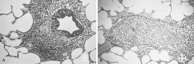
Among the most effective and probably least appreciated protective microbial factors is biofilm. Bacteria exist in either a planktonic (free floating) or sessile (attached) state; while it is the former state that characterizes C&S testing, but it is the latter state that enables persistence of the resident population, as well as the formation of biofilm.95-97 Biofilm is defined as a biopolymer, matrix-enclosed bacterial population in which bacteria adhere either to one another or to a surface.95 The outer layer of the biofilm may lose water such that it is hardened, thus providing better protection from the environment, including exposure to antimicrobials. The inner sactum of the biofilm is largely aqueous, composed of glycocalyx or slime (e.g., Staphylococcus spp.). In addition to passive diffusion, aqueous pores permeate the structure, allowing movement of nutrients and metabolic debris. Biofilm populations containing normal microflora in the skin or mucous membranes (e.g., urinary bladder) are lost with shedding of the skin (or bladder) surface or by the excretion of mucus; new cells and mucus are rapidly colonized by biofilm-forming bacteria. Microbes released from the surface may colonize new surfaces and subsequently produce new biofilms and new (e.g., persistent or recurring) infections. Bacterial communication during biofilm formation is sophisticated, involving quorum-sensing systems that ultimately may be targets of microbial therapy.96 Biofilm may facilitate and protect growth of normal or pathogenic flora on foreign surfaces and can facilitate subsequent translocation of microbes to otherwise sterile tissues. Persistent, chronic bacterial infections may reflect biofilm-producing bacteria; persistent inflammation associated with immune complexes contributes to clinical signs. Dental plaque is a prototypic example of the impact that biofilm might have on preventing antimicrobial penetration. Cystic fibrosis associated with Pseudomonas is a disease in which biofilm contributes to mortality. Pathogens associated with biofilm in veterinary medicine include, but are by no means limited to, Acinetobacter, Actinobacillus, Klebsiella, P. aeruginosa, and Staphylococcus (aureus and pseudintermedius).95 Glycocalyx may contribute to protective mechanisms of other organisms as well (e.g., sulfur granules and Nocardia; Figure 6-14). Not all pathogens associated with biofilm cause infection (e.g., urinary catheters). However, because they ultimately may be the source of infection, clinical resolution may not be possible until the biofilm is destroyed. Yet, its nature is difficult to predict based on the planktonic growth of individuals in cultures compared to the consortium that occurs in vivo.97 Catheters (urinary or intravascular), orthopedic fixation devices, and materials used in wound management are examples of surfaces on which biofilm might develop.

image

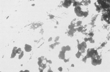
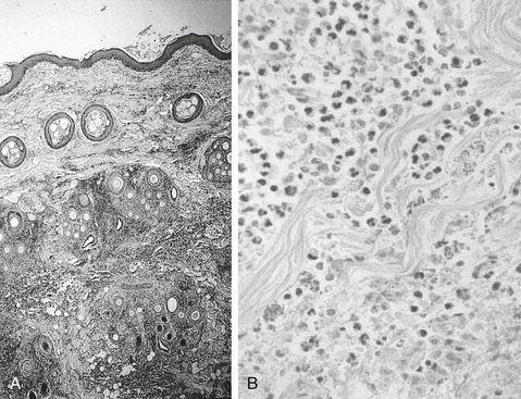
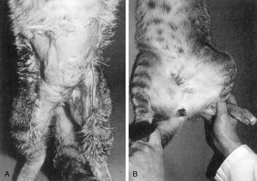
Figure 6-14 An example of combined host and microbial factors that negatively impacts therapy, Nocardia causes a marked inflammatory response by the host. Additionally, the organism causes secretion of calcium that combines with its biofilm, resulting in the formation of “sulfur” granules that protect the organisms from drug penetrations.

KEY POINT 6-20

Biofilm can form on many foreign or natural surfaces and may profoundly decrease the likelihood of successful antimicrobial therapy.

Antimicrobial Resistance

The role of resistance in therapeutic failure of antimicrobials is well established.23,98 The use of antimicrobials increasingly is associated with emergence of resistance. For each class of antimicrobial drugs approved for use in human medicine, resistance generally has emerged within 1 to 2 decades of use. Clinically relevant resistance toward sulfonamides, the first class of antimicrobials approved in the United States (1930s) was documented by the 1940s. Penicillins, tetracyclines, streptomycin (aminoglycoside), and erythromycin (macrolides) were all approved within a 10-year span, with resistance documented within 5 years for methicillin versus approximately 10 years for streptomycin. Resistance to nalidixic acid, the progenitor of fluoroquinolones (approved in 1950), took 3 decades to emerge, perhaps convincing manufacturers that resistance to fluoroquinolones would emerge very slowly. However, resistance to norfloxacin, the first fluoroquinolone approved in the United States, took less than 3 years to emerge, despite the fact that the lack of plasmid-mediated resistance was among the attributes of this class. Resistance to extended-spectrum cephalosporins emerged within 4 years of approval and to amoxicillin–clavulanic acid, within 5 years. Resistance to vancomycin, specifically developed to treat MRSA, emerged in its second decade of use.

KEY POINT 6-21

Antimicrobial resistance increasingly will prevent the successful empirical selection of antimicrobial drugs.

Inherent Versus Acquired Resistance

Antimicrobial resistance might be inherent to the microorganisms or acquired, either through chromosomal mutations or transfer of genetic information.99 Generally, spectrums of antimicrobials (listed on package inserts and elsewhere) reflect inherent resistance patterns rather than acquired resistance patterns. Examples include limited efficacy of aminoglycosides toward anaerobic organisms because the drugs must be actively transported into the cell (oxygen dependent) or the resistance of gram-positive organisms, which lack an outer cell membrane, to polymyxin B, which targets the same. Acquired resistance, on the other hand, generally renders a previously susceptible organism resistant. As such, it is not necessarily predictable and can occur during the course of therapy (leading to changes in a C&S pattern). More problematically, it is often shared among microbes.

Shared resistance among bacteria reflects the ability of bacteria to incorporate extrachromosomal DNA carrying the information for resistance from other organisms. Extrachromosomal DNA (including plasmids and bacteriophages) encode for resistance to multiple drugs and can be transmitted vertically (to progeny) or horizontally, across species and genera. Transposons are individual or clusters of resistance genes bound by integrons, which move resistance genes back and forth between chromosomes to plasmids. Consequently, bacterial resistance is extremely mobile and can spread rapidly.101 Among the mechanisms by which genetic resistance information is shared is (sexual) conjugation. Conjugation occurs particularly in gram-negative organisms and may be accompanied by genetic material that confers bacterial pathogenicity as well as altered metabolic functions. However, Enterococcus spp. and selected other gram-positive bacteria also transfer resistance to glycopeptides through conjugative transposons.101 Transduction, which requires a specific receptor, involves transfer of information by a bacterial virus (bacteriophage) and is implemented especially by Staphylococcus spp. Resistance, including methicillin resistance, can be transferred between coagulase-negative and -positive Staphylococcus.1 Transformation involves transfer of naked DNA from one lysed bacterium to another; this mechanism of transfer tends to be limited (in humans) to pneumococcal meningitis.

Although present for eons, acquired antimicrobial resistance increasingly is becoming problematic. The impact of antimicrobial resistance can be extensive. In some human intensive care units, selected isolates are characterized by a resistance prevalence of 86%. The impact of resistance on the patient includes increased morbidity, mortality, and increased hospital costs.107 Patterns of resistance have emerged in veterinary medicine, although differences appear to occur in the ability of organisms to develop resistance to an antimicrobial, varying with species and strain. Many organisms remain predictably susceptible to selected drugs (e.g., Brucella, Chlamydia), whereas others are becoming problematic (e.g., P. multocida). Several organisms traditionally have developed resistance that can rapidly impair efficacy of new antimicrobials (e.g., E. coli, K. pneumoniae, Salmonella, S. aureus, S. pneumoniae). In general, these organisms have developed multidrug resistance (MDR). MDR is now considered the normal response to antimicrobials for gram-positive cocci pneumococci, enterococci, and staphylococci.102 Among these, Staphylococcus spp. is considered most problematic: it is intrinsically virulent, is able to adapt to many different environmental conditions, increasingly is associated with resistance to other classes of antimicrobials, and tends to be associated with life-threatening infections.102,103 In a veterinary teaching hospital the percentage of patients with S. intermedius susceptible to cephalexin and amoxicillin–clavulanic acid decreased from a high of 96% in 2005 to < 60% in 2007, a trend that appears to be emerging in other veterinary hospitals.110

KEY POINT 6-22

Acquired resistance can occur during the course of antimicrobial therapy.

E. coli is among the organisms that have developed multi-drug resistance.104,105 Fluoroquinolone-resistant E. coli emerged as early as 1998, little over a decade after the approval of enrofloxacin for dogs or cats.28 Multidrug-resistant E. coli has emerged as a cause of nosocomial infections in dogs108 and UTIs in canine critical care patients.104,109 The presentation is similar to the that in human critical care patients, with risk factors such as sex (males), hospital stay, and previous antimicrobial therapy being similar for both.

Factors Contributing to the Emergence of Resistance

Development of antimicrobial resistance is facilitated by several factors111; among the most important is exposure to antimicrobials. In the individual patient, single-dose ciprofloxacin prophylaxis increased the prevalence of ciprofloxacin-resistant fecal E. coli from 3% to 12% in humans.112 Ciprofloxacin treatment for prostatitis resulted in posttreatment fecal colonization with quinolone-resistant E. coli that was genetically distinct from the infection-causing strains after treatment in 50% of the patients.113 Our laboratory has demonstrated that standard doses of either amoxicillin or enrofloxacin given orally will cause close to 100% of fecal E. coli to become resistant to the treatment drug within 3 to 9 days of therapy; for enrofloxacin the isolates generally are multidrug resistant. As with MRSA or MRSI (S. intermedius), the advent of resistance by E. coli and other gram-negative organisms has been associated with increased cephalosporin use.1

The gastrointestinal flora offers a natural environment that exemplifies the impact of antimicrobials on selection pressure. The normal flora of the gastrointestinal tract is extremely diverse, with anaerobes predominating. Among the aerobes, E. coli are the major gram-negative and Enterococcus the major gram-positive organisms.101 Environmental microbes maintain an ecologic niche through suppression of the competition by either consumption of nutrients or secretion of antibiotics. Therefore commensal organisms are constantly being exposed to antibiotics, and are “primed” to develop resistance.101 However, the microbes producing the antibiotic, as well as surrounding normal flora, are resistant to the antibiotic. Thus genes for resistance develop along with genes directing antibiotic production.

Rapid microbial turnover in the gastrointestinal tract supports the development of resistance by ensuring active DNA replication and thus mutation potential (see previous discussion). Chromosomal (DNA) mutations (10-14 to 10-10 per cell division) are DNA mistakes that have been missed by bacterial repair mechanisms. These mistakes occur spontaneously and randomly, regardless of whether the antibiotic is present. If the mutation that confers resistance to an antimicrobial occurs in the presence of the antimicrobial when it is administered to the patient, the surviving mutant, reflecting its single-step mutation, confers a low level of resistance (see the discussion of mutant prevention concentration). The MIC of the organism is likely to increase. Further microbial turnover and continued therapy can lead to multistep mutations and rapid emergence of high-level resistance characterized by increasingly higher MIC. Stepwise mutations can lead to specific resistance such as that demonstrated toward fluorinated quinolones (stepwise mutation in the DNA gyrase gene). Nonspecific mechanisms of resistance, including that shared among organisms, are more likely to result in MDR. Microflora of the gastrointestinal tract can serve as a reservoir of resistance genes; a single drug, via integrons, plasmids, and transposons, facilitates the rapid transfer of MDR among organisms. The gastrointestinal environment exemplifies a pattern whereby resistance can emerge as a result of a combination of selection pressure and mutation. Clinically, similar mechanisms of emerging resistance are likely to occur at sites of infection.

Mutant Prevention Concentration

Drlica and coworkers114 have hypothesized the mutant selection window, (see Figure 6-15) comprised of a lower threshold represented by the culture MIC of the infecting organism and an upper threshold or boundary, the MPC. Should a dose be designed such that drug concentrations fall within this window (i.e., between the MIC and MPC) at the site of infection, the mutant isolate is likely to emerge as a resistant colony. The practical application of the hypothesis explains the observed behavior of mycobacterium organisms toward fluoroquinolones (FQs). Increasing concentrations of the FQs inhibits the nonresistant (wild-type) organisms and colony numbers rapidly decrease. But this period of decline is followed by a plateau period of minimal or no growth. During this plateau phase, remaining resistant isolates recover and start to multiply again. The resistance of this emerging, second population presumably reflect a single-step (chromosomal or plasmid-mediated) mutation that resulted in an increase in the MIC to low-level resistance (e.g., MIC is close to the breakpoint). However, when these first-step mutants are exposed to even higher drug concentrations, a second rapid decline in numbers occurs, this time reflecting inhibition of the mutated, resistant organisms. Again, once sufficient bacteria recover, a second plateau occurs as the first-step mutants mutate. This stepwise or multistep mutation confers high level resistance (MIC exceeds the breakpoint several fold) that can be overcome only by very high concentrations of the FQ. The mutant selection window, which is to be avoided with initial therapy, describes drug concentrations on either side of the initial plateau for the single-step mutants. The lower boundary is defined by those drug concentrations sufficiently high to remove the majority of the wild-type competitors (MIC), whereas the higher boundary (the MPC) is defined by the concentrations necessary to inhibit the least susceptible (most resistant) isolates (the single-step mutants).115 Above this concentration, a second mutation step (which is very rare) would be required for a population of resistant organisms to develop; the risk of this happening is reduced by preventing microbial turnover (i.e., killing all isolates).

image

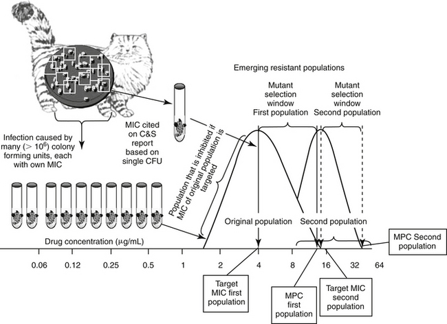
Figure 6-15 Stepwise mutation can emerge as a result of selection pressure induced by antimicrobial therapy that targets the minimum inhibitory concentration (MIC) of the infecting microbe. The mutant prevention concentration (MPC) is the concentration of drug that is necessary to inhibit first-step mutants, or the MIC of the least susceptible isolate in a resident population of pathogens. As the resident population or inoculum of wild (nonresistant) pathogen isolates reaches 108-10 colony-forming units (CFUs), some isolates will spontaneously mutate such that resistance emerges to the drug of interest. However, when cultured, the MIC reported for the population is likely to represent the mode (the most commonly reported MIC), which in a normally distributed population, is also the MIC50 for the population. In contrast, the MIC of the first-step mutant will be the high end of the population MIC range. This is the concentration that should be targeted to inhibit the entire population—that is, the MPC. If the dosing regimen is designed to target the the mutant selection window, that is, the MIC of the wild population rather than the MPC (the MIC of the first-step mutant)—treatment with the drug will inhibit all isolates at or below the MIC. The void in isolates will allow the remaining, more resistant first-step mutants to recover, particularly in patients not sufficiently healthy to suppress recovering microbes. As this new population expands, a second distribution curve emerges. If recultured, the MIC of the second first-step mutant population will be higher than the wild population. If the population reaches a sufficient size (e.g., 108 CFUs), a second, spontaneous mutation is likely to occur, resulting in a new, higher MPC. Targeting the MPC is particularly important when using drugs for which resistance emerges in response to mutations.

KEY POINT 6-23

The mutant prevention concentration (MPC) is the highest MIC of any of the colony-forming units causing infection in the patient. Failure to achieve this concentration may allow resistant microbes to emerge, particularly in the at-risk patient.

On the basis of this observation, Drlica and coworkers contend that MIC-based strategies used to design dosing regimens readily select for resistant mutants.115 Their contention is based on the observation that only one resistant mutation is needed for bacteria to grow in the presence of an antimicrobial and that infections generally contain an adequate number of CFUs for several first-step resistant mutants to be present prior to treatment. They coined the term MPC as an in vitro measure of preferred antimicrobial concentration target. If the MPC (rather than the MIC) is achieved at the site of infection, the risk of resistance is minimized because isolates that exceed the MPC concentration must have undergone a second concurrent resistance mutation step prior to therapy. As such, the MPC, not the MIC, would be the concentration targeted at the site of infection in the patient. Indeed, simply achieving the reported MIC of the infecting microbe at the site of infection is probably the approach that is most likely to yield clinically resistant organisms. Accordingly, consideration should be given to assuring that “dead bugs don’t mutate.” If the least susceptible of the isolates is inhibited with the dosing regimen, then the recovering population should not be resistant.

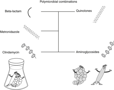
Drlica115 has demonstrated that MPCs do not correlate to MICs. In vitro, the MPC would be defined in vitro as the (lowest) drug concentration (in the media) that yields no recovered organisms when over 1010 CFUs (mimicking bacterial load in the patient) are plated. Currently, determining the MPC is costly, requiring multiple testing steps and large numbers of cells; for example, standard culture procedures are based on 106 CFUs, whereas determination of the MPC requires at least 108-10 CFUs. However, an MPC-based strategy to dosing clinically makes sense and should be an effective means of blocking the growth of first-step resistant mutants. Such a strategy would force wild-type cells to acquire two resistance mutations for growth, an event that is rare. Experimental in vitro data114 have confirmed that MPC levels of an FQ do indeed inhibit strains that harbor first-step gyrA mutations (the mechanism of microbial resistance to FQ).116 Application of the MPC is most appropriate for drugs and organisms that develop resistance by chromosomally mediated point mutations (e.g., the fluorquinolones).117 However, the spirit of targeting the MPC might be assumed even for other drugs, in order to minimize the impact of selection pressure on emergent resistant populations. The mutant selection window can be narrowed if more than two bacterial sites are targeted, such as might occur with combination antimicrobial therapy, or with drugs that simultaneously target more than one site (e.g., the FQs).114

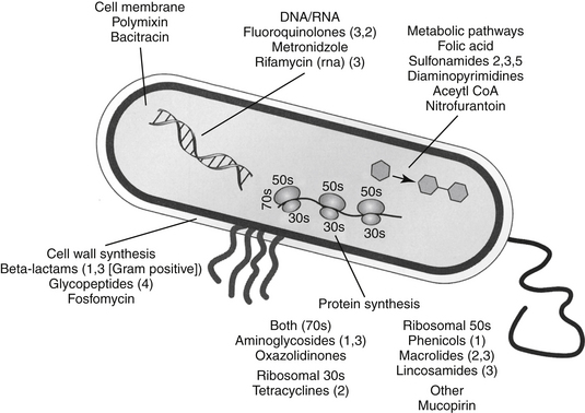
Biochemical Mechanisms of Resistance

Bacteria often respond to the presence of the antimicrobial by altering their physiology such that resistance occurs, often to multiple drugs. Microbes develop antimicrobial resistance by two primary mechanisms: modification of the target site or altered intracellular drug concentration. Methods by which intracellular drug concentration can be decreased include changes in porin sizes for gram-negative organisms (e.g., most drugs; see Figure 6-3). Porins are transmembrane proteins (e.g., OmpF) that form an aqueous channel that allows passive movement of large hydrophilic molecules. Porins are one of the few means by which drugs can gain access to intracellular targets. A change in porin size (i.e., by the addition of side chains that filter out drugs) or number increases antimicrobial resistance, as is demonstrated by the loss of the OprD protein that imparts resistance to imepenem by Pseudomonas spp. Closely associated with the porin proteins are efflux proteins that pump drug out of the organism; the pumps often are associated with porin proteins (e.g., FQs and tetracyclines). Most of these pumps are fueled by energy associated with proton exchange, the most notable in gram negative organisms being of the RND (resistance nodulation division) family. The best characterized in this family is the Acr-AB/TolC system, which is a complex bacterial stress response system that allows bacteria to pump out toxic molecules.101 These and other pump systems are often characterized by a wide range of substrate specificities and, along with porins, are a common mechanism whereby an isolate can express multidrug resistance. In contrast, a number of microbes generate enzymes that destroy antimicrobials (e.g., aminoglycoside acetylases, beta-lactamases that destroy penicillins or cephalosporins, transferases that destroy chloramphenicol); in such instances resistance conferred by these mechanisms is generally limited to a single drug or drug class. Enzymatic inactivation is more likely for natural drugs to which microbes have previously been exposed (and thus presented with a greater opportunity to develop enzymes). In contrast, enzymes are less likely to destroy synthetic drugs.118 However, plasmid-mediated enzymatic destruction of FQs has recently been described, once again highlighting the resourcefulness of bacteria.119 Changes in target structure are another major mechanism of resistance. Example targets that have been modified include, but are not limited to, cell wall proteins (e.g., penicillin-binding proteins [PBs], particularly for MRSA [PB2] or Enterococcus [PB5]), or binding sites (i.e., on ribosomes, as for aminoglycosides, or DNA gyrase for FQs).120,121 Organisms often are characterized by more than one mechanism of resistance. Multiple mechanisms are well documented for some organisms against selected beta-lactams and have been described against FQs (e.g., altered DNA gyrase and increased efflux pumps) and others. Resistance can be induced, as is exemplified by beta-lactamase formation in Staphylococcus spp. which greatly increases in the presence of a beta-lactam antibiotic, or for fluoroquinolone, for which efflux pump activity is markedly upregulated. Discussion of specific mechanisms of resistance will be addressed with the appropriate drugs (see Chapter 7).

Avoiding Antimicrobial Resistance

Among the approaches to reducing resistance are pharmacologic manipulations and changes in antimicrobial use practices. Pharmaceutical manufacturers have been able to manipulate antimicrobial drugs in a variety of ways such that resistance is minimized, and these options can be selected in an attempt to minimize resistance. For example, bacterial resistance has been decreased by synthesizing smaller molecules that can penetrate smaller porins (e.g., the extended-spectrum penicillins ticarcillin and piperacillin); synthesizing larger molecules that force the microbe to develop more than one point mutation (e.g., later-generation FQs), “protecting” the antimicrobial from enzymatic destruction (e.g., with clavulanic acid, which diverts the beta-lactamase from the penicillin); modifying the compound so that it is more difficult to destroy (e.g., amikacin, which is a larger and more difficult to reach molecule than gentamicin and carbapenems, later generation cephalosporins); and developing lipid-soluble compounds that are more able to achieve effective concentrations at the site of infection (e.g., doxycycline compared with other tetracyclines). Increasingly, drug design–based tactics will be implemented to minimize emergent resistance. Increasingly the role of the practitioner is equally important. A three “D”s approach might reduce the risk of emergent resistance: De-escalate antimicrobial use, design a treatment regimen that minimizes resistance (dead bugs don’t mutate), and decontaminate the environment through proper hygiene. These approaches are exemplified by strategies implemented by intensive care units to reduce antimicrobial resistance that often involve a multitiered approach (Box 6-3). Actions include the following:

Box 6-3 Reducing Transmission of Resistant Microbes (Decontamination)

Treating infected patients in order of least-at-risk to most-at-risk
Dedicated diagnostic or handling equipment
Proper bandaging of infected sites
Dedicated bandage areas
Protection during animal handling (disposable gloves, masks, gowns, eyewear)
Decreased contact with body fluids
Dedicated disposal for contaminated materials
Proper hand washing between patients
Easy access to hand sanitizers (alchohol based)
Strict asepsis during surgery
Dedicated cleaning materials
Clean in order of cleanest to dirtiest
Proper disinfection of the following:
Exposed or at-risk rooms
Tables, doors, counter surfaces, floors, and so on
Equipment (stethoscopes, keyboards, pens, and so on)
Isolate carriers (not always necessary)

KEY POINT 6-24

The goal of antimicrobial therapy is twofold: resolving clinical signs associated with infection and avoiding emergent resistance. The two goals are not mutually inclusive.

1. De-escalate. De-escalation begins with not using an antimicrobial when an alternate therapy (including no therapy) is more or perhaps equally effective. Enacting primary prevention by decreasing length of hospital stay, decreasing use of invasive devices, and implementing newer approaches (e.g., selective digestive decontamination and vaccine development).107 De-escalation also includes setting limits on the duration of antimicrobial therapy (see later discussion) and rotating the use of antimicrobial drugs on a regular schedule.107,125 De-escalation might also refer to changing from a higher to a lower tier category of drugs (following a “hit hard, get out quick”) in a critical patient.
2.Design: Improving appropriate antimicrobial use through proper dosing regimens includes selection of the most appropriate drug for the bug while narrowing the spectrum. This approach also should be applied to empirical antimicrobial therapy. Design of the dosing regimen should take into account the appropriate PDI for concentration or time-dependent drugs, and when possible, targeting the MPC. More controversial approaches to design include techniques implemented in hospitals include adhering to prescribed formularies or requiring prior approval for using certain antibiotics.
3. Decontaminate: Approaches intended to reduce bacterial exposure are among the most important to avoiding resistance. These include improving infection control through selective decontamination procedures, prevention of horizontal transmission through proper hand-washing technique, and use of gloves and gown, or prevention by reducing exposure to bandages or other contaminated materials by identifying proper work areas and disposal sites. Other approaches include, provision of soap alternatives, easy access to disinfectants (which should complement, not replace, hand washing) and improvement of the workload and facilities for health care workers. Improved information systems technology also plays a role. Each proposed or implemented strategy has theoretical benefits and limitations, but good data on their efficacy in controlling antimicrobial resistance are limited.107,125 However, it is clear that decreased antimicrobial use is associated with a decrease in the advent of resistance.

Risk factors for emerging resistance in the hospital or community setting include but are not limited to increased antimicrobial use, host factors such as severity of illness and length of stay, and lack of adherence to infection control practices.107 Consequently, among the de-escalation efforts implemented in human hospital and community environments is restricted antimicrobial use. In humans the increasing presence of drug-resistant bacterial infections among hospitalized patients is linked to the greater numbers of patients receiving inappropriate antimicrobial treatment.123 A recent on-line report found that in human medicine, antimicrobials were often prescribed despite infection being an infrequent cause of the illness (i.e., pharyngitis). Further, the chosen antimicrobial often was inappropriate for those bacteria potentially causing infection in the treated body system.124 Accordingly, reducing inappropriate antimicrobial use has become a priority in human medicine.

Among the more rational paradigms for antibacterial de-escalation, is an approach to empirical antimicrobial use in the hospital setting for patients with serious bacterial infections.123 Such antimicrobial de-escalation attempts to balance the need to provide appropriate initial antibacterial treatment while limiting the emergence of antimicrobial resistance. The goal of de-escalation in this setting is to prescribe an initial antimicrobial regimen that will cover the most likely bacterial pathogens associated with infection while minimizing the risk of emerging antimicrobial resistance.123 The three-pronged approach includes narrowing the antimicrobial regimen through culture, assessing isolate susceptibility for dose determination, and choosing the shortest course of therapy clinically acceptable. Judicious antimicrobial use combined with restricted use of ceftazidime led to a decreased antimicrobial resistance to beta-lactams, in general, in a human teaching hospital environment.126 Note that this strategy does not exclude the use of “big gun” antimicrobials. The approach of withholding use of high-impact drugs (e.g., meropenem or vancomycin) in patients whose need for effective therapy is critical to avoid emerging resistance that might limit drug use in later patients may not be rational or in the best interest of the patient. A more appropriate approach is to use the drug correctly. However, routine use of less powerful drugs is appropriate but only if these alternatives are just as effective. Regardless of the choice, once the decision is made to use an antimicrobial, attention must be paid to dosing regimens that minimize the advent of resistance by ensuring that infecting microbes are eradicated.

Another strategy to decrease the impact of antimicrobial use on resistance is a decreased duration of therapy (see the discussion of enhancing antimicrobial efficacy). One study in human critical care patients found that reducing the duration of antimicrobial therapy from 14 days to 10 days decreased the emergence of resistance.127 Increasingly, clinical trials will focus on demonstrating efficacy of shorter (i.e., < 5 to 7 days) treatment regimens.

Rapid detection of the correct microbe and the presence of resistance would facilitate the proper design of a therapeutic regimen. Genetic changes (e.g., mutations) that result in resistance lend themselves to molecular detection. However, molecular tests are often limited to those mutations characterized by few polymorphisms (e.g., MRSA, potentially MRSIG, and Enterococcus sp.). Generally, these tests require culture conditions that are often designed to facilitate expression of the resistant gene and are based on amplification techniques. Yet, as with culture, although they are able to determine phenotypic expression, they do not necessarily document the isolate as the cause of infection. Further, they generally do not detect low levels of resistance that increase the MIC but do not render the microbe as “resistant” by susceptibility testing.122 Topical therapy should be considered when possible. Therapeutic drug monitoring may be helpful for some drugs (e.g., aminoglycosides). With at-risk patients in whom emergent mutants may not be sufficiently suppressed. Drugs inherently more resistant to bacterial inactivation should be selected (e.g., amikacin rather than gentamicin). Combination antimicrobial therapy (e.g., beta-lactamase–protected antimicrobial combinations; combination of beta-lactams with aminoglycosides) also reduces the incidence of resistance; for example, the use of an FQ reduced the advent of resistance to cephalosporins in one study.33 Care should be taken in selecting a drug simply because of cost. Cost should be a factor only after other considerations have been taken into account. The cost of an excellent antimicrobial can be easily surpassed by the selection and use of several less expensive, but also less effective, antimicrobials.

Host Factors that Affect Antimicrobial Efficacy

Careful consideration must be given to host factors that can reduce concentrations of active drug at the site of infection.23,75,128 The impact of host factors on antimicrobial efficacy is often underestimated; such effects can be profound.

Among such host factors is distribution of the drug to the site of the infection (drug distribution is discussed under drug factors). Thus far, discussions on antimicrobial efficacy have been focused on achieving the MIC of the infecting isolate in the patient plasma. However, infections generally are not in plasma, and patients are not generally normal. The relationship between the MIC of the infecting organisms and drug concentrations achieved at the site of infection (both magnitude and duration) is so complex that predicting efficacy is difficult. Ultimately, mathematical models that integrate the major determinants of efficacy (bactericidal activity, relationship between PDC and MIC, duration of postantibiotic effect, and susceptibility versus resistance) may prove most predictive.129 The determinants of this relationship and the influence of drug, microbial, and host factors on efficacy warrant further discussion.

The MICBP of a drug is based on plasma Cmax, yet infections generally occur in tissues rather than plasma. More specifically, the site of infection generally is interstitial fluid. However, detection of drug in tissues is difficult, leading to PDC as the surrogate marker of tissue concentrations. In instances in which PDCs overestimate extracellular fluid, care must be taken to adjust doses. For such drugs C&S testing may overestimate efficacy of the drug (see the section on drug distribution). On the other hand, for some tissues, drug concentrations at the site may far exceed PDC (see below). Inflammation may profoundly alter drug efficacy (Table 6-7).23,128 Acute inflammation may initially increase drug delivery and drug concentration to the site of infection because of increased blood flow, increased capillary permeability, and increased protein release at the site (the latter effect increases the concentration of total, but not necessarily active, drug). However, chronic inflammation may do the opposite. Purulent exudate presents an acidic, hyperosmolar, and hypoxic environment that impairs the efficacy of many antimicrobials (Figure 6-16). Hemoglobin and degradative products of inflammation can bind antimicrobials.130 Selected drugs, including aminoglycosides (and probably highly protein-bound drugs) are bound to and thus inactivated by proteinaceous debris that accumulates with inflammation. Some antimicrobials can inhibit neutrophil function. Accumulation of cellular debris associated with the inflammatory process can present a barrier to passive antimicrobial distribution. The deposition of fibrous tissue at the infected site further impairs drug penetrance and distribution (Figure 6-17).

Table 6-7 Negative Effects of the Microenvironment on Antimicrobial Efficacy

Environmental Factor Effect
Acidic pH Penicillins inactivated at pH < 6.0
  Aminoglycosides and enrofloxacin more effective in alkaline pH
Hypertonicity/hyperosmolarity Impaired efficacy of beta-lactam antibiotics
Pus Acidic pH
  Hypertonic
  Hyperosmolar
  Protein binding of selected drugs
  Binding to sediment (aminoglycosides)
Low O2 tension Aminoglycosides inactive
  Growth of organisms slowed → decreased efficacy of bactericidal drugs
  Impaired phagocytic activity of leukocytes
Large inoculum Greater concentration of antimicrobial inactivating enzymes
  Greater concentration of drug molecules required
Leukocytes Impaired chemotaxis, phagocytosis, metabolism
image

Figure 6-16 The inflammatory response to bacteria is intended to support the host in overcoming an infection. However, the response can become a confounding factor. For example, the inflammation of pneumonia of bronchitis dilutes the drug, presents a barrier to passive diffusion, and may bind and thus inactivate the drug. Local pH and thus drug ionization may impair drug action, and generation of a decreased oxygen tension further decreases drug efficacy.

(Photo courtesy Bayer Animal Health.)

image

Figure 6-17 Deposition of fibrous tissue in deep pyoderma presents a barrier to drug penetration.

(Photo courtesy Bayer Animal Health.)

KEY POINT 6-25

Although the host inflammatory response initially may facilitate therapeutic success, it can ultimately profoundly decrease the likelihood of success.

Local pH becomes more acidic as degradative products such as lysosomes, nucleic acids, and other intracellular constituents from white blood cells accumulate. The efficacy of many antimicrobials can subsequently be impaired. In humans a pH level ranging from 5.5 to 6.8 can adversely affect both host defenses and antimicrobial activity. White blood cell oxidative bursts and phagocytosis are diminished in the presence of a low pH level. Some antimicrobials are inactive at a low pH level. Erythromycin loses all of its activity when pH is below 7. Similar effects have been reported for beta-lactam antibiotics. Although beta-lactam antibiotics are weak acids and therefore less ionized in an acidic environment, they are generally less effective at a pH 6. The activities of cefoxitin, piperacillin, and imipenem (or meropenem) are significantly less at pH 6 than at pH 6.5 with piperacillin being least affected. The activity of clindamycin is similarly decreased. In addition, the accumulation of some drugs in white blood cells that might otherwise facilitate efficacy is impaired in an acidic environment. Changes in pH also lead to changes in the concentration of un-ionized and thus active drug. Weak bases such as aminoglycosides and FQs are predominantly ionized in an acidic environment and are less effective than in a less acidic environment, in part because of impaired diffusibility.

Low tissue oxygen tension, which can accompany pus, reduces white blood cell phagocytic and killing activity; slows the growth of organisms, making them less susceptible to many drugs; and specifically prevents the efficacy of aminoglyosides, which depend on active transport into bacterial organisms. The aerobic component (i.e., facultative anaerobes) of a mixed infection may also be resistant to aminoglycoside therapy because the oxidative transport systems of such organisms (e.g., E. coli) may shut down in an anaerobic environment. Drugs that target cell walls, and beta-lactams in particular, are less effective in a hyperosmolar environment, which might occur as inflammatory debris accumulates and osmotic destruction of organisms is reduced.

Host response to infection and its impact on antimicrobial therapy may vary with the organ system infected. For example, in respiratory tract infections, mucus produced by the host can directly interfere with antimicrobial therapy. Aminoglycoside efficacy may be decreased by chelation with magnesium and calcium in the mucus. Antimicrobials may bind to glycoproteins, and mucus may present a barrier to passive diffusion. In addition, some antimicrobials may alter the function of the mucociliary apparatus, either by increasing mucous viscosity or by decreasing ciliary activity (e.g., tetracyclines).

Changes in the health of the host can lead to changes in drug disposition that can result in lower than anticipated PDCs (see Chapter 2).50 The volume to which a drug is distributed can be affected by the fluid compartments, which vary with age, species, and hydration status. Distribution to target organs can be affected profoundly by cardiovascular responses, particularly in the shock patient. Elimination of the drug must be considered when selecting antimicrobials for the critical patient. Changes in glomerular filtration cause parallel changes in renal excretion of drugs. Serum creatinine concentrations should be used to modify doses or intervals of potentially toxic drugs that are excreted renally (see Chapter 2).131 Likewise, severe changes in hepatic function may indicate selection of an antimicrobial drug not dependent on hepatic function for activation or excretion.

Host Factors That Facilitate Drug Efficacy

Host factors may also facilitate antimicrobial efficacy. Among the most important host factors are local and systemic defenses ranging from compounds that directly target microbes to healthy tissues that provide mechanical barriers and a competent immune system. The role of host defenses are beyond the scope of this chapter but cannot be underemphasized.

Other host factors that facilitate therapy include the accumulation of the drug in active form at the site of infection, which may facilitate antimicrobial efficacy and decrease the risk of resistance. Obvious examples include drugs that undergo renal or biliary excretion. For such drugs urine or bile concentrations (respectively) may exceed PDC thirtyfold to several hundredfold (see the discussion of treatment of urinary tract infections, Chapter 8). Another site of drug concentration is the phagocytic leukocyte (WBC), both in peripheral circulation and at the site of inflammation. Active concentrations of some antimicrobials (e.g., macrolides, lincosamides, and FQs) may increase concentrations 20 to 100 or more times the PDC.28,132-137 Phagocytic accumulation may facilitate treatment of intracellular infections (e.g., Brucella spp., cell wall–deficient organisms, intracellular parasites, and facultative intracellular organisms such as Staphylococcus spp.). Thus drugs that achieve only bacteriostatic concentrations in plasma may become bactericidal inside the cell, particularly against organisms that locate and survive inside cells (Figure 6-18). Additionally, accumulated drug released by dying phagocytes at the site of infection may increase concentrations to which the infecting microbe is exposed. Accumulation of drug inside WBC has been assumed as an explanation of the disconnect of azithromycin efficacy in pulmonary infections despite low PDCs.91 Note, however, that drug accumulation does not necessarily enhance drug efficacy. Often, the accumulated drug is sequestered into subcellular organelles, where it cannot reach the organism. In addition, the drug may become otherwise inactivated once inside the cell. The different mechanisms of action of these drugs may not occur in an anaerobic environment, and concentrations by the WBCs might be impaired in an anaerobic environment. The FQs are an example of a class of drugs whose uptake by WBCs is facilitated in an acidic environment; these drugs are distributed throughout the cytosol, where they remain active. The drug will leave the WBCs and enter a drug-free environment and thus may facilitate drug concentrations at the site of infection. Phagocytic WBCs with accumulated enrofloxacin delivered drug to inflamed tissue cages in dogs, demonstrating that accumulation may increase therapeutic response.137 Drugs that do not accumulate in WBCs include the beta-lactams, aminoglycosides, and metronidazole. Drugs that are moderately accumulated in WBCs include chloramphenicol (onefold to fivefold) and selected sulfonamides (threefold to fivefold).138

image

Figure 6-18 The intracellular location of organisms presents a barrier to drug penetration. Some organisms are obligate intracellular organisms, whereas others, such as Staphyloccocus spp., demonstrated cytologically (A) and by special stain of infected skin (B), may survive phagocytosis, serving to reinfect tissue once the phagocytic white blood cell has died.

Another potential facilitating host factor is infection at a site that is topically accessible. In such situations several 1000-fold concentrations of the MIC may be reached with topical administration. The rationale for collecting C&S data for such infections might be controversial, but identification of the organism and some indication of susceptibility is prudent, particularly if initial therapy fails.

Drug Factors that Affect Antimicrobial Efficacy

Mechanisms of Drug Action

Knowledge of the mechanism of action (see Figure 6-19) of a particular antimicrobial is important for several reasons:

1. The mechanism of action of a drug determines whether the antimicrobial can act in a bactericidal or bacteriostatic manner (assuming proper concentrations are achieved at the tissue site; see previous discussion). Drugs that are capable of bactericidal effects at therapeutic doses are listed in Table 6-7.
2. The mechanism of action may determine whether or not the drug is concentration-dependent or time-dependent, which will impact the design of the dosing regimen.
3. The therapeutic efficacy of some antimicrobials can be impaired by host factors that alter the mechanism of action of the drug. Knowledge of the mechanism of action will facilitate anticipation of therapeutic failure.
4. The mechanism of antimicrobial action often reflects the mechanism of resistance. Identifying mechanisms by which resistance might be avoided or minimized requires an appreciation of these mechanisms of action.
5. Understanding or anticipating selected host toxicities associated with antimcirobials can be improved by understanding their mechanism of action.
6. Understanding antimicrobial mechanisms of action provides a basis for the selection of antimicrobials to be used in combination. Such drugs should be selected on the basis of mechanisms of action that complement rather than antagonize one another (see Combination Antimicrobial Therapy section).
image

Figure 6-19 Targets of antimicrobial actions for the different classes of antimicrobial drugs. The number in parentheses refers to the major mechanism(s) of acquired resistance (other mechanisms also exist; see Chapter 7): 1 = enzymatic destruction (e.g., beta-lactamases for beta-lactams, acetylases for phenicols); 2 = increased efflux pump activity (may be associated with altered porin influx in gram-negative isolates); 3 = altered targeted site (e.g., mutations in DNA gyrase for fluoroquinolones or penicillin-binding proteins for gram-positive isolates); 4 = interfering protein and 5 = increased production of targeted metabolite. Decreased porin size is a common mechanism of resistance associated with increased efflux pump activity for many gram-negative isolates.

The cell wall is an important target for several antimicrobials, protecting the hypertonic intracellular environment of the organism from the hypotonic extracellular environment.23 A variety of proteins located in the cell wall (penicillin-bound proteins) are important in the formation of the cell wall during division of growth of the organisms. These proteins are the target of several antimicrobial agents. Destruction of the peptidoglycan layer, which provides support to the cell wall, increases the permeability of the cell wall to the hypotonic environment, resulting in osmotic lysis of the cell. Intracellular structures are also major targets for various antimicrobial agents. Binding of ribosomes, the site of protein synthesis in the cell, can either inhibit protein formation or result in the formation of faulty proteins that eventually prove detrimental to the organism. The nuclear material of microbes is another target: Interference with cellular DNA inhibits cellular division, as well as initial cellular functions. Generally, impaired DNA synthesis results in cell death. Other intracellular targets include selected metabolic pathways such as folic acid synthesis, which, when interfered with, prevents formation of materials vital to the microorganism.

Drug Disposition

Absorption

Care must be taken when selecting the antimicrobial that the disposition of the drug meets the needs of the patient (see earlier discussion of host factors). The availability of drug preparations determines drug selection in many instances because not all drugs are available for administration by all routes. To maximize plasma and thus tissue drug concentrations, intravenous administration is the preferred route for critically ill patients or difficult-to-penetrate tissues, with intramuscular and subcutaneous administration being second and third choices, respectively. Oral administration of antimicrobials, however, is preferred for long-term use, for nonhospitalized patients, and when drug therapy is targeting the gastrointestinal tract.

Note that although a drug may be 100% bioavailable after oral administration (i.e., the drug is completely absorbed), the rate of absorption may be sufficiently slow that the peak effect is minimized (although the duration of drug in circulation may be prolonged). Efficacy may be impaired, particularly for organisms with a high MIC or for concentration-dependent drugs. Slow-release preparations, either orally or parenterally administered, should be used cautiously because prolonged absorption (controlled rate of release) may be so slow that therapeutic concentrations are not achieved. The risk of resistance may be increased in such situations. Although slow-release products might improve compliance for time-dependent drugs, their use may also preclude shorter duration therapy. Topical administration is the sole route for drugs that are too toxic to the host to administer systemically. Care must be taken, however, with drugs applied to skin whose surface has been damaged. Sufficient drug absorption may occur to render the patient at risk of developing toxicity. Drugs applied to the ear canal may be ototoxic, particularly in the presence of a perforated tympanic membrane.

Distribution

Once in circulation, the antimicrobial must distribute well to target tissues (i.e., the site of infection). The principles determining drug distribution to and from tissues are discussed in Chapter 1, and movement of each antimicrobial is discussed in Chapter 7. Whereas sinusoidal capillaries, found primarily in the adrenal cortex, pituitary gland, liver, and spleen, present essentially no barrier to drug movement. Fenestrated capillaries such as those located in kidneys and endocrine glands contain pores (50 to 80 nm in size) that facilitate movement between plasma and interstitium. Because the ratio of capillary surface area to interstitial fluid volume is so large, unbound drug movement from plasma into the interstitium occurs very rapidly in these tissues.139,140 Continuous capillaries, such as those found in the brain, CSF, testes, and prostate, present a barrier of endothelial cells with tight junctions.139 Muscle, lungs, and adipose tissue also contain continuous capillaries.139-141 Therapeutic antimicrobial failure in a number of body systems in humans has been associated with failed drug penetration, including soft tissue infections, osteomyelitis, prostatitis, otitis, endocarditis, ocular infections, peridontitis, and sinusitis.141

KEY POINT 6-26

Interstitial fluid concentrations often parallel plasma concentrations for tissue with fenestrated capillaries. The same is not likely to be true for other tissues or in the presence of host or microbial factors that impair effective drug movement.

Models for detection of drugs in tissues focus, appropriately so, on interstitial (extracellular) concentrations.141,142 Methods that measure concentrations in tissue homogenates (including both intracellular and extracellular fluid) do not accurately represent interstitial concentrations. Extracellular fluids can be collected by a variety of methods, although a major limitation is the volume of fluid that can be collected. Detection of drug in fluids is often based on methods that require at least 1 mL or more of fluid. Of these models, those that are based on ultrafiltration techniques appear to be most accurate representations of extracellular fluid in the normal animal.143 Tissue cages that contain an inflammagen are reasonable methods to study the impact of inflammation on drug distribution.137 Determination of drug in tissues protected by specialized barriers is difficult, generally requiring anesthesia.144 If concentrations are compared with plasma, data must be based on the entire time versus concentration curve (i.e., AUC, Cmax) rather than single-point comparisons because drug does not distribute immediately into tissues. Care must also be taken to address the impact of protein binding, as can be demonstrated for cefovecin, a drug that is 90% to 99% bound to serum protein. Total serum concentrations are markedly higher than that in extraceullar fluid because the latter contains less protein.46

Doses for drugs generally should be higher when treating infections in tissues with continuous capillaries, particularly for water-soluble drugs. Comparison of MIC data with tissue drug concentrations may be useful when designing dosing regimens for such tissues.

Examples of different distribution patterns might be predicted somewhat based on Vd (Box 6-4; see also the section on antimicrobial drugs in Chapter 7). Although the Vd of a drug does not indicate to which tissues drug is distributed, it can be used to approximate likelihood of tissue penetration in that a lipid-soluble drug is more likely than a water-soluble drug to move beyond extracellular fluid. Urine and the central nervous system (CNS) offer two divergent examples of tissue penetration. Urine is easy to target by drugs that are renally eliminated. Other components of the urinary tract, such as the kidney and particularly the prostate, can, however, be more difficult to penetrate. Antimicrobial therapy of the CNS is very difficult, although success may be facilitated by inflammation, which enhances drug penetration. However, once inflammation resolves, drug distribution may again decrease. The blood–brain or CSF barrier represents a particularly challenging site because it not only prevents movement of antimicrobials into the CNS but also actively transports out or destroys some antimicrobials (i.e., penicillins and selected cephalosporins) (see Box 6-4). Care must be taken even with tissues normally characterized by excellent blood flow. For example, distribution of beta-lactams, aminoglycosides, and selected sulfonamides into bronchial secretions is generally <30% of that in plasma (see Chapter 8).130,145,146

Box 6-4 Tissue Distribution Pattern of Selected Drugs

Drugs Distributed to Extracellular Fluid (Vd ≤0.34 L/kg)

Beta-lactams
Aminoglycosides

Drugs Distributed to Total Body Water (Vd ≥0.6 L/kg)

Chloramphenicol
Clindamycin
Doxycycline/minocycline
Erythromycin
Fluorinated quinolones
Sulfonamides/trimethoprim

Drugs Concentrated in Urine

Beta-lactams
Aminoglycosides
Fluorinated quinolones
Sulfonamides/potentiated sulfonamides
Vancomycin

Drugs Concentrated in Bile

Clindamycin
Doxycycline/minocycline
Macrolides (erythromycin)
Rifampin

Drug Penetration of the Blood–Brain Barrier

Drugs that readily enter the cerebrospinal fluid (CSF)

Chloramphenicol
Doxycycline/minocycline (unbound)
Fluorinated quinolones (for some organisms)
Metronidazole
Rifampin
Sulfonamides/trimethoprim

Drugs that enter the CSF in the presence of inflammation

Penicillins
Selected cephalosporins (e.g., cefotaxime, ceftriaxone, ceftazidime)
Fluorinated quinolones
Vancomycin

Drugs that do not enter the CSF

Aminoglycosides
Carbenicillin
Cephalothin
Cefazolin
Cefotetan
Clindamycin
Erythromycin
Tetracycline

Drugs that Accumulate in White Blood Cells

Clindamycin
Erythromycin (macrolides)
Fluorinated quinolones
Rifampin

Lipid-soluble antimicrobials should be used for infections that are more difficult to treat, including those associated with tissue reaction or those caused by intracellular organisms, and when the site of infection presents a distribution barrier. Tissue distribution of aminoglycosides and most beta-lactam antimicrobials is limited to extracellular fluid; in contrast, many other antimicrobials (e.g., FQs, macrolides, and trimethoprim/sulfonamide combinations) are distributed well to all body tissues, including the prostate gland and eye. Enrofloxacin approximates or surpasses unity with plasma in many tissues.144 Imipenem (or meropenem), trimethoprim/sulfonamide, and FQs can achieve bactericidal concentrations for some infections in the CNS (particularly organisms with a low MIC); chloramphenicol will achieve bacteriostatic concentrations.147 Accumulation of antimicrobials in WBCs facilitates treatment of intracellular infections.132-137

Protein binding of a drug to plasma proteins may affect antimicrobial efficacy both in the patient and in vitro as data supporting drug selection and dose design are generated. Only unbound drug is pharmacologically active (see impact on cefovecin).45 In vivo, bound drug is retained in the vasculature; once in the interstitial fluids or inside the cell, the drug may again be bound and inactivated. In vivo C&S testing and determination of MIC occur in the absence of protein. Further, PK on which MICBP is based (Cmax being a major consideration) frequently is based on total drug, rather than the fraction of unbound. For a drug insignificantly protein bound, this disconnect is generally not significant. However, as the fraction of bound drug increases, C&S testing may markedly overestimate efficacy by the proportion of drug that is bound (i.e., a drug that is 50% protein-bound will actually yield an “active” Cmax that is 50% of the total). Clearance and Vd may be underestimated. Attempts should be made to base therapeutic decisions on unbound drug.130,148

Drug movement into bacteria must also be considered. The roles of drug pKa and the environmental pH of a target tissue on drug efficacy have already been addressed. Ionization may impair drug movement through the LPS for drugs that passively move through this layer.

Drug Elimination

The route through which the drug is eliminated is an important consideration for two reasons. First, if the site of infection is also a route of elimination for that drug, higher drug concentrations can be expected at the site. Second, if the drug is toxic to an organ of elimination, use of the drug should be avoided if the organ is already diseased. Also, if the drug is toxic to any tissue, the drug should be used cautiously in the presence of disease of the organ of elimination or dosing regimens should be appropriately modified.

Nonantimicrobial Effects of Antimicrobials

A number of antimicrobials influence various aspects of the immune system. The phagocytosis of drugs (e.g., macrolides, lincosamides, and FQs) was previously discussed.23,132-134,138 In addition to accumulation in WBCs, antimicrobials can influence WBC function. However, the effect can be variable. The negative effect of antimicrobials on phagocytic function has been well established, although the clinical relevance of this effect is less clear.149 Functions that are targeted include chemotaxis (increased, decreased, or unchanged by clindamycin, erythromycin, chloramphenicol, and lincomycin and decreased or unchanged by gentamicin), phagocytosis (increased by erythromycin and chloramphenicol and decreased by tobramycin and polymyxin B), oxidative burst (increased by clindamycin, cefotaxime, and quinolones and decreased by cefotaxime, trimethoprim/sulfonamides, chloramphenicol, and erythromycin), bacterial killing (increased by cefotaxime and decreased by sulfonamides and aminoglycosides), and cytokine production or activity (interleukin 1 [IL-1] increased by cefotaxime and cefaclor and IL-10 by erythromycin; IL-1 and tumor necrosis factor decreased by cefoxitin, erythromycin, and ciprofloxacin).138 Apoptosis of neutrophils may be accelerated.150

The clinical relevance of these potentially beneficial effects on phagocyte function is not clear, but relevance is supported by some studies. For example, long-term use of azithromycin appears to improve lung function in children with cystic fibrosis and is increasingly being included in its therapeutic regimen; the disease appears to progress more rapidly if azithromycin is not added to therapy. This effect of macrolides appears to target inflammation, because the effect occurs at concentrations below the MIC of the infecting organisms. Potential mechanisms include a reduction in IL-1β, IL-8, and neutrophils in bronchoalveolar lavage fluid.151,152 In addition to the antiinflammatory effects, macrolides appear to decrease Pseudomonas virulence by reducing the number of pili, thus altering adherence to tracheal epithelium, altering membrane proteins, and decreasing alginate formation.153,154

KEY POINT 6-27

Selected antimicrobials facilitate therapeutic success through immunomodulation or their ability to decrease virulence of the infecting microbe.

Antimicrobial Effects of Nonantimicrobial Drugs

Antimicrobial effects have been described for a number of nonantimicrobial drugs at plasma concentrations achieved when the drug is used for noninfective indications. For example, a number of phenothiazines, including those with antihistaminergic effects, are antibacterial. Because these effects occur both in vitro and in vivo, the effects cannot be attributed simply to immunomodulation. Chlorpromazine is antimycotic at concentrations much higher than can be achieved safely in plasma, but its accumulation over a hundredfold in macrophages containing phagocytized pathogens facilitates effective therapy at recommended doses.155 The less psychotically active thioridazine enhances the antimycotic activities of rifampin and streptomycin; between 2 and 3 months of use has been promoted as adjuvant therapy. Trifluoperazine and prochlorperazine inhibit S. aureus at concentrations of 10 to 50 μg/mL and selected other microbes (Shigella, Vibrio) at the same or higher concentrations and have demonstrated inhibitory effects in an animal model.156,157 Selected cardioactive drugs, including oxyfedrine and dobutamine, exhibit antimicrobial effects, again toward selected microbes.158 Amlodipine has broad antibacterial efficacy at concentrations as low as 5 to 10 μg/mL, with S. aureus being the most susceptible and gram-negative organisms (E. coli, Klebsiella, and Pseudomonas) requiring higher concentrations.159 Other drugs with demonstrated antimicrobial effects include the antispasmodic drug dicyclomine160 and selected nonsteroidal antiinflammatories.161 Among the dietary supplements with recognized antibacterial effects are the flavones. Flavone dietary supplements exhibited antibacterial activity to a variety of microbes in a mouse infection model.162,163 Chitosans have demonstrated efficacy toward a number of bacterial organisms, particularly gram-negative isolates at concentrations as low as 0.05 μg/mL.164 Several antifungal drugs have antibacterial properties, which are addressed in Chapter 9.

Adverse Drug Events and Antimicrobials

Actions that minimize host toxicity enhance therapeutic success. However, host cells are eukaryotic, whereas the bacteria are prokaryotic. As such, targets of antibacterial therapy are sufficiently different from mammalian cells that, as a class, antibacterials (but not antifungals) tend to be safe. For example, beta-lactam antibiotics are among the safest antimicrobials because they target cell walls, a structure not present in mammalian cells. Often, even if cellular structures are present in both microbe and host, differences in the structure will result in different antimicrobial binding properties. For example, sulfonamides and FQs tend to be safe because the antimicrobials have a much greater affinity for the bacterial target enzymes than the mammalian enzymes. As with other drugs, the incidence of predictable (type A) drug reactions to most antimicrobial therapy correlates with maximum or peak PDC. However, aminoglycoside-induced nephrotoxicity and ototoxicity are an exception; toxicity tends to be related to duration of exposure and is more likely if minimum or trough PDCs are above a maximum level.76,165,166 Occasionally, toxicity of antimicrobials does reflect their mechanism of action, if the microbial target occurs in mammalian cells and is structurally similar (see Chapter 7). For example, colistin and polymyxin target both microbial and host cell membranes. Administration of either drug is associated with a high incidence of nephrotoxicity (probably because drug is concentrated in renal tubular cells), and subsequently their use generally is limited to the topical route of administration. Drugs that inhibit protein synthesis by binding to ribosomes (e.g., tetracyclines, chloramphenicol) may cause (limited) antianabolic effects in the host at sufficiently high doses. For most antimicrobial drugs, host toxicity may occur through mechanisms unrelated to its mechanism of action, but as a result of targeting structures in host cells. Aminoglycosides cause nephrotoxicity and ototoxicity, not because of their ribosomal inhibition (their antibacterial mechanism of action) but because they actively accumulate in renal tubular (or otic hair) cells (as they do in bacterial organisms) and in lysosomes causing lysosomal disruption. Topical application is more likely to cause ototoxicity with aminoglycoside and other drugs (see Chapters 4 and 7). FQs cause retinal degeneration in cats, through mechanisms yet to be defined. Tilmicosin causes (potentially lethal) beta-adrenergic stimulation; the caustic nature of doxycycline can cause esophageal erosion in cats. Allergies are a less common adverse reaction caused by antimicrobials. Some drugs cause anaphylactoid reactions as a result of direct mast cell degranulation. True allergic reactions should be differentiated from anaphylactoid reactions (more common with intravenous administration of FQs). The latter may occur with the first dose and may be dose dependent. Anaphylactoid reactions can be minimized by administration of a small first dose before therapy. In contrast, drug-induced allergies generally require previous administration or a duration of therapy sufficient to allow antibody formation to the drug, which acts as a hapten (generally 10 to 14 days). Few drug allergies have been documented in animals. Among the most notorious are reactions to the potentiated sulfonamides.

KEY POINT 6-28

Because antimicrobial targets are prokaryotic and hosts are eukaryotic, adverse events seldom reflect the mechanism of antimicrobial activity.

Among the adverse reactions associated with antimicrobial use are those associated with drug interactions. Those most clinicaly relevant involve drug metabolizing enzymes. Examples of drugs that inhibit the metabolism of other drugs are the macrolides; chloramphenicol; and for selected drugs, the fluoroquinolones. In contrast, rifampin is an inducer. Increasingly, drugs that alter drug metabolizing enzymes are emerging as drugs that compete for or alter drug transport proteins (e.g., P-glycoprotein). Drug interactions involving antimicrobials are discussed with each class (see Chapter 8).

Adverse reactions to antimicrobials may reflect their antimicrobial success. Many orally administered drugs cause disruption of normal gastrointestinal microflora (see previous discussions). For example, the author has detected emergence of Clostridium perfringens in dogs treated with fosfomycin. Streptococcus spp. are generally associated with opportunistic infections. However, infections caused by members of this genus (S. pyogenes in humans and Streptococcus canis in animals) are associated with streptococcal toxic shock syndrome (STSS) and necrotizing fasciitis (NF).167 These syndromes appear to reflect the presence of lysogenic bacteriophage-encoded superantigen genes encoded in the bacterial organisms.167 The superantigen genes are powerful inducers of T-cell proliferation; the presence of the superantigens then causes release of host cytokines in quantities that may be sufficient to cause lethal effects. In one study a bacteriophage-encoded streptococcal superantigen gene was identified in the majority of S. canis isolates. Induction of these genes can lead to bacterial lysis and subsequent release of proinflammatory and other destructive cytokines. Indeed, use of the FQs has been associated with STSS and NF in dogs (see Chapter 7).168

Release of endotoxin is another example of seeming therapeutic success potentially leading to therapeutic failure (Figure 6-20). However, the clinical relevance of endotoxin release may be species dependent. Endotoxin release is a side effect of antimicrobials that occurs with therapeutic success, and it may influence antimicrobial selection for the patient infected with a large number of gram-negative organisms.84 Endotoxins cause further release of cytokines and other mediators of septic shock (see Chapter 8). Most of these effects are mediated by the inner lipid A component of the LPS molecule that becomes exposed after antimicrobial therapy. In human patients suffering from endotoxic shock, outcome of antimicrobial therapy has been related to plasma endotoxin levels. A number of antimicrobials cause release of endotoxin from gram-negative organisms. Attempts have been made to correlate the amount of endotoxin released to the class of antimicrobial and specifically to its mechanism of action.

image

Figure 6-20 Among the adverse reactions of antimicrobial therapy is release of bacterial toxins. The risk of damage to the host is greater with a large inoculum. In this example, rapid death of gram-negative organisms can result in rapid release of endotoxin. Drugs whose mechanism results in osmotic lysis (e.g., penicillins) are more likely to be associated with sufficient endotoxin release to cause harm to the patient.

Continued bacterial growth or rapid cell lysis and death have been suggested as important criteria for endotoxin release after antimicrobial therapy. In contrast, the rate of bacterial killing and antimicrobial efficacy do not appear to be related to the rate and amount of endotoxin release. The amount of endotoxin release varies among the antimicrobial classes and even within the classes. Release can be related to mechanism of action. Among the drugs traditionally used to treat septicemia, aminoglycosides have been associated with the least and beta-lactams with the greatest endotoxin release (with imipenem or meropenem causing the least amount of endotoxin release among the beta-lactams).169 The different amounts of endotoxin released by beta-lactams may reflect different affinities of the drugs for different penicillin-binding proteins. In vitro studies indicate that those beta-lactam antibiotics that specifically bind penicillin-binding protein (PBP)-3 are associated with endotoxin release, whereas those that bind PBP-2 cause little to no endotoxin release.170 The difference may reflect the fact that PBP-3 appears to form a complex with PBP-1, 4, and 7;171 binding of PBP-3 might thus affect a larger component of cell wall synthesis compared to binding of another PBP. The release of endotoxin by quinolones varies depending on the study. However, in a study of mouse E. coli peritonitis, imipenem (or meropenem) and ciprofloxacin caused less endotoxin release than did cefotaxime.84 Selected third-generation cephalosporins also appear to be associated with less endotoxin release: In a study of septicemic patients with acute pyelonephritis, the amount of endotoxin released did not differ among cefuroxime, ciprofloxacin, or netilmicin and each was deemed safe in the septicemic patient.172

KEY POINT 6-29

As a class, aminoglycosides are associated with the least and beta-lactams (excluding carbapenems and selected later generation cephalosporins) the most endotoxic release.

The release of endotoxin may also be dose (concentration) dependent. For example, endotoxin release is greater at half the recommended dose of ciprofloxacin (3 mg/kg versus 7 mg/kg ciprofloxacin) according to the previously described model.84 Actions that might minimize the sequelae of endotoxin release after antimicrobial therapy have not been established. Presumably, administering a dose more slowly may decrease the rate of endotoxin release. Binding and subsequent inactivation of endotoxin by antimicrobials have been documented, particularly for cationic antimicrobials (e.g., quinolones, aminoglycosides, and polymyxin).84,173

Enhancing Antimicrobial Efficacy

Selecting the Route

Drugs may be selected on the basis of their route of administration. Not all drugs are available for parenteral or oral administration. Parenteral, and particularly intravenous, administration is indicated for life-threatening infections or whenever tissue concentrations must be maximized. Parenteral drugs are also indicated for the vomiting animal. Oral drugs are indicated for long-term use, outpatient therapy, and treatment of gastrointestinal tract illness. Topical therapy may be selected to enhance drug delivery while minimizing toxicity. Topical therapy with lipid-soluble drugs might, however, best be limited to situations in which systemic therapy of the same drug is implemented, thus preventing development of subtherapeutic drug concentrations in tissues other than the site of topical application, as might occur if topical administration alone is implemented.

Designing the Dosing Regimen

Antimicrobial therapy must be implemented in a timely fashion. An effective dose of antimicrobials administered at the first appearance of a clinical infection has a much greater therapeutic effect than therapy initiated a week later; in critical care patients, hours can mean the difference between patient recovery or death. Dosing recommendations printed on the label generally might be followed for recently approved drugs; however, exceptions occur, particularly for older drugs as we learn more about optimizing antimicrobial therapy and identify changing patterns of susceptibility. In general, to maximize efficacy, doses should be increased particularly for serious or chronic infections, tissues that are difficult to penetrate, or infections associated with detrimental changes at the site of infection. Product labels may not reflect new findings regarding antimicrobial efficacy because pharmaceutical companies may choose not to incur the costs associated with gaining approval for a new label that reflects the new dosing regimen. Dose modification beyond that on the label should be based on C&S data, current literature, and clinical signs of the patient. Adverse reactions also should be considered. Although antimicrobials are safe as a class, several are associated with dose- or duration-dependent adversities, and client counseling with informed consent is indicated when off-label dosing presents potential harm to the patient.

The approach taken to determine a dosing regimen for a patient depends on the information that is available—that is, how much is needed (PD) and how much is achieved (PK) (Table 6-4). In each instance it is assumed that patient factors are well known.

A target Cmax can be calculated from MIC data that have been adjusted for time or concentration dependency. The dose of a drug administered intravenously is calculated as dose = target concentration Vd For orally administered drugs, the Vd must be corrected for bioavailability (F): dose = target concentration Vd/F (see Chapter 1). For antimicrobials the target concentration, or “what is needed,” is the MIC of the infecting microbe or a reasonable surrogate, such as the MIC90, modified as needed to account for host, drug, or microbial factors For a concentration-dependent drug, the MIC or MIC90 must be multiplied tenfold to achieve the targeted PDI Cmax/MIC ≥10. Thus for amikacin, a concentration-dependent drug, the targeted PDI for a patient infected with an E. coli with an MIC of 4 μg/mL is 40 μg/mL. If infection is in extracellular tissue and concentrations that are lower than in plasma are anticipated, the target Cmax plasma may need to be multiplied by 2 or more to achieve the target in tissues. Thus the target becomes 80 μg/mL.

For amikacin the reported Vd in dogs is 0.23 L/kg. Assume an infection is in the lungs, where drug concentrations reach 50% of PDC. The dose of amikacin to target a microbe causing infection in the lungs then would be 4 μg/mL (mg/L) 10 2 0.23 L/kg or 18.4 mg/kg. If the drug is given by a route other than intravenous, the dose must be modified further for bioavailability. For example, if amikacin is generally about 70% bioavailable (F=0.7) following subcutaneous administration, the subcutaneous dose for E. coli would be (4 μg/mL (mg/L) 10 2 0.23 L/kg)/0.7 = 27 mg/kg. As the MIC for this E. coli and amikacin was quite low, next consider the same approach for a P. aeruginosa with an MIC of 16 μg/mL. If the infection is in the upper respiratory tract (e.g., sinus of a cat), distribution will probably be <30% of that in plasma (multiply dose by 3). The calculated dose would be 16 mg/L 10 3.30.23 L/kg or 121 mg/kg. This is well beyond the recommended dose, and although it might be safe given once daily in a normal patient (drug concentrations would reach the target trough of 2 μg/mL by 6 to12 hours after dosing), the risk of adversity may outweigh the benefits of treatment with this dose. Combination therapy is indicated for this patient.

The design of a dosing regimen for a time-dependent antimicrobial is more complicated. For a time-dependent drug, the magnitude of Cmax depends on how many half-lives are to elapse between doses. The ratio of Cmax/MIC is important for determining the number of half-lives that can elapse before PDC = MIC. A good start is to multiply the MIC fourfold (Cmax/MIC = 4) to allow a duration of two half-lives for T > MIC (each doubling of the ratio or dose provides another half-life of T > MIC). The duration of the dosing interval then depends on the desired duration of T > MIC. For T > MIC = 50%, the duration of the dosing interval is twice the number of half-lives that T > MIC; in this case, (Cmax/MIC = 4), the dosing interval will be 4 half-lives. Although this sounds adequate, in reality, it may not be for drugs with a short half-life. For example, if the drug of interest is amoxicillin (half-life = 1 to 1.5 hr), the duration of the interval is 4 to 6 hrs, assuming all drug in plasma makes it to the site of infection. Thus, the ratio of Cmax/MIC may need to be higher for drugs with a short half-life if a convenient dosing interval is desired. Alternatively, a drug with a longer half-life can be chosen. Using cefpodoxime as an example, based on package insert data, the MIC90 of S. intermedius is 0.5 μg/mL. Peak concentrations at 10 mg/kg approximate 15 μg/mL, yielding a ratio Cmax/MIC of 30. The time that elapses before Cmax and MIC reach unity is just under 5 half-lives (30 to 15 to 7.5 to 3.5 to 1.75 to 0.75. With a half-life of 4.5 hours, T > MIC duration approximates 24 hours. Theoretically, if the target is T > MIC = 50%, a dosing interval of 48 hours might be possible. However, the PDI upon which time and concentration dependency are based are limited to a 24 hr period, thus a 24-hr-dosing interval is prudent. This is particularly true if the drug is targeting tough-to-penetrate tissues or inflammatory debris: the concentration might then be reduced to 10 μg/mL, yielding a Cmax/MIC of 10, or a duration of 2 half-lives, or 9 hours, for T > MIC. In this situation, a 24-hour dosing interval might be more appropriate; a 12-hour dosing interval might be prudent. Further, these calculations are based on a target of T > MIC of 50%. Although this target is often recommended, T > MIC of 75% to 100% might be better to minimize the risk of resistance, particularly in a patient at risk. Therapeutic drug monitoring can be used to establish or confirm a dose or interval for a drug for the individual patient and is ideally the basis of dose modification for critical patients. Unfortunately, few drugs (primarily the aminoglycosides and vancomycin) can be rapidly and accurately measured at a reasonable cost. The risks associated with these drugs, including the potential cost of using them at ineffective doses, however, may justify the cost.

Duration of Therapy

Among the most difficult decisions regarding antimicrobial therapy is the duration of administration. Generally, the duration of therapy should be 2 to 3 days beyond resolution of clinical signs. Indeed, if the dosing regimen is designed according to the saying “dead bugs don’t mutate,” then clinical signs of resolution should emerge rapidly. This is true, however, only if the clinical signs are discreet and able to respond rapidly. Such is not likely to be true in the absence of fever, or when radiographic resolution of inflammation or healing of inflamed skin are benchmarks. Not surprisingly, clinicians often adhere to the “longer is better” approach. However, emerging data in human medicine suggest a more pro-active approach to therapy duration reduction is prudent. Animal models have demonstrated that therapy beyond 5 days increases the intensity of drug therapy necessary to prevent emergent resistance.174 In human medicine a number of clinical studies have investigated the impact that reduced duration of therapy might have on efficacy and resistance. In general, the longer-is-better approach is not appropriate.175 Five days of therapy has been suggested as the upper limit in selected populations, including intrabdominal infections,175,176 community-acquired pneumonia,177 and other respiratory tract infections,178 and 3 days for pneumonia characterized by a low likelihood of becoming nosocomial.179 These studies demonstrate the increasing focus on the role of duration of therapy in the advent of resistance. However, their extrapolation to companion animals is not clear, in part because compliance differences might affect results. Exceptions for which duration of therapy might be longer include infection of sites characterized by poor local immunity (or the immunocompromised patient), tissues in which healing is prolonged, or in the presence of foreign bodies that facilitate antimicrobial growth. Exceptions also may apply to slow growing organisms.

KEY POINT 6-30

For uncomplicated infections the duration of therapy should be 5 to 7 days or less.

Combination Antimicrobial Therapy

Combination therapy can be used to achieve a broad antimicrobial spectrum for empirical therapy, treat a polymicrobial infection involving organisms not susceptible to the same drugs, reduce the likelihood of antimicrobial resistance, and reduce the risk of adverse drug reactions by minimizing doses of potentially toxic antimicrobials.23,24,26,137 Rational combination antimicrobial therapy may be the single most effective action taken to enhance antimicrobial efficacy for the chronic or serious infection. Primary reasons to avoid combination therapy include increases in risk of suprainfection, risk of toxicity (if both drugs are potentially toxic), high cost, and inconvenience to the patient.24

Synergism and Antagonism

Antimicrobials to be used in combination therapy should be selected rationally and based on target organisms as well as on mechanism of action (Table 6-8). Combinations might result in antagonistic, additive, or synergistic antimicrobial effects (Figure 6-21).180 Generally, these effects are defined by in in vitro systems; clinical relevance is more difficult to establish. Also, the combined effects of two or more antimicrobials are likely to differ with the organism. Avoidance of antagonism is particularly important for patients with inadequate host defenses.23,24,26,180 In general, bacteriostatic drugs that inhibit ribosomes and thus microbial growth (e.g., chloramphenicol, tetracyclines, erythromycin) should not be combined with drugs whose mechanism of action depends on protein synthesis such as growth of the organism (e.g., beta-lactams) or formation of a target protein. The bactericidal activity and continued degradation or destruction of the microbial target of beta-lactams and FQs depend on continued synthesis of bacterial proteins. Antagonistic effects have been well documented between beta-lactam antibiotics and inhibitors of ribosomal activity. The degree of antagonism between FQs and growth inhibitors is controversial; antagonism has been reported with the use of ciprofloxacin and chloramphenicol,180 but impaired efficacy was not detected in other studies.181 Antagonism between chloramphenicol and gentamicin has also been documented.180 Occasionally, the combination of a bacteriostatic ribosomal inhibitor and a drug whose efficacy depends on rapid growth might enhance efficacy, even though the “-cidal” drug will act only in a “static” fashion. For example, chloramphenicol enhances the efficacy of ampicillin toward Salmonella typhimurium and Staphylcoccus spp., presumably because it inhibits the production of beta-lactamases by the organisms that might otherwise destroy ampicllin.

Table 6-8 Examples of Synergistic Drug Combinations

Drug One Drug Two Organisms
Dicloxacillin Ampicillin, penicillin, cephalothins Escherichia coli, Klebsiella, Pseudomonas aeruginosa
β-Lactam: cephalothin, ampicillin, piperacillin, cefotaxime, cefamandole Aminoglycoside: gentamicin, amikacin Escherichia coli, Pseudomonas, aeruginosa, enterococci, others
Chloramphenicol Ampicillin Salmonella typhimurium, Staphylococcus aureus (effect is bacteriostatic in nature)
Penicillin Gentamicin Bacteroides melaninogenicus
Imipenem Vancomycin Staphylococcus aureus
β-Lactam, vancomycin Aminoglycoside Staphylococcus aureus
Trimethoprim/sulfonamide Imipenem, amikacin Nocardia asteroides (effect is bacteriostatic)
Imipenem Trimethoprim/sulfonamide, cefotaxime Nocardia asteroides (effect is bacteriostatic)
Ethambutol Rifampin, aminoglycosides, ciprofloxacin (enrofloxacin), clarithromycin Mycobacterium avium (effect is bacteriostatic)

From Wiedemann B, Atkinson BA: Susceptibility to antibiotics: species incidence and trends. In Lorian V, editor: Antibiotics in laboratory medicine, Baltimore, 1996, Williams & Wilkins, pp. 900-1168.

image

Figure 6-21 Combining antimicrobials can have different sequelae. Antagonistic antimicrobial combinations most commonly result when a drug that inhibits bacterial growth is combined with a drug whose action depends on rapid cell growth. Drugs that act at the same site may be antagonistic, additive, or synergistic (e.g., beta-lactams, depending which penicillin binding protein is targeted).

KEY POINT 6-31

The appropriate combination of two drugs characterized by resistance may render the microbe susceptible.

Chemical antagonism is also possible between two or more antimicrobials (see Chapter 2).181,182 Aminoglycosides and quinolones are chemically inactivated by penicillins at sufficient concentrations. Ticarcillin has been used therapeutically to reduce the risk of toxicity in a patient overdosed with an aminoglycoside.183 Chemical antagonism is unlikely in most clinical uses of these drugs. The risk of antagonism is increased, however, with simultaneous intravenous use of high doses of both ticarcillin and aminoglycosides, such as might occur if aminoglycosides are administered once daily. Potential chemical interactions between other antimicrobials should be identified before combination therapy. Certainly, antimicrobials should not be mixed in the same syringe or intravenous line unless a lack of antagonism has been confirmed.182

Drugs that have the same mechanism of action may act in an additive or synergistic fashion. For example, chloramphenicol and clindamycin bind the same 50S ribosomal subunit and will antagonize each other. Because tetracyclines bind to the 30S ribosomal subunit, combination with antimicrobials that target the 50S subunit might be considered (e.g., the phenicols, macrolides, and lincosamides) if there is scientific support. One study indicates an in vitro synergistic effect of the combined use of doxycycline and azithromycin against P. aeruginosa.183a

Additive effects probably occur when active metabolites are produced from an active parent compound, such as metabolism of enrofloxacin to ciprofloxacin.184 Antagonistic effects might occur, however, if the drugs compete for a limited number of target sites (e.g., chloramphenicol and erythromycin). In contrast, synergistic actions might occur if the antimicrobial targets are subtly different. For example, a combination of different beta-lactams generally results in additive antimicrobial activity. If the two antimicrobials target different PBPs, however, their combined effect may actually be synergistic (“double beta-lactam therapy”).185,186 In contrast, combinations of other beta-lactam antibiotics (including combining selected cephalosporins) are antagonistic.186 The different sequelae of combined beta-lactam therapy might be caused by the PBPs targeted by each drug.

Synergism between antimicrobials can occur if the two antimicrobials kill bacteria through independent mechanisms or through sequential pathways toward the same target.180,187 The combination of trimethoprim and a sulfonamide exemplifies synergism resulting from sequential actions in the same metabolic pathway (see discussion of potentiated sulfonamides) (see Chapter 7). Clavulanic acid “draws” the beta-lactamase activity of the microorganism away, allowing the protective beta-lactam to impair cell wall synthesis. Synergism between beta-lactams and aminoglycosides exemplifies synergism resulting from killing by independent pathways. Synergism is expected because their mechanisms of action complement one another, but efficacy is enhanced further because aminoglycoside movement into the bacteria is enhanced by increased cell wall permeability induced by the beta-lactam (Figure 6-22).180,188 Indeed, aminoglycoside activity against enterococci is adequate only when used synergistically with a cell wall–active antimicrobial, such as beta-lactams and vancomycin. Synergism also has been demonstrated against some strains of Enterobacteriaceae; P. aeruginosa; staphylococci, including MRSA; and other microorganisms. However, these organisms are not always inhibited by the combination of aminoglycoside and cell wall–active compounds. Indeed, antagonism has been described between aminoglycosides and beta-lactams against an MRSA, presumably because of induction of an aminoglycoside-modifying enzyme. Enhanced movement in a bacteria may occur with other drugs (e.g., potentiated sulfonamides, FQs) when combined with beta-lactams (see Figure 6-22). Rifampin is another drug for which combined use enchances antimicrobial efficacy of a number of drugs.

image

Figure 6-22 The combination of any number of drugs with a beta-lactam may result in synergistic antimicrobial effects. The prototypic example is a beta-lactam combined with an aminoglycoside, a class of water-soluble drugs whose movement through the cell to target ribosomes is limited. Changes in the cell wall permeability associated with the beta-lactam exposes the hypertonic (compared with the host) intracellular cytoplasm to the isotonic host, resulting in the influx of solutes into the organism. Intracellular access is thus facilitated for drugs also in the environment. Together, the two drugs are now more likely to kill the microbe. Such synergism has been documented in vitro between beta-lactams and a number of drugs, particularly those classified as bactericidal.

Combination therapy is a powerful tool for enhancing efficacy (Figure 6-23) as well as preventing resistance. Occasionally, the combination of drugs, which by themselves would not be expected to have efficacy against organisms not included in their spectrum, may exhibit efficacy against the organisms. For example, azithromycin and clarithromycin may exhibit synergistic effects with several other drugs against P. aeruginosa. When studied in patients with cystic fibrosis, the most active combinations demonstrating synergy were azithromycin combined with sulfadiazine/trimethoprim or doxycycline. Azithromycin occasionally demonstrated synergism against P. aeruginosa when combined with timentin, piperacillin/tazobactam, ceftazidime, meropenem, imipenem, ciprofloxacin, travofloxacin, chloramphenicol, and tobramycin.189 In the treatment of S. aureus, clindamycin inhibits early rapid killing of amikacin but acts synergistically with it at 24 to 48 hours.190

image

Figure 6-23 Atypical mycobacterium in a cat is associated with marked inflammation, including deposition of fibrous tissue deposition. This cat was successfully treated with a combination of sulfadiazine/trimethoprim and enrofloxacin after 3 months of therapy.

Polymicrobial Infections

Combination antimicrobial therapy may be selected because of the presence of a polymicrobial infection (Figure 6-24).23,24,77,191 Aminoglycosides or FQs are often combined with beta-lactams, metronidazole, or clindamycin to target both aerobic gram-positive and gram-negative infections or infections caused by both aerobes and anaerobes. The combined use of selected antimicrobials may result in therapy effective against a given microbe when either drug alone was ineffective.

image

Figure 6-24 Polymicrobial infections may require combination therapy. The quinolones and aminoglycosides offer excellent aerobic gram-negative coverage; the beta-lactams (especially penicillins), metronidazole, and clindamycin offer excellent gram-positive and anaerobic coverage.

Antimicrobial Prophylaxis

The prophylactic use of antimicrobials should be distinguished from treatment. The presence of infection or anticipated infection after bacterial contamination (e.g., an open fracture, contamination of abdominal contents with intestinal fluid) indicates the need for treatment rather than prophylaxis. If antimicrobial prophylaxis is to be implemented in anticipation of an invasive procedure (e.g., surgery), the following should serve as a basis for selection: The antimicrobial should target the most likely pathogenic organism, adequate concentrations of drug should be at the site of invasion before potential contamination, the antimicrobial should either have a long elimination half-life or be redosed during lengthy procedures, the least toxic drug should be selected, and the duration of therapy should be as short as possible.23,26

Prophylactic antimicrobials should not be used indiscriminately in the immunocompromised animal. The granulocytopenic patient is particularly predisposed to the development of suprainfection. Suprainfection occurs in 10% to 20% of human granulocytopenic patients receiving empirical broad-spectrum antimicrobials. Prolonging therapy increases the chance that suprainfection will occur.26 Prophylactic suppression of gastrointestinal flora is recommended in human patients who are profoundly granulocytopenic for more than 2 weeks. Traditional use of nonabsorbable antimicrobials effective against aerobic gram-negative organisms (e.g., neomycin) and drugs that target anaerobic organisms (e.g., metronidazole) are being replaced by use of trimethoprim/sulfonamide combinations or FQs.26 Trimethoprim/sulfonamide combinations are more palatable and less expensive, yet they are equally effective in preventing infections when compared with more expensive drugs in human critically ill patients. FQs allow persistence of anaerobic organisms in the gastrointestinal tract, thus reducing overgrowth of resistant gram-negative organisms and preventing rapid repopulation and overgrowth of aerobic gram-negative organisms as the antimicrobial is discontinued.

Other indications for medical prophylaxis include dentistry and prevention of recurrent, chronic infections (e.g., urinary tract, skin). The use of antimicrobials prophylactically for these conditions is discussed separately in the corresponding chapter.

Surgical Prophylaxis

Antimicrobial prophylaxis is defined as the administration of an antimicrobial agent in the absence of infection. The aim of antimicrobial prophylaxis is to reduce the number of viable bacteria present in the surgical wound to a level that normal host defenses can handle, thus preventing infection. Contaminating bacteria can enter the surgical wound from exogenous sources or the patient’s endogenous flora. Exogenous sources include surgical equipment, the surgery room, and surgical personnel. Duration of the surgical procedure plays a role in the incidence of wound infections, especially for procedures that last longer than 90 minutes.

Endogenous bacterial sources probably play a greater role in postoperative infections than exogenous sources. Endogenous sources include skin and mucosal surfaces that are transected during surgery. Hematogenous spread of bacteria may result from overt or occult septic foci or dental manipulations. Such sources should be either eliminated before surgery by appropriate therapeutic antimicrobial agents or avoided by not combining dental manipulations with surgery of body cavities (abdominal or thoracic) or orthopedic procedures.

Antimicrobial prophylaxis is not a substitute for good surgical practices, which include aseptic technique and gentle tissue handling. Considerations in the use of antimicrobial prophylaxis are the type of surgery, potential pathogens encountered, host competence, and pharmacologic and antibacterial properties of the antimicrobial agent.

Type of Surgery

Surgical wounds are classified as clean, clean-contaminated, contaminated, or dirty. Clean wounds are made under aseptic conditions, are closed primarily, and are not drained. Prophylactic antimicrobial therapy is not warranted for most clean procedures because bacterial contamination is minor, and the patient’s competence helps prevent wound infection. Possible indications for the use of antimicrobials in clean surgical procedures are when the consequences of infection would be catastrophic (e.g., total joint replacement) or when surgical implants are used.

Clean-contaminated wounds include those made in the gastrointestinal, genitourinary, or respiratory tract without significant intraoperative spillage. Also, clean procedures in which a break in sterile procedure occurred are considered clean-contaminated. Clean-contaminated wounds may benefit from prophylactic antimicrobial therapy, and consideration of the following factors seems appropriate when contemplating the use of perioperative antimicrobial therapy: number of resident bacteria encountered, amount of spillage expected, and impact of disease condition on bacterial colonization. Resident bacterial numbers vary depending on the site of the tract incised and the nature of disease. In the normal gastrointestinal tract, resident bacteria are numerous in the oropharyngeal cavity, distal ileum, and colon. Numbers are normally much lower in the distal esophagus, stomach, and most of the small intestine. The normal genitourinary tract above the distal urethra has low bacterial populations. The normal trachea and bronchi also have relatively sparse flora. Although amount of spillage cannot always be predicted preoperatively, prophylactic antimicrobials are probably indicated if the risk of intraoperative spillage seems high. Diseases, in general, tend to modify both bacterial numbers (usually increased numbers) and populations (usually more virulent forms).

Contaminated wounds include those in which there is acute, nonpurulent inflammation or those in which gross contamination from a hollow viscus occurs. Antimicrobial prophylaxis is generally warranted when surgery is performed on contaminated wounds. Also, the presence of extensive tissue damage or accumulation of blood within wounds may warrant prophylactic drug administration, because bacterial colonization is usually promoted.

Dirty or infected wounds benefit from irrigation with antiseptics. Chlorhexidine (0.05%) is an effective wound disinfectant for infected wounds. Use of antimicrobials (systemically, topically, or both) is generally indicated before surgery to treat an infected or dirty wound. Such use is more appropriately termed therapeutic antimicrobial therapy.

Potential Pathogens Encountered

The most frequently encountered pathogenic bacterial contaminants of surgical wounds are Staphylococcus spp. and E. coli. The most common skin bacteria are Staphylococcus spp., although many other organisms may be present as transient, topical flora. The oropharynx has a mixed population of gram-positive organisms (especially Staphylococcus spp., Streptococcus spp., and Actinomyces pyogenes), gram-negative organisms (Proteus, Pasteurella, Pseudomonas, and E. coli), and anaerobic organisms. The stomach and small intestine have very few organisms normally present, whereas the distal ileum and large intestine have large numbers of gram-negative (especially E. coli and Klebsiella, Pseudomonas, and Salmonella spp.) and anaerobic organisms. Potential pathogens encountered in the genitourinary tract include both gram-positive and gram-negative organisms (especially Staphylococcus and Streptococcus spp., E. coli, and Proteus and Pseudomonas spp.). Pathogens of the respiratory tract (especially lower respiratory tract) include both gram-positive organisms (Staphylococcus spp., Streptococcus spp., and A. pyogenes) and gram-negative organisms (Pseudomonas spp., E. coli, and Klebsiella, Pasteurella, and Enterobacter spp.).

Host Competence

Host resistance may be compromised systemically or locally. Patients with systemic immunodeficiency often have chronic, recurrent, or partially responsive infections. Prophylactic antimicrobial therapy is probably indicated for such patients regardless of the surgical procedure to be performed. Secondary immunodeficiencies have been associated with a variety of diseases, including hepatic or renal failure, hyperadrenocorticism, diabetes mellitus, and neoplasia. Other factors that may affect systemic host competence include advanced age, severe malnutrition, obesity, immunosuppressive drugs, and splenectomy.

Local factors of importance in the maintenance of host competence include tissue perfusion and tissue trauma. The competence of local defense mechanisms may be affected adversely by obstruction, neoplasia, ulceration, and hemorrhage. For example, the bacterial flora of a stagnant loop of jejunum caused by intestinal obstruction resembles that of the normal distal ileum (i.e., large numbers of resident bacteria). For the purposes of selecting perioperative antimicrobials, the clinician should accurately assess host competence before the surgical procedure.

Pharmacologic and Antibacterial Properties

The primary goal to be achieved by administration of prophylactic antimicrobial agents is to produce adequate concentrations of antimicrobial at the surgical incision site at the time of wound contamination. Also important is the concept that the major risk of contamination is at the time of surgery until a fibrin seal develops between wound edges (approximately 3 to 5 hours postoperatively). Factors of importance in the use of perioperative antimicrobials are absorption (timing and route of administration), distribution, and elimination characteristics. Absorption issues are of least concern with intravenously administered antimicrobials. For most antimicrobials distribution is relatively rapid and complete within 30 to 60 minutes after intravenous administration. The concentration of drug achieved in the tissue correlates with the concentration of free drug in the serum. Highly protein-bound drugs (i.e., little free drug in the serum) achieve lower tissue concentrations than do weakly bound agents (e.g., cefazolin, gentamicin, and ampicillin). Other factors such as lipid solubility, pH, and local environment may also influence tissue penetration of the drug. Elimination of most antimicrobials is principally by way of the kidneys. The rate of elimination determines the dosing interval that is selected. More rapidly eliminated drugs require more frequent administration. Cefazolin, for example, should be administered at 2-hour intervals during the surgical procedure to maintain adequate tissue and serum levels.

The following prophylactic antimicrobial regimen seems appropriate: an intravenous dose of drug given 30 to 60 minutes before incision (i.e., at anesthetic induction) and another dose given at the completion of the procedure. If the surgical procedure lasts longer than 3 hours, an additional intraoperative dose of antimicrobial should be given approximately 2 to 3 hours after the initial dose. There is no rationale for continuing antibiotic administration longer than 24 hours after surgery in the absence of documented infection. If infection is documented, therapeutic antimicrobial therapy is initiated.

The selected drug should be bactericidal for the pathogens that are most likely to contaminate the surgical site. First-generation cephalosporins (e.g., cefazolin) are generally as effective as and less expensive than second- and third-generation cephalosporins. Surgery of the lower gastrointestinal tract may require a more elaborate schedule of prophylactic drug administration, partly because of the presence of anaerobic organisms. A second-generation cephalosporin (e.g., cefoxitin) or an aminoglycoside/anaerobic combination (e.g., amikacin and clindamycin or gentamicin and amoxicillin) should be administered systemically. The use of oral antimicrobials for prophylaxis may not be prudent, in part, because peak concentrations are likely to be less than with intravenous administration, even if bioavailability is close to 100% (and many are not).

Inappropriate perioperative antimicrobial use has been shown to increase the incidence of complications. Examples of inappropriate perioperative antimicrobial use include use of antimicrobials for clean surgical procedures, initiation of prophylactic antimicrobials postoperatively, and continuation of antimicrobial administration for longer than 24 hours. Each of these actions risks the occurrence of one or more of the following complications: reduced efficacy, suprainfection, selection of resistant bacterial pathogens, greater client cost, and a potential for higher incidence of drug-associated complications.

Although surgical prophylaxis has been integrated into the perioperative surgical plans for veterinary patients, surprising little information supports its use. In one controlled study of dogs (n = 329) and cats (n = 544) undergoing clean and clean-contaminated surgical procedures, the postoperative infection rate did not differ in placebo (9.4%) compared with the cephalexin-pretreated group (8.9%).192 In another study investigating the impact of flushing in dogs undergoing total ear canal ablation, organisms were characterized by a higher incidence of antimicrobial resistance to cefazolin,29 suggesting that cefazolin may not be a rational choice in all presurgical candidates.

References

1. Dancer S.J. The problem with cephalosporins. J Antimicro Chemother. 2001;48:463-478.

2. Harrison P.F., Lederberg J. Antimicrobial resistance: issues and options, Forum on Emerging Infections Workshop Report, Institute of Medicine. Washington DC: National Academy Press; 1998.

3. Evans H.L., Lefrak S.N., Lyman J., et al. Cost of gram-negative resistance. Crit Care Med. 2007;35(1):89-95.

4. Divita D. Adding up antimicrobial use. J Am Vet Med Assoc. 2001;219:1071-1076.

5. O’Rourke K. Small animal veterinarians may come under scrutiny for antimicrobial use. J Am Vet Med Assoc. 2002;221:760-761.

6. Guardabassi L., Schwarz S., Lloyd D.H. Pet animals as reservoirs of antimicrobial-resistant bacteria. J Antimicro Chem. 2004;54:321-332.

7. Damborg P., Olsen K.E., Moller N.E., et al. Occurrence of campylobacter jejuni in pets living with human patients infected with C. jejuni. J Clin Microbiol. 2004;42(3):1363-1364.

8. Gortel K., Campbell K.L., Kakoma I., et al. Methicillin resistance among staphylococci isolated from dogs. Am J Vet Res. 1999;60(12):1526-1530.

9. Kania S.A., Williamson N.L., Frank L.A. Methicillin resistance of staphyloccci isolated from the skin of dogs with pyoderma. J Am Vet Med Assoc. 65, 2004. 1285–1268

10. Strommenger B., Kehrenberg C., Kettlitz C., et al. Molecular characterization of methicillin-resistant Staphylococcus aureus strains from pet animals and their relationship to human isolates. J Antimicrob Chemother. 2006;57:461-465.

11. Pottumarthy S., Schapiro J.M., Prentice J.L., et al. Clinical isolates of Staphylococcus intermedius masquerading as methicillin-resistant Staphylococcus aureus. J Clin Microbiol. 2004;42(12):5881-5884.

12. van Loo I., Huijsdens X., Tiemersma E., et al. Emergence of methicillin-resistant staphylococcus aureus of animal origin in humans. Emerg Infect Dis. 2007;13(12):1834-1839.

13. Johnson J.R., Clabots C., Kuskowski M.A. Multiple-host sharing, long-term persistence, and virulence of Escherichia coli clones from human and animal household members. J Clin Microbiol. 2008;46(12):4078-4082.

14. Miller K., O’Neill A.J., Chopra I. Escherichia coli mutators present an enhanced risk for emergence of antibiotic resistance during urinary tract infections. Antimicrob Agents Chemother. 2004;48:23-29.

15. Russo T.A., Johnson J.R. Medical and economic impact of extraintestinal infections due to Escherichia coli: an overlooked epidemic. Microbes Infect. 2003;5:449-456.

16. Marsik F.J., Parisi J.T., Blenden D.C. Transmissible drug resistance of Escherichia coli and Salmonella from humans, animals, and their rural environments. J Infect Dis. 1975;132(3):296-302.

17. Davies J. Microbes have the last word. A drastic re-evaluation of antimicrobial treatment is needed to overcome the threat of antibiotic-resistant bacteria. EMBO Rep. 2007;8(7):616-621.

18. Owens R.C., Fraser G.L., Stogsdill P. Antimicrobial stewardship programs as a means to optimize antimicrobial use. Pharmacotherapy. 2004;24(7):896-908.

19. Schlegel H.G., Jannasch H.W. Prokaryotes and their habitats. Dworkin M., editor. The prokaryotes: a handbook on the biology of bacteria, ed 3, vol 1. Philadelphia: Springer, 2006;137-184.

20. van Hal S.J., Stark D., Lockwood B., et al. Methicillin-resistant Staphylococcus aureus (MRSA) detection: comparison of two molecular methods (IDI-MRSA PCR assay and geno type MRSA direct PCR assay) with three selective MRSA agars (MRSA ID, MRSA Select, and CHROMagar MRSA) for use with infection-control swabs. J Clin Microbiol. 2007;45(8):2486-2490.

21. Casadevall A., Pirofski L. What is a pathogen? Ann Med. 2002;34:2-4.

22. Yoshikawa T.T. Empiric antimicrobial therapy. In: Ristuccia A.M., Cunha B.A., editors. Antimicrobial therapy. New York: Raven Press; 1984:125-135.

23. Chambers H.F. General principles of antimicrobial therapy. In Brunton L.L., Lazo J.S., Parker K.L., editors: Goodman & Gilman’s the pharmacological basis of therapeutics, ed 11, New York: McGraw-Hill, 2006.

24. Kapusnik J., Miller R.T., Sande M.A. Antibacterial therapy in critical care. In: Schoemaker W.C., Ayers S., Grenvik A., et al, editors. Textbook of critical care. ed 2. Philadelphia: Saunders; 1989:780-801.

25. Greene C.E. Infectious diseases of the dog and cat, ed 3. St Louis: Saunders; 2006.

26. Githaiga A., Ndirangu M., Peterson D.L. Infections in the immunocompromised patient. In Fink M., editor: Textbook of critical care, ed 5, Philadelphia: Saunders, 2005.

27. Hardie E.M. Sepsis versus septic shock. In: Murtaugh R.J., Kaplan P.M., editors. Veterinary emergency and critical care medicine. St Louis: Mosby, 1992.

28. Boothe D.M. The accumulation of pradofloxacin in phagocytes. Abstract. Berlin: 1st International Verafloxacin Symposium; 2006. 18

29. Hettlich B.F., Boothe H.W., Simpson R.B., et al. Effect of tympanic cavity evacuation and flushing on microbial isolates during total ear canal ablation with lateral bulla osteotomy in dogs. J Am Vet Med Assoc. 2005;227:748-755.

30. Boothe H.W., Howe L.M., Boothe D.M., Reynolds L.A., Carpenter M. Evaluation of outcomes in dogs treated for pyothorax: 46 cases (1983-2001). J Am Vet Med Assoc. 2010;236(6):657-663. Mar 15

31. Shaheen B.W., Boothe D.M., Oyarzabal O.A., Smaha T. Antimicrobial resistance profiles and clonal relatedness of canine and feline Escherichia coli pathogens expressing multidrug resistance in the United States. J Vet Intern Med. 2010;24(2):323-330. Mar-Apr

32. Turnidge J.D., Bell J.M. Antimicrobial susceptibility on solid media. In: Lorian V., editor. Antibiotics in laboratory medicine. Baltimore: Williams & Wilkins; 2005:8-60.

33. Schwaber M., Cosgrove S.E., Gold H. Fluoroquinolones protective against cephalosporin resistance in gram negative nosocomial pathogens. Emerg Infec Dis. 2004;10(1):94-99.

34. Ford A. Swab story—getting the most from wound cultures. CAP Today. 2009. http://www.thepathologycenter.org/Swab%20Story.pdf. Accessed November 12, 2009, at

35. Eliopoulos G.M., Ambrose P.G., Bradford P.A., et al. M23-A3 Development of in vitro susceptibility testing criteria and quality control parameters, Approved Guideline, ed 3. Wayne, Penn: Clinical and Laboratory Standards Institute; 2008.

36. Cockerill F.R., Wikler M.A., Rex J.G., et al. M100-S19 Performance standards for antimicrobial susceptibility testing; Nineteenth Informational Supplement, ed 3. Wayne, Penn: Clinical Laboratory Standards Institute; 2009.

37. Watts J.L., Apley M., Bade D.J., et al. M37-A3 Development of in vitro susceptibility testing criteria and quality control parameters for veterinary antimicrobial agents; Approved Guideline, ed 3. Wayne, Penn: Clinical Laboratory Standards Institute; 2008.

38. Watts J.L., Shryock T.R., Apley M., et al. M31-A2 Performance standards for antimicrobial disk and dilution susceptibility tests for bacteria isolated from animals, Approved Standard, ed 3. Wayne, Penn: Clinical Laboratory Standards Institute; 2008.

39. Arbeit R.D., Arbique J.C., Beall B., et al. MM11-A Molecular methods for bacterial strain typing, Approved Guidelines. Wayne, Penn: Clinical Laboratory Standards Institute; 2007.

40. Turnidge J.D., Bell J.M. Antimicrobial susceptibility on solid media. In: Lorian V., editor. Antibiotics in laboratory medicine. Baltimore: Williams & Wilkins; 2005:8-60.

41. Amsterdam D. Susceptibility testing of antimicrobials in liquid media. In: Lorian V., editor. Antibiotics in laboratory medicine. Baltimore: Williams & Wilkins; 2005:61-142.

42. Tenover F.C., Williams P.P., Stocker S., et al. Accuracy of six antimicrobial susceptibility methods for testing linezolid against staphylococci and enterococci. J Clin Microbiol. 2007;45(9):2917-2922.

43. Stegemann M.R., Passmore C.A., Sherington J., et al. Antimicrobial activity and spectrum of cefovecin, a new extended-spectrum cephalosporin, against pathogens collected from dogs and cats in Europe and North America. Antimicrob Agents Chemother. 2006;50(7):2286-2292.

44. Stegemann M.R., Sherington J., Blanchflower S. Pharmacokinetics and pharmacodynamics of cefovecin in dogs. J Vet Pharmacol Ther. 2006;29:501-511.

45. Mouton J.W., Dudley M.N., Cars O., et al. Standardization of pharmacokinetic/pharmacodynamic (PK/PD) terminology for anti-infective drugs: an update. J Antimicrob Chemother. 2005;55(5):601-607.

46. Wiedemann B., Atkinson B.A. Susceptibility to antibiotics: species incidence and trends. In: Lorian V., editor. Antibiotics in laboratory medicine. Baltimore: Williams & Wilkins; 1996:900-1168.

47. Levison M.E., Bush L.M. Pharmacodynamics of antimicrobial agents. Bactericidal and postantibiotic effects. Infect Dis Clin North Am. 1989;3:415-422.

48. Thompson W.L. Optimization of drug doses in critically ill patients. In: Schoemaker W.C., Ayers S., Grenvik A., et al, editors. Textbook of critical care. ed 2. Philadelphia: Saunders; 1989:1181-1210.

49. Brown S.A. Minimum inhibitory concentrations and postantimicrobial effects as factors in dosage of antimicrobial drugs. J Am Vet Med Assoc. 1987;191:871-872.

50. Schentag J.J., Nix D.E., Adelman M.H. Mathematical examination of dual individualization principles (I): Relationships between AUC above MIC and area under the inhibitory curve for cefmenoxime, ciprofloxacin and tobramycin. Ann Pharmacother. 1991;25:1050-1057.

51. Wikler M.A. The breakpoint. In: Lorian V., editor. Antibiotics in laboratory medicine. Baltimore: Williams & Wilkins; 1996:1-8.

52. Pitout J.D., Laupland K.B. Extended-spectrum beta-lactamase-producing Enterobacteriaceae: an emerging public-health concern. Lancet Infect Dis. 2008;8:159-166.

53. Tolun V., Küçükbasmaci O., Törümküney-Akbulut D., et al. Relationship between ciprofloxacin resistance and extended-spectrum beta-lactamase production in Escherichia coli and Klebsiella pneumoniae strains. Clin Microbiol Infect. 2004;10:72-75.

54. Boothe D.M., Boeckh A., Boothe H.W., et al. Plasma concentrations of enrofloxacin and its active metabolite ciprofloxacin in dogs following single oral administration of enrofloxacin at 7.5, 10, or 20 mg/kg. Vet Ther. 2002;3(4):409-419.

55. Corvaisier S., Mairie P.H., Bouvierd M.Y., et al. Comparisons between antimicrobial pharmacodynamic indices and bacterial killing as described by using the Zhi model. Antimicrob Agents Chemother. 42, 1998. 1731–173

56. Nicolau D.P. Predicting antibacterial response from pharmacodynamic and pharmacokinetic profiles. Infection. 2001;29(2):11-15.

57. McKinnon P.S., Davis S.L. Pharmacokinetic and pharmacodynamic issues in the treatment of bacterial infectious diseases. Eur J Clin Microbiol Infect Dis. 2004;23:271-288.

58. Craig W.A., Vogelman B. The postantibiotic effect. Ann Intern Med. 1987;106:900-902.

59. Craig W.A., Gudmundsson S. Postantibiotic effect. In: Lorian V., editor. Antibiotics in laboratory medicine. Baltimore: Williams & Wilkins; 1996:296-330.

60. Spivey J.M. Clinical frontiers: the postantibiotic effect. Clin Pharm. 1992;11:865-875.

61. Schentag J.J. Correlation of pharmacokinetic parameters to efficacy of antibiotics: relationships between serum concentrations, MIC values, and bacterial eradication in patients with gram-negative pneumonia. Scand J Infect Dis. 1991;74:218-234.

62. Vogelman B., Gudmundsson S., Leggett J., et al. Correlation of antimicrobial pharmacokinetic parameters with therapeutic efficacy in an animal model. J Infect Dis. 1988;158:831-847.

63. Athamna A., Athamna M., Medlej B., et al. In vitro post-antibiotic effect of fluoroquinolones, macrolides, beta-lactams, tetracyclines, vancomycin, clindamycin, linezolid, chloramphenicol, quinupristin/dalfopristin and rifampicin on Bacillus anthracis. J Antimicrob Chemother. 2004;53(4):609-615.

64. Hostacka A. Serum sensitivity and cell surface hydrophobicity of Klebsiella pneumoniae treated with gentamicin, tobramycin and amikacin. J Basic Microbiol. 1998;38(5-6):383-388.

65. Wang M.G., Zhang Y.Y., Zhu D.M., et al. Postantibiotic effects of eleven antimicrobials on five bacteria. Acta Pharmacologica Sinica. 2001;22(9):804-808.

66. O’Reilly T., Andes D.A., Ostergarrd N., et al. Evaluation of antimicrobials in experimental animal infections. In: Lorian V., editor. Antibiotics in laboratory medicine. Baltimore: Williams & Wilkins; 2005:654-718.

67. Mayer I., Nagy E. Post-antibiotic and synergic effects of fluoroquinolones and ceftazidime in combination against Pseudomonas strains. Acta Biologica Hungarica. 1994;33(5):937-947.

68. Sood P., Mandal A., Mishra B. Postantibiotic effect of a combination of antimicrobial agents on. Pseudomonas aeruginosa, Chemotherapy. 2000;46(3):173-176.

69. Majtan V., Majtanova L. Postantibiotic effect of some antibiotics on the metabolism of Pseudomonas aeruginosa. J Basic Microbiol. 1998;38(3):221-227.

70. Zhanel G.L., Karlowsky J.A., Hoban D.J., et al. Antimicrobial activity of subinhibitory concentrations of aminoglycosides against Pseudomonas aeruginosa as determined by the killing-curve method and the postantibiotic effect. Chemotherapy. 1991;37(2):114-121.

71. Jacobs M.R., Bajaksouzian S., Appelbaum P.C. Telithromycin post-antibiotic and post-antibiotic sub-MIC effects for 10 gram-positive cocci. J Antimicrob Chemother. 2003;52(5):809-812.

72. Levin S., Karakusis P.H. Clinical significance of antibiotic blood levels. In: Ristuccia A.M., Cuhna B.A., editors. Antimicrobial therapy. New York: Raven Press; 1984:113-124.

73. John J.F. What price success? The continuing saga of the toxic: therapeutic ratio in the use of aminoglycoside antibiotics. J Infect Dis. 1988;158:1-6.

74. Carbon C. Single-dose antibiotic therapy: what has the past taught us? J Clin Pharmacol. 1992;32:686-691.

75. Korzeniowski O.M. Effects of antibiotics on the mammalian immune system. Infect Dis Clin North Am. 1989;3:469-477.

76. Maller R., Ahrne H., Holmen C., Lausen I., et al. Once- versus twice-daily amikacin regimen: efficacy and safety in systemic gram-negative infections. J Antimicrob Chemother. 1993;31:939-948.

77. Nördstrom L., Ringberg H., Cronberg S., et al. Does administration of an aminoglycoside in a single daily dose affect its efficacy and toxicity? J Antimicrob Chemother. 1990;25:159-173.

78. Powell S.H., et al. Once-daily vs. continuous aminoglycoside dosing: efficacy and toxicity in animal and clinical studies of gentamicin, netilmicin, and tobramycin. J Infect Dis. 1983;147:918-932.

79. Blaser J. Efficacy of once- and thrice-daily dosing of aminoglycosides in in-vitro models of infection. J Antimicrob Chemother. 1991;27(Suppl C):21-28.

80. Vanhaeverbeek M., Siska G., Douchamps J., et al. Comparison of the efficacy and safety of amikacin once or twice-a-day in the treatment of severe gram-negative infections in the elderly. Int J Clin Pharmacol Ther. 1993;31:153-156.

81. Moore R.D., Lietman P.S., Smith C.R. Clinical response to aminoglycoside therapy: importance of the ratio of peak concentration to minimal inhibitory concentration. J Infect Dis. 1987;155:93-99.

82. Karlowsky J.A., Zhanel G.G., Davidson R.J. Postantibiotic effect in Pseudomonas aeruginosa following single and multiple aminoglycoside exposures in vitro. J Antimicrobial Chemother. 1994;33(5):937-947.

83. Barclay M.L., Begg E.J. Aminoglycoside adaptive resistance: importance for effective dosage regimens. Drugs. 2001;61(6):713-721.

84. Nitsche D., Schulze C., Oesser S., et al. Impact of effects of different types of antimicrobial agents on plasma endotoxin activity in gram-negative bacterial infections. Arch Surg. 1996;131(2):192-199.

85. Wetzstein H.G. The in vitro postantibiotic effect of enrofloxacin. Proc 18th World Buiatrics Congress, Bologna, Italy. 1994;18:615-618.

86. Marchbanks C.R., McKiel J.R., Gilbert D.H., et al. Dose ranging and fractionation of intravenous ciprofloxacin agianst Pseudomonas aeruginosa and Staphylococcus aureus in an in vitro model of infection. Antimicrob Agents Chemother. 1993;37:1756-1763.

87. Slavik R.S., Jewesson P.J. Selecting antibacterials for outpatient parenteral antimicrobial therapy: pharmacokinetic-pharmacodynamic considerations. Clin Pharmacokinet. 2003;42(9):793-817.

88. Preston S.L. The importance of appropriate antimicrobial dosing: pharmacokinetic and pharmacodynamic considerations. Ann Pharmacother. Jun 29, 2004;38(Suppl 9):S14-S18. Epub

89. MacGowan A.P., Bowker K.E. Continuous infusion of beta-lactam antibiotics. Clin Pharmacokinet. 1998;35(5):391-402.

90. Hoffman A., Danenberg H.D., Katzhendler I., et al. Pharmacodynamic and pharmacokinetic rationales for the development of an oral controlled-release amoxicillin dosage form. J Control Release. 1998;54(1):29-37.

91. Bishai W. Comparative effectiveness of different macrolides: clarithromycin, azithromycin, and erythromycin, Johns Hopkins Division of Infectious Diseases Antibiotic Guide. 2003. Johns Hopkins. Baltimore. http://hopkins-abxguide.org/. (last accessed June 2010)

92. Helfand M.S., Bonomo R.A. β-Lactamases: a survey of protein diversity. Curr Drug Targets Infect Disord. 2003;3:9-23.

93. Sobel J.D., Kaye D. Urinary tract infections. In Mandell G.L., editor: Mandell, Douglas, and Bennett’s principles and practice of infectious diseases, ed 7, Philadelphia: Churchhill Livingstone, 2010.

94. Coutee L., Alonso S., Reveneau N., et al. Role of adhesin release for mucosal colonization by a bacterial pathogen. J Exp Med. 2003;197(6):735-742.

95. Clutterbuck A.L., Woods E.J., Knottenbelt D.C., et al. Biofilms and their relevance to veterinary medicine. Vet Microbiol. 2007;121:1-17.

96. Hentzer M., Eberl L., Nielsen J., Givskov M. Quorum sensing: a novel target for the treatment of biofilm infections. BioDrugs. 2003;17(4):241-250.

97. Marsh P.D. Dental plaque: biological significance of a biofilm and community life-style. J Clin Periodontol. 2005;32(Suppl 6):7-15.

98. Tomasz A. Multiple antibiotic resistant pathogenic bacteria. N Engl J Med. 1994;330:1247-1251.

99. Rice L.B. Genetic and biochemical mechanisms of bacterial resistance to antimicrobial agents. In: Lorian V., editor. Antibiotics in laboratory medicine. Baltimore: Williams & Wilkins; 2005:441-508.

100. Fluit A.C., Schmitz F.J. Resistance integrons and super integrons. Clin Microbiol Infect. 2004;10:272-288.

101. Kariuki S., Hart C.A. Global aspects of antimicrobial-resistant enteric bacteria. Curr Opin Infect Dis. 2001;14:479-586.

102. Lowy F.D. Antimicrobial resistance: the example of Staphylococcus aureus. J Clin Invest. 2003;111:1265-1273.

103. Ruscher C., Lübke-Becker A., Wleklinski C.G., et al. Prevalence of methicillin-resistant staphylococcus pseudintermedius isolated from clinical samples of companion animals and equidaes. Vet Microbiol. 2009;136(1-2):197-201. Apr 14

104. Cooke C.L., Singer R.S., Jang S.S., et al. Enroflxoacin resistance in Eschericia coli isolated from dogs with urinary tract infections. J Am Vet Med Assoc. 2002;220:190-192.

105. Boothe D.M., Boeckh A., Simpson R.B., et al. Comparison of pharmacodynamic and pharmacokinetic indices of efficacy for 5 fluoroquinolones toward pathogens of dogs and cats. J Vet Intern Med. 2006;20:1297-1306.

106. Boothe D.M., Boeckh A., Boothe H.W. Evaluation of the distribution of enrofloxacin by circulating leukocytes to sites of inflammation in dogs. Am J Vet Res. 2009;70(1):16-22.

107. Hall C.S., Ost D.E. Effectiveness of programs to decrease antimicrobial resistance in the intensive care unit. Semin Respir Infect. 2003;18(2):112-121.

108. Sanchez S., McCrackin Stevenson M.A., Hudson C.R., et al. Characterization of multlidrug resistant Escherichia coli isolates associated with nosocomial infection in dogs. J Clin Microbiol. 2002;40(10):3586-3595.

109. Seguin M.A., Vaden S.L., Altier C., et al. Persistent urinary tract infections and reinfections in 100 dogs (1989-1999). J Vet Intern Med. 2003;17:622-631.

110. Sasaki T., Kikuchi K., Tanaka Y., et al. Methicillin-resistant Staphylococcus pseudintermedius in a veterinary teaching hospital. J Clin Microbiol. 2007;45(4):1118-1125.

111. Lautenbach E. Understanding studies of resistant organisms. Focus on epidemiologic methods. In: Owens R.C., Lautenbach E.L., editors. Antimicrobial resistance, problem pathogens and clinical countermeasures. New York: Informa Healthcare; 2008:61-74.

112. Hooton T. Fluoroquinolones and resistance in the treatment of uncomplicated urinary tract infection. Int J Antimicrob Agents. 2003;22:S65-S72.

113. Wagenlehner F., Stower-Hoffmann J., Schneider-Brachert W., et al. Influence of a prophylactic single dose of ciprofloxacin on the level of resistance of Escherichia coli to fluoroquinolones in urology. Int J Antimicrob Agents. 2000;15:207-211.

114. Drlica K. The mutant selection window and antimicrobial resistance. J Antimicrob Chem. 2003;52:11-17.

115. Drlica K., Zhao X., Blondeau J.M., et al. Low correlation between MIC and mutant prevention concentration. Antimicrob Agents Chemother. 2006;50(1):403-404.

116. Ginsburg A.S., Grosset J.H., Bishai W.R. Fluoroquinolones, tuberculosis and resistance. Lancet Infect Dis. 2003;3:432-442.

117. Smith H.J., Nichol K.A., Hoban D.J., et al. Stretching the mutant prevention concentration (MPC) beyond its limits. J Antimicrob Chemother. 2003;51:1323-1325.

118. Schmitz F.J., Higgins P.G., Mayer S. Activity of quinolones against gram-positive cocci: mechanisms of drug action and bacterial resistance. Eur J Clin Microbiol Infect Dis. 2002;21:647-659.

119. Robicsek A., Strahilevitz J., Jacoby G.A., et al. Fluoroquinolone-modifying enzyme: a new adaptation of a common aminoglycoside acetyltransferase. Nat Med. 2006;12:83-88.

120. Zhao X., Xu C., Domagala J., et al. DNA topoisomerase targets of the fluoroquinolones: a strategy for avoiding bacterial resistance. Microbiology. 1997;94:13991-13996.

121. Cohen S.P., McMurray L.M., Hooper D.C., et al. Cross resistance to fluoroquinolones in multiple antibiotic resistant Escherichia coli selected by tetracycline or chloramphenicol: decreased drug accumulation associated with membrane changes in addition to OmpF reduction. Antimicrob Agents Chemother. 1989;33:1318-1325.

122. Leven M. Molecular methods for the detection of antibacterial resistance genes. In: Lorian V., editor. Antibiotics in laboratory medicine. Baltimore: Williams & Wilkins; 2005:509-531.

123. Kollef M. Broad-spectrum antimicrobials and the treatment of serious bacterial infections: getting it right up front. Clin Infect Dis. 2008;47(Suppl 1):S3-S13.

124. Vega C.P. Efforts to reduce unnecessary antibiotic prescribing: are they worth it? A best evidence review. Fam Med. 2009. http://www.medscape.com/viewarticle/585456. accessed June 2010

125. Rice L.B. Controlling antibiotic resistance in the ICU: different bacteria, different strategies. Cleve Clin J Med. 2003;70(9):793-800.

126. Regal R.E., DePestel D.D., VandenBussche H.L. The effect of an antimicrobial restriction program on Pseudomonas aeruginosa resistance to beta-lactams in a large teaching hospital. Pharmacotherapy. 2003;23(5):618-624.

127. Marra A.R., de Almelda S.M., Correa L., et al. The effect of limiting antimicrobial therapy duration on antimicrobial resistance in the critical care setting. Am J Infect Control. Nov 3, 2008.

128. Brumbaugh G. Antimicrobial susceptibility and therapy: concepts and controversies, Parts I and II. Proc Am Coll Vet Intern Med. 1990;8:525-532.

129. Li R.C., Zhu Z.Y. The integration of four major determinants of antibiotic action: bactericidal activity, postantibiotic effect, susceptibility, and pharmacokinetics. J Chemother. 2002;14(6):579-583.

130. Bergan T. Pharmacokinetics of tissue penetration of antibiotics. Rev Infect Dis. 1981;3:45-66.

131. Lesar T.S., Zaske D.E. Modifying dosage regimens in renal and hepatic failure. In: Ristuccia A.M., Cuhna B.A., editors. Antimicrobial therapy. New York: Raven Press; 1984:95-111.

132. Easmon C.S.F., Crane J.P. Uptake of ciprofloxacin by macrophages. J Clin Pathol. 1985;38:442-444.

133. Tulkens P.M. Accumulation and subcellular distribution of antibiotics in macrophages in relation to activity against intracellular bacteria. In: Fass R.J., editor. Ciprofloxacin in pulmonology. San Francisco: W. Zuckschwerdt Verlag; 1990:12-20.

134. Hawkins E.H., Boothe D.E.M., Aucoin D., et al. Accumulation of enrofloxacin and its metabolite, ciprofloxacin, in alveolar macrophages and extracellular lung fluid of dogs. J Vet Pharmacol Ther. 1998;21:18-23.

135. Boeckh A., Boothe D.M., Wilkie S., et al. Time course of enrofloxacin and its active metabolite in peripheral leukocytes of dogs. Vet Ther. 2001;2:334-344.

136. Boothe H.W., Jones S.A., Wilkie W.S., et al. Evaluation of the concentration of marbofloxacin in alveolar macrophages and pulmonary epithelial lining fluid after administration in dogs. Am J Vet Res. 2005;66(10):1770-1774.

137. Boothe D.M., Boeckh A., Boothe H.W. Evaluation of the distribution of enrofloxacin by circulating leukocytes to sites of inflammation in dogs. Am J Vet Res. 2009;70(1):16-22.

138. Labro M.T. Interference of antibacterial agents with phagocyte functions: immunomodulation or “Immuno-Fairy Tales”? Clin Micro Review. 2000;40:625-650.

139. Ryan D.M. Pharmacokinetics of antibiotics in natural and experimental superficial compartments in animals and humans. J Antimicrob Chemother. 1993;31(Suppl D):1-16.

140. Barza M. Tissue directed antibiotic therapy: antibiotic dynamics in cells and tissues. Clin Infect Dis. 1994;19:910-915.

141. Müller M., dela Peña A., Derendorf H. Issues in pharmacokinetics and pharmacodynamics of anti-infective agents: distribution in tissue. Antimicrob Agents Chemother. 2004;48(5):1441-1453.

142. Bamberger D.M., Foxworth J.W., Bridewell D.L., et al. Extravascular antimicrobial distribution and the respective blood and urine concentrations in humans. In: Lorian V., editor. Antibiotics in laboratory medicine. Baltimore: Williams & Wilkins; 2005:719-848.

143. Brunner M., Derendorf H., Müller M. Microdialysis for in vivo pharmacokinetic/pharmacodynamic characterization of anti-infective drugs. Curr Opin Pharmacol. 2005;5(5):495-499.

144. Boothe D.M., Boeckh A., Boothe H.W., et al. Tissue concentrations of enrofloxacin and ciprofloxacin in anesthetized dogs following single intravenous administration. Vet Ther. 2001;2:120-128.

145. Braga P.C. Antibiotic penetrability into bronchial mucus: pharmacokinetic and clinical considerations. Curr Ther Res. 1989;49(2):300-327.

146. Bergogne-Bérézin E. Pharmacokinetics of antibiotics in respiratory secretions. In: Pennington J.E., editor. Respiratory infections: diagnosis and management. ed 2. New York: Raven Press; 1988:608-631.

147. LeFrock J.L., Prince R.A., Richards M.L. Penetration of antimicrobials into the cerebrospinal fluid and brain. In: Ristuccia A.M., Cuhna B.A., editors. Antimicrobial therapy. New York: Raven Press; 1984:397-413.

148. Craig W.A., Ebert S.C. Protein binding and its significance in antibacterial therapy. Infect Dis Clin North Am. 1989;3:407-414.

149. Labro M.T. Antibacterial agents—phagocytes: new concepts for old in immunomodulation. Int J Antimicrob Agents. 1998;10(1):11-21.

150. Shinkai M., Henke M.O., Rubin B.K. Macrolide antibiotics as immunomodulatory medications: proposed mechanisms of action. Pharmacol Ther. 2008;117(3):393-405.

151. Kourlas H. Anti-inflammatory properties of macrolide antibiotics. J Pharm Pract. 2006;19(5):326-329.

152. Amsden G.W. Anti-inflammatory effects of macrolides—an underappreciated benefit in the treatment of community-acquired respiratory tract infections and chronic inflammatory pulmonary conditions? J Antimicrob Chemother. 2005;55:10-21.

153. Baumann U., Fischer J.J., Gudowius P., et al. A Buccal adherence of Pseudomonas aeruginosa in patients with cystic fibrosis under long-term therapy with azithromycin. Infection. 2001;29(1):7-11.

154. Steinkamp G., Schmitt-Grohe S., Döring G., et al. Once-weekly azithromycin in cystic fibrosis with chronic Pseudomonas aeruginosa infection. Respir Med. 2008;102(11):1643-1653.

155. Amaral L., Kristiansen J.E., Viveiros M., et al. Activity of phenothiazines against antibiotic-resistant Mycobacterium tuberculosis: a review supporting further studies that may elucidate the potential use of thioridazine as anti-tuberculosis therapy. Antimicrob Chemother. 2001;47(5):505-511.

156. Mazumder R., Ganguly K., Dastidar S.G., et al. Trifluoperazine: a broad spectrum bactericide especially active on staphylococci and vibrios. Int J Antimicrob Agents. 2001;18(4):403-406.

157. Mazumder R., Chaudhuri S.R., Mazumder A. Antimicrobial potentiality of a phenothiazine group of antipsychotic drug-prochlorperazine. Indian J Exp Biol. 2002;40(7):828-830.

158. Mazumdar K., Ganguly K., Kumar K.A., et al. Antimicrobial potentiality of a new non-antibiotic: the cardiovascular drug oxyfedrine hydrochloride. Microbiol Res. 2003;158(3):259-264.

159. Kumar K.A., Ganguly K., Mazumdar K., et al. Amlodipine: a cardiovascular drug with powerful antimicrobial property. Acta Microbiol Pol. 2003;52(3):285-292.

160. Karak P., Kumar K.A., Mazumdar K., et al. Antibacterial potential of an antispasmodic drug dicyclomine hydrochloride. Indian J Med Res. 2003;118:192-196.

161. Annadurai S., Basu S., Ray S., et al. Antibacterial activity of the antiinflammatory agent diclofenac sodium. Indian J Exp Biol. 1998;36(1):86-90. 1998

162. Dastidar S.G., Mahapatra S.K., Ganguly K., et al. Antimicrobial activity of prenylflavanones. In Vivo. 2001;15(6):519-522.

163. Dastidar S.G., Manna A., Kumar K.A., et al. Studies on the antibacterial potentiality of isoflavones. Int J Antimicrob Agents. 2004;23(1):99-102.

164. No H.K., Park N.Y., Lee S.H., et al. Antibacterial activity of chitosans and chitosan oligomers with different molecular weights. Int J Food Microbiol. 2002;74(1-2):65-72.

165. Bennett W.M., Plamp C.E., Gilbert D.N., et al. The influence of dosage regimen on experimental gentamicin nephrotoxicity: dissociation of peak serum levels from renal failure. J Infect Dis. 1979;140:576-580.

166. Reiner N.E., Bloxham D.D., Thompson W.L. Nephrotoxicity of gentamicin and tobramycin given once daily or continuously in dogs. J Antimicrob Chemother. 1978;4(Suppl A):85-101.

167. Ingrey K.T., Ren J., Prescott J.F. A fluoroquinolone induces a novel mitogen-encoding bacteriophage in Streptococcus canis. Infect Immun. 2003;71(6):3028-3033.

168. Miller C.W., Prescott J.F., Mathews K.A., et al. Streptococcal toxic shock syndrome in dogs. J Am Vet Med Assoc. 1996;209:1421-1426.

169. Trautmann M., Zick R., Rukavina T., et al. Antibiotic-induced release of endotoxin: in-vitro comparison of meropenem and other antibiotics. J Antimicrob Chemother. 1998;41(2):163-169.

170. Holzenheimer R. The significance of endotoxin release in experimental and clinical sepsis in surgical patients—evidence for antibiotic-induced endotoxin release? Infection. 1998;26(2):77-84.

171. Bramhill D. Bacterial cell division. Annu Rev Cell Dev Biol. 1997;13:395-424.

172. Giamarellos-Bourboulis E.J., Perdios J., Gargalianos P., et al. Antimicrobial-induced endotoxaemia in patients with sepsis in the field of acute pyelonephritis. J Postgrad Med. 2003;49(2):118-122.

173. Aoki H., Kodama M., Tani T., et al. Treatment of sepsis by extracorporal elimination of endotoxin using polymyxin B-immobilized fiber. Am J Surg. 1994;167:412-417.

174. Tam V.H., Louie A., Fritsche T.R., et al. Impact of drug-exposure intensity and duration of therapy on the emergence of Staphylococcus aureus resistance to a quinolone antimicrobial. J Infect Dis. 2007;195:1818-1827.

175. Hedrick T.L., Evans H.L., Smith R.L., et al. Can we define the ideal duration of antibiotic therapy? Surg Infect (Larchmt). 2006;7(5):419-432.

176. Hedrick T.L., Sawyer R.G. Duration of antimicrobial therapy for intra-abdominal infections. Infect Med. 2004;21(10):506-510.

177. Kolditz M., Halank M., Höffken G. Short-course antimicrobial therapy for community-acquired penumonia. Treat Respir Med. 2005;4(4):231-239.

178. Goff D.A. Short-duration therapy for respiratory tract infections. Ann Pharmacother. 2004;38(9):S19-S23.

179. Dugan H.A., MacLaren R., Jung R. Duration of antimicrobial therapy for nosocomial pneumonia: possible strategies for minimizing antimicrobial use in intensive care units. J Clin Pharm Ther. 2003;28(2):123-129.

180. Eliopoulos G.M., Moellering R.C. Antimicrobial combinations. In: Lorian V., editor. Antibiotics in laboratory medicine. Baltimore: Williams & Wilkins; 1996:330-396.

181. Zeiler H.J., Voight W.H. Efficacy of ciprofloxacin in stationary-phase bacteria in vivo. Am J Med. 1987;82(5):87-90.

182. King Guide to Parenteral Admixtures. http://www.kingguide.com, 2009. Accessed December 16, 2009, at

183. Olin B.R. Drug facts and comparisons. New York: Lippincott; 1992. p. 346

183a. Saiman L., Chen Y., Yunhua G., et al. Synergistic activities of macrolide antibiotics against Pseudomonas aeruginosa, Burkholderia cepacia, Stenotrophomonas maltophilia, and Alcaligenes xylosoxidans isolated from patients with cystic fibrosis. Antimicrob Agents Chemother. 2002;46:1105-1107.

184. Kung K., Riond J.L., Wanner M. Pharmacokinetics of enrofloxacin and its metabolite ciprofloxacin after intravenous and oral administration of enrofloxacin in dogs. J Vet Pharmacol Ther. 1993;16:462-468.

185. Hopefl A.W. Overview of synergy with reference to double β-lactam combinations. Ann Pharmacother. 1991;25:972-977.

186. Pedler S.J., Bint A.J. Combinations of β-lactam antibiotics. Br Med J. 1984;288:1022-1023.

187. Richards R.M., Xing D.K. Investigation of synergism between combinations of ciprofloxacin, polymyxin, sulphadiazine and p-aminobenzoic acid. J Pharm Pharmacol. 1993;45:171-175.

188. Katou K., Nakamura A., Kato T., et al. Combined effects of panipenem and aminoglycosides on methicillin-resistant Staphylococcus aureus and Pseudomonas aeruginosa in vitro. Chemotherapy. 2005;51(6):387-391.

189. Saiman L., Liu Z., Chen Y. Synergistic activity of macrolide antibiotics paired with conventional antimicrobial agents against multiple antibiotic resistant Pseudomonas aeruginosa isolated from cytic fibrosis patients. Antimicrob Agents Chemother. 2002;46(4):1105-1107.

190. Watanakunakorn C., Glotzbecker C. Effects of combinations of clindamycin with gentamicin, tobramycin, and amikacin against Staphylococcus aureus. J Antimicrob Chemother. 1980;6:785-791.

191. Boothe D.M. Anaerobic infections in small animals. Probl Vet Med. 1990;2:330-347.

192. Daude-Lagrave A., Carozzo C., Fayolle P., et al. Infection rates in surgical procedures: a comparison of cefalexin vs. a placebo. Vet Comp Orthop Traumatol. 2001;14(3):146-150.

The author would like to acknowledge the input regarding culture and susceptibility testing and interpretation and infection control provided by Terri Hathcock, MS, Diagnostic Veterinary Microbiologist, Auburn University.

Staphylococcis intermedius is likely to be renamed Staphylococcus pseudintermedius. Until official, the term Staphylococcus intermedius group (SIG) will be used to refer to this organism.

Harry W. Boothe