Appendix A Review of Tooth Morphology
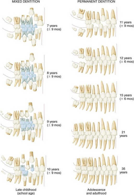
Appendix A includes color renditions of Figure 2-3Figure 2-4 considered in Chapter 2 on the eruption and development of the teeth. They can be used to demonstrate to patients the development of the dentitions from 5 months in utero to adolescence and adulthood.
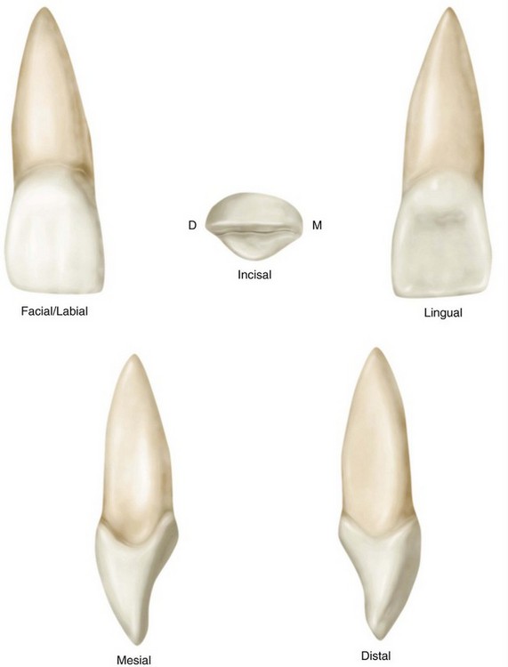
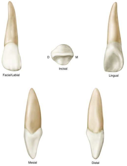
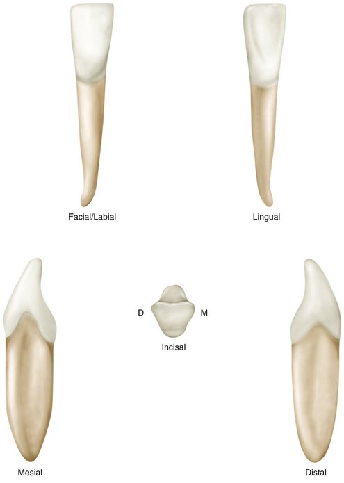
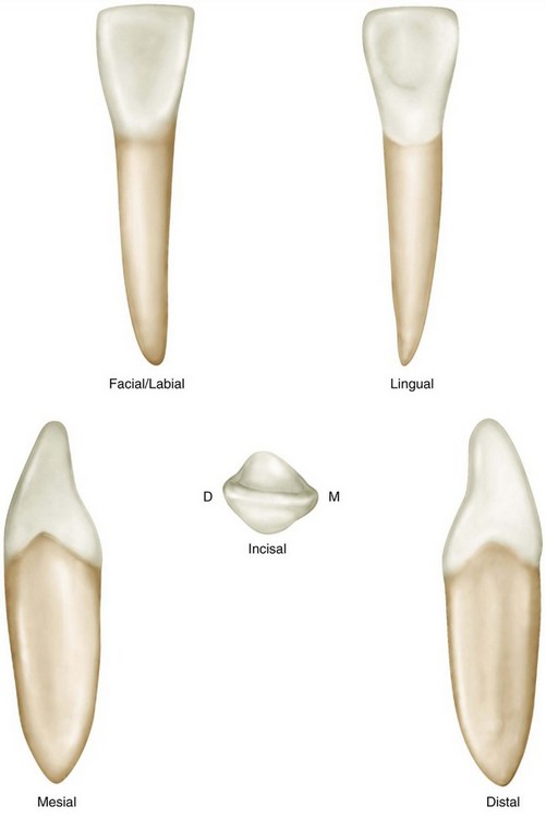
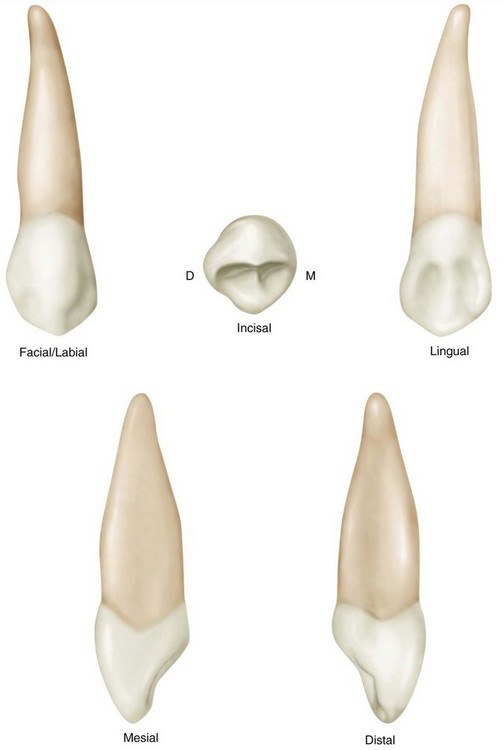
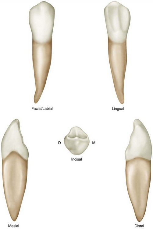
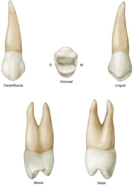
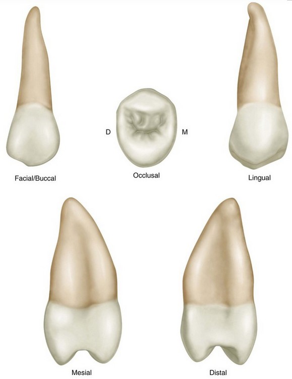
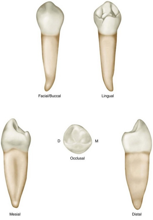
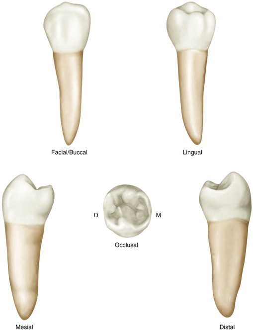
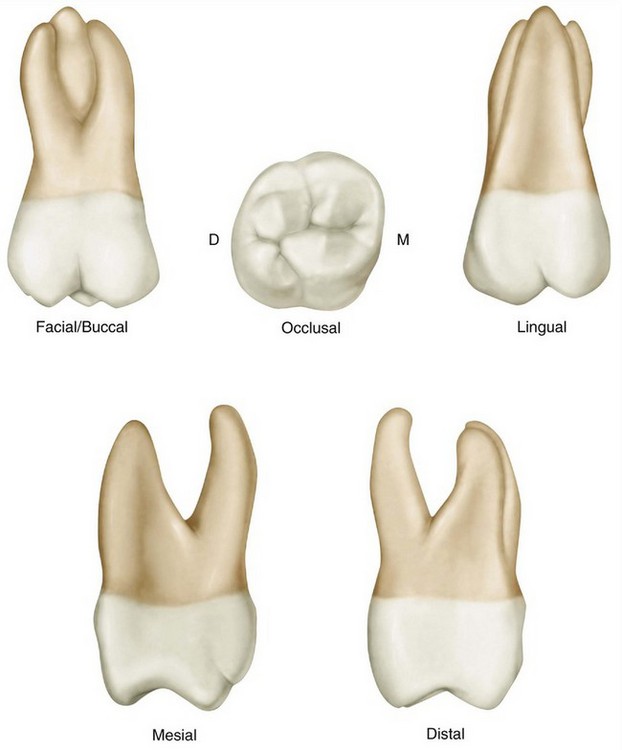
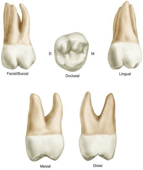
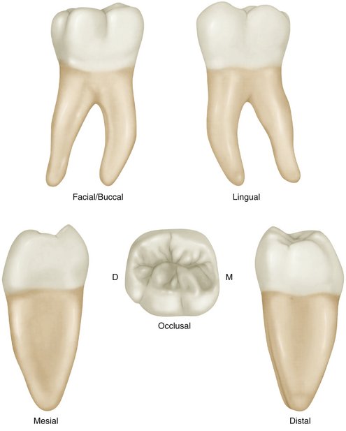
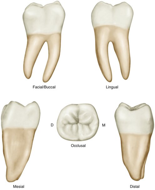
Also included are representative views of the facial/labial/buccal and occlusal/incisal aspects of all of the teeth considered in Chapter 6Chapter 7Chapter 8Chapter 9Chapter 10Chapter 11Chapter 12. Because of the proximity of these illustrations to the traits and characteristics of the teeth provided in Appendix B, page turns to earlier chapters can be minimized. It is also possible to view these same illustrations on the CD-ROM while examining the traits and characteristics in Appendix B.
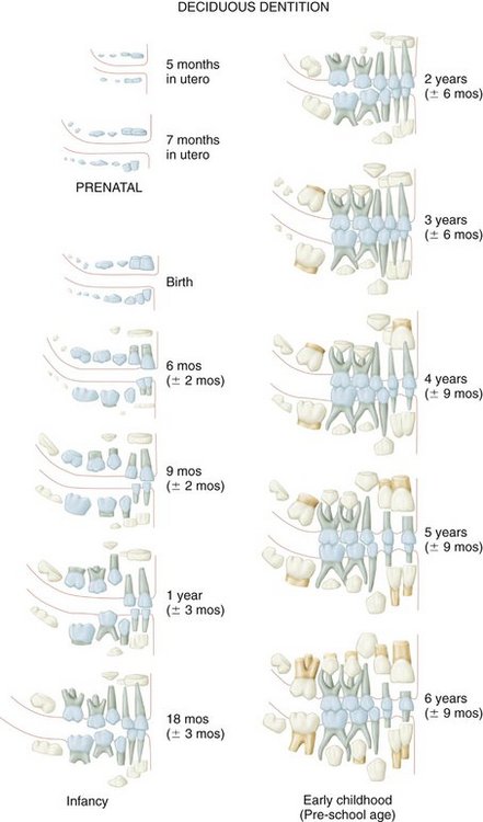
Appendix A-1 Development of the human dentition to the sixth year. The primary teeth are the blue ones in the illustration.
(Modified from Schour L, Massler M: The development of the human dentition, J Am Dent Assoc 28:1153, 1941.)