Peptic ulcers may be classified as acute or chronic, and peptic ulcer disease (PUD) is a chronic condition that affects the stomach or duodenum. Ulcers are described as gastric or duodenal and as primary or secondary. A gastric ulcer involves the mucosa of the stomach; a duodenal ulcer involves the pylorus or duodenum. Most primary ulcers occur in the absence of a predisposing factor and tend to be chronic, occurring more frequently in the duodenum. Stress ulcers result from the stress of a severe underlying disease or injury (e.g., severe burns, sepsis, increased intracranial pressure, severe trauma, multisystem organ failure) and are more frequently acute and gastric.
About 1.7% of children in general pediatric practices have PUD, and the disease represents about 3.4% per 10,000 of pediatric hospital admissions. Primary ulcers are more common in children older than 6 years, and stress ulcers are more common in infants younger than 6 months. Except for very young children, the incidence is two to three times greater in boys than in girls.
The exact cause is unknown, although infectious, genetic, and environmental factors are important. There is an increased familial incidence, and the disease is increased in persons with blood group O.
There is a significant relationship between the bacterium Helicobacter pylori (H. pylori) and ulcers. H. pylori is a microaerophilic, gram-negative, slow-growing, spiral-shaped, and flagellated bacterium known to colonize the gastric mucosa in about half of the population of the world (Cover and Blaser, 1996). It has been identified in 90% to 100% of adult patients with PUD. H. pylori synthesizes the enzyme urease, which hydrolyses urea to form ammonia and carbon dioxide. Ammonia then absorbs acid to form ammonium, thus raising the gastric pH. Also, its flagella allow the bacterium to swim across the viscous gastric mucus and reach the more neutral gastric pH below the mucus (Sgouros and Bergele, 2006). H. pylori may cause ulcers by weakening the gastric mucosal barrier and allowing acid to damage the mucosa. It is believed that it is acquired via the fecal-oral route, and this hypothesis is supported by finding viable H. pylori in feces.
In addition to ulcerogenic drugs, both alcohol and smoking contribute to ulcer formation. There is no conclusive evidence to implicate particular foods, such as caffeine-containing beverages or spicy foods, but polyunsaturated fats and fiber may play a role in ulcer formation. Psychologic factors may play a role in the development of PUD, and stressful life events, dependency, passiveness, and hostility have all been implicated as contributing factors.
Most likely, the pathology is due to an imbalance between the destructive (cytotoxic) factors and defensive (cytoprotective) factors in the GI tract. The toxic mechanisms include acid, pepsin, medications such as aspirin and nonsteroidal antiinflammatory drugs (NSAIDs), bile acids, and infection with H. pylori. The defensive factors include the mucus layer, local bicarbonate secretion, epithelial cell renewal, and mucosal blood flow. Prostaglandins play a role in mucosal defense because they stimulate both mucus and alkali secretion. The primary mechanism that prevents the development of peptic ulcer is the secretion of mucus by the epithelial and mucus glands throughout the stomach. The thick mucus layer acts to diffuse acid from the lumen to the gastric mucosal surface, thus protecting the gastric epithelium. The stomach and the duodenum produce bicarbonate, decreasing acidity on the epithelial cells and thereby minimizing the effects of the low pH (Chelimsky and Czinn, 2001). When abnormalities in the protective barrier exist, the mucosa is vulnerable to damage by acid and pepsin. Exogenous factors, such as aspirin and NSAIDs, cause gastric ulcers by inhibition of prostaglandin synthesis.
Zollinger-Ellison syndrome may occur in children who have multiple, large, or recurrent ulcers. This syndrome is characterized by hypersecretion of gastric acid, intractable ulcer disease, and intestinal malabsorption caused by a gastrin-secreting tumor of the pancreas.
Diagnosis is based on the history of symptoms, physical examination, and diagnostic testing. The focus is on symptoms such as epigastric abdominal pain, nocturnal pain, oral regurgitation, heartburn, weight loss, hematemesis, and melena (Box 24-9). History should include questions relating to the use of potentially causative substances such as NSAIDs, corticosteroids, alcohol, and tobacco. Laboratory studies may include a CBC to detect anemia, stool analysis for occult blood, liver function tests (LFTs), sedimentation rate, or CRP to evaluate IBD; amylase and lipase to evaluate pancreatitis; and gastric acid measurements to identify hypersecretion. A lactose breath test may be performed to detect lactose intolerance.
Radiographic studies such as an upper GI series may be performed to evaluate obstruction or malrotation. An upper endoscopy is the most reliable procedure to diagnose PUD. A biopsy is taken to determine the presence of H. pylori. It can also be diagnosed by a blood test that identifies the presence of the antigen to this organism. The C-urea breath test measures bacterial colonization in the gastric mucosa. This test is used to screen for H. pylori in adults and children. Polyclonal and monoclonal stool antigen tests are an accurate noninvasive method both for the initial diagnosis of H. pylori and for the confirmation of its eradication after treatment (Gisbert, de la Morena, and Abraira, 2006).
The major goals of therapy for children with PUD are to relieve discomfort, promote healing, prevent complications, and prevent recurrence. Management is primarily medical and consists of administration of medications to treat the infection and to reduce or neutralize gastric acid secretion.
Antacids are beneficial medications to neutralize gastric acid.
Histamine (H2) receptor antagonists (antisecretory drugs) act to suppress gastric acid production. Cimetidine (Tagamet), ranitidine (Zantac), and famotidine (Pepcid) are examples of these medications. These medications have few side effects.
PPIs, such as omeprazole and lansoprazole, act to inhibit the hydrogen ion pump in the parietal cells, thus blocking the production of acid. Controlled studies of these drugs have been done in adults, and these drugs are now commonly used to treat ulcers in children. They appear to be well tolerated and to have infrequent side effects (e.g., headache, diarrhea, nausea and vomiting).
Mucosal protective agents, such as sucralfate and bismuth-containing preparations, may be prescribed for PUD. Sucralfate is an aluminum-containing agent that forms a protective barrier over ulcerated mucosa to protect against acid and pepsin. Sucralfate is available in both pill and liquid forms. Because sucralfate blocks the absorption of other medications, it should be given separately from other medications.
Bismuth compounds are sometimes prescribed for the relief of ulcers, but they are used less frequently than PPIs. Although these compounds inhibit the growth of microorganisms, the mechanism of their activity is poorly understood. In combination with antibiotics, bismuth is effective against H. pylori. Although concern has been expressed about the use of bismuth salts in children because of potential side effects, none of these side effects has been reported when these compounds have been used in the treatment of H. pylori infection.
Triple drug therapy is the recommended treatment regimen for H. pylori (Ford, Delaney, Forman, and others, 2004). Combination therapy has demonstrated 90% effectiveness in eradication of H. pylori when compared with antibiotic monotherapy. Examples of drug combinations used in triple therapy are (1) bismuth, clarithromycin, and metronidazole; (2) lansoprazole, amoxicillin, and clarithromycin; and (3) metronidazole, clarithromycin, and omeprazole. Common side effects of medications include diarrhea, nausea, and vomiting. The yearly relapse rate of 80% for duodenal ulcer and 60% for gastric ulcer can be reduced to less than 5% after successful H. pylori eradication (Ford, Delaney, Forman, and others, 2004).
In addition to medications, the child with PUD should be given a nutritious diet and advised to avoid caffeine. Adolescents are warned about gastric irritation associated with alcohol use and smoking.
Children with an acute ulcer who have developed complications, such as massive hemorrhage, require emergency care. The administration of IV fluids, blood, or plasma depends on the amount of blood loss. Replacement with whole blood or packed cells may be necessary for significant loss.
Surgical intervention may be required for complications such as hemorrhage, perforation, or gastric outlet obstruction. Ligation of the source of bleeding or closure of a perforation is performed. A vagotomy and pyloroplasty may be indicated in children with recurring ulcers despite aggressive medical treatment.
Prognosis.: The long-term prognosis for PUD is variable. Many ulcers are successfully treated with medical therapy; however, primary duodenal peptic ulcers often recur. Complications such as GI bleeding can occur and extend into adult life. The effect of maintenance drug therapy on long-term morbidity remains to be established with further studies.
The primary nursing goal is to promote healing of the ulcer through compliance with the medication regimen. If an analgesic-antipyretic is needed, acetaminophen, not aspirin or NSAIDs, is used. Critically ill neonates, infants, and children in intensive care units should receive H2 blockers to prevent stress ulcers. Critically ill children receiving IV H2 blockers should have their gastric pH values checked at frequent intervals.
The role of stress in ulcer formation should be considered for nonhospitalized children with chronic illnesses. In children, many ulcers occur secondary to other conditions, and the nurse should be aware of family and environmental conditions that may aggravate or precipitate ulcers. Children may benefit from psychologic counseling and from learning how to cope constructively with stress.
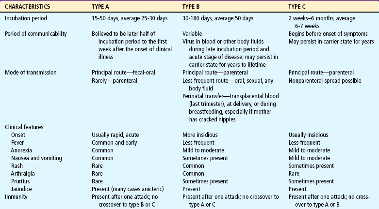
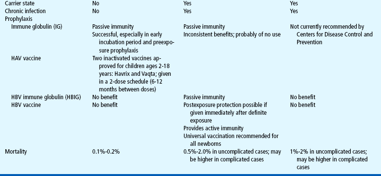
Hepatitis is an acute or chronic inflammation of the liver that can result from several different causes (e.g., virus, chemical or drug reaction, or other diseases). Nonviral causes of hepatitis include autoimmune hepatitis, Wilson disease, α1-antitrypsin deficiency, and steatohepatitis. The following six viruses cause 90% of cases of viral hepatitis (Table 24-4):
Hepatitis A.: HAV is the most common form of acute viral hepatitis in most parts of the world. It is a member of the picornavirus family. The virus produces a contagious disease transmitted primarily in contaminated stool spread via the fecal-oral route from person to person. HAV has been associated with miniepidemics in areas of poor hygiene and high population density. There is no chronic or carrier state. HAV infection affects individuals of all ages, but the highest incidence occurs among preschool- or school-age children younger than 15 years. Children may serve as the source of HAV infection in adults, such as in childcare center exposures. Usually HAV disease in children is mild. It is frequently anicteric and often subclinical. Infected children who show no symptoms may still spread the virus to others. HAV can be severe in children with immunodeficiency disorders. The incubation period is approximately 3 weeks. Although some cases may be prolonged, the prognosis is excellent. A highly effective vaccine for HAV was introduced in 1997; in 1999 the Advisory Committee on Immunization Practices recommended vaccination for all children over 24 months old living in the 11 states with the highest incidence of HAV (Amon, Darling, Fiore, and others, 2003; Balistreri, Chang, Ciocca, and others, 2002).
Hepatitis B.: HBV infection can occur as an acute or chronic infection and may range from being asymptomatic and limited to causing fatal fulminant (rapid and severe) hepatitis. HBV varies greatly throughout the world. High-prevalence areas have been identified in Africa and Asia; the United States is considered a low-prevalence area. Transmission is usually via the parenteral route through the exchange of blood or any bodily secretion or fluid. Infections from blood transfusion have been reduced as a result of blood product–screening procedures. Transplantation of organs, intimate physical contact, transmission from mother to infant, and the splashing of contaminated fluids into the mouth or eyes are other sources of infection. Adults whose occupations are associated with exposure to blood or blood products (such as health care workers) are at increased risk for infection and should receive HBV vaccination.
Most HBV infection in children is acquired perinatally. Newborns are at risk for hepatitis if the mother is infected with HBV or was a carrier of HBV during pregnancy. Possible routes of maternal-fetal or maternal-infant transmission include (1) leakage of virus across the placenta late in pregnancy or during labor and (2) ingestion of amniotic fluid or maternal blood. Infants who have HBV infection are more than 90% likely to become chronic carriers (Broderick and Jonas, 2003). The incubation period of HBV infection varies from 45 to 160 days.
HBV infection occurs in children and adolescents in the following high-risk groups:
Hepatitis C.: Approximately 1 in 50 people in the general population is positive for HCV antibodies (Centers for Disease Control and Prevention, 2001). About 0.2% to 0.4% of children younger than 12 years of age are infected with HCV. It is estimated that 4 million people in the United States are anti–HCV-positive. Approximately 7% of HCV-infected mothers transmit HCV to their newborns (Balistreri, Chang, Ciocca, and others, 2002). Another common route of infection is by percutaneous exposure, which occurs through transfusion of blood or blood products, transplantation of organs or tissues, or sharing of used needles. Transfusion-associated HCV infection is low, but a common cause of infection is injection drug use. The American Academy of Pediatrics (1998) suggests screening the following groups:
All infants born to HCV-infected women
Individuals who received blood products before 1992
The clinical course of HCV infection is variable. Incubation averages 6 to 7 weeks, with a range of 2 weeks to 6 months. Both acute and chronic HCV infection often produces only mild nonspecific symptoms or no symptoms at all (Bonkovsky and Mehta, 2001).
The length of time that maternal antibody is present in infants born to HCV-infected women must be considered, and screening should be done after the infant is 12 months old. However, a routine screening program, such as that for HBV, is not recommended. Current recommendations are to evaluate HCV-infected children at regular intervals to monitor for chronic hepatitis. Most children will be asymptomatic with evidence of chronic hepatitis on liver biopsy. Liver enzyme levels may fluctuate between periods of normal and elevated values.
Hepatitis D.: HDV is an important cause of acute and chronic liver disease. HDV is a defective RNA virus that requires the presence of HBV. HDV infection occurs primarily in hemophiliac patients and IV drug abusers. The incubation period is 2 to 8 weeks. Both acute and chronic forms are more severe than HBV infection and can lead to cirrhosis. Testing for HDV infection is recommended in children with chronic HBV infection or severe liver disease and in children with acute exacerbation of a previously stable liver disease.
Hepatitis E.: HEV infection is enterally transmitted. Transmission may occur through the fecal-oral route or from contaminated water. The incubation period is 2 to 9 weeks. This illness is uncommon in children, does not cause chronic liver disease, is not a chronic condition, and has no carrier state. The mortality rate resulting from submassive hepatic necrosis is low except in pregnant women in their third trimester, in whom mortality reaches 20%.
Hepatitis G.: HGV is a blood-borne virus that may also be transmitted by organ transplantation. High-risk groups include transfusion recipients, IV drug users, and individuals infected with HCV. Individuals with the virus are often asymptomatic, and most infections are chronic. The incubation period is unknown.
Diagnosis is based on the history (especially regarding possible exposure to a hepatitis virus); physical examination; and serologic markers (antibodies or antigens) indicating the presence of active infection with hepatitis A, B, or C or previous infection. Because the liver has a large functional reserve, abnormal laboratory tests may be the only indication of hepatitis. However, LFTs are not specific for the diagnosis of viral hepatitis. Although serum aspartate and alanine aminotransferase (AST and ALT) levels are markedly elevated in viral hepatitis, other diseases or conditions may cause their elevation. Serum bilirubin levels peak 5 to 10 days after clinical jaundice appears. When hepatitis is severe, albumin levels are depressed and prothrombin times are increased.
Diagnosis of viral hepatitis is based on the presence of specific viral markers. Diagnosis of acute HAV infection is based on the presence of anti-HAV immunoglobulin (immunoglobulin M [IgM]) antibody in the serum. HBV diagnosis depends on the presence of hepatitis B surface antigen (HBsAg) or anti-HBV core (anti-HBc) IgM antibody. Chronic HBV infection is associated with the persistence of HBsAg and HBV DNA markers. The diagnosis of HCV is based on the detection of anti-HCV antibodies and confirmation by polymerase chain reaction for hepatitis C RNA.
An abdominal ultrasound provides measurement of liver size, detection of cystic lesions and stones, and imaging of the gallbladder. Cholescintigraphy radionuclide imaging detects abnormalities in liver uptake, concentration, and excretory function. Finally, a liver biopsy aids in assessing the severity of the disease.
Pathologic changes occur primarily in the parenchymal cells of the liver and result in varying degrees of swelling, infiltration of liver cells by mononuclear cells, subsequent degeneration, necrosis, and fibrosis.
Hepatitis can be self-limited, and complete regeneration of liver cells without scarring may occur. However, some forms of hepatitis do not result in complete return of liver function. These include fulminant hepatitis, which is characterized by a severe, acute course and massive destruction of the liver, resulting in liver failure and death in 1 to 2 weeks. Subacute or chronic active hepatitis is characterized by progressive liver destruction, uncertain regeneration, scarring, and potential cirrhosis.
The initial anicteric (absence of jaundice) phase usually lasts 5 to 7 days and is often mistaken for influenza. Symptoms include nausea, vomiting, extreme anorexia, malaise, easy fatigability, arthralgia, skin rashes, slight to moderate fever, and epigastric or upper right quadrant abdominal pain. Dark urine is a symptom of the icteric (jaundice) phase. Pruritus may accompany jaundice and can be bothersome, but many children with acute viral hepatitis do not develop jaundice.
Treatment options for viral hepatitis are limited. The goals of management include early detection, recognition of chronic liver disease, support and monitoring, and prevention of spread of the disease.
HAV infection is an acute disease that resolves with support and management of symptoms. HBV and HCV treatment is directed at managing the viral load to prevent further destruction of the liver. Currently, HBV and HCV are treated with interferons, naturally occurring proteins that exert antiviral, antiproliferative, and immunomodulatory effects. A recent interferon formulation, pegylated interferon, can be administered once a week and has been found to sustain plasma levels and enhance viral suppression (Karnam and Reddy, 2003). Lamivudine and adefovir are two other interferon analogs that suppress the replication of HBV (Yuen and Lai, 2001). A combination of α-interferon and ribavirin has resulted in a sustained response in only 50% of patients with HBV and HCV (Waters and Nelson, 2006).
Another important aspect of the therapeutic management of hepatitis involves hospitalization. Hospitalization is necessary if coagulopathy or fulminant hepatitis is present.
Prevention.: Proper hand washing and standard isolation precautions can prevent the spread of hepatitis. Prophylactic use of standard immune globulin (Ig) is effective in preventing HAV infection in situations of preexposure (e.g., anticipated travel to areas where HAV is prevalent) or in situations of postexposure during the early part of the incubation period. Hepatitis B immune globulin (HBIg) is effective in preventing HBV infection after exposure. Ig and HBIg must be administered less than 2 weeks after exposure.
Vaccines have been developed to prevent HAV and HBV infection. HBV vaccination is recommended for all newborns and for high-risk groups. HAV is also recommended for high-risk groups (see Immunizations, Chapter 10). Active immunizations are not available against HCV. It is possible to prevent HDV infection by preventing HBV infection.
Prognosis.: The prognosis for children with hepatitis is variable and depends on the type of virus. HAV usually causes a mild and brief illness with no carrier state. HBV causes a wide spectrum of acute and chronic illness. Approximately 5% of individuals develop chronic hepatitis B each year, and about half of these develop fulminant liver failure, leading to death in the absence of liver transplantation (Kim, Gores, Benson, and others, 2005). Hepatocellular carcinoma is a potentially fatal complication of HBV infection. HCV causes acute hepatitis that progresses to chronic disease in more than 85% of affected individuals, with approximately 15% to 20% developing cirrhosis or hepatocellular carcinoma (Centers for Disease Control and Prevention, 2001; Richmond, Dunning, and Desmond, 2004). Chronic HCV infection is the leading indication for liver transplantation in adults in the United States (Regev and Schiff, 2000).
Nursing objectives depend on the severity of the hepatitis, the medical management, and factors influencing the control and transmission of the disease. Children with benign viral hepatitis are frequently cared for at home, and the clinic or office nurse must explain the medical therapy and control measures. If further assistance is needed for parents to comply with therapy, a public health nursing referral may be necessary.
A well-balanced diet and a realistic schedule of rest and activity adjusted to the child’s condition are encouraged. HAV is not infectious within a week after the onset of jaundice, and children may feel well enough to resume school. Parents are cautioned about administering any medication to the child, since normal dosages of many drugs may become dangerous because of the liver’s inability to detoxify and excrete them. Hand washing is the single most critical measure in reducing risk of transmission. The nurse should explain to parents and children the ways in which HAV (oral-fecal route) and HBV (parenteral route) are spread.
Nurses caring for young people with HBV infection and a known or suspected history of illicit drug use should help these teens realize the dangers of drug abuse. Nurses should stress the parenteral mode of transmission of hepatitis and encourage them to seek counseling through a drug program. HBV and HCV are chronic diseases that require frequent monitoring and management. Many communities have multidisciplinary clinics dedicated to the management of these diseases.
Cirrhosis occurs at the end stage of many chronic liver diseases, including biliary atresia (BA) and chronic hepatitis. Cirrhosis can also result from infectious, autoimmune, or toxic factors and from chronic diseases such as hemophilia and cystic fibrosis. A cirrhotic liver is irreversibly damaged.
Clinical manifestations in children are similar to those seen with all chronic liver disorders. Children exhibit jaundice, poor growth, anorexia, muscle weakness, and lethargy. Ascites, edema, GI bleeding, anemia, and abdominal pain may be present with impaired intrahepatic blood flow. Pulmonary function may be impaired because of pressure against the diaphragm from hepatosplenomegaly and ascites. Dyspnea and cyanosis may occur, especially on exertion. Intrapulmonary arteriovenous shunts may develop and cause hypoxemia. Spider angiomas and prominent blood vessels are often present on the upper torso.
Therapy is directed toward (1) frequent assessment of liver status with physical examination and LFTs and (2) management of specific complications. The only successful treatment for end-stage liver disease and liver failure may be liver transplantation, which has improved the prognosis substantially for many children with cirrhosis. Currently, the 1-year survival rate for liver transplantation is 85%. Increasing numbers of recipients are reaching their second decade after transplant. The increasing life span after transplantation is related to advances in surgical techniques and improved preoperative, intraoperative, and postoperative care (Atkison, Ross, Williams, and others, 2002).
Prognosis.: Liver transplantation has revolutionized the approach to liver cirrhosis. Liver failure and cirrhosis are indications for transplantation. Liver transplantation reflects the failure of other medical and surgical measures to prevent or treat cirrhosis. Careful monitoring of the child’s condition and quality of life is necessary to evaluate the need for and timing of transplantation (see Family Focus box).
Nursing care of the child with cirrhosis is determined by the cause of the cirrhosis, the severity of complications, and the prognosis. The prognosis for life is poor unless successful liver transplantation occurs. Nursing care of this child is similar to that for any child with a life-threatening illness (see Chapter 18). Hospitalization is usually required when complications occur.
BA is a destructive, idiopathic, inflammatory process that leads to fibrosis and obliteration of the biliary tree (Emerick and Whitington, 2006). BA has been detected in 3.7 in 10,000 live births (Chen, Chang, Du, and others, 2006). The disorder is more common in girls and preterm infants. In the United States, the incidence is twice as high in African-Americans as in Caucasian infants, and more common in Chinese than in either Japanese or Caucasian populations.
The exact cause of BA is unknown. Because BA has two distinct forms, postnatal and fetal-embryonic, different pathogenic mechanisms are suggested. Postnatal BA represents 65% to 90% of cases and is probably the result of infection or an immune-mediated mechanism. Jaundice, manifesting with yellow discoloration of the skin or sclerae, is the most common early symptom of BA. Jaundice, indicating cholestasis (the accumulation of compounds that cannot be excreted because of occlusion or obstruction of the biliary tree), can be visible at a total serum bilirubin concentration as low as 5.0 mg/dl. An abnormal direct bilirubin has been designated as greater than 1.0 mg/dl if the total bilirubin is less than 5 mg/dl or a value of direct bilirubin that represents more than 20% of the total bilirubin if it is greater than 5 mg/dl (Emerick and Whitington, 2006). Direct hyperbilirubinemia first appears after the resolution of physiologic jaundice. Jaundice is often associated with pale stool and dark urine. Histologic study demonstrates bile duct remnants and a progressive inflammatory process. In the fetal embryonic form of BA, which represents 10% to 35% of cases, there is a congenital absence of biliary ductal patency and an absence of bile duct remnants. Many infants have associated congenital anomalies. Varying degrees of cholestasis occur, resulting in retention of irritants and toxins. Injury to the liver occurs as the result of the inflammation caused by the cholestasis.
Early diagnosis is the key to the survival of the child with BA. Infants who undergo surgery in the first 60 days of life have an 80% chance of establishing bile flow. Between 60 to 90 days of life, the chance of reestablishing flow drops to 50%, and after 90 days to 10% (Chen, Chang, Du, and others, 2006). The typical infant is thriving, appears well, and has only very mild jaundice during the first 6 to 8 weeks (Emerick and Whitington, 2006) but will soon begin failing to thrive. Several clinical signs may indicate the presence of BA (Box 24-10). Blood tests should include a CBC, electrolytes, bilirubin, and liver enzymes. Additional laboratory analyses, including α1-antitrypsin level, TORCH titers (see Maternal Infections, Chapter 9), hepatitis serology, α-fetoprotein, urine cytomegalovirus, and a sweat test, are indicated to rule out other conditions that cause persistent cholestasis and jaundice. Abdominal ultrasonography allows inspection of the liver and biliary system. Hepatobiliary scintigraphy demonstrates biliary patency but does not provide diagnostic certainty. Endoscopic retrograde cholangiopancreatography (ERCP) is performed in very young infants. This procedure, which is done using general anesthesia, has an 80% reported diagnostic accuracy. Percutaneous liver biopsy is highly reliable when the biopsy contains specimens from a number of portal areas. Definitive diagnosis of BA is obtained during surgical laparotomy and an intraoperative cholangiogram.
The primary treatment of BA is hepatic portoenterostomy (Kasai procedure), in which a segment of intestine is anastomosed to the resected porta hepatis to attempt bile drainage. Bile drainage is achieved in approximately 80% to 90% of infants who undergo surgery when younger than 10 weeks of age (Ohi, 2001). However, progressive cirrhosis still occurs in many children, necessitating liver transplantation. Prophylactic antibiotics are given after the Kasai procedure to minimize the risk of ascending cholangitis.
Medical management is primarily supportive. It includes nutritional support with infant formulas that contain medium-chain triglycerides and essential fatty acids. Supplementation is usually required with fat-soluble vitamins; a multivitamin; and minerals, including iron, zinc, and selenium. Aggressive nutritional support with continuous tube feedings or TPN is indicated for moderate to severe failure to thrive. The enteral solution should be low in sodium. Ursodeoxycholic acid is used to treat pruritus and hypercholesterolemia.
Prognosis.: Untreated BA results in progressive cirrhosis and death in most children by 2 years of age. The Kasai procedure improves the prognosis but is not a cure. Biliary drainage can often be achieved if the surgery is done before the intrahepatic bile ducts are destroyed. Long-term survival has been reported in children who receive the Kasai procedure; however, even with successful bile drainage, many children ultimately develop liver failure.
Advances in surgical techniques and the use of immunosuppressive and antifungal drugs have improved the success of transplantation. The major obstacle continues to be a shortage of donor livers. Reduced-size, split-liver transplantation, retransplantation, and increased public awareness may improve donor organ availability in the future (see Ethical Case Study).
Nursing interventions for the child with BA include support of the family before, during, and after surgical procedures and education regarding the treatment plan. In the postoperative period of a portoenterostomy, nursing care is similar to that after major abdominal surgery. Family members need education relating to the proper administration of medications and nutritional therapy, including special formulas, vitamin and mineral supplements, tube feedings, or parenteral nutrition. Pruritus can often be relieved by drug therapy or comfort measures such as baths.
Children and their families also need psychosocial support. The uncertain prognosis, discomfort, and waiting for transplantation produce stress, and hospitalizations, pharmacologic therapy, and nutritional therapy impose financial burdens on the family. Families can receive help from the Children’s Liver Disease Foundation,* an organization that provides educational materials, programs, and support systems.
Clefts of the lip (CL) and palate (CP) are facial malformations that occur during embryonic development and are the most common congenital deformities of the head and neck. They may appear separately or, more often, together. CL results from failure of the maxillary and median nasal processes to fuse; CP is a midline fissure of the palate that results from failure of the two sides to fuse.
CL may vary from a small notch to a complete cleft extending into the base of the nose (Fig. 24-3). Clefts can be unilateral or bilateral. Deformed dental structures are associated with CL. CP alone occurs in the midline and may involve the soft and hard palates. When associated with CL, the defect may involve the midline and extend into the soft palate on one or both sides.
FIG. 24-3 Variations in clefts of lip and palate at birth. A, Notch in vermilion border. B, Unilateral cleft lip and palate. C, Bilateral cleft lip and palate. D, Cleft palate.
Cleft lip and palate (CL/P) is more common than CP alone and varies by ethnicity. The occurrence is 1 in 1000 births in Caucasians; 1 in 500 births in Native Americans and Asians, and 1 in 2000 births in African-Americans. CP occurs alone in only 1 in 2500 cases and does not display variation by ethnicity (Wilkins-Haug, 2003). Approximately 60% to 80% of children born with CL/P are male. Females have a higher frequency of isolated clefts of the secondary palate. Unilateral clefts are nine times more common than bilateral clefts and occur twice as frequently on the left side. Isolated bilateral CLs are uncommon; approximately 86% of those with bilateral CL also have palatal clefts. Approximately 68% of those with unilateral CLs have an associated palatal cleft (Kirschner and LaRossa, 2000).
Cleft deformities may be an isolated anomaly, or they may occur with a recognized syndrome. CL with or without CP is distinct from isolated CP. Clefts of the secondary palate alone are more likely to be associated with syndromes than is isolated CL or CL with CP.
CL and CP may be caused by exposure to teratogens such as alcohol, anticonvulsants, steroids, and retinoids, but there is little evidence to link isolated clefts to any single teratogenic agent with the exception of phenytoin. Use of phenytoin during pregnancy is associated with a tenfold increase in the incidence of CL. The incidence of CL among mothers who smoke during pregnancy is twice as great as the incidence in mothers who do not (Eppley, van Aalst, Robey, and others, 2005).
Cleft deformities represent a genetic defect in cell migration that results in a failure of the maxillary and premaxillary processes to come together between the third and twelfth week of embryonic development. Although often appearing together, CL and CP are distinct malformations embryologically, occurring at different times during the developmental process. Merging of the upper lip at the midline is completed between the seventh and eleventh weeks of gestation. Fusion of the secondary palate (hard and soft palate) takes place later, between the seventh and twelfth weeks of gestation. In the process of migrating to a horizontal position, the palates are separated by the tongue for a short time. If there is delay in this movement, or if the tongue fails to descend soon enough, the remainder of development proceeds but the palate never fuses.
CL with or without CP is apparent at birth. The defect elicits severe emotional reactions in parents. CP is less obvious than CL and may not be detected without a thorough assessment of the mouth. CP is identified when the examiner places a gloved finger directly on the palate. Clefts of the hard palate form a continuous opening between the mouth and the nasal cavity. The severity of the CP has an impact on feeding; the infant is unable to generate negative pressure and create suction in the oral cavity. This impairs feeding, even though in most cases the infant’s ability to swallow is normal.
Prenatal diagnosis with fetal ultrasound is not reliable until the soft tissues of the fetal face can be visualized at 13 to 14 weeks. The sensitivity of fetal ultrasound for facial clefting is almost 100% when CL/P is associated with other structural anomalies. An intact lip is the most difficult to diagnose prenatally (Wilkins-Haug, 2003).
Treatment of the child with CL is surgical and involves no long-term interventions other than possible scar revision. The management of CP involves the cooperative efforts of a multidisciplinary health care team, including pediatrics, plastic surgery, orthodontics, otolaryngology, speech/language pathology, audiology, nursing, and social work. Management is directed toward closure of the cleft(s), prevention of complications, and facilitation of normal growth and development in the child. Until recently, repair of cleft deformities in the neonate was not considered safe. Surgery is now possible in younger neonates because of advances in pediatric anesthesiology and neonatology. However, the infant must be free of any oral, respiratory, or systemic infections. In deformities of both the lip and palate, the palate is repaired first to avoid disrupting the lip after it has been repaired.
Surgical Correction of Cleft Lip.: The two most common procedures for repair of CL are the Tennison-Randall triangular flap (Z-plasty) and the Millard rotational advancement technique. The difference between these two is that the Tennison-Randall procedure crosses the philtral line and the Millard procedure advances a triangle of tissue in the upper third of the lip and does not cross the midline. Surgeons often use a combination of these two techniques to address individual differences. Improved surgical techniques have minimized scar retraction, and in the absence of infection or trauma, healing occurs with little scar formation. Optimal cosmetic results, however, are difficult to obtain in severe defects. Additional revisions may be necessary at a later age.
Surgical Correction of Cleft Palate.: CP repair was previously postponed until a later age than the repair of the CL to take advantage of palatal changes that take place with normal growth. With advanced surgical and anesthesia techniques, many surgeons are currently performing palatal repairs in the neonatal period (Sandberg, Magee, and Denk, 2002). The timing of repair remains controversial. Most surgeons prefer to close the cleft before the child develops faulty speech habits.
Prognosis.: Even with good anatomic closure, most children with CL/P have some degree of speech impairment that requires speech therapy. Physical problems result from inefficient functioning of the muscles of the soft palate and nasopharynx, improper tooth alignment, and varying degrees of hearing loss. Improper drainage of the middle ear, as a result of inefficient function of the eustachian tube, contributes to recurrent otitis media with scarring of the tympanic membrane, which leads to hearing impairment in many children with CP. Upper respiratory tract infections require immediate and meticulous attention, and extensive orthodontics and prosthodontics may be needed to correct malposition of teeth and maxillary arches.
Long-term problems are related to the child’s social adjustment. The better the physical care, the better is the chance for emotional and social adjustment, although the degree of residual disability are not always directly related to a satisfactory adjustment. Physical defects are a threat to the self-image, and abnormal speech quality is an impediment to social expression.
The immediate nursing problems for an infant with CL/P deformities are related to feeding and dealing with the parental reaction to the defect. Facial deformities are particularly disturbing to parents. CL is a visible defect that may produce a strong negative response in parents, which could influence maternal- and paternal-infant attachment. However, a study of infants with CL or CP indicated that maternal-infant attachment was not negatively affected when measured at 1 year of age (Speltz, Endriga, Fisher, and others, 1997). During the initial phase following birth, it is important for the nurse to emphasize not only on the infant’s physical needs, but also the parents’ emotional needs. The manner of handling the infant should convey to the parents that the infant is a precious human being. (See Chapter 18 for interventions in assisting parents in accepting a birth defect.) Throughout the course of therapy, parents need explanations of the immediate and long-range problems associated with CP. They may be unaware that more is involved than repairing the defect. Whenever possible, they should be referred to a comprehensive CP team.
Feeding.: Feeding the infant presents a challenge to nurses and parents. Growth failure in infants with CL or CP has been attributed to preoperative feeding difficulties. After surgical repair, most infants who have isolated CL or CP with no associated syndromes gain weight or achieve adequate weight and height for age.
CL or CP reduces the infant’s ability to suck, making bottle-feeding and breastfeeding difficult. In breastfeeding the CL or CP interferes with compression of the areola. Liquid taken into the mouth escapes via the cleft through the nose. Feeding is best accomplished with the infant’s head in an upright position, either held in the caregiver’s hand or cradled in the arm. Normal nipples are unsuitable for these infants, who are unable to generate the suction required. A variety of special “cleft palate” nipples have been devised and used with some success. However, large, soft nipples with large holes; Nursettes; or long, soft lamb’s nipples appear to offer the best means for nipple feeding. The newer “gravity flow” nipple attached to a squeezable plastic bottle allows formula to be deposited directly into the mouth in the same manner as with a bulb syringe. Success has also been achieved by modification of a standard nipple. A single small slit or crosscut is made in the end of the nipple with a sharp surgical blade or pair of sharp, thin scissors. The enlarged opening, which can be adjusted to the infant’s individual needs, allows the infant to swallow the formula easily, bypassing the suction problem.
The ESSR feeding technique also works well with these infants. The steps in ESSR are (1) Enlarge the nipple, (2) Stimulate the suck reflex, (3) Swallow fluid appropriately, and (4) Rest when the infant signals with facial expression. Infants fed with the ESSR method revealed a significantly greater increase in their mean weight before surgery than infants fed with traditional methods (Richard, 1994).
Using special or modified nipples or feeding techniques helps meet the infant’s sucking needs. Muscle development is important for later development of speech. During feeding, the nipple is positioned so that it is compressed by the infant’s tongue and existing palate. If a single-slit nipple is used, the slit is placed vertically so that the infant will be able to produce and stop a flow of milk by alternately opening and closing the opening. No matter which type of nipple is used, gentle, steady pressure on the base of the bottle reduces the chance of choking or coughing. The person feeding should resist the temptation to remove the nipple because of the noise the infant makes or fear that the infant will choke. These infants swallow excessive amounts of air, and they require frequent burping.
When the infant has trouble with nipple feeding, a rubber-tipped medicine dropper, Asepto syringe, or Breck feeder (a large syringe with soft rubber tubing) provides an efficient, safe feeding device. The rubber extension should be long enough to extend back into the mouth to reduce the likelihood of regurgitation through the nose. The formula is deposited on the back of the tongue, and the flow is controlled by bulb or syringe compression that is adjusted to the infant’s needs. With some infants, spoon feeding works best, especially if the formula is slightly thickened with cereal. After feeding, the infant is given water to rinse the mouth.
Many infants with CL/P can breastfeed. The nipple is positioned and stabilized well back in the oral cavity so that tongue action facilitates milk expression. The suction required to stimulate milk may be absent, so a breast pump may be useful before nursing to stimulate the let-down reflex.
Regardless of the feeding method used, the mother should begin feeding the infant as soon as possible, preferably after the initial nursery feeding. When maternal feeding is initiated early, the mother can help to determine the method best suited to her and the infant and can become adept in the technique before discharge from the hospital.
Preoperative Care.: In preparation for surgical repair, parents are frequently taught to accustom the infant to the needs of the early postoperative period, especially if surgery is delayed for several months. It is mandatory for the infant to be positioned on the back or side postoperatively. Most infants tolerate these positions well because they are accustomed to being supine for sleeping. It is also helpful to place the infant or child in arm restraints periodically before admission and to feed the infant with a rubber-tipped Asepto syringe or other device that will be used postoperatively.
Postoperative Care for Cleft Lip.: The major efforts in the postoperative period are directed toward protecting the operative site. After CL repair (cheiloplasty), a metal appliance or adhesive strips are securely taped to the cheeks to relax the surgical site and prevent tension on the suture line caused by crying or other facial movement. Efforts should be made to prevent crying as much as possible to avoid stress on the suture line. Elbow restraints to prevent the infant from rubbing or disturbing the suture line are applied immediately after surgery. It is advisable to pin the cuff of the restraints to the infant’s clothing to keep the restraints in place. Older infants who roll over require a jacket restraint in addition to restricting arm movement to prevent rolling on the abdomen and rubbing the face on the sheet, especially if the repair involves the lip. It is important to remove the elbow restraints periodically to exercise the arms, to provide relief from restrictions, to observe the skin for signs of irritation, and to provide an opportunity for cuddling and body contact. Restraints should be released one at a time. Sitting the infant in an infant seat provides a change of position and a different view of the environment. Adequate analgesia is required to relieve postoperative pain and to prevent restlessness.
Clear liquids are offered when the infant has fully recovered from the anesthesia, and feeding is resumed when tolerated. The suture site is carefully cleansed of formula or serosanguineous drainage as needed with a cotton-tipped swab dipped in saline. A thin layer of antibiotic ointment may be prescribed for application to the suture line after cleansing. Meticulous care of the suture line is essential because inflammation or infection will interfere with optimal healing and the ultimate cosmetic effect of the surgical repair. Gentle aspiration of mouth and nasopharyngeal secretions may be necessary to prevent aspiration and respiratory complications. An upright or infant seat position is helpful in the immediate postoperative period (especially for the infant who has difficulty handling secretions).
Postoperative Care of Cleft Palate.: The child with CP repair (palatoplasty) is allowed to lie on the abdomen immediately postoperatively. The child may resume feeding by bottle, breast, or cup shortly after surgery.
Oral packing may be secured to the palate after palatoplasty; this packing is usually removed after 2 to 3 days. Sometimes the infant will have difficulty breathing after surgery, since it is often necessary to alter an established pattern of breathing and adjust to breathing through the nose. This is frustrating but seldom requires more than positioning and support. The elbows may be restrained to keep the child’s hands away from the mouth. Parents are instructed to maintain elbow restraints at home until the palate is healed, usually in 4 to 6 weeks. They are instructed to remove the restraints (one at a time) frequently to allow the child to exercise the arms.
The nurse must assess the infant’s or child’s level of postoperative pain. Opioids may be prescribed initially, and acetaminophen may be given as needed thereafter. It is important to manage pain to decrease crying in infants with CL repair.
The older infant or child may be discharged on a blenderized or soft diet, and parents are instructed to continue the diet until the surgeon directs them otherwise. Parents are cautioned against allowing the child to eat hard items (such as toast, hard cookies, and potato chips) that can damage the repaired palate. The expected outcomes are described in the Nursing Process box.
Long-Term Care.: Children with CL/P often require a variety of services during recovery. Family members need support and encouragement by health professionals and guidance in activities that facilitate a normal outcome for their child. Parents frequently cite financial stress as a difficult issue. With the combined efforts of the family and the health team, most children achieve a satisfactory outcome. Many children with CL/P have surgical correction that creates a near normal–appearing lip and permits good function. Parents need to understand the function of therapy and the purpose and care of all appliances, as well as the importance of establishing good mouth care and proper brushing habits.
Throughout the child’s development, an important goal is the development of a healthy personality and self-esteem. Many communities have CP parents’ groups that offer help and support to families. Agencies that provide services and information for children with CL/P and their families include the American Cleft Palate–Craniofacial Association, the Cleft Palate Foundation, the Birth Defects Research for Children, the March of Dimes, and various state children’s medical services.
Congenital atresia of the esophagus and tracheoesophageal fistula (TEF) are rare malformations that result from failed separation of the esophagus and trachea by the fourth week of gestation. These defects occur as separate entities or in combination (Fig. 24-4). They have a fatal outcome without early diagnosis and treatment.
Esophageal atresia (EA) with or without an associated TEF is the most common esophageal malformation, occurring in approximately 1 in 3500 live births (Shaw-Smith, 2006). There appears to be an equal sex incidence, but the birth weight of most affected infants is significantly lower than average, and incidence of prematurity is unusually high. A history of maternal polyhydramnios is present in approximately 50% of infants with the defects. EA/TEF is often present with the VATER or VACTERL syndromes, acronyms for syndromes involving a combination of Vertebral, Anorectal, Cardiovascular, Tracheoesophageal, Renal, and Limb abnormalities. The cardiac and renal anomalies occur most frequently with EA/TEF.
The cause of EA/TEF is unknown. In the most frequently encountered form of EA and TEF (80% to 95% of cases), the proximal esophageal segment terminates in a blind pouch, and the distal segment is connected to the trachea or primary bronchus by a short fistula at or near the bifurcation (see Fig. 24-4, C). The second most common variety (5% to 8%) consists of a blind pouch at each end, widely separated and with no communication to the trachea (see Fig. 24-4, A). Less frequently, an otherwise normal trachea and esophagus are connected by a common fistula (see Fig. 24-4, E). Extremely rare anomalies involve a fistula from the trachea to the upper esophageal segment (see Fig. 24-4, B) or to both the upper and the lower segments (see Fig. 24-4, D).
The disorder is suspected on the basis of clinical manifestations (Box 24-11). EA should also be suspected in cases of maternal polyhydramnios. Although the diagnosis is established on the basis of clinical signs and symptoms, the exact type of anomaly is determined by radiographic studies. A radiopaque catheter is inserted into the hypopharynx and advanced until it encounters an obstruction. Chest films are taken to ascertain esophageal patency or the presence and level of a blind pouch. Sometimes fistulas are not patent, which makes them more difficult to diagnose. The presence of gas in the stomach or small bowel is indicative of a coexisting TEF.
EA is a surgical emergency. The treatment includes maintenance of a patent airway, prevention of pneumonia, gastric or blind pouch decompression, and surgical repair of the anomaly. When EA/TEF is suspected, the infant is immediately taken off oral intake, started on IV fluids, and placed in the position least likely to cause aspiration of either mouth or stomach secretions. Removal of secretions from the mouth and upper pouch requires frequent or continuous suction. Because aspiration pneumonia is almost inevitable and appears early, broad-spectrum antibiotic therapy is often instituted.
Primary surgical correction consists of a thoracotomy with division and ligation of the TEF and an end-to-side anastomosis of the esophagus. This may consist of one operation or be staged with two or more procedures. For infants who are preterm, have multiple anomalies, or are in poor condition, a staged procedure is preferred that involves palliative measures, including gastrostomy, ligation of the TEF, and provision of constant drainage of the esophageal pouch. A delayed esophageal anastomosis is usually attempted after several weeks to months when the upper pouch elongates. Further surgical techniques may be performed later to facilitate esophageal lengthening. If an esophageal anastomosis still cannot be accomplished, a cervical esophagostomy (to allow drainage of saliva) and gastrostomy are performed.
A primary anastomosis may be impossible because of insufficient length of the two segments of the esophagus. In these cases, an esophageal replacement procedure using a part of the colon, or gastric tube interposition may be necessary to bridge the missing esophageal segment. Endotracheal intubation may be required, since many infants (10% to 20%) also have tracheomalacia, a weakness in the tracheal wall that occurs when a dilated proximal pouch compresses the trachea in early fetal life or when the trachea does not develop normally because of a loss of intratracheal pressure.
Complications of a primary repair include an anastomotic leak, strictures resulting from tension or ischemia, esophageal motility disorders causing dysphagia, and GER.
Prognosis.: The prognosis is related to the birth weight, associated congenital anomalies, and time of diagnosis. The survival rate is nearly 100% in full-term infants without severe respiratory distress or other anomalies. In preterm low-birth-weight infants with associated anomalies, the incidence of complications is high.
Nursing responsibility for detection of this malformation begins immediately after birth. Ideally, the diagnosis should be made before the initial feeding, but often it is not. If fed, the infant swallows normally but suddenly coughs and struggles, and the fluid is aspirated or returns through the nose and mouth. For this reason, it is customary for the nurse to give the infant the first feeding of plain water or to be present when a parent feeds the child to observe the infant’s response. Early breastfeeding should not be prevented unless there is a strong suspicion of EA.
Cyanosis is usually the result of laryngospasm caused by overflow of saliva into the larynx from the proximal esophageal pouch. It normally clears after removal of the secretions from the oropharynx by suctioning. Any suspicion of TEF is reported immediately. The infant is placed in an incubator or a radiant warmer, and oxygen is administered to help relieve respiratory distress. Intubation and assisted mechanical ventilation may be necessary if the infant is in respiratory distress. When a newborn is suspected of having a TEF, the most desirable position is supine with the head elevated at least 30 degrees. This position minimizes the reflux of gastric secretions up the distal esophagus into the trachea and bronchi.
It is imperative that the source of aspiration be removed at once. Oral fluids are withheld, and the infant’s fluid needs are met parenterally or via gastrostomy. Until surgery the blind pouch is kept empty by intermittent or continuous suction through an indwelling nasal catheter that extends to the end of the pouch. The catheter needs attention because it has a tendency to become clogged with mucus. It is usually replaced daily. In the event that a staged repair is performed, a gastrostomy tube is inserted and left open so that air entering the stomach through the fistula can escape, thus minimizing the danger that gastric contents will be regurgitated into the trachea. The tube empties by gravity drainage. Feedings through the gastrostomy tube and irrigations with fluid are contraindicated before surgery in the infant with a distal TEF. Nursing interventions include respiratory assessment, airway management, thermoregulation, fluid and electrolyte management, and possibly nutritional support.
Postoperative Care.: Postoperative care is essentially the same as for any high-risk newborn (see Nursing Care of the High-Risk Newborn and Family, Chapter 9). The infant is returned to the radiant heater, and the gastrostomy tube is connected to gravity drainage until the infant can tolerate feedings. At this time the tube is elevated and secured at a point above the level of the stomach. This allows gastric secretions to pass to the duodenum, and swallowed air can escape through the open tube. Tracheal suction should be done only using a premeasured catheter and with extreme caution to avoid injury to the suture line. If tolerated, gastrostomy feedings may be started and continued until the esophageal anastomosis is healed. Before oral feedings are initiated and the chest tube is removed, a contrast study or esophagram is performed to verify the integrity of the esophageal anastomosis.
The initial attempt at oral feeding must be carefully observed to make certain that the infant can swallow without choking. Oral feedings are begun with sterile water, followed by frequent small feedings of formula. Until the infant can take a sufficient amount by mouth, gastrostomy feedings or parenteral nutrition may supplement oral intake. Infants are usually not discharged until they are taking oral fluids well and the gastrostomy tube is removed. However, the infant who has palliative surgery will be discharged with the gastrostomy tube in place. The nurse is responsible for making certain that the caregiver is educated and has practiced the care of the gastrostomy (see Chapter 22).
Special Problems.: Upper respiratory tract complications are a threat to life in both the preoperative and postoperative periods. In addition to pneumonia, there is a constant danger of respiratory distress resulting from atelectasis, pneumothorax, and laryngeal edema. Any persistent respiratory difficulty after removal of secretions is reported to the surgeon immediately. The infant is monitored for anastomotic leaks, as evidenced by purulent chest tube drainage, increased WBC count, and temperature instability.
In the infant awaiting esophageal replacement surgery, the catheter is removed and the upper esophageal segment is drained through a cervical esophagostomy. An esophagostomy is difficult to care for because the skin becomes irritated by moisture from the continuous discharge of saliva. Frequent removal of drainage and application of a layer of protective ointment may remedy the problem. A dressing or ostomy appliance may be applied to collect the drainage, and an enterostomal therapist can provide additional guidance to prevent or treat skin breakdown.
For the infant who requires esophageal replacement, nonnutritive sucking is provided by a pacifier. Sometimes small amounts of water or formula are given orally, and although the liquid drains from the esophagostomy, this process allows the infant to develop mature sucking patterns. Other appropriate oral stimulation prevents feeding aversions. Infants who remain NPO for an extended period or who have not received oral stimulation have difficulty eating by mouth after corrective surgery and may develop oral hypersensitivity and food aversion. They require patient, firm guidance to learn how to take food into the mouth and swallow after repair. A referral to a multidisciplinary feeding behavior program is often necessary.
As with any congenital anomaly, parents need support in adjusting to the child’s condition (see Chapter 18). One difficulty is the immediate transfer of the sick newborn to the intensive care unit and the length of hospitalization. Encouraging parents to visit the infant, participate in care when appropriate, and express their feelings regarding the infant’s condition facilitates the attachment process. The nurse in the intensive care unit should assume responsibility for ensuring that the parents are kept fully informed of the infant’s progress.
Preparing parents for discharge involves teaching them skills they will need at home. Parents are taught to observe for behaviors that indicate the need for suctioning and for signs of respiratory distress and constriction of the esophagus (e.g., poor feeding, dysphagia, drooling, regurgitation of undigested food). Discharge planning also includes obtaining the necessary equipment and home nursing services to provide home care.
A hernia is a protrusion of a portion of an organ or organs through an abnormal opening. The danger from herniation arises when the organ protruding through the opening is constricted to the extent that circulation is impaired or when the protruding organs encroach on and impair the function of other structures. A hernia that cannot be reduced easily is called an incarcerated hernia. A strangulated hernia is one in which the blood supply to the herniated organ is impaired. The herniations of concern are those that protrude through the diaphragm, the abdominal wall, or the inguinal canal (see also Genitourinary Tract Disorders and Defects, Chapter 27). The other hernias of significance to the pediatric age-groups are outlined in Table 24-5.
Obstruction in the GI tract occurs when the passage of nutrients and secretions is impeded by a constricted or occluded lumen or when there is impaired motility (paralytic ileus). Obstructions may be congenital or acquired. Many congenital obstructions such as atresia, imperforate anus, meconium plug, and meconium ileus usually appear in the neonatal period. Other obstructions of congenital etiology such as malrotation, HD, volvulus, incarcerated hernia, and Meckel diverticulum appear after the first few weeks of life. Intestinal obstruction from acquired causes such as intussusception, pyloric stenosis, and tumors may occur in infancy or childhood. Intestinal obstructions from any cause are characterized by similar signs and symptoms (Box 24-12).
Hypertrophic pyloric stenosis (HPS) occurs when the circumferential muscle of the pyloric sphincter becomes thickened, resulting in elongation and narrowing of the pyloric channel. This produces an outlet obstruction and compensatory dilation, hypertrophy, and hyperperistalsis of the stomach. This condition usually develops in the first 2 to 5 weeks of life, causing projectile nonbilious vomiting, dehydration, metabolic alkalosis, and failure to thrive. The precise etiology is unknown. The reported incidence is 2 to 9 per 1000 live births with a male/female ratio of 6:1 There is a genetic predisposition, and siblings and offspring of affected persons are at increased risk of developing HPS. It is more common in full-term than in preterm infants and is seen less frequently in African-American and Asian infants than in Caucasian infants.
The circular muscle of the pylorus thickens as a result of hypertrophy (increased size) and hyperplasia (increased mass). This produces severe narrowing of the pyloric canal between the stomach and the duodenum, causing partial obstruction of the lumen (Fig. 24-5, A). Over time, inflammation and edema further reduce the size of the opening, resulting in complete obstruction. The hypertrophied pylorus may be palpable as an olivelike mass in the upper abdomen. Pyloric stenosis is not a congenital disorder. There is now substantial evidence to support decreased expression of neuronal nitric oxide synthase in the nerve fibers of the pyloric circular muscle in infants with HPS (Huang, Tiao, Lee, and others, 2006). In most cases HPS is an isolated lesion; however, it may be associated with intestinal malrotation, esophageal and duodenal atresia, and anorectal anomalies.
The diagnosis of HPS is often made after the history and physical examination. The olivelike mass is easily palpated when the stomach is empty, the infant is quiet, and the abdominal muscles are relaxed. Vomiting usually occurs 30 to 60 minutes after feeding and becomes projectile as the obstruction progresses. Emesis is nonbilious, usually consisting of stale milk. Often these infants become dehydrated and lethargic and appear significantly malnourished.
If the diagnosis is inconclusive from the history and physical signs (Box 24-13), ultrasonography will demonstrate an elongated, sausage-shaped mass with an elongated pyloric channel. If ultrasound fails to demonstrate a hypertrophied pylorus, then upper GI radiography should be done to rule out other causes of vomiting. Laboratory findings reflect the metabolic alterations created by severe depletion of both fluid and electrolytes from extensive and prolonged vomiting. There are decreased serum levels of both sodium and potassium, although these may be masked by the hemoconcentration from ECF depletion. Of greater diagnostic value is a decrease in serum chloride levels and increases in pH and bicarbonate (carbon dioxide content) characteristic of metabolic alkalosis. The BUN level will be elevated as evidence of dehydration.
Surgical relief of the pyloric obstruction by pyloromyotomy is the standard treatment for this disorder. The procedure is performed through a right upper quadrant incision (laparotomy) and consists of a longitudinal incision through the circular muscle fibers of the pylorus down to, but not including, the submucosa (see Fig. 24-5, B). The procedure has a high success rate when infants receive careful preoperative preparation to correct fluid and electrolyte imbalances.
Feedings are usually begun 4 to 6 hours postoperatively, beginning with small, frequent feedings of glucose water or electrolyte solutions followed by formula or breast milk as tolerated. Another procedure, laparoscopy, may be performed for infants with HPS. The use of a small incision for the laparoscope results in shorter surgical time, more rapid postoperative feeding, and quicker discharge.
The diagnosis of HPS is considered in the very young infant who appears alert but fails to gain weight and has a history of vomiting after meals. Assessment is based on observation of eating behaviors and evidence of other characteristic clinical manifestations.
Preoperative Care.: Preoperatively the emphasis is placed on restoring hydration and electrolyte balance. Infants are usually given no oral feedings and receive IV fluids with glucose and electrolyte replacement based on laboratory serum electrolyte values. Careful monitoring of the IV infusion and diligent attention to intake, output, and urine specific gravity measurements are important. Vomiting and the number and character of stools are observed and recorded accurately.
Observations also include assessment of vital signs, particularly those that might indicate fluid or electrolyte imbalances. These infants are prone to metabolic alkalosis from loss of hydrogen ions and to potassium, sodium, and chloride depletion. The skin, mucous membranes, and daily weight are assessed for alterations in hydration status and water gain or loss.
If stomach decompression and gastric lavage are used preoperatively, the nurse is responsible for ensuring that the tube is patent and functioning properly and for measuring and recording the type and amount of drainage. The infant is usually fed with the head elevated if an NG tube is in place. Infants who are receiving IV fluids or have an NG tube for continuous drainage must be observed to prevent the needle or tube from becoming dislodged.
General hygienic care, with attention to the skin and mouth in dehydrated infants, is essential. Protection from infection is also important because infants with impaired nutritional status are more susceptible than normal newborns. Parental involvement is encouraged and promoted.
Postoperative Care.: Postoperative vomiting may occur, and most infants, even with successful surgery, exhibit some vomiting during the first 24 to 48 hours because of edema resulting from the surgery. IV fluids are administered until the infant can retain adequate amounts by mouth. Observation of physical signs, monitoring of IV fluids, careful observation, and recording of intake and output are maintained. The infant is also observed for evidence of pain, and appropriate analgesics are given. The NG tube may be maintained after surgery for a variable time.
Feedings are usually instituted soon after surgery, beginning with clear liquids containing glucose and electrolytes advancing to formula or breast milk as tolerated. They are offered slowly, in small amounts, and at frequent intervals as ordered by the practitioner. Observation and recording of feedings and the infant’s responses to feedings are a vital part of postoperative care. Positioning with the head elevated 30% is usually continued postoperatively. Care of the operative site consists of observation for any drainage or signs of inflammation and care of the incision as directed by the surgeon.
Parents are encouraged to remain with their child and become involved in the child’s care. Vomiting of a projectile nature is frightening to parents, and they often believe that they may have done something wrong or that surgery was not successful. Most parents need support and reassurance that the condition is caused by a structural problem and is in no way a reflection on their parenting skills and capacities.
Intussusception is the most common cause of acute intestinal obstruction in children less than 5 years. The peak age is 3 to 9 months (Huppertz, Soriano-Gaabarro, Grimprel, and others, 2006). Intussusception is more common in boys than in girls and in children with cystic fibrosis. Although specific intestinal lesions can be found in about 3% of these children, the cause is usually not known. More than 90% of intussusceptions do not have a pathologic lead point, such as a polyp, lymphoma, or Meckel diverticulum. The idiopathic cases are most likely a result of hypertrophy of intestinal lymphoid tissue secondary to viral infection. Some cases have been associated with administration of the first licensed rotavirus vaccine, the reassortant rhesus-human tetravalent rotavirus vaccine (RRV-IV; RotaShield), which led to its voluntary withdrawal in 1998. No such association has to date been reported from large phase III safety trials with the two new rotavirus vaccines currently being introduced.
Intussusception occurs when one portion of bowel invaginates into a more distant portion of the bowel, pulling the mesentery with it. Lymphatic and venous congestion and bowel wall edema can cause obstruction of the intestine, and infarction and perforation of the bowel wall can occur (Huppertz, Soriano-Gaabarro, Grimprel, and others, 2006). Venous engorgement also leads to leaking of blood and mucus into the intestinal lumen, forming the classic currant jelly stools. The most common site is the ileocecal valve (ileocolic), where the ileum invaginates into the cecum and colon (Fig. 24-6). Other forms include ileoileal (one part of the ileum invaginates into another section of the ileum) and colocolic (one part of the colon invaginates into another area of the colon, usually in the area of the hepatic or splenic flexure or at some point along the transverse colon).
Frequently subjective findings lead to the diagnosis (Box 24-14), which can be confirmed by ultrasound. Spontaneous reduction occurs in up to 10% of patients.
Conservative treatment consists of radiologist-guided pneumoenema (air enema) with or without water-soluble contrast or ultrasound-guided hydrostatic (saline) enema, the advantage of the latter being that no ionizing radiation is needed (Huppertz, Soriano-Gaabarro, Grimprel, and others, 2006). Recurrence of intussusception after conservative treatment occurs in about 1 in 10 patients; no predictable risk factors for recurrence have been identified.
IV fluids, NG decompression, and antibiotic therapy may be used before hydrostatic reduction is attempted. If these procedures are not successful, the child may require surgical intervention. Surgery involves manually reducing the invagination and, when indicated, resecting any nonviable intestine.
Prognosis.: Nonoperative reduction is successful in approximately 80% of cases (Huppertz, Soriano-Gaabarro, Grimprel, and others, 2006). Surgery is required for patients in whom the contrast enema is unsuccessful. With early diagnosis and treatment, serious complications and death are uncommon.
The nurse can help establish a diagnosis by listening to the parent’s description of the child’s physical and behavioral symptoms. It is not unusual for parents to state that they thought something was seriously wrong before others shared their concerns. The description of the child’s severe colicky abdominal pain combined with vomiting is a significant sign of intussusception.
As soon as a possible diagnosis of intussusception is made, the nurse prepares the parents for the immediate need for hospitalization, the nonsurgical technique of hydrostatic reduction, and the possibility of surgery. It is important to explain the basic defect of intussusception. A model of the defect is easily demonstrated by pushing the end of a finger on a rubber glove back into itself or using the example of a telescoping rod. The principle of reduction by hydrostatic pressure can be simulated by filling the glove with water, which pushes the “finger” into a fully extended position.
Physical care of the child does not differ from that for any child undergoing abdominal surgery. Even though nonsurgical intervention may be successful, the usual preoperative procedures, such as maintenance of NPO status, routine laboratory testing (CBC and urinalysis), signed parental consent, and preanesthetic sedation, are performed. For the child with signs of electrolyte imbalance, hemorrhage, or peritonitis, additional preparation, such as replacement fluids, whole blood or plasma, and NG suctioning, may be needed. Before surgery the nurse monitors all stools.
Postprocedural care includes observations of vital signs, blood pressure, intact sutures and dressing, and the return of bowel sounds. After spontaneous or hydrostatic reduction, the nurse observes for passage of water-soluble contrast material (if used) and the stool patterns, since the intussusception may recur. Children may be admitted to the hospital or monitored on an outpatient basis. A recurrence of intussusception is treated with the conservative reduction techniques described above, but a laparotomy is considered for multiple recurrences.
Because hospitalization may be the child’s first separation from the parents, it is important to preserve the parent-child relationship by encouraging rooming-in or extended visiting. It may be the parents’ first experience with hospitalization, necessitating their preparation for procedures such as IV therapy, frequent vital sign and blood pressure monitoring, dressings, and NPO. Because of the rapidity of the onset, diagnosis, and treatment, parents may feel stunned or numb. They may ask few questions, or they may constantly make inquiries, sometimes the same ones several times. If the nurse realizes the circumstances surrounding this condition, the parents’ reactions are more likely to be understood and accepted.
Malrotation of the intestine is due to the abnormal rotation of the intestine around the superior mesenteric artery during embryologic development. Malrotation may manifest in utero or may be asymptomatic throughout life. Infants may have intermittent bilious vomiting, recurrent abdominal pain, distention, or lower GI bleeding. Malrotation is the most serious type of intestinal obstruction because, if the intestine undergoes complete volvulus (the intestine twisting around itself), compromise of the blood supply will result in intestinal necrosis, peritonitis, perforation, and death.
It is imperative that malrotation and volvulus be diagnosed promptly and surgical treatment instituted quickly. An upper GI series is the definitive procedure to diagnose this condition.
Anorectal malformations include a number of anomalies of the genitourinary and pelvic organs. These malformations are among the more common congenital malformations caused by abnormal development, with an incidence of 1 in 2000 to 5000 live births (Hendren, 1998). The anus and rectum originate from an embryologic structure called the cloaca. Lateral growth of the cloaca forms the urorectal septum that separates the rectum dorsally from the urinary tract ventrally. The rectum and urinary tract separate completely by the seventh week of gestation. Anomalies that occur reflect the stage of development of these processes.
Imperforate anus includes several forms of malformation without an obvious anal opening. Many have a fistula from the distal rectum to the perineum or genitourinary system. Anorectal malformations may occur in isolation or as part of the VACTERL or VATER syndromes.
A persistent cloaca is a complex anorectal malformation in which the rectum, vagina, and urethra drain into a common channel that opens onto the perineum via the usual urethral site (Chien, Chen, Tui, and others, 2005). Cloacal exstrophy is a rare, severe defect in which there is externalization of the bladder and bowel through the abdominal wall. Often the genitalia are indefinite and chromosome studies are necessary to determine the child’s gender, which is almost always female. The exstrophic bladder is separated into two halves by the cecum; other features may include an omphalocele, imperforate anus, and, at times, a neural tube defect. With improved surgical techniques, survival rates for this condition are 88% to 90% (Smith, Woodard, Broecker, and others, 1997).
Anorectal anomalies are classified according to gender and abnormal anatomic features, including genitourinary and associated pelvic anomalies. The level of rectal descent is determined by the relationship of the termination of the bowel to the puborectalis sling of the levator ani musculature. Anorectal malformations are also classified according to the level of the malformation (high, intermediate, and low) (Table 24-6). About 50% of children with anorectal anomalies have a urologic problem.
TABLE 24-6
Classification of Anorectal Malformations
From Stephens FD, Smith ED: Classification, identification, and assessment of surgical treatment of anorectal anomalies, Pediatr Surg Int 1(4):200-205, 1986.
Checking for patency of the anus and rectum is a routine part of the newborn assessment and should include observations regarding the passage of meconium. Inspection of the perineal area reveals absence of the normal anal opening; however, the appearance of the perineum alone does not accurately predict the level of the lesion. Genitourinary and pelvic anomalies associated with anorectal malformations should be considered.
In the newborn the presence of meconium on the perineum does not always indicate anal patency (particularly in girls), since a fistula may be present and allow evacuation of meconium through the vagina. Fistulas may not be apparent at birth but may become obvious as peristalsis gradually forces the meconium through the fistula. Rectourinary fistulas should be suspected if there is meconium in the urine. Anal stenosis may not be identified until the child is older and comes to the physician with a history of difficult defecation, abdominal distention, and ribbonlike stools.
Abdominal ultrasound is performed to determine the existence of other malformations. An IV pyelogram and voiding cystourethrogram are recommended for an infant with a high malformation to identify anomalies of the urinary tract. Further examination is also indicated when there is evidence of urinary tract infection or other symptoms. If a syndrome is suspected, cardiac evaluation and spinal films should be obtained.
Successful treatment for anal stenosis is generally accomplished by manual dilations. The procedure is initiated by a physician and repeated on a regular basis by the nurses in the hospital. Parents are taught to continue the dilations at home. Perineal fistulas are treated by anoplasty during the newborn period. The opening is moved to the center of the external sphincter, and dilations are begun. More extensive defects are usually managed with a colostomy and corrective surgical repair performed later in the first year.
The type of defect, the sacral anatomy, and the quality of muscles influence the long-term prognosis. In general, if the newborn has a deep midline groove, two well-formed buttocks, and an anal dimple, the prognosis for bowel control is better than if the infant has a flat or “rocker” bottom and no midline groove because of associated neurologic problems. A functioning interior anal sphincter is important to achieve continence. In its absence, the child may need a bowel program to achieve socially acceptable bowel continence. Other potential complications after surgical treatment include strictures, recurrent rectourinary fistula, mucosal prolapse, and constipation.
The first nursing responsibility is identification of undetected anorectal malformations. A poorly developed anal dimple, a genitourinary fistula, or vertebral abnormalities suggest a high lesion. A newborn who does not pass a stool within 24 hours of birth requires further assessment. In addition, meconium that appears at an inappropriate orifice is reported. Preoperative care includes diagnostic evaluation, GI decompression, and IV fluids.
Nursing care after an anorectoplasty is directed toward healing the surgical site without infection or complications. Care involves keeping the anal area as clean as possible with scrupulous perineal care. A temporary dressing and drain may be placed initially to manage the continuous passage of stool. Protective ointments such as zinc oxide and occlusive dressings such as hydrocolloids decrease skin irritation from frequent loose stools. The preferred position is a side-lying prone position with the hips elevated or a supine position with the legs suspended at a 90-degree angle to the trunk to prevent pressure on perineal sutures.
There may be an NG tube for abdominal decompression and IV feedings. The infant is given formula when normal peristalsis is noted. Care of the infant with a colostomy involves frequent dressing changes, meticulous skin care, and correct application of a collection device (see Chapter 22).
Family Support, Discharge Planning, and Home Care.: Long-term follow-up is important for children with high malformations. After the definitive pull-through procedure, toilet training is delayed and complete continence is seldom achieved at the usual age of 2 to 3 years. Prevention of constipation is important, and breastfeeding is encouraged postoperatively. If a cow’s milk–based formula is used, a laxative may be prescribed. Bowel habit training, diet modification, and administration of stool softeners or fiber are important aspects of bowel management. Optimum bowel function may not be achieved until late childhood or adolescence. Support and reassurance are important during the slow progression to normal function.
Parents are instructed in perineal and wound care or care of the colostomy. Anal dilations may be necessary for some infants. Parents are advised to observe stooling patterns and notify the physician if there are any signs of anal stricture or complications.
Chronic diarrhea and malabsorption of nutrients characterize malabsorption syndromes. An important complication of malabsorption syndromes in children is failure to thrive. Most cases are classified according to the location of the supposed anatomic or biochemical defect. The term celiac disease is often used to describe a symptom complex with four characteristics: (1) steatorrhea (fatty, foul, frothy, bulky stools), (2) general malnutrition, (3) abdominal distention, and (4) secondary vitamin deficiencies.
Digestive defects are conditions in which the enzymes necessary for digestion are diminished or absent, such as (1) cystic fibrosis, in which pancreatic enzymes are absent; (2) biliary or liver disease, in which bile flow is affected; or (3) lactase deficiency, in which there is congenital or secondary lactose intolerance.
Absorptive defects are conditions in which the intestinal mucosal transport system is impaired. This may occur because of a primary defect (e.g., celiac disease) or secondary to inflammatory disease of the bowel that results in impaired absorption because bowel motility is accelerated (e.g., UC). Obstructive disorders (e.g., HD) also cause secondary malabsorption from enterocolitis.
Anatomic defects, such as extensive resection of the bowel or short-bowel syndrome (SBS), affect digestion by decreasing the transit time of substances and affect absorption by severely compromising the absorptive surface.
Celiac disease, also known as gluten-induced enteropathy, gluten-sensitive enteropathy, and celiac sprue, is an immune-mediated enteropathy of the proximal small intestine triggered by inappropriate immune response to ingested gluten and gluten-related proteins found in wheat, rye, and barley. Celiac disease is one of the most common lifelong disorders affecting approximately 1% of the general population. As many as 2 million to 3 million Americans may be affected by celiac disease, but only 70,000 to 80,000 carry the diagnosis. It is second only to cystic fibrosis as a cause of malabsorption in children. The age that this condition first appears and its prevalence have changed over the past 30 to 40 years. Celiac sprue used to be considered a disease of childhood, but adult presentation is becoming more common. It is seen more frequently in Europe than in the United States and is rarely reported in Asians or African-Americans (American Gastroenterological Association Medical Position Statement, 2001). The exact cause of celiac disease is unknown, but there appears to be an inherited predisposition with an influence by environmental factors.
Genetic predisposition is an essential factor in the development of celiac disease. Membrane receptors involved in preferential antigen presentation to CD4+ T cells play a crucial role in the immune response characteristic of celiac disease. Genes located on the HLA region of chromosome 6, namely HLA-DQ2 or HLA-DQ8, are found in almost 100% of those affected with celiac disease (Murdock and Johnston, 2005). Once the inflammatory reaction is activated by gluten, CD4+ T cells produce cytokines, which are likely to contribute to the intestinal damage. The damage consists of infiltration of the lamina propria, crypt hyperplasia, and villous atrophy and flattening. With sufficient villous atrophy, malabsorption occurs.
Classic symptoms of celiac disease are GI manifestations usually noted several months after the introduction of gluten-containing grains into the diet, typically between the ages of 6 months and 2 years (Box 24-15). Typically, children are seen with impaired growth, chronic diarrhea, abdominal distention, muscle wasting with hypotonia, poor appetite, and lack of energy. The clinical manifestations are usually insidious and chronic. The first evidence may be failure to thrive and diarrhea. Less typical presentation has been observed in children ages 5 to 7 years, who have abdominal pain; nausea; vomiting; bloating; constipation; or extraintestinal manifestations, including short stature, pubertal delay, iron deficiency, dental enamel defects, and abnormal LFTs. Older children have been found to have osteoporosis. Untreated celiac disease can evolve into celiac crisis, characterized by abdominal distention, explosive watery diarrhea, and dehydration with electrolyte imbalance, leading to hypotensive shock and lethargy.
The diagnosis of celiac disease is based on a biopsy of the small intestine demonstrating the characteristic changes of mucosal inflammation, crypt hyperplasia, and villous atrophy (Dieterich, Esslinger, and Schuppan, 2003). Within a day or two of instituting the diet, most children with celiac disease demonstrate a favorable response, including weight gain and improved appetite. Within a few weeks there is resolution of the diarrhea and steatorrhea.
Commercially available serologic tests for celiac disease include antigliadin antibodies of both the immunoglobulin A and G classes (IgA and IgG); antiendomysium IgA; and antitissue transglutaminase IgA and IgG antibodies for screening first-degree relatives of known celiac disease patients and those with known celiac disease–associated disorders such as type 1 diabetes, thyroiditis, arthritis, primary biliary cirrhosis, Down syndrome, Turner syndrome, Williams syndrome, and osteopenia or osteoporosis. False-positive results are likely when only one serologic test is used because patients with these disorders can also test positive for these antibodies. Use of more than one test increases diagnostic accuracy (Gelfond and Fasano, 2006). Ruling out total IgA deficiency is necessary to minimize false-negative results.
Treatment of chronic celiac disease is primarily dietary. Although the diet is called “gluten free,” it is actually low in gluten, since it is impossible to remove every source of this protein. Because gluten is found primarily in the grains of wheat and rye, but also in smaller quantities in barley and oats, these four foods are eliminated. Corn and rice become substitute grain foods.
Children with untreated celiac disease may have associated lactose intolerance related to intestinal mucosal lesions, which usually improves with gluten withdrawal and intestinal healing. Specific nutritional deficiencies are treated with appropriate supplements, including vitamins, iron, and calories.
The main nursing consideration is helping the child adhere to dietary management. Considerable time is involved in explaining to the child and the parents the disease process, the specific role of gluten in aggravating the condition, and those foods that must be restricted. Also, a lactose-free diet, which necessitates eliminating all milk products, may be needed initially. It is especially difficult to maintain a diet indefinitely when the child has no symptoms and temporary transgressions result in no difficulties. However, evidence indicates that most individuals who relax their diet experience a relapse of their disease and possibly exhibit growth retardation, anemia, or osteomalacia. There is also the risk of developing malignant lymphoma of the small intestine or other GI malignancies.
Although the chief source of gluten is cereal and baked goods, grains are frequently added to processed foods as thickeners or fillers. Gluten is also added to many foods as “hydrolyzed vegetable protein.” The nurse must advise parents to read carefully all ingredients on labels to avoid hidden sources of gluten. Many gluten-containing products are easily eliminated from the infant’s or young child’s diet, but monitoring the diet of a school-age child or adolescent is more difficult. Many “favorite” foods, such as hot dogs, pizza, and spaghetti, are chief offenders. Luncheon preparation away from home is particularly difficult, since bread, luncheon meats, and instant soups are not tolerated.
Generally, management includes a diet high in calories and proteins, with simple carbohydrates, such as fruits and vegetables, but low in fats. Initially the bowel may be inflamed as a result of the pathologic process, so high-fiber foods, such as nuts, raisins, raw vegetables, and raw fruits with skin, are avoided until inflammation has subsided. In a survey of 253 patients with celiac disease in New York State, Lee and Newman (2003) found that 86% experienced difficulties eating out and 82% experienced difficulties traveling because of the constraints of a gluten-free diet.
Several organizations and resources are available to help families cope with this condition. The Celiac Sprue Association/United States of America* provides support, guidance, and educational materials to families concerning a gluten-free diet, food sources, recipes, and travel information. Several published cookbooks contain gluten-free recipes.†
SBS is a malabsorptive disorder that occurs when there is decreased mucosal surface area, usually as a result of extensive resection of the small intestine. The most common causes of SBS in children include congenital anomalies (jejunal and ileal atresia, gastroschisis), ischemia (necrotizing enterocolitis), and trauma or vascular injury (volvulus [twisting of bowel on itself]). Other causes include volvulus that results in massive resection, long-segment HD, and omphalocele.
The prognosis for infants and children with SBS has dramatically improved in the past 25 years as a result of advances in parenteral nutrition and enteral feeding. Both the amount and the location of bowel lost are important in determining the severity of the condition. The preservation of the terminal ileum and ileocecal valve influences fluid and nutrient absorption and may avoid problems of bacterial overgrowth by preventing the entrance of bacteria from the colon into the small intestine.
The small intestine has significant capacity for adaptation after resection. During the adaptation process, the villus height increases (villous hyperplasia), and the cell number and absorptive surface area also increase. As villus length and the number of enterocytes available for absorption per centimeter of bowel increase, nutrient absorption increases. Intraluminal enteral feedings stimulate the adaptation process and maintain the structural and functional integrity of the small intestine.
The goals of treatment are (1) to preserve as much length of bowel as possible during surgery; (2) to maintain the child’s nutritional status, growth, and development while intestinal adaptation occurs; (3) to stimulate intestinal adaptation with enteral feeding; and (4) to minimize complications related to the disease process and therapy.
Nutritional support is the long-term focus of care. The initial phase of therapy includes TPN as the primary source of nutrition. The second phase is the introduction of enteral feeding, which usually begins as soon as possible after surgery. Elemental formulas containing glucose, sucrose and glucose polymers, hydrolyzed proteins, and medium-chain triglycerides facilitate absorption. Usually these formulas are given by continuous infusion through an NG or gastrostomy tube. As the enteral feedings are advanced, the TPN solution is decreased in terms of calories, amount of fluid, and total hours of infusion per day. The final phase of nutritional support occurs when growth and development are sustained exclusively by enteral feedings. When TPN is discontinued, there is a risk of nutritional deficiency secondary to malabsorption of fat-soluble vitamins (A, D, E, K) and trace minerals (iron, selenium, zinc). Serum vitamin and mineral levels should be obtained, and enteral supplementation of vitamins and minerals may be required. Pharmacologic agents have been used to reduce secretory losses. H2 blockers, PPIs, and octreotide inhibit gastric or pancreatic secretion. Cholestyramine is often prescribed to improve diarrhea that is associated with bile salt malabsorption. Growth factors have also been used to hasten adaptation and to enhance mucosal growth, but these uses are still experimental.
Numerous complications are associated with SBS and long-term TPN (see Chapter 22). Infectious, metabolic, and technical complications can occur. Catheter sepsis can occur after improper care of the catheter. The GI tract can also be a source of microbial seeding of the catheter. Bowel atrophy may foster increased intestinal permeability of bacteria. A lack of adequate sites for central lines may become a significant problem for the child in need of long-term TPN. Hepatic dysfunction, hepatomegaly with abnormal LFTs, and cholestasis may also occur.
Bacterial overgrowth is likely to occur when the ileocecal valve is absent or when stasis exists as a result of a partial obstruction or a dilated segment of bowel with poor motility. Alternating cycles of broad-spectrum antibiotics are used to reduce bacterial overgrowth. This treatment may also decrease the risk of bacterial translocation and subsequent central venous catheter infections. Other complications of bacterial overgrowth and malabsorption include metabolic acidosis and gastric hypersecretion.
Many surgical interventions, including intestinal valves, tapering enteroplasty or stricturoplasty, intestinal lengthening, and interposed segments, have been used to slow intestinal transit, reduce bacterial overgrowth, or increase mucosal surface area. Intestinal transplantation has been performed successfully in children. Only children with a permanent dependence on TPN or severe complications of long-term parenteral nutrition are candidates for transplantation.
Prognosis.: The prognosis for infants with SBS has improved with advances in TPN and with the understanding of the importance of intraluminal nutrition. Improved surgical techniques for the management of therapy-related problems and the development of more specific immunosuppressive medications for transplantation have all contributed to improved management. The prognosis depends in part on the length of the residual small intestine. An intact ileocecal valve also improves the prognosis. Infants and children with SBS die from TPN-related problems, such as fulminant sepsis or severe TPN cholestasis.
The most important components of nursing care are administration and monitoring of nutritional therapy. During TPN therapy, care must be taken to minimize the risk of complications related to the central venous access device (i.e., catheter infections, occlusions, dislodgment, or accidental removal). Care of the enteral feeding tubes and monitoring of enteral feeding tolerance are also important nursing responsibilities.
When long-term parenteral nutrition is required, preparing the family for home care is a major nursing responsibility that should be initiated early to prevent a lengthy hospitalization with subsequent problems such as family dysfunction and developmental delays. Many infants and children can be successfully cared for at home with enteral and parenteral nutrition when the family is prepared and provided with adequate support services. Follow-up by a multidisciplinary nutritional support service is essential. The nurse plays an active and important role in the success of a home nutrition program. Home infusion companies provide portable equipment, which enables the child and family to maintain a more normal lifestyle.
When hospitalization is prolonged, the child’s developmental and emotional needs must be met. This often requires special planning to promote normal family adjustment and adaptation of the hospital routines. Care of the hospitalized child is discussed in Chapter 21.
Abbott Laboratories. HUMIRA (adalimumab) for the management of moderate-to-severe Crohn’s disease. Abbott Park, Ill: The Laboratories, 2007.
Acra, SA, Ghishan, GK. Electrolyte fluxes in the gut and oral rehydration solutions. Pediatr Clin North Am. 1996;43(2):433–449.
Ahmad, T, Armuzzi, A, Bunce, M, et al. The molecular classification of the clinical manifestations of Crohn’s disease. Gastroenterology. 2002;122(4):854–866.
Alexander, F, Sarigol, S, DiFiore, J, et al. Fate of the pouch in 151 pediatric patients after ileal pouch anal anastomosis. J Pediatr Surg. 2003;38(1):328–332.
American Academy of Pediatrics. Multicenter, randomized, double-blind clinical trial to evaluate the efficacy and safety of a reduced osmolarity oral rehydration salts solution in children with acute watery diarrhea. Pediatrics. 2001;107(4):613–619.
American Academy of Pediatrics, Committee on Infectious Diseases. Hepatitis C virus infection. Pediatrics. 1998;101(3):481–485.
American Academy of Pediatrics, Provisional Committee on Quality Improvement, Subcommittee on Acute Gastroenteritis. Practice parameter: the management of acute gastroenteritis in young children. Pediatrics. 1996;97(3):424–435.
American Gastroenterological Association Medical Position Statement. Celiac sprue. Gastroenterology. 2001;120(6):1522–1525.
Amon, JJ, Darling, N, Fiore, AE, et al. Factors associated with hepatitis A vaccination among children 24-35 months of age: United States. Pediatrics. 2003;117(1):30–33.
Atkison, PR, Ross, BC, Williams, S, et al. Long-term results of pediatric liver transplantation in a combined pediatric and adult transplant program. Can Med Assoc J. 2002;166(13):1663–1671.
Balistreri, WF, Chang, MH, Ciocca, M, et al. Acute and chronic hepatitis: working group report of the First World Congress of Pediatric Gastroenterology, Hepatology, and Nutrition. J Pediatr Gastroenterol Nutr. 2002;35(Suppl 2):S62–S73.
Baron, ML. Crohn disease in children: this chronic illness can be painful and isolating, but new treatments may help. Am J Nurs. 2002;102(10):26–34.
Bonkovsky, HL, Mehta, S. Hepatitis C: a review and update. J Am Acad Dermatol. 2001;44(2):159–182.
Broderick, AL, Jonas, MM. Hepatitis B in children. Semin Liver Dis. 2003;23(1):59–68.
Cavataio, F, Guandalini, S. Gastroesophageal reflux. In: Guandalini S, ed. Essential pediatric gastroenterology and nutrition. New York: McGraw-Hill, 2005.
Centers for Disease Control and Prevention. National Center for HIV/AIDS, Viral Hepatitis, STD, and TB PreventionNational hepatitis C prevention strategy. http://www.cdc.gov/ncidod/diseases/hepatitis/c/plan/index.htm, 2001. [retrieved April 22, 2007, from].
Chelimsky, G, Czinn, S. Peptic ulcer disease in children. Pediatr Rev. 2001;22(10):349–355.
Chen, S-M, Chang, M-H, Du, J-C, et al. Screening for biliary atresia by infant stool color card in Taiwan. Pediatrics. 2006;117(4):1147–1154.
Chien, JC, Chen, SJ, Tui, CM, et al. Is urorectal septum malformation sequence a variant of the vertebral defects, anal atresia, tracheo-oesophageal fistula, renal defects and radial dysplasia association? Report of a case and a review of the literature. Eur J Pediatr. 2005;164(6):350–354.
Christian, DJ, Buyske, J. Current status of antireflux surgery. Surg Clin North Am. 2005;85(5):931–947.
Coffin, SE. Future vaccines: recent advances and future prospects. Primary Care. 2001;28(4):869–887.
Cover, TL, Blaser, MJ. Helicobacter pylori infection, a paradigm for chronic mucosal inflammation: pathogenesis and implications for eradication and prevention. Adv Int Med. 1996;41:85–117.
Dasgupta, R, Langer, JC. Hirschsprung disease. Curr Probl Surg. 2004;41(12):949–988.
Dieterich, W, Esslinger, B, Schuppan, D. Pathomechanisms in celiac disease. Int Arch Allergy Immunol. 2003;132(2):98–108.
DiLorenzo, C, Youssef, NN, Sigurdsson, L, et al. Visceral hyperalgesia in children with functional abdominal pain. J Pediatr. 2001;139(6):838–843.
Emerick, KM, Whitington, PF. Neonatal liver disease. Pediatr Ann. 2006;35(4):281–286.
Eppley, BL, van Aalst, J, Robey, A, et al. The spectrum of orofacial clefting. Plast Reconstr Surg. 2005;115(7):101e–114e.
Ford, AC, Delaney, BC, Forman, D, et al. Eradication therapy in Helicobacter pylori positive peptic ulcer disease: systematic review and economic analysis. Am J Gastroenterol. 2004;99(9):1833–1855.
Gelfond, D, Fasano, A. Celiac disease in the pediatric population. Pediatr Ann. 2006;35(4):275–279.
Gisbert, JP, de la Morena, F, Abraira, V. Accuracy of monoclonal stool antigen test for the diagnosis of H. pylori infection: a systematic review and meta-analysis. Am J Gastroenterol. 2006;101(8):1921–1930.
Hendren, WH. Pediatric rectal and perineal problems. Pediatr Clin North Am. 1998;45(6):1353–1371.
Holcomb, GW. Minimally invasive surgery. In Hoekelman RA, Adam H, Nelson N, et al, eds.: Primary pediatric care, ed 4, St Louis: Mosby, 2001.
Huang, L-T, Tiao, M-M, Lee, S-Y, et al. Low plasma nitrite in infantile hypertrophic pyloric stenosis patients. Dig Dis Sci. 2006;51(5):869–872.
Huppertz, H-I, Soriano-Gaabarro, M, Grimprel, E, et al. Intussusception among young children in Europe. Pediatr Infect Dis J. 2006;25(1):S22–S29.
Jabbar, A, Wright, RA. Gastroenteritis and antibiotic-associated diarrhea. Primary Care. 2003;30(1):63–80.
Jackson, PG, Gleiber, MA, Askar, R, et al. Predictors of outcome in 100 consecutive laparoscopic antireflux procedures. Am J Surg. 2001;181(3):231–235.
Jackson, WD, Grand, RJ. Crohn’s disease. In McMillan JA, DeAngelis CD, Feigin RD, et al, eds.: Oski’s pediatrics: principles and practice, ed 3, Philadelphia: Lippincott Williams & Wilkins, 1999.
Kaiser, S, Frenchner, B, Jorulf, HK. Suspected appendicitis in children: US and CT—a prospective randomized study. Radiology. 2002;223(3):633–638.
Karnam, US, Reddy, KR. Pegylated interferons. Clin Liver Dis. 2003;7(1):139–148.
Kim, WR, Gores, GJ, Benson, JT, et al. Mortality and hospitalization for hepatocellular carcinoma in the United States. Gastroenterology. 2005;129(2):486–493.
Kirschner, RE, LaRossa, D. Cleft lip and palate. Otolaryngol Clin North Am. 2000;33(6):1191–1215.
Kugathasan, S, Collins, N, Maresso, K, et al. CARD15 gene mutations and risk for early surgery in pediatric-onset Crohn’s disease. Clin Gastroenterol Hepatol. 2004;2(11):1003–1009.
Kugathasan, S, Judd, RH, Hoffman, RG, Wisconsin Pediatric Inflammatory Bowel Disease Alliance. Epidemiologic and clinical characteristics of children with newly diagnosed inflammatory bowel disease in Wisconsin: a statewide population-based study. J Pediatr. 2003;143(4):525–531.
Kwok, MY, Kim, MK, Gorelick, MH. Evidence-based approach to the diagnosis of appendicitis in children. Pediatr Emerg Care. 2004;20(10):690–698.
Lee, A, Newman, JM. Celiac diet: its impact on quality of life. J Am Diet Assoc. 2003;103(11):1533–1535.
Li, BU, Balint, JP. Cyclic vomiting syndrome: evolution in our understanding of a brain-gut disorder. Adv Pediatr. 2000;47:117–160.
Loening-Baucke, V, Pashankar, DS. A randomized, prospective, comparison study of polyethylene glycole 3350 without electrolytes and milk of magnesia for children with constipation and fecal incontinence. Pediatrics. 2006;118(2):528–535.
Malek, MA, Curns, AT, Holman, RC, et al. Diarrhea- and rotavirus-associated hospitalizations among children less than 5 years of age: United States, 1997 and 2000. Pediatrics. 2006;117(6):1887–1892.
Marshall, GS. Testing new rotavirus vaccines: efficacy and safety. In: The management and prevention of rotavirus. Thorofare, NJ: Vindico Medical Education; 2006.
Murdock, AM, Johnston, SD. Diagnostic criteria for coeliac disease: time for change? Eur J Gastroenterol Hepatol. 2005;17(1):41–43.
Ohi, R. Surgery for biliary atresia. Liver. 2001;21(3):175–182.
Ramaswamy, K, Jacobson, K. Infectious diarrhea in children. Gastroenterol Clin. 2001;30(3):611–624.
Regev, A, Schiff, ER. Viral hepatitis A, B, and C. Clin Liver Dis. 2000;4(1):47–71.
Richard, ME. Weight comparisons of infants with complete cleft lip and palate. Pediatr Nurs. 1994;20(20):191–196.
Richards, CA. C. difficile epidemic continuing to spread. Infect Dis Children. 2006:39–41.
Richmond, J, Dunning, P, Desmond, P. Hepatitis C: a medical and social diagnosis. Austral Nurs J. 2004;12(1):23–25.
Rubin, DT, Kavitt, RT. Surveillance for cancer and dysplasia in inflammatory bowel disease. Gastroenterol Clin North Am. 2006;35(3):581–604.
Rudolph, CD, Mazur, LJ, Liptak, GS, et al. Guidelines for evaluation and treatment of gastroesophageal reflux in infants and children: recommendations of the North American Society for Pediatric Gastroenterology and Nutrition. J Pediatr Gastroenterol Nutr. 2001;32(Suppl 2):S1–S31.
Ruemmele, FM, Targan, SR, Levy, G, et al. Diagnostic accuracy of serological assays in pediatric inflammatory bowel disease. Gastroenterology. 1998;115(4):822–829.
Rutter, MD, Saunders, BP, Wilkinson, KH, et al. Thirty-year analysis of colonoscopic surveillance program for neoplasia in ulcerative colitis. Gastroenterology. 2006;130(4):1030–1038.
Sagar, J, Kumar, V, Shah, DK. Meckel’s diverticulum: a systematic review. J R Soc Med. 2006;99(10):501–505.
Sandberg, SJ, Magee, WP, Denk, MJ. Neonatal cleft lip and cleft palate repair. AORN Online. 2002;75(3):488. [490-499, 501, 503-504, 506-508,].
Saps, M, Li, BU. Chronic abdominal pain of functional origin in children. Pediatr Ann. 2006;35(4):246. [249-256,].
Saps, M, Sztainberg, MO, DiLorenzo, C, et al. A prospective community based study of gastroenterological symptoms in school-age children. Gastroenterology. 2005;128(4 Suppl 2):A169.
Schwab, M, Schaeffeler, E, Marx, C, et al. Association between the C3435T MDRI gene polymorphism and susceptibility for ulcerative colitis. Gastroenterology. 2003;124(1):26–33.
Sgouros, SN, Bergele, C. Clinical outcome of patients with Helicobacter pylori infection: the bug, the host, or the environment? Postgrad Med J. 2006;82(967):338–342.
Shaw-Smith, C. Oesophageal atresia, tracheo-oesophageal fistula, and the VACTERL association: review of genetics and epidemiology. J Med Genet. 2006;43(7):545–554.
Silbermintz, A, Markowitz, J. Inflammatory bowel diseases. Pediatr Ann. 2006;35(4):268–274.
Smith, EA, Woodard, JR, Broecker, BH, et al. Current urologic management of cloacal exstrophy: experience with 11 patients. J Pediatr Surg. 1997;32(2):256–262.
Speltz, ML, Endriga, MC, Fisher, PA, et al. Early predictors of attachment in infants with cleft lip and/or palate. Child Dev. 1997;68(1):12–25.
Staat, MA. What is the disease burden associated with rotavirus? In: The management and prevention of rotavirus. Thorofare, NJ: Vindico Medical Education; 2006.
Steiner, MJ, DeWalt, DA, Byerley, JS, et al. Is this child dehydrated? JAMA. 2004;291(22):2746–2754.
Strahlman, RS. Appendicitis. In Hoekelman RA, Adam H, Nelson N, et al, eds.: Primary pediatric care, ed 4, St Louis: Mosby, 2001.
Suwandhi, E, Ton, MN, Schwarz, SM. Gastroesophageal reflux in infancy and childhood. Pediatr Ann. 2006;35(4):259–266.
Szajewska, H, Ruszcynski, M, Radzikowski, A. Probiotics in the prevention of antibiotic-associated diarrhea in children: a meta-analysis of randomized controlled trials. J Pediatr. 2006;149(3):367–372.
Waters, L, Nelson, M. New therapeutic options for hepatitis C. Curr Opin Infect Dis. 2006;19(6):615–622.
Wilkins-Haug, L. Prenatal diagnosis of orofacial clefts, Up to Date. http://www.uptodate.com, 2003. [retrieved from].
Yuen, MF, Lai, CL. Treatment of chronic hepatitis B. Lancet Infect Dis. 2001;1(4):383–393.
Zangwill, KM. Protecting against rotavirus disease and its complications. In: The management and prevention of rotavirus. Thorofare, NJ: Vindico Medical Education; 2006.
Zeiter, DK, Hyams, JS. Gastroesophageal reflux: pathogenesis, diagnosis, and treatment. Allergy Asthma Proc. 1999;20(1):45–49.
*386 Park Ave. South, 17th Floor, New York, NY 10016; (800) 932-2423; http://www.ccfa.org. In Canada: Crohn’s and Colitis Foundation of Canada, http://www.ccfc.ca.
†UOAA, PO Box 66, Fairview, TN 37062-0066; (800) 826-0826; http://www.uoaa.org. In Canada: United Ostomy Association of Canada, PO Box 825-50, Charles St. East, Toronto, Ontario M4Y 2N7; (416) 595-5452; fax: (416) 595-9924; http://www.ostomycanada.ca.
‡1500 Commerce Pkwy., Suite C, Mt. Laurel, NJ; (888) 224-9626; http://www.wocn.org.
*36 Great Charles St., Birmingham, B3 3JY, United Kingdom; (0121) 212-3839; fax: (0121) 212-4300; http://www.childliverdisease.org.
*PO Box 31700, Omaha, NE 68131-0700; (877) CSA-4CSA or (402) 558-0600; http://www.csaceliacs.org. In Canada: Canadian Celiac Association, 5170 Dixie Road, Suite 204, Mississauga, Ontario, L4W 1E3; (905) 507-6208; http://www.celiac.ca.
†A booklet, Pointers for Parents: Coping with Celiac Sprue, provides information on shopping, cooking, and living with an affected child and is available from the Clinical Dietetics Department, Children’s Memorial Hospital, 2300 Children’s Plaza, Chicago, IL 60614; (773) 880-4793.