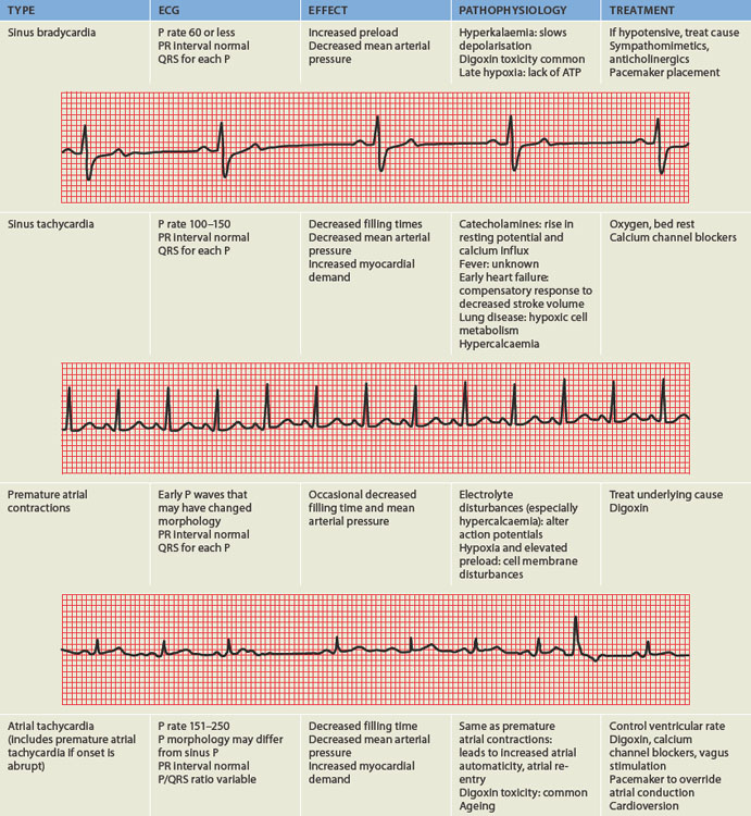
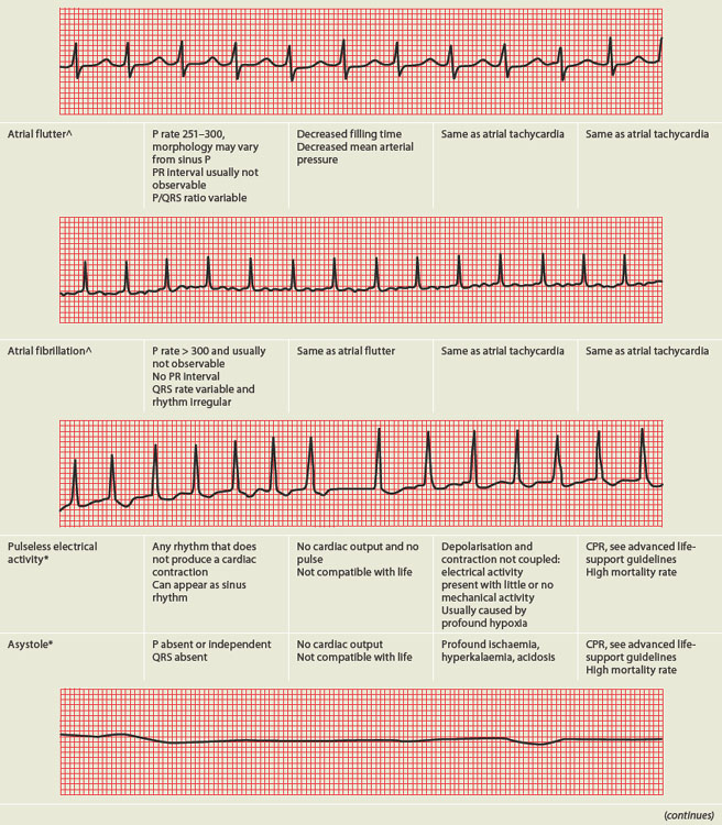
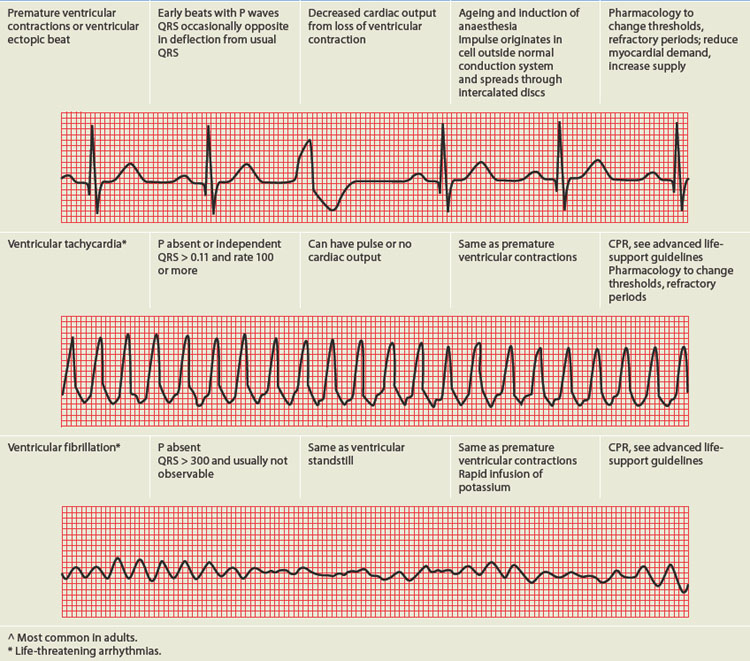
As you will recall, the pericardium is the outer layer of the heart, having approximately 10–30 mL of pericardial fluid to lubricate and protect the heart from infection and inflammation. Inflammation of the pericardium, known as pericarditis, is usually a response to other cardiac conditions, such as acute myocardial infarction or diseases of the thorax. The most common symptom arising from pericarditis is pain. Pericardial disease is often a localised manifestation of another disorder, such as infection (bacterial, viral, fungal or parasite); trauma or surgery; neoplasm; or a metabolic, immunological or vascular disorder (uraemia, rheumatoid arthritis, systemic lupus erythematosus).
Approximately 90% of cases of acute pericarditis are caused by viruses or are idiopathic.77 Acute pericarditis can also be caused by acute myocardial infarction, uraemia, cardiac surgery, some medications and autoimmune disorders. There are many forms of acute pericarditis, including serous, fibrinous, purulent and haemorrhagic. The pericardial membranes become inflamed and roughened and an exudate may develop. Figure 23-41 shows deposits of fibrin on the pericardium possibly following an acute myocardial infarction.
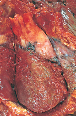
FIGURE 23-41 Fibrinous pericarditis.
Deposits of fibrin are shown on the pericardium.
Source: Kumar V. Robbins & Cotran pathologic basis of disease. 8th edn. Philadelphia: Saunders; 2010.
Symptoms include the sudden onset of severe retrosternal chest pain that worsens with respiratory movements and with lying down. The pain may radiate to the back as a result of irritation of the phrenic nerve (which innervates the trapezius muscles) as it traverses the pericardium.77 Individuals with acute pericarditis also report dysphagia, restlessness, irritability, anxiety, weakness and malaise.
Physical examination often discloses low-grade fever (< 38°C) and sinus tachycardia. Friction rub — a short, scratchy, grating sensation similar to the sound of sandpaper — may be heard at the cardiac apex and left sternal border and is diagnostic for pericarditis. The rub is caused by the roughened pericardial membranes rubbing against each other. Friction rubs are not present in approximately 15% of individuals with acute pericarditis or they may be intermittently heard and transient. Hypotension or the presence of pulsus paradoxus (an exaggerated decrease in systolic blood pressure with inspiration) is suggestive of cardiac tamponade (pericardial fluid impairing heart function; see below), which can be lethal. Electrocardiographic changes may reflect inflammatory processes through PR segment depression and diffuse ST segment elevation without Q waves, and they may remain abnormal for days or even weeks.
Treatment for uncomplicated acute pericarditis consists of relieving symptoms and includes anti-inflammatory agents. Exploration of the underlying cause is important. If pericardial effusion develops, aspiration of the excessive fluid may be necessary. Acute pericarditis is usually self-limiting.
Pericardial effusion — the accumulation of fluid in the pericardial cavity — can occur in all forms of pericarditis. The fluid may be a transudate, such as the serous effusion that develops with left heart failure, overhydration or hypoproteinaemia. More often, however, the fluid is an exudate, which reflects pericardial inflammation like that seen with acute pericarditis, heart surgery, some chemotherapeutic agents, infections and autoimmune disorders such as systemic lupus erythematosus.
Pericardial effusion, even in large amounts, is not necessarily clinically significant, except that it indicates an underlying disorder. If an effusion develops gradually, the pericardium can stretch to accommodate large quantities of fluid without compressing the heart. If the fluid accumulates rapidly, however, even a small amount (50–100 mL) may create sufficient pressure to cause cardiac compression, a serious condition known as cardiac tamponade. The danger is that pressure exerted by the pericardial fluid eventually will equal diastolic pressure within the heart chambers, which will interfere with right atrial filling during diastole. This causes increased venous pressure, systemic venous congestion, and signs and symptoms of right heart failure (distention of the jugular veins, oedema, hepatomegaly). Decreased atrial filling leads to decreased ventricular filling, decreased stroke volume and reduced cardiac output. Life-threatening circulatory collapse may occur.
An important clinical finding is pulsus paradoxus, in which there is a substantial decrease in systolic blood pressure during inspiration. Pulsus paradoxus in the setting of a pericardial effusion indicates tamponade and reflects impairment of diastolic filling of the left ventricle plus reduction of blood volume within all four cardiac chambers.
Other clinical manifestations of pericardial effusion are distant or muffled heart sounds, poorly palpable apical pulse, dyspnoea on exertion and dull chest pain. A chest X-ray film may disclose a water-bottle configuration of the cardiac silhouette. Doppler echocardiogram can detect an effusion as small as 20 mL.
Treatment of pericardial effusion or tamponade generally consists of pericardiocentesis (aspiration of excessive pericardial fluid) and treatment of the underlying condition. Persistent pain may be treated with analgesics, anti-inflammatory medications or steroids.
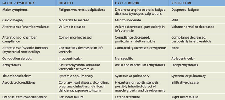
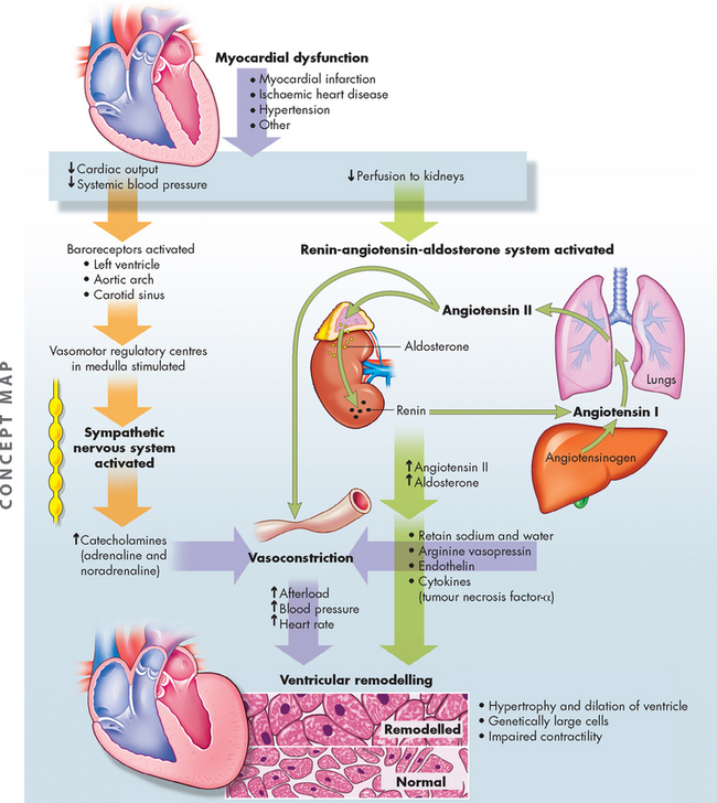
The cardiomyopathies are a diverse group of diseases that primarily affect the myocardium itself. Most are the result of remodelling caused by myocardial and neurohumoral responses (both neural and hormonal) to ischaemic events and hypertension. They may, however, be secondary to infectious disease, exposure to toxins, systemic connective tissue disease, infiltrative and proliferative disorders or nutritional deficiencies.78 However, many cases are idiopathic — that is, their cause is unknown. The cardiomyopathies are categorised as dilated (formerly, congestive), hypertrophic or restrictive, depending on their physiological effects on the heart (see Figure 23-42 and Table 23-10).
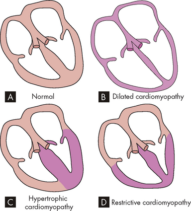
FIGURE 23-42 Diagram showing the three types of cardiomyopathy.
A Normal heart. B Dilated cardiomyopathy demonstrating enlargement of all four chambers. C Hypertrophic cardiomyopathy showing a thickened left ventricle. D Restrictive cardiomyopathy characterised by a small left ventricular volume.
Source: Copstead-Kirkhorn L-EC, Banasik JL. Pathophysiology. 4th edn. Philadelphia: Saunders; 2009.
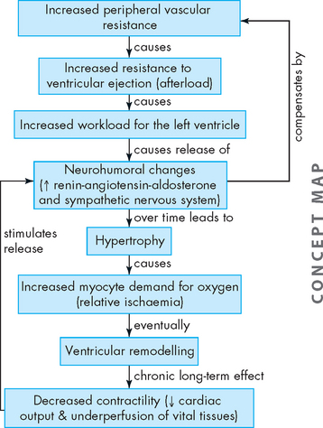
Disorders of the endocardium (the innermost lining of the heart wall) damage the heart valves, which are composed of endocardial tissue. Endocardial damage can be either congenital or acquired. The acquired forms cause inflammatory, ischaemic, traumatic, degenerative or infectious alterations of valvular structure and function. One of the most common causes of acquired valvular dysfunction is degeneration or inflammation of the endocardium secondary to rheumatic heart disease (see Table 23-11). Valvular stenosis occurs when the valve orifice is constricted and narrowed, so that blood cannot flow forwards and the workload of the cardiac chamber ‘behind’ the diseased valve increases (see Figure 23-43). Pressure within the ventricular or atrial chamber rises to overcome resistance to flow through the valve, causing the myocardium to work harder and producing myocardial hypertrophy.
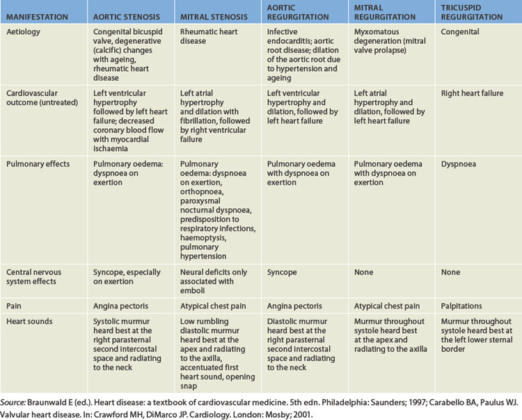
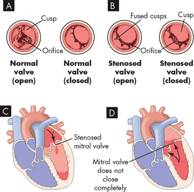
FIGURE 23-43 Valvular stenosis and regurgitation.
A Normal position of the valve leaflets, or cusps, when the valve is open and closed. B Open position of a stenosed valve (left) and open position of a closed regurgitant valve (right). C Haemodynamic effect of mitral stenosis. The stenosed valve is unable to open sufficiently during left atrial systole, inhibiting left ventricular filling. D Haemodynamic effect of mitral regurgitation. The mitral valve does not close completely during left ventricular systole, permitting blood to re-enter the left atrium.
In valvular regurgitation (also called insufficiency or incompetence), the valve leaflets, or cusps, fail to shut completely, permitting blood flow to continue even when the valve is supposed to be closed (see Figure 23-43). Blood can leak back into the chamber ‘upstream’, which increases the volume of blood the heart must pump and increases the workload of both the atrium and the ventricle. Increased volume leads to chamber dilation with cardiomegaly (see Figure 23-44) and increased workload leads to hypertrophy. Although all four heart valves may be affected, in adults those of the heart’s left side (the mitral and aortic valves) are far more commonly affected than those of the right (the tricuspid and pulmonary valves).
FIGURE 23-44 Cardiomegaly due to mitral and tricuspid regurgitation.
Note the enlarged heart, which is wider than normal. A On the posteroanterior chest X-ray there is marked enlargement of not only the left atrium (LA) but also the left ventricle (LV; seen as straightening of the left cardiac border), as well as right-sided enlargement, particularly of the right atrium (RA; seen by marked prominence of the right cardiac border). B On the lateral view of the chest the left ventricle can be seen overlapping the spine, and the right atrium and right ventricle (RV) have filled in the retrosternal space to more than the usual lower one-third.
Source: Metter FA. Essentials of radiology. 2nd edn. Philadelphia: Saunders; 2005.
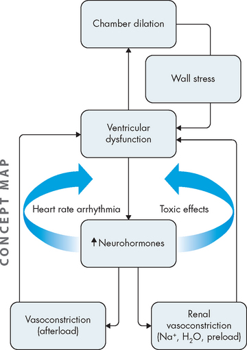
Valvular dysfunction stimulates chamber dilation and myocardial hypertrophy, both of which are compensatory mechanisms intended to increase the pumping capability of the heart but which lead to cardiac dysfunction over time. Eventually, myocardial contractility diminishes, the ejection fraction is reduced, diastolic pressure increases and the ventricles fail from overwork. Depending on the severity of the valvular dysfunction and the capacity of the heart to compensate, valvular alterations cause a range of symptoms and some degree of incapacitation (see Table 23-11).
Aortic stenosis is now the most common valvular abnormality, affecting nearly 2% of adults older than 65 years of age. It has three common causes:
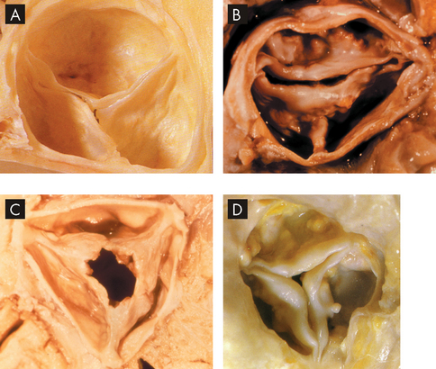
A Normal aortic valve. B Congenital bicuspid aortic stenosis. C Rheumatic aortic stenosis. D Calcified degenerative aortic stenosis.
Source: A & D Manabe H, Yutani C (eds). Atlas of valvular heart disease. Singapore: Churchill Livingstone; 1998. B & C Courtesy of William C Roberts, MD.
Aortic valve degeneration with ageing is associated with lipoprotein deposition in the tissue with chronic inflammation and leaflet calcification.80 The orifice of the aortic semilunar valve narrows, causing diminished blood flow from the left ventricle into the aorta. Outflow obstruction increases pressure within the left ventricle as it tries to eject blood through the narrowed opening.
Aortic stenosis tends to develop gradually. Clinical manifestations include decreased stroke volume, reduced systolic blood pressure and narrowed pulse pressure (the difference between systolic and diastolic pressure). Heart rate is often slow and pulses are faint. Left ventricular hypertrophy develops to compensate for the increased workload. Eventually, hypertrophy increases myocardial oxygen demand, which the coronary arteries may be unable to meet. In addition, aortic stenosis is frequently accompanied by atherosclerotic coronary disease, further contributing to inadequate coronary perfusion.80
Untreated aortic stenosis can lead to arrhythmias, myocardial infarction and heart failure. Echocardiography can be used to assess the severity of valvular obstruction before the onset of symptoms, and management almost always includes valve replacement with a prosthetic valve (see Figure 23-46), followed by long-term anticoagulation and prophylaxis for endocarditis, as needed.
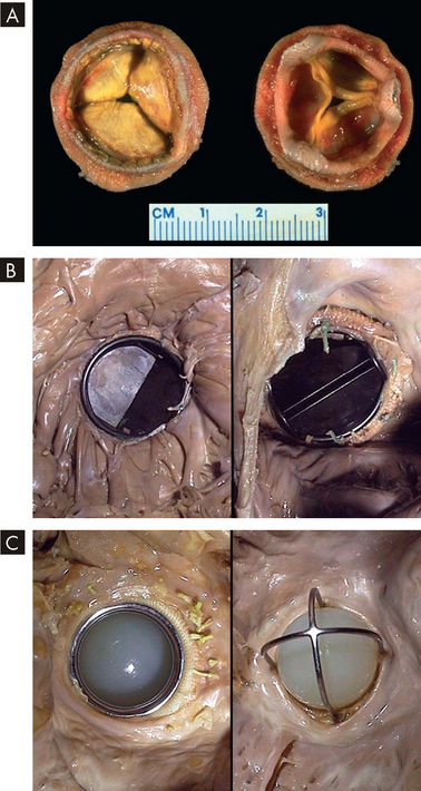
FIGURE 23-46 Various valve replacements.
A A porcine bioprosthesis. B A mechanical valve prosthesis of the tilting disc variety replacing the native mitral valve. C A mechanical valve prosthesis of the older ball-and-cage variety.
Source: Klatt EC. Robbins and Cotran atlas of pathology. 2nd edn. Philadelphia: Saunders; 2010.
Mitral stenosis impairs the flow of blood from the left atrium to the left ventricle. Mitral stenosis is most commonly caused by rheumatic heart disease. In fact, when stenosed mitral valves are examined after removal for mitral valve replacement, almost all have evidence of rheumatic changes.81 In addition, mitral stenosis is two to three times more common in women than in men.82 Autoimmunity in response to group A β-haemolytic streptococcal protein antigens leads to inflammation and scarring of the valvular leaflets. Scarring causes the leaflets to become fibrous and fused and the chordae tendineae become shortened.
Impedance to blood flow results in incomplete emptying of the left atrium and elevated atrial pressure as the chamber tries to force blood through the stenotic valve. Continued increases in left atrial volume and pressure cause atrial dilation and hypertrophy. The risk of developing atrial arrhythmias (especially fibrillation) and arrhythmia-induced thrombi is high. As mitral stenosis progresses, symptoms of decreased cardiac output occur, especially during exertion. Continued elevation of left atrial pressure and volume causes pressure to rise in the pulmonary circulation. If untreated, chronic mitral stenosis develops into pulmonary hypertension, pulmonary oedema and right ventricular failure.
Management includes anticoagulation and endocarditis prophylaxis along with β-blockers or calcium channel blockers to slow the heart rate. Mitral stenosis can often be repaired surgically but may require valve replacement (usually porcine; see Figure 23-46A) in advanced cases.
Aortic regurgitation results from an inability of the aortic valve leaflets to close properly during diastole resulting from abnormalities of the leaflets or the aortic root and annulus, or both.83 It can be congenital (bicuspid valve abnormalities) or acquired. Acquired aortic regurgitation can be caused by rheumatic heart disease, bacterial endocarditis, syphilis, hypertension, connective tissue disorders (e.g. Marfan’s syndrome), appetite-suppressing medications, trauma or atherosclerosis. In more than one-third of cases of aortic regurgitation there is no known cause.
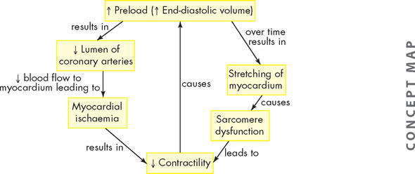
The haemodynamic abnormalities depend on the size of the ‘leak’. During systole, blood is ejected from the left ventricle into the aorta. During diastole, some of the ejected blood flows back into the left ventricle. Volume overload occurs in the ventricle because it receives blood from both the left atrium and the aorta during diastole. Over time, the end-diastolic volume of the left ventricle increases and myocardial fibres stretch to accommodate the extra fluid. Compensatory dilation permits the left ventricle to increase its stroke volume and maintain cardiac output. Ventricular hypertrophy also occurs as an adaptation to the increased volume and because of increased afterload created by the high stroke volume and resultant systolic hypertension. Ventricular dilation and hypertrophy eventually cannot compensate for aortic incompetence and heart failure develops.
Clinical manifestations include widened pulse pressure resulting from increased stroke volume and diastolic backflow. Other symptoms are usually associated with heart failure, which occurs when the ventricle can no longer pump adequately. Arrhythmias and endocarditis are common complications of aortic regurgitation. The severity of regurgitation can be estimated by echocardiography, and surgical management (valve replacement; see Figure 23-46) may be delayed for many years through careful use of vasodilators and inotropic agents.
Mitral regurgitation has many possible causes, including mitral valve prolapse (see below), rheumatic heart disease, infective endocarditis, acute myocardial infarction, connective tissue diseases (such as Marfan’s syndrome) and congestive cardiomyopathy (myocardial disease). Mitral regurgitation permits backflow of blood from the left ventricle into the left atrium during ventricular systole. Eventually, the left ventricular volume increases, causing it to become dilated and hypertrophied to maintain adequate cardiac output. The volume of backflow re-entering the left atrium gradually increases, causing atrial dilation and associated atrial fibrillation. As the left atrium enlarges, the valve structures stretch and become deformed, leading to further backflow. As mitral valve regurgitation progresses, left ventricular function may become impaired to the point of failure. Eventually, increased atrial pressure also causes pulmonary hypertension and failure of the right ventricle. Mitral incompetence is usually well tolerated — often for years — until ventricular failure occurs. Most clinical manifestations are caused by heart failure. The severity of regurgitation can be estimated by echocardiography, and surgical repair or valve replacement may become necessary (see Figure 23-46).
Tricuspid regurgitation is more common than tricuspid stenosis (narrowing) and is usually associated with failure and dilation of the right ventricle secondary to pulmonary hypertension. Tricuspid valve incompetence leads to volume overload in the right ventricle, increased systemic venous blood pressure and right heart failure. Pulmonary semilunar valve dysfunction can have the same consequences as tricuspid valve dysfunction.
In mitral valve prolapse syndrome one or both of the cusps of the mitral valve billow upwards (prolapse) into the left atrium during systole. The most common cause of mitral valve prolapse is degeneration of the leaflets in which the cusps are redundant, thickened and scalloped because of changes in tissue proteoglycans and infiltration by myofibroblasts.84 Mitral regurgitation occurs if the ballooning valve permits blood to leak into the atrium.
Many cases of mitral valve prolapse are completely asymptomatic. Symptomatic mitral valve prolapse can cause palpitations related to arrhythmias, tachycardia, light-headedness, syncope, fatigue (especially in the morning), lethargy, weakness, dyspnoea, chest tightness, hyperventilation, anxiety, depression, panic attacks and atypical chest pain. Many symptoms are vague and puzzling and are unrelated to the degree of prolapse.
Evaluation of mitral valve prolapse includes physical assessment and laboratory evaluation. Echocardiography is the procedure of choice for diagnosing the disorder. Most individuals with mitral valve prolapse have an excellent prognosis, do not develop symptoms and do not require any restriction in activity or medical management. However, some individuals have an increased risk for complications such as infective endocarditis, stroke and sudden death.
Rheumatic fever is a diffuse, inflammatory disease caused by a delayed exaggerated immune response to infection by the group A β-haemolytic streptococcus in genetically predisposed individuals. In its acute form, rheumatic fever is a febrile illness characterised by inflammation of the joints, skin, nervous system and heart.85 If untreated, rheumatic fever can cause scarring and deformity of cardiac structures resulting in rheumatic heart disease.
With increases in living standards and infection control, the incidence of rheumatic heart disease has been in decline in Western countries.85 However, Australia has the unenviable position of recording the highest rates of rheumatic heart disease in the world.86 Alarmingly, the rates are highest among Aboriginal people living in the Northern Territory and have been related to poor hygiene and standards of living compared to the non-Indigenous population87 — in fact, death rates from rheumatic heart disease are 15 and 23 times higher for Aboriginal men and women, respectively.88
Acute rheumatic fever can develop only as a sequel to pharyngeal infection by group A β-haemolytic streptococcus. Streptococcal skin infections do not progress to acute rheumatic fever, although both skin and pharyngeal infections can cause acute glomerulonephritis. This is because the strains of the microorganism that affect the skin do not have the same antigenic molecules in their cell membranes as those that cause pharyngitis and therefore do not elicit the same kind of immune response. Acute rheumatic fever is the result of an abnormal humoral and cell-mediated immune response to group A streptococcal cell membrane antigens called M proteins (see Figure 23-47).
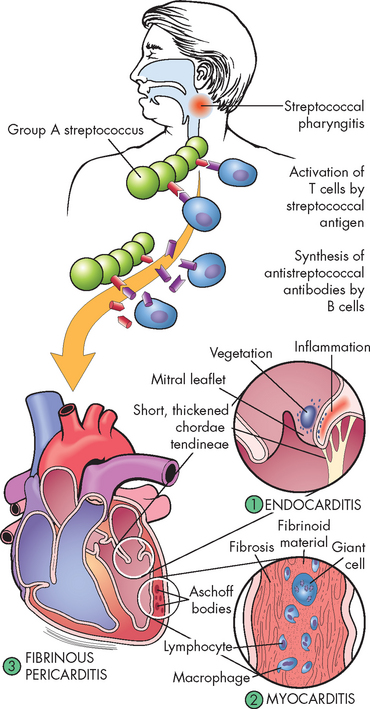
FIGURE 23-47 The pathogenesis and structural changes of rheumatic heart disease.
Beginning usually with a sore throat, rheumatic fever can develop only as a sequel to pharyngeal infection by group A β-haemolytic streptococcus. Suspected as a hypersensitivity reaction, it is proposed that antibodies directed against the M proteins of certain strains of streptococci cross-react with tissue glycoproteins in the heart, joints and other tissues. The exact nature of cross-reacting antigens has been difficult to define, but it appears that the streptococcal infection causes an autoimmune response against self-antigens. Inflammatory lesions are found in various sites; the most distinctive within the heart are called Aschoff bodies. The chronic sequelae result from progressive fibrosis because of healing of the inflammatory lesions and the changes induced by valvular deformities.
Source: Damjanov I. Pathology for the health professions. 3rd edn. St Louis: Saunders; 2006.
Diffuse, proliferative and exudative inflammatory lesions develop in the connective tissues, especially in the heart. The inflammation may subside before treatment, leaving behind damage to the heart valves and increasing the individual’s susceptibility to recurrent acute rheumatic fever after any subsequent streptococcal infections. Repeated attacks of acute rheumatic fever cause chronic proliferative changes (scarring) in the affected regions.
Approximately 10% of individuals with rheumatic fever develop rheumatic heart disease. The primary lesion usually involves the endocardium. Endocardial inflammation causes swelling of the valve leaflets, with secondary erosion along the lines of leaflet contact. Small beadlike clumps of vegetation containing platelets and fibrin are deposited on eroded valvular tissue and on the chordae tendineae (see Figure 23-48).
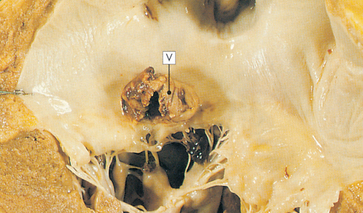
FIGURE 23-48 Mitral stenosis with vegetation.
Mitral stenosis and clumps of vegetation (V) containing platelets and fibrin are shown in this micrograph. The mitral leaflets are thickened and fused.
Source: Stevens A, Lowe J. Pathology. Edinburgh: Mosby; 2000.
Myocarditis (inflammation of the myocardium) may occur. Cardiomegaly and left heart failure may occur during episodes of untreated acute or recurrent rheumatic fever. Conduction defects and atrial fibrillation are often associated with rheumatic heart disease.
Many common clinical manifestations of acute rheumatic fever — fever, lymphadenopathy, arthralgia (painful joints), nausea, vomiting, epistaxis (nose bleeds), abdominal pain and tachycardia — are also associated with other disorders and are therefore not diagnostic of the disease. The major clinical manifestations of acute rheumatic fever are carditis, acute migratory polyarthritis (inflammation of more than one joint), chorea (a central nervous system disorder — see Chapter 9) and erythema marginatum (truncal rash), which may occur singly or in combination 1–5 weeks after streptococcal infection of the pharynx.
When combined with physical assessment findings, laboratory values lend significant support to the diagnosis of acute rheumatic fever. A positive throat culture for group A β-haemolytic streptococci can be an important finding when associated with certain physical signs. Elevated white blood cell count, erythrocyte sedimentation rate and C-reactive protein indicate inflammation. All three are usually increased at the time cardiac or joint symptoms begin to appear. They are more useful in identifying an acute inflammatory process and suggesting prognosis than in diagnosing acute rheumatic fever. These test results return towards normal levels as the inflammatory process resolves.
Therapy for acute rheumatic fever is aimed at eradicating the streptococcal infection and involves a 10-day regimen of oral penicillin or erythromycin administration. Non-steroidal anti-inflammatory drugs (NSAIDs) are used as anti-inflammatory agents for both rheumatic carditis and arthritis. Serious carditis may require adding cardiac glycosides, corticosteroids, diuretics and bed rest to the regimen. Surgical repair of damaged valves may be needed in chronic recurrent rheumatic fever and carditis leading to rheumatic heart disease.
Research suggests that a rheumatic recurrence will develop in 50–65% of children with known rheumatic fever if they have another group A streptococcal infection. To prevent a recurrence of acute rheumatic fever, continuous prophylactic antibiotic therapy may be necessary for as long as 5 years.85 Appropriate antibiotic therapy given within the first 9 days of infection usually prevents rheumatic fever.
Infective endocarditis is a general term used to describe infection and inflammation of the endocardium, especially the cardiac valves. Bacteria are the most common cause of infective endocarditis including Streptococcus viridans, Staphylococcus aureus, Staphylococcus epidermidis and group A β-haemolytic streptococci. Infective endocarditis was once a lethal disease, but morbidity and mortality diminished significantly with the advent of antibiotics and improved diagnostic techniques (see the box ‘Risk factors for ineffective endocarditis’).89
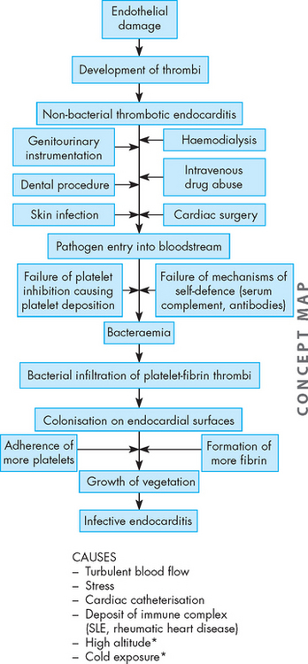
The pathogenesis of infective endocarditis requires at least three critical elements (see Figure 23-49): (1) the endocardium (e.g. heart valve) must be ‘prepared’, usually by endothelial damage, for microorganism colonisation; (2) blood-borne microorganisms must adhere to the damaged endocardial surface; and (3) the microorganisms must proliferate and promote the propagation of infective endocardial vegetation.
Endocarditis risk in children
Children with congenital heart disease are at risk for developing endocarditis. Although the risk is low, a transient bacteraemia has been noted to follow dental and surgical procedures and instrumentation involving mucosal surfaces. A blood-borne pathogen can settle in areas of the heart where there is high turbulence, an abnormal valve or vessel, or an artificial material such as a valve or homograft. Streptococcus viridans (α-haemolytic streptococci) is the most commonly found pathogen following dental or oral procedures. Enterococcus faecalis (enterococci) is the most common bacterium found following genitourinary and gastrointestinal tract surgery or instrumentation. The Infective Endocarditis Prophylaxis Expert Group convened by the Therapeutics Guidelines has provided updated guidelines for the prevention of bacterial endocarditis. The type and dose of antibiotic prophylaxis recommended depend on the procedure and the cardiac classification of risk for endocarditis.
Source: The Infective Endocarditis Prophylaxis Expert Group. Prevention of endocarditis. 2008 update from Therapeutic Guidelines: antibiotic version 13, and Therapeutic Guidelines: oral and dental version 1. Melbourne: Therapeutic Guidelines Limited; 2008.
Endocardial damage exposes the collagen within the endothelial basement membrane — this collagen attracts platelets and thereby stimulates thrombus formation on the membrane. This causes an inflammatory reaction. Infective endocarditis cannot develop unless microorganisms gain access to the bloodstream. They may enter the bloodstream during intravenous drug use, trauma or minor procedures such as dental cleaning or bladder catheterisation, or they may spread from uncomplicated upper respiratory or skin infections. A significant number of cases of infective carditis are healthcare–acquired infections in origin, especially those that result from genitourinary or gastrointestinal procedures or from surgical wound infections.
Once the endocardial surface is colonised, infected vegetations form (see Figure 23-50). Bacteria may accelerate fibrin formation by activating the coagulation cascade. Although endocardial tissue is constantly bathed in antibody-containing blood and is surrounded by scavenging monocytes and leucocytes, bacterial colonies are inaccessible to host defences because they are embedded in the protective fibrin clots. The lesions can form anywhere on the endocardium but usually occur on the endocardial surfaces of heart valves and surrounding structures.
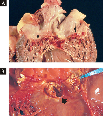
FIGURE 23-50 Infective endocarditis.
A Endocarditis of the mitral valve (subacute, caused by Streptococcus viridans). The large, friable vegetations are denoted by arrows. B Acute endocarditis of congenitally bicuspid aortic valve (caused by Staphylococcus aureus) with extensive cuspal destruction and ring abscess (arrow).
Source: Schoen FJ. Surgical pathology of removed natural and prosthetic heart valves. Human Pathol 1987; 18:558.
Infective endocarditis may be acute, subacute or chronic. It causes varying degrees of valvular dysfunction and may be associated with manifestations involving several organ systems (lungs, eyes, kidneys, bones, joints, central nervous system), making diagnosis exceedingly difficult. The ‘classic’ findings are fever and petechial lesions of the skin, conjunctiva and oral mucosa. Other manifestations include weight loss, back pain, night sweats and heart failure.
The criteria for the diagnosis of infective endocarditis include persistent bacteraemia, new heart murmurs, vascular complications and appropriate echocardiographic findings.90 Antimicrobial therapy is generally given for 4 to 6 weeks. Other drugs may be necessary to treat left heart failure secondary to valvular dysfunction. Surgery to remove infected tissue, with or without valve replacement, improves outcomes in many patients with infective endocarditis, especially those with severe heart failure or persistent bacteraemia despite antibiotic therapy.91
So far in this chapter we have examined the effects of pathophysiological changes to the blood vessels and the heart. These changes have typically altered the function of cardiac structures and we have explored how this impacts on mechanical contraction. In this section, we look at the pathophysiological changes that alter electrical conduction of the heart. Normal cardiac conduction requires the autorhythmic cells to produce action potentials that spread sequentially throughout conduction pathways in a coordinated fashion (refer to Chapter 22). Briefly, normal heart rhythm is generated by the sinoatrial (SA) node and travels through the atrioventricular node, down the atrioventricular bundle and through the bundle branches to the Purkinje fibres. These depolarisations initiate cardiac contraction and the heart oscillates through systole and diastole to maintain cardiac output. Cardiac conduction alterations may occur due to a variety of reasons, such as electrolyte disturbances, myocardial ischaemia and acute myocardial infarction, drug therapy and intrinsic problems of the autorhythmic cells.
Technically, the term dysrhythmia refers to a disturbance of heart rhythm. Although this term is entirely accurate and appropriate, in Australia and New Zealand we commonly refer to dysrhythmias as arrhythmias. The word arrhythmia actual means without cardiac rhythm (the prefix ‘a’ meaning no or without). Despite the fact that there are differences between the terms, they are often used interchangeably. However, we believe that dysrhythmia has not been universally accepted and so, for the purposes of consistency and to align with clinical terminology, we refer to alterations of cardiac conduction as arrhythmias.
Arrhythmias can range in severity from occasional ‘missed’ or rapid beats to serious disturbances that impair the pumping ability of the heart and that may result in heart failure and death. Arrhythmias can be caused by either an abnormal rate of impulse generation by the SA node or other pacemakers, or the abnormal conduction of impulses through the conduction system of the heart, including the myocardial cells themselves. Furthermore, arrhythmias can be non-pathological in origin, such as a missed beat in an athlete who has bradycardia (slowed heart rate) as a result of endurance training, ranging to potentially life-threatening conditions that may cause sudden death. Tables 23-12 and 23-13 explore the most common conduction alterations.
Atrial fibrillation is the most common arrhythmia and is most prevalent in the elderly population.92 It can occur due to a variety of reasons, although the treatment should be aggressive to prevent unwanted complications, such as blood clotting and pulmonary embolism or embolic strokes.
The life-threatening arrhythmias include ventricular tachycardia, ventricular fibrillation, asystole (without systole) and pulseless electrical activity. These arrhythmias cause cardiorespiratory arrest, as the heart has either stopped contractions or is abnormal to the extent that there is no cardiac output. Breathing cessation usually accompanies cardiac arrest. The management of these arrhythmias involves immediate life support. Evidence-based guidelines for adult and paediatric advanced resuscitation of these arrhythmias are included in Figure 23-51 and 23-52.
FIGURE 23-51 Adult cardiorespiratory arrest guidelines.
BLS = basic life support; VF = ventricular fibrillation; VT = ventricular tachycardia; PEA = pulseless electrical activity; CPR = cardiopulmonary resuscitation.
Source: Australian Resuscitation Council.
Heart failure is a syndrome encompassing several different types of cardiac dysfunction that result in inadequate perfusion of tissues with oxygen and blood-borne nutrients. Providing a definition of heart failure is difficult because consensus has been hard to achieve; however, it is now considered that both clinical features and an objective measure of abnormal ventricular function are required to adequately diagnose heart failure. The National Heart Foundation of Australia and the Cardiac Society of Australia and New Zealand have formulated the following definition:
Heart failure is a complex clinical syndrome with typical symptoms (e.g. dyspnoea, fatigue) that can occur at rest or on effort and is characterised by objective evidence of an underlying structural abnormality or cardiac dysfunction that impairs the ability of the ventricle to fill with or eject blood (particularly during physical activity). A diagnosis of heart failure may be further strengthened by improvement in symptoms in response to treatment.93
Heart failure occurs in up to 2% of all Australians;93 however, this figure is somewhat skewed because approximately 10% of sufferers are aged 65 years or older and more than 50% are aged 85 years and over.1,94 Most causes of heart failure result from dysfunction of the left ventricle (systolic and diastolic heart failure). The right ventricle may also be dysfunctional, especially in pulmonary disease (right ventricular failure). Coronary heart disease and acute myocardial infarction are documented in about two-thirds of systolic heart failure patients. Essential hypertension is also strongly associated with heart failure and is present in approximately two-thirds of all newly diagnosed cases.93 In the following sections we examine heart failure and its relationship to other cardiovascular diseases.
Left heart failure, commonly called congestive heart failure, can be further categorised as systolic heart failure or diastolic heart failure. These two types of heart failure can occur together or in isolation.
Systolic heart failure is defined as an inability of the heart to generate an adequate cardiac output to perfuse vital tissues. Cardiac output depends on the heart rate and stroke volume. Stroke volume is influenced by three major determinants: contractility, preload and afterload (refer to Chapter 22).
Contractility is reduced by diseases that disrupt myocyte activity. Myocardial infarction is the most common primary cause of decreased contractility; other causes include myocarditis and the cardiomyopathies. Secondary causes of decreased contractility, such as recurrent myocardial ischaemia and increased myocardial workload, contribute to inflammatory, immune and neurohumoral changes that mediate a process called ventricular remodelling (see Box 23-1).95,96 Ventricular remodelling results in hypertrophy and dilation of the myocardium and causes progressive myocyte contractile dysfunction over time (see Figure 23-53). When contractility is decreased, stroke volume falls and ventricular end-diastolic volume increases. This causes dilation of the heart and an increase in preload.
BOX 23-1 INFLAMMATION, IMMUNITY AND HUMORAL FACTORS IN THE PATHOGENESIS OF HEART FAILURE
The treatment of the haemodynamic abnormalities of heart failure can provide short-term improvement in symptoms but will not prevent the progression of myocardial dysfunction over time. Studies have shown that neurohumoral responses to heart failure (including changes in the renin-angiotensin-aldosterone system, catecholamines, natriuretic peptides, endothelin and nitric oxide) exert direct cardiotoxicity that results in progressive damage to the heart muscle. Drugs such as ACE inhibitors, angiotensin II receptor blockers and β-blockers can slow disease progression and are now the standard of care for heart failure. More recently, inflammatory cytokines such as tumour necrosis factor-alpha (TNF-α) and interleukins have been implicated in the pathogenesis of heart failure and its systemic complications. Early trials with anticytokine drugs are under way.
Source: Aukrust P et al. Inflammatory and anti-inflammatory cytokines in chronic heart failure: potential therapeutic implications. Ann Med 2005; 37(2):74–85; Mehra VC, Ramgolam VS, Bender JR. Cytokines and cardiovascular disease. J Leukocyte Biol 2005; 78(4):805–818; Mueller C et al. Inflammation and long-term mortality in acute congestive heart failure. Am Heart J 2006; 151(4):845–850; Torre-Amione G. Immune activation in chronic heart failure. Am J Cardiol 2005; 95(11A):3C–8C, discussion 38C–40C; Tousoulis D, Charakida M, Stefanadis C. Inflammation and endothelial dysfunction as therapeutic targets in patients with heart failure. Internat J Cardiol 2005; 100(3):347–353.
FIGURE 23-53 The pathophysiology of ventricular remodelling.
Myocardial dysfunction activates the renin-angiotensin-aldosterone and sympathetic nervous systems releasing neurohormones (angiotensin II, aldosterone, catecholamines and cytokines). These neurohormones contribute to ventricular remodelling.
Source: Carelock J, Clark AP. Heart failure: pathophysiologic mechanisms. Am J Nurs 2001; 101(12):27.
Preload increases with decreased contractility or an excess of plasma volume (intravenous fluid administration, renal failure, mitral valvular disease). Increases in preload can actually improve cardiac output, but as preload continues to rise, it causes a stretching of the myocardium that eventually can lead to dysfunction of the sarcomeres and decreased contractility. This relationship is described by the Frank-Starling law of the heart, which details the length–tension relationship between end-diastolic volume and stroke volume in the ventricle. (This was reviewed in Chapter 22 and shown in Figure 22-27.) However, with sarcomere dysfunction resulting in decreased contractility, increases in end-diastolic volume do not result in increases in stroke volume. In fact, decreased contractility leads to further increases in preload (see Figure 23-54), which causes a lowering of cardiac output for a given ventricular end-diastolic volume (see Figure 23-55). In this scenario, across the range of end-diastolic volumes, cardiac performance is less than normal, as increases in stroke volume do not occur to the same magnitude as the normal heart because of the damage to the myocytes.
FIGURE 23-55 The relationship between ventricular end-diastolic volume and stroke during normal contraction and heart failure.
The Frank-Starling law of the heart is the relationship between length (end-diastolic volume) and tension (stroke volume) in the heart. In the normal heart, increases in end-diastolic volume cause an increase in stroke volume, thereby increasing cardiac output. In heart failure, decreased contractility leads to reduced cardiac output, as increases in end-diastolic volume do not cause similar increases in stroke volume. Therefore, the curve is shifted downwards over the range of end-diastolic volumes.
Increased afterload is most commonly a result of increased peripheral vascular resistance, such as that seen with hypertension. With increased afterload, there is resistance to ventricular emptying and more work for the ventricle, which results in myocardial hypertrophy. Hypertrophy is mediated by angiotensin II and catecholamines and results in an increase in oxygen demand by the thickened myocardium. A state of relative ischaemia develops, which further contributes to changes in the myocytes themselves and ventricular remodelling (see Figure 23-56). In addition, hypertrophy results in the deposition of collagen between the myocytes, which can disrupt the integrity of the muscle, decrease contractility and make the ventricle more likely to dilate and fail.
As cardiac output falls, renal perfusion diminishes with activation of the renin-angiotensin-aldosterone system, which acts to increase both peripheral vascular resistance and plasma volume, thus further increasing afterload and preload. In addition, baroreceptors in the central circulation detect the decrease in perfusion and stimulate the sympathetic nervous system to cause yet more vasoconstriction and the hypothalamus to produce antidiuretic hormone. The neurohumoral aspects of systolic heart failure suggest that treatment must include inhibition of angiotensin, aldosterone and catecholamines to prevent long-term damage to the myocardium.95,96 Immune and inflammatory processes also play an important role in the pathogenesis of heart failure and its systemic complications (see Box 23-1). This vicious cycle of decreasing contractility, increasing preload and increasing afterload causes progressive worsening of systolic heart failure (see Figure 23-57).
FIGURE 23-57 The vicious cycle of heart failure.
Source: National Heart Foundation of Australia and Cardiac Society of Australia and New Zealand. Guidelines for the prevention, detection and management of chronic heart failure in Australia. Chronic Heart Failure Guidelines Expert Writing Panel; 2006.
The clinical manifestations of systolic heart failure are the result of pulmonary vascular congestion and inadequate perfusion of the systemic circulation. Individuals experience dyspnoea, orthopnoea (experiencing dyspnoea when lying flat — individuals compensate by sitting upright), cough of frothy sputum, fatigue, decreased urine output and oedema. Physical examination often reveals pulmonary oedema, hypotension or hypertension and evidence of underlying coronary heart disease or hypertension. The diagnosis can be further confirmed with echocardiography revealing decreased cardiac output and cardiomegaly.
Management of systolic left heart failure is aimed at interrupting the worsening cycle of decreasing contractility, increasing preload and increasing afterload.93 The acute onset of systolic left heart failure is most often the result of acute myocardial ischaemia and must be managed in conjunction with managing the underlying coronary disease. Oxygen, nitrate and morphine administration improves myocardial oxygenation and helps relieve coronary spasm while lowering preload through systemic venodilation. Intravenous inotropic drugs, such as dopamine or dobutamine, increase contractility and can help raise the blood pressure in hypotensive individuals. Diuretics reduce preload and ACE inhibitors, angiotensin receptor blockers and aldosterone blockers reduce both preload and afterload by decreasing aldosterone levels and reducing peripheral resistance. Short-acting intravenous β-blockers also have been found to reduce mortality in selected people.
Management of chronic left heart failure also relies on increasing contractility and reducing preload and afterload. The current evidence-based standard of care for individuals in Australia and New Zealand is outlined in Tables 23-14 and 23-15.
Table 23-14 NON-PHARMACOLOGICAL MANAGEMENT OF HEART FAILURE
GRADE OF RECOMMENDATION* | |
---|---|
Regular physical activity is recommended. All patients should be referred to a specifically designed physical activity program, if available. | B |
Patient support by a doctor and pre-discharge review and/or home visit by a nurse is recommended to prevent clinical deterioration. | A |
Patients frequently have coexisting sleep apnoea and, if suspected, patients should be referred to a sleep clinician as they may benefit from nasal CPAP. | D |
Patients who have an acute exacerbation, or are clinically unstable, should undergo a period of bed rest until their condition improves. | D |
Dietary sodium should be limited to below 2 g/day. | C |
Fluid intake should generally be limited to 1.5 L/day with mild to moderate symptoms, and 1 L/day in severe cases, especially if there is coexistent hyponatraemia. | C |
Alcohol intake should preferably be nil, but should not exceed 10–20 g a day (1 or 2 standard drinks). | D |
Smoking should be strongly discouraged. | D |
Patients should be advised to weigh themselves daily and to consult their doctor if their weight increases by more than 2 kg in a 2-day period, or if they experience dyspnoea, oedema or abdominal bloating. | D |
Patients should be vaccinated against influenza and pneumococcal disease. | B |
High-altitude destinations should be avoided. Travel to very humid or hot climates should be undertaken with caution, and fluid status should be carefully monitored. | C |
Sildenafil and other phosphodiesterase V inhibitors are generally safe in patients with heart failure. However, these medications are contraindicated in patients receiving nitrate therapy, or those who have hypotension, arrhythmias or angina pectoris. | C |
Obese patients should be advised to lose weight. | D |
A diet with reduced saturated fat intake and a high fibre intake is encouraged in patients with chronic heart failure. | D |
No more than 2 cups of caffeinated beverages per day recommended. | D |
Pregnancy should be avoided in patients with chronic heart failure. | D |
CPAP = continuous positive airway pressure. |
*GRADE OF RECOMMENDATION | DESCRIPTION |
---|---|
A | Rich body of high-quality RCT data |
B | Limited body of RCT data or high-quality non-RCT data |
C | Limited evidence |
D | No evidence available — panel consensus judgement |
RCT = randomised control trial. |
Source: National Heart Foundation of Australia and Cardiac Society of Australia and New Zealand. Guidelines for the prevention, detection and management of chronic heart failure in Australia. Chronic Heart Failure Guidelines Expert Writing Panel; 2006.
Table 23-15 PHARMACOLOGICAL MANAGEMENT OF HEART FAILURE
A ASYMPTOMATIC LEFT HEART FAILURE | GRADE OF RECOMMENDATION* |
---|---|
All patients with asymptomatic systolic LV dysfunction should be treated with an ACEI indefinitely, unless intolerant. | A |
Anti-hypertensive therapy should be used to prevent subsequent chronic heart failure in patients with elevated blood pressure. | A |
Preventative treatment with an ACEI may be considered in individual patients at high risk of ventricular dysfunction. | B |
β-blockers should be commenced early after a myocardial infarction, whether or not the patient has systolic ventricular dysfunction. | B |
Statin therapy should be used as part of a risk management strategy to prevent ischaemic events and subsequent chronic heart failure in patients who fulfil criteria for lipid lowering. | B |
ACEI = angiotensin converting enzyme (ACE) inhibitor; LV = left ventricular. |
B SYMPTOMATIC LEFT HEART FAILURE | GRADE OF RECOMMENDATION* |
---|---|
First-line agents | |
Diuretics should be used, if necessary, to achieve euvolaemia in fluid-overloaded patients. In patients with systolic LV dysfunction, diuretics should never be used as monotherapy, but should always be combined with an ACEI to maintain euvolaemia. | D |
Aldosterone receptor blockade with spironolactone is recommended for patients who remain severely symptomatic, despite appropriate doses of ACEIs and diuretics. | B |
Angiotensin II receptor antagonists may be used as an alternative in patients who do not tolerate ACEIs due to kinin-mediated adverse effects (e.g. cough). They should also be considered for reducing morbidity and mortality in patients with systolic chronic heart failure who remain symptomatic despite receiving ACEIs. | A |
Second-line agents | |
Digoxin may be considered for symptom relief and to reduce hospitalisation in patients with advanced chronic heart failure. It remains a valuable therapy in patients with atrial fibrillation. | B |
Hydralazine-isosorbide dinitrate combination should be reversed for patients who are truly intolerant of ACEIs and angiotensin II receptor antagonists, or for whom these agents are contraindicated and no other therapeutic option exists. | B |
Other agents | |
Amlodipine and felodipine can be used to treat co-morbidities such as hypertension and chronic heart disease in patients with systolic chronic heart failure. They have been shown to neither increase nor decrease mortality. | B |
LVEF = left ventricular ejection fraction; ACEI = angiotensin converting enzyme inhibitor. |
*GRADE OF RECOMMENDATION | DESCRIPTION |
---|---|
A | Rich body of high-quality RCT data |
B | Limited body of RCT data or high-quality non-RCT data |
C | Limited evidence |
D | No evidence available — panel consensus judgement |
RCT = randomised control trial. |
Source: National Heart Foundation of Australia and Cardiac Society of Australia and New Zealand. Guidelines for the prevention, detection and management of chronic heart failure in Australia. Chronic Heart Failure Guidelines Expert Writing Panel; 2006.
Diastolic heart failure is also known as heart failure with preserved systolic function. Diastolic heart failure can occur in isolation or along with systolic heart failure. Isolated diastolic heart failure is common and may account for up to 40% of all chronic heart failure cases.93 It is defined as pulmonary congestion (see Chapter 25) despite a normal stroke volume and cardiac output. It results from decreased compliance of the left ventricle and abnormal diastolic relaxation such that a normal left ventricular end-diastolic volume results in an increased left ventricular end-diastolic pressure. This pressure is reflected back into the pulmonary circulation and results in pulmonary oedema. The major causes of diastolic dysfunction include hypertension-induced myocardial hypertrophy and myocardial ischaemia with resultant ventricular remodelling.
Individuals with diastolic dysfunction present with dyspnoea on exertion, fatigue and evidence of pulmonary oedema. There may also be evidence of underlying coronary heart disease, hypertension or valvular disease. Diagnosis is made initially by echocardiography, which demonstrates poor ventricular filling with normal ejection fractions. Management is aimed at improving ventricular relaxation and prolonging diastolic filling times to reduce diastolic pressure.
Right heart failure can result from left heart failure when an increase in left ventricular filling pressure is reflected back into the pulmonary circulation. As pressure in the pulmonary circulation rises, the resistance to right ventricular emptying increases (see Figure 23-58). The right ventricle is poorly prepared to compensate for the increased afterload and dilates and fails. When this happens, pressure rises in the systemic venous circulation, resulting in peripheral oedema and hepatosplenomegaly (enlarged liver and spleen). Treatment relies on management of the left ventricular dysfunction. When right heart failure occurs in the absence of left heart failure, it is caused most commonly by diffuse hypoxic pulmonary disease such as chronic obstructive pulmonary disease (COPD), cystic fibrosis and adult respiratory distress syndrome (ARDS) (see Chapter 25). These disorders result in an increase in right ventricular afterload.
Congestive heart failure is a common complication of many congenital heart defects. Congestive heart failure in children can also be acquired, usually resulting from the cardiomyopathies.
In general, the pathophysiological mechanisms of congestive heart failure in infants and children are similar to those in adults. It is most often a result of decreased left ventricular systolic function and the associated left atrial and pulmonary venous hypertension and pulmonary venous congestion. The same compensatory mechanisms are activated in the face of inadequate cardiac output. Right ventricular failure is rare in childhood.
Left heart failure in infants is manifested as poor feeding and sucking, often leading to failure to thrive. In left heart failure, dyspnoea, tachypnoea and diaphoresis (excessive sweating) may be accompanied by retractions (inspiratory sinking in of the soft tissues on the chest wall), grunting and nasal flaring. Common skin changes, such as pallor or mottling, are often present (see Box 23-2). Treatment is aimed at decreasing cardiac workload and increasing the efficiency of the heart function. Congenital heart disease is managed with surgical repair. Medical management initially consists of diuretics. Agents that reduce afterload, such as captopril or enalapril and β-blockers, have recently been employed to further manage severe congestive heart failure.72,73
In shock, the cardiovascular system fails to perfuse the tissues adequately, resulting in widespread impairment of cellular metabolism. Because tissue perfusion can be disrupted by any factor that alters heart function, blood volume or blood pressure, shock has many causes and various clinical manifestations. Ultimately, however, a vicious cycle ensues and shock progresses to organ failure and death unless compensatory mechanisms reverse the process or clinical intervention succeeds. Untreated severe shock overwhelms the body’s compensatory mechanisms through positive feedback loops that initiate and maintain a downward physiological spiral.
The term multiple organ dysfunction syndrome (MODS) describes the failure of two or more organ systems after severe illness and injury and is a frequent complication of severe shock. The disease process is initiated and perpetuated by uncontrolled inflammatory and stress responses. It is progressive and is associated with significant mortality.97
The final common pathway in shock of any type is impairment of cellular metabolism. Figure 23-59 illustrates the pathophysiology of shock at the cellular level.
In all types of shock, the cell either is not receiving an adequate amount of oxygen or is unable to use oxygen. Without oxygen, the cell shifts from aerobic to anaerobic metabolism. Anaerobic metabolism is a less efficient method of extracting energy from carbon bonds and the cell begins to use its stores of ATP faster than stores can be replaced. Without ATP, the cell cannot maintain an electrochemical gradient across its selectively permeable membrane. Specifically, the cell cannot operate the sodium–potassium pump. Sodium and chloride accumulate inside the cell and potassium exits. Cells of the nervous system and myocardium are profoundly and immediately affected. The resting potentials of these cells are reduced and action potentials decrease in amplitude. Various clinical manifestations of impaired central nervous system and myocardial function result (see Figure 23-59).
As sodium moves into the cell, water follows. Throughout the body, the water drawn from the interstitium (the fluid compartment between the cells) into the cells is ‘replaced’ by water that is, in turn, drawn out of the vascular space. This decreases circulatory volume. Within the cells, water causes cellular oedema, which disrupts intracellular organelle membranes, leading to leakage of lysosomal contents. These enzymes injure the cells internally and can then leak into the interstitium.
Three positive feedback loops further impair oxygen use: (1) activation of the coagulation cascade; (2) decreased circulatory volume; and (3) toxin (lysosomal enzyme) release. The coagulation cascade activates the inflammatory response and causes clotting in the peripheral venous circulation leading to a decrease in venous return. Decreased blood volume causes the second positive feedback loop and magnifies decreased tissue perfusion in all types of shock. Toxin release, the third positive feedback loop, not only injures the cell that released it but also injures adjacent cells. By damaging the mechanisms of the surrounding cells, the lysosomal enzymes that leak from the cells extend the areas of impaired metabolism and cellular injury.
In addition to decreasing ATP stores, anaerobic metabolism affects the pH of the cell and metabolic acidosis develops. A compensatory mechanism enables the cardiac and skeletal muscles to use lactic acid as a fuel source, but only for a limited time. The decreasing pH (more acidic conditions) of the cell that is functioning anaerobically has serious consequences. Enzymes necessary for cellular function dissociate under acid conditions. Enzyme dissociation stops cell function, repair and division. As lactic acid is released systemically, blood pH drops, reducing the oxygen-carrying capacity of the blood. Therefore, less oxygen is delivered to the cells. Further acidosis triggers the release of more lysosomal enzymes because the low pH disrupts lysosomal membrane integrity — and the vicious cycle continues.
Impaired glucose use can be caused by either impaired glucose delivery or impaired glucose uptake by the cells (see Figure 23-59). The reasons for inadequate glucose delivery are the same as those outlined for inadequate oxygen delivery.
Some compensatory mechanisms activated by shock contribute to decreased glucose uptake by the cells. High serum levels of cortisol, thyroid hormone and catecholamines account for hyperglycaemia and insulin resistance, tachycardia, increased peripheral resistance and increased cardiac contractility. Cells shift to alternative processes (namely glycogenolysis, gluconeogenesis and lipolysis) to generate fuel for survival. Except in the liver, kidneys and muscles, body cells have extremely limited stores of glycogen. In fact, total body stores can fuel the metabolism for only about 10 hours. The depletion of fat and glycogen stores is not itself a cause of organ failure, but the energy costs of glycogenolysis and lipolysis are considerable and contribute to cellular failure.
The depletion of protein is a cause of organ failure. When gluconeogenesis causes proteins to be used for fuel, these proteins are no longer available to maintain cellular structure, function, repair and replication. As proteins are broken down, ammonia and urea are produced. Ammonia is toxic to living cells. Uraemia (high urea levels) develops and uric acid further disrupts cellular metabolism. Serum albumin and other plasma proteins are consumed for fuel first. Serum protein consumption decreases capillary osmotic pressure and contributes to the development of interstitial oedema, creating another positive feedback loop that decreases circulatory volume.
A final outcome of impaired cellular metabolism is the build-up of metabolic end products in the cell and interstitial spaces. Waste products are toxic to the cells and further disrupt cellular function and membrane integrity. Once a sufficiently large number of cells from vital organs have damage to their cellular membranes, leakage of lysosomal enzymes and ATP depletion, shock can be irreversible.
Cardiogenic shock is defined as ‘decreased cardiac output and evidence of tissue hypoxia in the presence of adequate intravascular volume’.98 Most cases of cardiogenic shock follow acute myocardial infarction, because the myocardium is damaged and therefore contractions are inadequate. Cardiogenic shock is often unresponsive to treatment, with a mortality of more than 70% reported. The pathophysiology of cardiogenic shock is outlined as a concept map in Figure 23-60.
FIGURE 23-60 Cardiogenic shock.
Shock becomes life-threatening when compensatory mechanisms (in blue) cause increased myocardial oxygen requirements. Renal and hypothalamic adaptive responses (i.e. renin-angiotensin-aldosterone and antidiuretic hormone) maintain or increase blood volume. The adrenal gland releases catecholamines (e.g. mostly adrenaline, some noradrenaline), causing vasoconstriction and increases in contractility and heart rate. These adaptive mechanisms, however, increase myocardial demands for oxygen and nutrients. These demands further strain the heart, which can no longer pump an adequate volume, resulting in shock and impaired metabolism. SVR = systemic vascular resistance.
The clinical manifestations of cardiogenic shock are caused by widespread impairment of cellular metabolism. They include impaired mentation, elevated preload in the systemic and pulmonary vasculature, systemic and pulmonary oedema, dusky skin colour, hypotension, oliguria (low urine output) and dyspnoea.98 Management of cardiogenic shock includes careful fluid and catecholamine administration.
Hypovolaemic shock is caused by loss of fluid volume: either whole blood (haemorrhage), plasma (burns) or interstitial fluid (diaphoresis, diabetes mellitus, emesis, diarrhoea or diuresis) in large amounts.99 Hypovolaemic shock begins to develop when intravascular volume has decreased by about 15% (see Figure 23-61 for details of haemorrhage).
FIGURE 23-61 The effects of haemorrhage on cardiac output and arterial pressure.
Controlled bleeding over 30 minutes demonstrates the changes in arterial pressure and cardiac output, which compensate until about 15% of blood is lost.
Source: Guyton AC, Hall JE. Textbook of medical physiology. 11th edn. Philadelphia: Saunders; 2006.
Hypovolaemia is offset initially by compensatory mechanisms (see Figure 23-62). Heart rate and peripheral vascular resistance increase, boosting both cardiac output and tissue perfusion pressures. Interstitial fluid moves into the vascular compartment. The liver and spleen add to blood volume by releasing stored red blood cells and plasma. In the kidneys, renin stimulates aldosterone release and the retention of sodium (and hence water), and antidiuretic hormone from the posterior pituitary gland also increases water retention. However, if the initial fluid or blood loss is great or if loss continues, compensation fails, resulting in decreased tissue perfusion. As in cardiogenic shock, oxygen and nutrient delivery to the cells is impaired and cellular metabolism fails. Anaerobic metabolism and lactate production result in lactic acidosis and serum and cellular electrolyte abnormalities.
FIGURE 23-62 Hypovolaemic shock.
This type of shock becomes life-threatening when compensatory mechanisms (in purple) are overwhelmed by continued loss of intravascular volume.
The clinical manifestations of hypovolaemic shock include poor skin turgor, thirst, oliguria, low preload, tachycardia, thready pulse, high peripheral vascular resistance and deterioration of mental status. The differences between the signs and symptoms of hypovolaemic shock and those of cardiogenic shock are mainly caused by differences in fluid volume and cardiac muscle health. Management begins with rapid fluid replacement with crystalloids and blood products.99 If adequate tissue perfusion cannot be restored promptly, systemic inflammation and multiple organ dysfunction are likely.
Neurogenic shock (sometimes called vasogenic shock) is the result of widespread and massive vasodilation that results from parasympathetic overstimulation and sympathetic understimulation (see Figure 23-63). This type of shock can be caused by any factor that stimulates parasympathetic stimulation or inhibits sympathetic stimulation of vascular smooth muscle. Trauma to the spinal cord or medulla and conditions that interrupt the supply of oxygen or glucose to the medulla can cause neurogenic shock by interrupting sympathetic activity. Depressive drugs, anaesthetic agents and severe emotional stress and pain are other causes. The loss of vascular tone results in ‘relative hypovolaemia’. Blood volume has not changed, but because of widespread vasodilation, the amount of space containing the blood has increased, so that peripheral vascular resistance decreases drastically, meaning that pressure in the vessels is inadequate to drive nutrients across the capillary membranes to the cells. In addition, bradycardia can occur with a decrease in cardiac output, which further contributes to hypotension and tissue hypoperfusion.100 As with other types of shock, this leads to impaired cellular metabolism. Management includes the careful use of fluids and vasopressors until blood pressure stabilises.101
Anaphylactic shock results from a widespread hypersensitivity reaction known as anaphylaxis. The basic physiological alteration is the same as that of neurogenic shock: vasodilation, peripheral pooling and relative hypovolaemia, leading to decreased tissue perfusion and impaired cellular metabolism (see Figure 23-64). Anaphylactic shock is often more severe than other types of shock because the hypersensitivity reaction that triggers vasodilation has other pathophysiological effects that rapidly involve the entire body.
Anaphylactic shock begins as an allergic reaction to an allergen. Common allergens known to cause reactions are insect venoms, shellfish, peanuts, latex and medications such as penicillin. In genetically predisposed individuals, allergens initiate a vigorous humoral immune response (type I hypersensitivity), which results in the production of large quantities of immunoglobulin E (IgE) antibody (refer to Chapter 15). Mast cells degranulate and release a large number of vasoactive and inflammatory cytokines. This provokes an extensive immune and inflammatory response, including vasodilation and increased vascular permeability, resulting in peripheral pooling and tissue oedema.102,103 Respiratory difficulty occurs due to constriction of the smooth muscle layers in airway walls.
The onset of anaphylactic shock is usually sudden and progression to death can occur within minutes unless emergency treatment is given. The first manifestations may be anxiety, difficulty breathing, gastrointestinal cramps, oedema, hives (urticaria) and sensations of burning or itching of the skin.102 A precipitous fall in blood pressure occurs, followed by impaired mentation. Treatment begins with removal of the antigen (if possible). Adrenaline is administered intramuscularly to cause vasoconstriction and reverse airway constriction. Fluids are given intravenously to reverse the relative hypovolaemia, and antihistamines and corticosteroids are given to stop the inflammatory reaction.
Septic shock is one component of a continuum of progressive dysfunctions called the systemic inflammatory response syndrome (SIRS). The syndrome begins with an infection that progresses to bacteraemia, then sepsis, severe sepsis, septic shock and finally multiple organ dysfunction syndrome. Consensus about definitions of each component was achieved in 1992 and revised in 2001; these definitions are presented in Table 23-16.104,105
Table 23-16 CAUSES AND DEFINITIONS OF SEPTIC SHOCK
CAUSE | DEFINITION |
---|---|
Infection | Microbial phenomenon characterised by an inflammatory response to the presence of microorganisms or the invasion of normally sterile host tissue by those microorganisms |
Bacteraemia | Presence of viable bacteria in the blood |
Systemic inflammatory response syndrome (SIRS) | A systemic inflammatory response to a variety of severe clinical insults manifested by two or more of the following signs: |
Sepsis | SIRS caused by infection plus some of the following: |
Severe sepsis | Sepsis associated with organ dysfunction |
Septic shock | Sepsis-induced hypotension or the requirement for vasopressors/inotropes (promote cardiac contractility) to maintain blood pressure despite adequate fluid resuscitation along with the presence of perfusion abnormalities that may include, but are not limited to, lactic acidosis, oliguria and acute alteration in mental status |
Multiple organ dysfunction syndrome | Presence of altered organ function in an acutely ill individual such that homeostasis cannot be maintained without intervention |
Source: Adapted from American College of Chest Physicians/Society of Critical Care Medicine Consensus Conference. Crit Care Med 1992; 20(6):864–874; Levy MM et al. SCCM/ES/CM/ACCP/ATS/SIS International Sepsis Definitions Conference. Crit Care Med 2003; 31(4):1250–1256.
Septic shock, a common cause of death of individuals in intensive care units, has a high mortality rate and can be caused by any class of microorganism. While it is mainly caused by bacteria, fungi and viruses, in almost one-third of cases the infectious organism is never identified.
Most septic shock begins when bacteria enter the bloodstream to produce bacteraemia. These bacteria may directly stimulate an inflammatory response or they may release toxic substances into the bloodstream. Gram-negative microorganisms release endotoxins and gram-positive microorganisms release exotoxins (see Chapter 14). These substances trigger the septic syndrome by interacting with macrophages and activate the complement system, the coagulation cascade, kinins and inflammatory cells (see Figure 23-65).106
The release of inflammatory mediators triggers intense cellular responses and the subsequent release of secondary mediators, including cytokines, prostaglandins, platelet-activating factor, oxygen-free radicals, nitric oxide and proteolytic enzymes. Chemotaxis, activation of granulocytes and reactivation of the phagocytic cells and inflammatory cascades result. This systemic inflammation, especially through the action of nitric oxide, leads to widespread vasodilation with compensatory tachycardia and increased cardiac output in the early stages of septic shock. As the disease progresses, inflammatory mediators, such as complement and interleukins, depress myocardial contractility such that cardiac output falls and tissue perfusion decreases.107 Tissue perfusion and cellular oxygen extraction are also affected by activation of the coagulation cascade through the action of platelet-activating factor and depletion of the endogenous anticoagulant protein C.
The inflammatory response can become overwhelming, leading to the systemic inflammatory response syndrome, which can progress to widespread tissue hypoxia and necrosis, leading to the multiple organ dysfunction syndrome.104 It has been determined that there is a parallel release of anti-inflammatory mediators and a depression in the immune response that accompanies the systemic inflammatory response syndrome, which contributes to the overall shock syndrome (see the box ‘Risk factors: inflammatory and anti-inflammatory mediators contributing to septic shock’).108–110 4 6
Clinical manifestations of septic shock are low arterial pressure, low peripheral vascular resistance from vasodilation, systemic oedema and an alteration in oxygen extraction by all cells. Tachycardia causes cardiac output to remain normal or become elevated, although myocardial contractility is reduced. Temperature instability is present, ranging from hyperthermia to hypothermia. Effects on other organ systems may result in deranged renal function, gastrointestinal mucosa changes that lead to the release of bacteria from the gut into the bloodstream, jaundice, clotting abnormalities, deterioration of mental status and adult respiratory distress syndrome. Treatment includes multiple drug antibacterial therapy, removal of the source of infection if one is found, fluid resuscitation and inotropic drugs to improve haemodynamic parameters. Many experimental treatments are under study; however, because the septic syndrome is incompletely understood, recommended treatment continues to evolve.
The clinical manifestations of shock are variable depending on the type of shock, and observable and measurable signs and symptoms are often conflicting in nature. Subjective complaints in shock are usually nonspecific. The individual may report feeling sick, weak, cold, hot, nauseated, dizzy, confused, afraid, thirsty and short of breath. Hypotension, characterised by a mean arterial pressure below 60 mmHg, is common to almost all shock states; however, it is a late sign of decreased tissue perfusion. Cardiac output and urinary output are usually variable early in shock states but generally become decreased as the shock syndrome progresses. Moreover, the feedback loops of intravascular clotting, decreased venous return and toxin release all contribute to a deterioration of cardiac output (see Figure 23-66).
The first treatment for shock is to discover and correct the underlying cause. General supportive treatment includes intravenous fluid administered to expand intravascular volume, inotrope drugs and supplemental oxygen. Further treatment depends on the cause and severity of the shock syndrome, which was discussed above with each type of shock. Once positive feedback loops are established, intervention in shock is difficult. Prevention and very early treatment offer the best prognosis.
Multiple organ dysfunction syndrome is the progressive dysfunction of two or more organ systems resulting from an uncontrolled inflammatory response to a severe illness or injury. The organ dysfunction can progress to organ failure and death (see Figure 23-67). Although sepsis and septic shock are the most common causes, any severe injury or disease process that activates a massive systemic inflammatory response in the host can initiate the multiple organ dysfunction syndrome. Clinical infection is not necessary for its development. Other common triggers are severe trauma, burns, acute pancreatitis, major surgery, circulatory shock, adult respiratory distress syndrome and necrotic tissue.
Multiple organ dysfunction syndrome is a relatively new diagnosis, first recognised as a distinct clinical syndrome in the mid-1970s. Today, it is the most common cause of mortality in intensive care units. Mortality for individuals with this syndrome is between 50% and 90% and it approaches 100% if there is failure of three or more organs.97 Moreover, mortality has not improved since the 1980s. People at greatest risk for developing the syndrome are the elderly and those with significant tissue injury or pre-existing disease (see the box ‘Risk factors: the development of multiple organ dysfunction syndrome’).
RISK FACTORS FOR INFLAMMATORY AND ANTI-INFLAMMATORY MEDIATORS CONTRIBUTING TO SEPTIC SHOCK
As a result of the initiating insult (sepsis, injury or disease), the neuroendocrine system is activated with the release of the stress hormones cortisol, adrenaline and noradrenaline into the bloodstream. The sympathetic nervous system is stimulated to compensate for complications resulting from the injury, such as fluid loss and hypotension. Vascular endothelial damage occurs as a direct result of injury or from damage by bacterial toxins and inflammatory mediators released into the circulation. The vascular endothelium becomes permeable, allowing fluid and protein to leak into the interstitial spaces, contributing to hypotension and hypoperfusion. When the endothelium is damaged, platelets and tissue thromboplastin are activated, resulting in systemic microvascular coagulation.
A massive systemic immune/inflammatory response then develops involving neutrophils, macrophages and mast cells (see Table 23-17). The pathways by which neutrophils and macrophages are activated vary and involve multiple events rather than individual triggers.
Table 23-17 CELLS OF INFLAMMATION AND MULTIPLE ORGAN DYSFUNCTION
CELL | ACTIVATORS | CONTRIBUTION TO MULTIPLE ORGAN DYSFUNCTION |
---|---|---|
Neutrophils | Complement, kinins, endotoxin, clotting factors | |
Macrophages | Complement, endotoxin, chemotactic factors | |
Mast cells | Direct injury, endotoxin, complement |
Source: Danesh J et al. C reactive protein and other circulating markers of inflammation in the prediction of coronary heart disease. N Engl J Med 2004; 350:1387–1397; Fichtlscherer S, Heeschen C, Zeiher AM. Inflammatory markers and coronary artery disease. Curr Opin Pharmacol 2004; 4:124–131; Karaduman M et al. Leptin, soluble interleukin-6 receptor, C-reactive protein and soluble vascular cell adhesion molecule-1 levels in human coronary atherosclerotic plaque. Clin Experim Immunol 2006; 143(3):452–457; Tomai F et al. Elevated C-reactive protein levels and coronary microvascular dysfunction in patients with coronary artery disease. Eur Heart J 2005; 26(20):2099–2105.
The numerous inflammatory and clotting processes operating in the multiple organ dysfunction syndrome cause maldistribution of blood flow and hypermetabolism. Oxygen delivery to the tissues decreases despite the increased systemic blood flow. Hypermetabolism with accompanying alterations in carbohydrate, fat and lipid metabolism is initially a compensatory measure to meet the body’s increased demands for energy. The alterations in metabolism affect all aspects of fuel utilisation. The net result of hypermetabolism is depletion of oxygen and fuel supplies.
Decreased oxygen delivery to the cells caused by the maldistribution of blood flow, coagulation, myocardial depression and the hypermetabolic state combine to create an imbalance in oxygen supply and demand. Tissue hypoxia ensues with cellular acidosis and impaired cellular function and results in the multiple organ failure.
There is often a predictable clinical pattern in the development of multiple organ dysfunction syndrome.111 After the initial event and aggressive resuscitation for approximately 24 hours, the individual develops a low-grade fever, tachycardia, dyspnoea, altered mental status and hyperdynamic and hypermetabolic states. The lung is often the first organ to fail, resulting in acute respiratory distress syndrome. Between 7 and 10 days, the hypermetabolic and hyperdynamic states intensify, bacteraemia is common, and signs of liver and kidney failure appear. During days 14 to 24, renal and liver failure becomes more severe and the gastrointestinal system shows evidence of dysfunction. Haematological failure and myocardial failure are usually later manifestations. Death may occur as early as 14 days or after a period of several weeks.
The clinical manifestations of individual organ failure within this syndrome are the result of inflammation and tissue hypoxia. Respiratory failure is characterised by tachypnoea, pulmonary oedema, use of accessory muscles and hypoxaemia. Liver failure, although developing early, is not clinically detectable until later stages of the syndrome, at which time jaundice, abdominal distension, liver tenderness, muscle wasting and hepatic encephalopathy (see Chapter 27) appear.
Progressive oliguria and oedema mark the development of renal failure. Anuria (no urine), hyperkalaemia and metabolic acidosis may occur if renal shutdown is severe. The gastrointestinal system is sensitive to ischaemic and inflammatory injury; clinical manifestations of bowel involvement are haemorrhage, ileus (impaired gut motility), malabsorption, diarrhoea or constipation, vomiting, anorexia and abdominal pain.
The signs and symptoms of cardiac failure in the hypermetabolic, hyperdynamic phase of the syndrome are similar to those of septic shock: tachycardia, bounding pulse, increased cardiac output, decreased peripheral vascular resistance and hypotension. In the terminal stages, hypodynamic circulation with bradycardia, profound hypotension and ventricular arrhythmias may develop. Ischaemia and inflammation are responsible for the central nervous system manifestations, which include apprehension, confusion, disorientation, restlessness, agitation, headache, decreased cognitive ability and memory, and decreased level of consciousness. When ischaemia is severe, seizures and coma can occur.
Because presently there is no specific therapy for multiple organ dysfunction syndrome, early detection is extremely important so that supportive measures can be initiated immediately. Frequent assessment of the clinical status of individuals at known risk is essential. Once organ failure develops, monitoring of laboratory values and haemodynamic parameters can also be used to assess the degree of impairment. Therapeutic management consists of prevention and support.
John is a 58-year-old male who is looking forward to retiring from work in the next few years. He works at the Department of Transport in a management position. He presented to the Accident and Emergency Department of a large teaching hospital today as he was experiencing chest pain, which lasted for approximately 25 minutes. He was immediately given oxygen therapy and a sublingual glyceryl trinitrate tablet. Blood was taken for testing, and he was put on ECG monitoring. Investigations revealed elevated LDL levels and low HDL levels. His total triglyceride levels were also elevated, and his blood pressure is elevated at 145/85 mmHg.
John explains that he has not been to the doctor for years as he has felt healthy, so he has not previously been diagnosed with hypertension. He started smoking in his late teens, but quit around the age of 30 when he and his wife started a family. He describes himself as having a pretty good diet, but further discussion indicates that he eats many meals containing pre-packaged food items and he eats fast food regularly for lunch at work. He also enjoys take-away meals or eating out at least once per weekend, but thinks this is normal when compared to others he knows. He does not exercise, but maintains that he was ‘really fit’ when he was younger. Overall, he has felt very well and cannot understand why he has suddenly experienced this episode today.